Embed Size (px)
Citation preview

Root Development and Absorption ofAmmonium and Nitrate from the
Rhizosphere
Arnold J. Bloom,1,* Paul A. Meyerhoff,1 Alison R. Taylor,1
and Thomas L. Rost2
1Department of Vegetable Crops, University of California, Davis, California 95616, USA; 2Section of Plant Biology,
University of California, Davis, California 95616, USA
ABSTRACT
Plant roots operate in an environment that is ex-
tremely heterogeneous, both spatially and tempo-
rally. Nonetheless, under conditions of limited
diffusion and against intense competition from soil
microorganisms, plant roots locate and acquire vital
nitrogen resources. Several factors influence the
mechanisms by which roots respond to ammonium
and nitrate. Nitrogen that is required for cell divi-
sion and expansion derives primarily from the apex
itself absorbing rhizosphere ammonium and nitrate.
Root density and extension are greater in nutrient
solutions containing ammonium than in those
containing nitrate as the sole nitrogen source. Root
nitrogen acquisition alters rhizosphere pH and re-
dox potential, which in turn regulate root cell pro-
liferation and mechanical properties. The net result
is that roots proliferate in soil zones rich in nitrogen.
Moreover, plants develop thinner and longer roots
when ammonium is the primary nitrogen source,
an appropriate strategy for a relatively immobile
nitrogen form.
Key words: ammonium; nitrate; roots growth;
development; rhizosphere; pH redox potential
INTRODUCTION
Nitrogen is the mineral element that plants require
in greatest amounts and whose availability often
limits plant productivity in natural and managed
ecosystems. To obtain nitrogen, plants may form
symbiotic relationships with bacteria or fungi,
scavenge amino acids from organic soils, or even
practice insectivory. Nonetheless, most plants ob-
tain the vast majority of their nitrogen through root
absorption of the inorganic ions ammonium (NH4+)
and nitrate (NO3)) from the soil solution.
The spatial and temporal availability of soil NH4+
and NO3) is highly heterogeneous. Within centim-
eters or over the course of a day, soil NH4+ and NO3
)
may vary by an order of magnitude (Jackson and
Bloom 1990). This heterogeneity derives from sev-
eral factors. The physical, chemical, and biological
processes that release or remove NH4+ and NO3
) from
the soil are complex. The discontinuous gaseous,
liquid, and solid phases in a soil limit movement of
nitrogen compounds. To survive under such heter-
Received: 2 December 2002; accepted: 21 January 2003; Online publica-
tion: 22 April 2003
Present address of Alison R. Taylor: The Marine Biological Association, The
Laboratory, Citadel Hill, Plymouth PL1 2PB, UK.
Subscribers can view Figures 3, 9, and 14 in this article in color at http://
www.springerlink.com/link/service/journals/00344/contents/03/0009/
index.html
*Corresponding author; e-mail: [email protected]
J Plant Growth Regul (2003) 21:416–431DOI: 10.1007/s00344-003-0009-8
416
ogeneity and under intense competition from soil
microorganisms, plant roots must be in the right
place, at the right time, and with the right mecha-
nisms in place. Plant roots can sense soil patches
enriched in nitrogen and, in response, alter their
growth and development. The following discusses
current research on the relationship between inor-
ganic nitrogen in the rhizosphere and root growth
and development.
ROOT GROWTH AND DEVELOPMENT
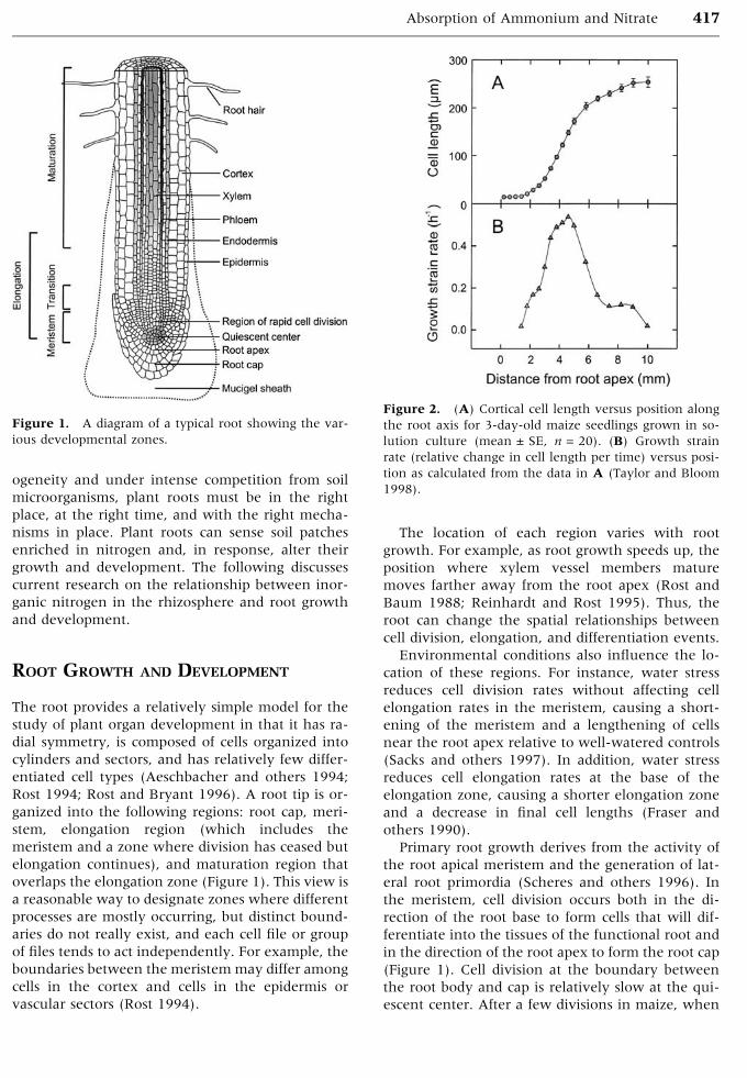
The root provides a relatively simple model for the
study of plant organ development in that it has ra-
dial symmetry, is composed of cells organized into
cylinders and sectors, and has relatively few differ-
entiated cell types (Aeschbacher and others 1994;
Rost 1994; Rost and Bryant 1996). A root tip is or-
ganized into the following regions: root cap, meri-
stem, elongation region (which includes the
meristem and a zone where division has ceased but
elongation continues), and maturation region that
overlaps the elongation zone (Figure 1). This view is
a reasonable way to designate zones where different
processes are mostly occurring, but distinct bound-
aries do not really exist, and each cell file or group
of files tends to act independently. For example, the
boundaries between the meristem may differ among
cells in the cortex and cells in the epidermis or
vascular sectors (Rost 1994).
The location of each region varies with root
growth. For example, as root growth speeds up, the
position where xylem vessel members mature
moves farther away from the root apex (Rost and
Baum 1988; Reinhardt and Rost 1995). Thus, the
root can change the spatial relationships between
cell division, elongation, and differentiation events.
Environmental conditions also influence the lo-
cation of these regions. For instance, water stress
reduces cell division rates without affecting cell
elongation rates in the meristem, causing a short-
ening of the meristem and a lengthening of cells
near the root apex relative to well-watered controls
(Sacks and others 1997). In addition, water stress
reduces cell elongation rates at the base of the
elongation zone, causing a shorter elongation zone
and a decrease in final cell lengths (Fraser and
others 1990).
Primary root growth derives from the activity of
the root apical meristem and the generation of lat-
eral root primordia (Scheres and others 1996). In
the meristem, cell division occurs both in the di-
rection of the root base to form cells that will dif-
ferentiate into the tissues of the functional root and
in the direction of the root apex to form the root cap
(Figure 1). Cell division at the boundary between
the root body and cap is relatively slow at the qui-
escent center. After a few divisions in maize, when
Figure 1. A diagram of a typical root showing the var-
ious developmental zones.
Figure 2. (A) Cortical cell length versus position along
the root axis for 3-day-old maize seedlings grown in so-
lution culture (mean ± SE, n = 20). (B) Growth strain
rate (relative change in cell length per time) versus posi-
tion as calculated from the data in A (Taylor and Bloom
1998).
Absorption of Ammonium and Nitrate 417
the root apex has grown away by about 0.1 mm,
root cells begin to divide more rapidly. Cell division
again tapers off at about 0.4 mm from the apex, and
the cells may expand equally in all directions. Some
have named this area the transition zone (Baluska
and others 1996, 2001).
The elongation zone begins 0.7–1.5 mm from the
apex (Figure 2). Here, cells rapidly extend in length
and undergo a final round of divisions to produce
the cylinder of endodermal cells. In the vascular
cylinder, the phloem begins to differentiate within 3
or 4 mm of the apex, reflecting the importance of
phloem function to cell division and elongation.
Carbohydrates that flow through the phloem to the
growing apices serve not only as an energy source
but as carbon skeletons for newly synthesized or-
ganic compounds and as osmoticants. The phloem
also supplies nitrogen to these tissues (Lazof and
others 1992). In the youngest tissues where the
phloem has not yet developed, namely, the meris-
tematic and transition zones, translocation of car-
bohydrate and nitrogen must rely heavily upon
symplastic diffusion, but symplastic diffusion alone
is too slow to support apical growth (Bret–Harte and
Silk 1994b). One explanation for the slow cell di-
vision in the quiescent center may be the limited
quantities of carbohydrates or nitrogen reaching
this centrally located region.
NITROGEN LIMITATIONS AT THE ROOT
APEX
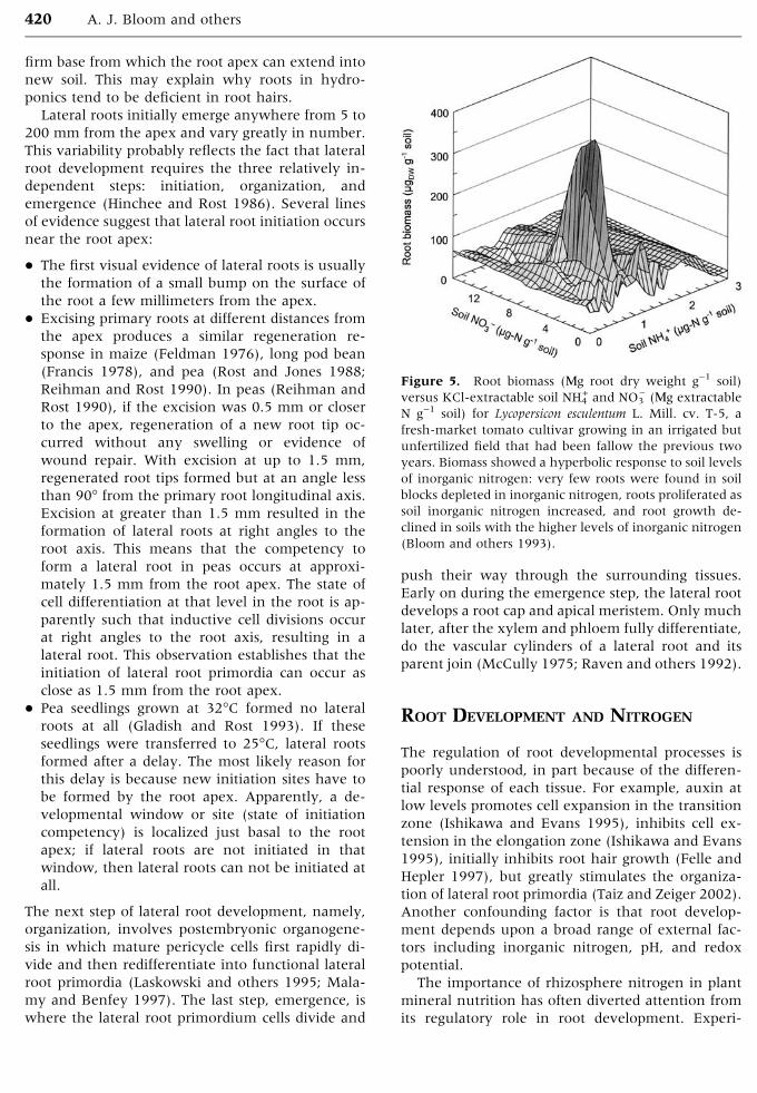
We have developed an in planta root extensiometer
to monitor growth, plasticity, and elasticity of the
Figure 3. Schematic of the in planta root extensiometer
that measures extension under stretching. A RVDT
monitors the position of the root cap, while a LVDT
monitors the position of the mature zone. A pipette tip is
glued to the root cap and a toothpick is glued to the
mature zone. Weights are placed near the tip to assess root
plasticity and elasticity. Nutrient solution flows from an
outlet near the top to bathe the root (J. Frensch and A.J.
Bloom, unpublished).
Figure 4. Length of the root elongation zone — the
difference between the position of the root apex and a
point on the root initially 15 mm from the apex—versus
time for 3-day-old maize seedlings whose roots were ex-
posed to l mM CaSO4, 200 MM KH2PO4, and either 100
MM NH4H2PO4 (NH4+), 100 MM KNO3 (NO3
)), or no ni-
trogen (No N). The labeled arrows indicate the times for
the NH4+ treatment at which designated weights were
added near the tip or 68 mOsm KCl was added to the
nutrient solution. The same series of weights and osmot-
icant were applied to the NO3) and No N treatments at the
times indicated by the shorter arrows. Adding 68 mOsm
KCl to the nutrient solution applies a known stress of 0.17
MPa on the cell wall and provides an independent cali-
bration of the stress induced by the weights (J. Frensch
and A.J. Bloom, unpublished).
418 A. J. Bloom and others

root elongation zone in response to different nu-
trient solutions (Figure 3). A pipette tip is attached
to the root cap with surgical-grade cyanoacrylic glue
and linked to a rotary voltage displacement trans-
ducer (RVDT) with a nylon thread. A piece of a
toothpick is glued to the basal end of the elongation
zone and linked to a linear variable differential
transformer (LVDT) with a thread. The difference
between the readings of the RVDT and the LVDT
indicates the length of the root elongation zone.
Changes in this length when weights are added near
the apex reflect root plasticity plus elasticity,
whereas the changes when weights are removed
reflect only elasticity. The root cuvette is tilted a few
degrees from vertical, and nutrient solution (1 mM
CaSO4, 200 MM KH2PO4, and either no nitrogen,
100 MM NH4H2PO4, or 100 MM KNO3 adjusted to
pH 6.5 with KOH) continuously flows down its
surface immersing the root. Our capability to assess
root mechanical properties in planta is unique; the
standard approach has been to assess tissue seg-
ments that have been frozen, thawed, abraded, and
then boiled (for example, see Wu and others 1996).
In preliminary experiments on 3-day-old maize
seedlings, the root elongation zone extended 1–3
mm h)1 (Figure 4 shows typical traces for three
treatments; the extension rates were calculated
once they reached a steady value after a perturba-
tion), rates comparable to those of plants growing in
vermiculite (Sharp and others 1990) or solution
culture (Taylor and Bloom 1998). This indicates that
the root extensiometer does not significantly dam-
age the root. Extension under NH4+ was significantly
faster (2.16 ± 0.07 mm h)1, n = 6) than under NO3)
(1.90 ± 0.13 mm h)1, mean ± SE, n = 6), which in
turn was significantly faster than under nitrogen
deprivation (1.67 ± 0.08 mm h)1). Exposure to 68
mOsm KCl diminished root extension under NO3)
(1.56 ± 0.12 mm h)1, n = 6), but not under NH4+ or
N deprivation. Applying a load of 1.2–5.2 g had no
significant effect on the rate of root extension and
the roots exhibited only an elastic response (data
not shown).
These results imply that growth of the root
elongation zone, even for plants with large nitrogen
reserves, is nitrogen limited. The meristem and
transition zones differ from more mature root zones
in that they lack fully differentiated phloem tissue
and, thus, cannot rapidly import nitrogen from
more mature tissues. For example, little of the NO3)
absorbed in the maturation zone moves toward the
apex (Siebrecht and others 1995). Consequently,
the nitrogen required for cell division and isotropic
cell expansion derives primarily from nitrogen that
the apical zones themselves absorb and assimilate.
Assimilation of NH4+ and NO3
) to glutamine con-
sume the equivalent of about 2 ATPs per NH4+ and
12 ATPs per NO3), respectively (Bloom and others
1992). In the carbohydrate-limited apical meristem
(Bret–Harte and Silk 1994a), the lower-energy re-
quirement for NH4+ assimilation may permit dividing
cells to maintain energy reserves above a critical
threshold. Absorption and assimilation of NH4+ and
NO3) also alter the redox poise of a cell and, thereby,
its division rate. This is discussed more fully below.
The presence of NO3) stimulated root elongation
under 0 mOsm KCl, but not under 65 mOsm KCl.
This suggests that NO3) may serve as an osmoticant,
a possibility that is also discussed below.
ROOT HAIRS AND LATERAL ROOTS
The maturation zone of a typical root starts between
5 and 20 mm from the apex (Figure 1). In this zone,
the xylem develops the capacity to translocate
substantial quantities of water and solutes to the
shoot. Root hairs also first appear in this region.
Although root hairs have become a model system
for the study of tip growth and associated ion fluxes
(see, for example, Gassmann and Schroeder 1994;
Ridge 1995; Felle and Hepler 1997; Schiefelbein
2000; Bibikova and Gilroy 2003), their primary
physiological function is still in question (Peterson
and Farquhar 1996; Raven and Edwards 2001).
Root hairs increase root surface area and should
enhance root water and nutrient absorption. They
have the capacity to absorb nutrients (Jungk 2001).
Exposure to low levels of nutrients may stimulate
root hair extension (nitrate, Fohse and Jungk 1983,
but see Ewens and Leigh 1985; phosphate, Bates
and Lynch 1996, but see Gahoonia and others
1999). Moreover, wild-type Arabidopsis plants ac-
quire phosphate more efficiently than mutants
lacking root hairs (Bates and Lynch 2001).
In contrast, root hairs may not enhance nutrient
absorption. They may be situated where the root
apex has already depleted the rhizosphere of water
and nutrients (Clarkson 1985; Jungk 2001) or may
grow so slowly and so close together that their own
depletion zones overlap (Clarkson 1991). This
would limit their effectiveness in absorbing water
from drier soils or immobile nutrients such as NH4+.
Cytoplasmic streaming is also relatively slow in root
hairs, impeding rapid transfer to the remainder of
the plant (Raven and Edwards 2001).
Root hairs may be involved in anchorage. Al-
though their ability to anchor an entire plant is
limited (Bailey and others 2002), they may serve to
anchor the root in the mature zone and provide a
Absorption of Ammonium and Nitrate 419
firm base from which the root apex can extend into
new soil. This may explain why roots in hydro-
ponics tend to be deficient in root hairs.
Lateral roots initially emerge anywhere from 5 to
200 mm from the apex and vary greatly in number.
This variability probably reflects the fact that lateral
root development requires the three relatively in-
dependent steps: initiation, organization, and
emergence (Hinchee and Rost 1986). Several lines
of evidence suggest that lateral root initiation occurs
near the root apex:
� The first visual evidence of lateral roots is usually
the formation of a small bump on the surface of
the root a few millimeters from the apex.
� Excising primary roots at different distances from
the apex produces a similar regeneration re-
sponse in maize (Feldman 1976), long pod bean
(Francis 1978), and pea (Rost and Jones 1988;
Reihman and Rost 1990). In peas (Reihman and
Rost 1990), if the excision was 0.5 mm or closer
to the apex, regeneration of a new root tip oc-
curred without any swelling or evidence of
wound repair. With excision at up to 1.5 mm,
regenerated root tips formed but at an angle less
than 90� from the primary root longitudinal axis.
Excision at greater than 1.5 mm resulted in the
formation of lateral roots at right angles to the
root axis. This means that the competency to
form a lateral root in peas occurs at approxi-
mately 1.5 mm from the root apex. The state of
cell differentiation at that level in the root is ap-
parently such that inductive cell divisions occur
at right angles to the root axis, resulting in a
lateral root. This observation establishes that the
initiation of lateral root primordia can occur as
close as 1.5 mm from the root apex.
� Pea seedlings grown at 32�C formed no lateral
roots at all (Gladish and Rost 1993). If these
seedlings were transferred to 25�C, lateral roots
formed after a delay. The most likely reason for
this delay is because new initiation sites have to
be formed by the root apex. Apparently, a de-
velopmental window or site (state of initiation
competency) is localized just basal to the root
apex; if lateral roots are not initiated in that
window, then lateral roots can not be initiated at
all.
The next step of lateral root development, namely,
organization, involves postembryonic organogene-
sis in which mature pericycle cells first rapidly di-
vide and then redifferentiate into functional lateral
root primordia (Laskowski and others 1995; Mala-
my and Benfey 1997). The last step, emergence, is
where the lateral root primordium cells divide and
push their way through the surrounding tissues.
Early on during the emergence step, the lateral root
develops a root cap and apical meristem. Only much
later, after the xylem and phloem fully differentiate,
do the vascular cylinders of a lateral root and its
parent join (McCully 1975; Raven and others 1992).
ROOT DEVELOPMENT AND NITROGEN
The regulation of root developmental processes is
poorly understood, in part because of the differen-
tial response of each tissue. For example, auxin at
low levels promotes cell expansion in the transition
zone (Ishikawa and Evans 1995), inhibits cell ex-
tension in the elongation zone (Ishikawa and Evans
1995), initially inhibits root hair growth (Felle and
Hepler 1997), but greatly stimulates the organiza-
tion of lateral root primordia (Taiz and Zeiger 2002).
Another confounding factor is that root develop-
ment depends upon a broad range of external fac-
tors including inorganic nitrogen, pH, and redox
potential.
The importance of rhizosphere nitrogen in plant
mineral nutrition has often diverted attention from
its regulatory role in root development. Experi-
Figure 5. Root biomass (Mg root dry weight g)1 soil)
versus KCl-extractable soil NH4+ and NO3
) (Mg extractable
N g)1 soil) for Lycopersicon esculentum L. Mill. cv. T-5, a
fresh-market tomato cultivar growing in an irrigated but
unfertilized field that had been fallow the previous two
years. Biomass showed a hyperbolic response to soil levels
of inorganic nitrogen: very few roots were found in soil
blocks depleted in inorganic nitrogen, roots proliferated as
soil inorganic nitrogen increased, and root growth de-
clined in soils with the higher levels of inorganic nitrogen
(Bloom and others 1993).
420 A. J. Bloom and others

ments in which roots grow through compartments
containing different levels of NH4+ or NO3
) (Robin-
son 1994) demonstrate that lateral roots proliferate
only within the highly localized region directly ex-
posed to these ions (Hackett 1972; Drew and others
1973; Drew 1975; Drew and Saker 1975; Grime and
others 1986; Sattelmacher and Thoms 1989; Bing-
ham and others 1997; Dunbabin and others 2001).
These results are inconsistent with strictly nutri-
tional effects. Lateral root primordia, given their
location, should have access to nitrogenous com-
pounds transported in the stele from either the
shoot or other root zones. Nitrogen absorbed as NO3)
in more basal regions may be translocated to the
maturation zone (Lazof and others 1992). Hence,
even when a root apex grows into nitrogen-de-
pleted regions, the lateral root primordia are likely
to be well nourished if more basal parts of the root
are exposed to high NO3) concentrations. External
levels of NO3), therefore, seem to serve as a signal in
lateral root development (Tischner 2000; McIntyre
2001; Forde 2002).
The role of NO3) as a signal has been most thor-
oughly explored in Arabidopsis. In this species, high
NO3) levels in the medium inhibit lateral root
elongation (Zhang and Forde 1998; Zhang and
others 1999; Linkohr and others 2002). If NO3) en-
richment is limited to a small zone, lateral root
elongation doubles in that zone. Mutants deficient
in NO3) reductase showed a similar response to local
NO3) enrichment indicating that the NO3
) stimula-
tion of lateral root growth can be independent of
nitrogen nutrition (Zhang and Forde 1998). Studies
on mutants, which are compromised in auxin pro-
duction or reception, indicated that this hormone
might (Zhang and others 1999; Marchant and oth-
ers 2002) or might not (Linkohr and others 2002)
mediate the influence of external NO3) on lateral
root elongation. An analogous study with ABA
mutants suggested that Arabidopsis has both ABA-
sensitive and -insensitive pathways that mediate
this response (Signora and others 2001).
Evidence for developmental coordination in the
response to local concentrations of nutrients comes
from several sources. For plants grown in split-root
hydroponic systems, raising nutrient concentrations
in one chamber increased the number of lateral
roots in that chamber and decreased the number in
the other chamber even though nutrient concen-
trations in the second chamber remained un-
changed (Gersani and Sachs 1992; Bingham and
others 1997). In two field experiments (Bloom and
others 1993), root growth of tomato showed a hy-
perbolic response to soil levels of inorganic nitrogen:
Very few roots were found in soil blocks depleted in
inorganic nitrogen, roots proliferated as soil inor-
ganic nitrogen increased, and root growth declined
in soils with the higher levels of inorganic nitrogen
(Figure 5). The optimal levels for root growth were
2 Mg NH4+–N g)1 soil and 8 Mg NO3
)–N g)1 soil.
Root development responds not only to the
quantity of inorganic nitrogen in the rhizosphere,
Table 1. The Plant Parameters (mean ± SE, n = 4) of Shoot and Root Biomass, Total Root Length,Root Branching, and Root Area for Lycopersicon esculentum L. Mill. cv. T5a
TreatmentbShoot biomass
(mgDW)
Root biomass
(mgDW)
Root length
(m)
Root branching
(roots m)1)
Root area
(cm2)
100 lM NH4+ 34.8 ± 1.5 13.2 ± 0.6 4.27 ± 0.19 34.9 ± 1.6 17.3 ± 0.8
200 lM NO3) 35.4 ± 3.0 9.1 ± 0.8 3.00 ± 0.23 28.1 ± 0.8 12.9 ± 1.5
aPlants were grown in solution culture for 12 days under constant levels of nitrogen nutrition and pH 6.0 ± 0.3.bThe two treatments developed similar shoot biomass indicating that nitrogen was not limiting (aBloom and others 1993).
Figure 6. Mass (mg dry weight per m root length) of
tissue at various distances from the apex of a maize
seminal root grown in medium containing either NO3) or
NH4+ plus NO3
). Sections from 10 roots were cut, dried, and
weighed in four separate experiments. Shown are the
mean ± SE for the four replicates (Taylor and Bloom
1998).
Absorption of Ammonium and Nitrate 421

but to nitrogen form NH4+ or NO3
) (Bloom 1997). In
a solution culture system that controlled NH4+ or
NO3) levels (Bloom and others 1993), shoot growth
of tomato was similar under both nitrogen forms,
but root growth (biomass, length, branching, or
area) was enhanced by NH4+ (Table 1). Root exten-
sion (Figure 4) and mass (Figure 6) of maize seed-
lings were greater in nutrient solutions containing
NH4+ than in those containing NO3
). Root apical
organization was independent of nitrogen source
(Figure 7). These results are consistent with the
hypothesis that NH4+ nutrition accelerates cell divi-
sion. We are currently testing this hypothesis.
NITROGEN AS A SIGNAL
We conducted an experiment to determine whether
NH4+ and NO3
) themselves serve as signals that de-
termine root proliferation. Maize seeds (Zea mays cv.
Dekalb) were surface-sterilized in 1% NaClO, rinsed
thoroughly with water, and placed in rows on ger-
mination paper, a thick paper toweling that is dif-
ficult for roots to penetrate. The germination paper
was rolled into a tube, placed upright with one end
in l mM CaSO4, and left in the dark at 25�C for 2
days. Seedlings with a primary root of 30 ± 1 mm
were transferred to a controlled environmental
chamber held at 22�C and 95% relative humidity.
The shoots received 600 Mmol quanta m)2 s)1 PAR
from metal-halide HID lamps over a 14-h day. The
roots were placed in light-impermeable boxes held
at 10� from vertical so that the roots grew down at a
slant along a Plexiglas surface covered with germi-
nation paper. A second strip of germination paper
was draped over the root. The bottom ends of both
strips of germination paper were immersed in a
Figure 7. Micrographs of root apices from 4-day-old maize seedlings grown on (A) 100 MM NH4+, (B) 100 MM NO3
), or
(C) a medium without nitrogen. These tissues appear similar in their organization. The bar in the middle frame indicates
the scale for all three micrographs. The photomicrographs were prepared by Dr. Carol Wenzel.
Figure 8. (A) Seminal root length, (B) lateral root
length, and (C) lateral root density (number per unit
length of seminal root) in maize plants that were treated
for 48 h with either 5 mM KC1, 5 mM KNO3, 0.1 mM
KCl, or 0.1 mM NH4Cl. Basal, treated, and apical refer to
the root regions of the seminal root that initiated before,
during, and after the treatments. Shown are the
mean ± SE for 12 plants (A.J. Bloom and P.A. Meyerhoff,
unpublished).
422 A. J. Bloom and others

reservoir that contained a nutrient solution that was
refreshed on a daily basis. For 2 days this solution
contained either 0.1 mM NH4Cl, 0.1 mM KCl, 5.0
mM KNO3, or 5.0 mM KCl. The lower concentra-
tions of NH4+ were to avoid ammonium toxicity.
Subsequently, all of the treatments received the
same nutrient solution [one-tenth strength of a
modified Hoagland solution (Bloom 2002)] for an
additional six days.
We measured the roots three times: first, before
the different treatments were applied; second, when
the different treatments ceased; and finally, six days
after the treatments. These measurements permit-
ted us to identify which parts of the seminal root
initiated before, during, or after the treatments.
Seminal root length and number of lateral roots
were assessed under a dissecting microscope. Some
of the roots were cleared under vacuum in etha-
nol:acetic acid (3:1) to count unemerged lateral root
primordia. We evaluated lateral root lengths using a
video camera and an image analysis system (AgVi-
sion Root and Leaf Analysis, Decagon Devices,
Pullman, WA) on roots that were stained with
Toluidine Blue to increase contrast.
There were several differences among the treat-
ments (Figure 8). In the root zone initiated during
the NO3) treatment, lateral roots were longer (Figure
8B). This response appeared to require direct ex-
posure of young laterals to external NO3) because
the lengths of lateral roots that initiated in the ab-
sence of NO3) were similar among all treatments
(Figure 8B). Our results on maize are consistent
with those on Arabidopsis introduced above in
which exposure of root patches to NO3) stimulated
lateral root elongation in the patch (Zhang and
Forde 1998; Zhang and others 1999; Linkohr and
others 2002). We also found in maize that exposure
to the higher osmotic treatments (5.0 mM KCl or
5.0 mM KNO3) enhanced lateral root density in
zones initiated before the treatments began (Figure
8C). All of the lateral root primordia eventually
emerged (data not shown). Seminal root lengths
were similar under all treatments (Figure 8A). Al-
together, these observations—higher lateral root
densities, all lateral roots emerge, and similar sem-
inal root lengths—imply that higher osmotic
strengths stimulated maize lateral root initiation.
Stimulation of root growth and development by
NO3) may derive in part from its role as an osmoti-
cant (McIntyre 2001). In maize, NO3) stimulated
extension of the seminal root under low osmotic
conditions but not under high (Figure 4). Exposing
a section of an Arabidopsis root to NO3) stimulated
lateral root elongation in proportion to the con-
centrations from 0.05 to 10 mM NO3) (Zhang and
others 1999; Linkohr and others 2002). Extension
of cells in the root �elongation zone� depends upon
osmotically driven uptake of water. Cells in the
elongation zone generate the required osmotic po-
tential through accumulating carbohydrates and K+
(Sharp and others 1990), but the counterions for
this K+ remain uncertain. Organic acids, such as
malate generated from dark CO2 fixation, may be
involved (Osmond 1976), but Cl) and particularly
NO3) are perhaps more suitable because accumula-
tion of these inorganic anions entails less metabolic
energy. Plant cells function normally under high
levels of NO3) (Goyal and Huffaker 1984). Moreo-
ver, NO3-stored in the cells of the elongation zone
can later serve as a nitrogen source as the tissue
matures (Lazof and others 1992). As an osmoticant,
NO3) would not behave like a typical chemical sig-
nal. In the standard model of a signal, once the
signal exceeds a certain low threshold, a specific
receptor in the cell membrane changes state and
starts a transduction cascade that generates a re-
sponse at a larger scale.
Another possible signal of inorganic nitrogen in
the soil is nitric oxide (NO). Soil microorganisms
readily convert NH4+ and NO3
) into NO. It serves as a
potent signal for many other animal and plant re-
sponses (Beligni and Lamattina 2001; Wendehenne
and others 2001). To examine the influence of NO
upon root development, we grew tomato for eight
days on a complete nutrient solution containing 100
MM NH4NO3 and then bubbled either air or 0.8
ppm NO (a level that roots might encounter in the
soil; D. Smart, personal communication) through
the solution for 10 h. We analyzed a number of root
parameters immediately after the treatments and
four days later and found no significant differences
between the treatments (Table 2).
These results argue that neither NH4+ NO3
), nor
NO serves as a direct signal for root development
rather some secondary effects of NH4+ and NO3
) may
be the actual stimulus perceived by the plant. For
example, NH4+ and NO3
) alter the pH and redox
potential of the rhizosphere, and roots may respond
to such alterations.
ACID GROWTH OF ROOTS
The �acid-growth hypothesis,� originally postulated
to explain auxin-induced growth (Cleland 1971;
Hager and others 1991; Rayle and Cleland 1992),
may apply to nitrogen-induced growth (Bloom
1997). Plant nitrogen metabolism alters rhizosphere
Absorption of Ammonium and Nitrate 423

pH: NH4+ assimilation releases protons, whereas
NO3) assimilation produces hydroxide ions (Raven
and Smith 1976; Allen 1988). Plants supplied with
NH4+ as the N source strongly acidify and those
supplied with NO3) slightly alkalinize the rhizo-
sphere (Smart and Bloom 1998). Through such pH
changes, rhizosphere NH4+ and NO3
) may affect cell
wall expansion. Acidity in the root apoplast loosens
the cell wall matrix (Taiz 1984; Edelmann and Fry
1992). Root growth zones correlate well with rhiz-
osphere acidification as detected with pH indicators
(Marschner 1995) and microelectrodes (Taylor and
Table 2. Relative Change per Day* for 8-day-old Tomatoes Exposed to Air or 0.8 ppm NO for 10 hMeasured 0 and 4 Days after Treatment (mean ± SE for 6 plants)
Lateral roots
Primary root length Total length Avg. length Mass
Treatment (cm cm)1 d)1) (cm cm)1 d)1) (cm cm)1 d)1) (roots cm)1) ())1 d)1
Air 0.105 ± 0.013 0.399 ± 0.020 0.286 ± 0.021 0.008 ± 0.027
NO 0.093 ± 0.008 0.438 ± 0.023 0.325 ± 0.010 0.020 ± 0.028
*(ln L1-ln L2)/(t1-t2)
Figure 9. Stainless-steel and glass root cuvette for the microelectrode experiments. The cuvette is positioned on the stage
of an inverted microscope and the root and microelectrode are viewed from below. Experimental solutions flow into the
chamber at one end and out at the other. When the flow is stopped, a ion depletion zone develops. A micromanipulator
positions the microelectrode tip at various distances from the root surface to monitor this zone. The left inset depicts a
multibarrel microelectrode. The three longer barrels are pulled from thin-wall glass capillaries: one contains NH4+-selective
LIX (liquid ion exchanger), another contains NO3) LIX, and the last contains a salt solution and serves as a local reference
electrode. The four shorter barrels (that is, three outer barrels and the center) are pulled from solid glass rods. The right
inset shows a scanning electron micrograph of the tip from a multibarrel ion-selective microelectrode. Each of the three
barrels with holes is filled with a liquid ion exchanger or salt solution. The total tip diameter to enclose the open barrels is
about 3 Mm (Colmer and Bloom 1998).
424 A. J. Bloom and others
Bloom 1998). Enhanced H+ efflux is detected on the
outer curved surface of roots responding to gravity
and is thought to contribute to more rapid cell ex-
pansion (Mulkey and Evans 1982).
To quantify the relationships among inorganic
nitrogen assimilation, rhizosphere pH, and root de-
velopment, we developed multibarrel ion-selective
microelectrodes that can simultaneously monitor
electrical potential and the activities of two ions
with high temporal and spatial resolution (Figure
9). Each of the ion-selective barrels is filled with a
liquid ion exchanger (LIX) consisting of an iono-
phore that is a lipophylic ion channel, a plasticizer
to solubilize the ionophore, and PVC dissolved in a
volatile solvent. A salt solution containing the ion of
interest is then backfilled behind the LIX, and a
chloridized silver wire is placed in the salt solution.
The silver wires from the three barrels fit into a
circular Teflon IC socket that fits into the head stage
of a multichannel, differential electrometer ampli-
fier (Bloom 1989).
With these electrodes, we monitor ion gradients
between 20 and 100 Mm from the surface of a root
to determine which areas along the root axis are
most active in NH4+, NO3
), and H+ fluxes (Colmer
and Bloom 1998; Taylor and Bloom 1998). This
approach, which assumes radial symmetry and ion
diffusion through water, pinpoints fluxes to within
800 Mm along the root axis with 95% certainty
(Henriksen and others 1992).
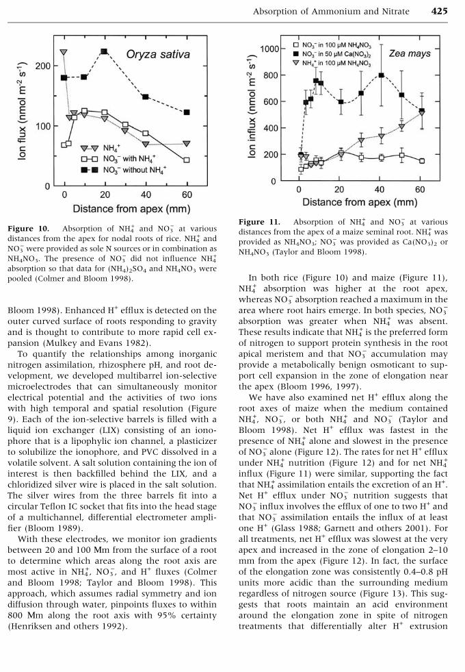
In both rice (Figure 10) and maize (Figure 11),
NH4+ absorption was higher at the root apex,
whereas NO3) absorption reached a maximum in the
area where root hairs emerge. In both species, NO3)
absorption was greater when NH4+ was absent.
These results indicate that NH4+ is the preferred form
of nitrogen to support protein synthesis in the root
apical meristem and that NO3) accumulation may
provide a metabolically benign osmoticant to sup-
port cell expansion in the zone of elongation near
the apex (Bloom 1996, 1997).
We have also examined net H+ efflux along the
root axes of maize when the medium contained
NH4+, NO3
), or both NH4+ and NO3
) (Taylor and
Bloom 1998). Net H+ efflux was fastest in the
presence of NH4+ alone and slowest in the presence
of NO3) alone (Figure 12). The rates for net H+ efflux
under NH4+ nutrition (Figure 12) and for net NH4
+
influx (Figure 11) were similar, supporting the fact
that NH4+ assimilation entails the excretion of an H+.
Net H+ efflux under NO3) nutrition suggests that
NO3) influx involves the efflux of one to two H+ and
that NO3) assimilation entails the influx of at least
one H+ (Glass 1988; Garnett and others 2001). For
all treatments, net H+ efflux was slowest at the very
apex and increased in the zone of elongation 2–10
mm from the apex (Figure 12). In fact, the surface
of the elongation zone was consistently 0.4–0.8 pH
units more acidic than the surrounding medium
regardless of nitrogen source (Figure 13). This sug-
gests that roots maintain an acid environment
around the elongation zone in spite of nitrogen
treatments that differentially alter H+ extrusion
Figure 10. Absorption of NH4+ and NO3
) at various
distances from the apex for nodal roots of rice. NH4+ and
NO3) were provided as sole N sources or in combination as
NH4NO3. The presence of NO3) did not influence NH4
+
absorption so that data for (NH4)2SO4 and NH4NO3 were
pooled (Colmer and Bloom 1998).
Figure 11. Absorption of NH4+ and NO3
) at various
distances from the apex of a maize seminal root. NH4+ was
provided as NH4NO3; NO3) was provided as Ca(NO3)2 or
NH4NO3 (Taylor and Bloom 1998).
Absorption of Ammonium and Nitrate 425

from the root. In comparison to the NH4+ treatments,
the surface of the root was more alkaline when NO3)
was the sole nitrogen source (Figure 13) so that the
more limited proton pumping under NO3) (Figure
12) still produced a similar decline in pH (Figure
13).
Shifting the nutrient solution that bathed a root
in an in planta root extensiometer (Figure 3) from
pH 6.5 to 5.6 increased root elasticity by 50% (data
not shown). The rate of root extension, however,
declined from 1.67 ± 0.08 to 1.51 ± 0.06 mm h)1
(mean ± SE, n = 6). Peters and Felle (1999) found
that lowering the pH of the medium stimulated root
elongation only in slowly growing maize plants. We
must conclude, as did these authors, that cell wall
pH may contribute to the control of root growth but
that other factors can override its influence.
REDOX POTENTIAL AND ROOT
DEVELOPMENT
The redox potential of the rhizosphere may reflect
not only which nitrogen compounds are present
and in what amounts, but how they were generat-
ed. As nitrogen cycles through various inorganic
and organic compounds in the soil, its oxidation
number ranges from )3 to +5. NH4+ is a moderate
reducing agent (Eh = )0.35 V), whereas NO3) is a
strong oxidizing agent (Eh = +0.74 V). Conversion of
NO3) to N2 may generate highly reactive oxidizing
agents such as O3 or O2) (Paul and Clark 1996). In
addition, ammonification (the process through
which soil microbes convert organic nitrogen into
NH4+) occurs under both reducing and oxidizing
conditions, whereas oxidizing conditions favor ni-
trification (microbial conversion of NH4+ to NO3
))
and reducing conditions favor denitrification (mi-
crobial conversion of NO3) to NO, N2O, or N2). The
net results are that soil NO3) is likely to accumulate
only when the soil redox potential is high and that
the relative availability of NH4+ should increase as
the soil redox potential declines with the maximum
absolute availability at moderate redox potentials.
Root activity alters rhizosphere redox potential
through respiratory oxygen consumption and ion
uptake or exudation. In particular, root absorption
and assimilation of NH4+ and NO3
) consume 0.31 mol
O2 mol)1 NH4+ and 1.5 mol O2 mol)1 NO3
), respec-
tively (Bloom and others 1992). Thus, when roots
use NO3) as a nitrogen source, the rhizosphere redox
potential declines more rapidly than when they use
NH4+.
Redox potential influences a wide range of cell
functions including the detoxification of free radi-
cals, the activity of many enzymes, and the ex-
pression of many genes (Luthje and others 1997;
Berczi and Møller 2000; Pfannschmidt and others
2001). To regulate internal redox potential, plant
cells use both a complex suite of cytosolic redox
buffers such as glutathione or ascorbic acid (May
and Leaver 1993; Noctor and others 2000) and
several plasma membrane oxidoreductases that
transfer electrons from cytosolic donors to extra-
cellular acceptors in the apoplast. These oxidore-
Figure 12. Net proton efflux at various distances from
the apex of a maize seminal root when the medium
contains NH4+, NO3
), or both forms of nitrogen (Taylor and
Bloom 1998).
Figure 13. The pH at 50 or 1000 Mm from the root
surface at various distances from the apex of a maize
seminal root. The root was bathed in a medium that
lacked nitrogen or contained NH4+, NO3
), or both forms of
nitrogen (Taylor and Bloom 1998).
426 A. J. Bloom and others

ductases are involved with signal transduction of
light (Gautier and others 1992, but see Taylor and
Assmann 2001; Mullineaux and Karpinski 2002),
membrane polarization and H+ excretion (Marre
and others 1988), generation of oxidative bursts in
response to wounding and pathogen attack (Auh
and Murphy 1995), hormonal regulation of cell
growth (Bottger and Hilgendorf 1988), and modifi-
cation of cell wall structure (Bradley and others
1992).
In plant roots, plasma membrane oxidoreduc-
tases have been detected and isolated based on
their ability to reduce artificial impermiant elec-
tron acceptors such as ferricyanide (HCF). All
Figure 14. Maize growing in 0.8% agar that contains 1 mM CaSO4, 1 MM KH2PO4, and either a pH indicator dye (0.12%
Bromocresol purple, left column), a redox indicator dye [800 MM K3Fe(CN)6 with 1 mM FeCl3, middle column], or both
dyes (right column). The rows contain replicates. Usually, the plates were covered with aluminum foil. Yellowing of the
pH dye and darkening of the redox dye indicate H+ extrusion and root reduction of the rhizosphere, respectively. The
shoots grew normally under all treatments, but the presence of the redox indicator dye inhibited lateral root development
(A.R. Taylor and A.J. Bloom, unpublished).
Absorption of Ammonium and Nitrate 427

plant roots studied so far have a constitutive NADH
oxidase or �standard oxidoreductase system� (Crane
and others 1991). This system can reduce external
electron acceptors of high redox potential and
may use O2 as the terminal electron acceptor. In
dicots and nongraminaceous plants, iron stress in-
duces a �turbo� ferric reductase system, but it remains
unclear whether the turbo system is distinct from
or reflects enhanced expression/activity of the
standard oxidoreductase system (Beinfait 1988).
Plasma membrane redox enzymes may regulate
cell elongation via structural changes in the cell
wall. Low extracellular concentrations (MM) of ar-
tificial electron acceptors, such as ascorbate free
radical or HCF, stimulate the standard oxidoreduc-
tase, H+ secretion, and root growth, presumably
through an acid growth mechanism and enhanced
nutrient absorption (Crane and others 1991; Gon-
zales–Reyes and others 1995). Higher levels (mM),
however, inhibited the growth of Lepidium roots
(Crane and others 1991) and the development of
maize lateral roots (Figure 14). This indicates that
regulation of cell wall loosening by pH and redox
activity is complex. Cell wall stiffening occurs as
part of plant pathogen defenses (hypersensitive re-
sponse) when membrane-associated peroxidases
generate H2O2 that crosslinks cell wall proteins
(Bradley and others 1992). Redox reactions also
diminish cell plasticity in the elongation zone of
coleoptiles (Bradley and others 1992).
Redox activity influences root elongation also
through cell proliferation. Cell division in Arabidopsis
root apical meristems depended upon the redox
potential of the cytosol (Sanchez–Fernandez and
others 1997). Apical initials had high cellular levels
of the reductant glutathione (GSH), whereas the
quiescent zone had low levels. Artificially increasing
GSH pools or exogenously applying GSH or ascorbic
acid stimulated cell division and growth. In maize,
root apical meristems showed a similar pattern; ac-
tively dividing meristematic cells had higher levels of
the cytosolic reductant ascorbic acid than cells in the
quiescent zone (Kerk and Feldman 1995).
We propose the following scenario to integrate
these phenomena: The redox potential of the rhiz-
osphere influences the relative availability of NH4+
and NO3) (moderate potentials favor NH4
+) and the
redox potential of the roots. Root redox potentials
and membrane-associated redox activities regulate
cell proliferation and extension and, thus, root
growth (moderate potentials favor growth). The
nitrogen demands of root growth stimulate NH4+ or
NO3) assimilation that, in turn, influences redox
potentials (NO3) rapidly lowers potentials). The in-
terplay between NH4+ and NO3
) and rhizosphere
redox potential may be partially responsible for the
observed large fluctuations in the relative availa-
bility of soil NH4+ and NO3
) and in root growth
(Jackson and Bloom 1990).
SUMMARY
Although NH4+ and NO3
) absorption and assimilation
by plants are only some of the many processes that
influence rhizosphere pH and redox potential or that
regulate root growth and development, root nitro-
gen acquisition is a primary determinant of plant
productivity and, as such, should be a central process
to integrate the responses of roots to their environ-
ment. The following factors influence the mecha-
nisms by which roots respond to NH4+ and NO3
):
1. Growth at the root apex may be nitrogen limited.
In maize seedlings with ample nitrogen reserves,
extension of the root apex increased in solutions
containing NH4+or NO3
). The apex differs from
mature root zones in that it lacks fully differen-
tiated phloem tissue and, thus, cannot rapidly
import nitrogen from other tissues. This suggests
that the nitrogen required for cell division and
expansion in the apex derives primarily from its
own absorption of rhizosphere NH4+ and NO3
).
2. Cell division in the root apical meristem may be
more rapid under NH4+ nutrition. Root density
and extension of maize seedlings were greater in
nutrient solutions containing NH4+ than in those
containing NO3) as the sole nitrogen source.
Perhaps in the carbohydrate-limited apical
meristem, the lower energy requirement for NH4+
assimilation versus NO3) assimilation permits
dividing cells to maintain energy reserves above
a critical threshold.
3. Root nitrogen acquisition alters rhizosphere pH
and redox potential, which in turn may regulate
root cell proliferation and mechanical properties.
Assimilation of NH4+ releases protons, whereas
that of NO3) produces hydroxide ions; plants
supplied with NH4+ strongly acidify and those
supplied with NO3) slightly alkalinize the rhizo-
sphere. An acid pH in the cell wall matrix in-
creases its elasticity. Moreover, NO3) assimilation
depletes reducing equivalents more than NH4+
assimilation. This may shift cellular redox po-
tential to a level that diminishes cell proliferation.
ACKNOWLEDGMENTS
We thank Wendy K. Silk for her insightful com-
ments, David R. Smart and Carol L. Wenzel for their
efforts on the NO experiments, Jurgend Frensch for
428 A. J. Bloom and others

his work with the root extensiometer, and Jamie
Brayton for her help in maize root growth experi-
ments. This research was supported in part by NSF
IBN-99-74927 and USDA NRI-CGP-2000-00647 to
AJB.
REFERENCES
Aeschbacher RA, Schiefelbein JW, Benfey PN. 1994. The genetic
and molecular basis of root development. Annu Rev Plant
Physiol Plant Mol Biol 45:25–45.
Allen S. 1988. Intracellular pH regulation in plants. ISI Atlas Sci
Animal Plant Sci 1:283–288.
Auh C-K, Murphy TM. 1995. Plasma membrane redox enzyme is
involved in the synthesis of O2- and H2O2 by Phytophthora
elicitor-stimulated rose cells. Plant Physiol 107:1241–1247.
Bailey PHJ, Currey JD, Fitter AH. 2002. The role of root system
architecture and root hairs in promoting anchorage against
uprooting forces in Allium cepa and root mutants of Arabidopsis
thaliana. J Exp Bot 53:333–340.
Baluska F, Volkmann D, Barlow PW. 1996. Specialized zones of
development in roots: View from the cellular level. Plant
Physiol 112:3–4.
Baluska F, Volkmann D, Barlow PW. 2001. A polarity crossroad in
the transition growth zone of maize root apices: Cytoskeletal and
developmental implications. J Plant Growth Regul 20:170–181.
Bates TR, Lynch JP. 1996. Stimulation of root hair elongation in
Arabidopsis thaliana by low phosphorus availability. Plant Cell
Environ 19:529–538.
Bates TR, Lynch JP. 2001. Root hairs confer a competitive ad-
vantage under low phosphorus availability. Plant Soil 236:243–
250.
Beinfait HF. 1988. The turbo reductase in plant plasma mem-
branes. In: Crane FL, Morre DJ, Low H, editors. Oxidoreduc-
tases in Control of Animal and Plant Growth. New York:
Plenum Press, p 89–93.
Beligni MV, Lamattina L. 2001. Nitric oxide in plants: The history
is just beginning. Plant Cell Environ 24:267–278.
Berczi A, Møller IM. 2000. Redox enzymes in the plant plasma
membrane and their possible roles. Plant Cell Environ
23:1287–1302.
Bibikova T, Gilroy S. 2003. Root hair development. J Plant
Growth Regul 21:383–415.
Bingham IJ, Blackwood JM, Stevenson EA. 1997. Site, scale and
time-course for adjustments in lateral root initiation in wheat
following changes in C and N supply. Ann Bot 80:97–106.
Bloom AJ. 1989. Continuous and steady-state nutrient absorp-
tion by intact plants. In: Torrey JG, Winship LJ, editors.
Application of Continuous and Steady State Methods to Root
Biology. Dordrecht: Kluwer Academic, p 147–163.
Bloom AJ. 1996. Nitrogen dynamics in plant growth Systems.
Life Support Biosphere Sci 3:35–41.
Bloom AJ. 1997. Interactions between inorganic nitrogen nutri-
tion and root development. Z Pflanzenernahr Bodenk 160:253–
259.
Bloom AJ. 2002. Mineral Nutrition. In: Taiz L, Zeiger E, editors.
Plant Physiology, 3rd ed. Sunderland, MA: Sinauer Associates,
p 67–86.
Bloom AJ, Jackson LE, Smart DR. 1993. Root growth as a func-
tion of ammonium and nitrate in the root zone. Plant Cell
Environ 16:199–206.
Bloom AJ, Sukrapanna SS, Warner RL. 1992. Root respiration
associated with ammonium and nitrate absorption and assim-
ilation by barley. Plant Physiol 99:1294–1301.
Bottger M, Hilgendorf F. 1988. Hormone action on transmem-
brane electron and H+ transport. Plant Physiol 86:1038–1043.
Bradley DJ, Kjellbom P, Lamb CJ. 1992. Elicitor- and wound-
induced oxidative cross-linking of a proline rich plant cell wall
protein: a novel, rapid defense response. Cell 70:21–30.
Bret–Harte MS, Silk WK. 1994a. Fluxes and deposition rates of
solutes in growing roots of Zea mays. J Exp Bot 45:1733–1742.
Bret–Harte MS, Silk WK. 1994b. Nonvascular, symplasmic dif-
fusion of sucrose cannot satisfy the carbon demands of growth
in the primary root tip of Zea mays L. Plant Physiol 105:19–33.
Clarkson DT. 1985. Factors affecting mineral nutrient acquisition
by plants. Annu Rev Plant Physiol 36:77–115.
Clarkson DT. 1991. Root structure and sites of ion uptake. In:
Waisel Y, Eshel A, Kafkai U, editors. Plant roots, the hidden
half. New York: Marcel Dekker, p 417–453.
Cleland RE. 1971. Cell wall extension. Annu Rev Plant Physiol
22:197–222.
Colmer TD, Bloom AJ. 1998. A comparison of NH4+ and NO3
) net
fluxes along roots of rice and maize. Plant Cell Environ 21:240–
246.
Crane FL, Morre DJ, Low HE, Bottger M. 1991. The oxidore-
ductase enzymes in plant plasma membranes. In: Crane FL,
Morre DJ, Low HE, editors. Oxidoreduction at the Plasma
Membrane: Relation to growth and transport. Boca Raton, FL:
CRC Press, p 21–33.
Drew MC. 1975. Comparison of the effects of a localized supply of
phosphate, nitrate, ammonium and potassium on the growth
of the seminal root system, and the shoot, in barley. New
Phytol 75:479–490.
Drew MC, Saker LR. 1975. Nutrient supply and the growth of the
seminal root system in barley. II. Localized compensatory in-
creases in lateral root growth and rates of nitrate uptake when
nitrate supply is restricted to only part of the root System. J Exp
Bot 26:79–90.
Drew MC, Saker LR, Ashley TW. 1973. Nutrient supply and the
growth of the seminal root system in barley. I. The effect of
nitrate concentration on the growth of axes and laterals. J Exp
Bot 24:1189–1202.
Dunbabin V, Rengel Z, Diggle A. 2001. The root growth response
to heterogeneous nitrate supply differs for Lupinus angustifolius
and Lupinus pilosus. Aust J Agric Res 52:495–503.
Edelmann HG, Fry SC. 1992. Kinetics of integration of xyloglucan
into the walls of suspension-cultured rose cells. J Exp Bot
43:463–470.
Ewens M, Leigh RA. 1985. The effect of nutrient solution com-
position on the length of root hairs of wheat (Triticum aestivum
L). J Exp Bot 36:713–724.
Feldman LF. 1976. The de novo origin of the quiescent center in
regenerating root apices of Zea mays. Planta 128:207–212.
Felle HH, Hepler PK. 1997. The cytosolic Ca2+ concentration
gradient of Sinapis alba root hairs as revealed by Ca2+-selective
microelectrode tests and fura-dextran ratio imaging. Plant
Physiol 114:39–45.
Fohse D, Jungk A. 1983. Influence of phosphate and nitrate
supply on root hair formation of rape, spinach and tomato
plants. Plant Soil 74:359–368.
Forde BG. 2002. Local and long-range signaling pathways regu-
lating plant responses to nitrate. Annu Rev Plant Physiol Plant
Mol Biol 53:203–224.
Francis D. 1978. Regeneration of meristematic activity following
decapitation of the root tip of Vicia faba L. New Phytol 81:357–
365.
Fraser TE, Silk WK, Rost TL. 1990. Effects of low water potential
on cortical cell length in growing regions of maize roots. Plant
Physiol 93:648–651.
Absorption of Ammonium and Nitrate 429

Gahoonia TS, Nielsen NE, Lyshede OB. 1999. Phosphorus (P)
acquisition of cereal cultivars in the field at three levels of P
fertilization. Plant Soil 211:269–281.
Garnett TP, Shabala SN, Smethurst PJ, Newman IA. 2001. Si-
multaneous measurement of ammonium, nitrate and proton
fluxes along the length of eucalypt roots. Plant Soil 236:55–62.
Gassmann W, Schroeder JI. 1994. Inward-rectifying K+ channels
in root hairs of wheat: A mechanism for aluminum-sensitive
low-affinity K+ uptake and membrane potential control. Plant
Physiol 105:1399–1408.
Gautier H, Vavasseur A, Laseve G, Boudet AM. 1992. Redox
processes in the blue light response of guard cell protoplasts of
Commelina communis L. Plant Physiol 98:34–38.
Gersani M, Sachs T. 1992. Development correlations between
roots in heterogeneous environments. Plant Cell Environ
15:463–469.
Gladish DK, Rost TL. 1993. The effects of temperature on primary
root growth dynamics and lateral root distribution in garden
pea (Pisum sativum L, Cv Alaska) Environ Exp Bot 33:243–258.
Glass ADM. 1988. Nitrogen uptake by plant roots. ISI Atlas Sci
Animal Plant Sci 1:151–156.
Gonzales-Reyes JA, Alcaın FJ, Caler JA, Serrano A, Cordoba F,
Navas P. 1995. Stimulation of onion root elongation by
ascorbate and ascorbate free radical in Allium cepa L. Proto-
plasma 84:31–35.
Goyal SS, Huffaker RC. 1984. Nitrogen toxicity in plants. In:
Hauck RD, editor. Nitrogen in Crop Production. Madison, WI:
ASA, CSSA, SSSA, p 97–118.
Grime JP, Crick JC, Rincon JE. 1986. The ecological significance
of plasticity. In: Jennings DH, Trewavas AJ, editors. Plasticity in
Plants. Cambridge: Company of Biologists Limited, p 5–29.
Hackett C. 1972. A method of applying nutrients locally to roots
under controlled conditions, and some morphological effects of
locally applied nitrate on the branching of wheat roots. Aust J
Biol Sci 25:1169–1180.
Hager A, Debus G, Edel H-G, Stransky H, Serrano R. 1991. Auxin
induces exocytosis and rapid synthesis of a high turnover pool
of plasma membrane H+-ATPase. Planta 185:527–537.
Henriksen GH, Raman DR, Walker LP, Spanswick RM. 1992.
Measurement of net fluxes of ammonium and nitrate at the
surface of barley roots using ion-selective microelectrodes. II.
Patterns of uptake along the root axis and evaluation of the
microelectrode flux estimation technique. Plant Physiol
99:734–747.
Hinchee MAW, Rost TL. 1986. The control of lateral root devel-
opment in cultured pea seedlings. I. The role of seeling organs
and plant growth regulators. Bot Gaz 147:137–147.
Ishikawa H, Evans ML. 1995. Specialized zones of development
in roots. Plant Physiol 109:725–727.
Jackson LE, Bloom AJ. 1990. Root distribution in relation to soil
nitrogen availability in field-grown tomatoes. Plant Soil
128:115–126.
Jungk A. 2001. Root hairs and the acquisition of plant nutrients
from soil. J Plant Nutr Soil Sci 164:121–129.
Kerk NM, Feldman LJ. 1995. A biochemical model for the initi-
ation and maintenance of the quiescent center: Implications for
organization of root meristems. Development 121:2825–2833.
Laskowski MJ, Williams ME, Nusbaum HC, Sussex IM. 1995.
Formation of lateral root meristems is a two-stage process.
Development 121:3303–3310.
Lazof DB, Rufty Jr TW, Redinbaugh MG. 1992. Localization of
nitrate absorption and translocation within morphological re-
gions of the corn root. Plant Physiol 100:1251–1258.
Linkohr BI, Williamson LC, Fitter AH, Leyser HMO. 2002. Nitrate
and phosphate availability and distribution have different ef-
fects on root system architecture of Arabidopsis. Plant J 29:751–
760.
Luthje S, Doering O, Heuer S, Luethen H, Boettger M. 1997.
Oxidoreductases in plant plasma membranes. Biochim Biophys
Acta 1331:81–102.
Malamy JE, Benfey PN. 1997. Organization and cell differentia-
tion in lateral roots of Arabidopsis thaliana. Development
124:33–44.
Marchant A, Bhalerao R, Casimiro I, Eklof J, Casero PJ, Bennett
G, Sandberg G. 2002. AUX1 promotes lateral root formation by
facilitating indole-3-acetic acid distribution between sink and
source tissues in the Arabidopsis seedling. Plant Cell 14:589–
597.
Marre MT, Moroni A, Albergoni FG, Marre E. 1988. Plasma-
lemma redox activity and H+ extrusion. Plant Physiol 87:25–29.
Marschner H. 1995. Mineral Nutrition of Higher Plants. 2nd ed.
London: Academic Press, p 889.
May M, Leaver CJ. 1993. Oxidative stimulation of glutathione
synthesis in Arabidopsis thaliana suspension cultures. Plant
Physiol 103:621–627.
McCully ME. 1975. The development of lateral roots. In: Torrey
DT, Clarkson DT, editors. The Development and Function of
Roots. London: Academic Press, p 105–124.
McIntyre GI. 2001. Control of plant development by limiting
factors: A nutritional perspective. Physiol Plantarum 113:165–
175.
Mulkey TJ, Evans ML. 1982. Suppression of asymmetric acid
efflux and gravitropism in maize roots treated with auxin
transport inhibitors or sodium orthovanadate. J Plant Growth
Regul 1:259–265.
Mullineaux P, Karpinski S. 2002. Signal transduction in response
to excess light: Getting out of the chloroplast. Curr Opin Plant
Biol 5:43–48.
Noctor G, Veljovic–Jovanovic S, Foyer CH. 2000. Peroxide
processing in photosynthesis: Antioxidant coupling and redox
signalling. Philos Trans R Soc London B Biol Sci 355:1465–
1475.
Osmond CB. 1976. Ion absorption and carbon metabolism in cells
of higher plants. In: Luttge U, Pitman MG, editors. Encyclo-
pedia of Plant Physiology. New Series. Berlin: Springer-Verlag,
p 347–372.
Paul EA, Clark FE. 1996. Soil Microbiology and Biochemistry.
2nd ed. San Diego: Academic Press, p 340.
Peters WS, Felle HH. 1999. The correlation of profiles of surface
pH and elongation growth in maize roots. Plant Physiol
121:905–912.
Peterson PJL, Farquhar ML. 1996. Roots hairs: Specialized tu-
bular cells extending root surfaces. Bot Rev 62:1–40.
Pfannschmidt T, Allen JF, Oelmueller R. 2001. Principles of redox
control in photosynthesis gene expression. Physiol Plantarum
112:1–9.
Raven JA, Edwards D. 2001. Roots: Evolutionary origins and
biogeochemical significance. J Exp Bot 52:381–401.
Raven JA, Smith FA. 1976. Nitrogen assimilation and transport in
vascular land plants in relation to intracellular pH regulation.
New Phytol 76:415–431.
Raven PH, Evert RF, Eichhorn SE. 1992. Biology of Plants. 5th ed.
New York: Worth Publishers, p 791.
Rayle DL, Cleland RE. 1992. The acid growth theory of auxin-
induced cell elongation is alive and well. Plant Physiol
99:1271–1274.
Reihman MA, Rost TL. 1990. Regeneration responses in pea roots
after tip excision at different levels. Am J Bot 77:1159–1167.
Reinhardt DH, Rost TL. 1995. On the correlation of primary root
growth and tracheary element size and distance from the tip in
430 A. J. Bloom and others

cotton seedlings grown under salinity. Environ Exp Bot 5:575–
588.
Ridge RW. 1995. Recent developments in the cell and molecular
biology of root hairs. J Plant Res 108:399–405.
Robinson D. 1994. The response of plants to non-uniform sup-
plies of nutrients. New Phytol 127:635–674.
Rost TL. 1994. Root tip organization and the spatial relationships
of differentiation events. In: Iqbal M, editor. Growth Patterns in
Vascular Plants. Portland, OR: Dioscorides Press, p 59–76.
Rost TL, Baum S. 1988. On the correlation of primary root length,
meristem size and protoxylem tracheary element position in
pea seedlings. Am J Bot 75:414–424.
Rost TL, Bryant JA. 1996. Root organization and gene expression
patterns. J Exp Bot 47:1613–1628.
Rost TL, Jones TJ. 1988. Pea root regeneration after tip excision at
different levels: Polarity of new growth. Ann Bot 61:513–523.
Sacks MM, Silk WK, Burman P. 1997. Effect of water stress on
cortical cell division rates within the apical meristem of primary
roots of maize. Plant Physiol 114:519–527.
Sanchez–Fernandez R, Fricker M, Corben LB, White NS, Sheard
CJ, Leaver CJ, Van Montagu M, Inze D, May MJ. 1997. Cell
proliferation and hair tip growth in the Arabidopsis root are
under mechanistically different forms of redox control. Proc
Natl Acad Sci USA 94:2745–2750.
Sattelmacher B, Thoms K. 1989. Root growth and 14C-translo-
cation into the roots of maize (Zea mays L.) as influenced by
local nitrate supply. Z Pflanzenernahr Bodenk 152:7–10.
Scheres B, McKhann HI, van den Berg C. 1996. Roots redefined:
Anatomical and genetic analysis of root development. Plant
Physiol 111:959–964.
Schiefelbein JW. 2000. Constructing a plant cell. The genetic
control of root hair development. Plant Physiol 124:1525–1531.
Sharp RE, Hsiao TC, Silk WK. 1990. Growth of the maize primary
root at low water potentials. 2. Role of growth and deposition
of hexose and potassium in osmotic adjustment. Plant Physiol
93:1337–1346.
Siebrecht S, Mack G, Tischner R. 1995. Function and contribu-
tion of the root tip in the induction of NO3) uptake along the
barley root axis. J Exp Bot 46:1669–1676.
Signora L, De Smet I, Foyer CH, Zhang H. 2001. ABA plays a
central role in mediating the regulatory effects of nitrate on
root branching in Arabidopsis. Plant J 28:655–662.
Smart DR, Bloom AJ. 1998. Investigations of ion absorption
during NH4+ exposure I. Relationship between H+ efflux and
NO3) absorption. J Exp Bot 49:95–100.
Taiz L. 1984. Plant cell expansion: regulation of cell wall
mechanical properties. Annu Rev Plant Physiol 35:585–
657.
Taiz L, Zeiger E. 2002. Plant Physiology. 3rd ed. Sunderland, MA:
Sinauer Associates, p 690.
Taylor AR, Assmann SM. 2001. Apparent absence of a redox
requirement for blue light activation of pump current in broad
bean guard cells. Plant Physiol 125:329–338.
Taylor AR, Bloom AJ. 1998. Ammonium, nitrate and proton
fluxes along the maize root. Plant Cell Environ 21:1255–
1263.
Tischner R. 2000. Nitrate uptake and reduction in higher and
lower plants. Plant Cell Environ 23:1005–1024.
Wendehenne D, Pugin A, Klessig DF, Durner J. 2001. Nitric ox-
ide: Comparative synthesis and signaling in animal and plant
cells. Trends Plant Sci 6:177–183.
Wu Y, Sharp RE, Durachko DM, Cosgrove DJ. 1996. Growth
maintenance of the maize primary root at low water potentials
involves increases in cell-wall extension properties, expansion
activity, and wall susceptibility to expansions. Plant Physiol
111:765–772.
Zhang H, Forde BG. 1998. An Arabidopsis MADS box gene that
controls nutrient-induced changes in root architecture. Science
279:407–409.
Zhang HM, Jennings A, Barlow PW, Forde BG. 1999. Dual
pathways for regulation of root branching by nitrate. Proc Natl
Acad Sci USA 96:6529–6534.
Absorption of Ammonium and Nitrate 431