Embed Size (px)
Citation preview

REVIEW ARTICLE
Role of Transesophageal Echocardiography-GuidedCardioversion of Patients With Atrial FibrillationAllan L. Klein, MD, FACC, R. Daniel Murray, PHD, Richard A. Grimm, DO, FACCCleveland, Ohio
Electrical cardioversion of patients with atrial fibrillation (AF) is frequently performed torelieve symptoms and improve cardiac performance. Patients undergoing cardioversion aretreated conventionally with therapeutic anticoagulation for three weeks before and four weeksafter cardioversion to decrease the risk of thromboembolism. A transesophageal echocardi-ography (TEE)-guided strategy has been proposed as an alternative that may lower stroke andbleeding events. Patients without atrial cavity thrombus or atrial appendage thrombus byTEE are cardioverted on achievement of therapeutic anticoagulation, whereas cardioversionis delayed in higher risk patients with thrombus. The aim of this review is to discuss the issuesand controversies associated with the management of patients with AF undergoing cardio-version. We provide an overview of the TEE-guided and conventional anticoagulationstrategies in light of the recently completed Assessment of Cardioversion Using Transesoph-ageal Echocardiography (ACUTE) clinical trial. The two management strategies comparablylower the patient’s embolic risk when the guidelines are properly followed. The TEE-guidedstrategy with shorter term anticoagulation may lower the incidence of bleeding complicationsand safely expedite early cardioversion. The inherent advantages and disadvantages of bothstrategies are presented. The TEE-guided approach with short-term anticoagulation isconsidered to be a safe and clinically effective alternative to the conventional approach, andit is advocated in patients in whom earlier cardioversion would be clinically beneficial. (J AmColl Cardiol 2001;37:691–704) © 2001 by the American College of Cardiology
Electrical cardioversion of patients with atrial fibrillation(AF) to normal sinus rhythm is frequently performed torelieve symptoms, improve cardiac performance and possi-bly decrease cardioembolic risk. However, the electricalcardioversion procedure itself has an inherent risk of strokedue to possible embolization of pre-existing thrombus in theleft atrial appendage (1). Patients undergoing electricalcardioversion are conventionally treated with therapeuticanticoagulation for three weeks before and four weeks aftercardioversion to decrease this risk (2). Transesophagealechocardiography (TEE) with short-term anticoagulationhas been proposed as an alternative strategy to guideanticoagulation management for patients with AF under-going electrical cardioversion (3,4). The lack of soundclinical trial data on stroke rates, bleeding risk and healthcare economics in randomized studies has fueled the con-fusion and controversy on the relative merits of the twostrategies.
Since the early 1990s, the TEE-guided approach withshort-term anticoagulation was suggested to have severalpotential advantages over the conventional management of
seven weeks of anticoagulation. First, TEE should be ableto detect left atrial appendage thrombi that are presumablyresponsible for embolic stroke after electrical cardioversion.Thus, sparing patients with thrombi from cardioversion mayreduce the incidence of embolic events. Second, in themajority of patients without left atrial appendage thrombi,earlier cardioversion may shorten the period of anticoagu-lation and lower the corresponding risk of bleeding compli-cations. The cost savings from lowered embolic and bleed-ing rates with earlier cardioversion for patients withoutthrombi may offset the cost of the TEE-guided strategy.Finally, it was postulated that earlier cardioversion mayincrease the likelihood of a successful return to and main-tenance of sinus rhythm.
In contrast, some investigators have pointed to thepotential disadvantages of the TEE-guided strategy (5–8).They maintain that the clinical efficacy of TEE-guidedmanagement has not been clearly demonstrated and the costis not justified in the absence of efficacy data. Defenders ofconventional management further argue that small thrombican be missed by TEE, the TEE examination is laborintensive and the expertise needed to detect thrombi maynot be suitable for community hospitals.
The two competing management strategies haveprompted written debate on the relative merits of theapproaches (6,9). Likewise, recent national cardiologymeetings have featured oral debates addressing the contro-versy (10–12).
From the Cardiovascular Imaging Section, Department of Cardiology, ClevelandClinic Foundation, Cleveland, Ohio. This study was supported by an AmericanMedical Association Education and Research Foundation Grant, an AmericanSociety of Echocardiography Outcomes Research Award and a Grant-in-Aid byAdvanced Technology Laboratories.
Manuscript received April 19, 1999; revised manuscript received October 6, 2000,accepted November 10, 2000.
Journal of the American College of Cardiology Vol. 37, No. 3, 2001© 2001 by the American College of Cardiology ISSN 0735-1097/01/$20.00Published by Elsevier Science Inc. PII S0735-1097(00)01178-5

In the absence of randomized data to guide the clinician,the American College of Chest Physicians (ACCP) practiceguidelines (2), the American College of Cardiology/American Heart Association (ACC/AHA) practice guide-lines on AF (13) and the ACC/AHA echocardiographypractice guidelines (14) have suggested that the TEE-guided approach may be an alternative to the conventionalstrategy (15). Recently, the Assessment of CardioversionUsing Transesophageal Echocardiography (ACUTE) mul-ticenter, randomized trial of .1,200 patients (15,16) hasconfirmed that this approach should be considered a strongalternative in the early cardioversion of patients with AF.
This review addresses the issues and controversies asso-ciated with the anticoagulation management of patientswith AF undergoing cardioversion. We discuss the basicmechanisms of stroke after electrical cardioversion for pa-tients with AF. We present the advantages and disadvan-tages of the conventional and TEE-guided anticoagulationapproaches, as well as the preliminary results of the recentlycompleted ACUTE trial. General recommendations basedon the prevailing data are presented to guide the clinician inthe management of these patients.Atrial fibrillation and stroke. Atrial fibrillation is the mostcommon sustained arrhythmia encountered in clinical prac-tice, with an overall prevalence of 0.4% in the generalpopulation (13,17–19), affecting 2.2 million Americans(18,20). The prevalence of AF increases markedly withadvancing age, affecting 0.2% of the population 25 to 34years old, 2% to 5% of those .60 years and 10% of those.80 years (18,19,21–23). The percentage of the populationthat is elderly is expected to rise over the next few decades,thus causing an increased burden of diseases related to AF(24,25).
One of the most important sequelae of AF is its associ-ation with thromboembolic disease and stroke. A four- tosixfold increase (15-fold with a history of rheumatic heartdisease) makes AF one of the most potent risk factors forstroke in the elderly and the most common cause of embolicstroke (19,26–29). The Framingham Heart Study and AFtrials showed an annual stroke rate of 4.2% and 4.5% peryear, respectively (29–31). The risk of stroke in nonvalvularAF varies with age, beginning at a rate of 1.5% in the sixthdecade and increasing two- to threefold with each increas-
ing decade, reaching 24% in the ninth decade (30). Moststrokes associated with AF are presumed to be from thrombiformed in the fibrillating left atrial appendage (32,33).Atrial fibrillation has also been associated with a twofoldincrease in both total and cardiovascular mortality (19). Inaddition, the morbidity of AF appears to be substantial(34,35). The consequences of AF include a decreasedfunctional capacity as a result of palpitations, fatigue,dyspnea or signs of myocardial ischemia; congestive heartfailure; pulmonary edema; or hypotension. Functional ca-pacity decreases in those patients in whom normal sinusrhythm cannot be restored (34,35). The rapid ventricularrates of AF may also impair ventricular function (36,37).Furthermore, continued AF causes mechanical and electri-cal remodeling. This remodeling may result in an increasedpropensity for continuing or recurring AF (38).
Nonvalvular AF is responsible for .75,000 strokes an-nually in the U.S. (28,39). Patients with AF spent anestimated average of five days in the hospital, at a cost of$4,800, which represents a total annual cost of $1 billion(40). Therefore, AF has an enormous impact in the U.S. byincreasing not only the incidence of stroke and mortality,but also medical costs, including acute hospital paymentsand total-care payments (24).Cardioversion and stroke. Since 1962, direct current(DC) cardioversion has been used to restore sinus rhythm inpatients with AF (41). Unfortunately, electrical cardiover-sion has a risk of clinical thromboembolism that rangesfrom 0% to 5.6% (without anticoagulation), usually in thefirst week after the procedure (42–67). Table 1 lists themajor studies of embolic events (since the 1960s) groupedby either electrical or chemical cardioversion. A summary ofthese studies suggests that there is little difference in theembolic risk between electrical and chemical cardioversions(1.4 6 1.3% vs. 1.2 6 1.0%).
A period of empiric anticoagulation before DC cardio-version may lower the risk of embolism, as shown in 1969(60), but because there are few prospective studies (4), theexact rate of embolism is unknown. Notably, the 5.6% riskof stroke in the week after cardioversion in nonanticoagu-lated patients is similar to the yearly incidence for patientswith chronic nonvalvular AF. Thus, cardioversion increasesor concentrates the embolic risk of AF by perhaps 50-foldduring the week after the procedure.Conventional anticoagulant strategy. Over the past de-cade, the ACCP recommended guidelines for managinganticoagulation around the time of cardioversion for AF(2,26,68). These guidelines constitute current conventionaltherapy and strongly recommend that all patients with AFfor more than two days be treated with warfarin for threeweeks before and continued until sinus rhythm has beenmaintained for four weeks after cardioversion. Since 1992,there has been little change in the guidelines, except that theTEE-guided approach may be used in selected circum-stances (2).
The rationale for conventional management is based on
Abbreviations and AcronymsACC 5 American College of CardiologyAHA 5 American Heart AssociationACCP 5 American College of Chest PhysiciansACUTE 5 Assessment of Cardioversion Using
Transesophageal Echocardiography trialAF 5 atrial fibrillationDC 5 direct currentINR 5 international normalized ratioTEE 5 transesophageal echocardiographyTTE 5 transthoracic echocardiography
692 Klein et al. JACC Vol. 37, No. 3, 2001TEE-Guided Cardioversion March 1, 2001:691–704

nonrandomized studies showing the efficacy of empiricanticoagulation in patients undergoing electrical cardiover-sion. The rationale for the three weeks of anticoagulationbefore electrical cardioversion is based on a study suggestingthat at least 14 days are needed for fibroblastic infiltrationand stabilization of a thrombus in the left atrial appendage(43). The four weeks of anticoagulation after cardioversionis supported by a Doppler echocardiographic study showingthat the transmitral atrial (A) wave does not return to
normal until three to four weeks after electrical cardiover-sion (69) and for the recurrence of AF (13).
Table 2 outlines the advantages and disadvantages of theconventional anticoagulation approach. The major advan-tage for the conventional strategy is that it is relatively easyto administer and especially suitable for community hospi-tals (13). The clinician is not dependent on the availabilityof multiplane TEE probes and level III personnel welltrained in performing TEE (11). Finally, when done cor-
Table 1. Reported Incidences of Embolic Events After Electrical and Chemical CardioversionFrom Atrial Fibrillation
StudyReference
Number n AC RxPercent
Embolism
Electrical cardioversionLown (1963) 44 50 Some 1.7Killip (1963) 45 62 In 45% 0.0Morris (1964) 48 70 In 6% 3.4Oram (1964) 49 100 Some 1.9Hurst (1964) 50 121 No 1.3Morris (1966) 51 108 Some 2.5Korsgren (1965) 52 138 Yes 0.0Halmos (1966) 53 175 No 0.4Selzer (1966) 54 189 No 2.1Lown (1967) 55 350 In 29% 0.9Resnekov (1967) 56 204 Some 0.6Hall (1968) 57 142 In 39% 0.8Radford (1968) 58 156 In 17% 0.0Aberg (1968) 59 207 Most 0.7Bjerkelund (1969) 60 437 Yes 1.1McCarthy (1969) 61 149 Some 1.6Henry (1976) 62 37 Some 5.6Roy (1986) 63 152 In 72% 1.3Arnold (1992) 64 454 Most 1.3
1.4 6 1.3*Chemical cardioversion
Sokolow (1956) 42 177 Some 1.3Goldman (1960) 43 400 No 1.5Freeman (1963) 46 100 Yes 0.0Rokseth (1963) 47 274 Yes 1.6Carlsson (1996) 65 1,152 Some 0.26Mitchell (1997) 66 110 Some 2.7
1.2 6 1.0*
*Mean value 6 SD.AC 5 anticoagulation; Rx 5 treatment.
Table 2. Advantages and Disadvantages of the Conventional Approach to Cardioversion ofPatients With Atrial Fibrillation
Advantages Disadvantages
● Use of warfarin for 3 to 4 weeks beforecardioversion may lower the stroke ratefrom 5.6% to a very low stroke rate of, 2% (5,60,64,70).
● Delaying cardioversion to normal sinus rhythm for 3to 4 weeks potentially decreases functional capacity(34,35).
● Relatively easy to administer with regularmonitoring of INRs (7,13).
● Prolonging treatment for 7 to 8 weeks increases therisk of bleeding complications (16,26,30,70–72).
● Suitable for community hospitals (7,13). ● Not followed by routine clinical practice, especially inthe elderly (65,73).
● The conventional approach has withstoodthe “test of time” since the 1960s.
● Patients who are at the highest risk for developingsystemic embolization who should receive moreprolonged or intensive anticoagulation are notroutinely identified (1,74).
INR 5 international normalized ratio.
693JACC Vol. 37, No. 3, 2001 Klein et al.March 1, 2001:691–704 TEE-Guided Cardioversion

rectly (target international normalized ratio [INR] 2 to 3),conventional management has “stood the test of time,” withlow stroke rates since the 1960s.
Unfortunately, the conventional strategy also has limita-tions (Table 2). There have never been any controlledstudies showing its efficacy (13,14,26,40,60,64). Further-more, anticoagulation guidelines are poorly followed inroutine clinical practice, especially in the elderly (64,65,70).
Delaying cardioversion to normal sinus rhythm for threeto four weeks may have a number of important sequelae.Time may decrease the likelihood of successful cardiover-sion and maintenance of normal sinus rhythm. Owing to anongoing atrial remodeling process, the return of atrialfunction is inversely related to the duration of the atrialarrhythmia, or “atrial fibrillation begets atrial fibrillation”(38). In one study, patients with AF for less than two weeksbefore cardioversion had a return of atrial function within24 h, as compared with patients with AF for more than sixweeks, who had a delay in the return of atrial function up tothree weeks. An intermediate group with AF of two to sixweeks needed one week for atrial function to return (71,72).Thus, the conventional management of anticoagulationnecessitates a delay in the return of atrial function andincreases the risk of postcardioversion thrombus formation.
Prolonging warfarin treatment for a total of seven to eightweeks increases the risk of major and minor bleedingcomplications (4,16,26,30,73–77). The period of anticoag-ulation in conventional management almost doubles thetotal duration of systemic anticoagulation, thus exposing thepatient to potentially greater bleeding complications. Infact, cardioversion patients may be at an increased risk,particularly during the second month of anticoagulation(73). The major nemesis of the conventional strategy is theneed for three weeks of therapeutic anticoagulation beforecardioversion and the need to “reset the three-week timeclock” because of sub-therapeutic anticoagulation or when ableeding complication occurs. In fact, more than 20% ofconventional patients may never make it to electrical car-dioversion while waiting the three weeks (4,16).
Major bleeding complications related to cardioversionhave been reported in 1% to 2% of patients, whereas minorbleeding has been reported in 6% to 18% of patients(4,16,72,73). These complications result in the physicianreducing the level of therapeutic anticoagulation (INR 2 to3), thus resetting the time clock of three weeks beforecardioversion or even stopping anticoagulation. A recentstudy noted that the anticoagulation ACCP guidelines arenot followed, especially in elderly patients (.65 years old)with AF undergoing cardioversion, because of the perceivedfear of the increased risk of bleeding complications (65).Thus, elderly patients may especially benefit from a TEE-guided approach to cardioversion with brief anticoagulation.
Furthermore, those patients who are at high risk (i.e.,those with left atrial cavity thrombi or left atrial appendagethrombi) who should receive more prolonged or intensive
anticoagulation are not routinely identified before undergo-ing the procedure (1,78).Role of TEE in detection of thrombus. Transesophagealechocardiography is considered the procedure of choice fordetecting left atrial appendage and left atrial cavity thrombi(79–81) (Fig. 1). Therefore, TEE-guided cardioversion wasinitially proposed as an earlier and safer alternative approachto the conventional therapy of seven weeks of anticoagula-tion (1,78,82).
Transesophageal echocardiography using monoplane, bi-plane or multiplane imaging can detect thrombi in both theleft atrium and left atrial appendage, with a high degree ofsensitivity and a specificity varying from 93% to 100%(72,81,83–88). In contrast, the less invasive transthoracicechocardiography has not been shown to be effective inidentifying left atrial cavity or left atrial appendage thrombi(73,89). In addition, TEE is uniquely suited to observespontaneous echocardiographic contrast, which is consid-ered to be a substrate for thrombus formation (90–92) andsystemic embolization (93–95). The safety profile of TEEhas been well documented: major complication rates are,0.02% (96,97). Therefore, many investigators have sug-gested that patients with AF who require cardioversion canbe screened effectively for thrombus before undergoingcardioversion. This technique may also be used to seriallymonitor the resolution of thrombus in the 10% to 15% ofpatients who had left atrial thrombus on the initial TEE(81). Recently, TEE has been used to evaluate the embolicrisk potential in high and low risk patients with AF(98–102).
Figure 1. Transesophageal echocardiographic image of a mobile andprotruding thrombus (arrow) located in the left atrial appendage of apatient with AF scheduled to undergo DC cardioversion. Cardioversionwas postponed in this patient. AF 5 atrial fibrillation; DC 5 directcurrent.
694 Klein et al. JACC Vol. 37, No. 3, 2001TEE-Guided Cardioversion March 1, 2001:691–704
Use of TEE does not obviate the use of anticoagulationin cardioversion. The initial studies that used TEE inpatients undergoing electrical cardioversion attempted todemonstrate that if thrombi could be excluded, anticoagu-lation before and after the procedure would not be necessary(33,103). The rationale for this earlier TEE strategy wasbased on the notion that the only potential source ofthromboembolism was a pre-existing cardiac thrombus thatmay embolize after successful cardioversion (43).
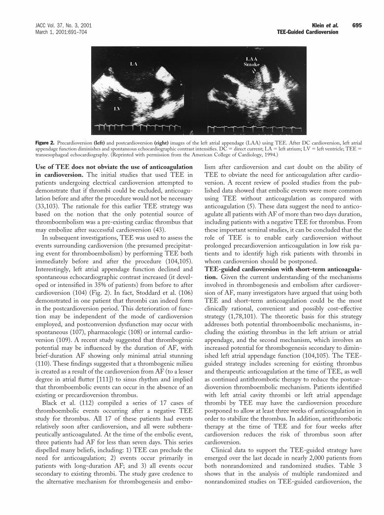
In subsequent investigations, TEE was used to assess theevents surrounding cardioversion (the presumed precipitat-ing event for thromboembolism) by performing TEE bothimmediately before and after the procedure (104,105).Interestingly, left atrial appendage function declined andspontaneous echocardiographic contrast increased (it devel-oped or intensified in 35% of patients) from before to aftercardioversion (104) (Fig. 2). In fact, Stoddard et al. (106)demonstrated in one patient that thrombi can indeed formin the postcardioversion period. This deterioration of func-tion may be independent of the mode of cardioversionemployed, and postconversion dysfunction may occur withspontaneous (107), pharmacologic (108) or internal cardio-version (109). A recent study suggested that thrombogenicpotential may be influenced by the duration of AF, withbrief-duration AF showing only minimal atrial stunning(110). These findings suggested that a thrombogenic milieuis created as a result of the cardioversion from AF (to a lesserdegree in atrial flutter [111]) to sinus rhythm and impliedthat thromboembolic events can occur in the absence of anexisting or precardioversion thrombus.
Black et al. (112) compiled a series of 17 cases ofthromboembolic events occurring after a negative TEEstudy for thrombus. All 17 of these patients had eventsrelatively soon after cardioversion, and all were subthera-peutically anticoagulated. At the time of the embolic event,three patients had AF for less than seven days. This seriesdispelled many beliefs, including: 1) TEE can preclude theneed for anticoagulation; 2) events occur primarily inpatients with long-duration AF; and 3) all events occursecondary to existing thrombi. The study gave credence tothe alternative mechanism for thrombogenesis and embo-
lism after cardioversion and cast doubt on the ability ofTEE to obviate the need for anticoagulation after cardio-version. A recent review of pooled studies from the pub-lished data showed that embolic events were more commonusing TEE without anticoagulation as compared withanticoagulation (5). These data suggest the need to antico-agulate all patients with AF of more than two days duration,including patients with a negative TEE for thrombus. Fromthese important seminal studies, it can be concluded that therole of TEE is to enable early cardioversion withoutprolonged precardioversion anticoagulation in low risk pa-tients and to identify high risk patients with thrombi inwhom cardioversion should be postponed.TEE-guided cardioversion with short-term anticoagula-tion. Given the current understanding of the mechanismsinvolved in thrombogenesis and embolism after cardiover-sion of AF, many investigators have argued that using bothTEE and short-term anticoagulation could be the mostclinically rational, convenient and possibly cost-effectivestrategy (1,78,101). The theoretic basis for this strategyaddresses both potential thromboembolic mechanisms, in-cluding the existing thrombus in the left atrium or atrialappendage, and the second mechanism, which involves anincreased potential for thrombogenesis secondary to dimin-ished left atrial appendage function (104,105). The TEE-guided strategy includes screening for existing thrombusand therapeutic anticoagulation at the time of TEE, as wellas continued antithrombotic therapy to reduce the postcar-dioversion thromboembolic mechanism. Patients identifiedwith left atrial cavity thrombi or left atrial appendagethrombi by TEE may have the cardioversion procedurepostponed to allow at least three weeks of anticoagulation inorder to stabilize the thrombus. In addition, antithrombotictherapy at the time of TEE and for four weeks aftercardioversion reduces the risk of thrombus soon aftercardioversion.
Clinical data to support the TEE-guided strategy haveemerged over the last decade in nearly 2,000 patients fromboth nonrandomized and randomized studies. Table 3shows that in the analysis of multiple randomized andnonrandomized studies on TEE-guided cardioversion, the
Figure 2. Precardioversion (left) and postcardioversion (right) images of the left atrial appendage (LAA) using TEE. After DC cardioversion, left atrialappendage function diminishes and spontaneous echocardiographic contrast intensifies. DC 5 direct current; LA 5 left atrium; LV 5 left ventricle; TEE 5transesophageal echocardiography. (Reprinted with permission from the American College of Cardiology, 1994.)
695JACC Vol. 37, No. 3, 2001 Klein et al.March 1, 2001:691–704 TEE-Guided Cardioversion

cardioversion-related embolic event rate is very low (0.35%[7 of 1,996 patients]) (4,16,103,113–116).Nonrandomized studies. An important nonrandomizedstudy (3) showed that early cardioversion after TEE couldbe performed when atrial thrombi were excluded. Theseinvestigators studied 230 patients with TEE before cardio-version of AF and detected atrial thrombi in 34 patients(15%). No clinically apparent embolic events were detectedafter cardioversion in 186 (95%) of 196 patients in whom noatrial thrombi were detected and who were spared long-term anticoagulation before cardioversion as a result of anegative TEE study for thrombus. In a more recent study of466 patients by the same investigators (114), thrombi weredetected in 13.9%, and 88% of the patients without thrombiwere converted successfully to sinus rhythm. However, onepatient (0.2%) who had been converted to sinus rhythmwith relatively low risk had a peripheral embolus six daysafter cardioversion. If the duration of AF was less than threeweeks, patients undergoing TEE-guided cardioversion wereless likely to have recurrent AF and were able to maintainsinus rhythm at one year, as compared with patients withAF longer than three weeks (81% vs. 60%; p 5 0.005)
(114). Other investigators (106,116) have described similarpromising results in nonrandomized patients undergoing aTEE-guided strategy.Randomized studies. The ACUTE pilot study was thefirst prospective, randomized study of 126 patients with AFfor longer than two days duration undergoing cardioversion(4). Patients from 10 international clinical sites were ran-domized to either a TEE-guided strategy with brief anti-coagulation (n 5 62) or the conventional anticoagulationapproach (n 5 64). The primary end point events wereischemic stroke, transient ischemic attack and systemicembolization for a four-week postcardioversion period. Thefeasibility, safety and other outcomes of the ACUTE pilotstudy are summarized in Table 4.
This pilot study demonstrated that a TEE-guided strat-egy was both feasible and safe as compared with conven-tional therapy (4). The TEE-guided strategy allowed earlycardioversion without embolization in 100% of the patientswithout thrombi on TEE. Atrial thrombi were detected inseven patients (13%), leading to postponement of electricalcardioversion. Notably, cardioversion was done much earlierwith the TEE-guided strategy than with the conventionalstrategy (0.6 weeks vs. 4.7 weeks), which highlights theissue of time (4.7 weeks) that is needed to achieve atherapeutic INR before cardioversion. Accordingly, theincidence of clinical instability and bleeding tended to beless in the TEE-guided group (1.6% vs. 7.8%; p 5 0.21).The ACUTE multicenter trial. The ACUTE multicenterstudy was a randomized clinical trial involving patientsundergoing electrical cardioversion of AF lasting longerthan two days. The study compared a TEE-guided strategycombined with short-term anticoagulation with a conven-tional anticoagulation strategy (15) (Fig. 3).
The study was an investigator-initiated trial lackingprincipal sponsorship. The termination before enrolling thetarget 3,000 patients was determined by the Data Safety and
Table 3. Summary of Studies of TransesophagealEchocardiography (TEE)-Guided Approach to Cardioversion ofAtrial Fibrillation, Including the Incidence of Thrombus byTEE and Recorded Embolic Events
StudyReferenceNumber n
AtrialThrombi
EmbolicEvents
Orsinelli (1993) 103 39 9 (23%) 1 (2.56%)Stoddard (1995) 113 206 37 (18%) 0Klein (1997) 4 126 7 (13%) 0Weigner (1998) 114 466 64 (13.9%) 1 (0.21%)Grimm (1998) 115 417 28 (7%) 0Corrado (1999) 116 123 11 (9%) 0ACUTE (2000) 16 619 79 (13.6%) 5 (0.81%)Total 1,996 235 (11.8%) 7 (0.35%)
ACUTE 5 Assessment of Cardioversion Using Transesophageal Echocardiography.
Table 4. Feasibility and Safety Outcome Summary for the ACUTE Pilot Study by Treatment Group
TEE-Guided Approach Conventional Approach p Value
Feasibility outcomesElectrical cardioversion 76% (47/62) (CI 63–86%) 58% (37/64) (CI 45–70%) 0.03Scheduled electrical cardioversion 94% (44/47) (CI 58–82%) 70% (26/37) (CI 29–54%) , 0.01Time from enrollment to electrical cardioversion (weeks) 0.6 (CI 0.3–0.9) 4.8 (CI 3.8–5.7) , 0.01Time from enrollment to normal sinus rhythm (weeks) 1.0 (CI 0.5–1.6) 4.3 (CI 3.0–5.6) , 0.01
Safety outcomesEmbolic event 0% (CI 0–5%) 2% (1/64) (CI 0–8%) . 0.20Cardioversion related death 0% (CI 0–5%) 0% (0/64) (CI 0–5%) . 0.20Hemodynamic instability and bleeding 2% (CI 0–8%) 8% (5/64) (CI 3–17%) . 0.20
Other outcomesPatients with TEE-detected RA or LA thrombi 13% (CI 5–24%) — — —Patients without thrombus by TEE and converted to
normal sinus rhythm75% (CI 62–86%) — — —
Normal sinus rhythm after DCC 85% (CI 51–76%) 76% (28/37) (CI 31–57%) . 0.20Normal sinus rhythm at 8 weeks 55% (CI 42–68%) 56% (37/64) (CI 43–69%) . 0.20Time from enrollment to follow-up (weeks) 5.7 (CI 5.1–63) 7.7 (CI 7.1–8.2) , 0.01
Data are presented as percentage (n) (95% confidence interval [CI]) or mean value (95% CI).DCC 5 direct current cardioversion; LA 5 left atrial; RA 5 right atrial; TEE 5 transesophageal echocardiography.Reprinted with permission from the American College of Physicians, 1997.
696 Klein et al. JACC Vol. 37, No. 3, 2001TEE-Guided Cardioversion March 1, 2001:691–704

Monitoring Board and the ACUTE Steering Committee,which reported low event rates and slow recruitment. Atotal of 1,222 patients were randomized from 70 clinicalinternational sites over a five-year period. The eight-weekstudy outcomes of the ACUTE study were presentedrecently as a late-breaking trial at the ACC’s 2000 ScientificSessions (16,117).
Of the 1,222 randomized patients, 619 were assigned tothe TEE-guided arm, whereas 603 were assigned to theconventional arm. Left or right atrial cavity and atrialappendage thrombi were detected by TEE in 76 patients(14%), resulting in postponement of the cardioversion. TheTEE-guided strategy allowed early (mean three days) suc-cessful cardioversion in 81% of the patients withoutthrombi. There were two patients with embolic eventsoccurring within the first week after cardioversion, whichwere associated with AF recurrence and subtherapeuticINRs (16,117).
Of the 603 patients assigned to the conventional arm,only 333 (55%) had electrical cardioversion (mean 31 days)and 80% were successful. There was one patient who had anembolic stroke occurring within the first week after cardio-version. Of the 270 patients who never underwent electricalcardioversion, the main reason in 127 patients (47%) wasspontaneous or chemical conversion. In contrast, 143 pa-tients never had cardioversion, for other reasons, includingmajor or minor bleeding, medical reasons and lost tofollow-up. There were also 32 patients (5%) who crossedover to the TEE arm because of hemodynamic instability(hypotension or congestive heart failure) (16).
In the ACUTE study, using intention-to-treat analysisthere was no difference in the composite end point of stroke,transient ischemic attack and peripheral embolism betweenthe TEE-guided arm and the conventional arms (0.81% vs.0.50%; p 5 0.50). However, there was a significant differ-ence in the composite end point of major and minorbleeding between the TEE-guided arm and the conven-tional arms (2.9% vs. 5.5%; p 5 0.02) (16).
An important unexpected outcome of the ACUTE mul-ticenter study was that it showed that the aggregate embolicevent rate for both arms of the study was much lower thanexpected (0.7%), and the aggregate composite hemorrhagiccomplication rate was higher than expected (4.2%). Com-pared with the conventional strategy, the TEE-guided armwith short-term anticoagulation decreased the compositerate for major and minor bleeding complications. However,there was no significant difference in 8-week maintenance ofnormal sinus rhythm, cardiac deaths or cardioversion-related deaths between the two arms. The results of thisrandomized study suggested that the TEE-guided approachwith short-term anticoagulation may be considered as aclinically effective alternative to the conventional anticoag-ulation strategy in the management of patients undergoingcardioversion (16,117).Registry of patients undergoing TEE-guided cardiover-sion. The ACUTE registry is a nonrandomized cohort ofpatients with AF from the Cleveland Clinic who underwentTEE-guided cardioversion and were followed for evidenceof stroke. In a recent review of the registry (115), atrialthrombi were detected and cardioversion was postponed in
Figure 3. Protocol design of the ACUTE multicenter study. Enrolled patients are randomly assigned to either the TEE-guided group or the conventionalgroup for an eight-week study period. (Reprinted with permission from Excerpta Medica, Inc., 1998.) ACUTE 5 Assessment of Cardioversion UsingTransesophageal Echocardiography; DCC5 direct current cardioversion; TEE 5 transesophageal echocardiography.
697JACC Vol. 37, No. 3, 2001 Klein et al.March 1, 2001:691–704 TEE-Guided Cardioversion

28 (7%) of the 417 patients. In contrast, cardioversion wasperformed in 388 patients (93%), 363 (94%) of whom weresuccessfully converted to normal sinus rhythm withoutembolic stroke (115). These results, using the TEE-guided(ACUTE) approach, have now been extended to over 600consecutive patients.Cost-effectiveness. In the absence of published studiesshowing the relative clinical effectiveness of the two strate-gies, cost-effectiveness may affect patterns of use. Using adecision analytic model, Seto et al. (118) assessed thecost-effectiveness of the TEE-guided strategy in hospital-ized patients with AF. The cost per quality-adjusted life yearwas compared for three different management strategies: 1)the conventional strategy with initial screening by trans-thoracic echocardiography (TTE); 2) initial TTE, followedby TEE and early cardioversion if an atrial thrombus isexcluded by TEE; and 3) initial TEE, followed by earlycardioversion if no atrial thrombus is detected. The TEE-guided early cardioversion without TTE was the least costly.Recently, they have extended their model and showed thatit was less costly to perform follow-up TEE to documentthrombus resolution for patients with thrombi on theirinitial TEE (119).Atrial thrombus resolution after prolonged cardiover-sion. Table 5 shows a number studies using serial TEEevaluation to document the resolution of thrombi after theinitial TEE (4,106,116,120–122) for these high risk pa-tients. The mechanism for reduction of embolic risk usinganticoagulant agents was thought to be organization andadherence of the thrombus to the left atrial appendage wall,but these TEE studies support atrial thrombus resolutionand prevention of new thrombus (4,116,120). The percent-age of thrombi resolved after three to four weeks ofanticoagulant therapy has varied widely, from 89% resolu-tion in one study (120) to 50% in the ACUTE pilot study(4) to 5% in another study (106). In a recent follow-up TEEstudy of 164 patients with left atrial thrombi, thrombusresolution occurred in 80% of the patients at a mean of 6.7weeks, with little further resolution on subsequent TEEstudies (122). These discrepant findings among differentstudies may relate to several factors, including differentpatient characteristics, duration of AF and diagnostic crite-ria for thrombus detection. In addition, the finding ofthrombus portends a poor outcome, with a risk of stroke or
embolic events of 10.4% per year or a death risk of 15.8%(123). Thus, there are many unresolved issues with regard tomanagement of residual thrombus detected by TEE.Discrepancy of thrombus detection and thromboembo-lism. There remains an inconsistency between the findingof thrombus by TEE in 10% to 15% of the patients with AFundergoing cardioversion and stroke rates ,1% after car-dioversion (1,4,76). There are several explanations for thisdiscrepancy: 1) most thrombi do not embolize and may berelated to the characteristics and location of the thrombus inthe left atrial appendage; 2) not all thromboembolic eventsare clinically apparent; and 3) some appendage thrombidetected by TEE represent false positive findings. Moreinvestigation is needed to determine the relationship be-tween thrombi by TEE and cardioversion-related embolicevents.Prediction of success of conversion to normal sinusrhythm. Initially, there was a lot of enthusiasm about theuse of left atrial appendage areas and flow velocities bypulsed wave Doppler echocardiography to predict the im-mediate and long-term success of cardioversion (1). How-ever, the results have been variable to date (124–128). Inone study (124), the left atrial appendage flow velocities(mean emptying velocity .19 cm/s), maximal left atrialappendage area and duration of AF were useful in predict-ing the initial recovery of sinus rhythm. However, anotherstudy showed that the left atrial appendage flow velocities(mean peak left atrial appendage emptying velocity .35cm/s) were not predictive of either the success of cardiover-sion or the maintenance of sinus rhythm at one-yearfollow-up (126). Thus, the overall data suggest that areduced peak left atrial appendage velocity (generally ,20cm/s) may play some role in predicting the decreased successof cardioversion and maintenance of normal sinus rhythm,but its unique contribution remains unknown.Disadvantages of the TEE-guided strategy. In the con-text of the findings of the ACUTE multicenter study andnonrandomized studies in nearly 2,000 patients, as dis-cussed earlier, the TEE-guided approach with short-termanticoagulation appears to be as feasible and safe as theconventional arm. However, there remain some importantdisadvantages of using the TEE-guided approach (Table 6).The ability of the TEE-guided strategy, as compared withthe conventional approach, to lower the stroke rate has not
Table 5. Previous Studies Documenting Resolution of Atrial Thrombus by SerialTransesophageal Echocardiography
Study(Reference no.) n
Frequency ofThrombus
AnticoagulationDuration
Atrial ThrombusResolved onSecond TEE
Stoddard 1995 (106) 21 NA 5 to 17 weeks 9/21 (43%)Collins 1995 (120) 18 NA 4 weeks (median) 16/18 (89%)Tsai 1997 (121) 8 10% NA 6/8 (75%)Klein 1997 (4) 7 13% 6 weeks 3/7 (43%)Jaber 2000 (122) 164 NA 6.7 weeks (mean) 131/164 (80%)Corrado 1999 (116) 11 11% 4 weeks (median) 9/11 (82%)
NA 5 not available; TEE 5 transesophageal echocardiography.
698 Klein et al. JACC Vol. 37, No. 3, 2001TEE-Guided Cardioversion March 1, 2001:691–704

been shown (5–7,9,16,129). It has been largely assumed thatdetecting a left atrial appendage thrombus and not cardio-verting the patient would avoid an embolic event. Becausethe embolic stroke rate was low (0.7%) (16), a subsequentclinical trial would require .10,000 patients for adequatestatistical power (15). Using this line of reasoning, one mayargue that stroke after cardioversion is so rare; therefore,why bother to spend the resources to detect thrombi(6,7,11)? Another argument is that the bleeding risk reduc-tion with the TEE-guided arm has been exaggerated.Although TEE-guided management cuts the time of anti-coagulation in half, Landefeld (130) has shown that thehighest risk for major bleeding is in the first three to fourweeks of outpatient warfarin therapy, which is common toboth strategies. Furthermore, often patients will need anti-coagulation beyond the seven- to eight-week cardioversionperiod, because they may have some of the important riskfactors for stroke (7) or they may have recurrent AF in thefirst three to six months after cardioversion (131–134).
Also, some investigators have argued that the additionalcost of the TEE procedure is not justified in the absence ofstrong efficacy data. It is estimated from industry sourcesthat between $50 and $100 million is spent on TEE forpatients with AF, without guidelines or criteria for thesepatients (135). Despite models showing potential cost-effectiveness (118), the preliminary ACUTE cost data onthe TEE-guided arm do not appear to be statisticallydifferent from the data on the conventional arm. TheTEE-guided strategy may potentially be more costly thanthe conventional approach because of the increased costsassociated with repeating TEE after four weeks of antico-agulation (119).
Despite the sensitivity of multiplane TEE being .95%,
TEE may miss thrombi ,2 mm, which may have thepotential to embolize, especially in the setting of thecomplex morphology of multilobed appendage (136). Incontrast, with the increasing use of TEE in the community,there may be a tendency for overcalling thrombi (falsepositive results) in the left atrial appendage. It may bedifficult to distinguish thrombi from artifacts, pectinatemuscles and severe spontaneous echocardiographic contrast,or “sludge,” in the left atrial appendage (15,137). Figure 4provides examples of some of the echocardiographic pitfallsin the recognition of left atrial appendage thrombus.
Another important issue is whether the TEE-guidedstrategy is suitable for community hospitals, where equip-ment and personnel resources as well as expertise (level IIIechocardiographers) may be limited (138,139). Transesoph-ageal echocardiography is a semi-invasive technique with apotential for esophageal perforation (96,97).National use of TEE-guided strategy. There is a growingperception that the use of the TEE-guided approach isincreasing nationally. We lack actual Medicare data, andtherefore national health statistics are not yet known.Murray et al. (140) performed a survey of 197 clinicalpractices in the U.S. to evaluate national use of this strategy.The survey revealed that the frequency of the TEE-guidedstrategy varies within institutions from 0% to 80%, with thehighest concentration in tertiary-care institutions and uni-versity hospitals. Overall, the TEE-guided procedure wasbeing used at least occasionally in 75% of the institutionssurveyed and may account for ;12% of all electrical cardio-versions for AF (140). At our own institution, the TEE-guided approach has remained relatively stable, accountingfor 14.7% of the 3,329 electrical cardioversions from 1991 to1997 (140). More recently, TEE-guided cardioversions
Table 6. Advantages and Disadvantages of the Transesophageal Echocardiography(TEE)-Guided Approach to Cardioversion of Patients With Atrial FibrillationUndergoing Cardioversion
Advantages Disadvantages
● Transesophageal echocardiography should beable to detect left atrial appendage thrombi,which increase the risk of embolic strokeafter electrical cardioversion. Thus, sparingpatients with thrombi from cardioversionmay reduce the incidence of embolic events.
● Transesophageal echocardiography is performedwithout any definite guidelines about whoshould receive the procedure (high risk vs. lowrisk) (12).
● In the majority of patients with left atrialappendage thrombi, earlier cardioversionmay shorten the period of anticoagulationand lower the corresponding risk of bleedingcomplications (16,117).
● Residual thrombus on repeat TEE maydiminish the cost-effectiveness of the TEE-guided approach (119).
● A TEE-guided approach may prove morecost-effective owing to the reduction inlaboratory monitoring costs and thereduction in bleeding complications.
● Transesophageal echocardiography requires alevel III–trained physician and availability ofexpensive echocardiographic machines.
● Earlier cardioversion is believed to increasethe likelihood of a successful return to andmaintenance of sinus rhythm (4).
● Transesophageal echocardiography may missthrombi that may embolize after cardioversion(6). In contrast, TEE may render false positiveresults by erroneously identifying spontaneousechocardiographic contrast, “sludge,” multilobedappendages or pectinate muscles as thrombus.
699JACC Vol. 37, No. 3, 2001 Klein et al.March 1, 2001:691–704 TEE-Guided Cardioversion

have increased dramatically at our institution and nowexceed 29% of all electrical cardioversions performed.Status of TEE-guided strategy in 2001. The current datasuggest that the TEE-guided strategy with short-termanticoagulation is a strong alternative to the conventionalstrategy (16,141). Clearly, with stroke rates ,1.0%, theclinician can utilize either management strategy in theirdaily practices, depending on the individual patient orphysician requirements. For example, the clinician at thecommunity hospital may not have the resources or TEEskills (level III) to perform the TEE-guided approach andmay elect to follow the conventional guidelines. In contrast,the hospital-based clinician may tend to perform the TEE-guided strategy more often, because skilled personnel andthe infrastructure are readily available.
From the available evidence, there are certain patientsubgroups that may benefit from the TEE-guided strategy.First, the inpatient with new-onset AF (,4 weeks dura-tion), regardless of risk profile, may benefit from earlycardioversion using the TEE-guided strategy. This may be
particularly important for high risk patients (such as thosewith congestive heart failure, previous embolism or hemo-dynamic instability) in whom the prompt return of normalsinus rhythm would be beneficial (142). Second, the high-risk patient may benefit from further risk stratification byTEE to identify left atrial appendage thrombus, severespontaneous echocardiographic contrast or complex ather-oma (143,144). The identification of a thrombus beforecardioversion would lead to cancellation of the cardioversionand more prolonged (and perhaps intense) anticoagulanttherapy. A repeat TEE would be necessary to show throm-bus resolution before cardioversion. Third, even for patientsin whom the likelihood of thrombus is low (100), eliminat-ing the need for prolonged anticoagulation pre-cardioversionby ruling out the presence of thrombus would allow earlycardioversion and avoid the delay for return to sinus rhythm.We await further data for the intermediate risk patient inwhom the detection rate of thrombus is not well described.Fourth, the inpatient would be better suited to have theTEE-guided approach with intravenous heparin, as com-
Figure 4. Pitfalls in TEE screening for thrombi using the TEE-guided approach to cardioversion, including pectinate muscles (arrows) in the left atrialappendage (A), multilobed appendage (arrows) (B) and viscous spontaneous echocardiographic contrast, or “sludge” (C). Pectinate muscle tissue, multilobedappendage or sludge can be mistaken for thrombi, rendering a false positive TEE screening result. TEE 5 transesophageal echocardiography.
700 Klein et al. JACC Vol. 37, No. 3, 2001TEE-Guided Cardioversion March 1, 2001:691–704

pared with the outpatient. The outpatient setting has somelimitations, because these patients would need three to fivedays of warfarin before the TEE-guided DC cardioversion,and thus the time benefit of early cardioversion is dimin-ished (4,7,31). Outpatient management may be improvedwith the use of low molecular weight heparin, which can beused as bridge therapy to warfarin (see later discussion)(145). Finally, the benefit of the TEE-guided strategyseems to be limited in the chronically anticoagulated patientwith persistent AF undergoing cardioversion.New developments. A major limitation of the TEE-guided approach with intravenous heparin (for inpatients) isthe extended length of the hospital stay to allow for overlapbetween the heparin and warfarin (four to five days).Recently, a newer TEE-guided strategy has been advocatedwith the use of low molecular weight heparin therapy to beused as bridge therapy for anticoagulation. This therapy hasthe potential to lower costs by treating patients as outpa-tients instead of inpatients (146), provide greater patientconvenience and increase quality of life, as compared withintravenous heparin. A randomized clinical pilot study(ACUTE II) is under way to compare TEE-guided cardio-version using low molecular weight heparin with the stan-dard intravenous unfractionated heparin in patients with AF(145). Recently, a TEE-stratified study was able to identify162 low risk patients with AF and atrial flutter lastinglonger than two days and safely perform immediate cardio-version with the use of low molecular weight heparin(dalteparin) to bridge warfarin therapy (147). The low riskpatients receiving immediate cardioversion maintained sinusrhythm better after one month, as compared with thepatients with prolonged precardioversion warfarin therapy.
In addition, there is work in progress involving directthrombin inhibitors and glycoprotein IIa/IIIb antiplatelettherapy for thrombotic prophylaxis of patients with AF.There is also growing interest in the use of chemicalcardioversion with ibutilide or dofetilide and TEE-guidedcardioversion of AF (148,149).
CONCLUSIONS
There remains an ongoing controversy about the manage-ment of patients with AF undergoing cardioversion. Thestandard of care is still conventional management, which isperformed by most cardiologists worldwide. Based on thefindings of both nonrandomized and randomized studies,the TEE-guided approach with short-term anticoagulationcan be considered a safe and clinically effective alternative toconventional management, especially for patients who maybenefit from early cardioversion.
AcknowledgmentsThe authors wish to thank Susan E. Jasper, BSN, ArielGoodman-Bizon, BA, and Marie D. Campbell for theirassistance in the preparation of this manuscript.
Reprint requests and correspondence: Dr. Allan L. Klein, TheCleveland Clinic Foundation, 9500 Euclid Avenue, Departmentof Cardiology/Desk F-15, Cleveland, Ohio 44195. E-mail:[email protected].
REFERENCES
1. Grimm RA, Stewart WJ, Black IW, Thomas JD, Klein AL. Shouldall patients undergo transesophageal echocardiography before electri-cal cardioversion of atrial fibrillation? J Am Coll Cardiol 1994;23:533–41.
2. Laupacis A, Albers G, Dalen J, Dunn MI, Jacobson AK, Singer DE.Antithrombotic therapy in atrial fibrillation. Chest 1998;114:579S–89S.
3. Manning WJ, Silverman DI, Keighley CS, Oettgen P, Douglas PS.Transesophageal echocardiographically facilitated early cardioversionfrom atrial fibrillation using short-term anticoagulation: final resultsof a prospective 4.5-year study. J Am Coll Cardiol 1995;25:1354–61.
4. Klein AL, Grimm RA, Black IW, et al. Cardioversion guided bytransesophageal echocardiography: the ACUTE pilot study—a ran-domized, controlled trial. Ann Intern Med 1997;126:200–9.
5. Moreyra E, Finkelhor RS, Cebul RD. Limitations of transesophagealechocardiography in the risk assessment of patients before nonanti-coagulated cardioversion from atrial fibrillation and flutter: an anal-ysis of pooled trials. Am Heart J 1995;129:71–5.
6. Prystowsky EN. Management of atrial fibrillation: simplicity sur-rounded by controversy. Ann Intern Med 1997;126:244–6.
7. Prystowsky EN. Perspectives and controversies in atrial fibrillation.Am J Cardiol 1998;82:3I–6I.
8. Autore C, Cartoni D, Piccininno M. Multiplane transesophagealechocardiography and stroke. Am J Cardiol 1998;81:79G–81G.
9. Klein AL, Murray RD, Grimm RA. Transesophageal echocar-diography-guided cardioversion: going for broke? Ann Intern Med1997;127:652–3.
10. Klein A, Gersh B. Should TEE be done routinely before cardiover-sion (debate). Presented at the American Society of Echocardiogra-phy Scientific Sessions; Chicago, IL: 2000.
11. Klein AL, Prystowsky EN. All patients with new-onset atrialfibrillation should undergo TEE prior to cardioversion (debate).Presented at the American College of Cardiology 47th AnnualScientific Session, Atlanta, Georgia, 1998.
12. Manning WJ, Gersh B. TEE-guided cardioversion should be theroutine approach; protagonist/antagonist (debate). Presented at theAmerican Heart Association Scientific Sessions, Dallas, Texas, 1998.
13. Prystowsky EN, Benson DW, Fuster V, et al. Management ofpatients with atrial fibrillation: A statement for healthcare profes-sionals from the subcommittee on electrocardiography and electro-physiology, American Heart Association. Circulation 1996;93:1262–77.
14. Cheitlin MD, Alpert JS, Armstrong WF, et al. ACC/AHA guide-lines for the clinical application of echocardiography: executivesummary. A report of the American College of Cardiology/AmericanHeart Association Task Force on Practice Guidelines (Committee onClinical Application of Echocardiography). J Am Coll Cardiol1997;29:862–79.
15. ACUTE Investigators. Design of a clinical trial for the assessment ofcardioversion using transesophageal echocardiography (ACUTEmulticenter study). Am J Cardiol 1998;81:877–83.
16. ACUTE Investigators. Assessment of Cardioversion Using Trans-esophageal Echocardiography (ACUTE) multicenter study: eight-week clinical outcomes. Presented at the American College ofCardiology Scientific Sessions, Late-Breaking Clinical Trial, 2000.
17. Ostrander L, Brandt RL, Kjelsberg M, et al. Electrocardiographicfindings among the adult population of a total natural community—Tecumseh, Michigan. Circulation 1965;31:888–98.
18. Feinberg WM, Blackshear JL, Laupacis A, Kronmal R, Hart RG.Prevalence, age distribution, and gender of patients with atrialfibrillation: analysis and implications. Arch Intern Med 1995;155:469–73.
19. Kannel WB, Abbott RD, Savage DD, McNamara PM. Coronaryheart disease and atrial fibrillation: the Framingham study. AmHeart J 1983;106:389–96.
701JACC Vol. 37, No. 3, 2001 Klein et al.March 1, 2001:691–704 TEE-Guided Cardioversion

20. Blackshear JL, Kopecky SL, Litin SC, Safford RE, Hammill SC.Management of atrial fibrillation in adults: prevention of thrombo-embolism and symptomatic treatment. Mayo Clin Proc 1996;71:150–60.
21. Kannel WB, Abbott RD, Savage DD, McNamara PM. Epidemio-logic features of chronic atrial fibrillation: the Framingham study.N Engl J Med 1982;306:1018–22.
22. Cairns JA, Connolly SJ. Nonrheumatic atrial fibrillation: risk ofstroke and role of antithrombotic therapy. Circulation 1991;84:469–81.
23. Furberg CD, Psaty BM, Manolio TA, Gardin JM, Smith VE,Rautaharju PM. Prevalence of atrial fibrillation in elderly subjects(the Cardiovascular Health Study). Am J Cardiol 1994;74:236–41.
24. Wolf PA. Prevention of stroke. Lancet 1998;352 Suppl 3:SIII15–8.25. Wolf PA, Mitchell JB, Baker CS, Kannel WB, D’Agostino RB.
Impact of atrial fibrillation on mortality, stroke and medical costs.Arch Intern Med 1998;158:229–34.
26. Laupacis A, Albers G, Dalen J, Dunn M, Feinberg W, Jacobson A.Antithrombotic therapy in atrial fibrillation. Chest 1995;108:352S–9S.
27. Wolf PA, Dawber TR, Thomas HJ, Kannel WB. Epidemiologicassessment of chronic atrial fibrillation and risk of stroke: theFramingham study. Neurology 1978;28:973–7.
28. Wolf PA, Abbott RD, Kannel WB. Atrial fibrillation: a majorcontributor to stroke in the elderly. The Framingham Study. ArchIntern Med 1987;147:1561–4.
29. Wolf PA, Abbott RD, Kannel WB. Atrial fibrillation as an indepen-dent risk factor for stroke: the Framingham study. Stroke 1991;22:983–8.
30. Atrial Fibrillation Investigators. Risk factors for stroke and efficacy ofantithrombotic therapy in atrial fibrillation: analysis of pooled datafrom five randomized controlled trials. Arch Intern Med 1994;154:1449–57.
31. Ezekowitz MD, Levine JA. Preventing stroke in patients with atrialfibrillation. JAMA 1999;281:1830–5.
32. Rittoo D, Sutherland G, Currie P, Starkey I, Shaw T. A prospectivestudy of left atrial spontaneous echo contrast and thrombus in 100consecutive patients referred for balloon dilation of the mitral valve.J Am Soc Echocardiogr 1994;7:516–27.
33. Black IW, Hopkins AP, Lee LC, Walsh WF. Evaluation oftransesophageal echocardiography before cardioversion of atrial fibril-lation and flutter in nonanticoagulated patients. Am Heart J 1993;126:375–81.
34. Gosselink AT, Crijns HJ, van den Berg MP, et al. Functionalcapacity before and after cardioversion of atrial fibrillation: a con-trolled study. Br Heart J 1994;72:161–6.
35. Gosselink AT, Bijlsma EB, Landsman ML, Crijns HJ, Lie KI.Long-term effect of cardioversion on peak oxygen consumption inchronic atrial fibrillation: a 2-year follow-up. Eur Heart J 1994;15:1368–72.
36. Grogan M, Smith H, Gersh B, Wood D. Left ventricular dysfunc-tion due to atrial fibrillation in patients initially believed to haveidiopathic dilated cardiomyopathy. Eur Heart J 1992;69:1570–3.
37. Shinbane JS, Wood MA, Jensen DN, Ellenbogen KA, FitzpatrickAP, Scheinman MM. Tachycardia-induced cardiomyopathy: a re-view of animal models and clinical studies. J Am Coll Cardiol1997;29:709–15.
38. Wijffels M, Kirchhof C, Dorland R, Allessie M. Atrial fibrillationbegets atrial fibrillation: a study in awake chronically instrumentedgoats. Circulation 1995;92:1954–68.
39. SPAF Investigators. Stroke Prevention in Atrial Fibrillation (SPAF)study: final results. Circulation 1991;84:527–39.
40. Nattel S. Newer developments in the management of atrial fibrilla-tion. Am Heart J 1995;130:1094–106.
41. Lown B, Amarasingham R, Neuman J. New method for terminatingcardiac arrhythmias. JAMA 1962;182:548–55.
42. Sokolow M, Ball RE. Factors influencing conversion of chronic atrialfibrillation with specific reference to serum quinidine concentration.Circulation 1956;14:568–85.
43. Goldman MJ. The management of chronic atrial fibrillation. ProgCardiovasc Dis 1960;2:465–79.
44. Lown B, Perlroth MG, Kaidbey S, Abe T, Harken DE. “Cardiover-sion” of atrial fibrillation: a report on the treatment of 65 episodes in50 patients. N Engl J Med 1963;269:325–31.
45. Killip T, Baer RA. Hemodynamic effects after reversion from atrialfibrillation to sinus rhythm by precordial shock. J Clin Invest1966;45:658–71.
46. Freeman I, Wexler J. Atrial fibrillation: anticoagulation and quinidi-nization. South Med J 1967;60:13–7.
47. Rokseth R, Storstein Q. Quinidine therapy of chronic auricularfibrillation: the occurrence and mechanism of syncope. Arch InternMed 1963;111:184–9.
48. Morris JJ, Peter RH, McIntosh HD, Kong Y, North WC. Experi-ence with cardioversion of atrial fibrillation and flutter. Am J Cardiol1964;14:94–100.
49. Oram S, Davies JPH. Further experience of electrical conversion ofatrial fibrillation to sinus rhythm: analysis of 100 patients. Lancet1964;1:1294–8.
50. Hurst JW, Paulk EA Jr, Proctor HD, et al. Management of patientswith atrial fibrillation. Am J Med 1964;37:728–41.
51. Morris JJ, Peter RH, McIntosh HD. Electrical conversion of atrialfibrillation: immediate and long-term results and selection of pa-tients. Ann Intern Med 1966;65:216–31.
52. Korsgren M, Leskinen E, Peterhoff V, et al. Conversion of atrialarrhythmias with DC shock: primary results and a follow-up inves-tigation. Acta Med Scand 1965;177 Suppl 431:1–40.
53. Halmos PB. Direct current cardioversion of atrial fibrillation. BrHeart J 1966;28:302–8.
54. Selzer A, Kelly JJ, Johnson RB, et al. Immediate and long-termresults of electrical conversion of arrhythmias. Prog Cardiovasc Dis1966;9:90–104.
55. Lown B. Electrical reversion of cardiac arrhythmias. Br Heart J1967;29:469–89.
56. Resnekov L. Haemodynamic studies before and after electricalconversion of atrial fibrillation and flutter to sinus rhythm. Br Heart J1967;29:700–8.
57. Hall JI, Wood DR. Factors affecting cardioversion of atrial arrhyth-mias with special reference to quinidine. Br Heart J 1968;30:84–90.
58. Radford MD, Evans DW. Long-term results of DC reversion ofatrial fibrillation. Br Heart J 1968;30:91–6.
59. Aberg H, Cullhed I. Direct current conversion of atrial fibrillation:long-term results. Acta Med Scand 1968;184:433–40.
60. Bjerkelund CJ, Orning OM. The efficacy of anticoagulant therapy inpreventing embolism related to DC electrical conversion of atrialfibrillation. Am J Cardiol 1969;23:208–16.
61. McCarthy C, Varghese PJ, Barritt DW. Prognosis of atrial arrhyth-mias treated by electrical counter shock therapy: a three-year follow-up. Br Heart J 1969;31:496–500.
62. Henry WL, Morganroth J, Pearlman AS, et al. Relation betweenechocardiographically determined left atrial size and atrial fibrillation.Circulation 1976;53:273–9.
63. Roy D, Marchand E, Gagne P, Chabot M, Cartier R. Usefulness ofanticoagulant therapy in the prevention of embolic complications ofatrial fibrillation. Am Heart J 1986;112:1039–43.
64. Arnold AZ, Mick MJ, Mazurek RP, Loop FD, Trohman RG. Roleof prophylactic anticoagulation for direct current cardioversion inpatients with atrial fibrillation or atrial flutter. J Am Coll Cardiol1992;19:851–5.
65. Carlsson J, Tebbe U, Rox J, et al. Cardioversion of atrial fibrillationin the elderly. Am J Cardiol 1996;78:1380–4.
66. Mitchell MA, Hughes GS, Ellenbogen KA, et al. Cardioversionrelated stroke rates in atrial fibrillation and atrial flutter. Circulation1997;96 Suppl I:I453.
67. Johnston SD, Trouton TG, Wilson C. A review of direct currentcardioversions for atrial arrhythmia. Ulster Med J 1998;67:19–24.
68. Laupacis A, Albers G, Dunn M, Feinberg W. Antithrombotictherapy in atrial fibrillation: third ACCP Conference on Antithrom-botic Therapy. Chest 1992;102:426S–53S.
69. Manning WJ, Leeman DE, Gotch PJ, Come PC. Pulsed Dopplerevaluation of atrial mechanical function after electrical cardioversionof atrial fibrillation. J Am Coll Cardiol 1989;13:617–23.
70. Schlicht JR, Davis RC, Naqi K, Cooper W, Rao BV. Physicianpractices regarding anticoagulation and cardioversion of atrial fibril-lation. Arch Intern Med 1996;156:290–4.
71. Manning WJ, Silverman DI, Katz SE, et al. Temporal dependence ofthe return of atrial mechanical function on the mode of cardioversionof atrial fibrillation to sinus rhythm. Am J Cardiol 1995;75:624–6.
72. Silverman DI, Manning WJ. Role of echocardiography in patients
702 Klein et al. JACC Vol. 37, No. 3, 2001TEE-Guided Cardioversion March 1, 2001:691–704
undergoing elective cardioversion of atrial fibrillation. Circulation1998;98:479–86.
73. Weinberg DM, Mancini J. Anticoagulation for cardioversion of atrialfibrillation. Am J Cardiol 1989;63:745–6.
74. Forfar J. A 7-year analysis of haemorrhage in patients on long termanticoagulant treatment. Br Heart J 1979;42:128–32.
75. Connolly SJ, Laupacis A, Gent M, Roberts RS, Cairns JA, Joyner C.Canadian Atrial Fibrillation Anticoagulation (CAFA) study. J AmColl Cardiol 1991;18:349–55.
76. Levine MN, Raskob G, Hirsch J. Hemorrhagic complications oflong-term anticoagulant therapy. Chest 1989;95 Suppl:36–5.
77. Levine MN, Raskob G, Landefeld S, Hirsh J. Hemorrhagic compli-cations of anticoagulant treatment. Chest 1995;108:276S–90S.
78. Leung DY, Grimm RA, Klein AL. Transesophageal echocardiog-raphy-guided approach to cardioversion of atrial fibrillation. ProgCardiovasc Dis 1996;39:21–32.
79. Mugge A, Kuhn H, Daniel WG. The role of transesophagealechocardiography in the detection of left atrial thrombi. Echocardi-ography 1993;10:405–17.
80. Mugge A, Kuhn H, Nikutta P, Grote J, Lopez JA, Daniel WG.Assessment of left atrial appendage function by biplane transesoph-ageal echocardiography in patients with nonrheumatic atrial fibrilla-tion: identification of a subgroup of patients at increased embolic risk.J Am Coll Cardiol 1994;23:599–607.
81. Manning WJ, Weintraub RM, Waksmonski CA, et al. Accuracy oftransesophageal echocardiography for identifying left atrial thrombi.Ann Intern Med 1995;123:817–22.
82. Manning WJ. Atrial fibrillation, transesophageal echo, electricalcardioversion, and anticoagulation. Clin Cardiol 1995;18:122–8.
83. Aschenberg W, Schlulter M, Kremer P, Schroder E, Siglow V,Bleifeld W. Transesophageal two-dimensional echocardiography forthe detection of left atrial appendage thrombus. J Am Coll Cardiol1986;7:163–6.
84. Mugge A, Daniel WG, Hausmann J, Godke J, Wagenbreth L,Lichtlen PR. Diagnosis of left atrial appendage thrombi by trans-esophageal echocardiography: clinical implications and follow-up.Am J Card Imaging 1990;4:173–9.
85. Acar J, Michel PL, Cormier B, Vahanian A, Iung B. Features ofpatients with severe mitral stenosis with respect to atrial rhythm:atrial fibrillation in predominant and tight mitral stenosis. ActaCardiol 1992;47:115–24.
86. Olson JD, Goldenberg IF, Pedersen W, et al. Exclusion of atrialthrombus by transesophageal echocardiography. J Am Soc Echocar-diogr 1992;5:52–6.
87. Hwang JJ, Chen JJ, Lin SC, et al. Diagnostic accuracy of left atrialappendage thrombi by transesophageal echocardiography for detect-ing left atrial thrombi in patients with rheumatic heart disease havingundergone mitral valve operations. Am J Cardiol 1993;72:677–81.
88. Fatkin D, Scalia G, Jacobs N, et al. Accuracy of biplane transesoph-ageal echocardiography in detecting left atrial thrombus. Am JCardiol 1996;77:321–3.
89. Schweizer P, Bardos P, Erbel R, et al. Detection of left atrial thrombiby echocardiography. Br Heart J 1981;45:148–56.
90. Beppu S, Nimura Y, Sakakibara H, Nagata S, Park YD, Izumi S.Smoke-like echo in the left atrial cavity in mitral valve disease: itsfeatures and significance. J Am Coll Cardiol 1985;6:744–9.
91. Iliceto S, Antonelli G, Sorino M, Biasco G, Rizzon P. Dynamicintracavitary left atrial echoes in mitral stenosis. Am J Cardiol1985;55:603–6.
92. Daniel WG, Nellessen U, Schroder E, et al. Left atrial spontaneouscontrast in mitral valve disease: an indicator for increased thrombo-embolic risk. J Am Coll Cardiol 1988;11:1204–11.
93. Erbel R, Stern H, Ehrenthal W, et al. Detection of spontaneousechocardiographic contrast within the left atrium by transesophagealechocardiography: spontaneous echocardiographic contrast. ClinCardiol 1986;9:245–52.
94. Black IW, Hopkins AP, Lee LC, Walsh WF. Left atrial spontaneousecho contrast: a clinical and echocardiographic analysis. J Am CollCardiol 1991;18:398–404.
95. Leung DY, Black IW, Cranney GB, Hopkins AP, Walsh WF.Prognostic implications of left atrial spontaneous echo contrast innonvalvular atrial fibrillation. J Am Coll Cardiol 1994;24:755–62.
96. Daniel WG, Erbel R, Kasper W, et al. Safety of transesophageal
echocardiography: a multicenter survey of 10,419 examinations.Circulation 1991;83:817–21.
97. Khandheira B, Seward J, Tajik A. Transesophageal echocardiogra-phy. Mayo Clin Proc 1994;69:856–63.
98. Stollberger C, Chnupa P, Kronik G, et al. Transesophageal echocar-diography to assess embolic risk in patients with atrial fibrillation.ELAT study group, Embolism in Left Atrial Thrombi. Ann InternMed 1998;128:630–8.
99. Zabalgoitia M, Halperin JL, Pearce LA, Blackshear JL, Asinger RW,Hart RG, the Stroke Prevention in Atrial Fibrillation III Investiga-tors. Transesophageal echocardiographic correlates of clinical risk ofthromboembolism in nonvalvular atrial fibrillation. J Am Coll Car-diol 1998;31:1622–6.
100. The SPAF III Writing Committee for the Stroke Prevention inAtrial Fibrillation Investigators. Patients with nonvalvular atrialfibrillation at low risk of stroke during treatment with aspirin: theStroke Prevention in Atrial Fibrillation III study. JAMA 1998;279:1273–7.
101. Main ML, Klein AL. Cardioversion in atrial fibrillation: indications,thromboembolic prophylaxis, and role of transesophageal echocardi-ography. J Thromb Thrombolysis 1999;7:53–60.
102. Blackshear JL, Zabalgoitia M, Pennock G, et al., the StrokePrevention and Atrial Fibrillation Transesophageal Echocardiogra-phy Investigators. Warfarin safety and efficacy in patients withthoracic aortic plaque and atrial fibrillation. Am J Cardiol 1999;83:453–5.
103. Orsinelli DA, Pearson AC. Usefulness of transesophageal echocar-diography to screen for left atrial thrombus before elective cardiover-sion for atrial fibrillation. Am J Cardiol 1993;72:1337–9.
104. Grimm RA, Stewart WJ, Maloney JD, et al. Impact of electricalcardioversion for atrial fibrillation on left atrial appendage functionand spontaneous echo contrast: characterization by simultaneoustransesophageal echocardiography. J Am Coll Cardiol 1993;22:1359–66.
105. Fatkin D, Kuchar DL, Thorburn CW, Feneley MP. Transesopha-geal echocardiography before and during direct current cardioversionof atrial fibrillation: evidence for “atrial stunning” as a mechanism ofthromboembolic complications. J Am Coll Cardiol 1994;23:307–16.
106. Stoddard MF, Dawkins PR, Prince CR, Longaker RA. Transesoph-ageal echocardiographic guidance of cardioversion in patients withatrial fibrillation. Am Heart J 1995;129:1204–15.
107. Grimm RA, Leung DY, Black IW, Stewart WJ, Thomas JD, KleinAL. Left atrial appendage “stunning” after spontaneous conversion ofatrial fibrillation demonstrated by transesophageal Doppler echocar-diography. Am Heart J 1995;130:174–6.
108. Falcone RA, Morady F, Armstrong WF. Transesophageal echocar-diographic evaluation of left atrial appendage function and sponta-neous contrast formation after chemical or electrical cardioversion ofatrial fibrillation. Am J Cardiol 1996;78:435–9.
109. Omran H, Jung W, Rabahieh R, et al. Left atrial chamber andappendage function after internal atrial defibrillation: a prospectiveand serial transesophageal echocardiographic study. J Am CollCardiol 1997;29:131–8.
110. Sparks PB, Jayaprakash S, Mond HG, Vohra JK, Grigg LE, KalmanJM. Left atrial mechanical function after brief duration atrial fibril-lation. J Am Coll Cardiol 1999;33:342–9.
111. Grimm RA, Stewart WJ, Thomas JD, Klein AL. Left atrial append-age “stunning” following electrical cardioversion of atrial flutter: anattenuated response as compared to atrial fibrillation as a mechanismfor a lower susceptibility to thromboembolic events. J Am CollCardiol 1997;29:582–9.
112. Black IW, Fatkin D, Sagar KB, et al. Exclusion of atrial thrombus bytransesophageal echocardiography does not preclude embolism aftercardioversion of atrial fibrillation: a multicenter study. Circulation1994;89:2509–13.
113. Stoddard MF, Dawkins PR, Prince CR, Ammash NM. Left atrialappendage thrombus is not uncommon in patients with acute atrialfibrillation and a recent embolic event: a transesophageal echocardio-graphic study. J Am Coll Cardiol 1995;25:452–9.
114. Weigner MJ, Patel U, Thomas LR, et al. Transesophageal echocar-diography facilitated cardioversion from atrial fibrillation: short-termsafety and implications regarding maintenance of sinus rhythm(abstr). Circulation 1998;98 Suppl I:I500.
115. Grimm RA, Agler DA, Vaughn SE, et al. TEE-guided anticoagu-
703JACC Vol. 37, No. 3, 2001 Klein et al.March 1, 2001:691–704 TEE-Guided Cardioversion
lation in patients undergoing electrical cardioversion of atrial fibril-lation: results from the ACUTE Registry (abstr). J Am Coll Cardiol1998;31 Suppl:353A.
116. Corrado G, Tadeo G, Beretta S, et al. Atrial thrombi resolution afterprolonged anticoagulation in patients with atrial fibrillation. Chest1999;115:140–3.
117. Kleiman NS, Califf RM. Results from late-breaking clinical trialsessions at ACCIS 2000 and ACC 2000. J Am Coll Cardiol2000;36:310–25.
118. Seto TB, Taira DA, Tsevat J, Manning WJ. Cost-effectiveness oftransesophageal echocardiographic-guided cardioversion: a decisionanalytic model for patients admitted to the hospital with atrialfibrillation. J Am Coll Cardiol 1997;29:122–30.
119. Seto TB, Taira DA, Manning WJ. Cardioversion in patients withatrial fibrillation and left atrial thrombi on initial transesophagealechocardiography: should transesophageal echocardiography be re-peated before elective cardioversion? A cost-effectiveness analysis.J Am Soc Echocardiogr 1999;12:508–16.
120. Collins LJ, Silverman DI, Douglas PS, Manning WJ. Cardioversionof nonrheumatic atrial fibrillation: reduced thromboembolic compli-cations with four weeks of precardioversion anticoagulation arerelated to atrial thrombus resolution. Circulation 1995;92:160–3.
121. Tsai LM, Lin LJ, Teng JK, Chen JH. Prevalence and clinicalsignificance of left atrial thrombus in nonrheumatic atrial fibrillation.Int J Cardiol 1997;58:163–9.
122. Jaber WA, Prior DL, Thamilarasan M, et al. Efficacy of anticoagu-lation in resolving left atrial and left atrial appendage thrombi: atransesophageal echocardiographic study. Am Heart J 2000;140:150–6.
123. Leung DY, Davidson PM, Cranney GB, Walsh WF. Thromboem-bolic risks of left atrial thrombus detected by transesophageal echo-cardiogram. Am J Cardiol 1997;79:626–9.
124. Manabe K, Oki T, Tabata T, et al. Transesophageal echocardio-graphic prediction of initially successful electrical cardioversion ofisolated atrial fibrillation: effects of left atrial appendage function. JpnHeart J 1997;38:487–95.
125. Omran H, Jung W, Schimpf R, et al. Echocardiographic parametersfor predicting maintenance of sinus rhythm after internal atrialdefibrillation. Am J Cardiol 1998;81:1446–9.
126. Perez Y, Duval AM, Carville C, et al. Is left atrial appendage flow apredictor for outcome of cardioversion of nonvalvular atrial fibrilla-tion? A transthoracic and transesophageal echocardiographic study.Am Heart J 1997;134:745–51.
127. Verhorst PM, Kamp O, Welling RC, Van Eenige MJ, Visser CA.Transesophageal echocardiographic predictors for maintenance ofsinus rhythm after electrical cardioversion of atrial fibrillation. Am JCardiol 1997;79:1355–9.
128. Agmon Y, Khandheria BK, Gentile F, Seward JB. Echocardio-graphic assessment of the left atrial appendage. J Am Coll Cardiol1999;34:1867–77.
129. Chambers J. Is it safe to cardiovert atrial fibrillation without antico-agulation if the transesophageal echocardiogram is normal? InternJ Cardiol 1998;63:107–9.
130. Landefeld C, Goldman L. Major bleeding in outpatients treated withwarfarin: incidence and prediction by factors known at the start ofoutpatient therapy. Am J Med 1989;87:144–52.
131. Golzari H, Cebul RD, Bahler RC. Atrial fibrillation: restoration andmaintenance of sinus rhythm and indications for anticoagulationtherapy. Ann Intern Med 1996;125:311–23.
132. Arnar DO, Danielsen R. Factors predicting maintenance of sinusrhythm after direct current cardioversion of atrial fibrillation andflutter: a reanalysis with recently acquired data. Cardiology 1996;87:181–8.
133. Van Gelder IC, Crijns HJ, Tieleman RG, et al. Chronic atrialfibrillation: success of serial cardioversion therapy and safety of oralanticoagulation. Arch Intern Med 1996;156:2585–92.
134. Tieleman RG, Van Gelder I, Crijns H, et al. Early recurrence ofatrial fibrillation after electrical cardioversion: a result of fibrillation-induced electrical remodeling of the atria? J Am Coll Cardiol1998;3:167–73.
135. Hart RG, Halperin JL. Atrial fibrillation and thromboembolism: adecade of progress in stroke prevention. Ann Intern Med 1999;13:688–95.
136. Veinot JP, Harrity PJ, Gentile F, et al. Anatomy of the normal leftatrial appendage: a quantitative study of age-related changes in 500autopsy hearts—implications for echocardiographic examination.Circulation 1997;96:3112–5.
137. Murray RD, Bashir M, Jasper SE, Grimm RA, et al. Left atrialappendage “sludge” in atrial fibrillation patients undergoing TEEscreening for cardioversion: the ACUTE registry (abstr). J Am SocEchocardiogr 2000;13:445.
138. Pearlman AS, Gardin J, Martin R, et al. Guidelines for physiciantraining in transesophageal echocardiography: recommendations ofthe American Society of Echocardiography Committee for PhysicianTraining in Echocardiography. J Am Soc Echocardiogr 1992;5:187–94.
139. Stewart WJ, Aurigemma GP, Bierman FZ, et al. Guidelines fortraining in adult cardiovascular medicine. Core cardiology trainingsymposium. Task force 4: training in echocardiography. J Am CollCardiol 1995;25:16–9.
140. Murray RD, Goodman A, Lieber E, et al. National use of thetransesophageal echocardiographic-guided approach to cardioversionfor patients in atrial fibrillation. Am J Cardiol 2000;85:239–44.
141. Klein AL. Emerging role of echocardiography in the evaluation ofpatients with atrial fibrillation into the new millennium. Echocar-diogr 2000;17:353–6.
142. Gersh BJ. Antithrombotic therapy in nonrheumatic/nonvalvularatrial fibrillation. J Cardiovasc Electrophysiol 1999;10:461–71.
143. The Stroke Prevention in Atrial Fibrillation (SPAF) Investigators,Committee on Echocardiography. Transesophageal echocardio-graphic correlates of thromboembolism in high-risk patients withnonvalvular atrial fibrillation. Ann Intern Med 1998;128:639–47.
144. Manning WJ, Douglas PS. Transesophageal echocardiography andatrial fibrillation: added value or expensive toy? Ann Intern Med1998;128:685–7.
145. Murray RD, Shah A, Jasper S, et al. Transesophageal echocardiog-raphy-guided enoxaparin antithrombotic strategy for cardioversion ofatrial fibrillation: the ACUTE II pilot study. Am Heart J 2000;139:e5.
146. Murray RD, Deitcher SR, Shah A, et al. Potential clinical efficacyand cost benefit of a TEE-guided low molecular weight heparin(enoxaparin) approach to antithrombotic therapy in patients under-going immediate cardioversion from atrial fibrillation. J Am SocEchocardiogr 2001. In press.
147. Roijer A, Eskilsson J, Olssen B. Transesophageal echocardiography-guided cardioversion of atrial fibrillation or flutter: selection of alow-risk group for immediate cardioversion. Eur Heart J 2000;21:837–47.
148. Murray KT. Ibutilide. Circulation 1998;97:493–7.149. Torp-Pedersen C, Moller M, Bloch-Thomsen PE, et al. Dofetilide
in patients with congestive heart failure and left ventricular dysfunc-tion. Danish Investigators of arrhythmia and mortality on dofetilidestudy group. N Engl J Med 1999;341:857–65.
704 Klein et al. JACC Vol. 37, No. 3, 2001TEE-Guided Cardioversion March 1, 2001:691–704