Embed Size (px)
Citation preview

www.elsevier.com/locate/ynbdi
Neurobiology of Disease 25 (2007) 17–26Role of the transcription factor E2F1 in CXCR4-mediatedneurotoxicity and HIV neuropathology
Saori Shimizu,a Muhammad Z. Khan,a Randi L Hippensteel,a Anjum Parkar,a
Ramesh Raghupathi,b and Olimpia Meuccia,⁎
aDepartment of Pharmacology and Physiology, Drexel University, College of Medicine, 245 North 15th Street, NCB 8804, Philadelphia, PA 19102, USAbDepartment of Neurobiology and Anatomy, Drexel University, College of Medicine, 2900 Queen Lane, Philadelphia, PA 19129, USA
Received 30 May 2006; revised 8 August 2006; accepted 15 August 2006Available online 28 September 2006
This study sought to determine the role of the transcription factorE2F1 in CXCR4-mediated neurotoxicity and HIV neuropathology. Westudied the effect of the HIV envelope protein gp120 on the expressionof E2F1-dependent apoptotic proteins in human and rodent neuronsand examined the expression pattern of E2F1 in the brain of HIV-infected individuals. Our findings suggest that in cultured neuronsgp120 increased E2F1 levels in the nucleus, stimulated its transcrip-tional activity and enhanced the expression of the E2F1 target proteinsCdc2 and Puma. Studies with neuronal cultures from E2F1 deficientmice demonstrated that the transcription factor is required for gp120-induced neurotoxicity and up-regulation of Cdc2 and Puma. Levels ofE2F1 protein were greater in the nucleus of neurons in brains of HIV-infected patients exhibiting dementia when compared to HIV-negativesubjects or HIV-positive neurologically normal patients. Overall, thesestudies indicate that E2F1 is primarily involved in CXCR4-mediatedneurotoxicity and HIV neuropathogenesis.© 2006 Elsevier Inc. All rights reserved.
Keywords: Neuronal death; Cdc2; Cell cycle proteins; Chemokines; gp120
Introduction
The chemokine receptor CXCR4 is virtually ubiquitous andplays important roles in many physiological processes, such asneuronal migration, survival and differentiation; this receptor hasalso been implicated in the pathogenesis of cancer and AIDS—including NeuroAIDS (Tran and Miller, 2003). Analogous to otherinflammatory disorders, neuronal injury and death are often found
Abbreviations: AIDS, acquired immunodeficiency syndrome; CNS,central nervous system; HAART, highly active antiretroviral therapy; HAD,HIV-associated dementia; HIV, human immunodeficiency virus; HIVE, HIVencephalopathy; Rb, retinoblastoma; SIVE, simian immunodeficiency virusencephalitis.⁎ Corresponding author. Fax: +1 215 762 2299.E-mail address: [email protected] (O. Meucci).Available online on ScienceDirect (www.sciencedirect.com).
0969-9961/$ - see front matter © 2006 Elsevier Inc. All rights reserved.doi:10.1016/j.nbd.2006.08.004
in AIDS patients (Adle-Biassette et al., 1995; Petito and Roberts,1995; Shi et al., 1996). Cellular alterations not necessarilyassociated to cell loss, such as dendritic pruning and synapticimpairment, are also thought to contribute to NeuroAIDS (Bellizziet al., 2005; Everall et al., 1999; Sa et al., 2004). Although highlyactive antiretroviral therapy (HAART) has greatly reduced HIVmortality and morbidity, the neurological complications of HIVinfection significantly affect the clinical management of treatedpatients, and HIV-dementia still represents a serious concern forpatients that have limited access to therapy (reviewed in Gonzalez-Scarano and Martin-Garcia, 2005; McArthur et al., 2003). Asneurons are not the primary targets of HIV in the brain – the virusmostly infects macrophages and microglia and, to a lesser extent,astroglia – the neuropathogenesis of AIDS is not completelyunderstood. A number of viral and cellular factors produced byinfected or activated cells over a long period of time appear to beresponsible for the neuronal damage caused by HIV (recentreviews: Gartner and Liu, 2002; Gonzalez-Scarano and Martin-Garcia, 2005; Jones and Power, 2006; Mattson et al., 2005). One ofthese factors is the HIV envelope protein gp120 that binds tochemokine receptors (i.e., CCR5 and CXCR4) on the surface oftarget cells, including neurons (Gartner and Liu, 2002; Gonzalez-Scarano and Martin-Garcia, 2005; Hesselgesser et al., 1998; Jonesand Power, 2006; Mattson et al., 2005). Several in vitro and in vivostudies have shown the neurotoxic potential of the envelope proteinand the involvement of chemokine receptors in this process(Gartner and Liu, 2002; Gonzalez-Scarano and Martin-Garcia,2005; Jones and Power, 2006; Mattson et al., 2005). Theinteraction of the co-receptors with the viral protein only partiallyresembles the interaction with their natural ligands (Farzan et al.,2002), suggesting that activation of chemokine receptors may leadto cell death. Indeed, both apoptotic and pro-survival actions ofchemokines have been reported (Cartier et al., 2005; Hesselgesseret al., 1998; Kaul and Lipton, 1999; Meucci et al., 1998). Thus,chemokine receptors may mediate both survival and apoptoticpathways in neurons.
Brain-derived viruses generally use CCR5 for entry (Gonza-lez-Scarano and Martin-Garcia, 2005), but viruses exploiting

18 S. Shimizu et al. / Neurobiology of Disease 25 (2007) 17–26
CXCR4 as co-receptor (or dual tropic viruses) are also found inthe CNS and able to infect macrophages (Yi et al., 2003). Theseviruses are highly neurotoxic in vitro (Gabuzda and Wang, 2000)and generally predominate in the late stages of the disease. Theincreased permeability of the BBB (blood brain barrier) at thesestages of disease contributes to exposure of the brain to peripheralviral envelopes or to other viral proteins and immune/inflamma-tory mediators, which can interact with neuronal and glialCXCR4 and alter their function. Given the crucial role of CXCR4in brain physiology (reviewed by Tran and Miller, 2003), thisaspect deserves consideration in spite of the predominant role ofCCR5-using viruses in brain infection.
Our previous studies suggest that activation of CXCR4 by itsendogenous ligand, the chemokine CXCL12/SDF, promotesexpression and activation of the retinoblastoma gene product(Rb) in cultured neurons (Khan et al., 2003). Rb is a majorregulator of neuronal survival and its action is primarily due to itsability to repress the transcription factor E2F1, thereby inhibitingthe expression of pro-apoptotic genes (DeGregori et al., 1997).Thus, deregulation of the CDK/Rb/E2F1 pathway may beinvolved in the negative effects of CXCR4 on neuronal survival.Interestingly, aberrant expression of these cell cycle proteins inpost-mitotic neurons has been associated with various neuro-pathological conditions, including HIV/SIV encephalitis (Jordan-Sciutto et al., 2002; Roberts et al., 2003). The aim of this studywas to determine whether E2F1 is involved in the neuronaldamage caused by HIVgp120. Our in vitro and ex vivo findingssuggest that E2F1 is required for the CXCR4-mediated neuro-toxicity caused by the HIV envelope protein, and that E2F1 mayplay an active role in the transcription of neuronal apoptotic genesin HIV-infected brains. As emerging evidence implies thatCXCR4/CXCL12 function may be regulated by different cellularand environmental factors (such as cross-talk with neuropeptidepathways and activation of metalloproteinases), these findingsalso support the hypothesis that alteration of neuronal CXCR4may be involved in additional neuroinflammatory/neuroimmunedisorders.
Materials and methods
Animals
All experiments involving animals were performed according toprotocols approved by the Institutional Animal Care and UseCommittee of Drexel University College of Medicine. E2F1-deficient mice (strain number 002785, E2F1−/−, Jackson Labora-tory, Bar Harbor, ME), obtained by inserting a cassette thatdisrupted the DNA binding and dimerization domain of the E2F1gene (Field et al., 1996), and their control mice B6129SF2/J (strainnumber 101045, E2F1+/+, Jackson Laboratory, Bar Harbor, ME)were bred separately. Genotyping was performed to confirm theabsence of E2F1 in the knockout mice by PCR analysis of DNAextracted from mice cerebella as previously described (Field et al.,1996) with minor modifications. Briefly, at the end of eachneuronal and glial preparation, each mouse cerebellum wasincubated overnight with 300μg of Proteinase K (Roche,Indianapolis, IN) in Blobel’s solution (50mM Tris, 100mM NaCl,5mM EDTA, 1% SDS), and DNA was extracted using Phenol/Chloroform/Isoamyl alcohol (Fisher Scientific, Pittsburgh, PA),followed by ethanol precipitation. Three primers (5′-GGATAT-GATTCTTGGACTTCTTGG-3′, 5′-CTAAATCTGACCAC-
CAAACGC-3′, 5′-CAAGTGCCAGCGGGGCTGCTA AAG-3′)were used to amplify the 172-bp DNA fragment for the wild-type (WT) and 227-bp DNA fragment for knockout (KO) mice.PCR samples were run on 3% agarose gel.
Neuronal cultures
Cortical neurons were obtained from the brains of either mouseor rat embryos and cultured in serum-free medium using thebilaminar co-culture system, as previously described (Khan et al.,2003; Meucci et al., 1998). In this model, pure neuronal culturesare grown in the presence of a separate glial feeder-layer (from thecortex of the same species), which supports their growth anddifferentiation. Thus, neurons can be separated from glia at anytime. Briefly, cortical neurons were plated on poly-L-lysine-coated15mm coverslips (35,000 cells) or 60mm dishes (1×10⁶ cells).Four hours after plating, the coverslips containing neurons weretransferred to a culture dish containing the glial monolayer and co-cultured for at least seven days. Treatments (with or without glia asindicated) usually started at the seventh day in vitro (DIV). Whenindicated the CXCR4 antagonist, AMD3100 (Sigma-Aldrich, St.Louis, MO), was added to the medium 15min before the gp120.
Human neuroblastoma SH-SY5Y cells were obtained from theAmerican Type Culture Collection (ATCC, Manassas, VA) andmaintained in Minimum Essential Medium/F12 medium with 10%Fetal Bovine Serum. Before treatment with gp120IIIB, SH-SY5Ycells were differentiated with retinoic acid (10μM) in order toinduce their neuronal phenotype—as previously described (Pahl-man et al., 1984). Both differentiated and undifferentiated cellsexpress CXCR4 and are sensitive to the toxic action of gp120IIIB(Hawkins et al., 1999).
Human brain tissue studies
A total of 23 brain autopsy samples from three groups ofpatients (Table 1) were obtained from four tissue banks of theNational NeuroAIDS Tissue Consortium (NNTC) (Morgello et al.,2001). Specimens included samples from the cerebral cortex and/or hippocampus of: HIV patients with or without neurologicalimpairment (i.e., here defined as HIV/HAD and HIV, respectively)and control patients (HIV-negative). Available information aboutage, sex, postmortem interval, viral load in plasma andcerebrospinal fluid (CSF) and CD4+ cell number were providedby NNTC. HIV/HAD patients were diagnosed by HIV RNAlevels, CD4 cell counts and neurological examination—and stagedbased on the Memorial Sloan-Kettering (MSK) score (Marder etal., 2003). Tissue was fixed in 10% formalin, embedded inparaffin and cut at 6–10μm for immunohistochemistry. After de-paraffinization and quenching of endogenous peroxidase, sectionswere incubated at 95°C in Antigen Retrival Solution (Dako,Carpinteria, CA) for 1h. Sections were then incubated in Tris-buffered saline (TBS) with 10% normal donkey serum (JacksonImmunoResearch, West Grove, PA) for an additional hour, andthen overnight with an anti-E2F1 monoclonal antibody (1:100,KH-95, Santa Cruz, CA). This was followed by incubation with abiotin-conjugated donkey anti-mouse antibody (1:250, JacksonImmuno Research, West Grove, PA), and Tyramide SignalAmplification (TSA) from Perkin Elmer (Wellesley, MA)according to the manufacturer’s instructions. Staining wasvisualized with Vector NovaRed (red-brown; Vector laboratories,Burlingame, CA). When indicated, staining for the cytoplasmic/
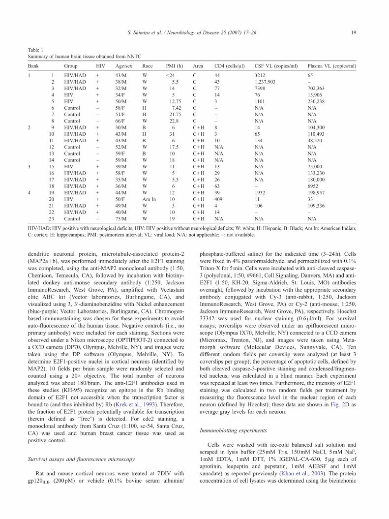
Table 1Summary of human brain tissue obtained from NNTC
Bank Group HIV Age/sex Race PMI (h) Area CD4 (cells/μl) CSF VL (copies/ml) Plasma VL (copies/ml)
1 1 HIV/HAD + 43/M W <24 C 44 3212 652 HIV/HAD + 38/M W 5.5 C 43 1,237,903 –3 HIV/HAD + 32/M W 14 C 77 7398 702,3634 HIV + 34/F W 5 C 14 76 15,9065 HIV + 50/M W 12.75 C 3 1101 230,2386 Control – 58/F H 7.42 C – N/A N/A7 Control – 51/F H 21.75 C – N/A N/A8 Control – 66/F W 22.8 C – N/A N/A
2 9 HIV/HAD + 30/M B 6 C+H 8 14 104,30010 HIV/HAD + 43/M H 31 C+H 3 65 110,49311 HIV/HAD + 43/M B 6 C+H 10 134 48,52012 Control – 52/M W 17.5 C+H N/A N/A N/A13 Control – 59/F B 10 C+H N/A N/A N/A14 Control – 59/M W 18 C+H N/A N/A N/A
3 15 HIV + 39/M W 11 C+H 13 N/A 75,00016 HIV/HAD + 58/F W 5 C+H 29 N/A 133,23017 HIV/HAD + 35/M W 5.5 C+H 26 N/A 180,00018 HIV/HAD + 36/M W 6 C+H 63 – 6952
4 19 HIV/HAD + 44/M W 12 C+H 39 1932 198,95720 HIV + 50/F Am In 10 C+H 409 11 3321 HIV/HAD + 49/M W 3 C+H 4 106 109,33622 HIV/HAD + 40/M W 10 C+H 14 – –23 Control – 75/M W 19 C+H N/A N/A N/A
HIV/HAD: HIV positive with neurological deficits; HIV: HIV positive without neurological deficits; W: white; H: Hispanic; B: Black; Am In: American Indian;C: cortex; H: hippocampus; PMI: postmortem interval; VL: viral load; N/A: not applicable; –: not available.
19S. Shimizu et al. / Neurobiology of Disease 25 (2007) 17–26
dendritic neuronal protein, microtubule-associated protein-2(MAP2a+b), was performed immediately after the E2F1 stainingwas completed, using the anti-MAP2 monoclonal antibody (1:50,Chemicon, Temecula, CA), followed by incubation with biotiny-lated donkey anti-mouse secondary antibody (1:250, JacksonImmunoResearch, West Grove, PA), amplified with Vectastainelite ABC kit (Vector laboratories, Burlingame, CA), andvisualized using 3, 3′-diaminobenzidine with Nickel enhancement(blue-purple; Vector Laboratories, Burlingame, CA). Chromogen-based immunostaining was chosen for these experiments to avoidauto-fluorescence of the human tissue. Negative controls (i.e., noprimary antibody) were included for each staining. Sections wereobserved under a Nikon microscope (OPTIPHOT-2) connected toa CCD camera (DP70, Olympus, Melville, NY), and images weretaken using the DP software (Olympus, Melville, NY). Todetermine E2F1-positive nuclei in cortical neurons (identified byMAP2), 10 fields per brain sample were randomly selected andcounted using a 20× objective. The total number of neuronsanalyzed was about 180/brain. The anti-E2F1 antibodies used inthese studies (KH-95) recognize an epitope in the Rb bindingdomain of E2F1 not accessible when the transcription factor isbound to (and thus inhibited by) Rb (Krek et al., 1993). Therefore,the fraction of E2F1 protein potentially available for transcription(herein defined as “free”) is detected. For cdc2 staining, amonoclonal antibody from Santa Cruz (1:100, sc-54, Santa Cruz,CA) was used and human breast cancer tissue was used aspositive control.
Survival assays and fluorescence microscopy
Rat and mouse cortical neurons were treated at 7DIV withgp120IIIB (200pM) or vehicle (0.1% bovine serum albumin/
phosphate-buffered saline) for the indicated time (3–24h). Cellswere fixed in 4% paraformaldehyde, and permeabilized with 0.1%Triton-X for 5min. Cells were incubated with anti-cleaved caspase-3 (polyclonal, 1:50, #9661, Cell Signaling, Danvers, MA) and anti-E2F1 (1:50, KH-20, Sigma-Aldrich, St. Louis, MO) antibodiesovernight, followed by incubation with the appropriate secondaryantibody conjugated with Cy-3 (anti-rabbit, 1:250, JacksonImmunoResearch, West Grove, PA) or Cy-2 (anti-mouse, 1:250,Jackson ImmunoResearch, West Grove, PA), respectively. Hoechst33342 was used for nuclear staining (0.6μl/ml). For survivalassays, coverslips were observed under an epifluorescent micro-scope (Olympus IX70, Melville, NY) connected to a CCD camera(Micromax, Trenton, NJ), and images were taken using Meta-morph software (Molecular Devices, Sunnyvale, CA). Tendifferent random fields per coverslip were analyzed (at least 3coverslips per group); the percentage of apoptotic cells, defined byboth cleaved caspase-3-positive staining and condensed/fragmen-ted nucleus, was calculated in a blind manner. Each experimentwas repeated at least two times. Furthermore, the intensity of E2F1staining was calculated in two random fields per treatment bymeasuring the fluorescence level in the nuclear region of eachneuron (defined by Hoechst); these data are shown in Fig. 2D asaverage gray levels for each neuron.
Immunoblotting experiments
Cells were washed with ice-cold balanced salt solution andscraped in lysis buffer (25mM Tris, 150mM NaCl, 5mM NaF,1mM EDTA, 1mM DTT, 1% IGEPAL-CA-630, 5μg each ofaprotinin, leupeptin and pepstatin, 1mM AEBSF and 1mMvanadate) as reported previously (Khan et al., 2003). The proteinconcentration of cell lysates was determined using the bicinchonic

20 S. Shimizu et al. / Neurobiology of Disease 25 (2007) 17–26
acid assay from Pierce (Rockford, IL). Equal amounts of proteinswere loaded in each lane, separated by SDS–PAGE and transferredto PVDF membranes for immunoblotting. The following primaryantibodies were used: anti-E2F1 (1:1000, KH-20, Sigma-Aldrich,St. Louis, MO), anti-Cdc2 (1:500, sc-54, Santa Cruz, CA), anti-Puma (1:1000, #4976, Cell Signaling, Danvers, MA), anti-BAD(1:1000, #9292 Cell Signaling, Danvers, MA) and anti-BADphospho S-128 (1:1000, ab5687, Abcam, Cambridge, MA), anti-CXCR4 (1:1000, H-118, Santa Cruz Biotechnology, Santa Cruz,CA), anti-NeuN (1:1000, MAB377, Chemicon, Temecula, CA)and anti-Actin (1:5000, A2066, Sigma-Aldrich, St. Louis, MO).The last two were used as loading controls for nuclear andcytoplasmic proteins, respectively. Bands were detected bychemiluminescence using Pierce reagents (SuperSignal WestFemto maximum sensitivity), according to the manufacture’sinstructions, and analyzed using the FluorChem 8900 apparatusfrom Alpha Innotech (San Leandro, CA).
Electrophoretic mobility shift assay (EMSA)
EMSA was performed as previously described (Khan et al.,2003). Nuclear extracts (2μg) were incubated with 3′ biotinylated-oligonucleotides containing a consensus binding site for E2F1(5′-ATTTAAGTTTCGCGCCCTTTCTCAA-3′) according to themanufacturer’s protocol (LightShift Chemiluminescent EMSA kit,Pierce). After incubation, samples were loaded on a 6% poly-acrylamide non-denaturing gel in 1% Tris–acetate–EDTA buffer.After transfer to the membrane, DNA was cross-linked to the
Fig. 1. Expression of E2F1 in the nucleus of cortical neuron in HIV/HAD patients: Fand HIV-negative control subjects were used to examine E2F1 expression by immunLow levels of E2F1 were detected in the brains of neurologically normal HIV patnucleus of cortical neurons in HIV/HAD brains (arrowhead in B; 40× in C). Doublpositive neurons (D) and quantify differences among the three groups of patients
membrane by UV light, and DNA–protein complexes weredetected by chemiluminescence.
Transfection and luciferase assays
Rat cortical neurons (7DIV) were transfected with the pGL2ANplasmid (gift from Dr. W Kaelin, Dana-Farber Cancer Institute)containing the luciferase cDNA sequence downstream of an E2F-dependent promoter (Neuman et al., 1994), along with the pEGFP,using Lipofectamine 2000 from Invitrogen (2μg per plasmid ineach dish containing 1 million cells). An E2F1 expression plasmid(pMax–E2F1) was transfected with the other plasmids in thepositive controls only (2μg/dish). The Bright-Glo™ (Promega,Madison, WI) reagents were used to detect luciferase activityaccording to the manufacturer’s protocol, using a spectrophoto-fluorometer (Wallac 1420 multilabel counter, Perkin Elmer,Wellesley, MA). Expression of pEGFP was measured to determinetransfection efficiency and all luciferase measurements werenormalized accordingly. Neurons were treated with gp120(200pM) 2–3h after the transfection.
Statistical analysis
One-way analysis of variance (ANOVA), followed by Neu-man–Keuls multiple comparison procedure, was used for analysisof survival assays and immunohistochemistry studies. Westernblots and Luciferase assays were analyzed using the Student’s ttest. All data are reported as mean±SEM.
rontal cortices from HIV-positive patients (with or w/o neurological deficits)ohistochemistry (as described in detail in theMaterials and methods section).ients (A) or control subjects (graph), whereas E2F1 was up-regulated in thee staining with the neuronal marker MAP2 was performed to identify E2F1-(E, ten fields/brain); ⁎p<0.05 vs. HIV; scale bars 5.0μm.

21S. Shimizu et al. / Neurobiology of Disease 25 (2007) 17–26
Results
Up-regulation of E2F1 in the nucleus of cortical neurons of HIV/HAD patients
As previously reported (Jordan-Sciutto et al., 2002), widespreadup-regulation of E2F1 was observed in both gray and white matterof HIV-infected patients that exhibited neurologic impairment(MSK 1–3; not shown). However, in the present study neuronalE2F1 was mainly expressed in the nucleus (Fig. 1), consistent withits traditional role in transcription. In contrast, neurons in the cortexof HIV patients that were neurologically normal (Fig. 1A) and ofHIV-negative patients (not shown), did not appear to contain
Fig. 2. Effect of HIV gp120 on E2F1 expression in rat primary neurons: Western bloup-regulated in the nucleus of rat primary cortical neurons after treatment with gp1cultures at 7DIV. Antibodies against the neuronal-specific nuclear protein NeuN ocytosolic (CE) extracts, respectively; Hoechst 33442 was used for nuclear staininglevels was observed after a few hours of treatment with gp120IIIB (generally 3h) aexperiments using total cell extracts from untreated (control) and gp120-treated neuintensity of E2F1 nuclear staining in individual neurons at each time points, as detepositive pre-apoptotic neurons, i.e., cleaved caspase-3-positive neurons showing he(arrows in C; blue bars in graph E, ^p<0.001 vs. control). Apoptotic neurons, i.e.,detected at later time (white bars in graph E; ⁎p<0.001 vs. control).
detectable levels of E2F1 protein. In order to quantify differencesin the expression of E2F1 within the three groups of patients, wecalculated the percentage of E2F1-positive neurons in specimensprovided by a single tissue bank (bank 1, see Table 1), thus keepingpotential variability due to the handling of tissue from differentsources to a minimum. These included samples from the frontalcortex of HIV/HAD patients (n=3), of HIV patients withoutneurological problems (n=2) and of HIV-negative control patients(n=3). The postmortem interval for these patients (4 males and 4females) was less than 24h and their age ranged from 32 to66years. Viral loads in the CSF and blood varied (Table 1).Neurons were identified by double staining with the cytoplasmic/dendritic neuronal marker MAP2 (Fig. 1D). A marked increase in
t analysis (A–B) and immunocytochemistry studies (C) indicate that E2F1 is20IIIB (200pM) for the indicated time. The viral protein was added to the co-r β-actin were used as markers in the immunoblots with nuclear (NE) andin the immunocytochemistry studies. A significant increase in E2F1 proteins shown by the graph in B, which reports the average of three independentrons (mean±SEM; ⁎p<0.05). Each scatter plot in graph D shows the averagermined by immunostaining (bars at each time represent mean values). E2F1-althy morphology, were also observed after short-term treatments with gp120cleaved caspase-3-positive neurons with condensed/fragmented nuclei, were

22 S. Shimizu et al. / Neurobiology of Disease 25 (2007) 17–26
the number of E2F1-positive neurons was observed in HIV/HADbrains, as compared to control or HIV-positive individuals withoutneurological deficits—suggesting that E2F1 is up-regulated in theneurons of HIV/HAD patients (Figs. 1B–E). No significantdifferences were found between control brains and brains fromHIV-positive subjects that were neurologically normal (Fig. 1E).
HIVIIIB-gp120 increases expression of E2F1 in primary corticalneurons
Previous data suggested that activation of neuronal CXCR4 byits natural ligand, the chemokine CXCL12, promotes Rb function(Khan et al., 2003), and that the neuroprotective effect of thechemokine is related to the inhibition of E2F1-mediated apoptosis.Unlike CXCL12, gp120IIIB (an X4-using viral protein) increasedRb phosphorylation in neurons (Khan et al., 2003), resulting in arelease of the inhibitory effect of Rb on E2F1 and favoringactivation of E2F1. In order to determine whether gp120IIIB alsoaffects E2F1 expression/localization in cultured neurons, proteinlevels of this transcription factor were measured in nuclear andcytosolic extracts of neurons treated with gp120IIIB. Immunoblotanalyses of nuclear and cytolosic fractions revealed that E2F1 waspredominantly localized to the nuclear fraction of post-mitoticneurons (Fig. 2). No significant changes in the subcellular
Fig. 3. HIV gp120 up-regulates E2F1 pro-apoptotic targets in rat neurons: Treatmneurons as determined by gene reporter assays (A, mean±SEM of one representpositive control for the assay. Elevated levels of transcriptional targets of E2F1 (Putreated with gp120 (B–C) as well as in neurons transfected with the E2F1 exprescultures with AMD3100 (100ng/ml) 15min before the addition of the gp120 (C).
distribution of E2F1 were observed after gp120IIIB treatment,whereas the nuclear levels of the protein were increased by the viralprotein (Fig. 2A/B). Similar results were obtained from immuno-cytochemistry-based analysis of E2F1 expression (Fig. 2C). Theseexperiments show low levels of E2F1 in control neurons and moreintense staining following gp120IIIB treatment (Fig. 2C). Asindicated in Fig. 2D, the average gray level of the E2F1 nuclearstaining in individual neurons gradually increased after gp120treatment. Consistent with previous studies (Khan et al., 2005),gp120 IIIB was found to induce neuronal cell death in these neurons(Fig. 2E). These experiments also showed that in gp120-treatedneurons E2F1 up-regulation could be detected before the nuclearalterations typical of apoptosis occurred (as assessed by stainingwith the antibody against active caspase-3 in neurons displayingmorphologically normal nuclei) (Fig. 2C, arrow). Thus, the increasein E2F1 caused by the viral protein appears to be a relatively earlyevent in the apoptotic process. An increase in the E2F1 protein isindeed already evident after a few hours of gp120 treatment (Fig.2A/B)—when neurons still appear viable. The early increase inE2F1 may indicate a stimulation of E2F1-dependent transcriptionof E2F1 by the HIV protein (Neuman et al., 1994). Finally, thegp120-induced increase of E2F1 seems to be a direct effect ofgp120 on neurons, as similar results were obtained when treatmentswere performed in the absence of glia (not shown).
ent with gp120IIIB (200pM) increased transcriptional activity in rat primaryative experiment; ⁎p<0.01 vs. control). E2F1 over-expression was used asma and Cdc2) were also found in neuronal extracts from primary rat culturession plasmid (D). The effect of gp120IIIB was blocked by pre-treatment of
23S. Shimizu et al. / Neurobiology of Disease 25 (2007) 17–26
HIVIIIB-gp120 stimulates transcription of E2F1-dependentapoptotic genes in neurons
To determine whether gp120IIIB stimulates the transcriptionalactivity of E2F1, cortical neurons were transfected with a plasmidcarrying the sequence of the luciferase gene under the control of anE2F1-dependent promoter (Neuman et al., 1994). A plasmidexpressing EGFP was co-transfected to monitor transfectionefficiency and for subsequent normalization of luciferase assays,whereas an E2F1 expression vector was used for the positivecontrols. These experiments showed that treatment with gp120IIIBsignificantly increased transcriptional activity in transfectedneurons (Fig. 3A). We additionally tested the effect of gp120IIIBon two endogenous E2F1 transcriptional targets, namely Cdc2(also known as Cdk1) and Puma, which are known to induceapoptosis in neurons (Hershko and Ginsberg, 2004; Konishi andBonni, 2003). The neuronal levels of these proteins were enhancedby treatment with gp120IIIB (Fig. 3B). This effect was blocked bythe CXCR4 antagonist AMD3100 (Fig. 3C), indicating thatCXCR4 mediates the action of gp120IIIB. Under the sameexperimental conditions, gp120 also induced a robust increase inthe phosphorylation of Bad protein on serine 128 (Fig. 3B), whichis direct target of Cdc2 (Konishi et al., 2002). Moreover, up-
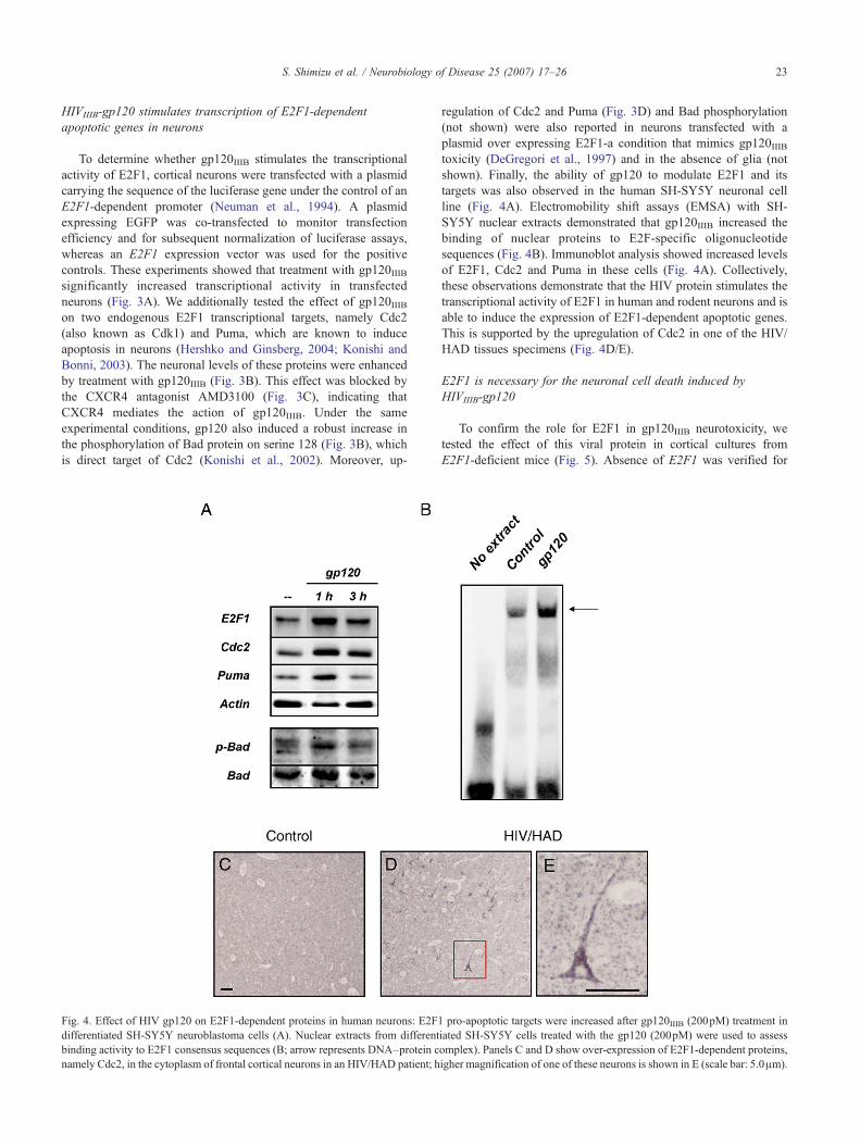
Fig. 4. Effect of HIV gp120 on E2F1-dependent proteins in human neurons: E2F1differentiated SH-SY5Y neuroblastoma cells (A). Nuclear extracts from differentbinding activity to E2F1 consensus sequences (B; arrow represents DNA–protein cnamely Cdc2, in the cytoplasm of frontal cortical neurons in an HIV/HAD patient; h
regulation of Cdc2 and Puma (Fig. 3D) and Bad phosphorylation(not shown) were also reported in neurons transfected with aplasmid over expressing E2F1-a condition that mimics gp120IIIBtoxicity (DeGregori et al., 1997) and in the absence of glia (notshown). Finally, the ability of gp120 to modulate E2F1 and itstargets was also observed in the human SH-SY5Y neuronal cellline (Fig. 4A). Electromobility shift assays (EMSA) with SH-SY5Y nuclear extracts demonstrated that gp120IIIB increased thebinding of nuclear proteins to E2F-specific oligonucleotidesequences (Fig. 4B). Immunoblot analysis showed increased levelsof E2F1, Cdc2 and Puma in these cells (Fig. 4A). Collectively,these observations demonstrate that the HIV protein stimulates thetranscriptional activity of E2F1 in human and rodent neurons and isable to induce the expression of E2F1-dependent apoptotic genes.This is supported by the upregulation of Cdc2 in one of the HIV/HAD tissues specimens (Fig. 4D/E).
E2F1 is necessary for the neuronal cell death induced byHIVIIIB-gp120
To confirm the role for E2F1 in gp120IIIB neurotoxicity, wetested the effect of this viral protein in cortical cultures fromE2F1-deficient mice (Fig. 5). Absence of E2F1 was verified for
pro-apoptotic targets were increased after gp120IIIB (200pM) treatment iniated SH-SY5Y cells treated with the gp120 (200pM) were used to assessomplex). Panels C and D show over-expression of E2F1-dependent proteins,igher magnification of one of these neurons is shown in E (scale bar: 5.0μm).

24 S. Shimizu et al. / Neurobiology of Disease 25 (2007) 17–26
each neuronal preparation by PCR (Fig. 5A). Neuronal culturesfrom both wild-type and knockout animals were treated with200pM gp120IIIB for 24h. The extent of neuronal death caused bygp120IIIB in the wild-type mouse cultures was comparable to theneurotoxicity reported in the rat cultures (Fig. 5B), though higherlevels of basal cell death were generally found in the mousecultures. However, the effect of the viral protein was abrogated incultures from E2F1-deficient animals (Fig. 5B), even when higherconcentration of gp120IIIB (400pM) were used (data not shown).Caspase-3 activation plus alteration of nuclear morphology wereused to evaluate neuronal cell death (Fig. 5B/C). As observed inrat neurons, gp120IIIB increased the total levels of the E2F1protein in mouse cultures within 3h of treatment, as determinedby Western blot (not shown). Primary neurons from E2F1knockout mice were resistant to gp120-induced apoptosis andlevels of Cdc2 and Puma were not altered by the viral protein inthese cultures (Fig. 5D). The protein levels of CXCR4 werecomparable in extracts from wild-type and knockout cultures (Fig.5D). These data suggest that up-regulation of Cdc2 and Pumafollowing gp120IIIB treatment requires E2F1 expression.
Fig. 5. E2F1 is necessary for HIVgp120-induced cell death: Neuronal cultures fromgp120-induced apoptosis. DNAwas extracted from the cerebellum of each animalKO, respectively, as expected). Cloned E2F1 was used as positive control. SurvivalKO cultures are resistant to gp120IIIB (B/C, ⁎p<0.0001 vs. control in wild-type anregulated after gp120 treatment in wild-type but not in KOmice-as determined byWprotein levels was observed between WT and KO mice (D).
Discussion
The studies described here demonstrate that activation ofCXCR4 by the HIV envelope protein gp120 stimulates the activityof the transcription factor E2F1 in neurons and suggest that thetranscription of E2F1-dependent genes may be involved in HIVneuropathology. The experiments on cultured neurons indicate thatHIVgp120 up-regulates E2F1 and induces the expression ofspecific target proteins regulated by this transcription factor. Theseeffects are inhibited by a specific CXCR4 antagonist and do notrequire the presence of glia, suggesting that the envelope proteinacts via neuronal CXCR4. The results with the E2F1 KO culturesrevealed that E2F1 is necessary to gp120-induced neurotoxicityand up-regulation of Cdc2 and Puma. Transcriptional up-regulationof apoptotic genes, such as Puma, is also known to be triggered byp53, another major transcription factor involved in HIV apoptosisthat is also regulated by CXCR4 (Castedo et al., 2002; Garden etal., 2004; Khan et al., 2005). However, these data suggest that p53is not sufficient to activate gp120-induced neurotoxic pathways inthe absence of E2F1 and may act in concert with (or downstream
E2F1 KO and wild-type mice were used to determine the role of E2F1 infor PCR analysis (A, bands 172bp and 227bp correspond to wild-type andassays performed as reported in the Materials and methods section show thatd vs. gp120 treatment in KO mice). E2F1 targets, Cdc2 and Puma were up-estern blot analysis with total neuronal cell extracts. No difference in CXCR4

25S. Shimizu et al. / Neurobiology of Disease 25 (2007) 17–26
of) E2F1. The involvement of p53 in HIV apoptosis has beenestablished in both neuronal and non-neuronal models (Castedo etal., 2002; Garden et al., 2004) and the two transcription factors caninteract in different ways. For instance, E2F1 can induce p53accumulation via inactivation of the Mdm2 protein, which targetsp53 for proteasome degradation (Tao and Levine, 1999).Phosphorylation of specific p53 serine residues by E2F1 targetsis another key step in E2F1-mediated apoptosis (Rogoff et al.,2002). Interestingly, stimulation of CXCR4 by gp120 increasesp53 protein levels and phosphorylation in cortical neurons (Khan etal., 2005).
The fact that high levels of free E2F1 (i.e., unbound to Rb) werefound in neurons that still appear morphologically normal, butwere already primed to death, suggests that E2F1 is recruited in theearly stages of gp120-induced neurotoxicity. These data are inagreement with previous observations that gp120 stimulates Rbphosphorylation and promotes the DNA binding activity of E2F1in neurons, as well as with reports showing increased Rbphosphorylation in the nucleus of neurons of SIVE/HIVE subjects(Jordan-Sciutto et al., 2002; Khan et al., 2003).
In contrast to earlier observations of increased E2F1 expressionin the cytosol of neurons in HIVE patients (Jordan-Sciutto et al.,2002), our studies indicate that E2F1 mostly localizes to the nucleiof neurons in both HIV/HAD patients and in neuronal culturestreated with HIVgp120. Though the same anti-E2F1 antibodieswere employed in the two studies, the different detection methodsand/or tissue preparation might account for this discrepancy.Furthermore, nuclear localization of E2F1 might be restricted tomore severe cases of encephalitis, such as those examined in thisstudy. Nonetheless, our in vitro studies (using additional antibodiesand alternative approaches) support the link between increasedE2F1 transcription in neurons and HIV neuropathogenesis.Stimulation of CXCR4 by the HIV envelope induces up-regulationof E2F1 and E2F1-dependent apoptotic proteins in neurons,including Cdc2/Cdk1 (Castedo et al., 2002; Konishi and Bonni,2003). This protein is of particular interest as it was found to bespecifically associated with E2F1-driven apoptotic pathways inneurons, but not involved in the activation of E2F1-target genesinvolved in DNA synthesis and replication (Konishi et al., 2002).Increased levels of Cdc2 in neurons were also found in the brain ofone HIV/HAD patient. This protein kinase catalyzes the phosphor-ylation of the BH3-only protein BAD at a distinct site (serine 128),thus inducing BAD-mediated apoptosis in primary neurons andopposing the anti-apoptotic effects of growth factors (Konishi etal., 2002). These data point to the role of E2F1-dependentapoptotic pathways in NeuroAIDS and raise the issue of whetherthis phenomenon may be restricted to CXCR4 activation.Considering the different impact of the two co-receptors onneuronal physiology, it would not be surprising that CCR5-usingenvelope proteins behave differently, but it remains to beestablished. A different behavior of the two co-receptors couldexplain why only a fraction of HIV-infected individuals developsHAD (even in the absence of HAART), despite the major role ofCCR5 in HIV brain infection.
However, other factors also need to be considered. For instance,the ability of different HIV strains to induce syncytia, rather thantheir co-receptor usage, may be relevant to neuropathogenesis.Indeed, recent studies reported that the presence of dying syncytiain the brain correlates with disease severity (Nardacci et al., 2005).Interestingly, these neuropathology studies also reported inductionof Puma in apoptotic neurons of these patients (Nardacci et al.,
2005). Syncytia are a common characteristic of HIV-infectedtissue, including the brain. Though typically the syncytium-inducing phenotype has been assigned to HIV strains thatefficiently induce syncytia in transformed T cells in vitro (i.e.,CXCR4-using strains), many R5 HIV-1 strains promote syncytiaformation in primary macrophages albeit with variable efficacy,and their different ability in forming syncytia has been suggested toassociate with their neuropathogenic potential (Gorry et al., 2002).On the other hand, although brain-derived viruses predominantlyuse CCR5, viruses that exclusively use CXCR4 for infection ofmicroglia/macrophages have been identified (Gorry et al., 2005)and rarely reported in the CSF (Yi et al., 2003). Thus, both CXCR4and CCR5-using viruses can potentially induce macrophagesyncytia and contribute to HAD, though adaptive viral evolutionappears to favor M-tropism of R5 HIV-1 strains (Gorry et al.,2005).
To conclude, we propose that the transcriptional activity ofE2F1 and its downstream effectors affect the survival and functionof neurons and contribute to the neurological deficits in HIVpatients. These apoptotic pathways could also be recruited by thenatural CXCR4 ligand (the chemokine CXCL12/SDF) underpathological conditions, such as upon activation of extracellularproteases (Jones and Power, 2006)—extending the clinicalrelevance of these observations to other neuroinflammatoryconditions. Indeed, we and others have previously reported thatchemokines affect cell cycle proteins expression in neurons (Khanet al., 2003; Khan et al., 2005). As E2F1 up-regulation appears tobe an early event in the neuropathogenesis, interfering with itsactivity might provide some level of therapeutic intervention. Thiscould be achieved by blocking Rb phosphorylation by cyclin-dependent kinase inhibitors (CDKI), which are currently in clinicaltrial for cancer therapy (Schwartz et al., 2005). Inhibition of CDKscan also prevent syncytia apoptosis of HIV-infected cells (Castedoet al., 2002), increasing the chances of success.
Acknowledgments
Supported by NIH grants, DA 19808 and DA15014 (OM), NS41561 (RR) and by the W.W. Charitable Trust (OM). The NNTC issupported by NIMH and NINDS, contact NIH-N01MH32002, NY:MH59724, UCLA: NS38841, UCSD: MH59745-06, TX: NS45491.
References
Adle-Biassette, H., Levy, Y., Colombel, M., Poron, F., Natchev, S.,Keohane, C., Gray, F., 1995. Neuronal apoptosis in HIV infection inadults. Neuropathol. Appl. Neurobiol. 21, 218–227.
Bellizzi, M.J., Lu, S.M., Masliah, E., Gelbard, H.A., 2005. Synaptic activitybecomes excitotoxic in neurons exposed to elevated levels of platelet-activating factor. J. Clin. Invest. 115, 3185–3192.
Cartier, L., Hartley, O., Dubois-Dauphin, M., Krause, K.H., 2005.Chemokine receptors in the central nervous system: role in braininflammation and neurodegenerative diseases. Brain Res. Brain Res.Rev. 48, 16–42.
Castedo, M., Roumier, T., Blanco, J., Ferri, K.F., Barretina, J., Tintignac,L.A., Andreau, K., Perfettini, J.L., Amendola, A., Nardacci, R., et al.,2002. Sequential involvement of Cdk1, mTOR and p53 in apoptosisinduced by the HIV-1 envelope. EMBO J. 21, 4070–4080.
DeGregori, J., Leone, G., Miron, A., Jakoi, L., Nevins, J.R., 1997. Distinctroles for E2F proteins in cell growth control and apoptosis. Proc. Natl.Acad. Sci. U. S. A. 94, 7245–7250.

26 S. Shimizu et al. / Neurobiology of Disease 25 (2007) 17–26
Everall, I.P., Heaton, R.K., Marcotte, T.D., Ellis, R.J., McCutchan, J.A.,Atkinson, J.H., Grant, I., Mallory, M., Masliah, E., 1999. Corticalsynaptic density is reduced in mild to moderate human immunodeficiencyvirus neurocognitive disorder. HNRC Group. HIV NeurobehavioralResearch Center. Brain Pathol. 9, 209–217.
Farzan, M., Babcock, G.J., Vasilieva, N., Wright, P.L., Kiprilov, E.,Mirzabekov, T., Choe, H., 2002. The role of post-translationalmodifications of the CXCR4 amino terminus in stromal-derived factor1 alpha association and HIV-1 entry. J. Biol. Chem. 277, 29484–29489.
Field, S.J., Tsai, F.Y., Kuo, F., Zubiaga, A.M., Kaelin Jr., W.G., Livingston,D.M., Orkin, S.H., Greenberg, M.E., 1996. E2F-1 functions in mice topromote apoptosis and suppress proliferation. Cell 85, 549–561.
Gabuzda, D., Wang, J., 2000. Chemokine receptors and mechanisms of celldeath in HIV neuropathogenesis. J. Neurovirology 6 (Suppl 1),S24–S32.
Garden, G.A., Guo, W., Jayadev, S., Tun, C., Balcaitis, S., Choi, J., Montine,T.J., Moller, T., Morrison, R.S., 2004. HIVassociated neurodegenerationrequires p53 in neurons and microglia. FASEB J. 18, 1141–1143.
Gartner, S., Liu, Y., 2002. Insights into the role of immune activation in HIVneuropathogenesis. J. Neurovirology 8, 69–75.
Gonzalez-Scarano, F., Martin-Garcia, J., 2005. The neuropathogenesis ofAIDS. Nat. Rev. Immunol. 5, 69–81.
Gorry, P.R., Taylor, J., Holm, G.H., Mehle, A., Morgan, T., Cayabyab, M.,Farzan, M., Wang, H., Bell, J.E., Kunstman, K., et al., 2002. IncreasedCCR5 affinity and reduced CCR5/CD4 dependence of a neurovirulentprimary human immunodeficiency virus type 1 isolate. J. Virol. 76,6277–6292.
Gorry, P.R., Churchill, M., Crowe, S.M., Cunningham, A.L., Gabuzda, D.,2005. Pathogenesis of macrophage tropic HIV-1. Curr. HIV Res. 3,53–60.
Hawkins, V., Shen, Q., Chiueh, C.C., 1999. Kynostatin and 17beta-estradiolprevent the apoptotic death of human neuroblastoma cells exposed toHIV-1 protease. J. Biomed. Sci. 6, 433–438.
Hershko, T., Ginsberg, D., 2004. Up-regulation of Bcl-2 homology 3 (BH3)-only proteins by E2F1 mediates apoptosis. J. Biol. Chem. 279,8627–8634.
Hesselgesser, J., Taub, D., Baskar, P., Greenberg, M., Hoxie, J., Kolson,D.L., Horuk, R., 1998. Neuronal apoptosis induced byHIV-1 gp120 andthe chemokine SDF-1 alpha is mediated by the chemokine receptorCXCR4. Curr. Biol. 8, 595–598.
Jones, G., Power, C., 2006. Regulation of neural cell survival by HIV-1infection. Neurobiol. Dis. 21, 1–17.
Jordan-Sciutto, K.L., Wang, G., Murphey-Corb, M., Wiley, C.A., 2002. Cellcycle proteins exhibit altered expression patterns in lentiviral-associatedencephalitis. J. Neurosci. 22, 2185–2195.
Kaul, M., Lipton, S.A., 1999. Chemokines and activated macrophages inHIV gp120-induced neuronal apoptosis. Proc. Natl. Acad. Sci. U. S. A.96, 8212–8216.
Khan, M.Z., Brandimarti, R., Musser, B.J., Resue, D.M., Fatatis, A.,Meucci, O., 2003. The chemokine receptor CXCR4 regulates cell-cycleproteins in neurons. J. Neurovirology 9, 300–314.
Khan, M.Z., Shimizu, S., Patel, J.P., Nelson, A., Le, M.T., Mullen-Przeworski, A., Brandimarti, R., Fatatis, A., Meucci, O., 2005.Regulation of neuronal P53 activity by CXCR 4. Mol. Cell. Neurosci.30, 58–66.
Konishi, Y., Bonni, A., 2003. The E2F-Cdc2 cell-cycle pathway specificallymediates activity deprivation-induced apoptosis of postmitotic neurons.J. Neurosci. 23, 1649–1658.
Konishi, Y., Lehtinen, M., Donovan, N., Bonni, A., 2002. Cdc2phosphorylation of BAD links the cell cycle to the cell death machinery.Mol. Cell 9, 1005–1016.
Krek, W., Livingston, D.M., Shirodkar, S., 1993. Binding to DNA and the
retinoblastoma gene product promoted by complex formation ofdifferent E2F family members. Science 262, 1557–1560.
Marder, K., Albert, S.M., McDermott, M.P., McArthur, J.C., Schifitto, G.,Selnes, O.A., Sacktor, N., Stern, Y., Palumbo, D., Kieburtz, K., et al.,2003. Inter-rater reliability of a clinical staging of HIV-associatedcognitive impairment. Neurology 60, 1467–1473.
Mattson, M.P., Haughey, N.J., Nath, A., 2005. Cell death in HIV dementia.Cell Death Differ. 12 (Suppl 1), 893–904.
McArthur, J.C., Haughey, N., Gartner, S., Conant, K., Pardo, C., Nath, A.,Sacktor, N., 2003. Human immunodeficiency virus-associated dementia:an evolving disease. J. Neurovirology 9, 205–221.
Meucci, O., Fatatis, A., Simen, A.A., Bushell, T.J., Gray, P.W., Miller, R.J.,1998. Chemokines regulate hippocampal neuronal signaling and gp120neurotoxicity. Proc. Natl. Acad. Sci. U. S. A. 95, 14500–14505.
Morgello, S., Gelman, B.B., Kozlowski, P.B., Vinters, H.V., Masliah, E.,Cornford, M., Cavert, W., Marra, C., Grant, I., Singer, E.J., 2001. TheNational NeuroAIDS Tissue Consortium: a new paradigm in brainbanking with an emphasis on infectious disease. Neuropathol. Appl.Neurobiol. 27, 326–335.
Nardacci, R., Antinori, A., Larocca, L.M., Arena, V., Amendola, A.,Perfettini, J.L., Kroemer, G., Piacentini, M., 2005. Characterization ofcell death pathways in human immunodeficiency virus-associatedencephalitis. Am. J. Pathol. 167, 695–704.
Neuman, E., Flemington, E.K., Sellers, W.R., Kaelin Jr., W.G., 1994.Transcription of the E2F-1 gene is rendered cell cycle dependent by E2FDNA-binding sites within its promoter. Mol. Cell. Biol. 14, 6607–6615.
Pahlman, S., Ruusala, A.I., Abrahamsson, L., Mattsson, M.E., Esscher, T.,1984. Retinoic acid-induced differentiation of cultured human neuro-blastoma cells: a comparison with phorbolester-induced differentiation.Cell Differ. 14, 135–144.
Petito, C.K., Roberts, B., 1995. Evidence of apoptotic cell death in HIVencephalitis. Am. J. Pathol. 146, 1121–1130.
Roberts, E.S., Zandonatti, M.A., Watry, D.D., Madden, L.J., Henriksen, S.J.,Taffe, M.A., Fox, H.S., 2003. Induction of pathogenic sets of genes inmacrophages and neurons in NeuroAIDS. Am. J. Pathol. 162,2041–2057.
Rogoff, H.A., Pickering, M.T., Debatis, M.E., Jones, S., Kowalik, T.F.,2002. E2F1 induces phosphorylation of p53 that is coincident with p53accumulation and apoptosis. Mol. Cell. Biol. 22, 5308–5318.
Sa, M.J., Madeira, M.D., Ruela, C., Volk, B., Mota-Miranda, A., Paula-Barbosa, M.M., 2004. Dendritic changes in the hippocampal formationof AIDS patients: a quantitative Golgi study. Acta Neuropathol. (Berl.)107, 97–110.
Schwartz, G.K., Weitzman, A., O’Reilly, E., Brail, L., de Alwis, D.P.,Cleverly, A., Barile-Thiem, B., Vinciguerra, V., Budman, D.R., 2005.Phase I and pharmacokinetic study of LY293111, an orally bioavailableLTB4 receptor antagonist, in patients with advanced solid tumors.J. Clin. Oncol. 23, 5365–5373.
Shi, B., De Girolami, U., He, J., Wang, S., Lorenzo, A., Busciglio, J.,Gabuzda, D., 1996. Apoptosis induced by HIV-1 infection of the centralnervous system. J. Clin. Invest. 98, 1979–1990.
Tao, W., Levine, A.J., 1999. Nucleocytoplasmic shuttling of oncoproteinHdm2 is required for Hdm2-mediated degradation of p53. Proc. Natl.Acad. Sci. U. S. A. 96, 3077–3080.
Tran, P.B., Miller, R.J., 2003. Chemokine receptors: signposts to braindevelopment and disease. Nat. Rev., Neurosci. 4, 444–455.
Yi, Y., Chen, W., Frank, I., Cutilli, J., Singh, A., Starr-Spires, L., Sulcove, J.,Kolson, D.L., Collman, R.G., 2003. An unusual syncytia-inducinghuman immunodeficiency virus type 1 primary isolate from the centralnervous system that is restricted to CXCR4, replicates efficiently inmacrophages, and induces neuronal apoptosis. J. Neurovirology 9,432–441.