Embed Size (px)
Citation preview

Mitochondrial Stress Engages E2F1Apoptotic Signaling to Cause DeafnessNuno Raimundo,1 Lei Song,2 Timothy E. Shutt,1 Sharen E. McKay,1 Justin Cotney,3 Min-Xin Guan,4 Thomas C. Gilliland,1
David Hohuan,2 Joseph Santos-Sacchi,2 and Gerald S. Shadel1,3,*1Department of Pathology2Department of Surgery (Otolaryngology), Department of Cellular and Molecular Physiology, and Department of Neurobiology3Department of GeneticsYale University School of Medicine, New Haven, CT 06520, USA4Insitute of Genetics, Zhejiang University, Hangzhou, Zhejiang, China 310058
*Correspondence: [email protected]
DOI 10.1016/j.cell.2011.12.027
SUMMARY
Mitochondrial dysfunction causes poorly under-stood tissue-specific pathology stemming fromprimary defects in respiration, coupled with alteredreactive oxygen species (ROS), metabolic signaling,and apoptosis. The A1555G mtDNA mutation thatcauses maternally inherited deafness disrupts mito-chondrial ribosome function, in part, via increasedmethylation of the mitochondrial 12S rRNA by themethyltransferase mtTFB1. In patient-derivedA1555G cells, we show that 12S rRNA hypermethyla-tion causes ROS-dependent activation of AMPkinase and the proapoptotic nuclear transcriptionfactor E2F1. This retrograde mitochondrial-stressrelay is operative in vivo, as transgenic-mtTFB1mice exhibit enhanced 12S rRNA methylation inmultiple tissues, increased E2F1 and apoptosis inthe stria vascularis and spiral ganglion neurons ofthe inner ear, and progressive E2F1-dependenthearing loss. This mouse mitochondrial diseasemodel provides a robust platform for decipheringthe complex tissue specificity of human mitochon-drial-based disorders, as well as the precise patho-genic mechanism of maternally inherited deafnessand its exacerbation by environmental factors.
INTRODUCTION
Mitochondria are multi-functional cellular organelles involved in
oxidative metabolism, ion homeostasis, signal transduction,
and apoptosis and hence contribute to human disease by
a variety ofmechanisms.Mitochondrial pathogenesis is complex
and involves maternally inherited diseases due to mutations in
mtDNA, as well asMendelian-inherited disease due tomutations
in nuclear genes required for mitochondrial function (DiMauro
and Schon, 2003). Furthermore, mitochondrial dysfunction is
implicated in more common disorders such as diabetes, heart
716 Cell 148, 716–726, February 17, 2012 ª2012 Elsevier Inc.
disease, cancer, neurodegeneration, and aging (Wallace, 2005;
Shadel, 2008), making efforts to better understand how mito-
chondria cause and exacerbate human disease pathology
imperative and of broad significance.
A major hurdle in understanding mitochondrial disease patho-
genesis is the current lack of understanding of the often-extreme
tissue specificity involved (DiMauro and Schon, 2003). Because
defective oxidative phosphorylation (OXPHOS) per se can be
pathogenic, this is often attributed to variable energetic thresh-
olds in different cell types. However, defective OXPHOS is also
frequently associated with increased mitochondrial reactive
oxygen species (ROS) that are a major cause of pathology
because they promote molecular damage, oxidative stress,
and cell death. Finally, mitochondria and ROS are involved in
signal transduction pathways (Hamanaka and Chandel, 2010)
that are likely an underappreciated downstream cause of
pathology due to mitochondrial dysfunction. For example,
a major signaling node that reacts to mitochondrial dysfunction
is the AMP-dependent protein kinase (AMPK) pathway, which
responds to cellular energy decline that can occur when
OXPHOS is disrupted (Hardie, 2007) and is regulated by ROS
(Emerling et al., 2009; Quintero et al., 2006).
A hallmark case of tissue-specific mitochondrial disease
pathology is maternally inherited deafness caused by the
A1555G mutation in human mtDNA (Prezant et al., 1993). This
relatively common mutation (Vandebona et al., 2009) causes
nonsyndromic and/or aminoglycoside-induced deafness and
occurs in the 12S rRNA gene, encoding the RNA component of
the small (28S) mitochondrial ribosome subunit. It is also in close
proximity to an evolutionarily conserved stem-loop that contains
two tandem adenine residues that are methylated by the site-
specific rRNA adenine N6-di-methyltransferase, h-mtTFB1/
TFB1M (McCulloch et al., 2002; Seidel-Rogol et al., 2003). This
methylation occurs during ribosome biogenesis in bacteria
(Pulicherla et al., 2009) and is essential in mice, the lack of which
disrupts mitochondrial 28S ribosome subunit assembly (Meto-
diev et al., 2009).
Hearing loss caused by the A1555G mutation is irreversible
(Prezant et al., 1993), implying death of critical, irreplaceable
cells in the inner ear by an unknown pathogenic cell-death
pathway. The A1555G mutation alters mitochondrial ribosome

function and translation (Cotney et al., 2009; Guan et al., 1996,
2000; Hobbie et al., 2008), which in turn causes OXPHOS
defects that are thought to contribute to deafness pathology.
However, mitochondrial translation and OXPHOS defects
observed in patient-derived primary and cytoplasmic hybrid
(cybrid) cell lines are mild and highly dependent on the nuclear
genetic background (Guan et al., 1996, 2000), suggesting that
other aspects of mitochondrial dysfunction may also be opera-
tive. Finally, polymorphisms near the TFB1M gene encoding
h-mtTFB1 are nuclear modifiers of the A1555G deafness pheno-
type (Bykhovskaya et al., 2004), suggesting a connection
between 12S rRNA methylation and hearing loss (Shadel,
2004b).
We recently showed that patient-derived A1555G cybrids are
hypermethylated at the 12S rRNA stem-loop methylated by
h-mtTFB1 (Cotney et al., 2009). These A1555G cybrids exhibit
defective mitochondrial biogenesis and membrane potential
and heightened sensitivity to stress-induced apoptosis (Cotney
et al., 2009), apparently due to disrupted coordination of overall
mitochondrial biogenesis with the assembly of mitochondrial
ribosomes (Cotney et al., 2007). Remarkably, these phenotypes
are shared by HeLa cells with wild-typemtDNA that instead have
hypermethylated 12S rRNA due to overexpression of h-mtTFB1,
leading us to conclude that hypermethylation of mitochondrial
ribosomes per se is a key molecular defect driving the apoptotic
phenotype (Cotney et al., 2009).
In the current study, we tested the hypothesis that this hyper-
methylation of mitochondrial ribosomes instigates a unique form
of mitochondrial stress signaling involved in the deafness
pathology of the A1555G mutation. We show that increased
mitochondrial ROS generated in A1555G cybrids activate the
proapoptotic nuclear transcription factor E2F1 in an AMPK-
dependent manner, and that overexpression of mtTFB1 in
mice (to model pathogenesis due to increased mitochondrial
12S rRNA methylation) is sufficient to cause progressive hearing
loss associated with tissue-specific upregulation of E2F1 and
apoptosis of critical cells of the inner ear.
RESULTS
A Mitochondrial Stress-Response Pathway ActivatesNuclear Transcription Factor E2F1We hypothesized that mitochondrial defects due to 12S rRNA
hypermethylation elicit a mitochondrial retrograde stress signal
(Butow and Avadhani, 2004) that explains the apoptotic suscep-
tibility we documented previously in A1555G and h-mtTFB1
methyltransferase overexpression cell lines (Cotney et al.,
2009). Therefore, we performed microarray analysis on these
cell lines, which exhibit 12S hypermethylation for different
reasons (i.e., due to the A1555G point mutation or overexpres-
sion of the h-mtTFB1), reasoning that this would define a
common transcriptional signature, which, in turn, would allow
elements of this mitochondrial stress-response pathway to be
deciphered.
Expression microarrays were performed on A1555G cybrids,
using analogous 143B cybrids containing the corresponding
wild-type mtDNA (‘‘A’’ at position 1555) as the negative control,
and on HeLa cells overexpressing h-mtTFB1 compared to
those expressing a methyltransferase-deficient G65A mutant
of h-mtTFB1 as a negative control (Figure 1A). We identified
genes that were significantly upregulated or downregulated in
the hypermethylation cell lines (Table S1 available online), which
we reasoned would include genes that were responding specif-
ically to 12S hypermethylation. We then analyzed the promoters
of this set of hypermethylation-responsive genes (Figure 1B) for
transcription factor binding sites and found that E2F1 sites were
the most frequently overrepresented in genes upregulated in
12S hypermethylation (Table 1). We next crossed our microar-
ray dataset with a published dataset identifying E2F1-respon-
sive genes and found significant overlap (Table S2), again
supporting E2F1 induction in our experimental system. We
also prepared a list of E2F1 targets by selecting human genes
whose promoter is predicted to contain cis-elements for
E2F1, and found that there is a significant enrichment of E2F1
targets in the transcripts induced by 12S rRNA hypermethyla-
tion (Table S2), again suggesting an increase in E2F1 transcrip-
tional activity.
E2F1 Mediates Proapoptotic Signaling in CellsContaining the Human Deafness-Associated A1555GMutationE2F1 is one of a family of eight E2F transcription factors that are
involved in cell cycle regulation through their cyclin-dependent
interactions with pocket proteins, including the tumor
suppressor Rb (DeGregori and Johnson, 2006). Of particular
significance, E2F1 is involved in proapoptotic signaling (Field
et al., 1996; Hou et al., 2000; Polager and Ginsberg, 2009).
This connection to cell death, in conjunction with our microarray
results, made E2F1 a likely candidate for a stress-responsive
protein that is responding to mitochondrial dysfunction caused
by the A1555G mutation and perhaps also the causative factor
in the observed apoptosis susceptibility in the 12S hyperme-
thylation cell lines (Cotney et al., 2009). Entirely consistent with
this concept, we confirmed E2F1 transcriptional induction in
cells with 12S rRNA hypermethylation by q-PCR (Figure S1A),
and found that the steady-state level of E2F1 protein was
increased in A1555G cybrids and in HeLa cells that overexpress
h-mtTFB1 compared to appropriate control lines (Figure 2A; Fig-
ure S1B). In addition, we observed a decrease in the steady-
state amount of Rb (Figure 2A; Figure S1B) and/or its increased
phosphorylation (Figure 2A; Figure S1B), either of which is
consistent with increased E2F1 activity in the 12S hypermethy-
lated cell lines.
Given the association of active E2F1 with apoptosis, we next
tested whether the upregulation of E2F1 per se in A1555G
cybrids is involved in their heightened sensitivity to apoptotic
cell death. In strong support of this concept, we found that
suppressing the hypermethylation of the 12S rRNA via knock-
down of h-mtTFB1 abolished E2F1 induction (Figure 2B; Fig-
ure S1C) and reduced the susceptibility of A1555G cybrids to
apoptosis (Figure 2C). Furthermore, reduction of E2F1 to near
wild-type levels by shRNA in the A1555G cybrids abrogated
the increase in apoptosis susceptibility, as measured by cas-
pase 3/7 activity (Figure 2D) and accumulation of the cleaved
form of PARP (Figure 2E; Figure S1D). Altogether, these results
indicate that the Rb-E2F1 apoptotic pathway responds to
Cell 148, 716–726, February 17, 2012 ª2012 Elsevier Inc. 717

Figure 1. Integrative Genomic Analysis Reveals Upregulation of Nuclear Transcription Factor E2F1 in Response to Mitochondrial Stress
Induced by Mitochondrial Ribosome Hypermethylation(A) Table showing the four cell lines on which we performed microarray analysis to develop a signature of the gene expression response to mitochondrial 12S
hypermethylation: two control lines with ‘‘basal’’ mitochondrial 12S hypermethylation (blue) and two experimental cell lines with 12S hypermethylation (orange),
with the number of biological replicates indicated. To the right of the table is the representative heat map of all replicates of the cell lines analyzed, with transcripts
that are induced or repressed depending on 12S hypermethylation status delineated. ‘‘wt’’ indicates wild-type with regard to either mtDNA or h-mtTFB1.
Transcripts whose expression was significantly different (t test p value < 0.05) between the two groups (‘‘basal’’ versus ‘‘hyper’’-methylated) represented a 12S
hypermethylation signature list that was analyzed further (total of 4,333 transcripts, 2,492 induced, and 1,841 repressed).
(B) The promoter regions (�500 to +100 bp of the predicted transcription start site, TSS) of the genes that were differentially expressed were analyzed for known
transcription factor binding sites. The Venn diagram indicates the top transcription factors identified in the repressed (blue) and induced (orange) genes. Since
E2F1 was the most overrepresented binding site in the genes induced by 12S rRNA hypermethylation, it became a focus of this study.
mitochondrial stress induced by hypermethylation of 12S rRNA
in mitochondrial ribosomes.
ROS-Dependent AMPK Activation Underlies E2F1Induction in A1555G CellsTo determine the proximal signal leading to E2F1 activation we
examined respiratory function in A1555G and corresponding
wild-type cybrids and found that the former have decreased
basal and maximal mitochondrial O2 consumption rates (Fig-
ure 3A). Furthermore, the respiratory chain in the A1555G cells
Table 1. Transcription Factors Whose cis-Elements Are
Detected in the Promoters of Transcripts Upregulated in
Response to 12S rRNA Hypermethylation, but Not in the
Promoters of the Downregulated Transcripts
Transcription
Factor
Upregulated
Target Genes
Downregulated
Target Genes p Value
E2F1 50 0 0.00010
CEBPA 43 0 0.00012
RUNX1T1 15 0 0.0010
IRF3 13 0 0.0018
E2F2 9 0 0.0128
The p value was calculated using Fisher’s exact test.
718 Cell 148, 716–726, February 17, 2012 ª2012 Elsevier Inc.
is reduced (Figure 3B), a condition conducive to increased
ROS production (e.g., superoxide) (Jones et al., 2005).
Accordingly, we found that A1555G cells have increased
mitochondrial superoxide, as measured by MitoSox (Figure 3C).
To determine if this increased superoxide was underlying
proapoptotic signaling to E2F1 in A1555G cybrids, we overex-
pressed mitochondrial superoxide dismutase 2 (SOD2) and
found that this eliminated E2F1 induction, increased the total
amount of Rb, decreased Rb hyperphosphorylation and elimi-
nated the apoptosis susceptibility in A1555G cells (Figures 3D
and 3E).
We next sought to determine howmitochondrial superoxide is
sensed and promotes E2F1 activation. AMPK is an established
sensor of mitochondrial dysfunction. Furthermore, its activity is
activated by superoxide (Quintero et al., 2006) and regulates
E2F1 by phosphorylating Rb (Dasgupta and Milbrandt, 2009)
making it a potential candidate in the stress pathway we have
elucidated. Accordingly, we found that A1555G cybrids have
increased active AMPKa (phospho-AMPKa) compared to wild-
type controls, and that AMPKa activity was restored close to
normal upon overexpression of SOD2 (Figure 3D). To determine
if the increased AMPK activity was linked to E2F1 induction in
A1555G cells, we treated A1555G cells with AMPK inhibitor
compound C, which was confirmed by reduced phosphorylation
of AMPK target acetyl-CoA carboxylase (ACC) (Figure 3F). Phar-
macological inactivation of AMPK in A1555G cybrids reduced

Figure 2. Hypermethylation of Mitochondrial 12S rRNA Leads to Activation of E2F1- and E2F1-Dependent Apoptosis
(A) Western blot analysis of whole-cell extracts for E2F1, Rb and phosphorylated-Rb, (Rb[T826]-P) in HeLa cells that overexpress h-mtTFB1 (mtTFB1-OE)
compared to cell containing an empty-vector negative control (pcDNA), and in human 143B cybrids containing wild-type (wt) or A1555G mtDNA. Tubulin was
probed as a loading control. Quantification is shown in Figure S1B.
(B) Effect of mtTFB1 knockdown (to reduce 12S hypermethylation) on E2F1 levels in A1555G cybrids by Western blot using tubulin as a loading control. Two
independent knockdown constructs were used (+), and a scrambled shRNA was used as a negative control (�). 143B cybrids with wild-type (wt) mtDNA treated
with the negative control shRNA are shown in lane 1. Quantification is shown in Figure S1C.
(C) Quantitation of apoptosis (caspase 3/7 activity) in 143B cybrids containing wild-type (wt) or A1555G mtDNA without (�) or with (+) mtTFB1 knocked down by
shRNA. Basal and induced apoptosis refer to the presence and absence of staurosporine. The values plotted represent mean ± standard deviation (n = 12), with t
test p values indicated.
(D) Quantitation of apoptosis (caspase 3/7 activity) in 143B cybrids containing wild-type (wt) or A1555G mtDNA without (�) or with (+) E2F1 knocked down by
shRNA. Basal and induced apoptosis refer to the presence and absence of staurosporine. The values plotted represent mean ± standard deviation (n = 12), with t
test p values indicated.
(E) Western blot analysis of full-length and cleaved PARP and of E2F1 in wild-type (wt) and A1555G treated with scrambled (�) or E2F1 (+) shRNA without (�) or
with (+) induction of apoptosis by staurosporine. E2F1, full-length (PARP) and caspase-3-cleaved forms of PARP are shown, with tubulin probed as a loading
control. Quantification is shown in Figure S1D.
Rb phosphorylation and E2F1 levels (Figure 3F), and lowered
apoptosis susceptibility of the A1555G cells (Figure 3G). Taken
together, these results strongly implicate AMPK as a ROS-sensi-
tive component of the mitochondrial stress-response pathway
that leads to proapoptotic E2F1 signaling in cells containing
the deafness-causing A1555G mutation.
Cell 148, 716–726, February 17, 2012 ª2012 Elsevier Inc. 719

Figure 3. A Proapoptotic Mitochondrial Stress-Signaling Pathway that Involves Mitochondrial ROS-Dependent Activation of E2F1 by
AMPK
(A) Basal and maximal (+ FCCP uncoupler) mitochondrial O2 consumption rates in 143B cybrids containing wild-type (wt) or A1555G mtDNA are shown. The
mean ± standard deviation (n = 24) is plotted, with t test p values indicated.
(B) Degree of respiratory chain reduction in the same cells as in (A). This is the ratio of basal to maximal respiration, the latter of which was considered
100% reduction (i.e., the chain handling the maximal amount of electrons). The mean ± standard deviation (n = 12) is plotted, with t test p values
indicated.
(C) Mitochondrial ROS in 143B cybrids containing wild-type (wt) or A1555G mtDNA as measured by FACS analysis of MitoSox staining. The mean fluorescence
intensity ± standard deviation is plotted (n = 3), with t test p values indicated.
(D) Western blot analysis of A1555G cybrids that overexpress SOD2. Immunoblotting of E2F1, Rb, phospho-Rb T826 and S807/811, AMPKa, and phospho-
AMPKa T172 are shown in A1555G cybrids transfected with the SOD2 overexpression vector (lanes 5 and 6), and in control cybrids with wild-type mtDNA
(‘‘wt,’’ lanes 1 and 2) and A1555G mtDNA (lanes 3 and 4) transfected with empty vector. Lanes 1 and 2, 3 and 4, and 5 and 6 represent independent biological
replicates. Tubulin was probed as a loading control.
720 Cell 148, 716–726, February 17, 2012 ª2012 Elsevier Inc.

Figure 4. Transgenic Mice that Overexpress the mtTFB1 rRNA Methyltransferase Exhibit 12S Hypermethylation and Progressive Hearing
Loss
(A) Western blot of mtTFB1 in wild-type (wt) and transgenic mtTFB1 (Tg-mtTFB1) mouse tissues using GAPDH as loading control.
(B) Quantification of the degree of 12S rRNA methylation in Tg-mtTFB1 mouse heart and brain tissues using a methylation-sensitive mitochondrial 12S rRNA
primer-extension assay (Cotney et al., 2009). The values plotted represent mean of methylated/unmethylated ± standard deviation (n = 3), with t test p values
indicated.
(C) Hearing thresholds determined by ABR analysis of Tg-mtTFB1 (red points) and control wild-type (wt), nontransgenic littermate control mice (black points) are
shown in the audiogram (frequency versus threshold curve). The threshold (in sound pressure level, dB SPL) was tested with 5 dB resolution and frequencies in
half octave steps from 32 to 2 kHz. The values plotted representmean ±SEM, with t test p values indicated (*p < 0.05; **p < 0.01; ***p < 0.001). Mice were tested at
ages 3–6 months (9 wt mice and 12 Tg-mtTFB1 mice were used).
(D) Same as in (C) except mice were tested at ages 9–12 months (5 wild-type mice and 6 Tg-mtTFB1 mice were used).
Transgenic Mice that Overexpress the MitochondrialrRNA Methyltransferase mtTFB1 Exhibit IncreasedMitochondrial 12S rRNAMethylation in Multiple Tissuesand Progressive Hearing LossThe cell culture studies above strongly implicate an E2F1-depen-
dent, proapoptotic mitochondrial stress signaling pathway
downstream of mitochondrial 12S hypermethylation as a poten-
tial pathogenic component of deafness caused by the A1555G
mtDNA mutation. To test this hypothesis directly, we generated
(E) Quantitation of apoptosis (caspase 3/7 activity) in 143B cybrids containing wi
SOD2 overexpression (gray bars). Basal and induced apoptosis refer to the pr
standard deviation (n = 12), with t test p values indicated.
(F) Effect of pharmacological AMPK inhibition on the levels of acetyl-CoA carb
A1555G cybrids (lanes 5 and 6), determined bywestern blot of whole-cell extracts
(A1555G cybrids) are the vehicle controls (�). Lanes 1 and 2, 3 and 4, and 5 and
(G) Quantitation of apoptosis (caspase 3/7 activity) in 143B cybrids containing wi
AMPK inhibition (gray bars). Basal and induced apoptosis refer to the presence
deviation (n = 12), with t test p values indicated.
transgenic mice that globally overexpress the mouse mtTFB1
rRNAmethyltransferase (Tg-mtTFB1) responsible for the specific
mitochondrial 12S methylation event implicated (Seidel-Rogol
et al., 2003). These animals are viable and fertile, and have
increased mtTFB1 protein in multiple tissues (Figure 4A) that
corresponds with increased 12S rRNA methylation (Figure 4B).
When we measured hearing function in Tg-mtTFB1 mice by the
auditory brainstem response (ABR) and compared them to
age-matched, nontransgenic littermate controls, we found that
ld-type (wt) mtDNA (white bars) or A1555G mtDNA without (black bars) or with
esence and absence of staurosporine. The values plotted represent mean ±
oxylase (ACC), phospho-ACC S79 (ACC-P), phospho-Rb T826, and E2F1 in
using tubulin as a loading control. Lanes 1 and 2 (wild-type cybrids) and 3 and 4
6 represent independent biological replicates.
ld-type (wt) mtDNA (white bars), or A1555G mtDNA without (black bars) or with
and absence of staurosporine. The values plotted represent mean ± standard
Cell 148, 716–726, February 17, 2012 ª2012 Elsevier Inc. 721
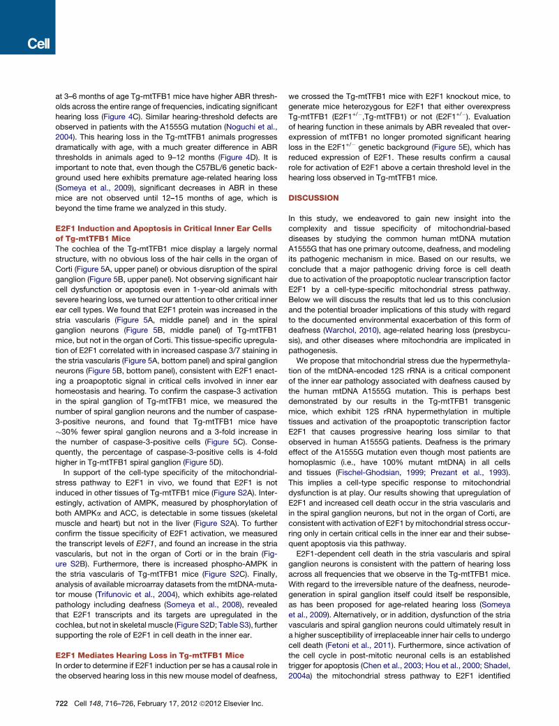
at 3–6 months of age Tg-mtTFB1 mice have higher ABR thresh-
olds across the entire range of frequencies, indicating significant
hearing loss (Figure 4C). Similar hearing-threshold defects are
observed in patients with the A1555G mutation (Noguchi et al.,
2004). This hearing loss in the Tg-mtTFB1 animals progresses
dramatically with age, with a much greater difference in ABR
thresholds in animals aged to 9–12 months (Figure 4D). It is
important to note that, even though the C57BL/6 genetic back-
ground used here exhibits premature age-related hearing loss
(Someya et al., 2009), significant decreases in ABR in these
mice are not observed until 12–15 months of age, which is
beyond the time frame we analyzed in this study.
E2F1 Induction and Apoptosis in Critical Inner Ear Cellsof Tg-mtTFB1 MiceThe cochlea of the Tg-mtTFB1 mice display a largely normal
structure, with no obvious loss of the hair cells in the organ of
Corti (Figure 5A, upper panel) or obvious disruption of the spiral
ganglion (Figure 5B, upper panel). Not observing significant hair
cell dysfunction or apoptosis even in 1-year-old animals with
severe hearing loss, we turned our attention to other critical inner
ear cell types. We found that E2F1 protein was increased in the
stria vascularis (Figure 5A, middle panel) and in the spiral
ganglion neurons (Figure 5B, middle panel) of Tg-mtTFB1
mice, but not in the organ of Corti. This tissue-specific upregula-
tion of E2F1 correlated with in increased caspase 3/7 staining in
the stria vascularis (Figure 5A, bottom panel) and spiral ganglion
neurons (Figure 5B, bottom panel), consistent with E2F1 enact-
ing a proapoptotic signal in critical cells involved in inner ear
homeostasis and hearing. To confirm the caspase-3 activation
in the spiral ganglion of Tg-mtTFB1 mice, we measured the
number of spiral ganglion neurons and the number of caspase-
3-positive neurons, and found that Tg-mtTFB1 mice have
�30% fewer spiral ganglion neurons and a 3-fold increase in
the number of caspase-3-positive cells (Figure 5C). Conse-
quently, the percentage of caspase-3-positive cells is 4-fold
higher in Tg-mtTFB1 spiral ganglion (Figure 5D).
In support of the cell-type specificity of the mitochondrial-
stress pathway to E2F1 in vivo, we found that E2F1 is not
induced in other tissues of Tg-mtTFB1 mice (Figure S2A). Inter-
estingly, activation of AMPK, measured by phosphorylation of
both AMPKa and ACC, is detectable in some tissues (skeletal
muscle and heart) but not in the liver (Figure S2A). To further
confirm the tissue specificity of E2F1 activation, we measured
the transcript levels of E2F1, and found an increase in the stria
vascularis, but not in the organ of Corti or in the brain (Fig-
ure S2B). Furthermore, there is increased phospho-AMPK in
the stria vascularis of Tg-mtTFB1 mice (Figure S2C). Finally,
analysis of available microarray datasets from the mtDNA-muta-
tor mouse (Trifunovic et al., 2004), which exhibits age-related
pathology including deafness (Someya et al., 2008), revealed
that E2F1 transcripts and its targets are upregulated in the
cochlea, but not in skeletal muscle (Figure S2D; Table S3), further
supporting the role of E2F1 in cell death in the inner ear.
E2F1 Mediates Hearing Loss in Tg-mtTFB1 MiceIn order to determine if E2F1 induction per se has a causal role in
the observed hearing loss in this new mouse model of deafness,
722 Cell 148, 716–726, February 17, 2012 ª2012 Elsevier Inc.
we crossed the Tg-mtTFB1 mice with E2F1 knockout mice, to
generate mice heterozygous for E2F1 that either overexpress
Tg-mtTFB1 (E2F1+/�,Tg-mtTFB1) or not (E2F1+/�). Evaluation
of hearing function in these animals by ABR revealed that over-
expression of mtTFB1 no longer promoted significant hearing
loss in the E2F1+/� genetic background (Figure 5E), which has
reduced expression of E2F1. These results confirm a causal
role for activation of E2F1 above a certain threshold level in the
hearing loss observed in Tg-mtTFB1 mice.
DISCUSSION
In this study, we endeavored to gain new insight into the
complexity and tissue specificity of mitochondrial-based
diseases by studying the common human mtDNA mutation
A1555G that has one primary outcome, deafness, and modeling
its pathogenic mechanism in mice. Based on our results, we
conclude that a major pathogenic driving force is cell death
due to activation of the proapoptotic nuclear transcription factor
E2F1 by a cell-type-specific mitochondrial stress pathway.
Below we will discuss the results that led us to this conclusion
and the potential broader implications of this study with regard
to the documented environmental exacerbation of this form of
deafness (Warchol, 2010), age-related hearing loss (presbycu-
sis), and other diseases where mitochondria are implicated in
pathogenesis.
We propose that mitochondrial stress due the hypermethyla-
tion of the mtDNA-encoded 12S rRNA is a critical component
of the inner ear pathology associated with deafness caused by
the human mtDNA A1555G mutation. This is perhaps best
demonstrated by our results in the Tg-mtTFB1 transgenic
mice, which exhibit 12S rRNA hypermethylation in multiple
tissues and activation of the proapoptotic transcription factor
E2F1 that causes progressive hearing loss similar to that
observed in human A1555G patients. Deafness is the primary
effect of the A1555G mutation even though most patients are
homoplasmic (i.e., have 100% mutant mtDNA) in all cells
and tissues (Fischel-Ghodsian, 1999; Prezant et al., 1993).
This implies a cell-type specific response to mitochondrial
dysfunction is at play. Our results showing that upregulation of
E2F1 and increased cell death occur in the stria vascularis and
in the spiral ganglion neurons, but not in the organ of Corti, are
consistent with activation of E2F1 bymitochondrial stress occur-
ring only in certain critical cells in the inner ear and their subse-
quent apoptosis via this pathway.
E2F1-dependent cell death in the stria vascularis and spiral
ganglion neurons is consistent with the pattern of hearing loss
across all frequencies that we observe in the Tg-mtTFB1 mice.
With regard to the irreversible nature of the deafness, neurode-
generation in spiral ganglion itself could itself be responsible,
as has been proposed for age-related hearing loss (Someya
et al., 2009). Alternatively, or in addition, dysfunction of the stria
vascularis and spiral ganglion neurons could ultimately result in
a higher susceptibility of irreplaceable inner hair cells to undergo
cell death (Fetoni et al., 2011). Furthermore, since activation of
the cell cycle in post-mitotic neuronal cells is an established
trigger for apoptosis (Chen et al., 2003; Hou et al., 2000; Shadel,
2004a) the mitochondrial stress pathway to E2F1 identified

Figure 5. Deafness Pathology in Tg-mtTFB1 Mice Involves Upregulation of E2F1 and Apoptosis in the Stria Vascularis and Spiral Ganglion
Neurons of the Inner Ear
(A) H&E staining of the organ of Corti and stria vascularis (top panels) from representative 1-year -old wild-type (wt, left column) and Tg-mtTFB1 mice (right
column). The middle panels show immunohistochemistry staining for E2F1 and the lower panels show immunohistochemistry for cleaved caspase-3 in similar
representative sections. White arrows indicate areas where significant changes in signal are observed. Stria vascularis (s.v.), outer hair cells (OHC), scale bar,
100 mm. See also Figure S2.
(B) Same as in (A) but sections that highlight staining in the spiral ganglion neurons are shown. Spiral ganglion (s.g.), scale bar, 100 mm.
(C) Quantification of cellular density in the spiral ganglion (the left side of the plot) and of caspase-3-positive spiral ganglion neurons (right side). The sections were
obtained from three wild-type and three Tg-mtTFB1 mice at 1 year of age, and six independent sections were counted for each animal. The values plotted
represent mean ± standard deviation, with t test p values indicated.
(D) Same as in (C), but expressing the percentage of caspase-3-positive cells in the spiral ganglion. The values plotted represent mean ± standard deviation, with t
test p values indicated.
(E) ABR analysis of E2F1+/� mice (black points) and E2F1+/�/Tg-mtTFB1 mice (red points) performed as described in Figure 4C. Mice were tested at ages
3–6months (eight E2F1+/�mice and ten E2F1+/�/Tg-mtTFB1micewere used). The values plotted represent mean ±SEM,with t test p values indicated (*p < 0.05).
herein may also may make some cells of the inner ear inherently
more susceptible to cell death due to being forced into the cell
cycle, as opposed to activation the E2F1 apoptosis pathway
per se. Based on these considerations, we propose that
A1555G patients would likewise be more prone to eventual
loss of critical inner ear cells via this mitochondrial stress
pathway to E2F1, explaining their irreversible hearing loss either
spontaneously, as a function of age, or in response to environ-
mental stimuli such as noise or aminoglycosides. Even though
our results strongly implicate 12S hypermethylation per se in
the deafness phenotype in mice, it is likely that defects in mito-
chondrial translation caused by the A1555G mutation, such as
infidelity (Hobbie et al., 2008), conspire with hypermethylation
to produce the precise deafness pathology in humans and/or
mediate its exacerbation by aminoglycoside antibiotics. There-
fore, it remains important to determine precisely which
Cell 148, 716–726, February 17, 2012 ª2012 Elsevier Inc. 723

mechanisms and cell types within the inner ear instigate deaf-
ness pathology in this new mouse model and in human
A1555G patients. A full understanding of these events may be
therapeutic in this regard and could help advance efforts to
regenerate hearing function.
Our results in A1555G cybrids also provide key insight into the
nature of the pathogenic mitochondrial stress-signaling
pathway. Specifically, hypermethylation of mitochondrial ribo-
somes, which are needed for translational and assembly of the
respiratory chain (Bonawitz et al., 2006), disrupts mitochondrial
respiration in amanner that increases ROS production (Figure 3).
We propose that increased mitochondrial superoxide is sensed
by AMPK (Emerling et al., 2009; Quintero et al., 2006), activation
of which relays the stress signal to E2F1. Since E2F1 activity is
associated with proapoptotic signaling and is necessary for the
enhanced cell death on A1555G cybrids (Figure 2D) and hearing
loss in Tg-mtTFB1 mice (Figure 5E), we speculate ROS- and
AMPK-dependent activation of E2F1 is the major mitochondrial
stress-signaling pathway involved under these circumstances.
Going forward, it will be important to determine precisely how
E2F1 is regulated by ROS and AMPK.
Themarked tissue specificity of E2F1 induction and, to a lesser
degree, of AMPK activation (Figure S2A), provides insight into
the complex mechanisms underlying tissue specificity of mito-
chondrial diseases. The requirement for multiple sequential
steps to fully activate the mitochondrial ROS-AMPK-E2F1
apoptotic pathway provides a variety of opportunities for
different tissues to suppress a pathogenic mechanism. For
example, tissues with lower OXPHOS activity or better redox
buffering could prevent initial activation of the pathogenic mech-
anism. Alternatively, in other tissues, AMPK may not respond to
ROS, E2F1 may not be activated by AMPK, or E2F1 may not be
a potent trigger of apoptosis, any of which would presumably
prevent a pathogenic outcome. These observations may be
generalizable, in that other pathogenic mitochondrial retrograde
signaling pathways likely exist with their differential activation or
readout in tissues contributing to the complex tissue specificity
observed in mitochondrial diseases.
Finally, the induction of E2F1 in response to a mitochondrial
malfunction may represent a paradigm in mitochondrial patho-
genesis where the cause of the disease is not the immediate
OXPHOS dysfunction, but instead themisinterpretation of result-
ing retrogradesignals generatedbymitochondria. Thepivotal role
of AMPKas a sensor of energy charge and nowas a ROS-depen-
dent regulator of E2F1 activity highlights the complexity of mito-
chondrial stress responses and the need for additional research
to uncover other such pathways. Finally, it is tempting to specu-
late that aberrant mitochondrial stress signaling may also be of
general significance in diseases and circumstances where mito-
chondrial dysfunctionandapoptosisare implicated, suchasheart
disease, cancer, neurodegenerative diseases and aging.
EXPERIMENTAL PROCEDURES
Cell Culture, shRNA-Mediated Gene Knockdown, FACS Analysis,
and 12S Methylation Assays
143B osteosarcroma cybrids containing either wild-type (A1555A) or mutant
(A1555G)mtDNA (Guan et al., 2001) were grown in DMEMhigh glucose (Sigma
724 Cell 148, 716–726, February 17, 2012 ª2012 Elsevier Inc.
D5648) plus 10% fetal calf serum at 37�Cand 5%CO2. Prior to all experiments,
cells were plated at 5,000 cells/cm2 and grown for 96 hr to 70%–80% conflu-
ence. When puromycin was used for selection, it was removed at least 24 hr
prior to harvesting cells. HeLa cells that overexpress h-mtTFB1 have been
described (Cotney et al., 2007, 2009).
Retroviral-mediated shRNA knockdown ofmtTFB1 and E2F1 by was carried
as described (Cotney et al., 2009) with the following modifications. Wild-type
or mutant 143B cybrids were transfected and plated at 5,000/cm2 in DMEM
with 10% fetal calf serum and 600 ng/ml puromycin and used for experiments
at 70%–80% confluence. Puromycin was removed 48 hr prior to analysis.
Target sequences ligated into the pSiren-RetroQ vector (Clonetech) for
mtTFB1 and E2F1 knockdown, as well the scrambled negative-control shRNA
used were described previously (Cotney et al., 2009; Goto et al., 2006). To
overexpress SOD2, cybrids were transfected with pCMV-SPORT6-SOD2
(Open Biosystems MMM4769-99609684) or pcDNA3.0 as a negative control
as described (Cotney et al., 2009). To inhibit AMPK, cybrids were treated for
16 hr with 10 mM of compound C (Calbiochem) and then used immediately
for experiments.
To analyze mitochondrial superoxide by FACS, wild-type and A1555G cy-
brids were treated with 50 nMMitoSOX (Invitrogen) for 20 min at 37�C, rinsed,and detached with trypsin. Fluorescence was measured using a FACScan (BD
Biotechnology) and analyzed using FlowJo software. Primer-extension anal-
ysis of mitochondrial 12S rRNA adenine dimethylation was performed on
1–5 mg of total cellular or tissue RNA prepared using the RNEasy Kit (QIAGEN)
as described (Cotney et al., 2007; Seidel-Rogol et al., 2003), except 2 mMeach
of dATP, dCTP, and dTTP and 0.5 mM ddGTP were used.
Western Blotting
Whole-cell extracts of cultured human cells were prepared in 1.5% n-dodecyl-
maltoside in PBS as described (Raimundo et al., 2009). Gels were loaded with
50 mg of total protein per well, and the following antibodies were used for
immunoblotting: E2F1 (Santa Cruz, sc193), mtTFB1/TFBM1 (McCulloch and
Shadel, 2003) or that provided by Dr. Craig Cameron, tubulin (Thermo,
DM1A), Rb (BD PharMingen, 554136), phospho-Rb (T826) (Abcam, ab4779-
50), phospho-Rb (S807/811) (Cell Signaling, 9308S), AMPKa (Cell Signaling,
2603), phospho-AMPKa (T172) (Cell Signaling, 2535S), acetyl-CoA carbox-
ylase (ACC) (Cell Signaling, 3676), phospho-ACC (S79) (Cell Signaling,
3661), SOD2 (Assay Designs, SOD-111), PARP (Cell Signaling, 9542), and
GAPDH (Ambion, AM4300). Western blots were performed on three to four
biological replicates and at least three technical replicates.
Apoptosis Assays
To induce E2F1-dependent apoptosis, cybrids were treated with 200 nM
staurosporine for 3 hr in the presence of 10mMwortmannin as described (Hall-
strom et al., 2008). Cells were then rinsed and used for two different apoptosis
assays. Caspase 3/7 activity was determined using a plate reader 1 hr after
treatment with Caspase-Glo reagent (Promega) as described by the manufac-
turer, and full-length and cleaved PARP were assayed by western blot.
Oxygen Consumption Analysis
Cells were plated in XF96 plates (SeaHorse Biosciences) at 10,000 cells/well
and the next day cellular O2 consumption was determined in a SeaHorse
Bioscience XF96 extracellular flux analyzer according tomanufacturer instruc-
tions. Cells were maintained at 37�C in normal growth medium without serum.
In some wells 8 mM of the mitochondrial uncoupler FCCP was added.
Microarrays and Data Analysis
Total cellular RNA from the cultured cells was prepared using an RNeasy mini
RNA extraction kit (QIAGENGmbH, Hilden, Germany) and used for the expres-
sion microarray procedure in conjunction with the Yale University W.M. Keck
Foundation Biotechnology Resource Laboratory. RNA integrity was first veri-
fied by an Agilent Bioanalyzer and then amplified, labeled and hybridized
onto GeneChip HG-U133 Plus 2.0 arrays (Affymetrix, Agilent, Santa Clara,
CA) using standard protocols recommended by the manufacturer, starting
from 5 mg of total RNA. Data were normalized by the RMA method (Irizarry
et al., 2003), using commercial software (GeneSpring, Agilent, Santa Clara,
California, CA). For each biological sample, two RNA samples were prepared

from two independently collected cell pellets. To analyze the expression data,
samples were grouped according to their 12S rRNA methylation status (Fig-
ure 1A). The four samples in the hypermethylation group were compared to
the four samples with basal methylation to control for nonspecific effects of
overexpression of mtTFB1 independent of increased 12S rRNA methylation
and in an attempt to subtract effects due to cell nuclear genetic background.
Transcripts whose expression was significantly changed between the two
groups formed the 12S rRNA hypermethylation list (4333 transcripts; Table
S1). The student’s t test was used to determine statistically significant changes
in expression, with a cutoff p value of 0.05 (Raimundo et al., 2009). The false-
positive rate was determined to be 3.8% as described (Hovatta et al., 2005).
The correlation of the Q-PCR with the microarray results for six genes tested
was R2 = 0.77 (Raimundo et al., 2009).
To determine which cis-elements were present in the promoters of the
genes up- or downregulated in 12S rRNA hypermethylated samples, we
used the Bibliosphere software (Genomatix Software GmbH, Munich,
Germany). To minimize false positives, a cutoff of at least two cis element-
gene cocitations was implemented. Enrichment in cis-elements of the genes
up- or downregulated was determined by Fisher’s exact test.
Generation and Analysis of Transgenic Mice that
Overexpress mtTFB1
A mouse mtTFB1 cDNA was amplified with primers containing EcoR1 and
EcoRV restriction sites and ligated into the vector pCAGGS that contains
a CMV immediate early-enhancer/chicken b-actin/rabbit b-globin promoter
(Niwa et al., 1991) and confirmed by sequencing. Purified pCAGGS/mtTFB1
plasmid was digested with SalI and PstI, to liberate a fragment containing
the enhancer, promoter, TFB1M ORF, and poly-A tail. This linear DNA frag-
ment was then used by the Yale Animal Genomic Services to microinject
pronuclear, C57BL/6J X SJL/J embryos. Seventy-four animals were born
from the six foster mothers. After genotyping, one female (F1) founder was
identified that contained the transgene. This female was then mated with
a wild-type C57BL/6J male to generate the second generation (F2) and then
serially to create subsequent generations. Female F3 to F5 transgenics and
control nontransgenic littermates were used for all of the experiments. All
procedures were IACUC-approved as determined by the Yale University
Animal Care and Use Committee.
For analysis of mouse samples, tissues from 9-month-old mice were har-
vested, immediately frozen in liquid nitrogen, and stored at �80C. The tissues
were ground using a mortar and pestle previously frozen at �80C, kept on dry
ice during the procedure. Whole-tissue homogenates were prepared as
described (Cotney et al., 2009). Primer extension analysis and q-PCR were
performed onRNApurified from these samemouse tissues using the RNAeasy
kit using the method already described for human cells, but with the following
mouse 12S-specific primer: 50-ATTATTCCAAGC-30. To generate Tg-mtTFB1/
E2F1+/� mice, a C57BL/6J male E2F1�/� was obtained from the Jackson
Laboratory (Bar Harbor, ME) and mated with F4 Tg-mtTFB1 females, yielding
E2F1+/� progeny with and without the mtTFB1 transgene.
Auditory Brainstem Response and Cochlear Histology
Detailed methods for the ABR analysis can be found in the Extended Experi-
mental Procedures. A total of 21 animals (9 Tg-mtTFB1 and 12 wild-type litter-
mates) between the ages of 3 and 6 months, and 11 animals between 9 and
12 months (5 wild-type, 6 Tg-mtTFB1), were used for auditory brainstem
response (ABR) testing. The E2F1+/� animals were tested at 3–6 months of
age (total of 20, 8 wild-type and 12 Tg-mtTFB1). In all experiments, age-
matched littermates were compared directly on the same day and by the
same experienced specialist blinded to the genotype of the mice. The mice
were subsequently combined into 3–6 and 9–12 groups for data presentation
and statistical analysis. Animals were anesthetized with chloral hydrate
(480 mg/kg IP) and all recordings were conducted in a sound-attenuating
chamber (Industrial Acoustics Corp) using a customized TDT3 system
(Tucker-Davis Technologies, Inc.). A t test was used to determine the statistical
significance of the threshold difference at each frequency.
The inner ear was prepared for histology by removing the temporal bone and
exposing and rupturing the bulla. The cochlea was perfused with 4% parafor-
maldehyde (in PBS) using a syringe outfitted with small diameter tubing
through the oval and round windows. The tissue was incubated overnight, de-
calcified in 10% EDTA in PBS for 3 days, and then paraffin embedded,
sectioned in 5 mm samples and mounted onto glass slides by the Yale
Histology section. Following deparaffinization and rehydration, sections
were used for immunohistochemistry and hematoxylin staining. Images were
obtained using an Olympus IX71 microscope and Metamorph image analysis
software. Nine independent cochlear sections were analyzed from three
1-year-old wild-type and three Tg-mtTFB1 mice. Spiral ganglion cells were
counted using the DAPI counterstain in an area of 100 3 100 pixels and
normalized to area (mm2). Cells displaying cytoplasmic foci of caspase-3
were considered caspase-3+.
ACCESSION NUMBERS
The raw data were deposited in the Gene Expression Omnibus repository
(accession #GSE33780).
SUPPLEMENTAL INFORMATION
Supplemental Information includes Extended Experimental Procedures, three
tables, and two figures and can be found with this article online at doi:10.1016/
j.cell.2011.12.027.
ACKNOWLEDGMENTS
This study was supported by grants R01 HL-059655 to G.S.S. and R01
DC000273 to J.S.-S. from the U.S. NIH and a UMDF fellowship to T.E.S. The
authors thank Zimei Zhang for animal husbandry, Yale Animal Genomic
Services for help in generating the Tg-mtTFB1 mice, and Dr. Susan Kaech
for comments on the manuscript, helpful discussions and access to the
SeaHorse instrument.
Received: September 6, 2011
Revised: October 31, 2011
Accepted: December 15, 2011
Published: February 16, 2012
REFERENCES
Bonawitz, N.D., Clayton, D.A., and Shadel, G.S. (2006). Initiation and beyond:
multiple functions of the human mitochondrial transcription machinery. Mol.
Cell 24, 813–825.
Butow, R.A., and Avadhani, N.G. (2004). Mitochondrial signaling: the retro-
grade response. Mol. Cell 14, 1–15.
Bykhovskaya, Y., Mengesha, E., Wang, D., Yang, H., Estivill, X., Shohat, M.,
and Fischel-Ghodsian, N. (2004). Human mitochondrial transcription factor
B1 as a modifier gene for hearing loss associated with the mitochondrial
A1555G mutation. Mol. Genet. Metab. 82, 27–32.
Chen, P., Zindy, F., Abdala, C., Liu, F., Li, X., Roussel, M.F., and Segil, N.
(2003). Progressive hearing loss in mice lacking the cyclin-dependent kinase
inhibitor Ink4d. Nat. Cell Biol. 5, 422–426.
Cotney, J., Wang, Z., and Shadel, G.S. (2007). Relative abundance of the
human mitochondrial transcription system and distinct roles for h-mtTFB1
and h-mtTFB2 in mitochondrial biogenesis and gene expression. Nucleic
Acids Res. 35, 4042–4054.
Cotney, J., McKay, S.E., and Shadel, G.S. (2009). Elucidation of separate, but
collaborative functions of the rRNA methyltransferase-related human mito-
chondrial transcription factors B1 and B2 in mitochondrial biogenesis reveals
new insight into maternally inherited deafness. Hum. Mol. Genet. 18, 2670–
2682.
Dasgupta, B., and Milbrandt, J. (2009). AMP-activated protein kinase phos-
phorylates retinoblastoma protein to control mammalian brain development.
Dev. Cell 16, 256–270.
Cell 148, 716–726, February 17, 2012 ª2012 Elsevier Inc. 725

DeGregori, J., and Johnson, D.G. (2006). Distinct and Overlapping Roles for
E2F Family Members in Transcription, Proliferation and Apoptosis. Curr.
Mol. Med. 6, 739–748.
DiMauro, S., and Schon, E.A. (2003). Mitochondrial respiratory-chain
diseases. N. Engl. J. Med. 348, 2656–2668.
Emerling, B.M., Weinberg, F., Snyder, C., Burgess, Z., Mutlu, G.M., Viollet, B.,
Budinger, G.R., and Chandel, N.S. (2009). Hypoxic activation of AMPK is
dependent on mitochondrial ROS but independent of an increase in AMP/
ATP ratio. Free Radic. Biol. Med. 46, 1386–1391.
Fetoni, A.R., Picciotti, P.M., Paludetti, G., and Troiani, D. (2011). Pathogenesis
of presbycusis in animal models: a review. Exp. Gerontol. 46, 413–425.
Field, S.J., Tsai, F.Y., Kuo, F., Zubiaga, A.M., Kaelin, W.G., Jr., Livingston,
D.M., Orkin, S.H., and Greenberg, M.E. (1996). E2F-1 functions in mice to
promote apoptosis and suppress proliferation. Cell 85, 549–561.
Fischel-Ghodsian, N. (1999). Mitochondrial deafness mutations reviewed.
Hum. Mutat. 13, 261–270.
Goto, Y., Hayashi, R., Kang, D., and Yoshida, K. (2006). Acute loss of transcrip-
tion factor E2F1 induces mitochondrial biogenesis in HeLa cells. J. Cell. Phys-
iol. 209, 923–934.
Guan, M.X., Fischel-Ghodsian, N., and Attardi, G. (1996). Biochemical
evidence for nuclear gene involvement in phenotype of non-syndromic deaf-
ness associated with mitochondrial 12S rRNA mutation. Hum. Mol. Genet. 5,
963–971.
Guan, M.X., Fischel-Ghodsian, N., and Attardi, G. (2000). A biochemical basis
for the inherited susceptibility to aminoglycoside ototoxicity. Hum.Mol. Genet.
9, 1787–1793.
Guan, M.X., Fischel-Ghodsian, N., and Attardi, G. (2001). Nuclear background
determines biochemical phenotype in the deafness-associated mitochondrial
12S rRNA mutation. Hum. Mol. Genet. 10, 573–580.
Hallstrom, T.C., Mori, S., and Nevins, J.R. (2008). An E2F1-dependent gene
expression program that determines the balance between proliferation and
cell death. Cancer Cell 13, 11–22.
Hamanaka, R.B., and Chandel, N.S. (2010). Mitochondrial reactive oxygen
species regulate cellular signaling and dictate biological outcomes. Trends
Biochem. Sci. 35, 505–513.
Hardie, D.G. (2007). AMP-activated/SNF1 protein kinases: conserved guard-
ians of cellular energy. Nat. Rev. Mol. Cell Biol. 8, 774–785.
Hobbie, S.N., Bruell, C.M., Akshay, S., Kalapala, S.K., Shcherbakov, D., and
Bottger, E.C. (2008). Mitochondrial deafness alleles confer misreading of the
genetic code. Proc. Natl. Acad. Sci. USA 105, 3244–3249.
Hou, S.T., Callaghan, D., Fournier, M.C., Hill, I., Kang, L., Massie, B., Morley,
P., Murray, C., Rasquinha, I., Slack, R., and MacManus, J.P. (2000). The
transcription factor E2F1 modulates apoptosis of neurons. J. Neurochem.
75, 91–100.
Hovatta, I., Tennant, R.S., Helton, R., Marr, R.A., Singer, O., Redwine, J.M.,
Ellison, J.A., Schadt, E.E., Verma, I.M., Lockhart, D.J., and Barlow, C.
(2005). Glyoxalase 1 and glutathione reductase 1 regulate anxiety in mice.
Nature 438, 662–666.
Irizarry, R.A., Bolstad, B.M., Collin, F., Cope, L.M., Hobbs, B., and Speed, T.P.
(2003). Summaries of Affymetrix GeneChip probe level data. Nucleic Acids
Res. 31, e15.
Jones, R.G., Plas, D.R., Kubek, S., Buzzai, M., Mu, J., Xu, Y., Birnbaum, M.J.,
and Thompson, C.B. (2005). AMP-activated protein kinase induces a p53-
dependent metabolic checkpoint. Mol. Cell 18, 283–293.
McCulloch, V., Seidel-Rogol, B.L., and Shadel, G.S. (2002). A human mito-
chondrial transcription factor is related to RNA adenine methyltransferases
and binds S-adenosylmethionine. Mol. Cell. Biol. 22, 1116–1125.
726 Cell 148, 716–726, February 17, 2012 ª2012 Elsevier Inc.
McCulloch, V., and Shadel, G.S. (2003). Human mitochondrial transcription
factor B1 interacts with the C-terminal activation region of h-mtTFA and stim-
ulates transcription independently of its RNA methyltransferase activity. Mol.
Cell. Biol. 23, 5816–5824.
Metodiev, M.D., Lesko, N., Park, C.B., Camara, Y., Shi, Y., Wibom, R., Hul-
tenby, K., Gustafsson, C.M., and Larsson, N.G. (2009). Methylation of 12S
rRNA is necessary for in vivo stability of the small subunit of the mammalian
mitochondrial ribosome. Cell Metab. 9, 386–397.
Niwa, H., Yamamura, K., and Miyazaki, J. (1991). Efficient selection for high-
expression transfectants with a novel eukaryotic vector. Gene 108, 193–199.
Noguchi, Y., Yashima, T., Ito, T., Sumi, T., Tsuzuku, T., and Kitamura, K. (2004).
Audiovestibular findings in patients with mitochondrial A1555G mutation.
Laryngoscope 114, 344–348.
Polager, S., and Ginsberg, D. (2009). p53 and E2f: partners in life and death.
Nat. Rev. Cancer 9, 738–748.
Prezant, T.R., Agapian, J.V., Bohlman, M.C., Bu, X., Oztas, S., Qiu, W.Q.,
Arnos, K.S., Cortopassi, G.A., Jaber, L., Rotter, J.I., et al. (1993). Mitochondrial
ribosomal RNA mutation associated with both antibiotic-induced and non-
syndromic deafness. Nat. Genet. 4, 289–294.
Pulicherla, N., Pogorzala, L.A., Xu, Z., O Farrell, H.C., Musayev, F.N., Scars-
dale, J.N., Sia, E.A., Culver, G.M., and Rife, J.P. (2009). Structural and func-
tional divergence within the Dim1/KsgA family of rRNA methyltransferases.
J. Mol. Biol. 391, 884–893.
Quintero, M., Colombo, S.L., Godfrey, A., and Moncada, S. (2006). Mitochon-
dria as signaling organelles in the vascular endothelium. Proc. Natl. Acad. Sci.
USA 103, 5379–5384.
Raimundo, N., Vanharanta, S., Aaltonen, L.A., Hovatta, I., and Suomalainen, A.
(2009). Downregulation of SRF-FOS-JUNB pathway in fumarate hydratase
deficiency and in uterine leiomyomas. Oncogene 28, 1261–1273.
Seidel-Rogol, B.L., McCulloch, V., and Shadel, G.S. (2003). Human mitochon-
drial transcription factor B1 methylates ribosomal RNA at a conserved stem-
loop. Nat. Genet. 33, 23–24.
Shadel, G.S. (2004a). Coupling the mitochondrial transcription machinery to
human disease. Trends Genet. 20, 513–519.
Shadel, G.S. (2004b). A dual-function mitochondrial transcription factor tunes
out deafness. Mol. Genet. Metab. 82, 1–3.
Shadel, G.S. (2008). Expression and maintenance of mitochondrial DNA: new
insights into human disease pathology. Am. J. Pathol. 172, 1445–1456.
Someya, S., Yamasoba, T., Kujoth, G.C., Pugh, T.D.,Weindruch, R., Tanokura,
M., and Prolla, T.A. (2008). The role of mtDNAmutations in the pathogenesis of
age-related hearing loss in mice carrying a mutator DNA polymerase gamma.
Neurobiol. Aging 29, 1080–1092.
Someya, S., Xu, J., Kondo, K., Ding, D., Salvi, R.J., Yamasoba, T., Rabinovitch,
P.S., Weindruch, R., Leeuwenburgh, C., Tanokura, M., and Prolla, T.A. (2009).
Age-related hearing loss in C57BL/6J mice is mediated by Bak-dependent
mitochondrial apoptosis. Proc. Natl. Acad. Sci. USA 106, 19432–19437.
Trifunovic, A., Wredenberg, A., Falkenberg, M., Spelbrink, J.N., Rovio, A.T.,
Bruder, C.E., Bohlooly-Y, M., Gidlof, S., Oldfors, A., Wibom, R., et al. (2004).
Premature ageing in mice expressing defective mitochondrial DNA poly-
merase. Nature 429, 417–423.
Vandebona, H., Mitchell, P., Manwaring, N., Griffiths, K., Gopinath, B., Wang,
J.J., and Sue, C.M. (2009). Prevalence of mitochondrial 1555A—>G mutation
in adults of European descent. N. Engl. J. Med. 360, 642–644.
Wallace, D.C. (2005). Amitochondrial paradigm ofmetabolic and degenerative
diseases, aging, and cancer: a dawn for evolutionary medicine. Annu. Rev.
Genet. 39, 359–407.
Warchol, M.E. (2010). Cellular mechanisms of aminoglycoside ototoxicity.
Curr. Opin. Otolaryngol. Head Neck Surg. 18, 454–458.