Embed Size (px)
Citation preview

Research Article
Robust Antitumor Effects of Combined Anti–CD4-Depleting Antibody and Anti–PD-1/PD-L1 ImmuneCheckpoint Antibody Treatment in MiceSatoshi Ueha1, Shoji Yokochi1,2, Yoshiro Ishiwata1,2, Haru Ogiwara1, Krishant Chand1,Takuya Nakajima1, Kosuke Hachiga1,2, Shigeyuki Shichino1,Yuya Terashima1, Etsuko Toda1,Francis H.W. Shand3, Kazuhiro Kakimi4, Satoru Ito1,2, and Kouji Matsushima1
Abstract
Depletion of CD4þ cells in tumor-bearing mice has strongantitumor effects. However, the mechanisms underlying theseeffects and the therapeutic benefits of CD4þ cell depletionrelative to other immunotherapies have not been fully evalu-ated. Here, we investigated the antitumor effects of an anti–CD4-depleting mAb as a monotherapy or in combination withimmune checkpoint mAbs. In B16F10, Colon 26, or Lewis lungcarcinoma subcutaneous tumor models, administration ofthe anti-CD4 mAb alone had strong antitumor effects thatwere superior to those elicited by CD25þ Treg depletion orother immune checkpoint mAbs, and which were completely
reversed by CD8þ cell depletion. CD4þ cell depletion led to theproliferation of tumor-specific CD8þ T cells in the draininglymph node and increased infiltration of PD-1þCD8þ T cellsinto the tumor, with a shift toward type I immunity within thetumor. Combination treatment with the anti-CD4 mAb andimmune checkpoint mAbs, particularly anti–PD-1 or anti–PD-L1 mAbs, synergistically suppressed tumor growth and greatlyprolonged survival. To our knowledge, this work represents thefirst report of robust synergy between anti-CD4 and anti–PD-1or anti–PD-L1 mAb therapies. Cancer Immunol Res; 3(6); 631–40.�2015 AACR.
IntroductionImmune checkpoint modulators such as those targeting cyto-
toxic T-lymphocyte–associated antigen-4 (CTLA-4) and pro-grammed cell death-1 (PD-1) have attracted attention due totheir extraordinary antitumor effects in patients with advancedmelanoma, lung cancer, and renal cancer (1, 2). An mAb againstCTLA-4 (ipilimumab) that enhances both early T-cell activationand CTL function was approved for treatment of patients withadvanced melanoma in the United States in 2011. An anti–PD-1mAb (nivolumab) that protects activated T cells from exhaustionin peripheral tissues was approved for treatment of patients withmelanoma in Japan and in the United States in 2014. In addition,other mAbs against CTLA-4 (tremelimumab), PD-1 (pembroli-zumab), and programmed death-ligand 1 (PD-L1, a ligand forPD-1) are currently undergoing clinical trials to evaluate their
antitumor efficacy. However, despite clear survival benefits in asubset of tumor patients, other groups of patients are refractory tothese single-agent therapies.
Combination therapies comprising immune checkpoint mod-ulators that have different points of action, targeting, for example,the activation and expansionof T cells in lymphoid tissues and theexhaustion and deletion of T cells in the effector site, representpromising strategies for tumor immunotherapy (1). Synergisticantitumor effects in advancedmelanomahave been reportedwitha combination of anti–CTLA-4 and anti–PD-1 mAbs (3). Theantitumor efficacy of other combinations of regulators of lym-phocyte activation and expansion (e.g., Lymphocyte activationgene-3/LAG-3,OX40/CD134) and of lymphocyte exhaustion anddeletion (e.g., T-cell immunoglobulin mucin-3/TIM-3, 4-1BB/CD137, B- and T-lymphocyte attenuator/BTLA, glucocorticoid-induced TNF-receptor/GITR) is currently under investigation.Because immune checkpoint modulators play both positive andnegative roles in the immune inhibitory pathway with someredundancy, identification of optimal therapeutic combinationsremains a considerable challenge.
Another approach to immune checkpointmodulation involvesdepleting immunosuppressive leukocyte populations such asforkhead box P3 (Foxp3)þCD25þ regulatory T cells (Treg), Th2cells, T regulatory (Tr) 1/3 cells (4), myeloid-derived suppressorcells (MDSC) and indoleamine-2,3-dioxygenase (IDO)þ plasma-cytoid DCs (pDC; refs. 5–7). Several groups have suggested thatdepletion of CD4þ cells, including Tregs, Th2 cells, Tr1/3 cells,and a subpopulation of MDSCs and pDCs, results in strongantitumor effects in mouse models due to the enhancement ofCTL responses (8–12). These antitumor effects may be associatedwith the modulation of multiple immune checkpoints caused by
1Department of Molecular Preventive Medicine, Graduate School ofMedicine, The University of Tokyo, Tokyo, Japan. 2IDAC Theranostics,Inc., Tokyo, Japan. 3Department of Pharmacology and Therapeutics,The University of Melbourne, Melbourne,Victoria, Australia. 4Depart-ment of Immunotherapeutics,TheUniversity of TokyoHospital,Tokyo,Japan.
Note: Supplementary data for this article are available at Cancer ImmunologyResearch Online (http://cancerimmunolres.aacrjournals.org/).
S. Ueha, S. Yokochi, and Y. Ishiwata contributed equally to this article.
CorrespondingAuthor:KoujiMatsushima, TheUniversity of Tokyo, 7-3-1 Hongo,Bunkyo-ku, Tokyo 1130033, Japan. Phone: 81-3-5841-3431; Fax: 81-3-5684-2297;E-mail: [email protected]
doi: 10.1158/2326-6066.CIR-14-0190
�2015 American Association for Cancer Research.
CancerImmunologyResearch
www.aacrjournals.org 631
on February 28, 2019. © 2015 American Association for Cancer Research. cancerimmunolres.aacrjournals.org Downloaded from
Published OnlineFirst February 20, 2015; DOI: 10.1158/2326-6066.CIR-14-0190

CD4þ cell depletion. However, the relative advantage of CD4þ
cell depletion over other immune checkpoint mAb-based treat-ments remains unclear. Encouraged by the positive reports sur-rounding the benefits of anti-CD4mAb treatment inmice, and bythe recent clinical data supporting anti–CTLA-4 and anti–PD-1mAb therapies, here, we examine whether treatments that com-bine an anti-CD4 mAb and immune checkpoint modulatorsproduce synergistic antitumor activity.
Thus, in the present study, we used comprehensive immuno-logic analyses to compare the antitumor effects of an anti–CD4-depleting mAb with those of a variety of mAbs against immunecheckpoint molecules, including PD-1, PD-L1, PD-L2, CTLA-4,OX40, LAG-3, TIM-3, BTLA, and GITR, in mouse subcutaneoustumor models. We also investigated the antitumor effects oftreatments that combined an anti-CD4 mAb and antibodiesagainst these immune checkpoint molecules. We report thattreatment with an anti-CD4mAb alone induces strong antitumoreffects and expansion of tumor-specific CD8þ T cells, and thatcombination of an anti-CD4mAb with anti–PD-1 or anti–PD-L1mAbs results in striking synergy in the suppression of tumorgrowth.
Materials and MethodsMouse
Seven-week-old female C57BL/6 and male BALB/c mice werepurchased from Japan SLC. Fluorescent ubiquitination-based cell-cycle indicator (Fucci) double transgenic mice were generated bycrossbreeding FucciG1-#639 and FucciS/G2/M-#474 animals(obtained from Dr. A. Miyawaki through the RIKEN BRC) asdescribed previously (13). Mice transgenic for the gp100 mela-noma antigen-specific Pmel-1-TCR or the ovalbumin-specific OT-I TCR were purchased from The Jackson Laboratory. Each exper-imental group contained 8mice except where otherwise specified.All animal experiments were conducted in accordance with insti-tutional guidelines with the approval of the Animal Care and UseCommittee of the University of Tokyo.
Cell lines and tumor modelsB16F10 and Lewis lung carcinoma (LLC) were obtained from
the ATCC. Colon 26 was obtained from the Cell Resource Centerfor Biomedical Research, Institute of Development, Aging, andCancer, Tohoku University. B16F10 cells expressing the truncatedform of human low-affinity nerve growth factor receptor(DhLNGFR/hCD271) were generated by retroviral transductionand two subsequent rounds of in vivo passaging (SupplementaryFig. S1). B16F10 cells (5�105/mouse), LLCcells (5�105/mouse),and Colon 26 cells (2 � 105/mouse) were inoculated s.c. into theright flanks of C57BL/6 or BALB/c mice. Tumor diameter wasmeasured twice weekly and used to calculate tumor volume(mm3) [(major axis; mm) � (minor axis; mm)2 � 0.5236].
In vivo antibody treatmentAnti-CD4 (cloneGK1.5), anti-CD8 (cloneYTS169.4), anti–PD-
1 (clone J43), anti–PD-L1 (clone 10F.9G2), anti–PD-L2 (cloneTY25), anti-OX40 (clone OX-86), anti–CTLA-4 (clone 9D9),anti–LAG-3 (clone C9B7W), anti-BTLA (clone 6A6), anti–TIM-3 (clone RMT3-23), anti-GITR (clone DTA-1), and anti-CD25(clone PC-61.5.3) mAbs were purchased from BioXcell. Antibo-dies were injected i.p. at a dose of 200 mg per mouse. Anti-CD4mAb (200 mg/mouse) was administered in a single dose or in
successive doses ondays 5 and9 after tumor inoculation. Immunecheckpoint antibodies (200 mg/mouse) were administered ondays 4, 8, 14, and 18 after tumor inoculation. Combinationtreatments with the anti-CD4 mAb and anti-immune checkpointantibodies were administered under the same conditions asrespective single-agent protocols.
Immunohistologic analysisImmunofluorescent staining was performed as described pre-
viously (14–16) using primary antibodies and the appropriatefluorophore-conjugated secondary Abs as listed in Supplemen-tary Table S1, then photographed using an SP5 confocal micro-scope (Leica Microsystems).
Flow cytometryIntravascular leukocytes were stained by i.v. injection of fluor-
ophore-conjugatedmAb (3mg/mouse) against CD45orCD45.2 3minutes before collecting tissues. Single-cell suspensions wereprepared by enzymatic or mechanical dissociation of tissues withorwithout subsequent density separation, as described previously(17, 18). Flow-Countfluorospheres (BeckmanCoulter)were usedto determine cell numbers and normalize cell concentrationsbefore antibody staining. Cells were pretreated with Fc Block(anti-mouse CD16/CD32 mAb; clone 2.4G2, BioXcell), thenstained with mix of fluorophore-conjugated anti-mouse mAbsas indicated in Supplementary Table S1. Data were acquired on aGallios flow cytometer (Beckman Coulter) and analyzed usingFlowJo software (version 9.7.5; FlowJo, LLC). Nonviable cellswere excluded from the analysis based on forward and side scatterprofiles and propidium iodide staining.
Quantitative reverse transcription real-time PCRTotal RNA was extracted using a RNeasy Mini kit (Qiagen) and
converted to cDNA using ReverTra Ace qPCR RT Master Mix withgDNA Remover (Toyobo) according to the manufacturer'sinstructions. Real-time quantitative PCR analysis was performedusing THUNDERBIRD Probe qPCRMix or THUNDERBIRD SYBRqPCR Mix (Toyobo), and an ABI 7500 sequence detector system(Life Technologies). The primers used for the PCR reaction arelisted in Supplementary Table S2. The expression levels of eachgene were normalized to Rps3 expression level for each sample.
Statistical analysisUnless otherwise stated, data are presented as mean � SE.
Statistical analyses were performed using GraphPad Prism soft-ware (version 6.0e; GraphPad Software). For comparisonsbetween groups in the in vivo study, we used one-way ANOVAwith the Dunnett post hoc test. For comparisons between themeans of two variables, we used paired Student t tests. Compar-isons of survival data between groups were made using the log-rank test after Kaplan–Meier analysis. A P value of <0.05 wasconsidered to be statistically significant.
ResultsAn optimized anti-CD4 mAb treatment protocol exerts robustantitumor effects
We began by optimizing the protocol for anti-CD4 mAbadministration in B16F10, LLC and Colon 26 tumor models.Mice bearing subcutaneous tumors received a single i.p. injectionof 200 mg anti-CD4 mAb 2 days before (day �2) or 0, 3, 5, or
Ueha et al.
Cancer Immunol Res; 3(6) June 2015 Cancer Immunology Research632
on February 28, 2019. © 2015 American Association for Cancer Research. cancerimmunolres.aacrjournals.org Downloaded from
Published OnlineFirst February 20, 2015; DOI: 10.1158/2326-6066.CIR-14-0190

9 days after tumor inoculation. In all three models, administra-tion of anti-CD4 mAb on days 3 and 5 significantly suppressedtumor growth (Supplementary Fig. S2A–S2C). B16F10 tumorgrowth, but not LLC and Colon 26 tumor growth, was alsoinhibited bymAb administration on days�2 and 0 (Supplemen-tary Fig. S2A). However, the growth of LLC and Colon 26 tumorswas not significantly affected by mAb administration at days �2and 0 (Supplementary Fig. S2B and S2C). Successive administra-tion of the anti-CD4mAb on days 5 and 9 resulted in the greatestinhibition of tumor growth in all three models (data not shown).Doses of anti-CD4 mAb (3.1 or 12.5 mg/mouse) that were insuf-ficient to cause CD4 lymphocyte depletion had no inhibitoryeffect on tumor growth in the melanoma model (SupplementaryFig. S2D and S2E). On the basis of these results, for subsequentstudies, we adopted a protocol of administering the anti-CD4mAb at a dose of 200 mg/mouse successively on days 5 and 9 aftertumor inoculation.
We next compared the antitumor effects of the anti-CD4 mAbagainst those of a variety of immune checkpoint mAbs (PD-1, PD-L1, PD-L2, CTLA-4, OX40, LAG-3, TIM-3, BTLA, and GITR) in theB16F10 model, because melanoma is a major target of anti-immune checkpoint antibody therapy.We found that twice-weeklyinjections of immune checkpoint antibodies were sufficient toproduce the same level of antitumor effect as achieved with dailyinjections (data not shown). Among themAbs tested, the anti-CD4mAb was the most effective single-agent treatment in terms oftumor growth inhibition and survival (Fig. 1A–C). Collectively,these results confirm the potent antitumor effects of anti-CD4mAbtreatment in mice and reveal a surprising advantage of anti-CD4mAb treatment over immune checkpoint mAb treatment.
Anti-CD4 mAb treatment depletes CD4þ T cells and pDCsTo determine which cells are depleted by anti-CD4 mAb
therapy, we next examined changes in cell populations withimmunosuppressive potential following anti-CD4 mAb admin-istration at day 5 inmice bearing B16F10 tumors. Flow cytometricanalysis revealed that numbers of CD4þ T cells, including Foxp3þ
CD25þ Tregs, decreased 50- to 100-fold over days 2 to 9 followinganti-CD4 mAb administration (7 to 14 days after tumor inocu-lation), as comparedwith cell numbers in phase-matcheduntreat-ed tumor-bearing mice (Supplementary Fig. S3A–S3C). WhenLLC tumor-bearing mice were administered anti-CD4 mAb ondays 5 and 9, CD4þ T cells disappeared from the blood until atleast day 15 after the first mAb administration (SupplementaryFig. S3D). pDCs, a subset of which are positive for CD4 and havebeen implicated in the suppression of antitumor immuneresponses (7), also decreased 3- to 10-fold over days 2 to 9following mAb treatment (Supplementary Fig. S3A–S3C). MDSCsubpopulations, including neutrophils and Ly-6Chi or Ly-6Clo
monocytes, were not significantly affected by mAb treatment(data not shown). These results indicate that CD4þ T cells(including Tregs) and pDCs are the targets of anti-CD4 mAbtherapy.
Anti-CD4 mAb treatment increases the number of tumor-infiltrating CD8þ T cells
We next investigated the effects of anti-CD4 mAb therapyon tumor-infiltrating CD8þ T-cell populations. Intravascularstaining (IVS) is a technique that allows circulating leukocytespresent in tissue blood vessels (which represent a proportionof total leukocytes recovered) to be distinguished from cells
Figure 1.Antitumor effects of anti-CD4 mAb treatment. Mice bearing B16F10 melanoma tumors were injected i.p. with anti-CD4 mAb (200 mg/mouse) on days 5 and 9 oranti-immune checkpointmAbs on days4, 8, 14, and 18 after tumor inoculation. A, tumor growth curves. B, tumor volumeon day 16 (top) or day 15 (bottom). C, survivalfollowing tumor inoculation (8 mice/group). A and B, data, mean � SE of 8 mice per group; � , P < 0.05; �� , P < 0.01; ��� , P < 0.001 (compared with control).
Anti-CD4 and Immune Checkpoint Antibody Synergy
www.aacrjournals.org Cancer Immunol Res; 3(6) June 2015 633
on February 28, 2019. © 2015 American Association for Cancer Research. cancerimmunolres.aacrjournals.org Downloaded from
Published OnlineFirst February 20, 2015; DOI: 10.1158/2326-6066.CIR-14-0190
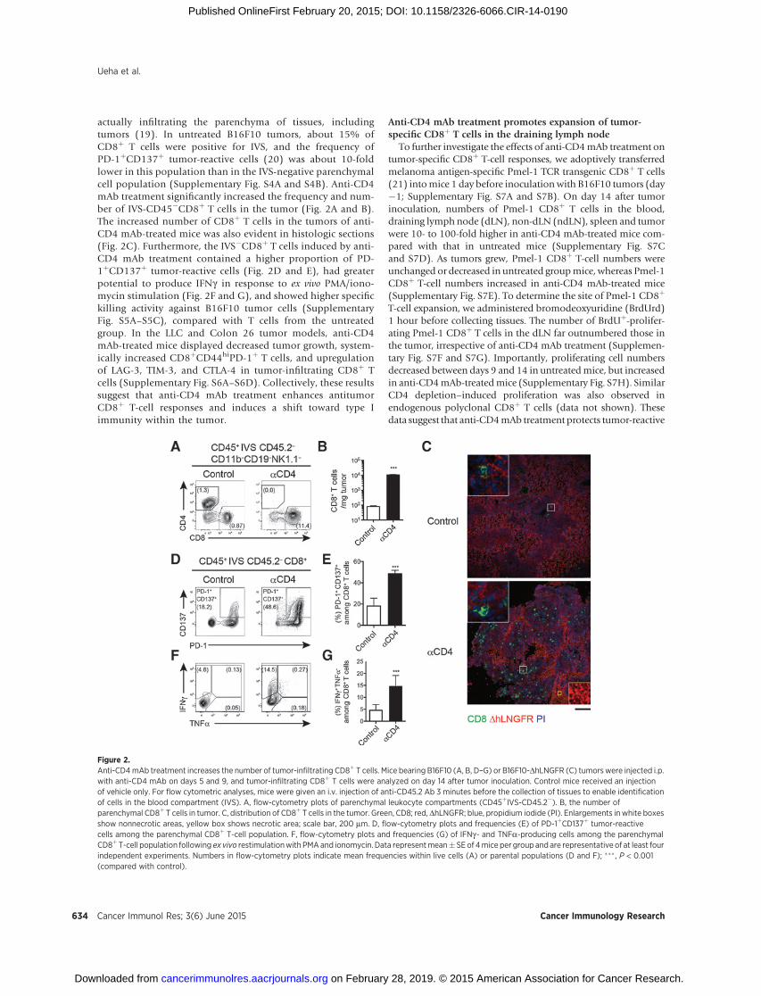
actually infiltrating the parenchyma of tissues, includingtumors (19). In untreated B16F10 tumors, about 15% ofCD8þ T cells were positive for IVS, and the frequency ofPD-1þCD137þ tumor-reactive cells (20) was about 10-foldlower in this population than in the IVS-negative parenchymalcell population (Supplementary Fig. S4A and S4B). Anti-CD4mAb treatment significantly increased the frequency and num-ber of IVS-CD45�CD8þ T cells in the tumor (Fig. 2A and B).The increased number of CD8þ T cells in the tumors of anti-CD4 mAb-treated mice was also evident in histologic sections(Fig. 2C). Furthermore, the IVS�CD8þ T cells induced by anti-CD4 mAb treatment contained a higher proportion of PD-1þCD137þ tumor-reactive cells (Fig. 2D and E), had greaterpotential to produce IFNg in response to ex vivo PMA/iono-mycin stimulation (Fig. 2F and G), and showed higher specifickilling activity against B16F10 tumor cells (SupplementaryFig. S5A–S5C), compared with T cells from the untreatedgroup. In the LLC and Colon 26 tumor models, anti-CD4mAb-treated mice displayed decreased tumor growth, system-ically increased CD8þCD44hiPD-1þ T cells, and upregulationof LAG-3, TIM-3, and CTLA-4 in tumor-infiltrating CD8þ Tcells (Supplementary Fig. S6A–S6D). Collectively, these resultssuggest that anti-CD4 mAb treatment enhances antitumorCD8þ T-cell responses and induces a shift toward type Iimmunity within the tumor.
Anti-CD4 mAb treatment promotes expansion of tumor-specific CD8þ T cells in the draining lymph node
To further investigate the effects of anti-CD4mAb treatment ontumor-specific CD8þ T-cell responses, we adoptively transferredmelanoma antigen-specific Pmel-1 TCR transgenic CD8þ T cells(21) intomice 1 day before inoculationwith B16F10 tumors (day�1; Supplementary Fig. S7A and S7B). On day 14 after tumorinoculation, numbers of Pmel-1 CD8þ T cells in the blood,draining lymph node (dLN), non-dLN (ndLN), spleen and tumorwere 10- to 100-fold higher in anti-CD4 mAb-treated mice com-pared with that in untreated mice (Supplementary Fig. S7Cand S7D). As tumors grew, Pmel-1 CD8þ T-cell numbers wereunchanged or decreased in untreated groupmice, whereas Pmel-1CD8þ T-cell numbers increased in anti-CD4 mAb-treated mice(Supplementary Fig. S7E). To determine the site of Pmel-1 CD8þ
T-cell expansion, we administered bromodeoxyuridine (BrdUrd)1 hour before collecting tissues. The number of BrdUþ-prolifer-ating Pmel-1 CD8þ T cells in the dLN far outnumbered those inthe tumor, irrespective of anti-CD4 mAb treatment (Supplemen-tary Fig. S7F and S7G). Importantly, proliferating cell numbersdecreased between days 9 and 14 in untreatedmice, but increasedin anti-CD4mAb-treatedmice (Supplementary Fig. S7H). SimilarCD4 depletion–induced proliferation was also observed inendogenous polyclonal CD8þ T cells (data not shown). Thesedata suggest that anti-CD4mAb treatment protects tumor-reactive
Figure 2.Anti-CD4mAb treatment increases the number of tumor-infiltrating CD8þ T cells. Mice bearing B16F10 (A, B, D–G) or B16F10-DhLNGFR (C) tumorswere injected i.p.with anti-CD4 mAb on days 5 and 9, and tumor-infiltrating CD8þ T cells were analyzed on day 14 after tumor inoculation. Control mice received an injectionof vehicle only. For flow cytometric analyses, mice were given an i.v. injection of anti-CD45.2 Ab 3 minutes before the collection of tissues to enable identificationof cells in the blood compartment (IVS). A, flow-cytometry plots of parenchymal leukocyte compartments (CD45þIVS-CD45.2�). B, the number ofparenchymal CD8þ T cells in tumor. C, distribution of CD8þ T cells in the tumor. Green, CD8; red, DhLNGFR; blue, propidium iodide (PI). Enlargements in white boxesshow nonnecrotic areas, yellow box shows necrotic area; scale bar, 200 mm. D, flow-cytometry plots and frequencies (E) of PD-1þCD137þ tumor-reactivecells among the parenchymal CD8þ T-cell population. F, flow-cytometry plots and frequencies (G) of IFNg- and TNFa-producing cells among the parenchymalCD8þT-cell population following ex vivo restimulationwith PMAand ionomycin. Data representmean� SEof 4mice per group and are representative of at least fourindependent experiments. Numbers in flow-cytometry plots indicate mean frequencies within live cells (A) or parental populations (D and F); ��� , P < 0.001(compared with control).
Ueha et al.
Cancer Immunol Res; 3(6) June 2015 Cancer Immunology Research634
on February 28, 2019. © 2015 American Association for Cancer Research. cancerimmunolres.aacrjournals.org Downloaded from
Published OnlineFirst February 20, 2015; DOI: 10.1158/2326-6066.CIR-14-0190

CD8þ T cells from deletion, a mechanism of peripheral tolerancein which the continuous and excessive exposure of antigen-specific T cells to cognate antigens eventually results in the lossof the antigen-specific T-cell clones.
To confirm the effects of anti-CD4 mAb treatment on theproliferation of CD8þ T cells, we used fluorescent ubiquitina-tion-based cell-cycle indicator (Fucci) double transgenic mice. InFucci mice, Fucci-orange (mKO2) and Fucci-green (mAG) areexpressed reciprocally in theG0–G1andS–G2–Mphases of the cellcycle, respectively (13, 18). In the B16F10 tumormodel, anti-CD4mAb treatment significantly increased the proportion of mAGþ
proliferating cells among CD8þCD44hi T cells in both the dLNand non-dLN, compared with the proportion of these cells inuntreated control mice (Supplementary Fig. S7I and S7J).
To determine whether this CD4 depletion–induced prolifera-tion was specific for tumor-specific CD8þ T cells or was a tumorantigen-independent response such as homeostatic proliferation(22), we adoptively transferred aCFSE-labeledmixture of Pmel-1,ovalbumin-specific OT-I, and polyclonal CD8þ T cells into B16tumor-bearing or tumor-freemice with or without anti-CD4mAbtreatment (Supplementary Fig. S8A). Pmel-1, but not OT-I orpolyclonal CD8þ T cells, selectively proliferated in the dLN of B16tumor-bearing mice (Supplementary Fig. S8B–S8E). These resultsindicate that CD4 depletion–induced T-cell expansion is specificfor tumor-specific CD8þ T cells. Collectively, these results suggestthat anti-CD4mAb treatment systemically increases the availabil-ity of tumor-specific CD8þ T cells by enhancing their proliferationin the dLN in a tumor-associated antigen-dependent manner.
Enhanced CD8þ T-cell responses underlie the antitumor effectsof anti-CD4 mAb treatment
To determine whether enhanced CTL responses are responsiblefor the antitumor effects of anti-CD4 mAb treatment, we admin-istered the anti-CD4 mAb together with an anti–CD8-depletingmAb. When the anti–CD8-depleting mAb was administeredtogether with the anti-CD4 mAb, the inhibitory effect of anti-CD4 mAb treatment on tumor growth was completely reversed(Fig. 3A and B). We also investigated whether treatment with ananti–CD25-depleting mAb, which is widely used to depleteFoxp3þCD25þ Tregs in mice (23), could produce the same effectas anti-CD4 mAb treatment. Under our administration protocol,tumor growth in the anti-CD25 mAb-treated group was almostequivalent to that observed in untreated mice (Fig. 3A and B).These results suggest that the tumor-specific CD8þ T cells that areinduced by anti-CD4 mAb treatment are responsible for the
antitumor effects of the treatment, and that anti-CD4 mAbtreatment might deplete immunosuppressive populations moreefficiently than anti-CD25 mAb treatment.
Combination treatment with anti-CD4 and anti–PD-1 or anti–PD-L1 mAbs synergistically enhances antitumor effects
Next, we examined whether synergistic antitumor effects couldbe achieved by supplementing anti-CD4 mAb treatment withvarious immune checkpoint mAbs, particularly those targetingthe exhaustion and deletion phase of the immune response. Wedevised a combination treatment protocol of anti-CD4mAbwithimmune checkpoint antibodies as depicted in Fig. 4A. Strikingly,combination treatment with anti-CD4 and anti–PD-L1 mAbs,and to a lesser extent anti-CD4 and anti–PD-1 mAbs, resulted indramatic synergistic inhibition of tumor growth in the B16F10melanoma model (Fig. 4B and C). Combination treatment withanti-CD4 and anti–CTLA-4, anti–TIM-3, anti-BTLA, and anti-GITR mAbs also had additive or synergistic effects (Fig. 4B andC), but anti–PD-L2, anti-OX40 and anti–LAG-3 mAbs producedno synergistic antitumor effect when combinedwith the anti-CD4mAb (Fig. 4B andC). Survivalwas also prolongedby combinationtreatment with anti-CD4 and anti–PD-L1 mAbs compared withanti-CD4 mAb monotherapy, but not by other combinations ofanti-CD4 and immune checkpoint mAbs (Fig. 4D). Importantly,depletion of CD8þ T cells completely abrogated the tumor growthinhibition induced by the combination of anti-CD4 and anti–PD-1or PD-L1mAbs, indicating thatCD8þT cells play a critical role inthe antitumor effects of the combination treatment (Fig. 4E).
To determine whether the synergistic antitumor effects of anti-CD4 and anti–PD-1 or anti–PD-L1 mAb treatment are commonto other tumor types andmouse strains, we examined the effect ofcombination treatment in the Colon 26 subcutaneous tumormodel in BALB/c mice. The anti–PD-1 or anti–PD-L1 mAb treat-ment alone did not inhibit tumor growth, whereas combinationtreatment with anti-CD4 and anti–PD-1 or anti–PD-L1 mAbsresulted in strong synergistic inhibition of tumor growth (Fig. 5Aand B). These effects were completely reversed by treatment withan anti–CD8-depleting mAb (Fig. 5B). Notably, we observedcomplete remission in 3 of 10 mice treated with the anti-CD4/anti–PD-1mAb combination, and in 6 of 10mice treatedwith theanti-CD4/anti–PD-L1 mAb combination. In addition, the 6 micethat rejected the tumor in the anti-CD4/anti–PD-L1 mAb-treatedgroup were resistant to rechallenge with Colon 26 tumor cells at adose five times higher than that used in the initial inoculation(Fig. 5C). Collectively, these results indicate that combination
Figure 3.CD8þ T cells play a pivotal role in theantitumor effects of anti-CD4 mAbtreatment. Mice bearing B16F10tumors were injected i.p. with anti-CD4, anti-CD8, and/or anti-CD25mAbs (200 mg/mouse) on days 5 and9 after tumor inoculation. A, tumorgrowth curves. B, tumor volume onday 15 after tumor inoculation. Data,mean � SE of 8 mice per group;�� , P < 0.05 (compared with control);††, P <0.01 (comparison as indicated).
Anti-CD4 and Immune Checkpoint Antibody Synergy
www.aacrjournals.org Cancer Immunol Res; 3(6) June 2015 635
on February 28, 2019. © 2015 American Association for Cancer Research. cancerimmunolres.aacrjournals.org Downloaded from
Published OnlineFirst February 20, 2015; DOI: 10.1158/2326-6066.CIR-14-0190

Figure 4.Combination treatment with anti-CD4 and anti–PD-1 or anti–PD-L1 mAbs has synergistic antitumor effects. Mice bearing B16F10 tumors received anti-CD4 mAb,anti-immune checkpoint mAb, or a combination of these, according to the treatment schedule shown in A. B, tumor volume on day 16 (left) or 15 (right); � , P < 0.05;�� , P < 0.01; ��� , P < 0.001 (compared with control); #, P ¼ 0.021 (compared with aCD4); ††, P < 0.01; †††, P < 0.001 (comparisons as indicated). C, tumorgrowth curves. D, survival plots representative of two independent experiments; � , P < 0.05; ��, P < 0.01; ��� , P < 0.001; ���� , P < 0.0001 (compared with control);†, P < 0.05; ††, P < 0.01; †††, P < 0.001 (compared with aCD4). E, anti-CD8 mAb was administered together with anti-CD4 mAb and tumor volumes were measuredon day 16; �� , P < 0.01 (compared with control). Data, mean � SE of 8 mice per group.
Ueha et al.
Cancer Immunol Res; 3(6) June 2015 Cancer Immunology Research636
on February 28, 2019. © 2015 American Association for Cancer Research. cancerimmunolres.aacrjournals.org Downloaded from
Published OnlineFirst February 20, 2015; DOI: 10.1158/2326-6066.CIR-14-0190

treatment with anti-CD4 and anti–PD-1 or anti–PD-L1mAbs hasa dramatic and robust antitumor effect that is mediated byantitumor CD8þ T cells.
Blockade of the PD-1/PD-L1 signaling axis increases thenumber of PD-1þ tumor-reactive CD8þ T cells in the circulation
Finally, we investigated the cellular andmolecularmechanismsunderlying the synergy between anti-CD4 and anti–PD-1 or anti–PD-L1 mAbs in the B16F10 melanoma model. Quantitative RT-PCR analysis of whole tumor tissue demonstrated that anti-CD4mAb treatment alone augmented expression of the antitumorcytokine genes Ifng and Tnf, the IFNg-inducible genes Cxcl10 andCd274/PD-L1 (24, 25), and genes encoding the proapoptoticmolecules Fasl, Prf1/perforin, and Gzmb/Granzyme B, comparedwith the expression levels of these genes in untreated tumors(Supplementary Fig. S9A and S9B). The upregulation of PD-L1 byanti-CD4 mAb treatment was also observed at the protein level(Supplementary Fig. S9C). However, no additive or synergisticeffects on gene expression were observed in groups receivingcombination treatment with anti-CD4 and anti–PD-1 or PD-L1mAbs. Consistent with these results, the proportion of IFNg-producing and TNFa-producing cells within the tumor-infiltrat-ing CD8þ T-cell population was equivalent between mice receiv-ing anti-CD4 mAb alone and mice receiving the combination ofanti-CD4 and anti–PD-1 or anti–PD-L1 mAbs (data not shown).
We next analyzed the effects of anti–PD-1 and anti–PD-L1mAbs on the PD-1þCD8þ T cells that increased in number in thesystemic circulation in response to anti-CD4 mAb treatment. Weexamined cell populations expressing the effector/memory T-cellmarker CD44 and the activation marker CD137. Combinationtreatment with anti-CD4 and anti–PD-L1 mAbs increased thefrequency ofCD44hiPD-1þ cells amongCD8þT cells in the blood,dLN and non-dLN, compared with that inmice receiving the anti-CD4 mAb alone (blood data shown in Fig. 6A and B). In bloodCD8þ T cells, expression levels of PD-1 on cells within the CD44hi
PD-1þ population and the frequency of PD-1þCD137þ cells weresignificantly higher inmice that received the combination of anti-CD4 and anti–PD-L1 mAbs compared with the correspondingexpression levels and frequency inmice that received the anti-CD4mAb alone (Fig. 6A–C). In contrast, combination treatment with
anti-CD4 and anti–PD-1 mAbs decreased the frequency of theCD44hiPD-1þ population among blood CD8þ T cells, anddecreased the expression levels of PD-1 on cells within theCD44hi PD-1þ population (Fig. 6A, E, and F). However, thefrequency of the CD44hiCD137þ tumor-reactive cell populationwas higher in mice receiving the combination of anti-CD4 andanti–PD-1 mAbs compared with mice receiving the anti-CD4mAb alone (Fig. 6A, E, and F), suggesting that anti–PD-1 mAbtreatment does not actually decrease the number of tumor-reac-tive CD8þ T cells in the blood, but rather decreases the level of PD-1 expression on these cells. On the other hand, the frequency ofPD-1þ cells among tumor-infiltrating CD8þ T cells in anti-CD4mAb-treated mice was not affected by treatment with anti–PD-1or anti–PD-L1 mAbs (Fig. 6D and G).
DiscussionThe recent success of anti–CTLA-4 and anti–PD-1 mAb thera-
pies in the clinic has highlighted the potential of immunotherapyfor the treatment of cancer (2, 3, 26–29). However, the develop-ment of immunotherapy forwidespread clinical use remains in itsearly stages. Extensive efforts have been directed toward enhanc-ing endogenous antitumor immunity by dampening the influ-ence of immunosuppressive mechanisms. Treatment strategieshave included combinations of antibodies with other antibodiesand with other immunotherapies or anticancer therapeutics. Inthe present study, we demonstrate that antibody-mediated deple-tion of CD4þ cells from tumor-bearing mice results in enhancedpolyclonal PD-1þCD137þ tumor-reactive and monoclonaltumor–specific Pmel-1 CD8þ T-cell responses, and strong inhi-bition of tumor growth. Combination treatment with the anti-CD4 mAb and various immune checkpoint mAbs, particularlyanti–PD-1 and anti–PD-L1 mAbs, revealed striking synergy insuppressing tumor growth and prolonging survival.
Several previous reports have described antitumor activity ofanti-CD4mAb treatment in solid tumormodels inC57BL/6mice,including subcutaneous tumors induced by inoculation with B16melanoma cells (9, 11, 12), recurrent TC1 lung cancer cells (30),or embryo cells expressing the adenovirus-derived E1A protein(10). Although the efficacy of immunotherapy in mouse tumor
Figure 5.Combination treatment with anti-CD4 and anti–PD-1 or anti–PD-L1 mAbs induces long-term antitumor CD8þ T-cell memory. Mice bearing Colon 26 tumors receivedanti-CD4, anti–PD-L1, anti–PD-1 or anti-CD8 mAbs or a combination of these according to the treatment schedule shown in Fig. 4A. A, tumor growth curves.B, tumor volume on day 18; �� , P < 0.01; ��� , P < 0.001 (compared with control); #, P¼ 0.029; ###, P < 0.001 (compared with aCD4); †††, P < 0.001 (comparisons asindicated). C, the 6 mice that achieved complete remission of Colon 26 tumors after anti-CD4 and anti–PD-L1 treatment were rechallenged on day 39 withColon 26 tumor cells at five times the cell number of the initial challenge. Arrow indicates day of rechallenge; � , P < 0.05; �� , P < 0.01 (compared with control). A andB, data, mean � SE of 10 mice per group.
Anti-CD4 and Immune Checkpoint Antibody Synergy
www.aacrjournals.org Cancer Immunol Res; 3(6) June 2015 637
on February 28, 2019. © 2015 American Association for Cancer Research. cancerimmunolres.aacrjournals.org Downloaded from
Published OnlineFirst February 20, 2015; DOI: 10.1158/2326-6066.CIR-14-0190

models often depends on tumor type, taken together, thesereports from independent groups and our results from the presentstudy suggest that anti-CD4 mAb treatment is likely to havebroad-spectrum antitumor activity against solid tumors. Optimi-zation of the anti-CD4 mAb administration protocol revealedrobust antitumor effects whenmice received themAbondays 3 or5, rather than when mice receive the mAb before tumor inocu-lation (day –2). These results suggest that pretreatment is notnecessary. However, priming and/or the preexistence of activatedCD8þ T cells are important for effective anti-CD4 mAb therapy.Although the mechanistic link between the timing of anti-CD4antibody administration and the efficacy of treatment remains tobe elucidated, administration of the antibody to patients withearly-stage cancer or whose tumor burden has been reduced bysurgical resection, irradiation or chemotherapeutics is likely to bemost beneficial.
A dose of anti-CD4 mAb sufficient to deplete most CD4þ cellswas required in order for antitumor effects to be observed. TheCD4þ cell population includes Foxp3þCD25þ Tregs, Th2 cells,Tr1/3 cells (4), and IDOþ immunosuppressive pDCs (7). Con-sidering that markedly increased proliferation of tumor-specificCD8þ T cells was observed in the dLN, anti-CD4mAb treatment islikely to augment proliferation of tumor-reactive CD8þ T cellsthrough the removal of these CD4þ immunosuppressive cellsfrom the dLN. In addition, anti-CD4mAb treatment increased theproportion of PD-1þCD137þ tumor-reactive cells and IFNg-pro-ducing cells among tumor-infiltrating CD8þ T cells in the B16F10model, suggesting that anti-CD4mAb treatment augmented boththe quantity and quality of tumor-specific CD8þ T-cell responses.We recently demonstrated that IFNg- and TNFa-induced cell-cycle
arrest is an important mechanism underlying the antitumoreffects induced by tumor-specific CD8þ T-cell transfer (31). Theshift toward IFNg-dominant type I immunity, which was evi-dent in the strong induction of IFNg and TNFa in tumor-infiltrating CD8þ T cells after anti-CD4 mAb treatment, is likelyto play a central role in the antitumor effects that we observed(32). Notably, depletion of CD25þ Tregs by administration ofan anti-CD25 mAb on days 5 and 9 after tumor inoculation didnot reproduce the antitumor effect of anti-CD4 mAb treatment.Because some Foxp3þ Tregs have low-to-negative CD25 expres-sion, residual Foxp3þCD25�/lo Tregs may have contributed tothis discrepancy. Moreover, the antitumor effects of anti-CD25mAb treatment have been reported to be optimal when themAb is administered before tumor inoculation (33, 34),because when it is administered after tumor inoculation, theanti-CD25 mAb depletes not only Tregs but also other activatedlymphocytes expressing CD25. The involvement of Treg andother CD4þ-immunosuppressive populations in the suppres-sion of CD8þ T-cell–mediated antitumor responses remains tobe elucidated.
The synergy that occurs in combination treatment with anti-CD4 and anti–PD-1 or anti–PD-L1 mAbs is likely due to theblockade of PD-1/PD-L1 signaling in PD-1þ activated CD8þ Tcells that are induced by anti-CD4 mAb treatment. We did notdetect any synergistic effect in terms of the quantity and quality ofthe tumor-infiltrating CD8þ T-cell response promoted by anti-CD4 and anti–PD-1 or anti–PD-L1 mAb treatment. However, thefrequency of the PD-1þCD137þ and CD44hiCD137þ tumor-reactive populations increased among CD8þ T cells in the bloodupon blockade of the PD-1/PD-L1 signaling axis. Considering
Figure 6.Anti–PD-L1 and anti–PD-1 treatments target PD-1þCD8þ T cells that are induced by anti-CD4 treatment. Mice bearing B16F10 tumors were treated with anti-CD4,anti–PD-L1, or anti–PD-1 mAbs, or a combination of these according to the treatment schedule shown in Fig. 4A. A, flow-cytometry plots of blood CD8þ T cells.B and E, proportions of CD44hi PD-1þ cells, PD-1þCD137þ cells or CD44hiCD137þ cells among blood CD8þ T cells on day 14. C and F, mean fluorescentintensity (MFI) of PD-1 expression on CD8þCD44hiPD-1þ cells in the blood. D and G, proportions of PD-1þ cells among tumor-infiltrating CD8þ T cells. B–D, showanti–PD-L1 mAb experiments; E–G, show anti–PD-1mAb experiments. Data, mean� SE of 4mice per group and are representative of two independent experiments;� , P < 0.05; �� , P < 0.01; ��� , P < 0.001.
Ueha et al.
Cancer Immunol Res; 3(6) June 2015 Cancer Immunology Research638
on February 28, 2019. © 2015 American Association for Cancer Research. cancerimmunolres.aacrjournals.org Downloaded from
Published OnlineFirst February 20, 2015; DOI: 10.1158/2326-6066.CIR-14-0190

that T cells continuously traffic between peripheral and secondarylymphoid tissues via the lymph–blood circulation, the block-ade of PD-1/PD-L1 signaling may prevent exhaustion or dele-tion of tumor-reactive PD-1þCD8þ T cells in the tumor andallow them to migrate into the dLN, thus sustaining antitumorCD8þ T-cell responses. In addition, anti-CD4 mAb treatmentincreased the number of IFNg-producing PD-1þCD8þ T cells inthe tumor, resulting in the upregulation of IFNg-induciblegenes, including PD-L1. Although the shift toward IFNg-dom-inant type-I immunity within the tumor contributes to theinhibition of tumor growth, it also promotes the exhaustionor deletion of tumor-infiltrating PD-1þCD8þ T cells by enhanc-ing PD-1/PD-L1 signaling. It is therefore likely that the synergyof the anti-CD4 and anti–PD-1 or anti–PD-L1 mAb combina-tion treatment arises due to the blockade of this adversenegative feedback mechanism.
We are in the process of developing a humanized anti-CD4mAb with potent antibody-dependent cell-mediated cytotox-icity as an anticancer therapeutic. Because CD4þ T cells playimportant roles in both humoral and cellular immunity, theheightened risk of infectious diseases that may be associatedwith transient CD4þ T-cell depletion should be carefully eval-uated in clinical trials. In addition, trials should seek to max-imize clinical efficacy and safety through rigorous optimizationof the antibody administration protocol. In preclinical studiesin nonhuman primates, no serious adverse effects were detectedafter several weeks of treatment with our humanized anti-human CD4 mAb that resulted in CD4þ T-cell depletion. Inaddition, no severe adverse effects have been observed duringphase II clinical trials for T-cell malignancy with long-termadministration of other humanized anti-CD4 mAbs (35, 36).Preexisting humoral immune mediators, such as immunoglob-ulin, plasma cells, and memory B cells, CD8þ T-cell responses,and unimpaired natural immunity, are likely to provide basalprotection against infectious diseases during CD4þ T-cell–depleting therapies. On the other hand, consideration shouldalso be given to the potential for the acute exacerbation ofchronic diseases associated with viral infection (e.g., hepatitis Cand B) due to excessive activation of effector and memoryCD8þ T cells after CD4þ cell depletion.
In conclusion, our study represents the first report of robustantitumor effects of combination treatment with an anti–CD4-depleting antibody and anti–PD-1 or anti–PD-L1 immunecheckpoint antibodies in mice. We have also characterized theimmunologic bases for the synergy between these agents.Recent clinical trials suggest that anti–PD-1, anti–PD-L1, or
anti–CTLA-4 mAbs, or combinations of these agents, are noteffective against all types of solid tumors. Our findings suggestthat combination treatment with an anti-CD4 mAb andimmune checkpoint mAbs, particularly anti–PD-1 or anti–PD-L1 mAbs, is likely to result in greater clinical efficacy againsta broader range of cancers.
Disclosure of Potential Conflicts of InterestS. Ueha has ownership interest (including patents) in IDAC Theranostics.
S. Yokochi is a manager and K. Hachiga is a researcher at IDAC Theranostics.K. Matsushima reports receiving a commercial research grant, has ownershipinterest (including patents), and is a consultant/advisory board member forIDAC Theranostics. No potential conflicts of interest were disclosed by the otherauthors.
Authors' ContributionsConception and design: S. Ueha, S. Yokochi, Y. Ishiwata, S. Ito, K. MatsushimaDevelopment of methodology: S. Ueha, S. Yokochi, Y. IshiwataAcquisition of data (provided animals, acquired and managed patients,provided facilities, etc.): S. Ueha, S. Yokochi, Y. Ishiwata, H. Ogiwara,K. Chand, K. Hachiga, Y. Terashima, E. Toda, K. KakimiAnalysis and interpretation of data (e.g., statistical analysis, biostatistics,computational analysis): S. Ueha, S. Yokochi, Y. Ishiwata, K. Chand,T. Nakajima, K. Hachiga, S. Shichino, S. Ito, K. MatsushimaWriting, review, and/or revision of the manuscript: S. Ueha, S. Yokochi,S. Shichino, F.H.W. Shand, S. Ito, K. MatsushimaAdministrative, technical, or material support (i.e., reporting or organizingdata, constructing databases): S. Ueha, S. Yokochi, H. Ogiwara, S. ShichinoStudy supervision: S. Ueha, K. Matsushima
AcknowledgmentsThe authors thank A. Miyawaki, A. Sakaue-Sawano, and the RIKEN
BioResource Center for providing FucciG1 and FucciS/G2/M mice; A. Hosoifor assistance with Pmel-1-B16F10 experiments; H. Yamazaki, K. Tsuji, andK. Yoshioka for animal care; A. Yamashita, S. Aoki, and S. Fujita for experttechnical assistance; and M. Otsuji, K. Takeda, and S. Shibayama for helpfuldiscussions.
Grant SupportThisworkwas supportedby the Japan Science andTechnologyAgencyCREST
program; Grants-in-Aid for Scientific Research (C) 25460491 (to S. Ueha) and(B) 25293113 (to K. Matsushima) from the Japanese Ministry of Education,Culture, Sports, Science and Technology; and Health and Labor ScienceResearch Grants for Research for Promotion of Cancer Control (AppliedResearch for Innovative Treatment of Cancer).
The costs of publication of this articlewere defrayed inpart by the payment ofpage charges. This article must therefore be hereby marked advertisement inaccordance with 18 U.S.C. Section 1734 solely to indicate this fact.
Received October 8, 2014; revised February 1, 2015; accepted February 15,2015; published OnlineFirst February 20, 2015.
References1. Pardoll DM. The blockade of immune checkpoints in cancer immuno-
therapy. Nat Rev Cancer 2012;12:252–64.2. Topalian SL,WeinerGJ, PardollDM.Cancer immunotherapy comes of age.
J Clin Oncol 2011;29:4828–36.3. Wolchok JD, Kluger H, Callahan MK, Postow MA, Rizvi NA, Lesokhin AM,
et al. Nivolumab plus ipilimumab in advanced melanoma. N Engl J Med2013;369:122–33.
4. Whiteside TL. Disarming suppressor cells to improve immunotherapy.Cancer Immunol Immunother 2012;61:283–8.
5. Alizadeh D, Larmonier N. Chemotherapeutic targeting of cancer-inducedimmunosuppressive cells. Cancer Res 2014;74:2663–8.
6. Camisaschi C, De Filippo A, Beretta V, Vergani B, Villa A, Vergani E, et al.Alternative activation of human plasmacytoid DCs in vitro and in
melanoma lesions: involvement of LAG-3. J Invest Dermatol 2014;134:1893–902.
7. Matta BM, Castellaneta A, Thomson AW. Tolerogenic plasmacytoid DC.European J Immunol 2010;40:2667–76.
8. Nagai H, Hara I, Horikawa T, Fujii M, Kurimoto M, Kamidono S, et al.Antitumor effects on mouse melanoma elicited by local secretion ofinterleukin-12 and their enhancement by treatment with interleukin-18.Cancer Invest 2000;18:206–13.
9. Nagai H, Hara I, Horikawa T, Oka M, Kamidono S, Ichihashi M.Elimination of CD4(þ) T cells enhances anti-tumor effect of locallysecreted interleukin-12 on B16 mouse melanoma and inducesvitiligo-like coat color alteration. J Invest Dermatol 2000;115:1059–64.
www.aacrjournals.org Cancer Immunol Res; 3(6) June 2015 639
Anti-CD4 and Immune Checkpoint Antibody Synergy
on February 28, 2019. © 2015 American Association for Cancer Research. cancerimmunolres.aacrjournals.org Downloaded from
Published OnlineFirst February 20, 2015; DOI: 10.1158/2326-6066.CIR-14-0190

10. den Boer AT, van Mierlo GJ, Fransen MF, Melief CJ, Offringa R, Toes RE.CD4þ T cells are able to promote tumor growth through inhibition oftumor-specific CD8þ T-cell responses in tumor-bearing hosts. Cancer Res2005;65:6984–9.
11. Yu P, Lee Y, Liu W, Krausz T, Chong A, Schreiber H, et al. Intratumordepletion of CD4þ cells unmasks tumor immunogenicity leading to therejection of late-stage tumors. J Exp Med 2005;201:779–91.
12. Choi BK, Kim YH, Kang WJ, Lee SK, Kim KH, Shin SM, et al. Mechanismsinvolved in synergistic anticancer immunity of anti-4–1BB and anti-CD4therapy. Cancer Res 2007;67:8891–9.
13. Sakaue-SawanoA, KurokawaH,Morimura T,Hanyu A,HamaH,OsawaH,et al. Visualizing spatiotemporal dynamics of multicellular cell-cycleprogression. Cell 2008;132:487–98.
14. Ueha S, YoneyamaH,Hontsu S, KurachiM,KitabatakeM,Abe J, et al. CCR7mediates the migration of Foxp3þ regulatory T cells to the paracorticalareas of peripheral lymph nodes through high endothelial venules. JLeukoc Biol 2007;82:1230–8.
15. Ueha S, Murai M, Yoneyama H, Kitabatake M, Imai T, Shimaoka T, et al.Intervention of MAdCAM-1 or fractalkine alleviates graft-versus-host reac-tion associated intestinal injurywhile preserving graft-versus-tumor effects.J Leukoc Biol 2007;81:176–85.
16. Shono Y, Ueha S,Wang Y, Abe J, KurachiM,Matsuno Y, et al. Bonemarrowgraft-versus-host disease: early destruction of hematopoietic niche afterMHC-mismatched hematopoietic stem cell transplantation. Blood 2010;115:5401–11.
17. Sawanobori Y, Ueha S, Kurachi M, Shimaoka T, Talmadge JE, Abe J, et al.Chemokine-mediated rapid turnover of myeloid-derived suppressor cellsin tumor-bearing mice. Blood 2008;111:5457–66.
18. Shand FH,Ueha S,OtsujiM,Koid SS, Shichino S, Tsukui T, et al. Tracking ofintertissue migration reveals the origins of tumor-infiltrating monocytes.ProcNatl Acad Sci U S A 2014;111:7771–6.
19. Anderson KG, Mayer-Barber K, Sung H, Beura L, James BR, Taylor JJ, et al.Intravascular staining for discrimination of vascular and tissue leukocytes.Nat Protoc 2014;9:209–22.
20. Ye Q, Song DG, Poussin M, Yamamoto T, Best A, Li C, et al. CD137accurately identifies and enriches for naturally occurring tumor-reactive Tcells in tumor. Clin Cancer Res 2014;20:44–55.
21. Overwijk WW, Theoret MR, Finkelstein SE, Surman DR, de Jong LA, Vyth-Dreese FA, et al. Tumor regression and autoimmunity after reversal of afunctionally tolerant state of self-reactive CD8þ T cells. J Exp Med2003;198:569–80.
22. Surh CD, Sprent J. Homeostasis of naive and memory T cells. Immunity2008;29:848–62.
23. Sakaguchi S. Regulatory T cells: history and perspective. Methods Mol Biol2011;707:3–17.
24. Dong H, Strome SE, Salomao DR, Tamura H, Hirano F, Flies DB, et al.Tumor-associated B7-H1 promotes T-cell apoptosis: a potential mecha-nism of immune evasion. Nature Med 2002;8:793–800.
25. Furuta J, Inozume T, Harada K, Shimada S. CD271 on melanoma cell isan IFN-gamma-inducible immunosuppressive factor that mediatesdownregulation of melanoma antigens. J Invest Dermatol 2014;134:1369–77.
26. Couzin-Frankel J. Breakthrough of the year 2013. Cancer immunotherapy.Science 2013;342:1432–3.
27. Hamid O, Robert C, Daud A, Hodi FS, HwuWJ, Kefford R, et al. Safety andtumor responses with lambrolizumab (anti-PD-1) in melanoma. N Engl JMed 2013;369:134–44.
28. Topalian SL, Drake CG, Pardoll DM. Targeting the PD-1/B7-H1(PD-L1)pathway to activate anti-tumor immunity. Curr Opin Immunol 2012;24:207–12.
29. TopalianSL,Hodi FS, Brahmer JR,Gettinger SN, SmithDC,McDermottDF,et al. Safety, activity, and immune correlates of anti-PD-1 antibody incancer. N Engl J Med 2012;366:2443–54.
30. Predina J, Eruslanov E, Judy B, Kapoor V, ChengG,Wang LC, et al. Changesin the local tumor microenvironment in recurrent cancers may explainthe failure of vaccines after surgery. Proc Natl Acad Sci U S A 2013;110:E415–24.
31. MatsushitaH,Hosoi A,Ueha S, Abe J, FujiedaN, TomuraM, et al. CytotoxicT lymphocytes block tumor growth both by lytic activity and IFN-gamma-dependent cell-cycle arrest. Cancer Immunol Res 2015;3:26–36.
32. Braumuller H,Wieder T, Brenner E, Assmann S, HahnM, AlkhaledM, et al.T-helper-1-cell cytokines drive cancer into senescence. Nature 2013;494:361–5.
33. Onizuka S, Tawara I, Shimizu J, Sakaguchi S, Fujita T, Nakayama E. Tumorrejection by in vivo administration of anti-CD25 (interleukin-2 receptoralpha) monoclonal antibody. Cancer Res 1999;59:3128–33.
34. Shimizu J, Yamazaki S, Sakaguchi S. Induction of tumor immunity byremoving CD25þCD4þ T cells: a common basis between tumor immunityand autoimmunity. J Immunol 1999;163:5211–8.
35. Kim YH, Duvic M, Obitz E, Gniadecki R, Iversen L, Osterborg A, et al.Clinical efficacy of zanolimumab (HuMax-CD4): two phase 2 studies inrefractory cutaneous T-cell lymphoma. Blood 2007;109:4655–62.
36. RiderDA,HavenithCE, de Ridder R, Schuurman J, FavreC, Cooper JC, et al.A human CD4monoclonal antibody for the treatment of T-cell lymphomacombines inhibition of T-cell signaling by a dual mechanism with potentFc-dependent effector activity. Cancer Res 2007;67:9945–53.
Cancer Immunol Res; 3(6) June 2015 Cancer Immunology Research640
Ueha et al.
on February 28, 2019. © 2015 American Association for Cancer Research. cancerimmunolres.aacrjournals.org Downloaded from
Published OnlineFirst February 20, 2015; DOI: 10.1158/2326-6066.CIR-14-0190
2015;3:631-640. Published OnlineFirst February 20, 2015.Cancer Immunol Res Satoshi Ueha, Shoji Yokochi, Yoshiro Ishiwata, et al. Treatment in Mice
PD-1/PD-L1 Immune Checkpoint Antibody−Antibody and Anti CD4-Depleting−Robust Antitumor Effects of Combined Anti
Updated version
10.1158/2326-6066.CIR-14-0190doi:
Access the most recent version of this article at:
Material
Supplementary
http://cancerimmunolres.aacrjournals.org/content/suppl/2015/02/20/2326-6066.CIR-14-0190.DC1
Access the most recent supplemental material at:
Cited articles
http://cancerimmunolres.aacrjournals.org/content/3/6/631.full#ref-list-1
This article cites 36 articles, 17 of which you can access for free at:
Citing articles
http://cancerimmunolres.aacrjournals.org/content/3/6/631.full#related-urls
This article has been cited by 8 HighWire-hosted articles. Access the articles at:
E-mail alerts related to this article or journal.Sign up to receive free email-alerts
Subscriptions
Reprints and
To order reprints of this article or to subscribe to the journal, contact the AACR Publications Department
Permissions
Rightslink site. Click on "Request Permissions" which will take you to the Copyright Clearance Center's (CCC)
.http://cancerimmunolres.aacrjournals.org/content/3/6/631To request permission to re-use all or part of this article, use this link
on February 28, 2019. © 2015 American Association for Cancer Research. cancerimmunolres.aacrjournals.org Downloaded from
Published OnlineFirst February 20, 2015; DOI: 10.1158/2326-6066.CIR-14-0190


![КОМПЛЕТ ЗАДАТАКА СА РЕШЕЊИМА МАТЕМАТИКА · CJ1e4H za 1-ueHa cyJIYHAapa H3HOCM 600 ÃHHapa. [10 noeHa] AaKJ1e, ueHa jeaHor cyJTYHaapa je 200 AMHapa](https://img.dokumen.tips/doc/110x75/60e67eac1b26426c6e479933/oe-oe-oeoe-cj1e4h.jpg)


