Embed Size (px)
Citation preview
RISK FACTORS FOR BK VIRUS NEPHROPATHY IN RENAL TRANSPLANT RECIPIENTS – THE ROLE OF URETERAL TRAUMA.
MPH CAPSTONE PROJECT JOHNS HOPKINS UNIVERSITY, BLOOMBERG SCHOOL OF PUBLIC HEALTH.
Abraham Thomas, M.D. MPH candidate.
Advisors
John McGready
Duvuru Geetha
1
Executive Summary
BK virus associated nephropathy (BKVAN) is an important emerging infectious disease in the
renal transplant population. The risk factors for the development of BKVAN are not clearly
defined.
I propose that trauma to the donor ureter due to the placement of indwelling stents is an
independent risk factor for the development of BKVAN. Stent placement may facilitate the
ascending spread of BKV from latent sites in the urinary epithelium to the renal cortex, where
active disease can develop in a susceptible host.
In 2002 14,523 kidney transplants were performed in the United States. The unadjusted graft
survival at 3 years is approximately 83%. BKVAN is a rare condition with a prevalence of 2-6%,
and it generally develops within the first two years after transplantation. BKVAN has a poor
prognosis and up to 40% of affected patients lose graft function within one year. Current
strategies to treat BKVAN including reduction of immunosupression and use of anti-viral drugs
have not been very successful.
This paper discusses the design, conduct and analysis of a case –control study that was
conducted to test the hypothesis that stent placement is a risk factor for BKVAN. The objectives
of the study are to provide data to support this hypothesis as well as to provide more information
about the clinical course and outcomes of this rare disease.
Analysis of the study results reveals a statistically significant association between stent use and
BKVAN. This novel finding is clinically important as it may lead to changes in existing surgical
practices in kidney transplantation. These results will help to guide the design of larger
prospective clinical trials that are necessary to confirm the association between stent use and BK
virus associated nephropathy.
2
Introduction
Over the past two decades the incidence of infectious complications following organ
transplantation has declined. However, in recent years BK virus associated nephropathy
(BKVAN) has emerged as an important complication of renal transplantation. This disease can
cause premature allograft failure in renal transplant recipients.
BK virus is a small non-enveloped DNA virus belonging to the polyomavirus family. This
family includes two human pathogens, JC virus and BK virus, both of which were first isolated
from immunocompromised patients (1). BK virus was first isolated in 1971 from the urine of a
Sudanese renal transplant patient (with the initials B.K.) who presented with ureteral stenosis (2)
Human infections with BK virus are common in childhood and are largely asymptomatic.
Seroconversion occurs in more than 90% of cases by the age of 20 (3). After primary infection
the virus remains latent in the urinary epithelium and lymphoid cells. Reactivation may occur in
the presence of intense immunosupression associated with conditions like organ transplantation,
primary immunodeficiency diseases, human immunodeficiency virus infection and following
immunotherapy for malignancy.
In the field of kidney transplantation, the emergence of BKVAN in the 1990s seems to coincide
with the widespread use of newer potent immunosuppressive drugs such as tacrolimus and
mycophenolate mofetil (MMF). Cases of BKVAN have been reported from most major
transplant centers around the world. However the risk factors associated with the development of
BKVAN are not clearly defined. The disease may or may not be related to the use of a specific
immunosuppressive agent. It is also interesting to note that there is a relative absence of BKVAN
in recipients of non-renal transplants who receive therapy with similar immunosuppressive
agents.
3
The disease can affect up to 6% of all kidney transplant recipients. BKVAN can develop at
varying times in the post transplantation period (average 40 weeks, range 6-150 weeks) (4, 5).
Clinically BKVAN presents with acute allograft dysfunction manifested by a rise in the serum
creatinine (Scr). Diagnostic tests that are being used currently include urine cytology and viral
assays. However to confirm the diagnosis of BKVAN a renal biopsy which demonstrates
classical histopathological and immunohistochemical findings is necessary. At present there is no
effective treatment for BKVAN. Several investigators have tried antiviral therapy and
modification of the immunosupression regimen to delay the progression of this disease. The
prognosis of BKVAN is poor and progression of the disease to irreversible graft failure has been
reported in up to 45% of all cases. (5,6,7)
Objectives
The primary objective of this study is to investigate the association between ureteral trauma
secondary to the use of indwelling stents and the development of BKVAN in renal transplant
recipients.
Secondary Objectives
1) To determine other risk factors for BKVAN in renal transplant recipient.
2) To describe the clinical course and outcomes of patients with BKVAN.
3) To compare graft survival in patients with BKVAN and controls without BKVAN.
Hypothesis
Ureteral trauma is an independent risk factor for the development of BKVAN. It facilitates the
ascending route of spread of BK virus from the urothelium to the renal cortex, where active
disease may develop in a susceptible host.
4
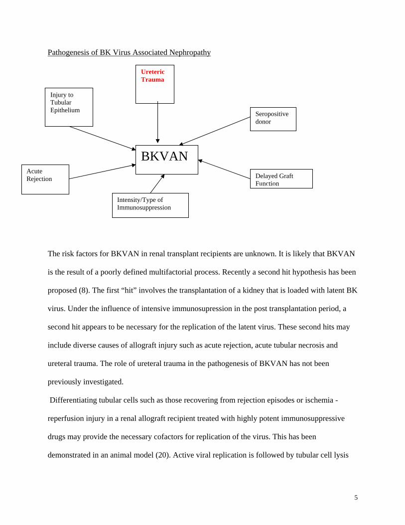
Pathogenesis of BK Virus Associated Nephropathy
BKVAN
Seropositive donor
Ureteric Trauma
Intensity/Type of Immunosuppression
Delayed Graft Function
Acute Rejection
Injury to Tubular Epithelium
The risk factors for BKVAN in renal transplant recipients are unknown. It is likely that BKVAN
is the result of a poorly defined multifactorial process. Recently a second hit hypothesis has been
proposed (8). The first “hit” involves the transplantation of a kidney that is loaded with latent BK
virus. Under the influence of intensive immunosupression in the post transplantation period, a
second hit appears to be necessary for the replication of the latent virus. These second hits may
include diverse causes of allograft injury such as acute rejection, acute tubular necrosis and
ureteral trauma. The role of ureteral trauma in the pathogenesis of BKVAN has not been
previously investigated.
Differentiating tubular cells such as those recovering from rejection episodes or ischemia -
reperfusion injury in a renal allograft recipient treated with highly potent immunosuppressive
drugs may provide the necessary cofactors for replication of the virus. This has been
demonstrated in an animal model (20). Active viral replication is followed by tubular cell lysis
5
and development of invasive disease manifested characterized by interstitial inflammation and
acute tubular necrosis.
The specific purpose of this study is to determine the role of ureteral trauma in the pathogenesis
of BKVAN. The best-known site of BK virus latency after a primary infection is the urogenital
tract. Viral sequences can be detected in up to 50% of human kidneys (26) and BK virus
associated ureteral stenosis has been reported in renal allograft recipients (27,28). Hence it
appears that if the virus remains in the lower urinary tract, patients remain asymptomatic or
develop pyuria and rarely ureteric stenosis. When the virus spreads to the renal cortex and is
reactivated then symptomatic disease that manifests as acute renal failure is possible. In biopsy
specimens the virus has been shown to be abundant in the medulla and distal tubule cells. This
finding also suggests that the virus spreads from the lower urinary tract to the renal cortex.
However, it is unclear how or why the BK virus spreads from the urothelium to the renal cortex.
Ureteral trauma induced by the placement of indwelling stents may play a role in facilitating
ascending spread of the BK virus from the urothelium to the renal cortex.
Ureteric Stents and Kidney Transplantation
Urological complications after kidney transplantation like obstruction of the ureter and urine
leakage at the vesicoureteric anastomosis can cause significant morbidity and mortality. With
improvements in surgical techniques the incidence of these complications has decreased to 5-10
% (9).
Placement of ureteric stents is common practice in renal transplantation. Retrospective studies
have suggested that this practice is beneficial and decreases the incidence of urine leaks (10).
However randomized controlled clinical trials of routine stenting in low -urological -risk kidney
transplantation show that the procedure is unnecessary and does not reduce the risk of urological
6
complications (11). At present selective stenting based on the judgement of the surgical team is
recommended in renal transplantation.
At our institution the decision to use a ureteric stent is made by the transplant surgeon at the time
of surgery. The usual indications for stent placement during kidney transplantation at this
institution include:
1) Lack of urine output or decreased urine flow from the transplanted kidney after the
anastomosis is completed.
2) Certain characteristics of the recipient bladder including a thin bladder wall, a large floppy
bladder or small, shrunken and scarred bladders.
3) Individual preference of the operating surgeon.
Flexible silastic stents with a 6 French diameter and a length of 16-20 cm are usually used. The
stents are removed after 4-6 weeks. This procedure is usually performed by outpatient
cystoscopy in the urology clinic.
Histology of the Ureter
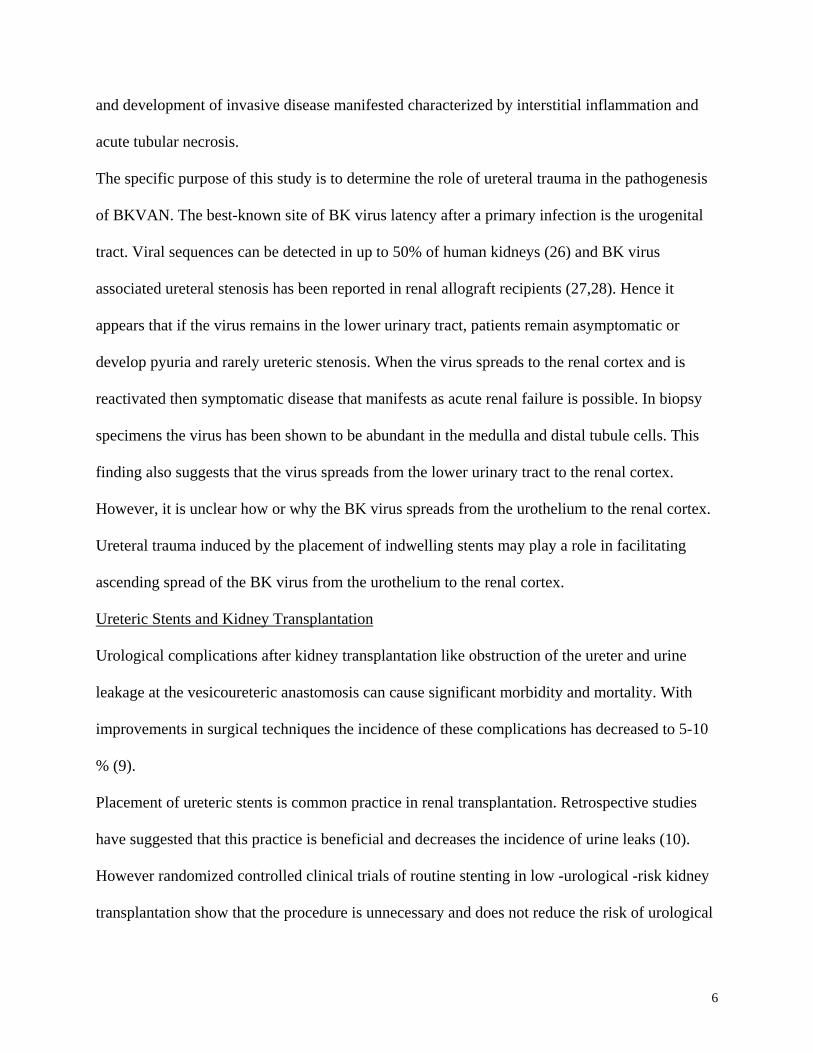
The ureter consists of intricately arranged bundles of smooth muscle within an inner lining of
lamina propria and urothelium and an outer sheath of fibrous tunica adventitia (figure 1). The
indwelling stent lies in close contact with the uroepithelium for several weeks. The biomaterial
in the stent can cause changes in the urinary epithelium. In animal models the biological
reactions to double -J stents include superficial epithelial destruction with erosions and
ulcerations of the transitional epithelium and inflammatory reactive changes. The latter include
mononuclear inflammatory infiltrate in the epithelium and lamina propria, edema of the lamina
propria, mucous metaplasia and transitional epithelial hyperplasia.
7
Figure 1 – Normal ureter
These changes were noted after intubation of the animal ureters for 6 weeks (12,13,14).
Similar reactive urothelial changes are expected to occur in the transplanted donor ureter as a
result of mechanical trauma and chemical inflammation secondary to the intra-operative
placement of indwelling stents. Following these reactive urothelial changes, epithelial
regeneration occurs.
We postulate that regenerating and differentiating uroepithelial cells may mediate enhanced
entry of BK virus into cells by upregulating cell surface receptors.
Very little is known about the behavior of the BK virus at the cellular level. However the cellular
interactions of the Simian Virus 40(SV 40) have been well described (21,22)
The SV40 virus is a polyoma virus that infects Rhesus monkeys. It shares 69% DNA sequence
identity with the BKV (23). The SV40 specific receptor comprises MHC class 1 proteins (16).
It has recently been recognized that SV40 enters the host cell through caveolae (22). Caveolae
are small (50-70nm) flask-shaped vesicles present in the cell surface of many cell types (24)
Drachenberg, et al have reported that the process by which BK virus enters the renal tubular cell
8
is morphologically very similar to the caveolae dependant endocytosis of SV 40 (25).
The virus can then spread from cell to cell in the uroepithelium by a receptor-mediated
mechanism. This process allows the virus to spread from the collecting ducts to cells for which
the BK virus is tropic like tubular cells and parietal epithelial cells. (18) Hence this mechanism
facilitates the ascending route of viral spread from the urothelium to the renal cortex.
However receptor mediated viral entry into a cell is not sufficient to cause viral replication.
Instead additional second hits like allograft injury secondary to acute tubular necrosis and acute
rejection may be necessary to trigger viral replication.
Methods
Study Design: A matched case control study was used to test the hypothesis that ureteric trauma
due to stent placement is associated with BKVAN.
Setting: Renal transplant clinics at Johns Hopkins Hospital and Johns Hopkins Bayview Medical
Center. One hundred and seventy eight kidney transplants were performed at this institution in
2002.
Subjects.
Twenty cases of BK virus associated nephropathy (BKVAN) were identified by retrospective
analysis of all kidney transplant recipients at Johns Hopkins Hospital between January 1996 and
August 2003.
A case was defined as a renal transplant recipient who had histological evidence of BKVAN in a
kidney biopsy performed in the post -transplant period for allograft dysfunction. The histological
diagnosis was made by the observation of BK virus induced cytopathic changes in the renal
tubular epithelium and confirmed by electron microscopy and immunohistochemical stain for the
BK virus (SV 40 antibody; Access Biomedical, Campo CA).
9
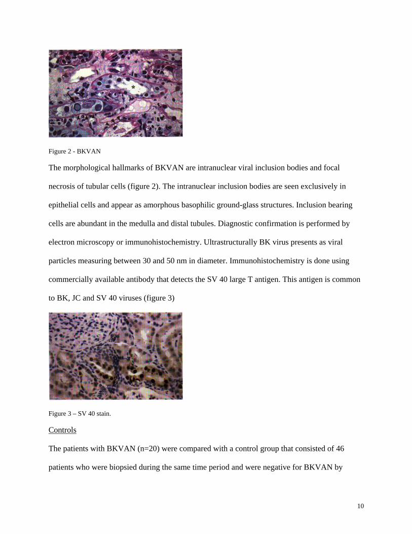
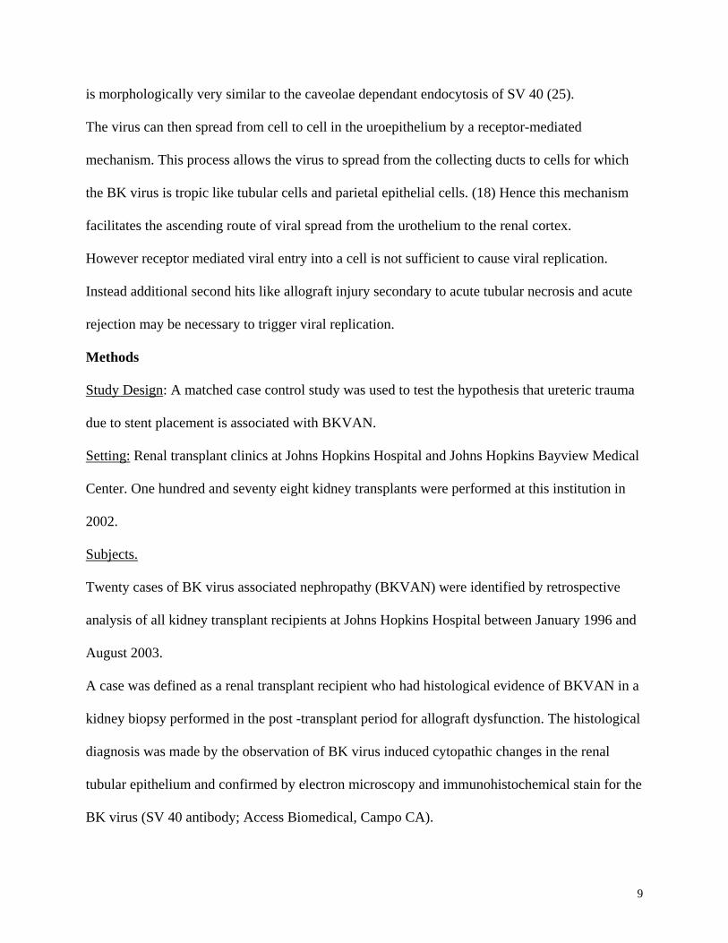
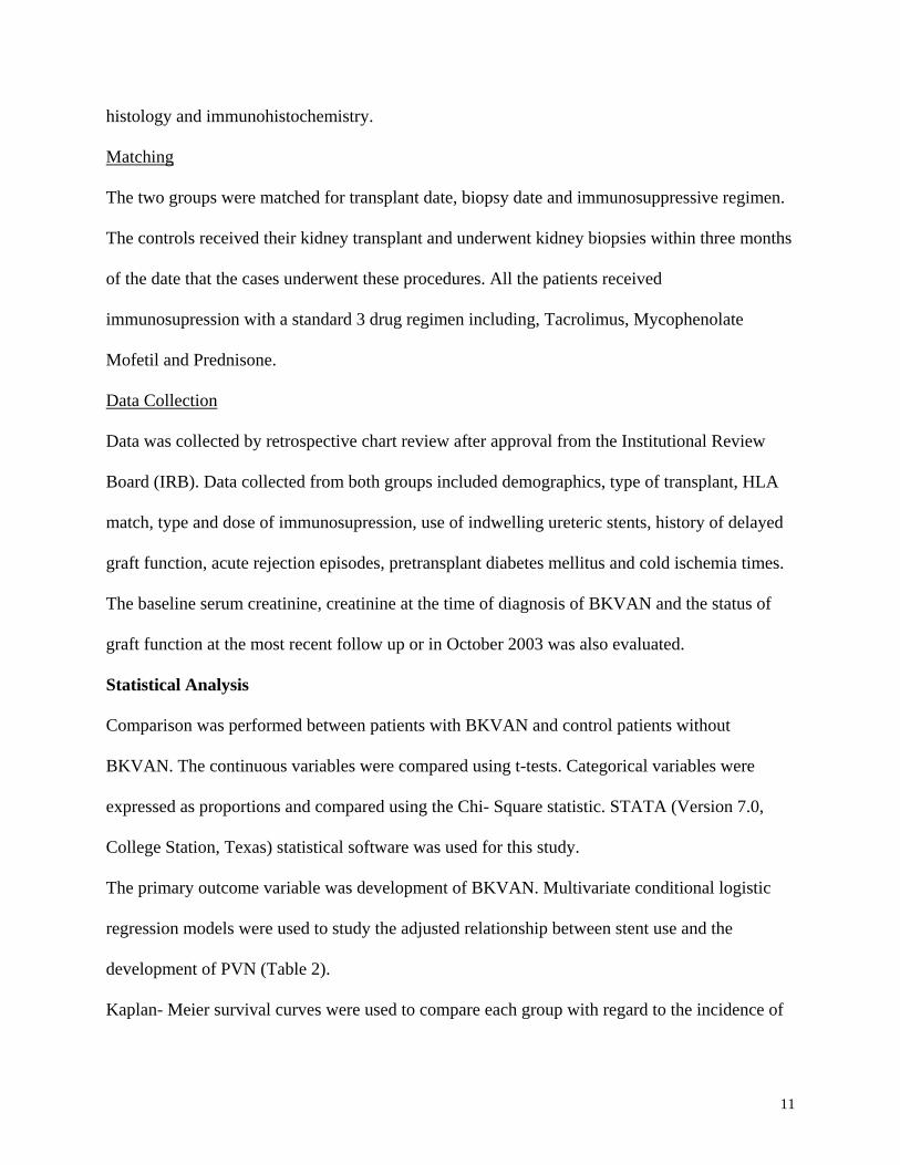
Figure 2 - BKVAN
The morphological hallmarks of BKVAN are intranuclear viral inclusion bodies and focal
necrosis of tubular cells (figure 2). The intranuclear inclusion bodies are seen exclusively in
epithelial cells and appear as amorphous basophilic ground-glass structures. Inclusion bearing
cells are abundant in the medulla and distal tubules. Diagnostic confirmation is performed by
electron microscopy or immunohistochemistry. Ultrastructurally BK virus presents as viral
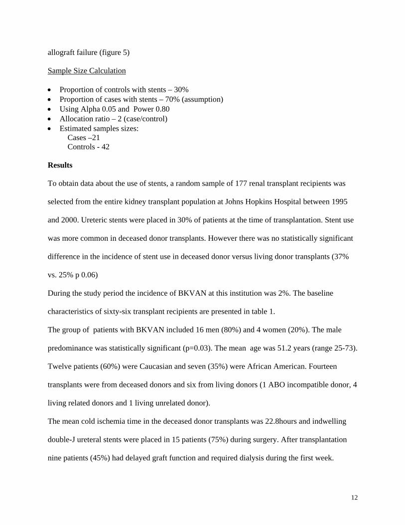
particles measuring between 30 and 50 nm in diameter. Immunohistochemistry is done using
commercially available antibody that detects the SV 40 large T antigen. This antigen is common
to BK, JC and SV 40 viruses (figure 3)
Figure 3 – SV 40 stain.
Controls
The patients with BKVAN (n=20) were compared with a control group that consisted of 46
patients who were biopsied during the same time period and were negative for BKVAN by
10
histology and immunohistochemistry.
Matching
The two groups were matched for transplant date, biopsy date and immunosuppressive regimen.
The controls received their kidney transplant and underwent kidney biopsies within three months
of the date that the cases underwent these procedures. All the patients received
immunosupression with a standard 3 drug regimen including, Tacrolimus, Mycophenolate
Mofetil and Prednisone.
Data Collection
Data was collected by retrospective chart review after approval from the Institutional Review
Board (IRB). Data collected from both groups included demographics, type of transplant, HLA
match, type and dose of immunosupression, use of indwelling ureteric stents, history of delayed
graft function, acute rejection episodes, pretransplant diabetes mellitus and cold ischemia times.
The baseline serum creatinine, creatinine at the time of diagnosis of BKVAN and the status of
graft function at the most recent follow up or in October 2003 was also evaluated.
Statistical Analysis
Comparison was performed between patients with BKVAN and control patients without
BKVAN. The continuous variables were compared using t-tests. Categorical variables were
expressed as proportions and compared using the Chi- Square statistic. STATA (Version 7.0,
College Station, Texas) statistical software was used for this study.
The primary outcome variable was development of BKVAN. Multivariate conditional logistic
regression models were used to study the adjusted relationship between stent use and the
development of PVN (Table 2).
Kaplan- Meier survival curves were used to compare each group with regard to the incidence of
11
allograft failure (figure 5)
Sample Size Calculation
• Proportion of controls with stents – 30% • Proportion of cases with stents – 70% (assumption) • Using Alpha 0.05 and Power 0.80 • Allocation ratio – 2 (case/control) • Estimated samples sizes: Cases –21 Controls - 42
Results
To obtain data about the use of stents, a random sample of 177 renal transplant recipients was
selected from the entire kidney transplant population at Johns Hopkins Hospital between 1995
and 2000. Ureteric stents were placed in 30% of patients at the time of transplantation. Stent use
was more common in deceased donor transplants. However there was no statistically significant
difference in the incidence of stent use in deceased donor versus living donor transplants (37%
vs. 25% p 0.06) During the study period the incidence of BKVAN at this institution was 2%. The baseline
characteristics of sixty-six transplant recipients are presented in table 1.
The group of patients with BKVAN included 16 men (80%) and 4 women (20%). The male
predominance was statistically significant (p=0.03). The mean age was 51.2 years (range 25-73).
Twelve patients (60%) were Caucasian and seven (35%) were African American. Fourteen transplants were from deceased donors and six from living donors (1 ABO incompatible donor, 4
living related donors and 1 living unrelated donor). The mean cold ischemia time in the deceased donor transplants was 22.8hours and indwelling
double-J ureteral stents were placed in 15 patients (75%) during surgery. After transplantation
nine patients (45%) had delayed graft function and required dialysis during the first week.
12
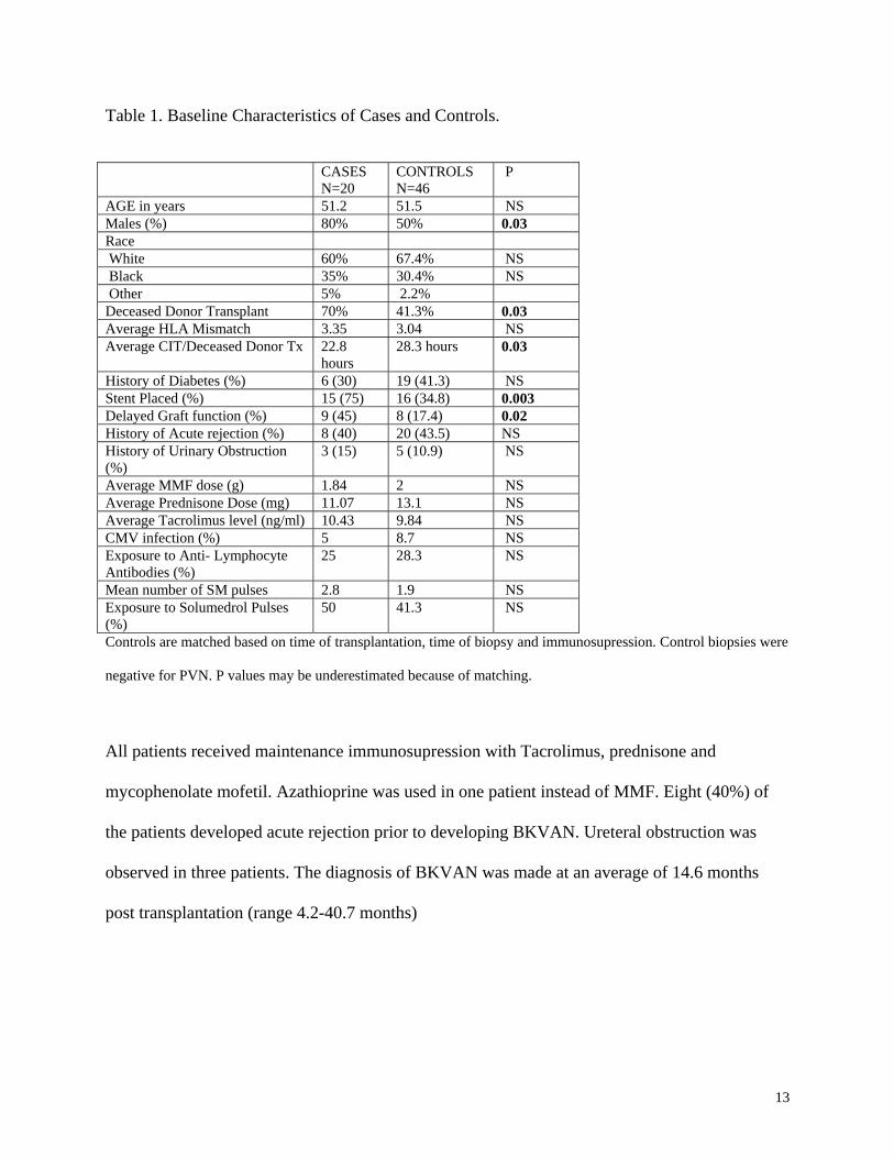
Table 1. Baseline Characteristics of Cases and Controls.
CASES
N=20 CONTROLS N=46
P
AGE in years 51.2 51.5 NS Males (%) 80% 50% 0.03 Race White 60% 67.4% NS Black 35% 30.4% NS Other 5% 2.2% Deceased Donor Transplant 70% 41.3% 0.03 Average HLA Mismatch 3.35 3.04 NS Average CIT/Deceased Donor Tx 22.8
hours 28.3 hours 0.03
History of Diabetes (%) 6 (30) 19 (41.3) NS Stent Placed (%) 15 (75) 16 (34.8) 0.003 Delayed Graft function (%) 9 (45) 8 (17.4) 0.02 History of Acute rejection (%) 8 (40) 20 (43.5) NS History of Urinary Obstruction (%)
3 (15) 5 (10.9) NS
Average MMF dose (g) 1.84 2 NS Average Prednisone Dose (mg) 11.07 13.1 NS Average Tacrolimus level (ng/ml) 10.43 9.84 NS CMV infection (%) 5 8.7 NS Exposure to Anti- Lymphocyte Antibodies (%)
25 28.3 NS
Mean number of SM pulses 2.8 1.9 NS Exposure to Solumedrol Pulses (%)
50 41.3 NS
Controls are matched based on time of transplantation, time of biopsy and immunosupression. Control biopsies were
negative for PVN. P values may be underestimated because of matching.
All patients received maintenance immunosupression with Tacrolimus, prednisone and
mycophenolate mofetil. Azathioprine was used in one patient instead of MMF. Eight (40%) of
the patients developed acute rejection prior to developing BKVAN. Ureteral obstruction was
observed in three patients. The diagnosis of BKVAN was made at an average of 14.6 months
post transplantation (range 4.2-40.7 months)
13
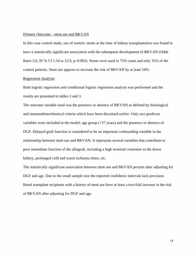
Primary Outcome – stent use and BKVAN
In this case control study, use of ureteric stents at the time of kidney transplantation was found to
have a statistically significant association with the subsequent development of BKVAN (Odds
Ratio 5.6, 95 % CI 1.54 to 22.9, p=0.003). Stents were used in 75% cases and only 35% of the
control patients. Stent use appears to increase the risk of BKVAN by at least 54%.
Regression Analysis
Both logistic regression and conditional logistic regression analysis was performed and the
results are presented in tables 2 and 3.
The outcome variable used was the presence or absence of BKVAN as defined by histological
and immunohistochemical criteria which have been discussed earlier. Only two predictor
variables were included in the model; age group (>57 years) and the presence or absence of
DGF. Delayed graft function is considered to be an important confounding variable in the
relationship between stent use and BKVAN. It represents several variables that contribute to
poor immediate function of the allograft, including a high terminal creatinine in the donor
kidney, prolonged cold and warm ischemia times, etc.
The statistically significant association between stent use and BKVAN persists after adjusting for
DGF and age. Due to the small sample size the reported confidence intervals lack precision.
Renal transplant recipients with a history of stent use have at least a two-fold increase in the risk
of BKVAN after adjusting for DGF and age.
14
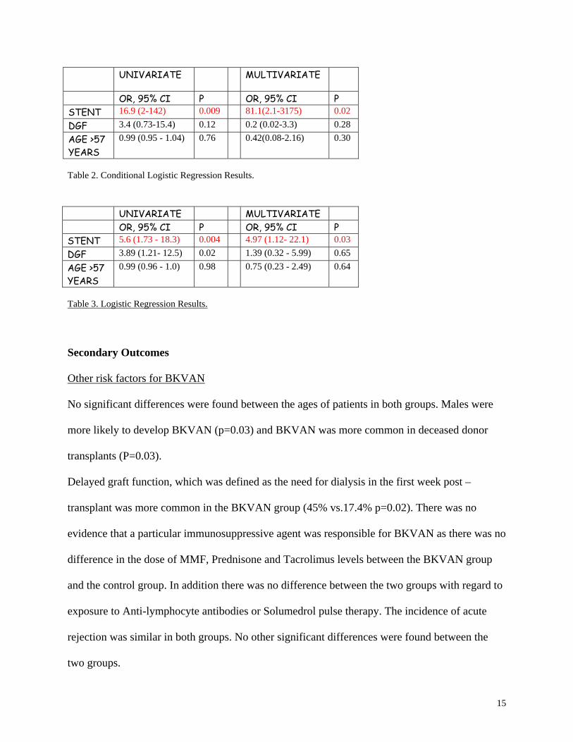
UNIVARIATE MULTIVARIATE OR, 95% CI P OR, 95% CI P STENT 16.9 (2-142) 0.009 81.1(2.1-3175) 0.02 DGF 3.4 (0.73-15.4) 0.12 0.2 (0.02-3.3) 0.28 AGE >57 YEARS
0.99 (0.95 - 1.04) 0.76 0.42(0.08-2.16) 0.30
Table 2. Conditional Logistic Regression Results. UNIVARIATE MULTIVARIATE OR, 95% CI P OR, 95% CI P STENT 5.6 (1.73 - 18.3) 0.004 4.97 (1.12- 22.1) 0.03 DGF 3.89 (1.21- 12.5) 0.02 1.39 (0.32 - 5.99) 0.65 AGE >57 YEARS
0.99 (0.96 - 1.0) 0.98 0.75 (0.23 - 2.49) 0.64
Table 3. Logistic Regression Results.
Secondary Outcomes
Other risk factors for BKVAN No significant differences were found between the ages of patients in both groups. Males were
more likely to develop BKVAN (p=0.03) and BKVAN was more common in deceased donor
transplants (P=0.03).
Delayed graft function, which was defined as the need for dialysis in the first week post –
transplant was more common in the BKVAN group (45% vs.17.4% p=0.02). There was no
evidence that a particular immunosuppressive agent was responsible for BKVAN as there was no
difference in the dose of MMF, Prednisone and Tacrolimus levels between the BKVAN group
and the control group. In addition there was no difference between the two groups with regard to
exposure to Anti-lymphocyte antibodies or Solumedrol pulse therapy. The incidence of acute
rejection was similar in both groups. No other significant differences were found between the
two groups.
15
Clinical course and outcomes
After the diagnosis of BKVAN was confirmed immunosupression was reduced in 18 patients
and remained unchanged in 2 patients. Immunosupression was reduced in a uniform manner. The
dose of MMF was either reduced by 50% or the drug was stopped. Tacrolimus dose was reduced
in 17 patients to obtain trough levels of 5-7ng/ml. In one patient cyclosporine was substituted for
Tacrolimus. The prednisone dose was reduced to 5-7.5 mg /day in all patients.
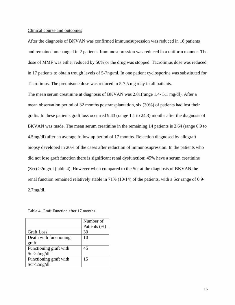
The mean serum creatinine at diagnosis of BKVAN was 2.81(range 1.4- 5.1 mg/dl). After a
mean observation period of 32 months postransplantation, six (30%) of patients had lost their
grafts. In these patients graft loss occurred 9.43 (range 1.1 to 24.3) months after the diagnosis of
BKVAN was made. The mean serum creatinine in the remaining 14 patients is 2.64 (range 0.9 to
4.5mg/dl) after an average follow up period of 17 months. Rejection diagnosed by allograft
biopsy developed in 20% of the cases after reduction of immunosupression. In the patients who
did not lose graft function there is significant renal dysfunction; 45% have a serum creatinine
(Scr) >2mg/dl (table 4). However when compared to the Scr at the diagnosis of BKVAN the
renal function remained relatively stable in 71% (10/14) of the patients, with a Scr range of 0.9-
2.7mg/dl.
Table 4. Graft Function after 17 months.
Number of Patients (%)
Graft Loss 30 Death with functioning graft
10
Functioning graft with Scr>2mg/dl
45
Functioning graft with Scr<2mg/dl
15
16
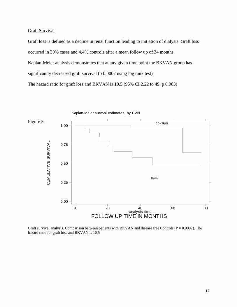
Graft Survival
Graft loss is defined as a decline in renal function leading to initiation of dialysis. Graft loss
occurred in 30% cases and 4.4% controls after a mean follow up of 34 months
Kaplan-Meier analysis demonstrates that at any given time point the BKVAN group has
significantly decreased graft survival (p 0.0002 using log rank test)
The hazard ratio for graft loss and BKVAN is 10.5 (95% CI 2.22 to 49, p 0.003)
Figure 5. Graft Survival Analysis
Kaplan-Meier survival estimates, by PVN
CU
MU
LATI
VE
SU
RV
IVA
L
FOLLOW UP TIME IN MONTHSanalysis time
0 20 40 60 80
0.00
0.25
0.50
0.75
1.00CONTROL
CASE
Graft survival analysis. Comparison between patients with BKVAN and disease free Controls (P = 0.0002). The hazard ratio for graft loss and BKVAN is 10.5
17
DISCUSSION
BKVAN is an important cause of premature graft loss in kidney transplant recipients. The
disease appears to be more common in elderly Caucasian males with a history of delayed graft
function. However multiple factors lead to the development and progression of BKVAN in the
setting of intense immunosupression.
This study has revealed a novel association between ureteral trauma due to stent placement and
the subsequent development of BKVAN. It is the first study in the kidney transplantation
literature to address this issue. The next question that needs to be answered is whether this
association is causal or not. There is clearly a temporal relationship between stent placement and
BKVAN and this association is biologically plausible.
However due to the observational nature of this study it is possible that the observed association
is due at least in some part to confounding variables. Regression analysis was used to account for
2 known confounding variables namely age and DGF, but there are probably other unknown
variables that influence this relationship. Another limitation of the study is the choice of controls.
They may not be entirely representative of the general transplant population, because I only
selected patients who had renal allograft biopsies. Hence the controls are more likely to have had
some form of allograft dysfunction, which led to the biopsy itself. One way to deal with this
problem would be to include renal transplant recipients who have never been biopsied as
controls. However unless newer diagnostic techniques are developed, it is not possible to rule out
BKVAN without doing a biopsy first.
The results of this study should help investigators to develop prospective clinical trials to
confirm the association between ureteral trauma and BKVAN.Based on these results alone it is
not possible to make recommendations for clinical practice. However the routine practice of
18
stenting in kidney transplantation of low –risk patients needs to be reevaluated.
Another important clinical finding in this study is that judicious reduction of immunosupression
has a role in the management of BKVAN. This treatment option appears to be a reasonable one
for the short- term preservation of graft function especially if the diagnosis of BKVAN is made
before significant renal dysfunction develops. Moreover the effects of newer therapies for
BKVAN have to be compared with the option of careful reduction in immunosupression.
19
References
1) Shah KV. Polyomaviruses. In: Fields BN, Knipe DM, Howley PM, editors Virology. 3rd edition. Philadelphia;
Lippincott-Raven, 1996.p.2027-43
2) Gardner SD, Field Am, Coleman DV, Hulme B. New human papovavirus (B.K.) isolated from urine after renal
transplantation Lancet 1971; 1:1253-7.
3) Dorries K New Aspects in the pathogenesis of polyomavirus induced disease. Advances viral research 1997; 48;
205-261.
4) Nickeleit V, Hirsch HH, Binet EF, et al. Polyomavirus infection of renal allograft recipients: from latent
infection to manifest disease. Journal of American Society of Nephrology 1999; 10:1080-9
5) Thomas A, Geetha D. Outcomes of BK Virus Associated Nephropathy following reduction in
immunosupression. Abstract to be presented at the American Transplant Congress May 14, 2004.
6) Binet I, Nickeleit V, Hirsch HH. Polyomavirus infections in transplant recipients. Current opinions in organ
transplant 2000; 5:210-221
7) Ramos E, Drachenberg C, Papadimitriou J, Hamze O, Fink J, et al. Clinical Course of Polyoma Virus
Nephropathy in 67 Renal Transplant Patients
8) Nasimul Ahsan Graft Vol 5 December 2002 S6
9) Makisalo H, Eklund B, Salmeta H, et al. Urological complications after 2084 consecutive kidney
transplantations. Transplant Proceedings 1997; 29:152.
10) Koo seen Lin LC, Bewick M, Koffman CG. Primary Use of a Double J Silicone stent in renal transplantation.
British Journal of Urology 1993; 150:1375).
11) Dominguez J, Clase C, Mahalati K, et al Transplantation vol. 70, 597-601, No. 4, August 27,2000
12) Cormio L, Talja M, Koivusalo A, et al Biocompatibility of Various Indwelling Double J stents: J Urology Vol.
153 (2). Feb 1995, 494-496)
13) Marx M, Bettman M, Bridge S, et al. The effect of various indwelling catheter materials on the normal canine
ureter. J Urology 139:180,1988
14) Juha Lumiaho, Antero Heino, Timo Pietilainen, et al. The journal of Urology vol. 164,1360-1363 October 2000
15) Atwood CJ. Cellular receptors for the polyomaviruses. In: Khalili K, Stoner GL editors. Human
Polyomaviruses: molecular and clinical perspectives. New York: Wiley -Liss; 2001.p.179-96
20
16) Atwood WJ, Norkin LC. Class 1 Major Histocompatibility proteins as cell surface receptors for simian virus 40.
J Virology 1989; 63:4474-7
17) Salunke DM, Caspar DL, Garcea RL. Self-Assembly of purified polyomavirus capsid protein VP1. Cell
1986; 46; 895-904)
18) Nickeleit V, Steiger J, Mihatsch M. BK virus infection after kidney transplantation Graft December 2002 vol. 5
S 47 to S 57)
19) Randhawa P Vats A, Shapiro R, et al. BK Virus: Discovery, Epidemiology, and Biology. Graft December 2002
Vole 5 S 19 to S27
20) Atencio IA, Shadan FF, Zhou XJ, et al. Adult Mouse Kidneys become Permissive to acute polyoma virus
infection and reactivate persistent infections in response to cellular damage and regeneration j Virology 1993;
67:1424-32)
21) Pullmans L, Kartenbeck J, Helenius A. Caveolar endocytosis of SV 40 reveals a new 2 step vesicular transport
pathway to the endoplasmic reticulum. Natural Cell Biology 2001; 3:473-483
22) Norkin LC, Anderson HA, Wolfrom SA. Caveolar Endocytosis of SV 40 is followed by Brefeldin A –sensitive
transport to the ER, where the virus disassembles Journal of Virology 2002; 76:5256-5266
23) Yang RC, Wu R, BK virus DNA complete nucleotide sequence of a human tumor virus Science 1979; 206; 456-
62.
24) Bruns RR, Palade GE. Studies on Blood Capillaries J Cell Biology 1968; 37:244-276
25) Drachenberg C, Papadimitriou J, Wali R, et al BK Polyoma Virus Allograft Nephropathy: Ultrastructural
Features from Viral Cell Entry to Lysis
26) Heritage J, Chesters PM, Mccance DJ.The persistence of papova BK dna sequences in normal human renal
tissue. J medical virology 1981; 8:143-150)
27) Mathur VS, Olson JL, Darragh TM, et al. Polyomavirus induced interstitial nephritis in two renal transplant
recipients: Case reports and review of the literature. American Journal of Kidney Diseases 29:754-758,1997
28) Coleman DV, Mackenzie EF, Gardner, SD, et al; BK infection and ureteric stenosis in renal allograft recipients.
J clinical path 31:338-347,1978
----------------------------------------------------------------------
21

22

23