Embed Size (px)
Citation preview
REVIEWS
When a pathogenic microorganism first infects its host,there is usually a dramatic activation of the INNATE andADAPTIVE immune responses, which can result in diseasesymptoms. If the pathogen and the host survive thisinitial interaction, the adaptive host immune systemusually clears the invading offender. However, somepathogenic bacteria are capable of maintaining infec-tions in mammalian hosts even in the presence ofinflammation, specific antimicrobial mechanisms anda robust adaptive immune response, and can thereforebe described as giving rise to persistent infection1,2 (BOX 1;
see TABLE 1 for a list of bacteria that cause persistentinfections in humans). For example, Helicobacter pyloriinhabits the human gastric mucosa and persistenceof this bacterium in its host can be life-long3;Mycobacterium tuberculosis can establish long-terminfections that can manifest as acute or chronic disease,or can be clinically asymptomatic with the potential tobecome reactivated later4,5; and Salmonella entericaserovar Typhi (S. typhi) causes systemic infection(typhoid fever) that involves colonization of theRETICULOENDOTHELIAL SYSTEM (RES). Some individualswho are infected with S. typhi become life-long car-riers, periodically shedding large numbers of bacteriain their stools. Persistently infected carriers serve asthe reservoir for these pathogens, and the carrier state is
an essential feature that is required for survival of thebacteria within a restricted host population.
Persistent colonization with these bacterialpathogens is usually not clinically apparent. However,even in the absence of clinical symptoms, infectionposes some risk to the host. Individuals who areinfected with M. tuberculosis are at risk of reactivationof the pathogen to produce an active disease state thatcan be life-threatening. A significant proportion ofpeople who are infected with H. pylori develop peptic orduodenal ulcers, or even gastric cancer6. In addition,individuals carrying S. typhi have an increased risk ofdeveloping hepatobiliary cancer7. The long-term resi-dence of the bacteria in a privileged host niche — suchas the MACROPHAGE vacuole or gastric mucosal layer —poses several fundamental biological questions. Forexample, what is the replicative and metabolic state ofthe bacteria during persistent asymptomatic infection,and how do these organisms manage to escape clear-ance for so long in the presence of the host immuneresponse? We are only now beginning to understandthe bacterial and host factors that are involved in thehost–pathogen interaction during persistent infection,and the answers to these questions are likely to providenew and exciting directions for research in the fields ofmicrobial pathogenesis and immunology.
PERSISTENT BACTERIAL INFECTIONS:THE INTERFACE OF THE PATHOGENAND THE HOST IMMUNE SYSTEMDenise M. Monack, Anne Mueller and Stanley Falkow
Abstract | Persistent bacterial infections involving Mycobacterium tuberculosis, Salmonellaenterica serovar Typhi (S. typhi) and Helicobacter pylori pose significant public-health problems.Multidrug-resistant strains of M. tuberculosis and S. typhi are on the increase, and M. tuberculosis and S. typhi infections are often associated with HIV infection. This reviewdiscusses the strategies used by these bacteria during persistent infections that allow them tocolonize specific sites in the host and evade immune surveillance. The nature of the host immuneresponse to this type of infection and the balance between clearance of the pathogen andavoidance of damage to host tissues are also discussed.
INNATE IMMUNE RESPONSE
A cellular defence reaction thatcounteracts invading pathogens,such as bacteria and viruses. Ituses interferon-dependentsignalling and leads to theactivation of genes that areresponsible for bactericidal orantiviral responses.
ADAPTIVE IMMUNE RESPONSE
This involves specificity andimmunological memory. It ismediated by T and B cellsthrough the activation ofcytotoxic CD8+ T cells forpathogen killing or byinteraction with CD4+ T cells for antibody production.
NATURE REVIEWS | MICROBIOLOGY VOLUME 2 | SEPTEMBER 2004 | 747
Department of Microbiologyand Immunology,Stanford School of Medicine,Stanford University, Stanford,California 94305, USA.Correspondence to D.M.M.e-mail: [email protected]:10.1038/nrmicro955
RETICULOENDOTHELIAL
SYSTEM
A diffuse system of cells thathelps the body fight infectionand eliminate cellular debristhrough the action of phagocyticcells (such as macrophages),Kupffer cells in the liver andreticular cells of the spleen, bonemarrow and lymph nodes.
MACROPHAGES
Cells of the mononuclear-phagocyte system that canphagocytose foreign particulatematerial. Macrophages arepresent in many tissues and areimportant for nonspecificimmune reactions.
DENDRITIC CELLS
Professional antigen-presentingcells that take up proteins andpresent peptide antigens to T cells in conjunction withaccessory molecules thatstimulate T-cell activation.They are characterized by manylong, thin processes extendingfrom the cell body.
748 | SEPTEMBER 2004 | VOLUME 2 www.nature.com/reviews/micro
R E V I E W S
Most individuals resolve infection with M. tuberculosissoon after the onset of adaptive immunity12. However,in some infected individuals, the organisms are nevercompletely cleared by the immune response4.Persistently infected individuals can harbour bacteriafor many years, and even throughout their life.Infected individuals are at risk of experiencing theconversion of an asymptomatic infection into a highlycontagious, clinically active and potentially deadlydisease state that is known as reactivation TB. The riskof conversion from an asymptomatic infection to onethat is clinically active is greatest soon after the initialinfection, and this occurs most often in immunolog-ically compromised individuals, such as newborns,the elderly and those who are infected with HIV13,14.
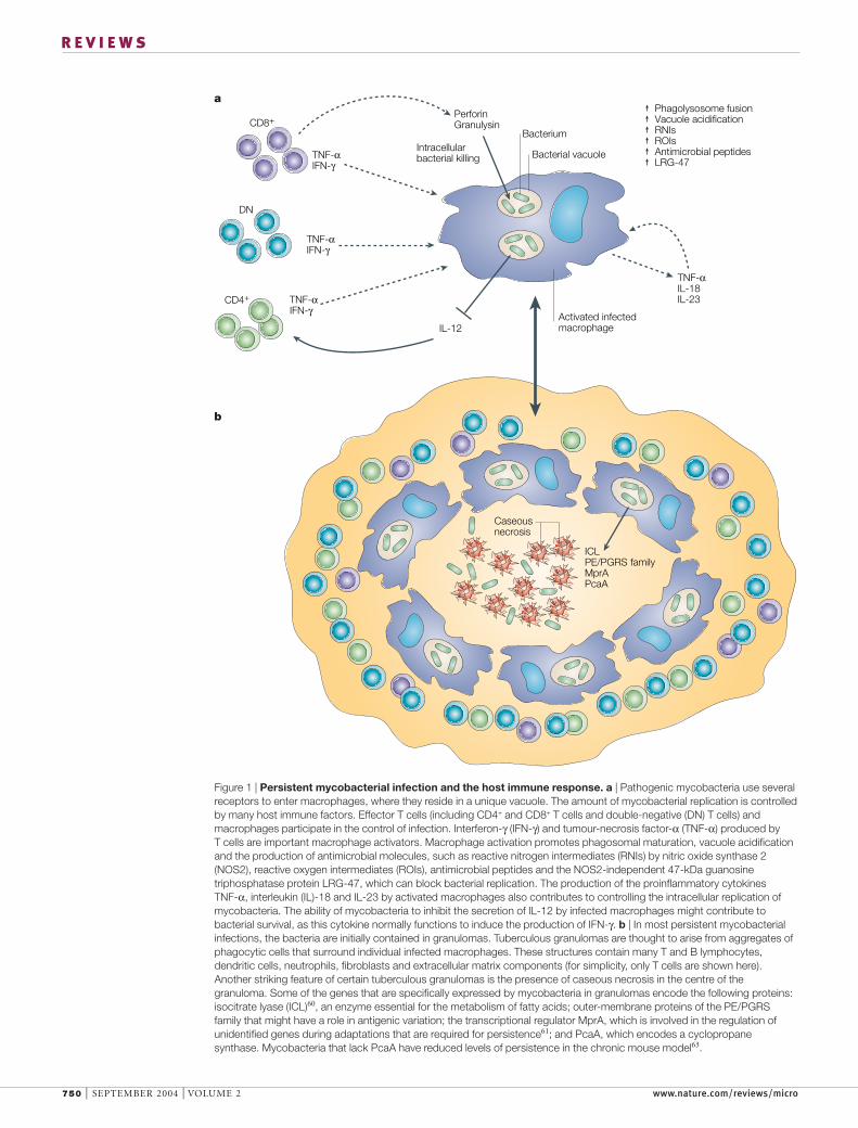
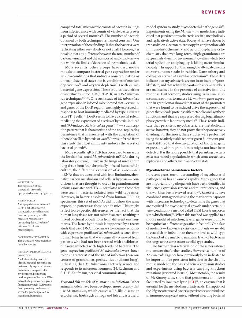
Mycobacterial survival at the immune interfacePersistent mycobacteria reside in granulomas. Althoughthe exact location of viable latent mycobacteria duringpersistent infections remains controversial, bacteria areoften found inside macrophages within granulomas,which are formed in response to persistent intracellularpathogens15 (FIG. 1). Tuberculous granulomas in humansand mice contain an organized collection of differenti-ated macrophages, T lymphocytes, some B lymphocytes,dendritic cells, neutrophils, fibroblasts and extracellularmatrix components16,17. Granulomas are thought toarise initially from aggregates of mononuclear phago-cytes that surround individual infected macrophages.These macrophages become activated, and in manycases several macrophages fuse to form giant cells,which are also formed in response to other persistent
The ability to cause persistent infection is a funda-mental aspect of the interaction between many diverseviral, bacterial and eukaryotic pathogens and theirmammalian hosts. This review is not intended toaddress all or even a significant proportion of thesepathogens. Rather, we will discuss several aspects ofthree persistent bacterial pathogens: H. pylori, a pre-dominantly extracellular pathogen, and M. tuberculosisand S. typhi, which are both facultative intracellularpathogens. We believe that the information that isemerging from these studies will provide an insight intothe general features shared by all microorganisms thathave adapted to persist in the face of a highly evolvedhost immune system.
Persistent mycobacterial infectionsPathogenic mycobacteria cause several long-terminfections in their respective hosts. M. tuberculosiscauses tuberculosis (TB), one of the oldest knownhuman infectious diseases, and this bacterium is esti-mated to infect one-third of the global population8.Primary infection with M. tuberculosis involves replica-tion of the organism at the initial pulmonary site ofinfection. This is followed by bacillaemia, in whichsmall numbers of bacteria are disseminated to theextrapulmonary organs — such as the regional lymphnodes — as well as to uninfected portions of the lung,by a mechanism that may involve the migration ofM. tuberculosis within DENDRITIC CELLS9,10.Adaptive immu-nity and restriction of bacterial growth occurs after thisdissemination and is probably promoted by the arrivalof bacteria in extrapulmonary lymphoid organs4,11.
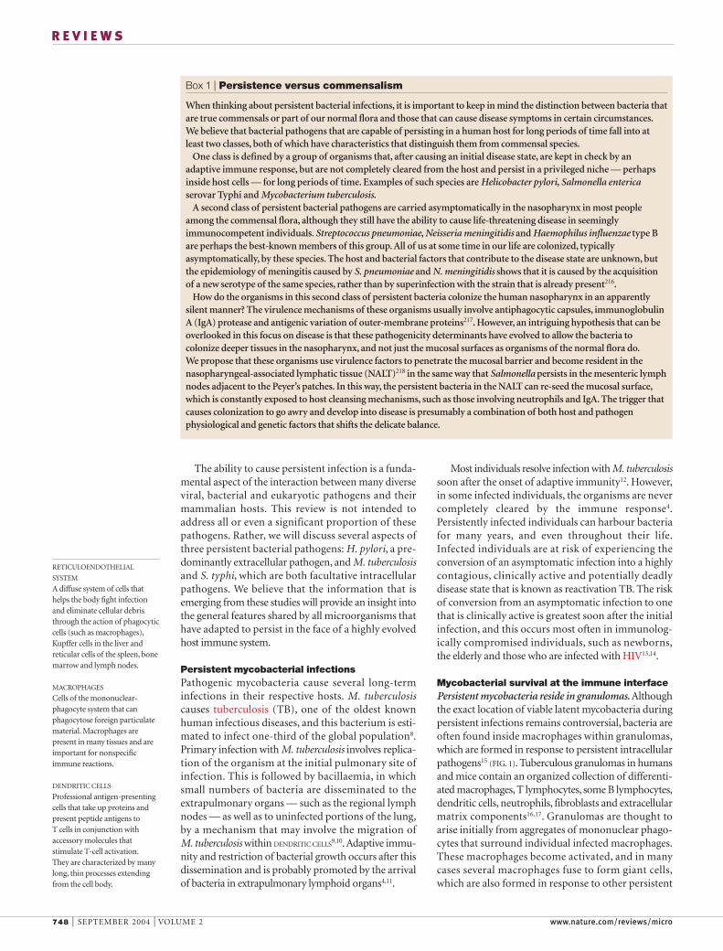
Box 1 | Persistence versus commensalism
When thinking about persistent bacterial infections, it is important to keep in mind the distinction between bacteria thatare true commensals or part of our normal flora and those that can cause disease symptoms in certain circumstances.We believe that bacterial pathogens that are capable of persisting in a human host for long periods of time fall into atleast two classes, both of which have characteristics that distinguish them from commensal species.
One class is defined by a group of organisms that, after causing an initial disease state, are kept in check by anadaptive immune response, but are not completely cleared from the host and persist in a privileged niche — perhapsinside host cells — for long periods of time. Examples of such species are Helicobacter pylori, Salmonella entericaserovar Typhi and Mycobacterium tuberculosis.
A second class of persistent bacterial pathogens are carried asymptomatically in the nasopharynx in most peopleamong the commensal flora, although they still have the ability to cause life-threatening disease in seeminglyimmunocompetent individuals. Streptococcus pneumoniae, Neisseria meningitidis and Haemophilus influenzae type Bare perhaps the best-known members of this group. All of us at some time in our life are colonized, typicallyasymptomatically, by these species. The host and bacterial factors that contribute to the disease state are unknown, butthe epidemiology of meningitis caused by S. pneumoniae and N. meningitidis shows that it is caused by the acquisitionof a new serotype of the same species, rather than by superinfection with the strain that is already present216.
How do the organisms in this second class of persistent bacteria colonize the human nasopharynx in an apparentlysilent manner? The virulence mechanisms of these organisms usually involve antiphagocytic capsules, immunoglobulinA (IgA) protease and antigenic variation of outer-membrane proteins217. However, an intriguing hypothesis that can beoverlooked in this focus on disease is that these pathogenicity determinants have evolved to allow the bacteria tocolonize deeper tissues in the nasopharynx, and not just the mucosal surfaces as organisms of the normal flora do.We propose that these organisms use virulence factors to penetrate the mucosal barrier and become resident in thenasopharyngeal-associated lymphatic tissue (NALT)218 in the same way that Salmonella persists in the mesenteric lymphnodes adjacent to the Peyer’s patches. In this way, the persistent bacteria in the NALT can re-seed the mucosal surface,which is constantly exposed to host cleansing mechanisms, such as those involving neutrophils and IgA. The trigger thatcauses colonization to go awry and develop into disease is presumably a combination of both host and pathogenphysiological and genetic factors that shifts the delicate balance.
NATURE REVIEWS | MICROBIOLOGY VOLUME 2 | SEPTEMBER 2004 | 749
R E V I E W S
role in persistence in vivo, and is a feature that distin-guishes pathogenic from non-pathogenic strains22.
As infected macrophages are the main reservoir ofinfection by pathogenic mycobacteria, studies of thebiology and biogenesis of the mycobacteria-containingphagosome have generated information that is importantfor the understanding of the cell biology, immunologyand microbiology of these pathogens23. Many groupshave described the trafficking of mycobacteria withinunactivated macrophages in tissue-culture experiments.In brief, mycobacteria interfere with phagosome matu-ration by blocking the fusion of nascent phagosomeswith endosomal and lysosomal compartments and bycausing alterations in membrane proteins that normallypromote the formation of an acidic phagolysosome.The steps involved in this process are beyond the scopeof this review, but are described in detail in REF. 24
In addition to the ability to block phagosome matu-ration in some circumstances, pathogenic mycobacteriahave also evolved mechanisms that allow them to persistin macrophage phagolysosomes, within granulomas, inthe presence of a host immune response. A recent studyindicated that within frog granulomas ~60% of intactbacteria of the species Mycobacterium marinum —which is closely related to M. tuberculosis — resided inphagolysosomes, and the level of phagolysosomal fusioncorrelated with the level of macrophage activation25.Therefore, it is possible that mycobacteria have at leasttwo mechanisms of adaptation to intramacrophagesurvival: restriction of phagolysosomal fusion early ininfection and adaptation to phagolysosomal fusionwithin the activated macrophages of granulomas later
infections, particularly those caused by viruses15.T lymphocytes and other immune cells are recruitedearly during the process of granuloma formation18. Thelesion that is formed is sealed off from surroundingtissue by epithelioid cells, which have tightly inter-digitated cell membranes that form zipper-like arraysand link adjacent cells, and which can also be fibroticand calcified. In the centre of granulomas there is usu-ally an area of caseous necrosis — a region of cellulardebris that has a distinct appearance.
How does M. tuberculosis survive within these lesionsfor so many years? One hypothesis is that persistentbacteria are either in a non-replicative state or only havelow levels of replication within the amorphous debris atthe caseous centre of the granuloma19. Evidence inhumans that the persisting bacteria are in a dormantstate comes from the results of culturing and stainingdiseased tissues from patients who have undergonechemotherapy, which might have resulted in false-negative culturing results19-21. An alternative hypothesisas to how a constant bacterial load is maintained is thatthere is a balance between active bacterial replicationand killing by the immune system. This is an active areaof research and is discussed in more detail below.
Survival within macrophages. Pathogenic mycobacteriainitiate long-term infection by entering host macro-phages22, after which they cause extensive remodellingof the PHAGOSOMAL environment to prevent the normalmaturation of this organelle into an acidic, hydrolyticcompartment23. The ability of pathogenic mycobacteriato replicate and/or survive in macrophages has an essential
PHAGOSOME
A membrane-bound,cytoplasmic vacuole formedaround particles that areingested by phagocytosis.
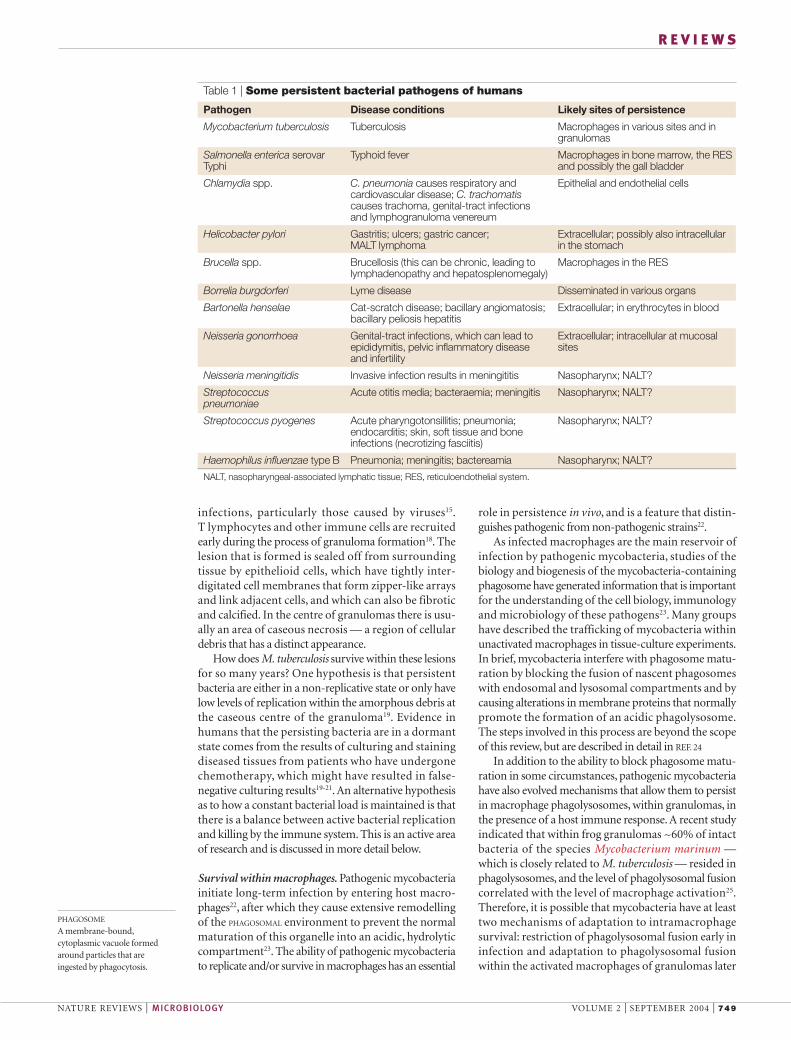
Table 1 | Some persistent bacterial pathogens of humans
Pathogen Disease conditions Likely sites of persistence
Mycobacterium tuberculosis Tuberculosis Macrophages in various sites and ingranulomas
Salmonella enterica serovar Typhoid fever Macrophages in bone marrow, the RES Typhi and possibly the gall bladder
Chlamydia spp. C. pneumonia causes respiratory and Epithelial and endothelial cellscardiovascular disease; C. trachomatiscauses trachoma, genital-tract infections and lymphogranuloma venereum
Helicobacter pylori Gastritis; ulcers; gastric cancer; Extracellular; possibly also intracellular MALT lymphoma in the stomach
Brucella spp. Brucellosis (this can be chronic, leading to Macrophages in the RESlymphadenopathy and hepatosplenomegaly)
Borrelia burgdorferi Lyme disease Disseminated in various organs
Bartonella henselae Cat-scratch disease; bacillary angiomatosis; Extracellular; in erythrocytes in bloodbacillary peliosis hepatitis
Neisseria gonorrhoea Genital-tract infections, which can lead to Extracellular; intracellular at mucosal epididymitis, pelvic inflammatory disease sitesand infertility
Neisseria meningitidis Invasive infection results in meningititis Nasopharynx; NALT?
Streptococcus Acute otitis media; bacteraemia; meningitis Nasopharynx; NALT?pneumoniae
Streptococcus pyogenes Acute pharyngotonsillitis; pneumonia; Nasopharynx; NALT?endocarditis; skin, soft tissue and bone infections (necrotizing fasciitis)
Haemophilus influenzae type B Pneumonia; meningitis; bactereamia Nasopharynx; NALT?
NALT, nasopharyngeal-associated lymphatic tissue; RES, reticuloendothelial system.
750 | SEPTEMBER 2004 | VOLUME 2 www.nature.com/reviews/micro
R E V I E W S
a
b
CD4+
DN
TNF-αIFN-γ
TNF-αIFN-γ
CD8+PerforinGranulysin
Intracellularbacterial killing
IL-12Activated infectedmacrophage
Bacterial vacuole
Bacterium
Phagolysosome fusionVacuole acidificationRNIsROIsAntimicrobial peptidesLRG-47
TNF-αIL-18IL-23
Caseousnecrosis
TNF-αIFN-γ
ICLPE/PGRS familyMprAPcaA
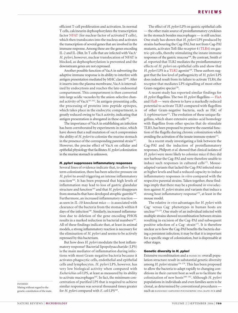
Figure 1 | Persistent mycobacterial infection and the host immune response. a | Pathogenic mycobacteria use severalreceptors to enter macrophages, where they reside in a unique vacuole. The amount of mycobacterial replication is controlledby many host immune factors. Effector T cells (including CD4+ and CD8+ T cells and double-negative (DN) T cells) andmacrophages participate in the control of infection. Interferon-γ (IFN-γ) and tumour-necrosis factor-α (TNF-α) produced by T cells are important macrophage activators. Macrophage activation promotes phagosomal maturation, vacuole acidificationand the production of antimicrobial molecules, such as reactive nitrogen intermediates (RNIs) by nitric oxide synthase 2(NOS2), reactive oxygen intermediates (ROIs), antimicrobial peptides and the NOS2-independent 47-kDa guanosinetriphosphatase protein LRG-47, which can block bacterial replication. The production of the proinflammatory cytokines TNF-α, interleukin (IL)-18 and IL-23 by activated macrophages also contributes to controlling the intracellular replication ofmycobacteria. The ability of mycobacteria to inhibit the secretion of IL-12 by infected macrophages might contribute tobacterial survival, as this cytokine normally functions to induce the production of IFN-γ. b | In most persistent mycobacterialinfections, the bacteria are initially contained in granulomas. Tuberculous granulomas are thought to arise from aggregates ofphagocytic cells that surround individual infected macrophages. These structures contain many T and B lymphocytes,dendritic cells, neutrophils, fibroblasts and extracellular matrix components (for simplicity, only T cells are shown here).Another striking feature of certain tuberculous granulomas is the presence of caseous necrosis in the centre of thegranuloma. Some of the genes that are specifically expressed by mycobacteria in granulomas encode the following proteins:isocitrate lyase (ICL)60, an enzyme essential for the metabolism of fatty acids; outer-membrane proteins of the PE/PGRSfamily that might have a role in antigenic variation; the transcriptional regulator MprA, which is involved in the regulation ofunidentified genes during adaptations that are required for persistence61; and PcaA, which encodes a cyclopropanesynthase. Mycobacteria that lack PcaA have reduced levels of persistence in the chronic mouse model63.
NATURE REVIEWS | MICROBIOLOGY VOLUME 2 | SEPTEMBER 2004 | 751
R E V I E W S
important in controlling intracellular M. tuberculosisreplication34–37. However, a proportion of the bacteriaare clearly still able to survive in macrophages, perhapsby a mechanism that involves inhibiting STAT1-mediated IFN-γ transcriptional responses38 and/orsuppressing the secretion of IL-12 — a proinflamma-tory CYTOKINE that acts to amplify IFN-γ production39,40
— which may in part be mediated by the M. tuberculosisSnm secretion pathway41. It is also likely that the abilityto resist killing by antimicrobial peptides contributes tomycobacterial survival in macrophages42.
Animal models of mycobacterial persistenceFrom both a therapeutic and an epidemiological view-point, the study of persistent mycobacterial infectionsin animal models is very important given the difficul-ties of studying latent TB in humans. Some researchhas been done using guinea pigs, which are able toarrest the initial acute phase of bacterial replicationduring M. tuberculosis infection43. However, althoughthe resulting pathology resembles that seen in humandisease, these animals succumb to the pathologicalconsequences of infection43. Persistent infection canalso occur after intratracheal infection of cynomolgusmonkeys44,45. Although it is likely that this closely mim-ics human infections, high costs limit the widespreaduse of this non-human primate model. Therefore,many research groups commonly use mouse modelsof M. tuberculosis infection or frog and fish models ofM. marinum infection. Although much information canbe gained from these models, care should be taken inextrapolating the results obtained with these modelsdirectly to human TB.
in infection. It is still unclear how the bacteria sensethese different intramacrophage environments; theidentification of the bacterial effector proteins that areinvolved in this should provide considerable insightinto this crucial stage of mycobacterial pathogenesis.
In further support of the proposal that mycobacteriause different strategies according to the circumstances,recent publications indicate that mycobacteria havetemporal and immune-response-triggered differencesin gene expression both in activated macrophages invitro and in macrophages isolated from infected tis-sue26–31. In addition, a recent study showed differentialmycobacterial survival in type 1 (interleukin-23 (IL-23)-producing) and type 2 (IL-10-producing) macrophages32.The immune status of the macrophage therefore hasan important role in bacterial persistence. Indeed, thecontrol of bacterial growth in murine models of latencyrequires interferon-γ (IFN-γ), tumour-necrosis factor-α(TNF-α) and nitric oxide (NO)16, all of which can alterthe environment of the bacteria-containing phagosomeand lead to killing of the pathogen.
IFN-γ is a crucial component of immunity to TB asit activates infected host macrophages, which directlyinhibit the replication of M. tuberculosis. The impor-tance of this molecule in the control of mycobacterialinfections is highlighted by the discovery of IFN-γ-related genetic mutations that predispose affectedindividuals to active TB, as well as to other infectionsthat are caused by intracellular bacterial species, suchas Salmonella spp.33 (BOX 2; TABLE 2). IFN-γ induces theexpression of NO synthase 2 (NOS2) and of the newlyidentified, NOS2-independent, 47-kDa guanosinetriphosphatase protein LRG-47, and both pathways are
CYTOKINES
Low-molecular-weight proteinsthat are important forimmunity, inflammation anddevelopment, and whichcontribute to thepathophysiology of acute andchronic infections.
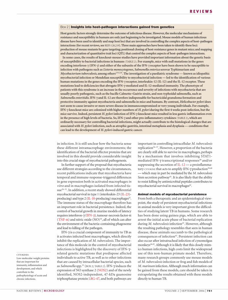
Box 2 | Insights into host–pathogen interactions gained from genetics
Host genetic factors strongly determine the outcome of infectious disease. However, the molecular mechanisms ofresistance and susceptibility in humans are only just beginning to be investigated. Mouse models of human infectiousdisease have been used to identify and map host loci that are involved in controlling the complex aspects of host–pathogeninteractions (for recent reviews, see REFS 128,219). Three main approaches have been taken to identify these loci:production of mouse mutants by gene targeting; positional cloning of host-resistance genes in mutant mice; and mappingand characterization of quantitative trait loci (QTL) that control the complex aspects of host–pathogen interactions.
In some cases, the results of knockout-mouse studies have provided important information about the genetic basisof susceptibility to bacterial infections in humans (TABLE 2). For example, mice with null mutations in the genesencoding interferon-γ (IFN-γ) and either of the subunits of the IFN-γ receptor have been shown to be susceptible toinfection with pathogens such as Listeria monocytogenes, Salmonella enterica serovar Typhimurium andMycobacterium tuberculosis, among others219,220. The investigation of a paediatric syndrome — known as idiopathicmycobacterial infection or Mendelian susceptibility to mycobacterial infection — led to the identification of varioushuman mutations in the genes encoding the IFN-γ receptor, interleukin-12 (IL-12) and the IL-12 receptor. Thesemutations lead to deficiencies that abrogate IFN-γ-mediated and IL-12-mediated immunity. The phenotype ofpatients with this syndrome is an increase in the occurrence and severity of infections with mycobacteria that areusually poorly pathogenic, such as the bacille Calmette–Guérin strain, and non-typhoidal salmonella, such asSalmonella enteritidis. IFN-γ and IL-12 are therefore indispensable for bactericidal granuloma formation andprotective immunity against mycobacteria and salmonella in mice and humans. By contrast, Helicobacter pylori doesnot seem to cause invasive or more severe disease in immunocompromised or very young individuals. For example,IFN-γ-knockout mice are colonized with higher numbers of H. pylori during the first 4 weeks post-infection, but themice survive. Indeed, persistent H. pylori infection of IFN-γ-knockout mice resulted in less gastric inflammation evenin the presence of high levels of bacteria. So, IFN-γ and other pro-inflammatory cytokines (TABLE 2), which areordinarily necessary for controlling bacterial infections, might actually contribute to the histological changes that areassociated with H. pylori infection, such as atrophic gastritis, intestinal metaplasia and dysplasia — conditions thatcan lead to the development of H. pylori-induced gastric cancer.
752 | SEPTEMBER 2004 | VOLUME 2 www.nature.com/reviews/micro
R E V I E W S
response, followed by a stable maintenance at high levelsin the lung over many months. The mice seem healthyuntil the disease reactivates, which can take place asmuch as 18 months later48. This chronic model resem-bles latency in humans in that it depends on the hostimmune response to contain the infection. However,unlike latent TB in humans, this model results in largenumbers of bacteria, which leads to pulmonary damagethat steadily accumulates in the lungs of chronicallyinfected animals49.
Although these mouse models have limitations,several groups have used them to address the meta-bolic state of persistent mycobacteria. Rees and Hart
Mouse models of M. tuberculosis infection. Severalmouse models of TB latency have been established46–48.The Cornell mouse model (also known as the drug-induced model) involves partial clearance of M. tuber-culosis infection by incomplete chemotherapy; theinfection is reduced to a point at which no bacterialcolonies are recovered46. Drug intervention is needed toinduce the latent state, which is not necessary in humandisease. The low-dose mouse model of latent TB (alsoknown as the chronic or plateau model) involves aerosolinfection or infection by intravenous routes. This resultsin an initial acute phase of bacterial replication that iscontrolled by the onset of an adaptive immune
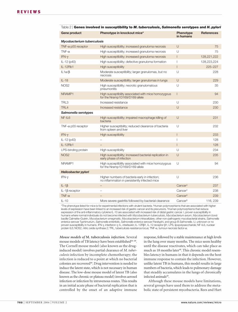
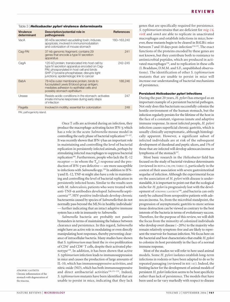
Table 2 | Genes involved in susceptibility to M. tuberculosis, Salmonella serotypes and H. pylori
Gene product Phenotype in knockout mice* Phenotype Referencesin humans
Mycobacterium tuberculosis
TNF-α p55 receptor High susceptibility; increased granuloma necrosis U 75
TNF-α High susceptibility; increased granuloma necrosis U 75
IFN-γ High susceptibility; increased granuloma necrosis I 128,221,222
IL-12 (p40) High susceptibility; defective granuloma formation I 128,223,224
IL-12Rb1 High susceptibility I 225–227
IL1α/β Moderate susceptibility; larger granulomas, but no U 228necrosis
IL-18 Moderate susceptibility; larger granulomas in lungs U 229
NOS2 High susceptibility; necrotic granulomatous U 35pneumonitis
NRAMP1 High susceptibility associated with mice homozygous I 94for the Nramp1D169/D169 allele
TRL3 Increased resistance U 230
TRL4 Increased resistance U 230
Salmonella serotypes
NF-IL6 High susceptibility; impaired macrophage killing of U 231bacteria
TNF-α p55 receptor Higher susceptibility; reduced clearance of bacteria U 232from spleen and liver
IFN-γ High susceptibility I 233
IL-12 (p40) – I 128
IL-12Rb1 – I 128
LPS-binding protein High susceptibility U 234
NOS2 High susceptibility; increased bacterial replication in U 235early phase of infection
NRAMP1 High susceptibility associated with mice homozygous U 94for the Nramp1D169/D169 allele
Helicobacter pylori
IFN-γ Higher numbers of bacteria early in infection; U 236no inflammation in persistently infected mice
IL-1β – Cancer‡ 237
IL-1β receptor – Cancer‡ 238
TNF-α – Cancer‡ 239
IL-10 More severe gastritis followed by bacterial clearance Cancer§ 116, 239
*The phenotype listed for mice is for experimental infections with virulent bacteria. ‡Human polymorphisms that are associated with higherlevels of expression have been linked to an increased risk of gastric cancer and its precursors. §Human polymorphisms that reduceexpression of the anti-inflammatory cytokine IL-10 are associated with increased risk of distal gastric cancer. I, proven susceptibility inhumans where normal individuals do not become infected with Mycobacterium tuberculosis, Mycobacterium avium, Mycobacterium bovisbacille Calmette–Guérin, Mycobacterium smegmatis, Mycobacterium intracellulare, other non-pathogenic mycobacterial strains, Salmonellaenterica serovar Typhimurium, Salmonella enteritidis, Salmonella enterica serovar Paratyphi, and group B Salmonella. U, unknown or noproven susceptibility in humans. IFN-γ, interferon-γ; IL, interleukin; IL-12Rβ1, IL-12 receptor β1; LPS, lipopolysaccharide; NF-IL6, nuclearprotein IL6; NOS2, nitric oxide synthase 2; TRL, tuberculosis-resistance locus; TNF-α, tumour-necrosis factor-α.
NATURE REVIEWS | MICROBIOLOGY VOLUME 2 | SEPTEMBER 2004 | 753
R E V I E W S
model system to study mycobacterial pathogenesis22.Experiments using the M. marinum model have indi-cated that persistent mycobacteria are in a metabolicallyand replicatively active state. Bouley et al. have shown bytransmission electron microscopy in conjunction withimmunohistochemistry and acid phosphatase cyto-chemistry that even long-term, single granulomas aresurprisingly dynamic environments, within which bac-terial replication and phagocytic killing occur simulta-neously25. In support of this, using the attenuated BACILLE
CALMETTE–GUÉRIN strain in rabbits, Dannenberg andcolleagues arrived at a similar conclusion56. These dataindicate that mycobacteria are not in an inert or ‘spore-like’ state, and that relatively constant bacterial numbersare maintained in the presence of an active immuneresponse. Furthermore, studies using DIFFERENTIAL FLUO-
RESCENCE INDUCTION to analyse M. marinum gene expres-sion in granulomas showed that most of the promotersthat were found to be induced drive the expression ofgenes that encode proteins with metabolic and syntheticfunctions and that are expressed during logarithmic-phase growth in laboratory media57. These results indi-cate that persistent mycobacteria are metabolicallyactive; however, they do not prove that they are activelydividing. Furthermore, these studies were performedusing the relatively stable reporter green fluorescent pro-tein (GFP), so that downregulation of bacterial geneexpression within granulomas might not have beendetected. It is therefore possible that persisting bacteriaexist as a mixed population, in which some are activelyreplicating and others are in an inactive state.
Mycobacterial persistence factorsIn recent years, our understanding of mycobacterialpathogenesis has advanced rapidly. Many genes thatare important for pathogenesis have been identified invirulence expression screens and mutant screens, andthis work has been reviewed recently22. Sasseti et al. havecombined mutagenesis using the mariner transposonwith microarray technology to determine the genes thatare required for mycobacterial growth under certain invitro conditions (a method called TraSH, for transposonsite hybridization)58. When this method was applied to amouse model of infection, several genes were found tobe required at different stages after infection59. One classof mutants — known as persistence mutants — are ableto establish an infection to the same level as wild-typebacteria, but are unable to maintain levels of bacteria inthe lungs to the same extent as wild-type strains.
The further characterization of these persistencemutants awaits further studies. However, a number ofM. tuberculosis genes have previously been indicated tobe important for persistent infection in the chronicmouse model on the basis of gene-expression studiesand experiments using bacteria carrying knockoutmutations (reviewed in REF. 5). Most notably, the resultsof McKinney et al. show that persistence in mice isfacilitated by isocitrate lyase (ICL)60, an enzyme that isessential for the metabolism of fatty acids. Disruption ofthe icl gene attenuated bacterial persistence and virulencein immunocompetent mice, without affecting bacterial
compared total microscopic counts of bacteria in lungsfrom infected mice with counts of viable bacteria overa period of several months50. The number of bacteriaobtained by both techniques remained constant. Oneinterpretation of these findings is that the bacteria werereplicating either very slowly or not at all. However, it ispossible that any difference between the total number ofbacteria visualized and the number of viable bacteria wasnot within the limits of detection of the methods used.
More recently, other groups have used mousemodels to compare bacterial gene expression underin vitro conditions that induce a non-replicating ordormant bacterial state (that is, conditions of nutrientdeprivation51 and oxygen depletion52) with in vivobacterial gene expression. These studies used eitherquantitative real-time PCR (qRT-PCR) or cDNA microar-ray techniques26,27,30. One such study of M. tuberculosisgene expression in infected mice showed that α-CRYSTALLIN
and genes of the DosR regulon are highly expressed inresponse to host immunity mediated by type 1 HELPER T
CELLS (TH1 cells)27. DosR seems to have a crucial role in
mediating the expression of a series of hypoxia-inducedand NO-induced M. tuberculosis genes53–55 — a transcrip-tion pattern that is characteristic of the non-replicatingpersistence that is associated with the adaptation oftubercle bacilli to hypoxia in vitro52. It was inferred fromthis study that host immunity induces the arrest ofbacterial growth27.
More recently, qRT-PCR has been used to measurethe levels of selected M. tuberculosis mRNAs duringlaboratory culture, in vivo in the lungs of mice and inlung tissue from four chronically infected humans26. Inculture, the differential expression of M. tuberculosismRNAs that are associated with iron limitation, alter-native carbon metabolism and cellular hypoxia — con-ditions that are thought to occur in granulomatouslesions associated with TB — correlated with those thatwere seen in bacteria isolated from wild-type mice.However, in bacteria isolated from human TB lungspecimens, this set of mRNAs did not show the sameexpression patterns as those seen in mice. This mightreflect host-specific differences or the fact that thehuman lung tissue was not microdissected, resulting inmixed bacterial populations from different environ-ments. The latter hypothesis is supported by a recentstudy that used DNA microarrays to examine genome-wide expression profiles of M. tuberculosis isolated fromhuman lung tissue that was surgically removed frompatients who had not been treated with antibiotics,but were infected with high levels of bacteria. Thegene-expression profiles of M. tuberculosis were shownto be characteristic of the site of infection (caseouscentres of granulomas, pericavities or distant lung),indicating that M. tuberculosis actively senses andresponds to its microenvironment (H. Rachman andS. H. E. Kaufmann, personal communication).
Frog and fish models of M. marinum infection. Otheranimal models have been developed more recently thatuse M. marinum, which causes a TB-like disease inectothermic hosts such as frogs and fish and is a useful
α-CRYSTALLIN
The expression of thischaperonin protein isupregulated in vitro by hypoxia.
HELPER T CELLS
A subpopulation of activatedCD4+ T cells that secretecharacteristic cytokines andfunction primarily in cell-mediated responses bypromoting the activation ofcytotoxic T cells andmacrophages.
BACILLE CALMETTE–GUÉRIN
The attenuated Mycobacteriumbovis live vaccine.
DIFFERENTIAL FLUORESCENCE
INDUCTION
A selection strategy used toidentify bacterial genes that arepreferentially expressed when abacterium is in a particularenvironment. By insertingrandom pieces of bacterial DNAin front of a promoterless greenfluorescent protein (GFP) gene,flow cytometry can be used toscreen for genes expressed inspecific environments.
754 | SEPTEMBER 2004 | VOLUME 2 www.nature.com/reviews/micro
R E V I E W S
of T-cell subsets in these two phases of infection4.Whether or not this is the case, CD4+ T cells have animportant but as yet undefined role in the control ofpersistent infection67. Although other activities of CD8+
T cells, in addition to IFN-γ production, might also beimportant for the control of persistent infections68-70,further studies are needed to determine the exact rolesof these cells.
The production of immunosuppressive cytokines —such as IL-10 and transforming growth factor-β (TGF-β)— has been documented in humans with active TB71-73,and IL-10 production is increased in the lungs of micethat show chronic mycobacterial infection. This indi-cates that the ability of IL-10 to downregulate theimmune response might contribute to the reactivationof chronic M. tuberculosis infection74. In animal models,the proinflammatory cytokine TNF-α has a key role inhost responses against TB75,76, including granulomaformation and the containment of disease18,77.Furthermore, treatments with antibodies that neutralizeTNF-α cause reactivation of TB in a mouse model oflatent infection78 and in human latent infection, asshown by the clinical observation of reactivation of TBduring the treatment of autoimmune disease79. TNF-αtherefore has a significant role in the control of persis-tent M. tuberculosis infections and, as discussed below, isequally important in persistent Salmonella infections.
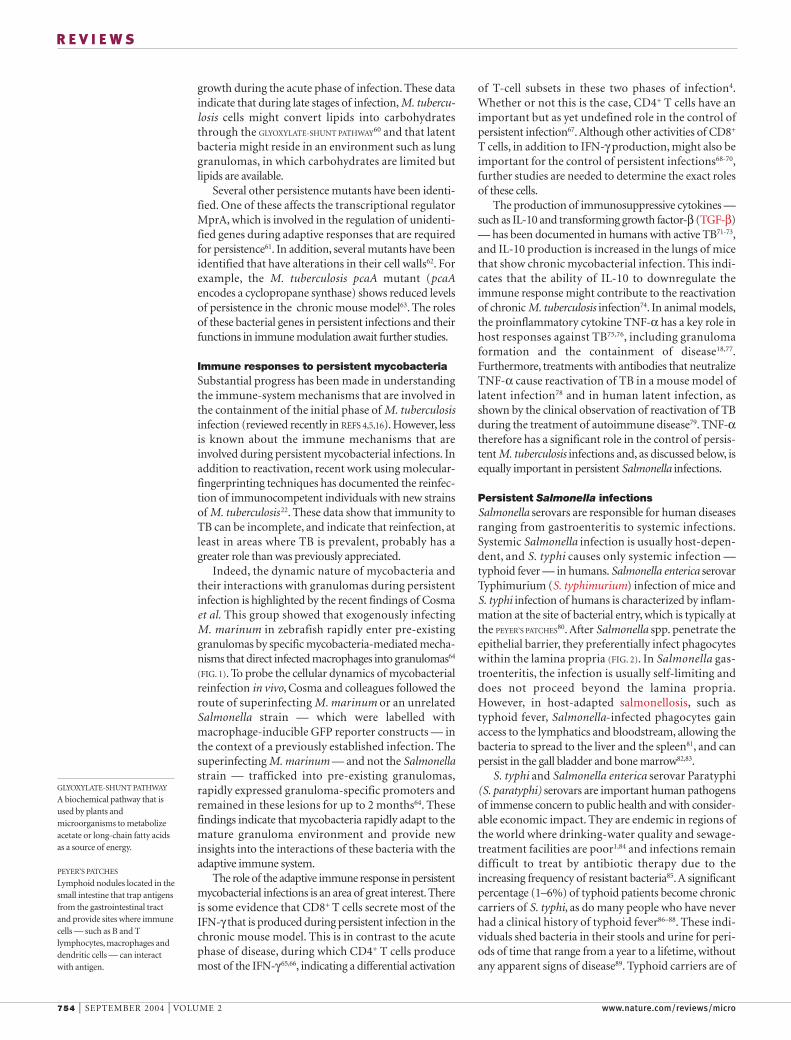
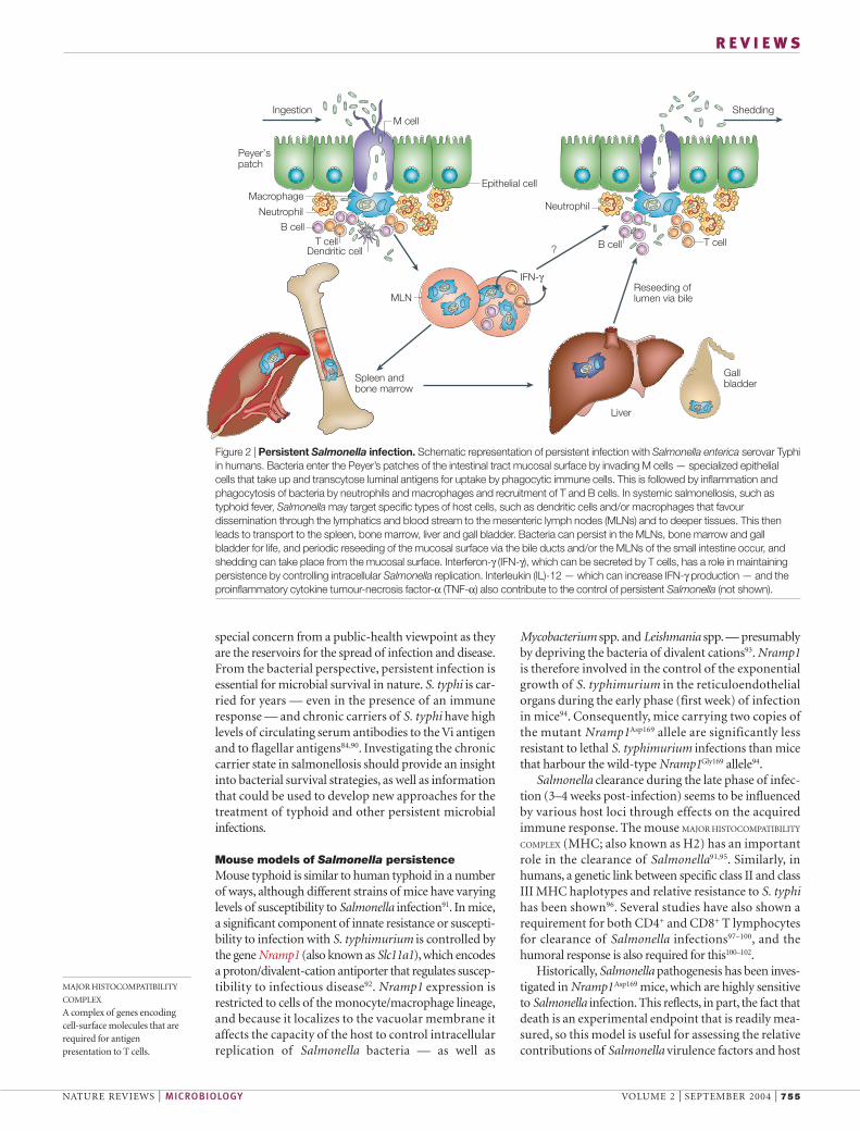
Persistent Salmonella infectionsSalmonella serovars are responsible for human diseasesranging from gastroenteritis to systemic infections.Systemic Salmonella infection is usually host-depen-dent, and S. typhi causes only systemic infection —typhoid fever — in humans. Salmonella enterica serovarTyphimurium (S. typhimurium) infection of mice andS. typhi infection of humans is characterized by inflam-mation at the site of bacterial entry, which is typically atthe PEYER’S PATCHES80. After Salmonella spp. penetrate theepithelial barrier, they preferentially infect phagocyteswithin the lamina propria (FIG. 2). In Salmonella gas-troenteritis, the infection is usually self-limiting anddoes not proceed beyond the lamina propria.However, in host-adapted salmonellosis, such astyphoid fever, Salmonella-infected phagocytes gainaccess to the lymphatics and bloodstream, allowing thebacteria to spread to the liver and the spleen81, and canpersist in the gall bladder and bone marrow82,83.
S. typhi and Salmonella enterica serovar Paratyphi(S. paratyphi) serovars are important human pathogensof immense concern to public health and with consider-able economic impact. They are endemic in regions ofthe world where drinking-water quality and sewage-treatment facilities are poor1,84 and infections remaindifficult to treat by antibiotic therapy due to theincreasing frequency of resistant bacteria85. A significantpercentage (1–6%) of typhoid patients become chroniccarriers of S. typhi, as do many people who have neverhad a clinical history of typhoid fever86–88. These indi-viduals shed bacteria in their stools and urine for peri-ods of time that range from a year to a lifetime, withoutany apparent signs of disease89. Typhoid carriers are of
growth during the acute phase of infection. These dataindicate that during late stages of infection, M. tubercu-losis cells might convert lipids into carbohydratesthrough the GLYOXYLATE-SHUNT PATHWAY60 and that latentbacteria might reside in an environment such as lunggranulomas, in which carbohydrates are limited butlipids are available.
Several other persistence mutants have been identi-fied. One of these affects the transcriptional regulatorMprA, which is involved in the regulation of unidenti-fied genes during adaptive responses that are requiredfor persistence61. In addition, several mutants have beenidentified that have alterations in their cell walls62. Forexample, the M. tuberculosis pcaA mutant (pcaAencodes a cyclopropane synthase) shows reduced levelsof persistence in the chronic mouse model63. The rolesof these bacterial genes in persistent infections and theirfunctions in immune modulation await further studies.
Immune responses to persistent mycobacteriaSubstantial progress has been made in understandingthe immune-system mechanisms that are involved inthe containment of the initial phase of M. tuberculosisinfection (reviewed recently in REFS 4,5,16). However, lessis known about the immune mechanisms that areinvolved during persistent mycobacterial infections. Inaddition to reactivation, recent work using molecular-fingerprinting techniques has documented the reinfec-tion of immunocompetent individuals with new strainsof M. tuberculosis 22. These data show that immunity toTB can be incomplete, and indicate that reinfection, atleast in areas where TB is prevalent, probably has agreater role than was previously appreciated.
Indeed, the dynamic nature of mycobacteria andtheir interactions with granulomas during persistentinfection is highlighted by the recent findings of Cosmaet al. This group showed that exogenously infecting M. marinum in zebrafish rapidly enter pre-existinggranulomas by specific mycobacteria-mediated mecha-nisms that direct infected macrophages into granulomas64
(FIG. 1). To probe the cellular dynamics of mycobacterialreinfection in vivo, Cosma and colleagues followed theroute of superinfecting M. marinum or an unrelatedSalmonella strain — which were labelled withmacrophage-inducible GFP reporter constructs — inthe context of a previously established infection. Thesuperinfecting M. marinum — and not the Salmonellastrain — trafficked into pre-existing granulomas,rapidly expressed granuloma-specific promoters andremained in these lesions for up to 2 months64. Thesefindings indicate that mycobacteria rapidly adapt to themature granuloma environment and provide newinsights into the interactions of these bacteria with theadaptive immune system.
The role of the adaptive immune response in persistentmycobacterial infections is an area of great interest. Thereis some evidence that CD8+ T cells secrete most of theIFN-γ that is produced during persistent infection in thechronic mouse model. This is in contrast to the acutephase of disease, during which CD4+ T cells producemost of the IFN-γ65,66, indicating a differential activation
GLYOXYLATE-SHUNT PATHWAY
A biochemical pathway that isused by plants andmicroorganisms to metabolizeacetate or long-chain fatty acidsas a source of energy.
PEYER’S PATCHES
Lymphoid nodules located in thesmall intestine that trap antigensfrom the gastrointestinal tractand provide sites where immunecells — such as B and Tlymphocytes, macrophages anddendritic cells — can interactwith antigen.
NATURE REVIEWS | MICROBIOLOGY VOLUME 2 | SEPTEMBER 2004 | 755
R E V I E W S
Mycobacterium spp. and Leishmania spp. — presumablyby depriving the bacteria of divalent cations93. Nramp1is therefore involved in the control of the exponentialgrowth of S. typhimurium in the reticuloendothelialorgans during the early phase (first week) of infectionin mice94. Consequently, mice carrying two copies ofthe mutant Nramp1Asp169 allele are significantly lessresistant to lethal S. typhimurium infections than micethat harbour the wild-type Nramp1Gly169 allele94.
Salmonella clearance during the late phase of infec-tion (3–4 weeks post-infection) seems to be influencedby various host loci through effects on the acquiredimmune response. The mouse MAJOR HISTOCOMPATIBILITY
COMPLEX (MHC; also known as H2) has an importantrole in the clearance of Salmonella91,95. Similarly, inhumans, a genetic link between specific class II and classIII MHC haplotypes and relative resistance to S. typhihas been shown96. Several studies have also shown arequirement for both CD4+ and CD8+ T lymphocytesfor clearance of Salmonella infections97–100, and thehumoral response is also required for this100–102.
Historically, Salmonella pathogenesis has been inves-tigated in Nramp1Asp169 mice, which are highly sensitiveto Salmonella infection. This reflects, in part, the fact thatdeath is an experimental endpoint that is readily mea-sured, so this model is useful for assessing the relativecontributions of Salmonella virulence factors and host
special concern from a public-health viewpoint as theyare the reservoirs for the spread of infection and disease.From the bacterial perspective, persistent infection isessential for microbial survival in nature. S. typhi is car-ried for years — even in the presence of an immuneresponse — and chronic carriers of S. typhi have highlevels of circulating serum antibodies to the Vi antigenand to flagellar antigens84,90. Investigating the chroniccarrier state in salmonellosis should provide an insightinto bacterial survival strategies, as well as informationthat could be used to develop new approaches for thetreatment of typhoid and other persistent microbialinfections.
Mouse models of Salmonella persistenceMouse typhoid is similar to human typhoid in a numberof ways, although different strains of mice have varyinglevels of susceptibility to Salmonella infection91. In mice,a significant component of innate resistance or suscepti-bility to infection with S. typhimurium is controlled bythe gene Nramp1 (also known as Slc11a1), which encodesa proton/divalent-cation antiporter that regulates suscep-tibility to infectious disease92. Nramp1 expression isrestricted to cells of the monocyte/macrophage lineage,and because it localizes to the vacuolar membrane itaffects the capacity of the host to control intracellularreplication of Salmonella bacteria — as well as
MAJOR HISTOCOMPATIBILITY
COMPLEX
A complex of genes encodingcell-surface molecules that arerequired for antigenpresentation to T cells.
SheddingIngestion
Peyer’spatch
?
M cell
Neutrophil
MacrophageEpithelial cell
B cellT cell
Dendritic cell
Neutrophil
B cell T cell
IFN-γReseeding of lumen via bile
Gall bladder
Liver
Spleen andbone marrow
MLN
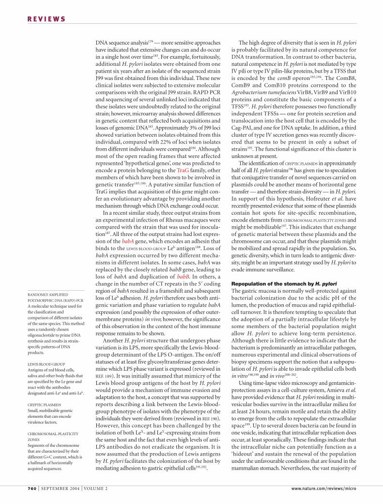
Figure 2 | Persistent Salmonella infection. Schematic representation of persistent infection with Salmonella enterica serovar Typhiin humans. Bacteria enter the Peyer’s patches of the intestinal tract mucosal surface by invading M cells — specialized epithelialcells that take up and transcytose luminal antigens for uptake by phagocytic immune cells. This is followed by inflammation andphagocytosis of bacteria by neutrophils and macrophages and recruitment of T and B cells. In systemic salmonellosis, such astyphoid fever, Salmonella may target specific types of host cells, such as dendritic cells and/or macrophages that favourdissemination through the lymphatics and blood stream to the mesenteric lymph nodes (MLNs) and to deeper tissues. This thenleads to transport to the spleen, bone marrow, liver and gall bladder. Bacteria can persist in the MLNs, bone marrow and gallbladder for life, and periodic reseeding of the mucosal surface via the bile ducts and/or the MLNs of the small intestine occur, andshedding can take place from the mucosal surface. Interferon-γ (IFN-γ), which can be secreted by T cells, has a role in maintainingpersistence by controlling intracellular Salmonella replication. Interleukin (IL)-12 — which can increase IFN-γ production — and theproinflammatory cytokine tumour-necrosis factor-α (TNF-α) also contribute to the control of persistent Salmonella (not shown).
756 | SEPTEMBER 2004 | VOLUME 2 www.nature.com/reviews/micro
R E V I E W S
trafficking of the Salmonella phagosome has beenanalysed in unactivated tissue-culture macrophages andit has been concluded that most Salmonella-containingvacuoles do not interact extensively with late endosomesand lysosomes113. Studies of intracellular Salmonellagene expression in unactivated macrophages haveshown that numerous virulence and SOS-responsegenes show significant changes in expression inresponse to the vacuolar environment114. However, thetrafficking and gene expression patterns of persistentintracellular Salmonella have not yet been investi-gated. Furthermore, the fate of macrophages that arepersistently infected with Salmonella is not known,nor is it clear how the bacteria infect new host cellsover time. It is possible that bacteria persist withinmacrophages for the lifetime of the host cell and theninfect a new macrophage. However, S. typhimurium isable to induce host-cell death in vivo115,116, providing apotential mechanism by which Salmonella can escapefrom an infected cell to infect neighbouring cells.S. typhimurium mediates macrophage death by at leasttwo mechanisms. One mechanism involves rapidmacrophage death that requires the type III secretionsystem (TTSS) that is encoded by the Salmonella PATHO-
GENICITY ISLAND SPI1 (REF. 117). The potential role of SPI1and SPI1-mediated macrophage cytotoxicity in persis-tent S. typhimurium infections is under investigation.
S. typhimurium can also induce macrophage deaththat occurs approximately 18 hours after infection. Thisdelayed macrophage death requires another TTSS thatis encoded by a second pathogenicity island, SPI2, and isused inside host cells118,119. It is possible that dead ordying macrophages containing S. typhimurium arephagocytosed by other macrophages that are recruitedto the site of infection, which then serve as a safe havenin which Salmonella can survive while avoiding extra-cellular host defences. It is also possible that the SPI2-mediated mechanism of cell death is not active duringpersistent infection of macrophages. Indeed, differentialexpression of SPI genes could be a strategy used by per-sistent Salmonella120. It is clear that SPI2 is required toavoid the effects of PHAGOCYTIC OXIDASE (PHOX) duringinfection of macrophages121 and to initiate systemicinfection122,123, but its role and the role of individualSPI2-secreted effector molecules in the continuingpresence of persistent of S. typhimurium is not yet known.
Persistent Salmonella and the immune responseMice that are persistently infected with S. typhimuriumhave high anti-Salmonella antibody titres106. Thismight represent a deliberate infection-associated shiftfrom a T
H1 to a T
H2 response, which might be involved
in keeping the numbers of bacteria inside eachmacrophage lower in the persistent S. typhimuriummodel than those reported in previous studies of acuteinfections in Nramp1-susceptible mice in which themice died124. However, the adaptive immune responsealso provides positive feedback to the innate immunesystem through the synthesis of cytokines that eitherincrease effector-cell numbers or activate these cells toproduce an increased antibacterial response.
immune responses in an acute infection. Althoughacute S. typhimurium infections have been well charac-terized using this model, it is not suitable for studies oflong-term carriage, as the mice either die rapidly fromrelatively low doses of Salmonella or attain sterilizingimmunity. Previous studies using specific S. typhimuriummutant strains — such as an attenuated strain that isunable to synthesize aromatic amino acids de novo(aroA– strain) or a mutant in polynucleotide phospho-rylase (PNPase) that has altered virulence gene expres-sion — in Nramp1-deficient strains of mice haveshown that these bacterial mutants can colonize micefor as long as 2 months103–105. Although these modelsare useful for understanding the development of pro-tective immunity to Salmonella, they have not added agreat deal to the understanding of the biology andpathogenesis of natural persistent Salmonella infectionswith wild-type bacteria.
Persistent Salmonella infection can be effectivelystudied using the 129sv mouse strain — which carries awild-type Nramp1 allele — and wild-type S. typh-imurium. Oral infection of 129sv mice results in systemicinfection that, in most cases, does not lead to death of thehost. Persistent infection in this model is characterizedby sporadic excretion of bacteria in stools and long-termcarriage of S. typhimurium in low numbers within clas-sical granulomatous lesions, which arise in the spleen,liver, gall bladder and mesenteric lymph nodes(MLNs)106. The data obtained from studies using persis-tently infected mice indicate that the most common siteof chronic carriage of S. typhimurium is the MLNs106.Indeed, this is often the only site from which viableSalmonella can be recovered.
Chronic infections with S. typhi and Salmonellaenterica serovar Dublin are classically associated withlong-term excretion of bacteria and localization in thegall bladder107–109. Although humans that carrySalmonella chronically often have BILIARY-TRACT disease,this condition is not an absolute requirement for devel-opment of the carrier state88,110. A previous studyshowed that S. typhi was carried exclusively in MLNs50 days after oral infection of chimpanzees111. In thecase of Salmonella enterica serovar Pullorum, it wasrecently shown that bacteria are carried in the spleenand reproductive tract, specifically in the ovaries andoviducts of hens112. In a recent study from our ownlaboratory, we found the main site of chronic carriage ofS. typhimurium in mice to be the MLNs, and not the gallbladder106. These studies indicate that the true reservoirof persistent bacterial carriage might change in responseto the host immune status and the underlying disease, asituation that might also apply to human infections.
Persistent Salmonella in macrophagesThe ability of Salmonella to survive in macrophages isrequired for systemic colonization of the host. Indeed,chronically infected humans and mice harbourSalmonella within the reticuloendothelial system forlong periods of time, and our group has shown that thepersistent bacteria reside in low numbers within MOMA2+
MACROPHAGES residing in the MLNs106. The intracellular
BILIARY TRACT
Includes the gall bladder and bileducts, which make and transportbile. Bile contains salts ordetergents that disrupt bacterialmembranes; it also activatesautolysins that digestpeptidoglycan.
MOMA2+ MACROPHAGES
MOMA2 is expressed in thecytoplasm of monocytes andmacrophages. MOMA2+
macrophages can be found inthe splenic red pulp, in thecortex of the thymus, in thesubcapsule and medullaryregions of lymph nodes and insites of acute and chronicinflammation.
PATHOGENICITY ISLANDS
Large (10 –50-kb) insertions inthe bacterial chromosome thatencode virulence determinants.They are thought to be acquiredby horizontal transfer.
PHAGOCYTIC OXIDASE (PHOX)
Production of reactive oxygenintermediates, which can killbacteria directly or after reactingwith chlorine, is mediated by theNADPH oxidase system locatedin the membrane of themacrophage and includes thePHOX enzyme.
NATURE REVIEWS | MICROBIOLOGY VOLUME 2 | SEPTEMBER 2004 | 757
R E V I E W S
genes that are specifically required for persistence.S. typhimurium strains that are deficient for mig-14,virK and somA are able to replicate in unactivatedmacrophages and establish infections in mice; how-ever, these mutants begin to be cleared in BALB/c micebetween 7 and 10 days post-infection134,135. The exactfunctions of the proteins encoded by these genes arenot known, but they contribute both to resistance toantimicrobial peptides, which are produced in acti-vated macrophages136, and to replication in these cells(I. Bradshaw, D.M.M. and S.F., unpublished observa-tions). The identification of other S. typhimuriummutants that are unable to persist in mice willincrease our understanding of bacterial mechanismsof persistence.
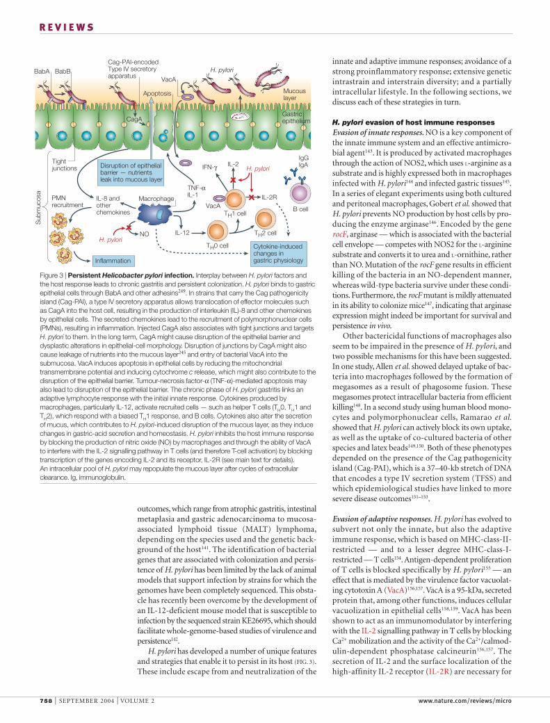
Persistent Helicobacter pylori infectionsDuring the past 20 years, H. pylori has emerged as animportant example of a persistent bacterial pathogen.Not only does this bacterium successfully colonize thehostile environment of the human stomach, but theinfection regularly persists for the lifetime of the host inthe face of a constant, vigorous innate and adaptiveimmune response. In most infected people, H. pyloriinfection causes superficial chronic gastritis, which isusually clinically asymptomatic, although histologi-cally apparent. However, a significant subset ofinfected individuals are at risk of the subsequentdevelopment of duodenal and peptic ulcers, and 1% ofthose that are infected will develop adenocarcinoma orlymphoma of the stomach137.
Most basic research in the Helicobacter field hasfocused on the study of bacterial virulence determinants(reviewed in REFS 6,138,139; TABLE 3), particularly in thecontext of their association with severe gastrointestinalsequelae of infection. Although the experimental focuson the association of H. pylori with disease is under-standable, it is important to point out that the ecologicalniche for H. pylori is progressively lost with the devel-opment of ATROPHIC GASTRITIS140, and bacteria can onlyrarely be cultured from seropositive patients with ade-nocarcinoma. So, from the microbial standpoint, theprogression of asymptomatic gastritis to more serioustissue destruction can be viewed as contrary to the bestinterests of the bacteria in terms of evolutionary success.Therefore, for the purpose of this review, we will shiftthe focus from the minority of colonized individualswho develop overt disease (~20%) to the majority whoremain relatively symptom-free and are likely to repre-sent the reservoir for human infection. We focus here onthe bacterial and host characteristics that enable H. pylorito colonize its host persistently in the face of a normalimmune response.
Most of the studies we will refer to here used animalmodels. Some H. pylori isolates establish long-terminfections in rodents or have been adapted to do so byrepeated passaging (reviewed in REF. 141). Indeed, thelimiting factor for the development of animal models ofpersistent H. pylori infection seems to be host specificityrather than lack of persistence. The models that havebeen used so far vary markedly with respect to disease
Once T cells are activated during an infection, theyproduce the macrophage-activating factor IFN-γ, whichhas a role in the acute Salmonella mouse model incontrolling the early phase of bacterial replication125–127.It was recently shown that IFN-γ has an important rolein maintaining and controlling the level of bacterialreplication in persistently infected animals, perhaps bystimulating infected macrophages to suppress bacterialreplication106. Furthermore, people who lack the IL-12receptor — in whom the T
H1 response and the pro-
duction of IFN-γ are defective — are more susceptibleto infections with Salmonella spp.128 In addition to IFN-γ and IL-12, TNF-α might also have a role in maintain-ing and controlling the level of bacterial replication inpersistently infected hosts. Similar to the results seenwith M. tuberculosis, patients who were treated withanti-TNF-α antibodies developed Salmonella septi-caemia129. HIV-positive individuals develop chronicbacteraemia caused by species of Salmonella that do notnormally pass beyond the MLNs in healthy individuals1
— further indicating that an intact adaptive immunesystem has a role in immunity to Salmonella.
Salmonella bacteria are probably not passivebystanders in terms of maintaining the balance betweenclearance and persistence. In this regard, Salmonellamight have an active role in modulating or even directlymanipulating host responses, thereby preventing clear-ance of intracellular bacteria. Many studies have shownthat S. typhimurium may limit the in vivo proliferationof CD4+ and CD8+ T cells, despite their activated phe-notype130. In addition, it has been shown that active S. typhimurium infection leads to immunosuppressionin mice and causes the production of large amounts ofIL-10, which has immunosuppressive activities, andnitric oxide (NO), which has both immunosuppressiveand direct antibacterial activities126,131–133. Indeed,S. typhimurium mutants have been identified that areunable to persist in mice, indicating that they lack
ATROPHIC GASTRITIS
Chronic inflammation of thestomach with degeneration ofthe mucosa.
Table 3 | Helicobacter pylori virulence determinants
Virulence Description/potential role in Referencesdeterminant pathogenesis
VacA 95-kDa secreted vacuolating toxin; induces 160–163,240apoptosis; involved in immunomodulation and colonization of mouse stomach
Cag-PAI 37-kb genomic fragment; contains 29 241genes that encode a type IV secretion apparatus
CagA 120-kD protein; translocated into host cell by 242–245type IV secretion apparatus encoded on Cag-PAI; phosporylated in host cell and binds SHP-2 tyrosine phosphatase; disrupts tightjunctions; epidemiologic link to cancer
BabA 78-kDa outer membrane protein; binds to 188,246fucosylated Lewis B blood group antigen;mediates adhesion to epithelial cells andpossibly stomach epithelium
Urease Resists acidic conditions in the stomach; activates 247innate immune responses during early stepsof infection
Flagella Involved in motility; essential for colonization 248
PAI, pathogenicity island.
758 | SEPTEMBER 2004 | VOLUME 2 www.nature.com/reviews/micro
R E V I E W S
innate and adaptive immune responses; avoidance of astrong proinflammatory response; extensive geneticintrastrain and interstrain diversity; and a partiallyintracellular lifestyle. In the following sections, wediscuss each of these strategies in turn.
H. pylori evasion of host immune responsesEvasion of innate responses. NO is a key component ofthe innate immune system and an effective antimicro-bial agent143. It is produced by activated macrophagesthrough the action of NOS2, which uses L-arginine as asubstrate and is highly expressed both in macrophagesinfected with H. pylori144 and infected gastric tissues145.In a series of elegant experiments using both culturedand peritoneal macrophages, Gobert et al. showed thatH. pylori prevents NO production by host cells by pro-ducing the enzyme arginase146. Encoded by the generocF, arginase — which is associated with the bacterialcell envelope — competes with NOS2 for the L-argininesubstrate and converts it to urea and L-ornithine, ratherthan NO. Mutation of the rocF gene results in efficientkilling of the bacteria in an NO-dependent manner,whereas wild-type bacteria survive under these condi-tions. Furthermore, the rocF mutant is mildly attenuatedin its ability to colonize mice147, indicating that arginaseexpression might indeed be important for survival andpersistence in vivo.
Other bactericidal functions of macrophages alsoseem to be impaired in the presence of H. pylori, andtwo possible mechanisms for this have been suggested.In one study, Allen et al. showed delayed uptake of bac-teria into macrophages followed by the formation ofmegasomes as a result of phagosome fusion. Thesemegasomes protect intracellular bacteria from efficientkilling148. In a second study using human blood mono-cytes and polymorphonuclear cells, Ramarao et al.showed that H. pylori can actively block its own uptake,as well as the uptake of co-cultured bacteria of otherspecies and latex beads149,150. Both of these phenotypesdepended on the presence of the Cag pathogenicityisland (Cag-PAI), which is a 37–40-kb stretch of DNAthat encodes a type IV secretion system (TFSS) andwhich epidemiological studies have linked to moresevere disease outcomes151–153.
Evasion of adaptive responses. H. pylori has evolved tosubvert not only the innate, but also the adaptiveimmune response, which is based on MHC-class-II-restricted — and to a lesser degree MHC-class-I-restricted — T cells154. Antigen-dependent proliferationof T cells is blocked specifically by H. pylori155 — aneffect that is mediated by the virulence factor vacuolat-ing cytotoxin A (VacA)156,157. VacA is a 95-kDa, secretedprotein that, among other functions, induces cellularvacuolization in epithelial cells158,159. VacA has beenshown to act as an immunomodulator by interferingwith the IL-2 signalling pathway in T cells by blockingCa2+ mobilization and the activity of the Ca2+/calmod-ulin-dependent phosphatase calcineurin156,157. Thesecretion of IL-2 and the surface localization of thehigh-affinity IL-2 receptor (IL-2R) are necessary for
outcomes, which range from atrophic gastritis, intestinalmetaplasia and gastric adenocarcinoma to mucosa-associated lymphoid tissue (MALT) lymphoma,depending on the species used and the genetic back-ground of the host141. The identification of bacterialgenes that are associated with colonization and persis-tence of H. pylori has been limited by the lack of animalmodels that support infection by strains for which thegenomes have been completely sequenced. This obsta-cle has recently been overcome by the development ofan IL-12-deficient mouse model that is susceptible toinfection by the sequenced strain KE26695, which shouldfacilitate whole-genome-based studies of virulence andpersistence142.
H. pylori has developed a number of unique featuresand strategies that enable it to persist in its host (FIG. 3).These include escape from and neutralization of the
Gastricepithelium
Mucouslayer
H. pylori
CagA
PMNrecruitment
Sub
muc
osa
TNF-αIL-1
IL-12
Macrophage
Apoptosis
TH0 cell
TH1 cell
TH2 cell
IFN-γ
Cytokine-inducedchanges ingastric physiologyInflammation
NO
BabA BabB
IL-2
IL-2R
IgGIgA
VacA
Cag-PAI-encodedType IV secretoryapparatus VacA
Tightjunctions
IL-8 andotherchemokines B cell
Disruption of epithelialbarrier — nutrients leak into mucous layer
H. pylori
H. pylori
Figure 3 | Persistent Helicobacter pylori infection. Interplay between H. pylori factors andthe host response leads to chronic gastritis and persistent colonization. H. pylori binds to gastricepithelial cells through BabA and other adhesins249. In strains that carry the Cag pathogenicityisland (Cag-PAI), a type IV secretory apparatus allows translocation of effector molecules suchas CagA into the host cell, resulting in the production of interleukin (IL)-8 and other chemokinesby epithelial cells. The secreted chemokines lead to the recruitment of polymorphonuclear cells(PMNs), resulting in inflammation. Injected CagA also associates with tight junctions and targets H. pylori to them. In the long term, CagA might cause disruption of the epithelial barrier anddysplastic alterations in epithelial-cell morphology. Disruption of junctions by CagA might alsocause leakage of nutrients into the mucous layer245 and entry of bacterial VacA into thesubmucosa. VacA induces apoptosis in epithelial cells by reducing the mitochondrialtransmembrane potential and inducing cytochrome c release, which might also contribute to thedisruption of the epithelial barrier. Tumour-necrosis factor-α (TNF-α)-mediated apoptosis mayalso lead to disruption of the epithelial barrier. The chronic phase of H. pylori gastritis links anadaptive lymphocyte response with the initial innate response. Cytokines produced bymacrophages, particularly IL-12, activate recruited cells — such as helper T cells (TH0, TH1 andTH2), which respond with a biased TH1 response, and B cells. Cytokines also alter the secretionof mucus, which contributes to H. pylori-induced disruption of the mucous layer, as they inducechanges in gastric-acid secretion and homeostasis. H. pylori inhibits the host immune responseby blocking the production of nitric oxide (NO) by macrophages and through the ability of VacAto interfere with the IL-2 signalling pathway in T cells (and therefore T-cell activation) by blockingtranscription of the genes encoding IL-2 and its receptor, IL-2R (see main text for details). An intracellular pool of H. pylori may repopulate the mucous layer after cycles of extracellularclearance. Ig, immunoglobulin.
NATURE REVIEWS | MICROBIOLOGY VOLUME 2 | SEPTEMBER 2004 | 759
R E V I E W S
The effect of H. pylori LPS on gastric epithelial cells— the other main source of proinflammatory cytokinesin the stomach besides macrophages — is still unclear.One study has shown that H. pylori LPS produced bystrains harbouring the Cag-PAI, but not from Cag-PAImutants, activates Toll-like receptor 4 (TLR4) on gas-tric-pit cells, thereby stimulating the innate immuneresponses of the gastric mucosa168. By contrast, Smith etal. reported that TLR2 mediates the proinflammatoryeffects of H. pylori on epithelial cells and show that H. pylori LPS is a TLR2 agonist169. These authors sug-gest that the low level of pathogenicity of H. pylori LPSdoes indeed result from its failure to activate TLR4, thereceptor that mediates LPS signalling of most otherGram-negative species170.
A recent study has reported similar findings for H. pylori flagellins. The two H. pylori flagellins — FlaAand FlaB — were shown to have a markedly reducedpotential to activate TLR5 compared with flagellinsof other Gram-negative bacteria, such as FliC ofS. typhimurium171. The evolution of these unique fla-gellins, which share extensive amino-acid homologywith flagellins from other species that do stimulateTLR5, has been proposed to preserve the essential func-tion of the flagella during chronic colonization whileavoiding the activation of the innate immune system171.
In a recent study investigating a link between theCag-PAI and the induction of proinflammatoryresponses, Philpott et al. showed that clinical isolates ofH. pylori were more likely to colonize mice if they didnor harbour the Cag-PAI and were therefore unable toinduce such responses in cultured cells172. Mouse-adapted variants that lacked the Cag-PAI infected miceat higher levels and had a reduced capacity to induceinflammatory responses in vitro compared with therespective parental strains. Taken together, these find-ings imply that there may be a profound in vivo selec-tion against H. pylori strains and variants that induce astrong host inflammatory response172, at least in themouse model.
The relative in vivo advantages for H. pylori withCag+ versus Cag– phenotypes in human hosts areunclear173,174. One study of an individual infected withmultiple strains showed recombination between strainsresulting in excision of the Cag-PAI and subsequentpositive selection of a Cag– strain175. It is thereforeunclear as to how the Cag-PAI benefits the bacteria dur-ing a persistent infection; it may be that it is importantfor a specific stage of colonization, but is dispensable atother stages.
Genetic diversity in H. pyloriExtensive recombination and a PANMICTIC overall popu-lation structure result in substantial genetic diversityamong H. pylori strains176–179. This has been proposedto allow the bacteria to adapt rapidly to changing con-ditions in their current host as well as to facilitate thecolonization of new hosts180–182. Although H. pyloripopulations in individuals and even families seem to beclonal, as determined by conventional procedures —such as RANDOMLY AMPLIFIED POLYMORPHIC DNA (RAPD) PCR and
efficient T-cell proliferation and activation. In normalT cells, calcineurin dephosphorylates the transcriptionfactor NFAT (for nuclear factor of activated T cells),which then translocates into the nucleus and activatesthe transcription of several genes that are involved in theimmune response. Among these are the genes encodingIL-2 and IL-2Rα. In T cells that are infected with VacA+
H. pylori, however, nuclear translocation of NFAT isblocked, as dephosphorylation is prevented and thedownstream genes are not expressed.
Another possible function of VacA in subverting theadaptive immune response is its ability to interfere withantigen presentation mediated by MHC class II160. Afterit inserts into the plasma membrane, VacA is internal-ized by endocytosis and reaches the late-endosomalcompartment. This compartment is then convertedinto large acidic vacuoles by the anion-selective chan-nel activity of VacA161,162. In antigen-presenting cells,the processing of proteins into peptide epitopes,which takes place in the endocytic compartment, isgreatly reduced owing to VacA activity, indicating thatantigen presentation is abrogated in these cells160.
The importance of VacA in establishing an infectionhas been corroborated by experiments in mice, whichhave shown that a null mutation of vacA compromisesthe ability of H. pylori to colonize the murine stomachin the presence of the corresponding parental strain163.However, the precise effect of VacA on cellular andepithelial physiology that facilitates H. pylori colonizationin the murine stomach is unknown.
H. pylori suppresses inflammatory responsesSeveral lines of evidence indicate that, to allow long-term colonization, there has been selective pressure onH. pylori to avoid triggering an intense inflammatoryreaction164. It has been proposed that high levels ofinflammation may lead to loss of gastric glandularstructure and function165 and that H. pylori disappearsfrom stomachs that have developed atrophic gastritis140.Furthermore, an increased inflammatory reaction —as seen in IL-10-knockout mice — is associated withclearance of the bacteria from the stomach within 8days of the infection166. Similarly, increased inflamma-tion due to deletion of the gene encoding PHOXresults in a marked reduction in bacterial numbers167.All of these findings indicate that, at least in animalmodels, a strong inflammatory reaction is necessary forthe elimination of H. pylori and seems to be activelyrepressed by this bacterium.
But how does H. pylori modulate the host inflam-matory response? Bacterial lipopolysaccharide (LPS)is the main mediator of inflammation during infec-tions with most Gram-negative bacteria because itactivates phagocytic cells, endothelial and epithelialcells and lymphocytes. H. pylori LPS, however, hasvery low biological activity when compared withEscherichia coli LPS, at least as measured by its abilityto activate macrophages165. In fact, the minimum con-centration of purified LPS that is required to achievesimilar responses was several thousand times greaterfor H. pylori compared with E. coli LPS165.
PANMIXIS
Mating without regard to thegenetic constitution of the mate.
760 | SEPTEMBER 2004 | VOLUME 2 www.nature.com/reviews/micro
R E V I E W S
The high degree of diversity that is seen in H. pyloriis probably facilitated by its natural competence forDNA transformation. In contrast to other bacteria,natural competence in H. pylori is not mediated by typeIV pili or type IV pilin-like proteins, but by a TFSS thatis encoded by the comB operon193,194. The ComB8,ComB9 and ComB10 proteins correspond to theAgrobacterium tumefaciens VirB8, VirB9 and VirB10proteins and constitute the basic components of aTFSS193. H. pylori therefore possesses two functionallyindependent TFSSs — one for protein secretion andtranslocation into the host cell that is encoded by theCag-PAI, and one for DNA uptake. In addition, a thirdcluster of type IV secretion genes was recently discov-ered that seems to be present in only a subset ofstrains195. The functional significance of this cluster isunknown at present.
The identification of CRYPTIC PLASMIDS in approximatelyhalf of all H. pylori strains196 has given rise to speculationthat conjugative transfer of novel sequences carried onplasmids could be another means of horizontal genetransfer — and therefore strain diversity — in H. pylori.In support of this hypothesis, Hofreuter et al. haverecently presented evidence that some of these plasmidscontain hot spots for site-specific recombination,encode elements from CHROMOSOMAL PLASTICITY ZONES andmight be mobilizable197. This indicates that exchangeof genetic material between these plasmids and thechromosome can occur, and that these plasmids mightbe mobilized and spread rapidly in the population. So,genetic diversity, which in turn leads to antigenic diver-sity, might be an important strategy used by H. pylori toevade immune surveillance.
Repopulation of the stomach by H. pyloriThe gastric mucosa is normally well-protected againstbacterial colonization due to the acidic pH of thelumen, the production of mucus and rapid epithelial-cell turnover. It is therefore tempting to speculate thatthe adoption of a partially intracellular lifestyle bysome members of the bacterial population mightallow H. pylori to achieve long-term persistence.Although there is little evidence to indicate that thebacterium is predominantly an intracellular pathogen,numerous experimental and clinical observations ofbiopsy specimens support the notion that a subpopu-lation of H. pylori is able to invade epithelial cells bothin vitro198,199 and in vivo200–202.
Using time-lapse video microscopy and gentamicin-protection assays in a cell-culture system, Amieva et al.have provided evidence that H. pylori residing in multi-vesicular bodies survive in the intracellular milieu forat least 24 hours, remain motile and retain the abilityto emerge from the cells to repopulate the extracellularspace199. Up to several dozen bacteria can be found inone vesicle, indicating that intracellular replication doesoccur, at least sporadically. These findings indicate thatthe intracellular niche can potentially function as a‘hideout’ and sustain the renewal of the populationunder the unfavourable conditions that are found in themammalian stomach. Nevertheless, the vast majority of
DNA sequence analysis179 — more sensitive approacheshave indicated that extensive changes can and do occurin a single host over time183. For example, fortuitously,additional H. pylori isolates were obtained from onepatient six years after an isolate of the sequenced strainJ99 was first obtained from this individual. These newclinical isolates were subjected to extensive molecularcomparisons with the original J99 strain. RAPD PCRand sequencing of several unlinked loci indicated thatthese isolates were undoubtedly related to the originalstrain; however, microarray analysis showed differencesin genetic content that reflected both acquisitions andlosses of genomic DNA183. Approximately 3% of J99 locishowed variation between isolates obtained from thisindividual, compared with 22% of loci when isolatesfrom different individuals were compared184. Althoughmost of the open reading frames that were affectedrepresented ‘hypothetical genes’, one was predicted toencode a protein belonging to the TraG family, othermembers of which have been shown to be involved ingenetic transfer185,186. A putative similar function ofTraG implies that acquisition of this gene might con-fer an evolutionary advantage by providing anothermechanism through which DNA exchange could occur.
In a recent similar study, three output strains froman experimental infection of Rhesus macaques werecompared with the strain that was used for inocula-tion187. All three of the output strains had lost expres-sion of the babA gene, which encodes an adhesin thatbinds to the LEWIS BLOOD GROUP Leb antigen188. Loss ofbabA expression occurred by two different mecha-nisms in different isolates. In some cases, babA wasreplaced by the closely related babB gene, leading toloss of babA and duplication of babB. In others, achange in the number of CT repeats in the 5’ codingregion of babA resulted in a frameshift and subsequentloss of Leb adhesion. H. pylori therefore uses both anti-genic variation and phase variation to regulate babAexpression (and possibly the expression of other outer-membrane proteins) in vivo; however, the significanceof this observation in the context of the host immuneresponse remains to be shown.
Another H. pylori structure that undergoes phasevariation is its LPS, more specifically the Lewis-blood-group determinant of the LPS O-antigen. The on/offstatuses of at least five glycosyltransferase genes deter-mine which LPS phase variant is expressed (reviewed inREF. 189). It was initially assumed that mimicry of theLewis blood group antigens of the host by H. pyloriwould provide a mechanism of immune evasion andadaptation to the host, a concept that was supported byreports describing a link between the Lewis-blood-group phenotype of isolates with the phenotype of theindividuals they were derived from (reviewed in REF. 190).However, this concept has been challenged by theisolation of both LeX- and LeY-expressing strains fromthe same host and the fact that even high levels of anti-LPS antibodies do not eradicate the organism. It isnow assumed that the production of Lewis antigensby H. pylori facilitates the colonization of the host bymediating adhesion to gastric epithelial cells191,192.
RANDOMLY AMPLIFIED
POLYMORPHIC DNA (RAPD) PCR
A molecular technique used forthe classification andcomparison of different isolatesof the same species. This methoduses a randomly chosenoligonucleotide to prime DNAsynthesis and results in strain-specific patterns of DNAproducts.
LEWIS BLOOD GROUP
Antigens of red blood cells,saliva and other body fluids thatare specified by the Le gene andreact with the antibodiesdesignated anti-Lea and anti-Leb.
CRYPTIC PLASMIDS
Small, mobilizable geneticelements that can encodevirulence factors.
CHROMOSOMAL-PLASTICITY
ZONES
Segments of the chromosomethat are characterized by theirdifferent G+C content, which isa hallmark of horizontallyacquired sequences.
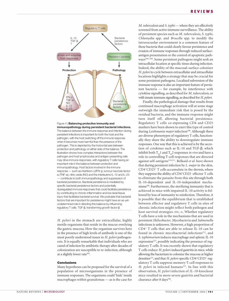
NATURE REVIEWS | MICROBIOLOGY VOLUME 2 | SEPTEMBER 2004 | 761
R E V I E W S
M. tuberculosis and S. typhi — where they are effectivelyscreened from active immune surveillance. The abilityof persistent species such as M. tuberculosis, S. typhi,Chlamydia spp. and Brucella spp. to modify theintravacuolar environment is a common feature ofthese bacteria that could clearly favour persistence andevasion of immune responses through reduced surface-antigen presentation or the control of apoptotic path-ways204–208. Some persistent pathogens might seek anintracellular location at specific times during infection.Indeed, the ability of the mucosal-surface colonizer H. pylori to cycle between extracellular and intracellularlocations highlights a strategy that may be crucial forsome persistent pathogens. Localized subversion of theimmune response is also an important feature of persis-tent bacteria — for example, by interference withcytokine signalling, as described for M. tuberculosis, orwith innate immune signalling, as described for H. pylori.
Finally, the pathological damage that results fromcontinued macrophage activation will at some stageoutweigh the immediate risk that is posed by theresidual bacteria, and the immune response mightturn itself off, allowing bacterial persistence.Regulatory T cells co-expressing CD4 and CD25markers have been shown to exert this type of controlduring Leishmania major infection209. Although thereare diverse phenotypes of regulatory T cells, function-ally they share the ability to downregulate immuneresponses. One way that this is achieved is by the secre-tion of cytokines such as IL-10 and TGF-β, whichinhibit both T
H1 and T
H2 responses in vivo and have a
role in controlling T-cell responses that are directedagainst self-antigens210,211. Belkaid et al. have shownthat during persistent infection by L. major in the skin,CD4+CD25+ T cells accumulate in the dermis, wherethey suppress the ability of CD4+CD25– effector T cellsto eliminate the parasite from this site through bothIL-10-dependent and IL-10-independent mecha-nisms209. Furthermore, the sterilizing immunity that isachieved in mice with impaired IL-10 activity is fol-lowed by loss of immunity to reinfection. Therefore, itis possible that the equilibrium that is establishedbetween effector and regulatory T cells in sites ofchronic infection might reflect both pathogen andhost survival strategies (FIG. 4). Whether regulatory T cells have a role in the mechanisms that are used inpersistent Helicobacter, Mycobacteria and Salmonellainfections is unknown. However, a high proportion ofCD4+ T cells that are able to release IL-10 can befound in chronic mycobacterial infections212, and S. typhimurium induces macrophage and splenic IL-10expression213, possibly indicating the presence of reg-ulatory T cells. It was recently shown that regulatoryT cells reduce H. pylori-induced gastritis in mice, whileallowing the bacterium to colonize the mucosa at higherdensities214, and that H. pylori-specific CD4+CD25+ reg-ulatory T cells suppress memory T-cell responses toH. pylori in infected humans215. In line with thisobservation, H. pylori infection of IL-10-knockoutmice resulted in more severe gastritis and bacterialclearance after 8 days166.
H. pylori in the stomach are extracellular, highlymotile organisms that reside in the mucus overlyingthe gastric mucosa. How the organism survives herein the presence of high levels of antibody is one of themost poorly understood issues in H. pylori pathogen-esis. It is equally remarkable that individuals who arecured of infection by antibiotic therapy after decades ofcolonization are susceptible to re-infection, althoughat a slightly lower rate203.
ConclusionsMany hypotheses can be proposed for the survival of apopulation of microorganisms in the presence ofimmune responses. The organisms could ‘hide’ insidemacrophages within granulomas — as is the case for
IFN-γTNF-αNOIL-12IL-23
Regulatory T cells
Innate and adaptivehost responses Persistent bacteria
PathologyProtection
IL-10TGF-β?
Bacterial persistencefactors
Bacterial persistencefactors
Figure 4 | Balancing protective immunity andimmunopathology during persistent bacterial infections.The balance between the immune response and infection duringpersistent infections is important for both the host and thepathogen, with the host switching off the immune responsewhen it becomes more harmful than the presence of thepathogen. This is depicted by the horizontal axis betweenprotection and pathology on either side of the balance. Theillustration shows how complex interactions between thepathogen and host lymphocytes and antigen-presenting cellsmay drive immune responses, with regulatory T cells having animportant role in the balance between protection andimmunopathology. Host factors involved in the immuneresponse — such as interferon-γ (IFN-γ), tumour-necrosis factor-α (TNF-α), nitric oxide (NO) and the interleukins IL-12 and IL-23— contribute to both immunopathology and suppression ofbacterial persistence. Bacterial persistence is mediated byspecific bacterial persistence factors and potentiallydysregulated immune responses that could facilitate persistenceby contributing to chronic inflammation and low-level tissueinjury that facilitates bacterial survival. We postulate that bacterialfactors that are important for persistence might have an as-yet-undetermined role in directing this balance by influencing regulatory T cells. TGF-β, transforming growth factor-β.
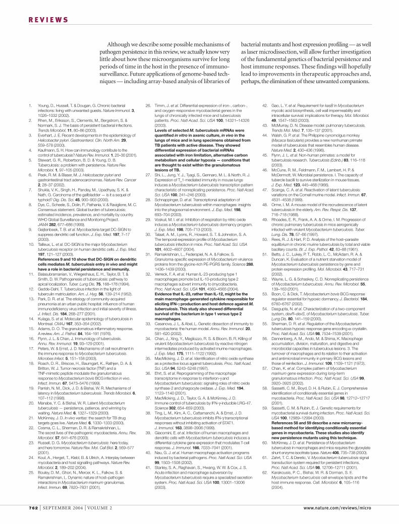
762 | SEPTEMBER 2004 | VOLUME 2 www.nature.com/reviews/micro
R E V I E W S
bacterial mutants and host expression profiling — as wellas laser microdissection, will allow further investigationof the fundamental genetics of bacterial persistence andhost immune responses. These findings will hopefullylead to improvements in therapeutic approaches and,perhaps, the elimination of these unwanted companions.
Although we describe some possible mechanisms ofpathogen persistence in this review,we actually know verylittle about how these microorganisms survive for longperiods of time in the host in the presence of immuno-surveillance. Future applications of genome-based tech-niques — including array-based analysis of libraries of
1. Young, D., Hussell, T. & Dougan, G. Chronic bacterialinfections: living with unwanted guests. Nature Immunol. 3,1026–1032 (2002).
2. Rhen, M., Eriksson, S., Clements, M., Bergstrom, S. &Normark, S. J. The basis of persistent bacterial infections.Trends Microbiol. 11, 80–86 (2003).
3. Everhart, J. E. Recent developments in the epidemiology ofHelicobacter pylori. Gastroenterol. Clin. North Am. 29,559–578 (2000).
4. Kaufmann, S. H. How can immunology contribute to thecontrol of tuberculosis? Nature Rev. Immunol. 1, 20–30 (2001).
5. Stewart, G. R., Robertson, B. D. & Young, D. B.Tuberculosis: a problem with persistence. Nature Rev.Microbiol. 1, 97–105 (2003).
6. Peek, R. M. & Blaser, M. J. Helicobacter pylori andgastrointestinal tract adenocarcinomas. Nature Rev. Cancer2, 28–37 (2002).
7. Shukla, V. K., Singh, H., Pandey, M., Upadhyay, S. K. &Nath, G. Carcinoma of the gallbladder — is it a sequel oftyphoid? Dig. Dis. Sci. 45, 900–903 (2000).
8. Dye, C., Scheele, S., Dolin, P., Pathania, V. & Raviglione, M. C.Consensus statement. Global burden of tuberculosis:estimated incidence, prevalence, and mortality by country.WHO Global Surveillance and Monitoring Project. JAMA 282, 677–686 (1999).
9. Geijtenbeek, T. B. et al. Mycobacteria target DC-SIGN tosuppress dendritic cell function. J. Exp. Med. 197, 7–17(2003).
10. Tailleux, L. et al. DC-SIGN is the major Mycobacteriumtuberculosis receptor on human dendritic cells. J. Exp. Med.197, 121–127 (2003).References 9 and 10 show that DC-SIGN on dendriticcells mediates M. tuberculosis entry in vivo and mighthave a role in bacterial persistence and immunity.
11. Balasubramanian, V., Wiegeshaus, E. H., Taylor, B. T. &Smith, D. W. Pathogenesis of tuberculosis: pathway toapical localization. Tuber. Lung Dis. 75, 168–178 (1994).
12. Gedde-Dahl, T. Tuberculous infection in the light oftuberculin matriculation. Am. J. Hyg. 56, 139–214 (1952).
13. Park, D. R. et al. The etiology of community-acquiredpneumonia at an urban public hospital: influence of humanimmunodeficiency virus infection and initial severity of illness.J. Infect. Dis. 184, 268–277 (2001).
14. Kulaga, S. et al. Molecular epidemiology of tuberculosis inMontreal. CMAJ 167, 353–354 (2002).
15. Adams, D. O. The granulomatous inflammatory response. A review. Am. J. Pathol. 84, 164–191 (1976).
16. Flynn, J. L. & Chan, J. Immunology of tuberculosis. Annu. Rev. Immunol. 19, 93–129 (2001).
17. Peters, W. & Ernst, J. D. Mechanisms of cell recruitment inthe immune response to Mycobacterium tuberculosis.Microbes Infect. 5, 151–158 (2003).
18. Roach, D. R., Briscoe, H., Baumgart, K., Rathjen, D. A. &Britton, W. J. Tumor necrosis factor (TNF) and a TNF-mimetic peptide modulate the granulomatousresponse to Mycobacterium bovis BCG infection in vivo.Infect. Immun. 67, 5473–5476 (1999).
19. Parrish, N. M., Dick, J. D. & Bishai, W. R. Mechanisms oflatency in Mycobacterium tuberculosis. Trends Microbiol. 6,107–112 (1998).
20. Manabe, Y. C. & Bishai, W. R. Latent Mycobacteriumtuberculosis — persistence, patience, and winning bywaiting. Nature Med. 6, 1327–1329 (2000).
21. McKinney, J. D. In vivo veritas: the search for TB drugtargets goes live. Nature Med. 6, 1330–1333 (2000).
22. Cosma, C. L., Sherman, D. R. & Ramakrishnan, L. The secret lives of the pathogenic mycobacteria. Annu. Rev.Microbiol. 57, 641–676 (2003).
23. Russell, D. G. Mycobacterium tuberculosis: here today, and here tomorrow. Nature Rev. Mol. Cell Biol. 2, 569–577(2001).
24. Koul, A., Herget, T., Klebl, B. & Ullrich, A. Interplay betweenmycobacteria and host signalling pathways. Nature Rev.Microbiol. 2, 189–202 (2004).
25. Bouley, D. M., Ghori, N., Mercer, K. L., Falkow, S. &Ramakrishnan, L. Dynamic nature of host–pathogeninteractions in Mycobacterium marinum granulomas. Infect. Immun. 69, 7820–7831 (2001).
26. Timm, J. et al. Differential expression of iron-, carbon-,and oxygen-responsive mycobacterial genes in the lungs of chronically infected mice and tuberculosispatients. Proc. Natl Acad. Sci. USA 100, 14321–14326 (2003).Levels of selected M. tuberculosis mRNAs werequantified in vitro in axenic culture, in vivo in thelungs of mice and in lung specimens obtained fromTB patients with active disease. They showeddifferential expression of bacterial mRNAsassociated with iron limitation, alternative carbonmetabolism and cellular hypoxia — conditions thatare thought to exist within the granulomatouslesions of TB.
27. Shi, L., Jung, Y. J., Tyagi, S., Gennaro, M. L. & North, R. J.Expression of TH1-mediated immunity in mouse lungsinduces a Mycobacterium tuberculosis transcription patterncharacteristic of nonreplicating persistence. Proc. Natl Acad.Sci. USA 100, 241–246 (2003).
28. Schnappinger, D. et al. Transcriptional adaptation ofMycobacterium tuberculosis within macrophages: insightsinto the phagosomal environment. J. Exp. Med. 198,693–704 (2003).
29. Voskuil, M. I. et al. Inhibition of respiration by nitric oxideinduces a Mycobacterium tuberculosis dormancy program.J. Exp. Med. 198, 705–713 (2003).
30. Talaat, A. M., Lyons, R., Howard, S. T. & Johnston, S. A. The temporal expression profile of Mycobacteriumtuberculosis infection in mice. Proc. Natl Acad. Sci. USA101, 4602–4607 (2004).
31. Ramakrishnan, L., Federspiel, N. A. & Falkow, S.Granuloma-specific expression of Mycobacterium virulenceproteins from the glycine-rich PE-PGRS family. Science 288,1436–1439 (2000).
32. Verreck, F. A. et al. Human IL-23-producing type 1macrophages promote but IL-10-producing type 2macrophages subvert immunity to (myco)bacteria. Proc. Natl Acad. Sci. USA 101, 4560–4565 (2004).Evidence that IL-23, rather than IL-12, might be themain macrophage-generated cytokine responsible foreliciting IFN-γ production and host defence against M.tuberculosis. This study also showed differentialsurvival of the bacterium in type 1 versus type 2macrophages.
33. Casanova, J. L. & Abel, L. Genetic dissection of immunity tomycobacteria: the human model. Annu. Rev. Immunol. 20,581–620 (2002).
34. Chan, J., Xing, Y., Magliozzo, R. S. & Bloom, B. R. Killing ofvirulent Mycobacterium tuberculosis by reactive nitrogenintermediates produced by activated murine macrophages.J. Exp. Med. 175, 1111–1122 (1992).
35. MacMicking, J. D. et al. Identification of nitric oxide synthaseas a protective locus against tuberculosis. Proc. Natl Acad.Sci. USA 94, 5243–5248 (1997).
36. Ehrt, S. et al. Reprogramming of the macrophagetranscriptome in response to interferon-γ andMycobacterium tuberculosis: signaling roles of nitric oxidesynthase-2 and phagocyte oxidase. J. Exp. Med. 194,1123–1140 (2001).
37. MacMicking, J. D., Taylor, G. A. & McKinney, J. D. Immune control of tuberculosis by IFN-γ-inducible LRG-47.Science 302, 654–659 (2003).
38. Ting, L. M., Kim, A. C., Cattamanchi, A. & Ernst, J. D.Mycobacterium tuberculosis inhibits IFN-γ transcriptionalresponses without inhibiting activation of STAT1. J. Immunol. 163, 3898–3906 (1999).
39. Giacomini, E. et al. Infection of human macrophages anddendritic cells with Mycobacterium tuberculosis induces adifferential cytokine gene expression that modulates T-cellresponse. J. Immunol. 166, 7033–7041 (2001).
40. Nau, G. J. et al. Human macrophage activation programsinduced by bacterial pathogens. Proc. Natl Acad. Sci. USA99, 1503–1508 (2002).
41. Stanley, S. A., Raghavan, S., Hwang, W. W. & Cox, J. S.Acute infection and macrophage subversion byMycobacterium tuberculosis require a specialized secretionsystem. Proc. Natl Acad. Sci. USA 100, 13001–13006(2003).
42. Gao, L. Y. et al. Requirement for kasB in Mycobacteriummycolic acid biosynthesis, cell wall impermeability andintracellular survival: implications for therapy. Mol. Microbiol.49, 1547–1563 (2003).
43. McMurray, D. N. Disease model: pulmonary tuberculosis.Trends Mol. Med. 7, 135–137 (2001).
44. Walsh, G. P. et al. The Philippine cynomolgus monkey(Macaca fasicularis) provides a new nonhuman primatemodel of tuberculosis that resembles human disease.Nature Med. 2, 430–436 (1996).
45. Flynn, J. L. et al. Non-human primates: a model fortuberculosis research. Tuberculosis (Edinb.) 83, 116–118(2003).
46. McCune, R. M., Feldmann, F. M., Lambert, H. P. &McDermott, W. Microbial persistence. I. The capacity oftubercle bacilli to survive sterilization in mouse tissues. J. Exp. Med. 123, 445–468 (1966).
47. Scanga, C. A. et al. Reactivation of latent tuberculosis:variations on the Cornell murine model. Infect. Immun. 67,4531–4538 (1999).
48. Orme, I. M. A mouse model of the recrudescence of latenttuberculosis in the elderly. Am. Rev. Respir. Dis. 137,716–718 (1988).
49. Rhoades, E. R., Frank, A. A. & Orme, I. M. Progression ofchronic pulmonary tuberculosis in mice aerogenicallyinfected with virulent Mycobacterium tuberculosis. Tuber.Lung. Dis. 78, 57–66 (1997).
50. Rees, R. J. & Hart, P. D. Analysis of the host–parasiteequilibrium in chronic murine tuberculosis by total and viablebacillary counts. Br. J. Exp. Pathol. 42, 83–88 (1961).
51. Betts, J. C., Lukey, P. T., Robb, L. C., McAdam, R. A. &Duncan, K. Evaluation of a nutrient starvation model ofMycobacterium tuberculosis persistence by gene andprotein expression profiling. Mol. Microbiol. 43, 717–731(2002).
52. Wayne, L. G. & Sohaskey, C. D. Nonreplicating persistenceof Mycobacterium tuberculosis. Annu. Rev. Microbiol. 55,139–163 (2001).
53. Boon, C. & Dick, T. Mycobacterium bovis BCG responseregulator essential for hypoxic dormancy. J. Bacteriol. 184,6760–6767 (2002).
54. Dasgupta, N. et al. Characterization of a two-componentsystem, devR–devS, of Mycobacterium tuberculosis. Tuber.Lung Dis. 80, 141–159 (2000).
55. Sherman, D. R. et al. Regulation of the Mycobacteriumtuberculosis hypoxic response gene encoding α-crystallin.Proc. Natl Acad. Sci. USA 98, 7534–7539 (2001).
56. Dannenberg, A. M., Ando, M. & Shima, K. Macrophageaccumulation, division, maturation, and digestive andmicrobicidal capacities in tuberculous lesions. 3. Theturnover of macrophages and its relation to their activationand antimicrobial immunity in primary BCG lesions andthose of reinfection. J. Immunol. 109, 1109–1121 (1972).
57. Chan, K. et al. Complex pattern of Mycobacteriummarinum gene expression during long-termgranulomatous infection. Proc. Natl Acad. Sci. USA 99,3920–3925 (2002).
58. Sassetti, C. M., Boyd, D. H. & Rubin, E. J. Comprehensiveidentification of conditionally essential genes inmycobacteria. Proc. Natl Acad. Sci. USA 98, 12712–12717(2001).
59. Sassetti, C. M. & Rubin, E. J. Genetic requirements formycobacterial survival during infection. Proc. Natl Acad. Sci.USA 100, 12989–12994 (2003).References 58 and 59 describe a new microarray-based method for identifying conditionally essentialgenes in mycobacteria. These studies also identifynew persistence mutants using this technique.
60. McKinney, J. D. et al. Persistence of Mycobacteriumtuberculosis in macrophages and mice requires the glyoxylateshunt enzyme isocitrate lyase. Nature 406, 735–738 (2000).
61. Zahrt, T. C. & Deretic, V. Mycobacterium tuberculosis signaltransduction system required for persistent infections. Proc. Natl Acad. Sci. USA 98, 12706–12711 (2001).
62. Karakousis, P. C., Bishai, W. R. & Dorman, S. E.Mycobacterium tuberculosis cell envelope lipids and thehost immune response. Cell. Microbiol. 6, 105–116(2004).
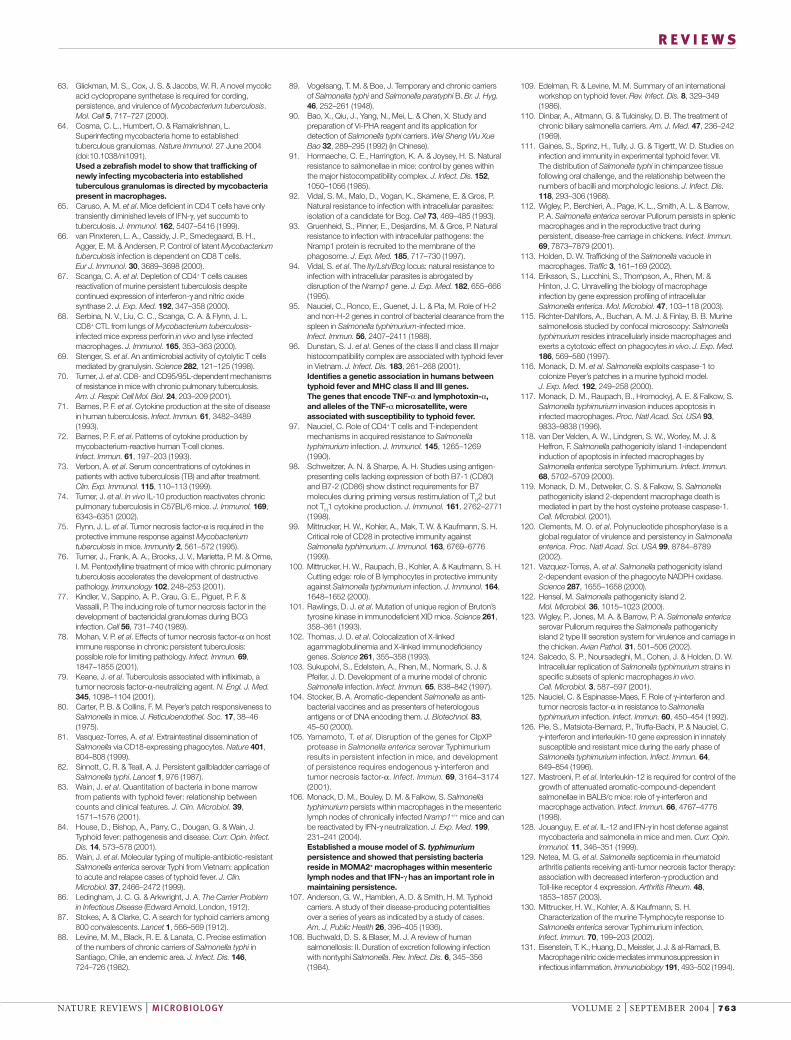
NATURE REVIEWS | MICROBIOLOGY VOLUME 2 | SEPTEMBER 2004 | 763
R E V I E W S
63. Glickman, M. S., Cox, J. S. & Jacobs, W. R. A novel mycolicacid cyclopropane synthetase is required for cording,persistence, and virulence of Mycobacterium tuberculosis.Mol. Cell 5, 717–727 (2000).
64. Cosma, C. L., Humbert, O. & Ramakrishnan, L.Superinfecting mycobacteria home to establishedtuberculous granulomas. Nature Immunol. 27 June 2004(doi:10.1038/ni1091).Used a zebrafish model to show that trafficking ofnewly infecting mycobacteria into establishedtuberculous granulomas is directed by mycobacteriapresent in macrophages.
65. Caruso, A. M. et al. Mice deficient in CD4 T cells have onlytransiently diminished levels of IFN-γ, yet succumb totuberculosis. J. Immunol. 162, 5407–5416 (1999).
66. van Pinxteren, L. A., Cassidy, J. P., Smedegaard, B. H.,Agger, E. M. & Andersen, P. Control of latent Mycobacteriumtuberculosis infection is dependent on CD8 T cells. Eur J. Immunol. 30, 3689–3698 (2000).
67. Scanga, C. A. et al. Depletion of CD4+ T cells causesreactivation of murine persistent tuberculosis despitecontinued expression of interferon-γ and nitric oxidesynthase 2. J. Exp. Med. 192, 347–358 (2000).
68. Serbina, N. V., Liu, C. C., Scanga, C. A. & Flynn, J. L. CD8+ CTL from lungs of Mycobacterium tuberculosis-infected mice express perforin in vivo and lyse infectedmacrophages. J. Immunol. 165, 353–363 (2000).
69. Stenger, S. et al. An antimicrobial activity of cytolytic T cellsmediated by granulysin. Science 282, 121–125 (1998).
70. Turner, J. et al. CD8- and CD95/95L-dependent mechanismsof resistance in mice with chronic pulmonary tuberculosis.Am. J. Respir. Cell Mol. Biol. 24, 203–209 (2001).
71. Barnes, P. F. et al. Cytokine production at the site of diseasein human tuberculosis. Infect. Immun. 61, 3482–3489(1993).
72. Barnes, P. F. et al. Patterns of cytokine production bymycobacterium-reactive human T-cell clones. Infect. Immun. 61, 197–203 (1993).
73. Verbon, A. et al. Serum concentrations of cytokines inpatients with active tuberculosis (TB) and after treatment.Clin. Exp. Immunol. 115, 110–113 (1999).
74. Turner, J. et al. In vivo IL-10 production reactivates chronicpulmonary tuberculosis in C57BL/6 mice. J. Immunol. 169,6343–6351 (2002).
75. Flynn, J. L. et al. Tumor necrosis factor-α is required in theprotective immune response against Mycobacteriumtuberculosis in mice. Immunity 2, 561–572 (1995).
76. Turner, J., Frank, A. A., Brooks, J. V., Marietta, P. M. & Orme,I. M. Pentoxifylline treatment of mice with chronic pulmonarytuberculosis accelerates the development of destructivepathology. Immunology 102, 248–253 (2001).
77. Kindler, V., Sappino, A. P., Grau, G. E., Piguet, P. F. &Vassalli, P. The inducing role of tumor necrosis factor in thedevelopment of bactericidal granulomas during BCGinfection. Cell 56, 731–740 (1989).
78. Mohan, V. P. et al. Effects of tumor necrosis factor-α on hostimmune response in chronic persistent tuberculosis:possible role for limiting pathology. Infect. Immun. 69,1847–1855 (2001).
79. Keane, J. et al. Tuberculosis associated with infliximab, atumor necrosis factor-α-neutralizing agent. N. Engl. J. Med.345, 1098–1104 (2001).
80. Carter, P. B. & Collins, F. M. Peyer’s patch responsiveness toSalmonella in mice. J. Reticuloendothel. Soc. 17, 38–46(1975).
81. Vasquez-Torres, A. et al. Extraintestinal dissemination ofSalmonella via CD18-expressing phagocytes. Nature 401,804–808 (1999).
82. Sinnott, C. R. & Teall, A. J. Persistent gallbladder carriage ofSalmonella typhi. Lancet 1, 976 (1987).
83. Wain, J. et al. Quantitation of bacteria in bone marrowfrom patients with typhoid fever: relationship betweencounts and clinical features. J. Clin. Microbiol. 39,1571–1576 (2001).
84. House, D., Bishop, A., Parry, C., Dougan, G. & Wain, J.Typhoid fever: pathogenesis and disease. Curr. Opin. Infect.Dis. 14, 573–578 (2001).
85. Wain, J. et al. Molecular typing of multiple-antibiotic-resistantSalmonella enterica serovar Typhi from Vietnam: applicationto acute and relapse cases of typhoid fever. J. Clin.Microbiol. 37, 2466–2472 (1999).
86. Ledingham, J. C. G. & Arkwright, J. A. The Carrier Problemin Infectious Disease (Edward Arnold, London, 1912).
87. Stokes, A. & Clarke, C. A search for typhoid carriers among800 convalescents. Lancet 1, 566–569 (1912).
88. Levine, M. M., Black, R. E. & Lanata, C. Precise estimationof the numbers of chronic carriers of Salmonella typhi inSantiago, Chile, an endemic area. J. Infect. Dis. 146,724–726 (1982).
89. Vogelsang, T. M. & Boe, J. Temporary and chronic carriersof Salmonella typhi and Salmonella paratyphi B. Br. J. Hyg.46, 252–261 (1948).
90. Bao, X., Qiu, J., Yang, N., Mei, L. & Chen, X. Study andpreparation of Vi-PHA reagent and its application fordetection of Salmonella typhi carriers. Wei Sheng Wu XueBao 32, 289–295 (1992) (in Chinese).
91. Hormaeche, C. E., Harrington, K. A. & Joysey, H. S. Naturalresistance to salmonellae in mice: control by genes withinthe major histocompatibility complex. J. Infect. Dis. 152,1050–1056 (1985).
92. Vidal, S. M., Malo, D., Vogan, K., Skamene, E. & Gros, P.Natural resistance to infection with intracellular parasites:isolation of a candidate for Bcg. Cell 73, 469–485 (1993).
93. Gruenheid, S., Pinner, E., Desjardins, M. & Gros, P. Naturalresistance to infection with intracellular pathogens: theNramp1 protein is recruited to the membrane of thephagosome. J. Exp. Med. 185, 717–730 (1997).
94. Vidal, S. et al. The Ity/Lsh/Bcg locus: natural resistance toinfection with intracellular parasites is abrogated bydisruption of the Nramp1 gene. J. Exp. Med. 182, 655–666(1995).
95. Nauciel, C., Ronco, E., Guenet, J. L. & Pla, M. Role of H-2and non-H-2 genes in control of bacterial clearance from thespleen in Salmonella typhimurium-infected mice. Infect. Immun. 56, 2407–2411 (1988).
96. Dunstan, S. J. et al. Genes of the class II and class III majorhistocompatibility complex are associated with typhoid feverin Vietnam. J. Infect. Dis. 183, 261–268 (2001).Identifies a genetic association in humans betweentyphoid fever and MHC class II and III genes. The genes that encode TNF-α and lymphotoxin-α, and alleles of the TNF-α microsatellite, wereassociated with susceptibility to typhoid fever.
97. Nauciel, C. Role of CD4+ T cells and T-independentmechanisms in acquired resistance to Salmonellatyphimurium infection. J. Immunol. 145, 1265–1269(1990).
98. Schweitzer, A. N. & Sharpe, A. H. Studies using antigen-presenting cells lacking expression of both B7-1 (CD80)and B7-2 (CD86) show distinct requirements for B7molecules during priming versus restimulation of TH2 butnot TH1 cytokine production. J. Immunol. 161, 2762–2771(1998).
99. Mittrucker, H. W., Kohler, A., Mak, T. W. & Kaufmann, S. H.Critical role of CD28 in protective immunity againstSalmonella typhimurium. J. Immunol. 163, 6769–6776(1999).
100. Mittrucker, H. W., Raupach, B., Kohler, A. & Kaufmann, S. H.Cutting edge: role of B lymphocytes in protective immunityagainst Salmonella typhimurium infection. J. Immunol. 164,1648–1652 (2000).
101. Rawlings, D. J. et al. Mutation of unique region of Bruton’styrosine kinase in immunodeficient XID mice. Science 261,358–361 (1993).
102. Thomas, J. D. et al. Colocalization of X-linkedagammaglobulinemia and X-linked immunodeficiencygenes. Science 261, 355–358 (1993).
103. Sukupolvi, S., Edelstein, A., Rhen, M., Normark, S. J. &Pfeifer, J. D. Development of a murine model of chronicSalmonella infection. Infect. Immun. 65, 838–842 (1997).
104. Stocker, B. A. Aromatic-dependent Salmonella as anti-bacterial vaccines and as presenters of heterologousantigens or of DNA encoding them. J. Biotechnol. 83,45–50 (2000).
105. Yamamoto, T. et al. Disruption of the genes for ClpXPprotease in Salmonella enterica serovar Typhimuriumresults in persistent infection in mice, and developmentof persistence requires endogenous γ-interferon andtumor necrosis factor-α. Infect. Immun. 69, 3164–3174(2001).
106. Monack, D. M., Bouley, D. M. & Falkow, S. Salmonellatyphimurium persists within macrophages in the mesentericlymph nodes of chronically infected Nramp1+/+ mice and canbe reactivated by IFN-γ neutralization. J. Exp. Med. 199,231–241 (2004).Established a mouse model of S. typhimuriumpersistence and showed that persisting bacteriareside in MOMA2+ macrophages within mesentericlymph nodes and that IFN-γ has an important role inmaintaining persistence.
107. Anderson, G. W., Hamblen, A. D. & Smith, H. M. Typhoidcarriers. A study of their disease-producing potentialitiesover a series of years as indicated by a study of cases. Am. J. Public Health 26, 396–405 (1936).
108. Buchwald, D. S. & Blaser, M. J. A review of humansalmonellosis: II. Duration of excretion following infectionwith nontyphi Salmonella. Rev. Infect. Dis. 6, 345–356(1984).
109. Edelman, R. & Levine, M. M. Summary of an internationalworkshop on typhoid fever. Rev. Infect. Dis. 8, 329–349(1986).
110. Dinbar, A., Altmann, G. & Tulcinsky, D. B. The treatment ofchronic biliary salmonella carriers. Am. J. Med. 47, 236–242(1969).
111. Gaines, S., Sprinz, H., Tully, J. G. & Tigertt, W. D. Studies oninfection and immunity in experimental typhoid fever. VII. The distribution of Salmonella typhi in chimpanzee tissuefollowing oral challenge, and the relationship between thenumbers of bacilli and morphologic lesions. J. Infect. Dis.118, 293–306 (1968).
112. Wigley, P., Berchieri, A., Page, K. L., Smith, A. L. & Barrow,P. A. Salmonella enterica serovar Pullorum persists in splenicmacrophages and in the reproductive tract duringpersistent, disease-free carriage in chickens. Infect. Immun.69, 7873–7879 (2001).
113. Holden, D. W. Trafficking of the Salmonella vacuole inmacrophages. Traffic 3, 161–169 (2002).
114. Eriksson, S., Lucchini, S., Thompson, A., Rhen, M. &Hinton, J. C. Unravelling the biology of macrophageinfection by gene expression profiling of intracellularSalmonella enterica. Mol. Microbiol. 47, 103–118 (2003).
115. Richter-Dahlfors, A., Buchan, A. M. J. & Finlay, B. B. Murinesalmonellosis studied by confocal microscopy: Salmonellatyphimurium resides intracellularly inside macrophages andexerts a cytotoxic effect on phagocytes in vivo. J. Exp. Med.186, 569–580 (1997).
116. Monack, D. M. et al. Salmonella exploits caspase-1 tocolonize Peyer’s patches in a murine typhoid model. J. Exp. Med. 192, 249–258 (2000).
117. Monack, D. M., Raupach, B., Hromockyj, A. E. & Falkow, S.Salmonella typhimurium invasion induces apoptosis ininfected macrophages. Proc. Natl Acad. Sci. USA 93,9833–9838 (1996).
118. van Der Velden, A. W., Lindgren, S. W., Worley, M. J. &Heffron, F. Salmonella pathogenicity island 1-independentinduction of apoptosis in infected macrophages bySalmonella enterica serotype Typhimurium. Infect. Immun.68, 5702–5709 (2000).
119. Monack, D. M., Detweiler, C. S. & Falkow, S. Salmonellapathogenicity island 2-dependent macrophage death ismediated in part by the host cysteine protease caspase-1.Cell. Microbiol. (2001).
120. Clements, M. O. et al. Polynucleotide phosphorylase is aglobal regulator of virulence and persistency in Salmonellaenterica. Proc. Natl Acad. Sci. USA 99, 8784–8789(2002).
121. Vazquez-Torres, A. et al. Salmonella pathogenicity island 2-dependent evasion of the phagocyte NADPH oxidase.Science 287, 1655–1658 (2000).
122. Hensel, M. Salmonella pathogenicity island 2. Mol. Microbiol. 36, 1015–1023 (2000).
123. Wigley, P., Jones, M. A. & Barrow, P. A. Salmonella entericaserovar Pullorum requires the Salmonella pathogenicityisland 2 type III secretion system for virulence and carriage inthe chicken. Avian Pathol. 31, 501–506 (2002).
124. Salcedo, S. P., Noursadeghi, M., Cohen, J. & Holden, D. W.Intracellular replication of Salmonella typhimurium strains inspecific subsets of splenic macrophages in vivo. Cell. Microbiol. 3, 587–597 (2001).
125. Nauciel, C. & Espinasse-Maes, F. Role of γ-interferon andtumor necrosis factor-α in resistance to Salmonellatyphimurium infection. Infect. Immun. 60, 450–454 (1992).
126. Pie, S., Matsiota-Bernard, P., Truffa-Bachi, P. & Nauciel, C.γ-interferon and interleukin-10 gene expression in innatelysusceptible and resistant mice during the early phase ofSalmonella typhimurium infection. Infect. Immun. 64,849–854 (1996).
127. Mastroeni, P. et al. Interleukin-12 is required for control of thegrowth of attenuated aromatic-compound-dependentsalmonellae in BALB/c mice: role of γ-interferon andmacrophage activation. Infect. Immun. 66, 4767–4776(1998).
128. Jouanguy, E. et al. IL-12 and IFN-γ in host defense againstmycobacteria and salmonella in mice and men. Curr. Opin.Immunol. 11, 346–351 (1999).
129. Netea, M. G. et al. Salmonella septicemia in rheumatoidarthritis patients receiving anti-tumor necrosis factor therapy:association with decreased interferon-γ production and Toll-like receptor 4 expression. Arthritis Rheum. 48,1853–1857 (2003).
130. Mittrucker, H. W., Kohler, A. & Kaufmann, S. H.Characterization of the murine T-lymphocyte response toSalmonella enterica serovar Typhimurium infection. Infect. Immun. 70, 199–203 (2002).
131. Eisenstein, T. K., Huang, D., Meissler, J. J. & al-Ramadi, B.Macrophage nitric oxide mediates immunosuppression ininfectious inflammation. Immunobiology 191, 493–502 (1994).
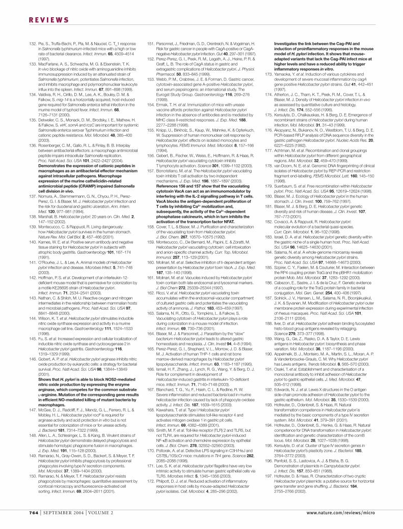
764 | SEPTEMBER 2004 | VOLUME 2 www.nature.com/reviews/micro
R E V I E W S
132. Pie, S., Truffa-Bachi, P., Pla, M. & Nauciel, C. TH1 responsein Salmonella typhimurium-infected mice with a high or lowrate of bacterial clearance. Infect. Immun. 65, 4509–4514(1997).
133. MacFarlane, A. S., Schwacha, M. G. & Eisenstein, T. K. In vivo blockage of nitric oxide with aminoguanidine inhibitsimmunosuppression induced by an attenuated strain ofSalmonella typhimurium, potentiates Salmonella infection,and inhibits macrophage and polymorphonuclear leukocyteinflux into the spleen. Infect. Immun. 67, 891–898 (1999).
134. Valdivia, R. H., Cirillo, D. M., Lee, A. K., Bouley, D. M. &Falkow, S. mig-14 is a horizontally acquired, host-inducedgene required for Salmonella enterica lethal infection in themurine model of typhoid fever. Infect. Immun. 68,7126–7131 (2000).
135. Detweiler, C. S., Monack, D. M., Brodsky, I. E., Mathew, H.& Falkow, S. virK, somA and rcsC are important for systemicSalmonella enterica serovar Typhimurium infection andcationic peptide resistance. Mol. Microbiol. 48, 385–400(2003).
136. Rosenberger, C. M., Gallo, R. L. & Finlay, B. B. Interplaybetween antibacterial effectors: a macrophage antimicrobialpeptide impairs intracellular Salmonella replication. Proc. Natl Acad. Sci. USA 101, 2422–2427 (2004).Demonstrates the expression of cationic peptides inmacrophages as an antibacterial effector mechanismagainst intracellular pathogens. Macrophageexpression of the murine cathelicidin-relatedantimicrobial peptide (CRAMP) impaired Salmonellacell division in vivo.
137. Nomura, A., Stemmermann, G. N., Chyou, P. H., Perez-Perez, G. I. & Blaser, M. J. Helicobacter pylori infection andthe risk for duodenal and gastric ulceration. Ann. Intern.Med. 120, 977–981 (1994).
138. Marshall, B. Helicobacter pylori: 20 years on. Clin. Med. 2,147–152 (2002).
139. Montecucco, C. & Rappuoli, R. Living dangerously: how Helicobacter pylori survives in the human stomach.Nature Rev. Mol. Cell Biol. 2, 457–466 (2001).
140. Karnes, W. E. et al. Positive serum antibody and negativetissue staining for Helicobacter pylori in subjects withatrophic body gastritis. Gastroenterology 101, 167–174(1991).
141. O’Rourke, J. L. & Lee, A. Animal models of Helicobacterpylori infection and disease. Microbes Infect. 5, 741–748(2003).
142. Hoffman, P. S. et al. Development of an interleukin-12-deficient mouse model that is permissive for colonization bya motile KE26695 strain of Helicobacter pylori. Infect. Immun. 71, 2534–2541 (2003).
143. Nathan, C. & Shiloh, M. U. Reactive oxygen and nitrogenintermediates in the relationship between mammalian hostsand microbial pathogens. Proc. Natl Acad. Sci. USA 97,8841–8848 (2000).
144. Wilson, K. T. et al. Helicobacter pylori stimulates induciblenitric oxide synthase expression and activity in a murinemacrophage cell line. Gastroenterology 111, 1524–1533(1996).
145. Fu, S. et al. Increased expression and cellular localization ofinducible nitric oxide synthase and cyclooxygenase 2 inHelicobacter pylori gastritis. Gastroenterology 116,1319–1329 (1999).
146. Gobert, A. P. et al. Helicobacter pylori arginase inhibits nitricoxide production by eukaryotic cells: a strategy for bacterialsurvival. Proc. Natl Acad. Sci. USA 98, 13844–13849(2001).Shows that H. pylori is able to block NOS2-mediatednitric oxide production by expressing the enzymearginase, which competes for the common substrateL-arginine. Mutation of the corresponding gene resultsin efficient NO-mediated killing of mutant bacteria bymacrophages.
147. McGee, D. J., Radcliff, F. J., Mendz, G. L., Ferrero, R. L. &Mobley, H. L. Helicobacter pylori rocF is required forarginase activity and acid protection in vitro but is notessential for colonization of mice or for urease activity. J. Bacteriol 181, 7314–7322 (1999).
148. Allen, L. A., Schlesinger, L. S. & Kang, B. Virulent strains ofHelicobacter pylori demonstrate delayed phagocytosis andstimulate homotypic phagosome fusion in macrophages. J. Exp. Med. 191, 115–128 (2000).
149. Ramarao, N., Gray-Owen, S. D., Backert, S. & Meyer, T. F.Helicobacter pylori inhibits phagocytosis by professionalphagocytes involving type IV secretion components. Mol. Microbiol. 37, 1389–1404 (2000).
150. Ramarao, N. & Meyer, T. F. Helicobacter pylori resistsphagocytosis by macrophages: quantitative assessment byconfocal microscopy and fluorescence-activated cellsorting. Infect. Immun. 69, 2604–2611 (2001).
151. Parsonnet, J., Friedman, G. D., Orentreich, N. & Vogelman, H.Risk for gastric cancer in people with CagA-positive or CagA-negative Helicobacter pylori infection. Gut 40, 297–301 (1997).
152. Perez-Perez, G. I., Peek, R. M., Legath, A. J., Heine, P. R. &Graff, L. B. The role of CagA status in gastric andextragastric complications of Helicobacter pylori. J. Physiol.Pharmacol. 50, 833–845 (1999).
153. Webb, P. M., Crabtree, J. E. & Forman, D. Gastric cancer,cytotoxin-associated gene A-positive Helicobacter pylori,and serum pepsinogens: an international study. TheEurogst Study Group. Gastroenterology 116, 269–276(1999).
154. Ermak, T. H. et al. Immunization of mice with ureasevaccine affords protection against Helicobacter pyloriinfection in the absence of antibodies and is mediated byMHC class II-restricted responses. J. Exp. Med. 188,2277–2288 (1998).
155. Knipp, U., Birkholz, S., Kaup, W., Mahnke, K. & Opferkuch,W. Suppression of human mononuclear cell response byHelicobacter pylori: effects on isolated monocytes andlymphocytes. FEMS Immunol. Med. Microbiol. 8, 157–166(1994).
156. Gebert, B., Fischer, W., Weiss, E., Hoffmann, R. & Haas, R.Helicobacter pylori vacuolating cytotoxin inhibits T lymphocyte activation. Science 301, 1099–1102 (2003).
157. Boncristiano, M. et al. The Helicobacter pylori vacuolatingtoxin inhibits T cell activation by two independentmechanisms. J. Exp. Med. 198, 1887–1897 (2003).References 156 and 157 show that the vacuolatingcytotoxin VacA can act as an immunomodulator byinterfering with the IL-2 signalling pathway in T cells.VacA blocks the antigen-dependent proliferation of T cells by inhibiting Ca2+ mobilization and,subsequently, the activity of the Ca2+-dependentphosphatase calcineurin, which in turn inhibits theactivation of the transcription factor NFAT.
158. Cover, T. L. & Blaser, M. J. Purification and characterizationof the vacuolating toxin from Helicobacter pylori. J. Biol. Chem. 267, 10570–10575 (1992).
159. Montecucco, C., De Bernard, M., Papini, E. & Zoratti, M.Helicobacter pylori vacuolating cytotoxin: cell intoxicationand anion-specific channel activity. Curr. Top. Microbiol.Immunol. 257, 113–129 (2001).
160. Molinari, M. et al. Selective inhibition of Ii-dependent antigenpresentation by Helicobacter pylori toxin VacA. J. Exp. Med.187, 135–140 (1998).
161. Molinari, M. et al. Vacuoles induced by Helicobacter pyloritoxin contain both late endosomal and lysosomal markers.J. Biol Chem 272, 25339–25344 (1997).
162. Ricci, V. et al. Helicobacter pylori vacuolating toxinaccumulates within the endosomal-vacuolar compartmentof cultured gastric cells and potentiates the vacuolatingactivity of ammonia. J. Pathol. 183, 453–459 (1997).
163. Salama, N. R., Otto, G., Tompkins, L. & Falkow, S.Vacuolating cytotoxin of Helicobacter pylori plays a roleduring colonization in a mouse model of infection. Infect. Immun. 69, 730–736 (2001).
164. Blaser, M. J. & Parsonnet, J. Parasitism by the “slow”bacterium Helicobacter pylori leads to altered gastrichomeostasis and neoplasia. J. Clin. Invest 94, 4–8 (1994).
165. Perez-Perez, G. I., Shepherd, V. L., Morrow, J. D. & Blaser,M. J. Activation of human THP-1 cells and rat bone marrow-derived macrophages by Helicobacter pylorilipopolysaccharide. Infect. Immun. 63, 1183–1187 (1995).
166. Ismail, H. F., Zhang, J., Lynch, R. G., Wang, Y. & Berg, D. J.Role for complement in development of Helicobacter-induced gastritis in interleukin-10-deficientmice. Infect. Immun. 71, 7140–7148 (2003).
167. Blanchard, T. G., Yu, F., Hsieh, C. L. & Redline, R. W. Severe inflammation and reduced bacteria load in murinehelicobacter infection caused by lack of phagocyte oxidaseactivity. J. Infect. Dis. 187, 1609–1615 (2003).
168. Kawahara, T. et al. Type I Helicobacter pylorilipopolysaccharide stimulates toll-like receptor 4 andactivates mitogen oxidase 1 in gastric pit cells. Infect. Immun. 69, 4382–4389 (2001).
169. Smith, M. F. et al. Toll-like receptor (TLR) 2 and TLR5, butnot TLR4, are required for Helicobacter pylori-induced NF-κB activation and chemokine expression by epithelialcells. J. Biol. Chem. 278, 32552–32560 (2003).
170. Poltorak, A. et al. Defective LPS signaling in C3H/HeJ andC57BL/10ScCr mice: mutations in Tlr4 gene. Science 282,2085–2088 (1998).
171. Lee, S. K. et al. Helicobacter pylori flagellins have very lowintrinsic activity to stimulate human gastric epithelial cells viaTLR5. Microbes Infect. 5, 1345–1356 (2003).
172. Philpott, D. J. et al. Reduced activation of inflammatoryresponses in host cells by mouse-adapted Helicobacterpylori isolates. Cell. Microbiol. 4, 285–296 (2002).
Investigates the link between the Cag-PAI andinduction of proinflammatory responses in the mousemodel of H. pylori infection. It shows that mouse-adapted variants that lack the Cag-PAI infect mice athigher levels and have a reduced ability to triggerinflammatory responses in vitro.
173. Yamaoka, Y. et al. Induction of various cytokines anddevelopment of severe mucosal inflammation by cagAgene positive Helicobacter pylori strains. Gut 41, 442–451(1997).
174. Atherton, J. C., Tham, K. T., Peek, R. M., Cover, T. L. &Blaser, M. J. Density of Helicobacter pylori infection in vivoas assessed by quantitative culture and histology. J. Infect. Dis. 174, 552–556 (1996).
175. Kersulyte, D., Chalkauskas, H. & Berg, D. E. Emergence ofrecombinant strains of Helicobacter pylori during humaninfection. Mol. Microbiol. 31, 31–43 (1999).
176. Akopyanz, N., Bukanov, N. O., Westblom, T. U. & Berg, D. E.PCR-based RFLP analysis of DNA sequence diversity in thegastric pathogen Helicobacter pylori. Nucleic Acids Res. 20,6221–6225 (1992).
177. Achtman, M. et al. Recombination and clonal groupingswithin Helicobacter pylori from different geographicalregions. Mol. Microbiol. 32, 459–470 (1999).
178. van Doorn, N. E. et al. Genomic DNA fingerprinting of clinicalisolates of Helicobacter pylori by REP-PCR and restrictionfragment end-labelling. FEMS Microbiol. Lett. 160, 145–150(1998).
179. Suerbaum, S. et al. Free recombination within Helicobacterpylori. Proc. Natl Acad. Sci. USA 95, 12619–12624 (1998).
180. Blaser, M. J. Ecology of Helicobacter pylori in the humanstomach. J. Clin. Invest. 100, 759–762 (1997).
181. Blaser, M. J. & Berg, D. E. Helicobacter pylori geneticdiversity and risk of human disease. J. Clin. Invest. 107,767–773 (2001).
182. Covacci, A. & Rappuoli, R. Helicobacter pylori: molecular evolution of a bacterial quasi-species. Curr. Opin. Microbiol. 1, 96–102 (1998).
183. Israel, D. A. et al. Helicobacter pylori genetic diversity withinthe gastric niche of a single human host. Proc. Natl Acad.Sci. USA 98, 14625–14630 (2001).
184. Salama, N. et al. A whole-genome microarray revealsgenetic diversity among Helicobacter pylori strains. Proc. Natl Acad. Sci. USA 97, 14668–14673 (2000).
185. Szpirer, C. Y., Faelen, M. & Couturier, M. Interaction betweenthe RP4 coupling protein TraG and the pBHR1 mobilizationprotein Mob. Mol. Microbiol. 37, 1283–1292 (2000).
186. Cabezon, E., Sastre, J. I. & de la Cruz, F. Genetic evidenceof a coupling role for the TraG protein family in bacterialconjugation. Mol. Gen. Genet. 254, 400–406 (1997).
187. Solnick, J. V., Hansen, L. M., Salama, N. R., Boonjakuakul,J. K. & Syvanen, M. Modification of Helicobacter pylori outermembrane protein expression during experimental infectionof rhesus macaques. Proc. Natl Acad. Sci. USA 101,2106–2111 (2004).
188. Ilver, D. et al. Helicobacter pylori adhesin binding fucosylatedhisto-blood group antigens revealed by retagging. Science 279, 373–377 (1998).
189. Wang, G., Ge, Z., Rasko, D. A. & Taylor, D. E. Lewisantigens in Helicobacter pylori: biosynthesis and phasevariation. Mol. Microbiol. 36, 1187–1196 (2000).
190. Appelmelk, B. J., Monteiro, M. A., Martin, S. L., Moran, A. P.& Vandenbroucke-Grauls, C. M. Why Helicobacter pylorihas Lewis antigens. Trends Microbiol. 8, 565–570 (2000).
191. Osaki, T. et al. Establishment and characterisation of amonoclonal antibody to inhibit adhesion of Helicobacterpylori to gastric epithelial cells. J. Med. Microbiol. 47,505–512 (1998).
192. Edwards, N. J. et al. Lewis X structures in the O antigenside-chain promote adhesion of Helicobacter pylori to thegastric epithelium. Mol. Microbiol. 35, 1530–1539 (2000).
193. Hofreuter, D., Odenbreit, S. & Haas, R. Naturaltransformation competence in Helicobacter pylori ismediated by the basic components of a type IV secretionsystem. Mol. Microbiol. 41, 379–391 (2001).
194. Hofreuter, D., Odenbreit, S., Henke, G. & Haas, R. Naturalcompetence for DNA transformation in Helicobacter pylori:identification and genetic characterization of the comBlocus. Mol. Microbiol. 28, 1027–1038 (1998).
195. Kersulyte, D. et al. Cluster of type IV secretion genes inHelicobacter pylori’s plasticity zone. J. Bacteriol. 185,3764–3772 (2003).
196. Penfold, S. S., Lastovica, A. J. & Elisha, B. G.Demonstration of plasmids in Campylobacter pylori. J. Infect. Dis. 157, 850–851 (1988).
197. Hofreuter, D. & Haas, R. Characterization of two crypticHelicobacter pylori plasmids: a putative source for horizontalgene transfer and gene shuffling. J. Bacteriol. 184,2755–2766 (2002).
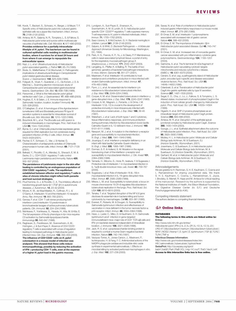
NATURE REVIEWS | MICROBIOLOGY VOLUME 2 | SEPTEMBER 2004 | 765
R E V I E W S
198. Kwok, T., Backert, S., Schwarz, H., Berger, J. & Meyer, T. F.Specific entry of Helicobacter pylori into cultured gastricepithelial cells via a zipper-like mechanism. Infect. Immun.70, 2108–2120 (2002).
199. Amieva, M. R., Salama, N. R., Tompkins, L. S. & Falkow, S.Helicobacter pylori enter and survive within multivesicularvacuoles of epithelial cells. Cell. Microbiol. 4, 677–690 (2002).Provides evidence for a partially intracellularlifestyle of H. pylori. The bacterium can be found incultured epithelial cells residing in multivesicularbodies, where it survives for at least 24 hours andfrom where it can emerge to repopulate theextracellular space.
200. Kazi, J. L. et al. Ultrastructural study of Helicobacter pylori-associated gastritis. J. Pathol 161, 65–70 (1990).
201. Bode, G., Malfertheiner, P. & Ditschuneit, H. Pathogeneticimplications of ultrastructural findings in Campylobacterpylori related gastroduodenal disease. Scand. J. Gastroenterol. 142, S25–S39 (1988).
202. Foliguet, B., Vicari, F., Guedenet, J. C., De Korwin, J. D. &Marchal, L. Scanning electron microscopic study ofCampylobacter pylori and associated gastroduodenallesions. Gastroenterol. Clin. Biol. 13, 65B–70B (1989).
203. Parsonnet, J. What is the Helicobacter pylori globalreinfection rate? Can. J. Gastroenterol. 17, 46B–48B (2003).
204. Hughes, E. A. & Galan, J. E. Immune response toSalmonella: location, location, location? Immunity 16,325–328 (2002).
205. O’Callaghan, D. et al. A homologue of the Agrobacteriumtumefaciens VirB and Bordetella pertussis Ptl type IVsecretion systems is essential for intracellular survival ofBrucella suis. Mol. Microbiol. 33, 1210–1220 (1999).
206. Boschiroli, M. L. et al. The Brucella suis virB operon isinduced intracellularly in macrophages. Proc. Natl Acad. Sci.USA 99, 1544–1549 (2002).
207. Byrne, G. I. et al. Chlamydia pneumoniae expresses genesrequired for DNA replication but not cytokinesis duringpersistent infection of HEp-2 cells. Infect. Immun. 69,5423–5429 (2001).
208. Fischer, S. F., Schwarz, C., Vier, J. & Hacker, G.Characterization of antiapoptotic activities of Chlamydiapneumoniae in human cells. Infect. Immun. 69, 7121–7129(2001).
209. Belkaid, Y., Piccirillo, C. A., Mendez, S., Shevach, E. M. &Sacks, D. L. CD4+CD25+ regulatory T cells controlLeishmania major persistence and immunity. Nature 420,502–507 (2002).The persistence of Leishmania major in the skin afterhealing is controlled by an endogenous population ofCD4+CD25+ regulatory T cells. The equilibriumestablished between effector and regulatory T cells insites of chronic infection might reflect both parasiteand host survival strategies.
210. Prud’homme, G. J. & Piccirillo, C. A. The inhibitory effects oftransforming growth factor-β-1 (TGF-β1) in autoimmunediseases. J. Autoimmun. 14, 23–42 (2000).
211. Moore, K. W., de Waal Malefyt, R., Coffman, R. L. &O’Garra, A. Interleukin-10 and the interleukin-10 receptor.Annu. Rev. Immunol. 19, 683–765 (2001).
212. Gerosa, F. et al. CD4+ T cell clones producing bothinterferon-γ and interleukin-10 predominate inbronchoalveolar lavages of active pulmonary tuberculosispatients. Clin. Immunol. 92, 224–234 (1999).
213. Nishikawa, F., Yoshikawa, S., Harada, H., Kita, M. & Kita, E.The full expression of the ity phenotype in ityr mice requiresC3 activation by Salmonella lipopolysaccharide.Immunology 95, 640–647 (1998).
214. Raghavan, S., Fredriksson, M., Svennerholm, A. M.,Holmgren, J. & Suri-Payer, E. Absence of CD4+CD25+
regulatory T cells is associated with a loss of regulationleading to increased pathology in Helicobacter pylori-infected mice. Clin. Exp. Immunol. 132, 393–400 (2003).The influence of CD4+CD25+ cells on H. pyloricolonization in a mouse model of infection wasanalysed. This showed that these cells reduceimmunopathology, possibly by reducing the activationof IFN-γ-producing CD4+ T cells, even at the expenseof a higher H. pylori load in the gastric mucosa.
215. Lundgren, A., Suri-Payer, E., Enarsson, K., Svennerholm, A. M. & Lundin, B. S. Helicobacter pylori-specific CD4+ CD25high regulatory T cells suppress memoryT-cell responses to H. pylori in infected individuals. Infect.Immun. 71, 1755–1762 (2003).
216. Saez-Llorens, X. & McCracken, G. H. Bacterial meningitis inchildren. Lancet 361, 2139–2148 (2003).
217. Salyers, A. & Whitt, D. Bacterial Pathogensis — A MolecularApproach (American Society for Microbiology, Washington,DC, 1994).
218. Park, H. S., Francis, K. P., Yu, J. & Cleary, P. P. Membranouscells in nasal-associated lymphoid tissue: a portal of entryfor the respiratory mucosal pathogen group Astreptococcus. J. Immunol. 171, 2532–2537 (2003).
219. Lengeling, A., Pfeffer, K. & Balling, R. The battle of twogenomes: genetics of bacterial host/pathogen interactionsin mice. Mamm. Genome 12, 261–271 (2001).
220. Mastroeni, P. et al. Interleukin 18 contributes to hostresistance and γ-interferon production in mice infected withvirulent Salmonella typhimurium. Infect. Immun. 67,478–483 (1999).
221. Flynn, J. L. et al. An essential role for interferon-γ inresistance to Mycobacterium tuberculosis infection. J. Exp. Med. 178, 2249–2254 (1993).
222. Cooper, A. M. et al. Disseminated tuberculosis in interferon-γgene-disrupted mice. J. Exp. Med. 178, 2243–2247 (1993).
223. Cooper, A. M., Magram, J., Ferrante, J. & Orme, I. M.Interleukin 12 (IL-12) is crucial to the development ofprotective immunity in mice intravenously infected withMycobacterium tuberculosis. J. Exp. Med. 186, 39–45(1997).
224. Wakeham, J. et al. Lack of both types 1 and 2 cytokines,tissue inflammatory responses, and immune protectionduring pulmonary infection by Mycobacterium bovis bacilleCalmette–Guerin in IL-12-deficient mice. J. Immunol. 160,6101–6111 (1998).
225. Newport, M. J. et al. A mutation in the interferon-γ-receptorgene and susceptibility to mycobacterial infection. N. Engl. J. Med. 335, 1941–1949 (1996).
226. Jouanguy, E. et al. Interferon-γ-receptor deficiency in aninfant with fatal bacille Calmette–Guerin infection. N. Engl. J. Med. 335, 1956–1961 (1996).
227. Dorman, S. E. & Holland, S. M. Mutation in the signal-transducing chain of the interferon-γ receptor andsusceptibility to mycobacterial infection. J. Clin. Invest 101,2364–2369 (1998).
228. Yamada, H., Mizumo, S., Horai, R., Iwakura, Y. & Sugawara, I.Protective role of interleukin-1 in mycobacterial infection inIL-1α/β double-knockout mice. Lab. Invest. 80, 759–767(2000).
229. Sugawara, I. et al. Role of interleukin-18 (IL-18) inmycobacterial infection in IL-18-gene-disrupted mice. Infect. Immun. 67, 2585–2589 (1999).
230. Mitsos, L. M. et al. Susceptibility to tuberculosis: a locus onmouse chromosome 19 (Trl-4) regulates Mycobacteriumtuberculosis replication in the lungs. Proc. Natl Acad. Sci.USA 100, 6610–6615 (2003).
231. Tanaka, T. et al. Targeted disruption of the NF-IL6 genediscloses its essential role in bacteria killing and tumorcytotoxicity by macrophages. Cell 80, 353–361 (1995).
232. Everest, P., Roberts, M. & Dougan, G. Susceptibility toSalmonella typhimurium infection and effectiveness ofvaccination in mice deficient in the tumor necrosis factor-αp55 receptor. Infect. Immun. 66, 3355–3364 (1998).
233. Hess, J., Ladel, C., Miko, D. & Kaufmann, S. H. Salmonellatyphimurium aroA– infection in gene-targetedimmunodeficient mice: major role of CD4+ TCR-αβ cells andIFN-γ in bacterial clearance independent of intracellularlocation. J. Immunol. 156, 3321–3326 (1996).
234. Jack, R. S. et al. Lipopolysaccharide-binding protein isrequired to combat a murine Gram-negative bacterialinfection. Nature 389, 742–745 (1997).
235. Vazquez-Torres, A., Jones-Carson, J., Mastroeni, P.,Ischiropoulos, H. & Fang, F. C. Antimicrobial actions of theNADPH phagocyte oxidase and inducible nitric oxidesynthase in experimental salmonellosis. I. Effects onmicrobial killing by activated peritoneal macrophages in vitro.J. Exp. Med. 192, 227–236 (2000).
236. Sawai, N. et al. Role of γ-interferon in Helicobacter pylori-induced gastric inflammatory responses in a mouse model.Infect. Immun. 67, 279–285 (1999).
237. El-Omar, E. M. et al. Interleukin-1 polymorphismsassociated with increased risk of gastric cancer. Nature 404, 398–402 (2000).
238. El-Omar, E. M. The importance of interleukin-1β inHelicobacter pylori associated disease. Gut 48, 743–747(2001).
239. El-Omar, E. M. et al. Increased risk of noncardia gastriccancer associated with pro-inflammatory cytokine genepolymorphisms. Gastroenterology 124, 1193–1201(2003).
240. Galmiche, A. et al. The N-terminal 34-kDa fragment ofHelicobacter pylori vacuolating cytotoxin targetsmitochondria and induces cytochrome c release. EMBO J. 19, 6361–6370 (2000).
241. Censini, S. et al. cag, a pathogenicity island of Helicobacterpylori, encodes type I-specific and disease-associatedvirulence factors. Proc. Natl Acad. Sci. USA 93,14648–14653 (1996).
242. Odenbreit, S. et al. Translocation of Helicobacter pyloriCagA into gastric epithelial cells by type IV secretion.Science 287, 1497–1500 (2000).
243. Segal, E. D., Cha, J., Lo, J., Falkow, S. & Tompkins, L. S.Altered states: involvement of phosphorylated CagA in theinduction of host cellular growth changes by Helicobacterpylori. Proc. Natl Acad. Sci. USA 96, 14559–14564(1999).
244. Higashi, H. et al. SHP-2 tyrosine phosphatase as anintracellular target of Helicobacter pylori CagA protein.Science 295, 683–686 (2002).
245. Amieva, M. R. et al. Disruption of the epithelial apical-junctional complex by Helicobacter pylori CagA. Science300, 1430–1434 (2003).
246. Guruge, J. L. et al. Epithelial attachment alters the outcomeof Helicobacter pylori infection. Proc. Natl Acad. Sci. USA95, 3925–3930 (1998).
247. Mobley, H. L. in Helicobacter pylori: Molecular and CellularBiology (eds Achtman, M. & Suerbaum, S.) (Horizon Scientific, Wymondham, 2001).
248. Josenhans, C. & Suerbaum, S. in Helicobacter pylori:Molecular and Cellular Biology (eds Achtman, M. &Suerbaum, S.) (Horizon Scientific, Wymondham, 2001).
249. Gerhard, M. et al. in Helicobacter pylori: Molecular andCellular Biology (eds Achtman, M. & Suerbaum, S.) (Horizon Scientific, Wymondham, 2001).
AcknowledgementsWe are grateful to H. Rachman, S. H. E. Kaufmann, C. Cosma andL. Ramakrishnan for sharing unpublished data. We thank S. H. E. Kaufmann, C. Cosma, L. Ramakrishnan, E. Joyce, I. Brodsky, S. Merrell, R. Haas and M. Amieva for critical reading of the manuscript. Research by the authors is supported by the National Institutes of Health, the Ellison Medical Foundation,the Digestive Disease Center (to S.F.) and DeutscheForschungsgemeinschaft (to A.M.).
Competing interests statementThe authors declare no competing financial interests.
Online links
DATABASESThe following terms in this article are linked online to:Entrez: http://www.ncbi.nlm.nih.gov/entrez/Helicobacter pylori | IFN-γ | IL-2 | IL-2R | IL-10 | IL-12 | IL-23 |LRG-47 | Mycobacterium marinum | Mycobacterium tuberculosis |NFAT | NOS2 | Nramp1 | S. typhi | S. typhimurium | TGF-β | TLR2 |TLR4 | TNF-αInfectious Disease Information:http://www.cdc.gov/ncidod/diseases/index.htmHIV | salmonellosis | tuberculosis | typhoid feverSwissProt: http://ca.expasy.org/sprot/babA | babB | FlaA | FlaB | ICL | mig-14 | rocF | TraG | VacA | virKAccess to this interactive links box is free online.