Embed Size (px)
Citation preview
O
R
N
R
A2
I
Seitbm
f
1d
rthopaedics & Traumatology: Surgery & Research (2013) 99S, S124—S139
Available online at
www.sciencedirect.com
EVIEW ARTICLE
euromuscular scoliosis
. Vialle ∗, C. Thévenin-Lemoine, P. Mary
rmand-Trousseau Hospital, Pediatric Orthopedic and Repair Surgery Department, Pierre-and-Marie-Curie University, Paris 6,6, Avenue du Dr-Arnold-Netter, 75012 Paris, France
Accepted: 29 October 2012
KEYWORDSNeuromuscularscoliosis;Muscular dystrophy;Spinal muscularatrophy;Cerebral palsy
Summary Scoliosis is a common deformity in many types of neuromuscular disease. Severespinal curvature can cause difficulty in sitting. Conservative and surgical treatment of neu-romuscular scoliosis differs from idiopathic scoliosis, being more complex and with a highercomplications rate. Non-surgical measures rarely fully control progressive scoliosis, but aim toprevent spinal deformities secondary to muscular hypotonia or contracture. Twenty-four hourbracing should be adjusted throughout growth, and may induce functional impairment and lossof independence. Corrective surgery requires multidisciplinary management and perioperativescreening. Pelvic obliquity is commonly associated with neuromuscular scoliosis, making sit-ting difficult: correction needs to be considered during surgical planning. The goal of surgicalcorrection is to obtain and maintain a well-balanced spine above a well-positioned pelvis. Pre-operative multidisciplinary assessment enables potential problems of terrain to be anticipated.
Respiratory function investigation will guide possible non-invasive perioperative ventilation.Nutritional and psychosocial assessment should also be incorporated in this preparation, asshould overall postoperative care. Implementing this overall strategic planning can achieve agood surgical and functional result in the vast majority of cases.© 2012 Elsevier Masson SAS. All rights reserved.pfct
ntroduction
pinal deformity is a key issue in the management of sev-ral neurological or muscular pathologies. Such pathologiesnduce control deficits or muscle weakness, which are often
he main symptoms. The clinical picture may sometimese associated with muscle retraction, sensitivity disorder,ental retardation and digestive, cardiac or respiratory∗ Corresponding author. Tel.: +33 1 71 73 89 07;ax: +33 1 44 73 69 42.
E-mail address: [email protected] (R. Vialle).
E
T
aic
877-0568/$ – see front matter © 2012 Elsevier Masson SAS. All rights reoi:10.1016/j.otsr.2012.11.002
roblems. Comprehensive management is thus complex butundamental. The various pathologies have many points inommon, in terms both of evolution and of assessment andreatment.
tiological forms
able 1 presents the main causes of spinal deviation.
The Scoliosis Research Society (SRS) classifies thems neuropathic, with central or peripheral motor neuronnvolvement or both, or myopathic [1], a heterogeneouslass of pathologies of very variable functional impact [2—5].
served.
Neuromuscular scoliosis S125
Table 1 Main neuromuscular etiologies of spinal deformity.
Central neurological causesCentral motor neuron involvement
Cerebral palsyHereditary ataxia (Friedreich, etc.)SyringomyeliaOther central causes (encephalopathy, Rett’s syndrome, etc.)
Peripheral neurological causesPeripheral motor neuron involvement
Acute anterior poliomyelitisInfantile spinal amyotrophyHereditary motor and sensory neuropathyHereditary sensory and vegetative neuropathy (familial dysautonomia)
Mixed central and peripheral neurological causes Medullary lesionMyelodysplasiaMyelomeningocele
Neuromuscular junction (motor end-plate) Myasthenia
Muscular causes Duchenne myopathyOther muscular dystrophy
ogry
aocsIal
Pd
C
Cienmeom‘
Arthr
Such ‘‘secondary’’ spinal deformity occurs much morefrequently during the course of these pathologies than does‘‘idiopathic’’ scoliosis in the general population: prevalenceranges from 25 to 100% according to etiology (Table 2).
Spinal deformity pathogenesis
Spinal deformity cannot be attributed to trunk muscle weak-ness alone; indeed, in certain pathologies, trunk musclehypertonia rather than paralysis is primarily implicated. Incentral neurologic pathology, for example, spinal deformitymay be induced by disharmonious control of trunk muscu-lature around the spinal axis, progressively worsening dueto a lack of effective muscular compensation mechanisms(Fig. 1).
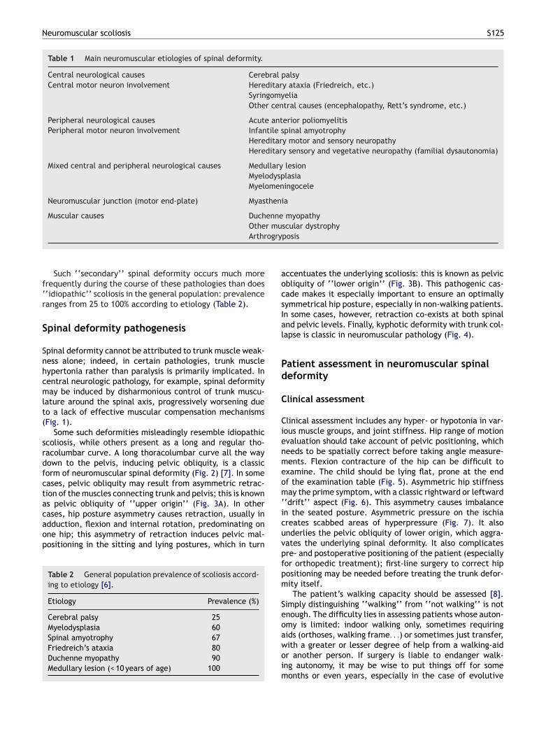
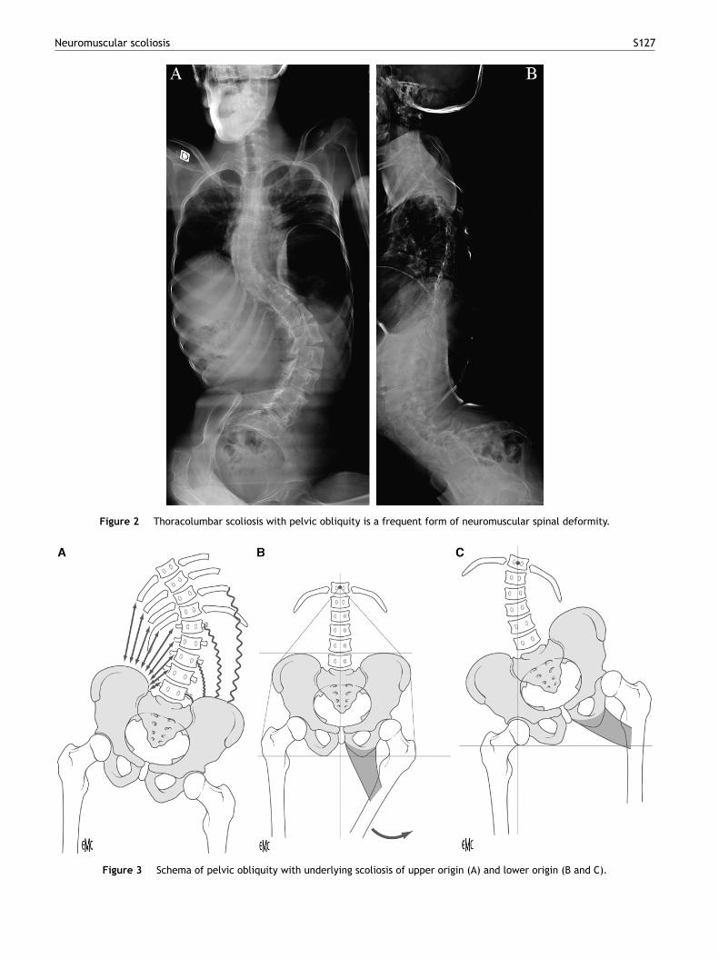
Some such deformities misleadingly resemble idiopathicscoliosis, while others present as a long and regular tho-racolumbar curve. A long thoracolumbar curve all the waydown to the pelvis, inducing pelvic obliquity, is a classicform of neuromuscular spinal deformity (Fig. 2) [7]. In somecases, pelvic obliquity may result from asymmetric retrac-tion of the muscles connecting trunk and pelvis; this is knownas pelvic obliquity of ‘‘upper origin’’ (Fig. 3A). In other
cases, hip posture asymmetry causes retraction, usually inadduction, flexion and internal rotation, predominating onone hip; this asymmetry of retraction induces pelvic mal-positioning in the sitting and lying postures, which in turnTable 2 General population prevalence of scoliosis accord-ing to etiology [6].
Etiology Prevalence (%)
Cerebral palsy 25Myelodysplasia 60Spinal amyotrophy 67Friedreich’s ataxia 80Duchenne myopathy 90Medullary lesion (< 10 years of age) 100
icuvpfpm
Seoawoim
posis
ccentuates the underlying scoliosis: this is known as pelvicbliquity of ‘‘lower origin’’ (Fig. 3B). This pathogenic cas-ade makes it especially important to ensure an optimallyymmetrical hip posture, especially in non-walking patients.n some cases, however, retraction co-exists at both spinalnd pelvic levels. Finally, kyphotic deformity with trunk col-apse is classic in neuromuscular pathology (Fig. 4).
atient assessment in neuromuscular spinaleformity
linical assessment
linical assessment includes any hyper- or hypotonia in var-ous muscle groups, and joint stiffness. Hip range of motionvaluation should take account of pelvic positioning, whicheeds to be spatially correct before taking angle measure-ents. Flexion contracture of the hip can be difficult to
xamine. The child should be lying flat, prone at the endf the examination table (Fig. 5). Asymmetric hip stiffnessay the prime symptom, with a classic rightward or leftward
‘drift’’ aspect (Fig. 6). This asymmetry causes imbalancen the seated posture. Asymmetric pressure on the ischiareates scabbed areas of hyperpressure (Fig. 7). It alsonderlies the pelvic obliquity of lower origin, which aggra-ates the underlying spinal deformity. It also complicatesre- and postoperative positioning of the patient (especiallyor orthopedic treatment); first-line surgery to correct hipositioning may be needed before treating the trunk defor-ity itself.The patient’s walking capacity should be assessed [8].
imply distinguishing ‘‘walking’’ from ‘‘not walking’’ is notnough. The difficulty lies in assessing patients whose auton-my is limited: indoor walking only, sometimes requiringids (orthoses, walking frame. . .) or sometimes just transfer,
ith a greater or lesser degree of help from a walking-aidr another person. If surgery is liable to endanger walk-ng autonomy, it may be wise to put things off for someonths or even years, especially in the case of evolutive
S126 R. Vialle et al.
Figure 1 Schema (J. Dubousset/R. Vialle) of progressive trunk imbalance induced by asymmetric application of muscular mainte-n runk
nl
sttt
iiih
lTig
al
R
Ctr
•
ance forces. Imbalance progresses to the point of inevitable t
euromuscular pathology, so that vertebral arthrodesis noonger entails a further loss of independence.
Static examination may be performed with the patientitting at the edge of a table, with the help of another persono maintain the seated posture if necessary. This examina-ion provides better assessment of trunk deformity and ofhe sagittal and frontal components of the collapse.
Dynamic trunk examination assesses deformity reducibil-ty. The spine should be studied level by level in lateralnclination and in rotational movement. Curvature reducibil-ty can also be judged by trying to raise the patient by theead (Fig. 8).
Prone examination should be systematic, with the patient
ying at the end of the table with lower limbs in flexion.his displays residual curvature after eliminating abnormal-ty due to limb-length discrepancy, pelvic asymmetry andravitational effects. Curvature reducibility can be judged
•
collapse.
gain in lateral inclination, as can the flexibility of the ilio-umbar angles.
espiratory assessment and management
areful respiratory assessment should be conducted sys-ematically in neuromuscular spinal deformity, for threeeasons:
spinal deformity can impact ventilation mechanics,especially when severe and associated with thoracic
hypokyphosis, thoracolumbar kyphosis or rib-cage defor-mity;the underlying neurologic pathology may, in itself or dueto its evolution, impair ventilation;
Neuromuscular scoliosis S127
Figure 2 Thoracolumbar scoliosis with pelvic obliquity is a frequent form of neuromuscular spinal deformity.
Figure 3 Schema of pelvic obliquity with underlying scoliosis of upper origin (A) and lower origin (B and C).

S128 R. Vialle et al.
F ly toA -ray,
•
iiica
vo
kmtw
ta
ti
tnttPm
toococp
igure 4 Evolution of postural hypotonia leading progressive. Lateral spinal X-ray, standing, age 8 years. B. Lateral spinal X
treatment, whether conservative or surgical, may haveimmediate respiratory impact that can sometimes beenduring or definitive.
Respiratory assessment can now rely on objective exam-nations. Conservative as well as surgical treatment maynduce sudden decompensation of a respiratory state thats usually fragile. Associated swallowing disorder, a poorlyontrolled epileptic state and fragile nutritional status areggravating factors.
Regular respiratory rehabilitation can be associated to aariety of instrumental techniques, each of which has itswn particular benefit (Fig. 9).
Intermittent positive pressure breathing devices (alsonown as ‘‘pressure relaxers’’) such as the Alpha 200 helpaintain rib-cage flexibility and improve thoracic amplia-
ion. This technique is intended for stiff spinal deformity
ith thoracic hypokyphosis or severe chest deformity.‘‘Cough-assist’’ devices are useful in elevated risk ofracheobronchial congestion [9]: hypotonic patients (spinalmyotrophy, muscular dystrophy). They are especially effec-
C
Mt
lumbar kyphosis in a boy with Duchenne muscular dystrophy. seated, age 10 years.
ive when the patient is bed-ridden or fatigued, as during themmediate postoperative course.
Non-invasive ventilation improves spontaneous ventila-ion quality by a mechanical ventilation aid via an oral orasal mask connected up to an assisted breathing appara-us [10]. It may be used ahead of heavy surgery and duringhe first postoperative months in the most fragile patients.reoperative assessment and family education allow imple-entation under good conditions [11].Invasive ventilation via tracheotomy may be required by
he severity of respiratory insufficiency or swallowing dis-rder with recurrent congestion. In patients at high riskf respiratory decompensation, tracheotomy should be dis-ussed among the multidisciplinary team before decidingn any vertebral arthrodesis. This avoids emergency tra-heotomy, performed under poor conditions, in case ofostoperative respiratory distress.
ardiac assessment
inimal cardiac assessment ahead of arthrodesis is manda-ory in neuromuscular spinal deformity [12].

Neuromuscular scoliosis S129
Figure 5 Clinical hip examination with flexion contracture measurement in ventral decubitus with pelvis at table edge. A. Clinicalphotograph of child’s position. B. Diagram of flexion contracture measurement.
Figure 6 Diagram of pelvic obliquity reducibility assessment in ventral decubitus (superior view). A. Spontaneous position, withpelvic obliquity and hip malpositioning. B. Good pelvic obliquity reducibility. C. Incomplete pelvic obliquity reducibility underasymmetric left lower limb traction.

S130 R. Vialle et al.
Figure 7 Pelvic obliquity inducing asymmetric stress to ischial weight-bearing points in seated posture. A. Imbalance with excessright ischium pressure. B. Compensatory trunk imbalance by upper limbs. C. Upper limb compensation impossible, requiring helpfrom another person to maintain upright trunk.
xplo
d
D
Figure 8 Clinical reducibility of collapse can be e
Table 3 presents the various types of myopathy with car-iac involvement.
Myocardial contractility is systematically impaired inuchenne muscular dystrophy. Onset may be sudden, at 10
ob
c
Table 3 Cardiac involvements in muscular dystrophy.
Usual name Genetic abnormality
Duchenne muscular dystrophy DMD gene
Becker muscular dystrophy DMD gene
Emery-Dreifuss muscular dystrophy EMD and LMNA gene
Limb-girdle or Erb muscular dystrophy Polygenic, recessiveor dominant
Steinert myotonia DMPK gene
Rett’s syndrome MECP2 gene
red by manual trunk elongation in seated posture.
r 11 years of age; surgical stabilization of the spine shoulde rapidly undertaken [13,14].
In Steinert myotonic dystrophy, conduction disorderan be screened by Holter or preoperative intracavitary
Location Type of cardiac involvement
Xp2.1 Cardiac insufficiencyXP2.1 Cardiac insufficiency
s Chromosome X,Chromosome 1
Conduction disorder,arrhythmia
Linked to X Cardiac insufficiency
Chromosome 9 Conduction disorderChromosome X Cardiac dysautonomia,
rhythm disorder

Neuromuscular scoliosis S131
Figure 9 A. Alpha 200 device for positive pressure passive ampliation. B. Cough-assist device for bronchopulmonary decongestion.n by
mnbs
utpm
C. Non-invasive ventilation by nasal mask. D. Invasive ventilatio
recording. A preoperative electrosystolic training probemay sometimes be necessary to prevent peroperativearrhythmia [15].
Trophic assessment and digestive or urinarydisorder
In general, in case of weight-loss or stagnation during the
growth period, nutritional deficiency should be primar-ily suspected, but also impaired respiratory function ordepression syndrome. During the months preceding spinalsurgery, particular attention should be paid to nutritionalssmc
tracheotomy.
anagement. In the most difficult cases, with history of mal-utrition and limited patient cooperation, nocturnal feedingy nasogastric tube or by gastrostomy should be consideredeveral weeks or months ahead of scheduled arthrodesis.
Preoperative management is mandatory for any chronicrinary infection. Urine sterilization and rigorous implemen-ation of a precise catheterization or self-catheterizationrotocol can limit infection risk, notably in case ofedullary involvement (paraplegia, myelomeningocele). In
elf-catheterization, precise assessment of implementationhould precede arthrodesis: altered trunk shape and sizeay cause difficulty for some patients, altering the technical
onditions of self-catheterization.

S132
Figure 10 CT slice confirming right middle lobe bronchusstretching (arrows) in contact with vertebral body in girl withe
I
Ipctda
orfiol
pocpw
tasil
T
A
Pc
Ptc
wttrgfltp(
Ec
I(dbGt
amtmrt
sbsfa
Et
TlIcnttec
tqs[
W
volutive scoliosis with thoracic lordosis.
maging
n non-walking hypotonic patients, imaging in the seatedosture, where the deformity is accentuated by weight,an be useful. Although realistic with respect to the pos-ural deformity, such imaging is poorly reproductible foriachronic comparison, and images taken in lying posturere preferable.
Specific ‘‘bending’’ views are needed to assess stiffeningf the different spinal levels. AP views under asymmet-ic traction (applied to a single lower limb) can assess therontal reducibility of pelvic obliquity. Complete corrections demonstrated by alignment of the line through the footf the two sacroiliac interlines with respect to the shoulderine [16].
Preoperative MRI is essential in suspected medullaryathology, even when longstanding (paraplegia, traumaticr non-traumatic quadriplegia), to detect a possible asso-iated syringomyelic cavity that may induce per- orostoperative neurologic aggravation, especially frequenthen medullary function is partially conserved [17,18].
Thoracic CT is recommended in spinal deformity withhoracic hypokyphosis or lordosis; the caliber of bronchinterior to the spinal convexity is often reduced [19]. Inevere cases, such bronchial ‘‘stretching’’ (Fig. 10) maynduce reversible or irreversible atelectasis and reducedung volume.
reatment
voiding spinal deformity: prevention
revention is the keystone of early management of manyhildren and adolescents with neuromuscular disease.
Stjt
R. Vialle et al.
reventing retraction and pathologic posture of therunk and above all of the limbs is the foundation ofomprehensive orthopedic management.
Countering asymmetric hip posture is the most effectiveay of countering the development of pelvic obliquity and
hus of scoliosis of lower origin. Countering flexion contrac-ure of the hips prevents the lumbar and lumbar-sacralegion stiffening into hyperlordosis [20,21]. Conversely, pro-ressive retraction of the hamstring muscles induces not onlyexion contracture of the knee and increasing hindrance ofhe upright stance, but also progressive retroversion of theelvic pedestal and progressive kyphosis of the lumbar spineFig. 11).
volutive spinal deformity: the role ofonservative treatment
n certain pathologies inducing severe muscular deficiencyquadriplegia, type 1 and 2 spinal amyotrophy), orthope-ic treatment should be very early. It begins with passiveracing, correcting the spine by traction exerted by aarchois-type corset between the two fixed points consti-
uted by the pelvis and the skull (Fig. 12).Some patients with central (cerebral palsy, cerebellar
taxia) or peripheral neurologic disorder (neuropathy) oruscular pathology involving only mild deficiency may be
reated using the more conventional Chenau corset or aolded corset for nocturnal hypercorrection. Other neu-
ologic disorders, such as dystonia, are not amenable toreatment by corset.
In most cases, conservative treatment of neuromuscularpinal deformity is only an interim measure awaiting verte-ral arthrodesis [22]; it is nevertheless effective in limitinguch consequences as impaired pulmonary development andunction, enabling future surgery to be as simple and limiteds possible.
volved spinal deformity: the role of surgicalreatment
o operate or not to operate? — Technical and ethicalimitsmprovement in surgical and, first and foremost, intensiveare, pneumologic, cardiologic and anesthesiologic tech-iques now allows surgical solutions of varying complexityo be offered to a very large number of patients, includinghe most fragile. Treatment options should be discussed asarly as possible with the patient and his or her family andlose environment [23].
Several studies have demonstrated objective postopera-ive functional improvement based on self-administereduality-of-life questionnaires, confirming the benefit ofurgical management, even in the most difficult cases24].
hen to operate?
ome deformities are strongly evolutive and difficult to con-ain conservatively. Relatively early surgery may thus beustified. Even so, conservative treatment should be con-inued to the very end if development of the chest and
Neuromuscular scoliosis S133
nd lu mu
ls
Figure 11 Hamstring retraction inducing pelvic retroversion ahips in extension, confirms complete reducibility and the purely
increased respiratory capacity can still be obtained by thegrowth of the trunk. In some poorly controlled deformities,
progressive spinal distraction rods, usually associated toclassical corset treatment, may be useful. The mechanicalresistance thresholds of these devices, however, frequentlyar
Figure 12 Conservative treatment sequence for severe spinal deamyotrophy. A. Correction begins with progressive axial traction by ctrunk collapse by detraction cast. C. Stabilization of correction in Ga
mbar kyphosis in seated posture (A). View in lateral decubitus,scular postural origin of the deformity.
ead to complications, particularly at the spinal fixationites.
The clinical and radiological criteria of spinal maturityre a matter of debate in neuromuscular disease [25]. Tri-adiate cartilage closure is a good sign of axial skeletal
formity with pelvic obliquity in a 9-year-old girl with spinalranial halo. B. Supplementary correction of pelvic obliquity andrchois corset with occipitomental extension.

S134 R. Vialle et al.
Figure 13 Severely evolved spinal deformity in a 15-year-old boy with cerebral palsy and spastic quadriplegia. A andB. Preoperative X-rays. C and D. Postoperative X-rays after anterior release of lumbar scoliosis convexity and posterior osteosynthesis.Despite initial severity, overall trunk, shoulder line and seated pelvis alignment is satisfactory.

Neuromuscular scoliosis S135
Figure 14 Example of correction by progressive bending and segmental instrumentation by pedicle screws in a 13-year-old boywith congenital muscular dystrophy. A and B. Preoperative X-rays. C and D. Postoperative X-rays.

S136 R. Vialle et al.
Figure 15 Surgical correction strategy in neurological scoliosis with pelvic obliquity by segmental T-assembly. A and B. Correctionof spinal deformity. C. Pelvic instrumentation with 21 pedicle screws in sacrum and two in the iliac wings, connected by a horizontalrod. D. Correction of pelvic obliquity by union connectors between pelvic and spinal assemblies. E. Final result after implant locking.

ictwb
CTrgcir
ooop
sebpp
CnmOsaf
ictb
C
Neeu
fslt
if
b‘p
Neuromuscular scoliosis
maturity, but may be late in case of resistant hip dislocationsecondary to the neuromuscular pathology.
Optimal scheduling of surgery is thus determined bycompromise: not too late, so as to have the least severeand most reducible deformity possible (Fig. 13), but not tooearly, to limit the risk of thoracic hypertrophy and restrictedlung volume.
Specificities of surgical strategy and techniqueSurgical techniques. Pediatric spinal deformity surgery hasbenefited in recent years from technological progress andever more radical operative strategies developed by teamsthat also treat adult deformity [26,27]. Segmental pedicularscrewing, especially in the apical region, provides 3D spinecontrol [28—31], preventing evolutive deformity followingarthrodesis when the operated spine still conserves growthpotential (crankshaft phenomenon [32]). It thus avoids pre-liminary epiphysiodesis. Multiple anchorage with implants ateach level of vertebral arthrodesis is a good solution to poorbone quality, with risk of mechanical assembly failure, inosteoporotic patients. Correction byin-situ progressive con-touring of the rods is effective, distributing stress over allthe implanted levels (Fig. 14). Sublaminar implants suchas the Universal Clamp may be applied in the deformityconcavity to limit the risk of screw detachment during trans-lation of the concavity toward the stem [33].
The two objectives are optimal correction of the spinaldeformity and pelvic obliquity. The aim is to achieve frontalalignment of the pelvic and scapular belts. In severe pelvicobliquity, an effective technique is to position the patientin asymmetric traction on a Cotrel table [6]. Preliminaryrelease of the deformity convexity is justified only in thoserare cases where residual pelvic obliquity exceeds 10◦
on preoperative traction views [16,34]: if the obliquity isreducible on asymmetric traction view, the benefit of pre-liminary release is greatly outweighed by the risks incurred,notably in terms of postoperative morbidity [35].Pelvic obliquity correction. Correcting the pelvic obliquityrequires the spinal assembly to be extended down to thepelvis. Numerous surgical techniques have been described,and complete mastery comes only after a long learning curve[36].
The pelvic-spinal assembly should enable isolatedsequential correction of pelvic positioning with respect tothe spinal assembly (Fig. 15). The iliosacral screwing tech-nique developed at the Saint-Vincent-de-Paul Hospital islimited by the need for specific connectors [16,37]. More-over, poor sacrum bone quality may also greatly impairfixation with such implants. The traditional pelvic extensiontechniques (e.g. Galvestone) also fail in some cases due topoor anchorage of the spinal assembly down to the pelvis[38,39].
Segmental techniques using pedicle screws or specificiliac extension screws provide good quality anchorage andfreedom in the means of fixation. Ideally, several pelvicanchorages (sacral and iliac) are combined and by meansof rod segments, so as to ‘‘share’’ mechanical risk during
correction maneuvers [34,40] (Fig. 13).Postoperative care. Postoperative management usuallybegins with a few days or weeks of intensive care; this isa critical period in which postoperative respiratory andftmc
S137
nfectious complications are not rare. A bivalve protectionorset may be useful for early verticalization without stresso the assembly or bone, which remain fragile. Severaleeks spent in a rehabilitation center are usually requiredefore discharge home.
omplicationshe morbidity induced by surgical correction of neu-omuscular spinal deformity is considerable, and farreater than in idiopathic deformity. The SRS databaseonfirmed the high prevalence (> 17%) of general andnfectious complications, with a non-negligible mortalityisk [41].
Prevention of respiratory complications is primarily basedn good respiratory assessment and management well aheadf surgery [42]. Non-invasive ventilation or tracheotomyften succeed in avoiding what could be insurmountableostoperative problems.
Prevention of neurological complications (always pos-ible in these cases) justifies the use of peroperativelectrophysiological monitoring, and is technically feasi-le, especially in case of peripheral neurologic or muscularathology, but more difficult in central pathologies (cerebralalsy).
Prevention of infectious complications is trickier [43].ertain risk factors are known and can be attacked (cuta-eous colonization, chronic urinary or pulmonary infection,alnutrition, poor oral-dental or cutaneous status, etc.).ther risk factors can be taken into account and dealt with,uch as surgery time and peroperative bleeding. Evolutionfter early surgical revision and prolonged antibiotherapy isavorable in most cases [44].
Severe respiratory or hemodynamic complications lead-ng to death were reported in 0.3% of cases [41], andoncerned patients with particularly fragile health status;hey should be clearly explained to the patient’s familyefore operating.
onclusions
euromuscular spinal deformity constitutes a broad, het-rogeneous nosological category in which the specificity ofach case is essentially defined by the repercussions of thenderlying neurologic or muscular pathology.
Global multidisciplinary management is the essentialoundation for treatment strategy. Full involvement of theurgical team from the outset helps avoid the insidious evo-ution toward severe deformity that occurs in absence ofreatment.
Conservative management is often difficult and demand-ng. It may be poorly tolerated and further impair theunctional potential of the disabled child or adolescent.
Surgical treatment provides a definitive and radicalut effective solution in severe deformity. It is not‘pointlessly aggressive’’, as physicians and physiothera-ists still tend to see it, but provides real benefit, both
unctionally and in terms of pain. It is, however, onlyhe last stop in a long period of preparation and assess-ent, weighing benefit against risks that are usually underontrol.

S
D
Tc
A
TtHtPt
R
[
[
[
[
[
[
[
[
[
[
[
[
[
[
[
[
[
[
[
[
[
[
[
[
[
[
[
138
isclosure of interest
he authors declare that they have no conflicts of interestoncerning this article.
cknowledgments
he author thanks the medical and surgical teams ofhe Armand-Trousseau Hospital (Paris), Raymond-Poincaréospital (Garches), National Hospital (Saint-Maurice) andhe Functional Rehabilitation Center of Saint-Fargeau-onthierry and Villiers-sur-Marne for their close collabora-ion in the global management of spinal deformity patients.
eferences
[1] McCarthy RE. Management of neuromuscular scoliosis. OrthopClin North Am 1999;30:435—49.
[2] Nadeau A, Kinali M, Main M, et al. Natural history of Ullrichcongenital muscular dystrophy. Neurology 2009;73:25—31.
[3] Jungbluth H. Multi-minicore disease. Orphanet J Rare Dis2007;2:31.
[4] Muntoni F, Bushby K, Manzur AY. Muscular dystrophy campaignfunded workshop on management of scoliosis in Duchennemuscular dystrophy 24 January 2005, London, UK. NeuromusculDisord 2006;16:210—9.
[5] McDonald CM, Abresch RT, Carter GT, et al. Profiles of neuro-muscular diseases. Duchenne muscular dystrophy. Am J PhysMed Rehabil 1995;74:S70—92.
[6] Berven S, Bradford DS. Neuromuscular scoliosis: causes ofdeformity and principles for evaluation and management.Semin Neurol 2002;22:167—78.
[7] Vialle R, Delecourt C, Morin C. Surgical treatment of scolio-sis with pelvic obliquity in cerebral palsy: the influence ofintraoperative traction. Spine 2006;31:1461—6.
[8] Cottalorda J. Neuropédiatrie à l’usage de l’Orthopédiste. Con-férences d’Enseignement de la SOFCOT. Paris: Elsevier ed;2010, p. 231—50.
[9] Fauroux B, Guillemot N, Aubertin G, et al. Physiologic ben-efits of mechanical insufflation-exsufflation in children withneuromuscular diseases. Chest 2008;133:161—8.
10] Fauroux B, Lofaso F. Non-invasive mechanical ventilation: whento start for what benefit? Thorax 2005;60:979—80.
11] Fauroux B, Aubertin G, Clement A, Lofaso F, Bonora M.Which tests may predict the need for non-invasive venti-lation in children with neuromuscular disease? Respir Med2009;103:574—81.
12] Pruijs JE, van Tol MJ, van Kesteren RG, van Nieuwenhuizen O.Neuromuscular scoliosis: clinical evaluation pre- and postop-erative. J Pediatr Orthop B 2000;9:217—20.
13] Roberto R, Fritz A, Hagar Y, et al. The natural history of car-diac and pulmonary function decline in patients with Duchennemuscular dystrophy. Spine 2011;15:1009—17.
14] Hahn F, Hauser D, Espinosa N, Blumenthal S, Min K. Scoliosiscorrection with pedicle screws in Duchenne muscular dystro-phy. Eur Spine J 2008;17:255—61.
15] Nigro G, Politano L, Passamano L, et al. Cardiac treatment inneuromuscular diseases. Acta Myol 2006;25:119—23.
16] Zahi R, Vialle R, Abelin K, et al. Spinopelvic fixation withiliosacral screws in neuromuscular spinal deformities: results
in a prospective cohort of 62 patients. Childs Nerv Syst2010;26:81—6.17] Hermanns H, Lipfert P, Meier S, et al. Cortical somatosensory-evoked potentials during spine surgery in patients with
[
R. Vialle et al.
neuromuscular and idiopathic scoliosis under propofol-remifentanil anaesthesia. Br J Anaesth 2007;98:362—5.
18] Williamson JB, Galasko CS. Spinal cord monitoring during oper-ative correction of neuromuscular scoliosis. J Bone Joint SurgBr 1992;74:870—2.
19] Dubousset J, Wicart P, Pomero V, Barois A, Estournet B. Spinalpenetration index: new three-dimensional quantified refer-ence for lordoscoliosis and other spinal deformities. J OrthopSci 2003;8:41—9.
20] Vialle R, Khouri N, Glorion C, Lechevallier J, Morin C. Lum-bar hyperlordosis of neuromuscular origin: pathophysiology andsurgical strategy for correction. Int Orthop 2007;31:513—23.
21] Vialle R, Khouri N, Guillaumat M. Lumbar hyperlodosis incerebral palsy: anatomic analysis and surgical strategy for cor-rection. Childs Nerv Syst 2006;22:704—9.
22] Olafsson Y, Saraste H, Al-Dabbagh Z. Brace treatment in neu-romuscular spine deformity. J Pediatr Orthop 1999;19:376—9.
23] Sarwark J, Sarwahi V. New strategies and decision making inthe management of neuromuscular scoliosis. Orthop Clin NorthAm 2007;38:485—96 [v].
24] Watanabe K, Lenke LG, Daubs MD, et al. Is spine deformitysurgery in patients with spastic cerebral palsy truly beneficial?A patient/parent evaluation. Spine 2009;34:2222—32.
25] Gupta MC, Wijesekera S, Sossan A, et al. Reliability ofradiographic parameters in neuromuscular scoliosis. Spine2007;32:691—5.
26] Suh SW, Modi HN, Yang J, Song HR, Jang KM. Posteriormultilevel vertebral osteotomy for correction of severe andrigid neuromuscular scoliosis: a preliminary study. Spine2009;34:1315—20.
27] Sponseller PD, Jain A, Lenke LG, et al. Vertebral columnresection in children with neuromuscular spine deformity.Spine 2011;23:860—4.
28] Modi HN, Suh SW, Song HR, Lee SH, Yang JH. Correction ofapical axial rotation with pedicular screws in neuromuscularscoliosis. J Spinal Disord Tech 2008;21:606—13.
29] Modi HN, Suh SW, Srinivasalu S, Mehta S, Yang JH. Comparisonof apical axial derotation between adolescent idiopathic andneuromuscular scoliosis with pedicle screw instrumentation.Asian Spine J 2008;2:74—80.
30] Modi HN, Suh SW, Fernandez H, Yang JH, Song HR. Accu-racy and safety of pedicle screw placement in neuromuscularscoliosis with free-hand technique. Eur Spine J 2008;17:1686—96.
31] Modi HN, Suh SW, Song HR, Fernandez HM, Yang JH. Treatmentof neuromuscular scoliosis with posterior-only pedicle screwfixation. J Orthop Surg Res 2008;3:23.
32] Dohin B, Dubousset JF. Prevention of the crankshaft phe-nomenon with anterior spinal epiphysiodesis in surgicaltreatment of severe scoliosis of the younger patient. Eur SpineJ 1994;3:165—8.
33] Jouve JL, de Gauzy JS, Blondel B, et al. Use of the Uni-versal Clamp for deformity correction and as an adjunct tofusion: preliminary results in scoliosis. J Child Orthop 2009;11:89—94.
34] Zahi R, Thevenin-Lemoine C, Rogier A, et al. The ‘‘T-construct’’ for spinopelvic fixation in neuromuscular spinaldeformities. Preliminary results of a prospective series of 15patients. Childs Nerv Syst 2011;27:1931—5.
35] Auerbach JD, Spiegel DA, Zgonis MH, et al. The correc-tion of pelvic obliquity in patients with cerebral palsy andneuromuscular scoliosis: is there a benefit of anterior releaseprior to posterior spinal arthrodesis? Spine 2009;34:E766—74.
36] Zeller R. Scoliose neurologique avec bassin oblique. Con-
férences d’Enseignement de la SOFCOT. Paris: ExpansionScientifique ed.; 1998, p. 217—33.37] Miladi LT, Ghanem IB, Draoui MM, Zeller RD, Dubousset JF.Iliosacral screw fixation for pelvic obliquity in neuromuscular

[
[
E179—85.
Neuromuscular scoliosis
scoliosis. A long-term follow-up study. Spine 1997;22:1722—9.
[38] Gau YL, Lonstein JE, Winter RB, Koop S, Denis F. Luque-Galveston procedure for correction and stabilization ofneuromuscular scoliosis and pelvic obliquity: a review of 68patients. J Spinal Disord 1991;4:399—410.
[39] Cotton LA. Unit rod segmental spinal instrumentation for thetreatment of neuromuscular scoliosis. Orthop Nurs 1991;10:17—23.
[40] Carroll EA, Shilt JS, Jacks L. MW construct in fusion for neuro-muscular scoliosis. Eur Spine J 2007;16:373—7.
[41] Reames DL, Smith JS, Fu KM, et al. Complications in the surgicaltreatment of 19,360 cases of pediatric scoliosis: a review of the
[
S139
Scoliosis Research Society Morbidity and Mortality database.Spine 2011;36:1484—91.
42] Gill I, Eagle M, Mehta JS, et al. Correction of neuromuscularscoliosis in patients with preexisting respiratory failure. Spine2006;31:2478—83.
43] Master DL, Connie PK, Jochen SH, Armstrong DG, ThompsonGH. Wound infections after surgery for neuromuscular sco-liosis: risk factors and treatment outcomes. Spine 2011;36:
44] Bachy M, Bouyer B, Vialle R. Infections after spinal correctionand fusion for spinal deformities in childhood and adolescence.Int Orthop 2012;36:465—9.


![An introduction to ADOLESCENT SCOLIOSIS - Medacta · Less common forms of adolescent scoliosis[2] are: Congenital Scoliosis: A fairly rare spine abnormality detected at birth Neuromuscular](https://img.dokumen.tips/doc/110x75/5baaa84309d3f2196d8cf3bc/an-introduction-to-adolescent-scoliosis-medacta-less-common-forms-of-adolescent.jpg)