Embed Size (px)
Citation preview
Scoliosis Awareness in Oman
مرض الجنف في سلطنة عمان عن التوعية
To whom it may concern
Scoliosis Awareness in Oman is a group of volunteers that aim to raise awareness on Scoliosis and its
non-surgical treatments. We have been researching and recently tried a treatment by CLEAR Institute. It
is a non-surgical treatment via spinal rehabilitation based on chiropractic principles.
On 2nd of January 2013, Tariq Suhail Al Shaibani, started his two weeks treatment at one of CLEAR’s
Institute certified doctors in Singapore. On the course of the two weeks intensive treatment he had a
significant improvement even though that another issue was discovered regarding his leg discrepancy.
After his two weeks treatment, Tariq Suhail returned for a one week follow up. In the course of four
days, he had improved by 30% and while he is on his treatment an improvement of up to 80% is
achieved.
Three other Omani patients have had visited the clinic in Singapore, as well. They all achieved significant
improvements. We are glad to confirm that CLEAR treatment works, and can cure Scoliosis. It does take
time, a lot of effort and patience but it works very effectively.
We strongly advise CLEAR’s treatment than surgery. We are ready to help coordinate with other
patients from the Oman which will help save accommodation costs significantly. Copies of the X-Rays
and other necessary documents are attached to this letter.
Regards,
Tariq Suhail Al Shaibani, Founder & President of Scoliosis Awareness in Oman
www.scoliosisoman.com [email protected] (+968)95-2715-96 OMN
The Treatment:
The treatment is for two weeks at first time. It’s for around 6-8 hours a day. 9pm – 12pm then 1pm to 4pm. It’s for five and a half days a week (Saturday is a half day).
- CLEAR Institute (Dr Clayton Stitzel) The CLEAR Institute has developed a specialized treatment protocol for scoliosis called, MIX, FIX, SET.
The soft tissue (spinal discs, ligaments, muscles, etc) slowly begin to adapt to the abnormal spinal
position, essentially “locking” the scoliosis in place. It becomes necessary to “unlock” the spinal position
from the maladapted soft tissue (MIX), so the spine can be repositioned (FIX), and finally the soft tissue
can be re-trained (SET) to hold the spine in the new straighter position.
The MIX, FIX, SET protocol is relatively painless, but the patient may experience mild stretching
discomfort during the initial onset of treatment as the body adapts and heals in its newly corrected
position. Some patients have reported mild muscle soreness the day after treatment, but it's extremely
rare that a patient is unable to complete treatment due to physical discomfort.
The scoliotic spine has tight muscles, ligaments and tendons that have evolved over time with the
scoliosis. The first step to correcting scoliosis is to "loosen them up," through exercise, also known as the
MIX, which increases spinal flexibility, which is one of the key factors in scoliosis reduction. We use
specialized equipment that has been designed solely for this purpose. This process is generally painless
and relaxing for the patient.
About CLEAR Institute:
CLEAR Institute Website: http://www.clear-institute.org/
Website by CLEAR Institute Doctors: http://www.treatingscoliosis.com/
Facebook Page: http://www.facebook.com/TreatingScoliosis?fref=ts
Dr Will Kalla, Precision Spine Singapore:
Website: http://www.precision-spine.com/ Address
350 Orchard Road
Shaw House #13-02
Singapore 238868
Telephone: (65) 6737 0515
Email Clinic: [email protected]
Contact Details: Website: www.scoliosisoman.com Email: [email protected]
Tel: +968 95271596
Facebook: http://www.facebook.com/scoliosisoman
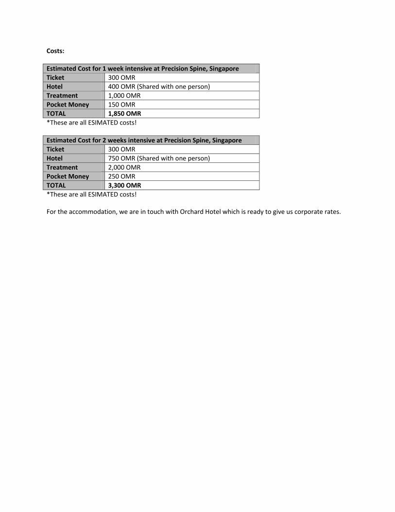
Costs:
Estimated Cost for 1 week intensive at Precision Spine, Singapore
Ticket 300 OMR
Hotel 400 OMR (Shared with one person)
Treatment 1,000 OMR
Pocket Money 150 OMR
TOTAL 1,850 OMR
*These are all ESIMATED costs!
Estimated Cost for 2 weeks intensive at Precision Spine, Singapore
Ticket 300 OMR
Hotel 750 OMR (Shared with one person)
Treatment 2,000 OMR
Pocket Money 250 OMR
TOTAL 3,300 OMR
*These are all ESIMATED costs! For the accommodation, we are in touch with Orchard Hotel which is ready to give us corporate rates.
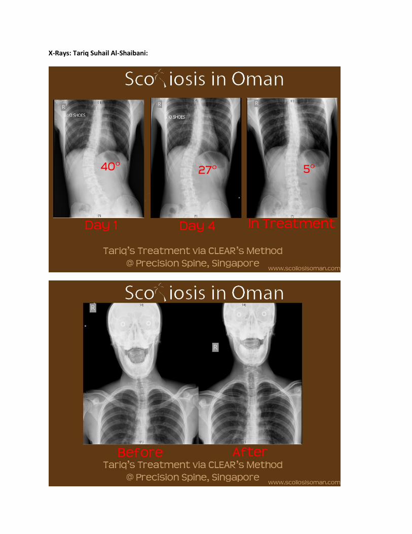
X-Rays: Tariq Suhail Al-Shaibani:
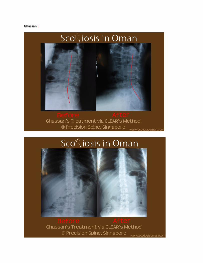
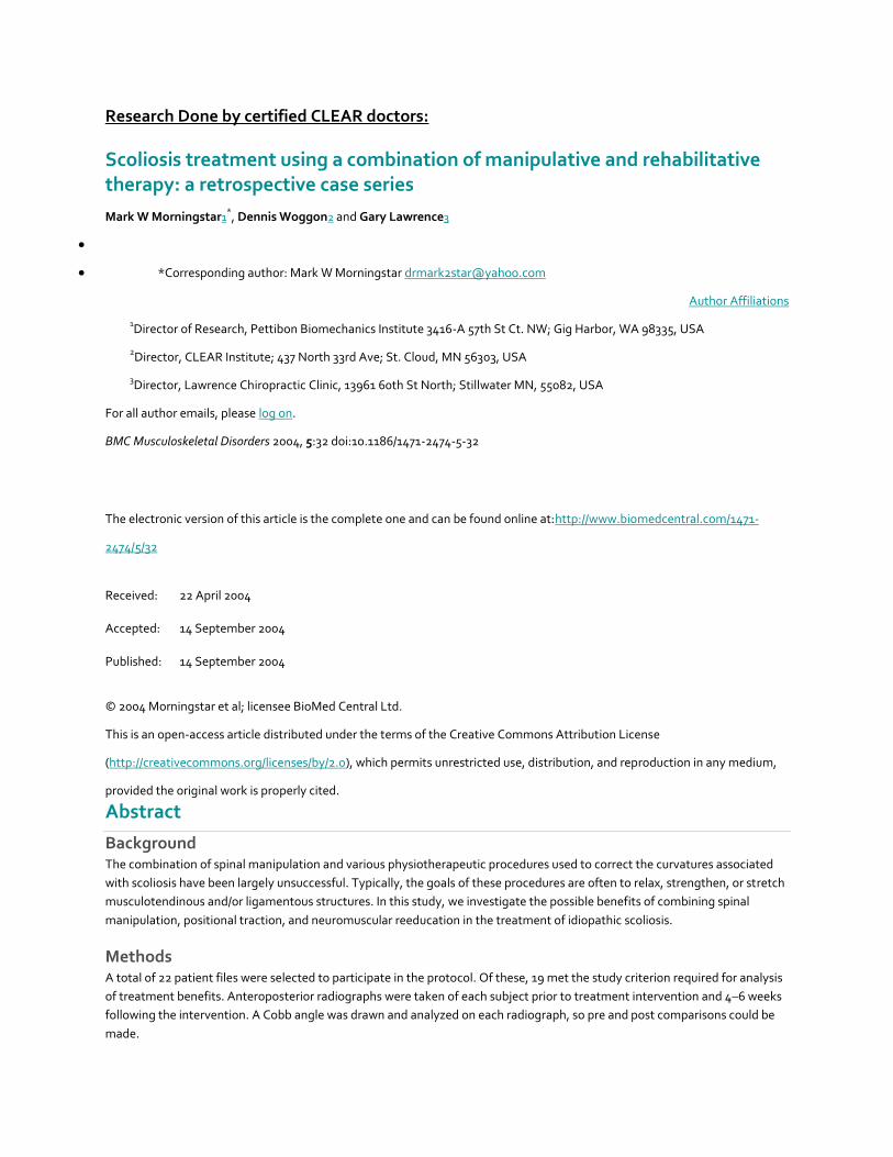
Ghassan :
Research Done by certified CLEAR doctors:
Scoliosis treatment using a combination of manipulative and rehabilitative therapy: a retrospective case series
Mark W Morningstar1*, Dennis Woggon2 and Gary Lawrence3
*Corresponding author: Mark W Morningstar [email protected]
Author Affiliations
1Director of Research, Pettibon Biomechanics Institute 3416-A 57th St Ct. NW; Gig Harbor, WA 98335, USA
2Director, CLEAR Institute; 437 North 33rd Ave; St. Cloud, MN 56303, USA
3Director, Lawrence Chiropractic Clinic, 13961 60th St North; Stillwater MN, 55082, USA
For all author emails, please log on.
BMC Musculoskeletal Disorders 2004, 5:32 doi:10.1186/1471-2474-5-32
The electronic version of this article is the complete one and can be found online at:http://www.biomedcentral.com/1471-
2474/5/32
Received: 22 April 2004
Accepted: 14 September 2004
Published: 14 September 2004
© 2004 Morningstar et al; licensee BioMed Central Ltd.
This is an open-access article distributed under the terms of the Creative Commons Attribution License
(http://creativecommons.org/licenses/by/2.0), which permits unrestricted use, distribution, and reproduction in any medium,
provided the original work is properly cited.
Abstract
Background The combination of spinal manipulation and various physiotherapeutic procedures used to correct the curvatures associated
with scoliosis have been largely unsuccessful. Typically, the goals of these procedures are often to relax, strengthen, or stretch
musculotendinous and/or ligamentous structures. In this study, we investigate the possible benefits of combining spinal
manipulation, positional traction, and neuromuscular reeducation in the treatment of idiopathic scoliosis.
Methods A total of 22 patient files were selected to participate in the protocol. Of these, 19 met the study criterion required for analysis
of treatment benefits. Anteroposterior radiographs were taken of each subject prior to treatment intervention and 4–6 weeks
following the intervention. A Cobb angle was drawn and analyzed on each radiograph, so pre and post comparisons could be
made.
Results After 4–6 weeks of treatment, the treatment group averaged a 17° reduction in their Cobb angle measurements. None of the
patients' Cobb angles increased. A total of 3 subjects were dismissed from the study for noncompliance relating to home care
instructions, leaving 19 subjects to be evaluated post-intervention.
Conclusions The combined use of spinal manipulation and postural therapy appeared to significantly reduce the severity of the Cobb angle
in all 19 subjects. These results warrant further testing of this protocol.
Background
In the MEDLINE- indexed literature, chiropractic treatment has shown to be largely ineffective at significantly reducing
scoliotic curvatures. Chiropractic treatment for scoliosis typically consists of spinal manipulation, electric stimulation, some
form of isotonic, active exercises, and shoe lifts [1]. However, Lantz et al [2] has shown that these procedures, when applied
over a one-year duration, were not sufficient to significantly reduce the Cobb angle of a scoliotic curvature.
The treatment in this study focuses on the reduction of scoliosis by manipulative and rehabilitative methods not commonly
used by most chiropractors. The major difference in this treatment compared to others is that stimulation of the involuntary
postural reflexes is utilized in the clinic setting as well as in home care. Many of the proposed etiologies of idiopathic scoliosis
are neurological in origin, including brain asymmetry [3], neural axis deformities [4], and central nervous system processing
errors [5]. Additionally, many coexistent neurological alterations are present in scoliosis patients, such as visual deficiency [6]
and decreased postural stability [7,8]. Therefore, the goals of the proposed treatment are not only to reduce the scoliotic
curvatures, but also to rehabilitate any underlying postural and neurological weaknesses or imbalances. Previous chiropractic
authors have investigated the effectiveness of various physiotherapeutic modalities in the treatment of scoliosis, such as
Pilates [9], stretching and massage [10], therapeutic exercises[11], orthotics [2], and ultrasound or electric stimulation [1]. The
purpose of the present study is to investigate any possible benefits from combining manipulative and rehabilitative techniques
from a randomized sample collected from various chiropractic facilities. Preliminary evidence [12] suggests that these
procedures may be beneficial for reducing the curvatures associated with scoliosis.
Methods
A nonrandomized set of 22 patients participated in the study. The age range of the subject group was 15–65 years of age. The
patients were selected from 3 different chiropractic facilities in the United States. Patients were evaluated according to their
chief complaint at initial presentation. Patients were excluded from the study if neoplasm, malignancy, fracture, scoliosis
secondary to genetic disorders, or previous arthrodesis were identified.
Each patient was examined radiographically for location and severity of scoliosis with standing anteroposterior full spine
imaging. All patients removed their shoes for the imaging. Cobb angles were drawn on each radiograph to identify the degree
of curvature present. A specific treatment plan was created based upon the results of each patient's radiographic
measurements before and after a sample trial of the proposed clinical procedures. Initially, standing lateral cervical, nasium,
lateral lumbar, and anteroposterior lumbopelvic views were taken. These views were taken to quantify forward head posture,
cervical lordosis, lumbar lordosis, the sacral base angle, and the Cobb angle of the major lateral curvature. We decided to use
the radiographic positioning and analysis outlined by Harrison et al [13-16], due to its previously published reliability. After
these images were taken, each patient was fitted with a 4-lb anterior headweight. They were instructed to walk around with
the headweight for 10 minutes. After 10 minutes, a follow-up lateral cervical radiograph was taken while wearing the anterior
headweight. The purpose of this lateral stress view is to evaluate the potential improvement in cervical lordosis and reduction
in forward head posture from using these procedures [17,18]. The basis for this aspect of the protocol is based upon the
inherent properties of a curved column. In the spine, lateral spinal displacements may occur when the normal sagittal spinal
curves [19-22] are flattened, reversed, or accentuated. These curves are necessary for the overall strength and flexibility of the
curved spinal column, according to the Delmas Index [23]. Therefore, the proposed treatment is intended to restore a normal
cervical and lumbar lordosis, and reduce forward head posture before the scoliotic curvatures are addressed.
The specific manipulative and rehabilitative procedures used in this study are designed to both reduce the scoliotic curvature
and theoretically retrain the involuntary neuromuscular, reflexive control of posture and balance. However, the specific
neurological effects, if any, remain to be investigated. Some of the procedures have been separately introduced or
tested [17,18,24-26].
The manipulative procedures included an upper cervical adjustment designed to mobilize the atlantal-occipital joint with the
use of a percussive instrument. This technique is shown in Figure 1. This technique is delivered to patients whose lateral
cervical radiographs demonstrated atlanto-occipital flexion. If atlanto-occipital extension was present on the initial lateral
cervical radiograph, a -Z drop piece was used to mobilize the occiput into flexion. This is also shown in Figure 1. An anterior
thoracic adjustment was administered with the patient's thoracic cage rotated opposite to the rotational displacement. A
thoracic drop piece was also used to mobilize and correct the smaller upper thoracic curvature. Side posture lumbopelvic
adjustments were delivered bilaterally to correct the rotational component of the pelvic misalignment. These side-posture
manipulations were performed on a 30°-incline bench to help pre-stress the spine out of its existing scoliotic curvatures.
Figure 1. The picture on the left demonstrates the mechanically assisted manipulation used when a
patient's skull is restricted in extension on lateral cervical radiograph. The picture on the right is the procedure used when the
skull flexion is restricted.
Certain traction procedures are also employed. These procedures are delivered using high-density foam blocks to pre-stress
the spine into specific positions so ligament deformation and stress-relaxation can take place. Supine pelvic blocking was
performed on each patient for 15 minutes. The position of the blocks was determined by each patient's pelvic rotation on
radiograph and posture analysis. One block is placed under the iliac crest of the posterior ilium, and the other block is placed
under the femoral head of the opposite, anteriorly-rotated ilium. Figure 2 illustrates the position of the pelvic blocks. The
rehabilitative procedures, demonstrated in Figure 3, included the use of head, shoulder, and hip weighting devices. These
devices may be used while simultaneously performing specific balancing exercises. These exercises include the use of a
Pettibon Wobble Chair®
and a Posturomed®
[17]. Tjernstrom et al [27] showed that repeated performance of a postural
alteration induces a long-term motor memory for achieving that novel postural position.
Figure 2. This picture shows the placement of the pelvic blocks for an anterior right ilium. The blocks are
placed opposite of the pelvic rotation.
Figure 3. The above picture illustrates a sample placement of the Pettibon Bodyweighting System. Here
we have an anterior headweight, right shoulderweight, and left-back and right-front hipweights.
The position of the body weighting was also determined radiographically for each patient. Initially, hipweights and
shoulderweights were applied according to each patient's posture analysis. Anteroposterior cervicothoracic and lumbopelvic
views were taken while wearing the head and body weighting. Since changes in spinal position are not reliably seen by
visualization [28,29], these stress radiographs were taken to confirm their corrective effects.
The attending physician treated each patient 3 times per week for the first 4–6 weeks. A total of 3 physicians performed the
treatment intervention for all patients. However, each patient did not receive identical treatment at all visits. The physicians
performed only those manipulative procedures that were deemed necessary based upon a visual posture analysis at the
beginning of each treatment session. However, the rehabilitative procedures remained constant throughout the study for all
patients.
Specific home care exercise programs were taught to each patient. These exercises were performed on a daily basis. Each
patient was instructed to wear the head and body weighting twice daily for 15 minutes each time. Secondly, each patient was
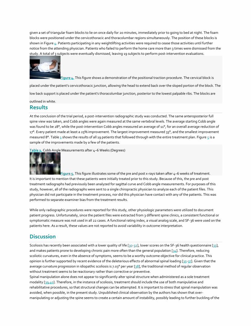
given a set of triangular foam blocks to lie on once daily for 20 minutes, immediately prior to going to bed at night. The foam
blocks were positioned under the cervicothoracic and thoracolumbar regions simultaneously. The position of these blocks is
shown in Figure 4. Patients participating in any weightlifting activities were required to cease those activities until further
notice from the attending physician. Patients who failed to perform the home care more than 3 times were dismissed from the
study. A total of 3 subjects were eventually dismissed, leaving 19 subjects to perform post-intervention evaluations.
Figure 4. This figure shows a demonstration of the positional traction procedure. The cervical block is
placed under the patient's cervicothoracic junction, allowing the head to extend back over the sloped portion of the block. The
low back support is placed under the patient's thoracolumbar junction, posterior to the lowest palpable ribs. The blocks are
outlined in white.
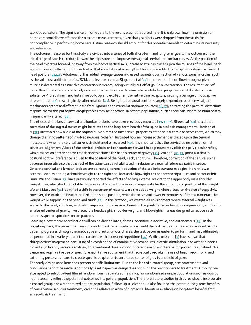
Results
At the conclusion of the trial period, a post-intervention radiographic study was conducted. The same anteroposterior full
spine view was taken, and Cobb angles were again measured at the same vertebral levels. The average starting Cobb angle
was found to be 28°, while the post-intervention Cobb angles measured an average of 11°, for an overall average reduction of
17°. Every patient made at least a 25% improvement. The largest improvement measured 33°, and the smallest improvement
measured 8°. Table 1 shows the results of all 19 patients that followed through with the entire treatment plan. Figure 5 is a
sample of the improvements made by a few of the patients.
Table 1. Cobb Angle Measurements after 4–6 Weeks (Degrees)
Figure 5. This figure illustrates some of the pre and post x-rays taken after 4–6 weeks of treatment.
It is important to mention that these patients were initially treated prior to this study. Because of this, the pre and post
treatment radiographs had previously been analyzed for sagittal curve and Cobb angle measurements. For purposes of this
study, however, all of the radiographs were sent to a single chiropractic physician to analyze each of the patient files. This
physician did not participate in the treatment process, nor did this physician have contact with any of the patients. This was
performed to separate examiner bias from the treatment results.
While only radiographic procedures were reported for this study, other physiologic parameters were utilized to document
patient progress. Unfortunately, since the patient files were extracted from 3 different spine clinics, a consistent functional or
symptomatic measure was not used in all 22 cases. A functional rating index, a visual analog scale, and SF-36 were used on the
patients here. As a result, these values are not reported to avoid variability in outcome interpretation.
Discussion
Scoliosis has recently been associated with a lower quality of life [30-32], lower scores on the SF-36 health questionnaire [33],
and makes patients prone to developing chronic pain more often than the general population [34]. Therefore, reducing
scoliotic curvatures, even in the absence of symptoms, seems to be a worthy outcome objective for clinical practice. This
opinion is further supported by recent evidence of the deleterious effects of abnormal spinal loading [35-37]. Given that the
average curvature progression in idiopathic scoliosis is 7.03° per year [38], the traditional method of regular observation
without treatment seems to be reactionary rather than corrective or preventive.
Spinal manipulation alone does not appear to significantly alter spinal structure when administered as a sole treatment
modality [39,40]. Therefore, in the instance of scoliosis, treatment should include the use of both manipulative and
rehabilitative procedures, so that structural changes can be attempted. It is important to stress that spinal manipulation was
avoided, when possible, in the present study. Unpublished clinical observation by the authors has shown that over-
manipulating or adjusting the spine seems to create a certain amount of instability, possibly leading to further buckling of the
scoliotic curvature. The significance of home care to the results was not reported here. It is unknown how the omission of
home care would have affected the outcome measurements, given that 3 subjects were dropped from the study for
noncompliance in performing home care. Future research should account for this potential variable to determine its necessity
and relevance.
The outcome measures for this study are divided into a series of both short-term and long-term goals. The outcome of the
initial stage of care is to reduce forward head posture and improve the sagittal cervical and lumbar curves. As the position of
the head migrates forward, or away from the body's vertical axis, increased strain is placed upon the muscles of the head, neck
and shoulders. Cailliet and Zohn indicated that an additional 10 inch/lbs of leverage is added to the spinal system in a forward
head posture [41,42]. Additionally, this added leverage causes increased isometric contraction of various spinal muscles, such
as the splenius capitis, trapezius, SCM, and levator scapula. Sjogaard et al [43] reported that blood flow through a given
muscle is decreased as a muscles contraction increases, being virtually cut off at 50–60% contraction. The resultant lack of
blood flow forces the muscle to rely on anaerobic metabolism. As anaerobic metabolism progresses, metabolites such as
substance P, bradykinin, and histamine build up and excite chemosensitive pain receptors, causing a barrage of nociceptive
afferent input [44], resulting in dysafferentation [45]. Being that postural control is largely dependant upon cervical joint
mechanoreceptors and afferent input from ligament and musculotendinous sources [46,47], correcting the postural distortions
responsible for this pathophysiologic process may be beneficial in patient populations, such as scoliosis, where postural control
is significantly altered [48].
The effects of the loss of cervical and lumbar lordosis have been previously reported [19,35-37]. Rhee et al [49] noted that
correction of the sagittal curves might be related to the long-term health of the spine in scoliosis management. Harrison et
al [35] illustrated how a loss of the sagittal curve alters the mechanical properties of the spinal cord and nerve roots, which may
change the firing patterns of involved neurons. Schafer illustrated how an increased demand is placed upon the cervical
musculature when the cervical curve is straightened or reversed [50]. It is important that the cervical spine be in a normal
structural alignment. A loss of the cervical lordosis and concomitant forward head posture may elicit the pelvo-ocular reflex,
which causes an anterior pelvic translation to balance the head's center of gravity [51]. Wu et al [52,53] point out that in
postural control, preference is given to the position of the head, neck, and trunk. Therefore, correction of the cervical spine
becomes imperative so that the rest of the spine can be rehabilitated in relation to a normal reference point in space.
Once the cervical and lumbar lordoses are corrected, coronal reduction of the scoliotic curvatures begins. Here this was
accomplished by adding a shoulderweight to the right shoulder and a hipweight to the anterior right ilium and posterior left
ilium. Wu and Essien [53] have previously reported the effects of adding external weight to the upper body via a shoulder
weight. They identified predictable patterns in which the trunk would compensate for the amount and position of the weight.
Wu and MacLeod [52] identified a shift in the center of mass toward the added weight when placed on the side of the pelvis.
However, the trunk and head remained in the same position, while the pelvis and lower extremities shifted to counteract the
weight while supporting the head and trunk [52]. In this protocol, we created an environment where external weight was
added to the head, shoulder, and pelvic regions simultaneously. Knowing the predictable patterns of compensatory shifting to
an altered center of gravity, we placed the headweight, shoulderweight, and hipweights in areas designed to reduce each
patient's specific spinal distortion patterns.
Learning a new motor coordination skill can be divided into 3 phases: cognitive, associative, and autonomous [54]. In the
cognitive phase, the patient performs the motor task repetitively to learn until the task requirements are understood. As the
patient progresses through the associative and autonomous phases, the task becomes easier to perform, and may ultimately
be performed in a variety of practical contexts with decreased repetitions [54]. While Lantz et al [2] have shown that
chiropractic management, consisting of a combination of manipulative procedures, electric stimulation, and orthotic inserts
did not significantly reduce a scoliosis, this treatment does not incorporate these physiotherapeutic procedures. Instead, this
treatment requires the use of specific rehabilitative equipment that theoretically recruits the use of head, neck, trunk, and
extremity postural reflexes to create specific adaptation to an altered center of gravity and field of gaze.
The study design used here does present specific limitations. Due to the lack of a control group, comparative data and
conclusions cannot be made. Additionally, a retrospective design does not blind the practitioners to treatment. Although we
attempted to select patient files at random from 3 separate spine clinics, nonrandomized sample populations such as ours do
not necessarily reflect the potential outcomes in a general population. Therefore, future studies in this area should incorporate
a control group and a randomized patient population. Follow-up studies should also focus on the potential long-term benefits
of conservative scoliosis treatment, given the relative scarcity of biomedical literature available on long-term benefits from
any scoliosis treatment.
Conclusions
Within the design limitations of the present study, the combined use of manipulative and neuromuscular rehabilitation
seemed to reduce scoliotic curvatures in 19 subjects by an average of 17°. This reduction took place within a 4 to 6-week
period. Although this treatment was not tested over the long term, the magnitude of the present results warrants further
studies into its effectiveness. This treatment should also be tested on specific types of scoliosis in follow-up trials. A long-term
investigation of this protocol is desirable.
Competing interests
This manuscript was submitted by Spinal Technologies, a BioMed Central institutional member. The rehabilitation equipment
used in this study is patented by Burl R Pettibon, DC and Spinal Technologies. MWM is the Director of Research for the
Pettibon Biomechanics Institute, and an active postgraduate instructor for Spinal Technologies. MWM does not receive
monetary compensation for this position. Rather, he is granted funding from Spinal Technologies to obtain biomedical
literature and statistician services. DW is a past postgraduate instructor for Spinal Technologies, founder and director of the
CLEAR Institute, and CEO of the Flex Neck Company. GL is an active postgraduate instructor for Spinal Technologies. The
authors receive lecture fees for each continuing education seminar conducted. All 3 authors maintain private chiropractic
practices from where all of the patient files in this study were taken. None of the above companies donated, funded, or
reimbursed any monies or equipment for this study. None of the authors have any ownership in Spinal Technologies or its
subsidiary companies, and none will gain any financial interest as a result of this paper.
Authors' contributions
Each author worked on one-third of the patient population. The first author was responsible for collecting the data and putting
our findings into written format.
Acknowledgements
The authors would like to thank Darin Weeks and Cassi Little for procedure demonstration.
References
1. Feise RJ: An inquiry into chiropractors' intention to treat adolescent idiopathic scoliosis: A telephone survey.
J Manipulative Physiol Ther 2001, 24:177-182. PubMed Abstract | Publisher Full Text
2. Lantz CA, Chen J: Effect of chiropractic intervention on small scoliotic curves in younger subjects: a time-series
cohort design.
J Manipulative Physiol Ther 2001, 24:385-393. PubMed Abstract | Publisher Full Text
3. Niesluchowski W, Dabrowska A, Kedzior K, Zagrajek T: The potential role of brain asymmetry in the development
of adolescent idiopathic scoliosis: a hypothesis.
J Manipulative Physiol Ther 1999, 22:540-544. PubMed Abstract | Publisher Full Text
4. Dobbs MB, Lenke L, Szymanski DA, Morcuende JA, Weinstein SL, Bridwell KH, Sponseller PD: Prevalence of neural
axis abnormalities in patients with infantile idiopathic scoliosis.
J Bone Joint Surg Am 2002, 84-A:2230-2234. PubMed Abstract | Publisher Full Text
5. Lowe TG, Edgar M, Margulies JY, Miller NH, Raso VJ, Reinker KA, Rivard CH: Etiology of idiopathic scoliosis:
current trends in research.
J Bone Joint Surg Am 2000, 82-A:1157-1168. PubMed Abstract | Publisher Full Text
6. Catanzariti JF, Salomez E, Bruandet JM, Thevenon A: Visual Deficiency and Scoliosis.
Spine 2001, 26:48-52. PubMed Abstract | Publisher Full Text
7. Nault ML, Allard P, Hinse S, Le Blanc R, Caron O, Labelle H, Sadeghi H: Relations between standing stability and
body posture parameters in adolescent idiopathic scoliosis.
Spine 2002, 27:1911-1917. PubMed Abstract | Publisher Full Text
8. Chen PQ, Wang JL, Tsuang YH, Liao TL, Huang PI, Hang YS: The postural stability control and gait pattern of
idiopathic scoliosis adolescents.
Clin Biomech 1998, 13(Suppl 1):S52-S58. Publisher Full Text
9. Blum CL: Chiropractic and pilates therapy for the treatment of adult scoliosis.
J Manipulative Physiol Ther 2002, 25:e3. PubMed Abstract | Publisher Full Text
10. Tarola GA: Manipulation for the control of back pain and curve progression in patients with skeletally mature
idiopathic scoliosis: two cases.
J Manipulative Physiol Ther 1994, 17:253-257. PubMed Abstract
11. Golembiewski GV, Catanzaro DJ: Scoliosis reduction utilizing an exercise.
J Vertebral Subluxation Res 2001, 4:31-36.
12. Morningstar MW, Strauchman MN, Gilmour G: Idiopathic Scoliosis Treatment Using the Pettibon Corrective
Procedures: A Case Report.
J Chiropr Med, in press.
13. Harrison DE, Harrison DD, Colloca CJ, Betz J, Janik TJ, Holland B: Repeatability over time of posture, radiograph
positioning, and radiograph line drawing: an analysis of six control groups.
J Manipulative Physiol Ther 2003, 26:87-98. PubMed Abstract | Publisher Full Text
14. Troyanovich SJ, Harrison SO, Harrison DD, Harrison DE, Payne MR, Janik TJ, Holland B:Chiropractic biophysics
digitized radiographic mensuration analysis of the anteroposterior lumbopelvic view: a reliability study.
J Manipulative Physiol Ther 1999, 22:309-315. PubMed Abstract | Publisher Full Text
15. Jackson BL, Harrison DD, Robertson GA, Barker WF: Chiropractic biophysics lateral cervical film analysis
reliability.
J Manipulative Physiol Ther 1993, 16:384-391. PubMed Abstract
16. Troyanovich SJ, Harrison DE, Harrison DD, Harrison SO, Janik T, Holland B: Chiropractic biophysics digitized
radiographic mensuration analysis of the anteroposterior cervicothoracic view: a reliability study.
J Manipulative Physiol Ther 2000, 23:476-482. PubMed Abstract | Publisher Full Text
17. Saunders ES, Woggon D, Cohen C, Robinson DH: Improvement of cervical lordosis and reduction of forward head
posture with anterior head weighting and proprioceptive balancing protocols.
J Vertebral Subluxation Res 2003, 4:000.
18. Morningstar MW, Strauchman MN, Weeks DA: Spinal manipulation and anterior headweighting for the correction
of forward head posture and cervical hypolordosis: a pilot study.
J Chiropr Med 2003, 2:51-55.
19. Harrison DE, Harrison DD, Troyanovich SJ, Harmon S: A normal spinal position: it's time to accept the evidence.
J Manipulative Physiol Ther 2000, 23:623-644. PubMed Abstract | Publisher Full Text
20. Harrison DD, Janik TJ, Troyanovich SJ, Harrison DE, Colloca CJ: Evaluation of the assumptions used to derive an
ideal normal cervical spine model.
J Manipulative Physiol Ther 1997, 20:246-254. PubMed Abstract
21. Harrison DE, Janik TJ, Harrison DD, Cailliet R, Harmon SF: Can the thoracic kyphosis be modeled with a simple
geometric shape? The results of circular and elliptical modeling in 80 asymptomatic patients.
J Spinal Disord Tech 2002, 15:213-220. PubMed Abstract | Publisher Full Text
22. Harrison DD, Cailliet R, Janik TJ, Troyanovich SJ, Harrison DE, Holland B: Elliptical modeling of the sagittal lumbar
lordosis and segmental rotation angles as a method to discriminate between normal and low back pain
subjects.
J Spinal Disord 1998, 11:430-439. PubMed Abstract
23. Kapandji IA: The physiology of the joints. The trunk and vertebral column. Volume 3. 5th edition. Churchill Livingstone;
1974:235-236.
24. Morningstar MW: Cervical hyperlordosis correction: a novel treatment method for mid thoracic pain.
J Chiropr Med 2003, 2:111-115.
25. Morningstar MW: Strength gains through lumbar lordosis restoration.
J Chiropr Med 2003, 2:137-141.
26. West DT, Mathews RS, Miller MR, Kent GM: Effective management of spinal pain in one hundred seventy-seven
patients evaluated for manipulation under anesthesia.
J Manipulative Physiol Ther 1999, 22:299-308. PubMed Abstract | Publisher Full Text
27. Tjernstrom F, Fransson PA, Hafstrom A, Magnusson M: Adaptation of postural control to perturbations- a process
that initiates long-term motor memory.
Gait Posture 2002, 15:75-82. PubMed Abstract | Publisher Full Text
28. Johnson GM: The correlation between surface measurements of head and neck posture and the anatomic
position of the upper cervical vertebrae.
Spine 1998, 23:921-927. PubMed Abstract | Publisher Full Text
29. Fedorak C, Ashworth N, Marshall J, Paull H: Reliability of the visual assessment of cervical and lumbar lordosis:
how good are we?
Spine 2003, 28:1857-1859. PubMed Abstract | Publisher Full Text
30. Shapiro GS, Taira G, Boachie-Adjei O: Results of surgical treatment of adult idiopathic scoliosis with low back
pain and spinal stenosis: a study of long-term clinical radiographic outcomes.
Spine 2003, 28:358-363. PubMed Abstract | Publisher Full Text
31. Danielsson AJ, Nachemson AL: Radiologic findings and curve progression 22 years after treatment for adolescent
idiopathic scoliosis: comparison of brace and surgical treatment with matching control group of straight
individuals.
Spine 2001, 26:516-525. PubMed Abstract | Publisher Full Text
32. Freidel K, Petermann F, Reichel D, Steiner A, Warschburger P, Weiss HR: Quality of life in women with idiopathic
scoliosis.
Spine 2002, 27:E87-E91. PubMed Abstract | Publisher Full Text
33. Schwab F, Dubey A, Pagala M, Gamez L, Farcy JP: Adult scoliosis: a health assessment analysis by SF-36.
Spine 2003, 28:602-606. PubMed Abstract | Publisher Full Text
34. Weinstein SL, Dolan LA, Spratt KF, Peterson KK, Spoonamore MJ, Ponseti IV: Health and function of patients with
untreated idiopathic scoliosis: a 50-year natural history study.
JAMA 2003, 289:559-567. PubMed Abstract | Publisher Full Text
35. Harrison DE, Cailliet R, Harrison DD, Troyanovich SJ, Harrison SO: A review of biomechanics of the central nervous
system- part III: spinal cord stresses from postural loads and their neurologic effects.
J Manipulative Physiol Ther 1999, 22:399-410. PubMed Abstract | Publisher Full Text
36. Harrison DE, Cailliet R, Harrison DD, Troyanovich SJ, Harrison SO: A review of biomechanics of the central nervous
system-part II: spinal cord strains from postural loads.
J Manipulative Physiol Ther 1999, 22:322-332. PubMed Abstract | Publisher Full Text
37. Harrison DE, Cailliet R, Harrison DD, Troyanovich SJ, Harrison SO: A review of biomechanics of the central nervous
system-part I: spinal canal deformations resulting from changes in posture.
J Manipulative Physiol Ther 1999, 22:227-234. PubMed Abstract | Publisher Full Text
38. Chuah SL, Kareem BA, Selvakumar K, Oh KS, Borhan Tan A, Harwant S: The natural history of scoliosis: curve
progression of untreated curves of different aetiology, with early (mean 2 year) follow up in surgically treated
curves.
Med J Malaysia 2001, 56(Suppl C):37-40. PubMed Abstract
39. Harrison DD, Jackson BL, Troyanovich SJ, Robertson G, DeGeorge D, Barker WF: The efficacy of cervical extension-
compression traction combined with diversified manipulation and drop table adjustments in the rehabilitation
of cervical lordosis: a pilot study.
J Manipulative Physiol Ther 1994, 17:454-464. PubMed Abstract
40. Harrison DD, Harrison DE, Troyanovich SJ: Structural Rehabilitation of the spine and posture: rationale for
treatment beyond the resolution of symptoms.
J Manipulative Physiol Ther 1998, 21:37-50. PubMed Abstract
41. Cailliet R:
In Neck and arm pain. 2nd edition. Edited by Davis FA. 1981.
42. Zohn DA:
In Musculoskeletal pain diagnosis and treatment. 2nd edition. Edited by Little Brown. 1988.
43. Sjogaard G, Savard G, Juel C: Muscle blood flow during isometric activity and its relation to muscle fatigue.
Eur J Appl Physiol Scand 1988, 57:327-335.
44. Travell JG, Simons D: Myofascial pain and dysfunction: the trigger point manual. Williams & Wilkins; 1973.
45. Seaman DR, Winterstein JF: Dysafferentation: a novel term to describe the neuropathophysiological effects of
joint complex dysfunction. A look at likely mechanisms of symptom generation.
J Manipulative Physiol Ther 1998, 21:267-280. PubMed Abstract
46. Grod JP, Diakow PR: Effect of neck pain on verticality perception: A cohort study.
Arch Phys Med Rehabil 2002, 83:412-415. PubMed Abstract | Publisher Full Text
47. Dietz V, Muller R, Colombo G: Locomotor activity in spinal man: significance of afferent input from joint and load
receptors.
Brain 2002, 125:2626-2634. PubMed Abstract | Publisher Full Text
48. Gauchard GC, Lascombes P, Kuhnast M, Perrin PP: Influence of different types of progressive idiopathic scoliosis
on static and dynamic postural control.
Spine 2001, 26:1052-1058. PubMed Abstract | Publisher Full Text
49. Rhee JM, Bridwell KH, Won DS, Lenke LG, Chotigavanichaya C, Hanson DS: Sagittal plane analysis of adolescent
idiopathic scoliosis.
Spine 2002, 27:2350-2356. PubMed Abstract | Publisher Full Text
50. Schafer RC: Clinical biomechanics: musculoskeletal actions and reactions. 2nd edition. Williams & Wilkins; 1987.
51. Lewit K:
Muscular and articular factors in movement restriction. Manual Medicine. 1985, 1:83-85.
52. Wu G, MacLeod M: The control of body orientation and center mass location under asymmetrical loading.
Gait Posture 2001, 13:95-101. PubMed Abstract | Publisher Full Text
53. Wu G, Essien I: The regulation of human orientation and center of mass in the frontal plane during upright
stance.
In Proceedings of North America Congress of Biomechanics. Waterloo, Canada 1988.
54. Harbst KB, Wilder PA: Neurophysiologic, motor control, and motor learning basis of closed kinetic chain
exercise.
Orthop Phys Ther Clin N Am 2000, 9:137-149.