Embed Size (px)
Citation preview

Hindawi Publishing CorporationProstate CancerVolume 2013, Article ID 103547, 11 pageshttp://dx.doi.org/10.1155/2013/103547
Review ArticleHypofractionated External-Beam Radiotherapy forProstate Cancer
L. Chinsoo Cho,1 Robert Timmerman,2 and Brian Kavanagh3
1 University of Minnesota Medical Center, 420 Delaware Street SE, MMC-494, Minneapolis, MN 55455, USA2Department of Radiation Oncology, University of Texas Southwestern Medical Center at Dallas, 5801 Forest Park Road,Dallas, TX 75390, USA
3Department of Radiation Oncology, University of Colorado School of Medicine, Anschutz Cancer Pavilion, Campus Mail Stop F-706,1665 Aurora Court, Suite 1032, Aurora, CO 80045, USA
Correspondence should be addressed to Brian Kavanagh; [email protected]
Received 30 April 2012; Accepted 13 October 2012
Academic Editor: Rami Ben-Yosef
Copyright © 2013 L. Chinsoo Cho et al.This is an open access article distributed under the Creative CommonsAttribution License,which permits unrestricted use, distribution, and reproduction in any medium, provided the original work is properly cited.
There are radiobiological rationales supporting hypofractionated radiotherapy for prostate cancer. The recent advancements intreatment planning and delivery allow sophisticated radiation treatments to take advantage of the differences in radiobiologyof prostate cancer and the surrounding normal tissues. The preliminary results from clinical studies indicate that abbreviatedfractionation programs can result in successful treatment of localized prostate cancer without escalation of late toxicity.
1. Introduction
Prostate cancer is the most common cancer diagnosed inAmericanmen after non-melanomatous skin cancer. Accord-ing to the American Cancer Society estimate, there will bemore than 241,000 new cases of prostate cancer in the UnitedStates in 2012. Approximately 28,000 men in the USA willdie of prostate cancer, making it the second leading cause ofcancer death in this country [1].
In most cases the prostate cancer is organ-confined atthe time of initial diagnosis [2]. Radical prostatectomy andradiotherapy, either given as a seed implant or external beamradiation therapy, are the accepted standard options for treat-ing the primary tumor itself, and androgen deprivation maybe added selectively for certain cases with an intermediate orhigh risk of dissemination based on clinical and pathologicfeatures evident at the time of diagnosis. Regarding the spe-cific option of external beam radiotherapy, the current widelyaccepted standard regimen for organ-confined prostate can-cer in the USA involves approximately eight weeks of frac-tionated treatments with a daily dose of 1.8–2.0Gy to a totaldose in the range of 70–80Gy.At some centers the treatments,also called fractions, are given over 9-10 weeks [3].
Although many patients have been successfully treatedwith radiotherapy regimens of this nature, the optimal radi-ation schedule for the curative treatment of prostate cancerremains an unsettled question. For patients with clinicalfeatures suggesting at least an intermediate level of aggres-siveness, a moderate dose escalation has been demonstratedto improve biochemical control with acceptable toxicity usingcontemporary radiotherapy techniques [4, 5]. Unfortunately,dose escalation using a conventionally fractionated treatmentschedule requires a lengthened treatment course that is lessconvenient for patients and more costly for government andprivate insurance carriers. Emerging evidence accumulatingfrom multiple recent studies indicates that more convenientand efficient shortened courses of radiotherapy for prostatecancer yield outcomes that are equivalent and possiblysuperior to the lengthier standard regimens. The scientificrationale for such “hypofractionated” treatment lies in theunique radiobiologic properties of prostate cancer.
2. Radiobiologic Rationale
The radiobiological basis of hypofractionation for prostatecancer assumes that the prostate cancer cells respond to
2 Prostate Cancer
radiotherapy in a manner that can be mathematically mod-eled with a classic linear-quadratic equation:
𝑆 = 𝑆0𝑒−𝛼𝐷−𝛽𝐷
2
, (1)
where 𝐷 is the dose given, and 𝑆 represents the cell survivalafter radiotherapy for initial cell population, 𝑆
𝑜. The con-
stants, 𝛼 and 𝛽, represent linear and quadratic componentsof the equation [6]. The equation may be rearranged toyield an estimate of the relative biological potency, called thebiological effective dose (BED), of a fractionated course oftherapy involving 𝑛 individual treatments:
BED = 𝑛𝐷(1 + 𝐷𝛼/𝛽) . (2)
The ratio 𝛼/𝛽, which has units of Gy, characterizes theradiation sensitivity of a particular cell type. The 𝛼/𝛽 ratio isgenerally assumed to be approximately 10Gy formost tumorsand early-responding normal tissues and less than 5Gy forlate-responding tissues.
Conventional fractionation of 1.8–2.0Gy/day is based onthe premise that the therapeutic ratio, defined as the chanceof eradicating tumor cells divided by the risk of normaltissue injury in late-responding normal tissue, is optimized byusing small doses per fraction. The reason is that the tumorcell response, proportional to the BED as determined in theequation above, is generally less influenced by fraction sizethan is the BED for the late-responding normal tissues thatsurround the targeted tumor. However, in prostate cancer,tumor cellsmay have lower 𝛼/𝛽 than the surrounding normaltissues, and the opposite condition applies. The 𝛼/𝛽 ratiofor prostate cancer is widely believed to be in the range 1–4Gy [7–12]. In this context, it has been hypothesized that thetherapeutic ratio would be enhanced by increasing the doseper fraction to the prostate cancer above the standard range[8–12].
It is noteworthy that the analyses that have yielded low𝛼/𝛽 ratios for prostate cancer have sometimes involvedcomparisons of low dose rate brachytherapy with externalbeam radiotherapy, and assorted mathematical assumptionshave been made [26–29] A comprehensive discussion ofrepair kinetics and other factors that influence the 𝛼/𝛽estimates is beyond the scope of the present paper, and thereader is referred to some of the various studies related tothis issue for additional discussion [10, 28, 30–35]. An addedcomplexity is that it is not clear whether the traditional linearquadratic model continuing to model closely the radiationdose-response in tumors applies for fractional doses in therange of 6–8Gy or higher [36–38]. Regardless, the aforemen-tioned theoretical analyses, taken together, have informed thedevelopment of the hypothesis that there might be mean-ingful clinical advantage in the administration of radiationdoses of greater than 2Gy per fraction in the managementof prostate cancer with external beam radiation therapy, andnumerous clinical studies related to this hypothesis have beenreported.
3. Clinical Application of Hypofractionationfor Prostate Cancer
One of the early experiences using hypofractionation forprostate cancer came from Europe. Over 200 patients weretreated at St. Thomas Hospital in London with hypofraction-ated radiotherapy to a dose of 55Gy in 12 fractions and laterto doses of 36Gy in 6 fractions with low rectal and urologicalcomplications [39, 40]. Investigators of this retrospectivereview advocated 6 fractions in 3 weeks. Since the earlyreports of hypofractionation, there has been a steady increasein reports of hypofractionated radiotherapy for prostate can-cer. Some have decreased the number of fractions modestly,and others have reduced it to only five sessions [41–50].
In the United States, Kupelian et al. [47] first reportedtheir institutional experience using hypofractionation totreat safely an initial 100 consecutive patients with localizedprostate cancer. Subsequently, an expanded experienceinvolving 770 patientswas reported [48]. All patients receivedintensity-modulated radiation therapy (IMRT) guided bydaily prostate localization with a transabdominal ultrasoundsystem. Patients were treated to total dose of 70Gy in 28daily fractional dose of 2.5 Gy. Biochemical failure, usingboth the ASTRO consensus definition [51] and the RTOGPhoenix definition (nadir+2 ng/mL) [52], was the studyendpoint. The Radiation Therapy Oncology Group (RTOG)Morbidity System was used to assess treatment-relatedgastrointestinal (GI) and genitourinary (GU) morbidity.Fifty-one patients (51%) received androgen deprivationtherapy for a period no longer than 6 months. The medianfollowup was 66 months.The 5-year biochemical relapse freesurvival (bRFS) rates were 85% by the ASTRO Consensusdefinition and 88% by the RTOG Phoenix definition. Resultswere also reported according to prognostic groups. For low,intermediate and high-risk disease, the 5-year bRFS ratesusing ASTRO consensus definition were 97%, 88%, and70%. The corresponding 5-year bRFS rates using the RTOGPhoenix definition were 97%, 93%, and 75%, respectively.The acute rectal toxicity scores were 0 in 20, 1 in 61, and 2 in19 patients. A great majority of patients experienced grade0-1 acute gastrointestinal (GI)/genitourinary (GU) toxicities.The actuarial late grade 3 rectal and urinary toxicity rate at 5years was 3% and 1%, respectively. Routine implementationof IMRT with tight PTVmargins and IGRT are likely to havecontributed to reduce the treatment-related toxicities.
A recently completed RTOG 0415 Phase III trial assignedrandomized patients to either the hypofractionation treat-ment strategy from the report by Kupelian et al. (70Gy in 28fractions) or to 73.8Gy in 41 fractions of 1.8 Gy daily doses.Three-dimensional conformal radiotherapy and IMRT wereallowed. The trial was restricted to those patients with low-risk prostate cancer (T1-2, and PSA <10 ng/mL, and Gleasonscore 2–6). Mature results of the study are not yet available.
A Canadian hypofractionated randomized trial hasbeen reported by Lukka et al. [13] The trial compared aconventional dose of 66Gy in 33 fractions, considered lowby contemporary standards, to a hypofractionated regimenof 52.5Gy in 20 fractions in men with low- and intermediate-risk prostate cancer. The dose per fraction was 2.625Gy,
Prostate Cancer 3
slightly higher than the fractional dose used by Kupelian etal. CT based treatment planning was done but contemporarytechnique such as IMRT was not used. In this trial, the 5-yearrate of failure (biochemical or clinical) was higher in thehypofractionated arm compared to the standard fraction-ation arm (60% versus 53%, 𝑃 < 0.05). The inferior resultseen with hypofractionated treatment may be explained bythe fact that for any 𝛼/𝛽 ratio >0.2, the BED of 52.5Gy in20 fractions is expected to be lower than the BED of 66Gyin 33 fractions. At a median followup of 5.7 years, there wasno difference in 5-year actuarial rate of late grade 3 or higherGI/GU toxicity between the two arms.
Pollack et al. [14] reported results of a hypofractionatedtrial in patients with intermediate to high-risk features. Thefractional dose was 2.7Gy per treatment. Intermediate riskwas defined as total Gleason’s score of 7, PSA between10–20 ng/mL, or ≥3 biopsy cores of combined Gleason’s score≥5, as long as no high-risk features were present. High riskwas defined as Gleason’s score 8–10, Gleason’s score 7 in ≥4cores, clinical T3 disease, or PSA >20 ng/mL. Up to 4 monthsof androgen-deprivation prior to randomization were per-mitted. However, it was discontinued after enrollment forpatients with intermediate risk and continued for 2 years forthose with high-risk. The clinical target volume (CTV) forintermediate-risk patients included the prostate and proximalseminal vesicles (approximately 9mm). In high-risk patients,the CTV included at least 50% of the seminal vesicles,prostate, and any extraprostatic extension. In the high-riskpatients, separate CTV and PTV were designed to treatthe distal portions of the seminal vesicles, periprostatic,periseminal vesicle, external iliac, obturator, and internal iliaclymph nodes. Treatments were delivered using IMRT withhypofractionated doses to normal tissues calculated usingan estimated 𝛼/𝛽 ratio of 1.5. The trial compared 76Gy inconventional 2.0Gy fractions to 70.2Gy in 2.7Gy fractions.The hypofractionated arm was estimated to be equivalent to84.4Gy in 2.0Gy fractions and designed to be equivalentto 8Gy increase over the standard fractionated total dose of76Gy.The 5-year result of the trial was recently reported [15].There were 303 assessable patients entered between 2002 and2006. There were 152 patients assigned to receive standardfractionation and 151 patients assigned to receive hypofrac-tionation. Median followup was greater than 60 monthsin both arms. The rates of biochemical failure using theNadir+2 ng/mL definition and clinical failure, consisting ofeither local-regional failure or distant metastasis (LRF/DM),and deaths without failure were reported. There were nostatistically significant differences between the treatmentarms in terms of biochemical failure, any failure, or lateside effects. The 5-year rates of any failure for standardfractionation and hypofractionation were 14.4% and 13.9%,respectively.There were no statistically significant differencesin GI toxicity between the arms. However, the GU toxicitywas higher among patients who received hypofractionation at5 years. Grade ≥2 gastrointestinal toxicities were seen in 5%and 6.8% of the conventional and hypofractionated groups,respectively, but transient genitourinary grade ≥2 toxicitiesoccurred in 8.3% and 18.3%, respectively (𝑃 = 0.028), thoughthey persisted in less than 10% at five years [60]. Factors
associated with increased GU toxicity included pretreatmentAUA symptoms score. Patients with AUA greater than 10at baseline had increased risk for grade ≥2 toxicities, 11%versus 34%, respectively. Length of ADT did not influenceGU toxicity.
An Australian trial reported by Yeoh et al. compared amodest dose of 64Gy in 32 treatments to hypofractionatedarm of 55Gy in 20 treatments in men with favorable-riskprostate cancer [16, 61]. The fractional dose in this trial was2.75Gy. Two hundred seventeen patients with T1-2 prostatecarcinomas were randomized to either the standard or thehypofractionated arm between 1996 and 2006. Treatmentswere predominantly four-field box technique with cus-tomized blocks using 6–23MV photons. At a median fol-lowup of 90 months, biochemical relapse-free survival(bRFS) was significantly better with hypofractionation whenPhoenix definition was used (53% versus 34%, 𝑃 < 0.5).However, there was no difference in bRFS rates when olderASTROdefinitionwas used (44%versus 44%).Morbiditywasmeasured with the LENT-SOMA questionnaires. Gastroin-testinal and genitourinary toxicity did not differ significantlybetween fractionation schedules. Investigators found thatthe conventional fractionation independently predicted forworse biochemical failure and genitourinary symptoms at 4years.
Coote et al. [17] reported a British dose escalation studyfor prostate cancer using hypofractionated IMRT regimenusing a higher dose per fraction. There were 60 patients withT2-3N0M0 adenocarcinoma of prostate, and either Gleason’sscore ≥7 or PSA 20–50 ng/L. Patients received 57–60Gy tothe prostate in 19-20 fractions using five-field IMRT. Alltreatments were delivered with a fractional dose of 3Gy, 5days per week. The target volumes included prostate andseminal vesicle without incorporation of regional lymphnodes. All patients received neoadjuvant hormonal therapyfor 3months before radiotherapy to amaximum of 6months.Toxicity was assessed 2 years post radiotherapy using theRTOG criteria, LENT/SOMA, and UCLA prostate indexassessment tools. There was no acute RTOG grade 3 or 4toxicity. At 2 years, there were 4% grade 2 GI and 4.25%grade 2 GU toxicity.There was no grade 3 or 4 GI toxicity butone patient developed grade 3 GU toxicity at 2 years. UCLAindex data showed a slight improvement in urinary func-tion at 2 years compared with pretreatment. LENT/SOMAassessments demonstrated worsening of bowel function at 2years. Patients receiving 60Gy were more likely to developproblems with bowel function than those receiving 57Gy.
Martin et al. [18] reported a prospective Phase II trialalso using 3Gy per fraction to treat 92 patients between 2001and 2004. Eligible patients had clinical stage T1c-T2c N0M0with Gleason’s score ≥ 6 and various PSA levels. The studywas designed to maintain a biologic equivalent rectal dose of120Gy
3, assuming 𝑎/𝛽 ratio of 3 for late normal tissue effects.
The treatments were delivered with 3Gy per fraction, 5 daysperweek for 4 consecutiveweeks to aminimumdose of 60Gyto the CTV (entire prostate and the base of seminal vesicles).With a median follow-up of 38 months, severe acute toxicity(grade 3–4) was rare, occurring in only 1 patient. There wasno ≥3 late toxicity. The rate of biochemical control was 97%

4 Prostate Cancer
at 14 months by the Phoenix definition and 76% at 3 years bythe older ASTRO consensus definition.
Recently, Dearnaley et al. [19] reported the preplannedinterim safety analysis of randomized multicenter Conven-tional or Hypofractionated High-Dose Intensity-ModulatedRadiotherapy for Prostate Cancer (CH HiP) trial. At thetime of analysis, 444 patients with localized prostate cancerbetween 2002 and 2006 had been enrolled. The eligiblepatients had clinical T1b–T3a N0M0 prostate cancer, PSA<30 ng/mL, Gleason’ score ≤ 7, WHO performance status of0-1, and estimated risk of lymph-node involvement <30%.Patients received androgen suppression for 3–6 monthsbefore and during radiotherapy, but it was optional for menwith low-risk disease. Patients in the control group received74Gy in 37 fractions. Patients in the hypofractionated groupsreceived either 60Gy in 20 fractions or 57Gy in 19 fractions.All treatments were 5 fractions per week. Treatment for bothstandard and hypofractionated was planned and deliveredusing an integrated simultaneous-boost techniquewith targetvolumes designed to deliver 80% of the total dose to theprostate and base or all seminal vesicles 96% to the prostatewith 0.5–1 cm margin, and 100% to the prostate with a 0–0.5 cm margin. Regional lymph nodes were not included inthe treatment target volumes. With median follow-up of 50.5months, 4.3% in the standard fractionation (74Gy) group hadGI toxicity RTOG grade ≥2 at 2 years. The GI toxicity waslower in both hypofractionated treatment arms: 3.6% in the60Gy group and the 1.4% in the 57Gy group. RTOG GUtoxicity grade ≥2 were seen in 2.2% of patients in the 74Gygroup, 2.2% of patients in the 60Gy group, and none in the57Gy group at 2 years. There were no statistically significantdifferences in cumulative incidence of side-effects betweenthe groups. A future report will include mature biochemicalcontrol rates data.
A phase III trial from Italycompared the toxicity andefficacy of hypofractionated (62Gy in 3.1 Gy daily doses, 4times per week) versus conventional fractionation radiother-apy (80Gy in 2Gy daily doses, 5 times per week) in patientswith high-risk prostate cancer [20].
One hundred sixty-eight patients were randomized toreceive three-dimensional conformal radiotherapy to theprostate and seminal vesicles. All patients received a 9-monthcourse of total androgen deprivation. The median follow-up was 32 and 35 months in the hypofractionation andconventional fractionation arms, respectively. No differencewas found for late toxicity between the two treatment groups.There were 17% and 16% grade 2 GI toxicity and 14% and11% GU toxicity at 3 years in the hypofractionation andconventional fractionation groups, respectively. The 3-yearfreedom from biochemical failure rates were 87% and 79% inthe hypofractionation and conventional fractionation groups,respectively (𝑃 = 0.035). The authors concluded that withequivalent late toxicity between the two treatment groups, thehypofractionated treatment resulted in better PSA control.
Other investigators have used similar 3Gy per fractions inhypofractionated treatments. A randomized trial comparingthe toxicity and efficacy of hypofractionated and convention-ally fractionated external-beam radiotherapy from Lithuaniawas reported by Norkus et al. [21, 22] Forty-four patients in
the standard treatment arm were irradiated with 74Gy in 37fractions, and 47 patients in the hypofractionated arm weregiven 13 fractions of 3Gy with additional 4 fractions of 4.5 Gyto a total dose of 57Gy. The clinical target volume includesthe prostate and a base of seminal vesicles. There was nosignificant difference in PSA response during the first-yearfollow-up. No acute grade 3 or 4 toxicities were observed.The grade 2 GU acute toxicity was significantly lower in thehypofractionated arm: 19.1% versus 47.7% (𝑃 = 0.003). Themedian duration of overall GI acute toxicity was also shorterwith hypofractionation: 3 versus 6 weeks (𝑃 = 0.017). Thefollow up is too short to draw any conclusion regarding thisstudy.
Soete et al. [23] used even higher dose per fraction, 3.5 Gy,to treat 36 patients in a phase II trial. Patients were treatedwith 56Gy in 16 fractions over 4 weeks. Acute toxicities werescored using theRTOG/EORTCcriteria and the internationalprostate symptom index. None of the patients experiencedgrade 3-4 toxicity. The grade 2 GU and GI toxicities were in44% and 36%, respectively. All GU and the majority of GIsymptoms had resolved 2 months after treatment. Althoughno grade 3-4 side effects were observed, the investigatorsnoted an increase of grade 1-2 early side effects as comparedto a conventional regimen.
Ritter et al. [24, 29] reported preliminary results ofa multi-center phase I/II clinical trial that explored theincreasingly hypofractionated radiation therapy for localizedprostate cancer. The three increasing hypofractionated levelswere 64.7Gy in 22 fractions (2.94Gy/fraction), 58.08Gy in16 fractions (3.63Gy/fraction), and 51.6Gy in 12 fractions(4.3 Gy/fraction). These regimens were designed to maintainequivalent predicted late toxicity of approximately 76Gy in38 fractions. When the tumor 𝛼/𝛽 of 1.5 Gy is assumed, the2Gy equivalent dose is estimated to be 82Gy. Fractionaldoseswere increasedwhen acceptable acute and late toxicitieswere noted. All patients were treated with tomotherapy orlinac based IMRT with daily image guidance. Three hundredseven patients with favorable to intermediate risk prostatecancer was accrued. Median follow-up for depending on thefractional dose level ranged from 16 to 42 months. Acutegrade 2 GU symptoms occurred in 20–30% of patients.Four to 9% of patients experienced grade ≥2 GI symptomsduring treatment, but it declined to 2% by 2 years. Actuarialrectal bleeding at 2 years did not differ significantly betweenfractional dose levels. The rate of rectal bleeding was 8%, butall resolved either spontaneously or with minor intervention.The 5-year, biochemical progression free survival (bPFS) forlevel 1 was 94.7%, with no difference between fractionationdose schedules (𝑃 = 0.95).
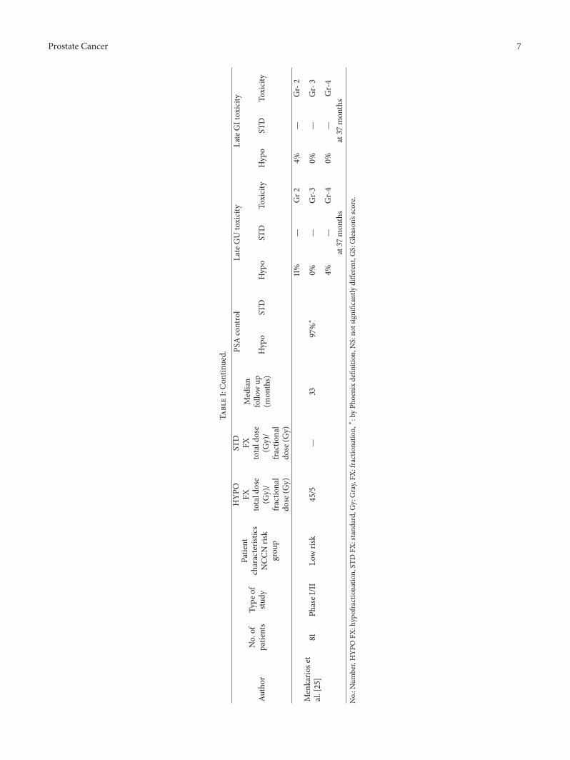
Menkarios et al. [25] treated 80 patients in a multi-institution phase I/II trial of three-dimensional conformalradiation therapy (3D-CRT) for favorable-risk group prostatecancer (T1a-T2a, Gleason≤ 6 and PSA <10 ng/mL). Thepatients received 5Gy weekly for a total dose of 45Gy(5Gy X 9). Primary end-points were feasibility and late GItoxicity by RTOG scale, while secondary end-points includedacute GI toxicity, acute and late genitourinary (GU) toxicity,biochemical control, and survival. At a median follow-upof 33 months, there was no acute GU grade 4 toxicity. The
Prostate Cancer 5
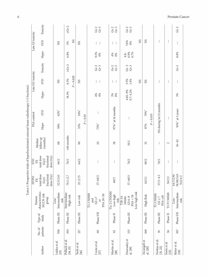
rates of grade 1, 2, and 3 acute GU toxicities were 29%, 31%,and 5%, respectively. There was no acute GI grade 3 or 4toxicity. Acute GI grade 1 and grade 2 toxicities were 30% and14%, respectively. Cumulative late grade ≥3 GI toxicity at 3years was 11%. The three-year actuarial biochemical controlrate was 97%. Prospective trials of hypofractionations usinggreater than 5 fractions are summarized in Table 1.
4. Stereotactic Body Radiation Therapy forProstate Cancer
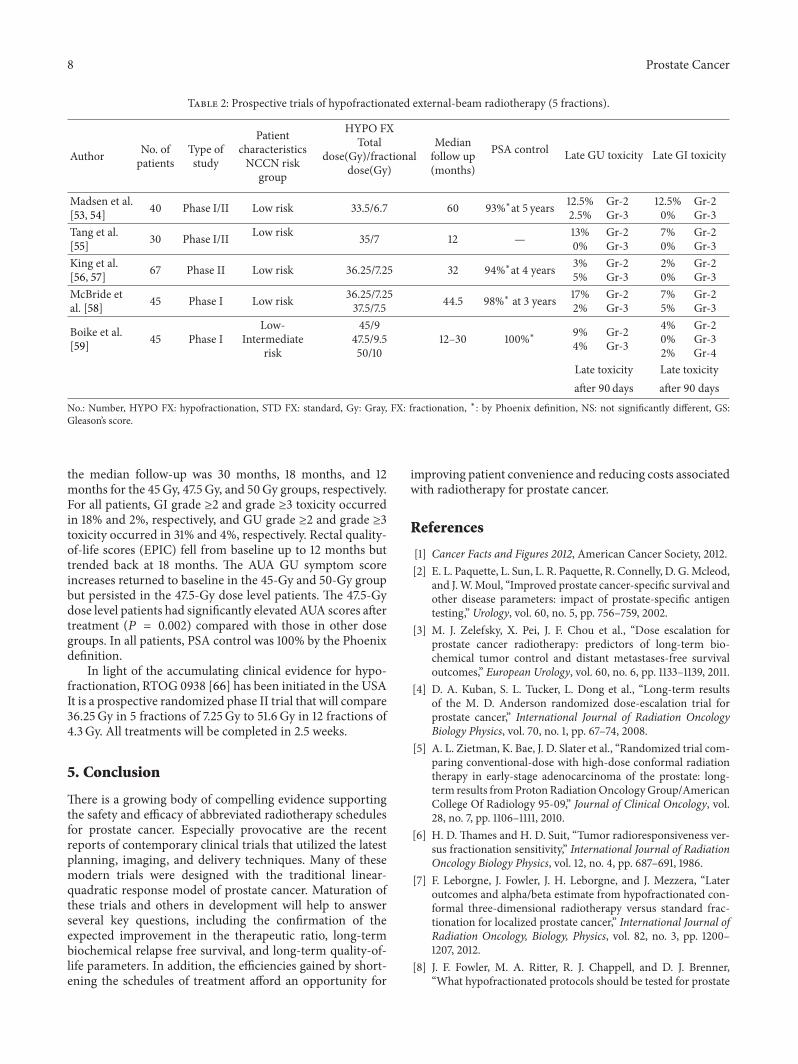
Stereotactic Body Radiation Therapy (SBRT) involves anultra-abbreviated treatment regimen of 5 or fewer fractionsadministered using image guidance and precise treatmentdelivery techniques. SBRT has been established as safe andefficacious for early stage lung cancer and selected patientswith oligometastatic cancer [62, 63], and it has also beenexplored in the treatment of prostate cancer [43–46, 50].SBRT can be delivered safely using any of several commer-cially available treatment systems. In some cases the dosedelivery involves a combination of multiple non-coplanarbeams aimed at the target, and in other systems the deliveryis accomplished with static intensity-modulated beams orrotating modulated arcs. The selective prospective trials ofSBRT for prostate cancer is listed in Table 2.
The first prospective trial of SBRT for prostate cancer waspublished by Madsen and colleagues [53, 54], who treated40 patients with SBRT using a daily dose of 6.7Gy to a totaldose of 33.5 (6.7 Gy X 5) Gy. The fractionation schedule wascalculated to be equivalent to 78Gy in 2Gy fractions usingan estimated 𝛼/𝛽 ratio of 1.5. At the median follow-up of 41months, there were no instances of grade 3 GI toxicity andonly a single episode of acute grade 3 GU toxicity. There wasno grade 3 or higher late toxicities. The PSA control rate was90% by the Phoenix definition.
Tang et al. [55] used slightly higher dose per fraction(7Gy) to treat 30 men a phase I/II study. The eligible menhad low-risk prostate cancer and received 5 weekly dose of7Gy to a total dose of 35Gy.The SBRT technique consisted ofintensity-modulated radiotherapy (IMRT) with daily imageguidance using implanted gold fiducials. All patients hadat least 6 months of follow-up. The treatments were welltolerated and there was no grade 3 or 4 GI/GU toxicity.Although there were initial grade 2 toxicities (13% GU and7% GI), these scores returned to or improved over baseline at6 months. The biochemical control rate was not available atthe time of initial reporting.
King et al. [56, 57] reported a follow-up of a phase IItrial. The treatment consisted of SBRT with a total doseof 36.25Gy in 5 fractions using the Cyberknife treatmentplatform. There were 67 patients treated between 2003 and2009. Eligible patients had low- to favorable-intermediate riskfeatures, including PSA ≤10, total Gleason’s score of 6 or 7,and clinical Stage T1c–T2b. At median follow-up of 2.7 years,there were no grade 4 toxicities. RTOG grade 3, 2, and 1bladder toxicitieswere seen in 3% (2 patients), 5% (3 patients),and 23% (13 patients), respectively. Rectal grade 3, 2, and 1toxicities were seen in 0, 2% (1 patient), and 12.5% (7 patients),respectively. The 4-year PSA relapse-free survival was 94%.
Katz et al. [50, 64] reported an experience of SBRTtreatment given to 304 patients with clinically localizedprostate cancer. Most received 5 fractions of 7.25Gy (totaldose 36.25Gy). At a median follow-up of 40 months (range,9–58months), 10 patients died of other causes and 9 were lostto follow-up.The 4-year actuarial freedom from biochemicalfailure is 98.5%, 93.0%, and 75%, for the low-, intermediate-,and high-risk groups. Late toxicity included 4.2% RTG grade2 rectal, 7.8% grade 2 urinary, and 1.4% grade 3 urinary.Mean Expanded Prostate Cancer Index Composite (EPIC)score for urinary and bowel QOL declined at 1 month post-treatment and returned to baseline by 2 years. Mean EPICsexual QOL declined by 23% at 1 month. Eighty percent ofthe patients potent at baseline remained potent at the time ofrecent analysis.
McBride et al. [58] reported a Phase I multi-institutionaltrial of SBRT, also using the CyberKnife delivery plat-form. Patients had National Comprehensive Cancer Net-work (NCCN)-defined, low-risk prostate adenocarcinoma(Gleason score, 2–6; clinical stage T1c-T2a; PSA ≤10 ng/mL).Eligible patients had prostate size ≤80 cc by ultrasound mea-surement and had American Urological Association (AUA)symptom scores ≤15. Thirty-four patients received 37.5Gydelivered in 5 fractions (7.5 Gy per fraction), 9 patientsreceived 36.25Gy in 5 fractions (7.25Gy per fraction), and2 patients received other regimens. All treatments werecompleted within 10 days with a minimum of 12 hoursbetween fractions. The planning target volume (PTV) wasprostate only with 3–5mm expansion. No patient receivedandrogen deprivation. With the median follow-up of 44.5months, none of the patients experience biochemical failureby Phoenix (nadir+2) definition. Thirteen patients experi-enced PSA bounces. The mean PSA bounce was 1.07 ng/mL.There was an episode of late grade 3 urinary obstructionrequiring TURP, and there were 2 (5%) episodes of late grade3 proctitis. SHIM, AUA, and EPIC scores were used to assessquality of life in 56% of the patients who filled out thequestionnaires. In addition to the decrease in sexual function,there also was a small late decline in EPIC Bowel scores.However, there were no statistically significant changes inAUA scores or EPIC Urinary scores.
In addition to the progressively larger dose per fractionused in the previously mentioned clinical studies, researchersat University of Texas at Southwestern Medical Center atDallas (UTSW) conducted an animal experiment usinghigher doses per fraction. Tumor bearing nude mice wasgiven 15Gy, 22.5Gy, or 45Gy in 3 weekly fractions. Only the45Gy group demonstrated sustained PSA and tumor volumedecreases in most mice [65].
This preclinical data supported the clinical trial launchedby Boike and colleagues at UTSW [59], who conducted aphase I study that escalated the total doses from 45Gy to50Gy in 5 fractions. Eligible patients included those withprostate size ≤60 cm3, Gleason score ≤6 with PSA ≤20,Gleason’s score of 7 with PSA ≤15, ≤T2b, and AmericanUrological Association (AUA) score ≤15.The total dose levelswere 45Gy, 47.5 Gy, and 50Gy in 5 fractions. All patients weretreated with a minimum of 36 hours between fractions withno more than 3 fractions per week. At the time of the report,
6 Prostate Cancer
Table1:Prospectivetria
lsof
hypo
fractio
natedexternal-beam
radiotherapy
(>5fractio
ns).
PSAcontrol
LateGUtoxicity
LateGItoxicity
Author
No.of
patie
nts
Type
ofstu
dy
Patie
ntcharacteris
tics
NCC
Nris
kgrou
p
HYP
OFX
totald
ose
(Gy)/
fractio
nal
dose
(Gy)
STD
FXtotald
ose
(Gy)/
fractio
nal
dose
(Gy)
Median
follo
wup
(mon
ths)
Hypo
STD
Hypo
STD
Toxicity
Hypo
STD
Toxicity
Lukk
aetal.
[13]
936
PhaseIII
Low-
Interm
ediate
risk
52.5/2.63
66/2
6858%
62%∗
NS
NS
Pollack
etal.
[14,15]
303
PhaseIII
Interm
ediate
Highris
k70.2/2.7
76/2
>60
mon
ths
18.3%
8.3%
≥Gr-2
6.8%
5%≥Gr-2
P=0.028
NS
Yeoh
etal.
[16]
217
PhaseIII
Lowris
k55
/2.75
64/2
9053%
34%∗
NS
NS
P<0.05
Coo
teetal.
[17]
60Ph
aseI/II
T2-3
N0M
0and
GS≥7
orPS
A20–50
57–6
0/3
—24
73%∗
—4% 4%
— —Gr-2
Gr-3
9.5%
0%— —
Gr-2
Gr-3
Martin
etal.
[18]
92Ph
aseII
T1c-2CN
XM0
Low-high
risk
60/3
—38
97%∗at14
mon
ths
3% 0%— —
Gr-2
Gr-3
4% 0%— —
Gr-2
Gr-3
Dearnaley
etal.[19]
153
PhaseIII
T1B-3A
N0M
0GS6–
8PS
A<50
Low-highris
k
57–6
0/3
74/2
50.5
—4.82–9
%0.7–4.2%
3.5% 1.4%
Gr-2
Gr-3
4.8–
6.9%
0.7%
7.6%
0%Gr-2
Gr-3
NS
NS
Arcangeliet
al.[20]
168
PhaseIII
HighRisk
62/3.1
80/2
3287%
70%∗
NS
NS
P=0.035
Norku
setal.
[21,22]
91Ph
aseIII
T1-3
N0M
0GS≤7
PSA≤10
57/3–4
.574/2
—NSdu
ring1st12mon
ths
——
Soetee
tal.
[23]
36Ph
aseII
T1-T3N0M
056/3.5
—2
——
—
Ritte
retal.
[24]
307
PhaseI/II
Low-
Interm
ediate
risk
64.7/2.94
58.08/3.63
51.6/4.3
—16–4
295%∗at5years
3%—
Gr-2
8.8%
—Gr-2
Prostate Cancer 7
Table1:Con
tinued. PS
Acontrol
LateGUtoxicity
LateGItoxicity
Author
No.of
patie
nts
Type
ofstu
dy
Patie
ntcharacteris
tics
NCC
Nris
kgrou
p
HYP
OFX
totald
ose
(Gy)/
fractio
nal
dose
(Gy)
STD
FXtotald
ose
(Gy)/
fractio
nal
dose
(Gy)
Median
follo
wup
(mon
ths)
Hypo
STD
Hypo
STD
Toxicity
Hypo
STD
Toxicity
11%
—Gr2
4%—
Gr-2
Menkario
set
al.[25]
81Ph
aseI/II
Lowris
k45/5
—33
97%∗
0%—
Gr-3
0%—
Gr-3
4%—
Gr-4
0%—
Gr-4
at37
mon
ths
at37
mon
ths
No.:N
umber,HYP
OFX
:hypofractionatio
n,ST
DFX
:stand
ard,Gy:Gray,FX
:fractionatio
n,∗:byPh
oenixdefin
ition
,NS:no
tsignificantly
different,G
S:Gleason’sscore.
8 Prostate Cancer
Table 2: Prospective trials of hypofractionated external-beam radiotherapy (5 fractions).
Author No. ofpatients
Type ofstudy
PatientcharacteristicsNCCN risk
group
HYPO FXTotal
dose(Gy)/fractionaldose(Gy)
Medianfollow up(months)
PSA control Late GU toxicity Late GI toxicity
Madsen et al.[53, 54] 40 Phase I/II Low risk 33.5/6.7 60 93%∗at 5 years 12.5%
2.5%Gr-2Gr-3
12.5%0%
Gr-2Gr-3
Tang et al.[55] 30 Phase I/II Low risk 35/7 12 — 13%
0%Gr-2Gr-3
7%0%
Gr-2Gr-3
King et al.[56, 57] 67 Phase II Low risk 36.25/7.25 32 94%∗at 4 years 3%
5%Gr-2Gr-3
2%0%
Gr-2Gr-3
McBride etal. [58] 45 Phase I Low risk 36.25/7.25
37.5/7.5 44.5 98%∗ at 3 years 17%2%
Gr-2Gr-3
7%5%
Gr-2Gr-3
Boike et al.[59] 45 Phase I
Low-Intermediate
risk
45/947.5/9.550/10
12–30 100%∗ 9%4%
Gr-2Gr-3
4%0%2%
Gr-2Gr-3Gr-4
Late toxicity Late toxicityafter 90 days after 90 days
No.: Number, HYPO FX: hypofractionation, STD FX: standard, Gy: Gray, FX: fractionation, ∗: by Phoenix definition, NS: not significantly different, GS:Gleason’s score.
the median follow-up was 30 months, 18 months, and 12months for the 45Gy, 47.5Gy, and 50Gy groups, respectively.For all patients, GI grade ≥2 and grade ≥3 toxicity occurredin 18% and 2%, respectively, and GU grade ≥2 and grade ≥3toxicity occurred in 31% and 4%, respectively. Rectal quality-of-life scores (EPIC) fell from baseline up to 12 months buttrended back at 18 months. The AUA GU symptom scoreincreases returned to baseline in the 45-Gy and 50-Gy groupbut persisted in the 47.5-Gy dose level patients. The 47.5-Gydose level patients had significantly elevated AUA scores aftertreatment (𝑃 = 0.002) compared with those in other dosegroups. In all patients, PSA control was 100% by the Phoenixdefinition.
In light of the accumulating clinical evidence for hypo-fractionation, RTOG 0938 [66] has been initiated in the USAIt is a prospective randomized phase II trial that will compare36.25Gy in 5 fractions of 7.25Gy to 51.6Gy in 12 fractions of4.3 Gy. All treatments will be completed in 2.5 weeks.
5. Conclusion
There is a growing body of compelling evidence supportingthe safety and efficacy of abbreviated radiotherapy schedulesfor prostate cancer. Especially provocative are the recentreports of contemporary clinical trials that utilized the latestplanning, imaging, and delivery techniques. Many of thesemodern trials were designed with the traditional linear-quadratic response model of prostate cancer. Maturation ofthese trials and others in development will help to answerseveral key questions, including the confirmation of theexpected improvement in the therapeutic ratio, long-termbiochemical relapse free survival, and long-term quality-of-life parameters. In addition, the efficiencies gained by short-ening the schedules of treatment afford an opportunity for
improving patient convenience and reducing costs associatedwith radiotherapy for prostate cancer.
References
[1] Cancer Facts and Figures 2012, American Cancer Society, 2012.[2] E. L. Paquette, L. Sun, L. R. Paquette, R. Connelly, D. G.Mcleod,
and J.W.Moul, “Improved prostate cancer-specific survival andother disease parameters: impact of prostate-specific antigentesting,” Urology, vol. 60, no. 5, pp. 756–759, 2002.
[3] M. J. Zelefsky, X. Pei, J. F. Chou et al., “Dose escalation forprostate cancer radiotherapy: predictors of long-term bio-chemical tumor control and distant metastases-free survivaloutcomes,” European Urology, vol. 60, no. 6, pp. 1133–1139, 2011.
[4] D. A. Kuban, S. L. Tucker, L. Dong et al., “Long-term resultsof the M. D. Anderson randomized dose-escalation trial forprostate cancer,” International Journal of Radiation OncologyBiology Physics, vol. 70, no. 1, pp. 67–74, 2008.
[5] A. L. Zietman, K. Bae, J. D. Slater et al., “Randomized trial com-paring conventional-dose with high-dose conformal radiationtherapy in early-stage adenocarcinoma of the prostate: long-term results fromProtonRadiationOncologyGroup/AmericanCollege Of Radiology 95-09,” Journal of Clinical Oncology, vol.28, no. 7, pp. 1106–1111, 2010.
[6] H. D. Thames and H. D. Suit, “Tumor radioresponsiveness ver-sus fractionation sensitivity,” International Journal of RadiationOncology Biology Physics, vol. 12, no. 4, pp. 687–691, 1986.
[7] F. Leborgne, J. Fowler, J. H. Leborgne, and J. Mezzera, “Lateroutcomes and alpha/beta estimate from hypofractionated con-formal three-dimensional radiotherapy versus standard frac-tionation for localized prostate cancer,” International Journal ofRadiation Oncology, Biology, Physics, vol. 82, no. 3, pp. 1200–1207, 2012.
[8] J. F. Fowler, M. A. Ritter, R. J. Chappell, and D. J. Brenner,“What hypofractionated protocols should be tested for prostate

Prostate Cancer 9
cancer?” International Journal of Radiation Oncology BiologyPhysics, vol. 56, no. 4, pp. 1093–1104, 2003.
[9] D. J. Brenner, “The linear-quadratic model is an appropriatemethodology for determining isoeffective doses at large dosesper fraction,” Seminars in Radiation Oncology, vol. 18, no. 4, pp.234–239, 2008.
[10] D. J. Brenner, A. A. Martinez, G. K. Edmundson, C. Mitchell,H. D.Thames, and E. P. Armour, “Direct evidence that prostatetumors show high sensitivity to fractionation (low 𝛼/𝛽 ratio),similar to late-responding normal tissue,” International Journalof Radiation Oncology Biology Physics, vol. 52, no. 1, pp. 6–13,2002.
[11] R. G. Dale and B. Jones, “Is the 𝛼/𝛽 for prostate tumors reallylow? In regard to Fowler et al., IJROBP 2001;50:1021–1031,”International Journal of Radiation Oncology Biology Physics, vol.52, no. 5, pp. 1427–1428, 2002.
[12] J. Z. Wang, M. Guerrero, and X. A. Li, “How low is the 𝛼/𝛽ratio for prostate cancer?” International Journal of RadiationOncology Biology Physics, vol. 55, no. 1, pp. 194–203, 2003.
[13] H. Lukka, C. Hayter, J. A. Julian et al., “Randomized trial com-paring two fractionation schedules for patients with localizedprostate cancer,” Journal of Clinical Oncology, vol. 23, no. 25, pp.6132–6138, 2005.
[14] A. Pollack, A. L. Hanlon, E. M. Horwitz et al., “Dosimetry andpreliminary acute toxicity in the first 100 men treated forprostate cancer on a randomized hypofractionation dose esca-lation trial,” International Journal of Radiation Oncology BiologyPhysics, vol. 64, no. 2, pp. 518–526, 2006.
[15] A. Pollack, G. Walker, and M. Buyyounouski, “Five year resultsof a randomized external beam radiotherapy hypofractionationtrial for prostate cancer,” International Journal of RadiationOncology Biology Physics, vol. 81, p. S1, 2011.
[16] E. E. Yeoh, R. J. Botten, J. Butters, A. C. Di Matteo, R. H. Hol-loway, and J. Fowler, “Hypofractionated versus conventionallyfractionated radiotherapy for prostate carcinoma: final resultsof phase III randomized trial,” International Journal of RadiationOncology, Biology, Physics, vol. 81, pp. 1271–1278, 2010.
[17] J. H. Coote, J. P. Wylie, R. A. Cowan, J. P. Logue, R. Swindell,and J. E. Livsey, “Hypofractionated intensity-modulated radio-therapy for carcinoma of the prostate: analysis of toxicity,”International Journal of Radiation Oncology Biology Physics, vol.74, no. 4, pp. 1121–1127, 2009.
[18] J. M. Martin, T. Rosewall, A. Bayley et al., “Phase II trial ofhypofractionated image-guided intensity-modulated radio-therapy for localized prostate adenocarcinoma,” InternationalJournal of Radiation Oncology Biology Physics, vol. 69, no. 4, pp.1084–1089, 2007.
[19] D. Dearnaley, I. Syndikus, G. Sumo et al., “Conventional versushypofractionated high-dose intensity-modulated radiotherapyfor prostate cancer: preliminary safety results from the CHHiPrandomised controlled trial,”The Lancet Oncology, vol. 13, no. 1,pp. 43–54, 2012.
[20] G. Arcangeli, B. Saracino, S. Gomellini et al., “A Prospectivephase III randomized trial of hypofractionation versus conven-tional fractionation in patients with high-risk prostate cancer,”International Journal of Radiation Oncology Biology Physics, vol.78, no. 1, pp. 11–18, 2010.
[21] D. Norkus, A. Miller, J. Kurtinaitis et al., “A randomized trialcomparing hypofractionated and conventionally fractionatedthree-dimensional external-beam radiotherapy for localizedprostate adenocarcinoma: AAAAA report on acute toxicity,”Strahlentherapie und Onkologie, vol. 185, no. 11, pp. 715–721,2009.
[22] D. Norkus, A. Miller, A. Plieskiene, E. Janulionis, and K. P.Valuckas, “A randomized trial comparing hypofractionatedand conventionally fractionated three-dimensional conformalexternal-beam radiotherapy for localized prostate adenocarci-noma: a report on the first-year biochemical response,” Medic-ina, vol. 45, no. 6, pp. 469–475, 2009.
[23] G. Soete, S. Arcangeli, G. De Meerleer et al., “Phase II study ofa four-week hypofractionated external beam radiotherapy regi-men for prostate cancer: report on acute toxicity,” Radiotherapyand Oncology, vol. 80, no. 1, pp. 78–81, 2006.
[24] M. A. Ritter, J. D. Forman, P. A. Kupelian et al., “A phase I/II trialof increasingly hypofractionated radiation therapy for prostatecancer,” International Journal of Radiation Oncology BiologyPhysics, vol. 75, pp. S80–S81, 2009.
[25] C. Menkarios, T. Vigneault, N. Brochet et al., “Toxicity reportof once weekly radiation therapy for low-risk prostate adeno-carcinoma: preliminary results of a phase I/II trial,” RadiationOncology, vol. 6, no. 1, article 112, 2011.
[26] D. J. Brenner and E. J. Hall, “Fractionation and protraction forradiotherapy of prostate carcinoma,” International Journal ofRadiation Oncology Biology Physics, vol. 43, no. 5, pp. 1095–1101,1999.
[27] G. M. Duchess and L. J. Peters, “What is the 𝛼/𝛽 ratio forprostate cancer? Rationale for hypofractionated high-dose-ratebrachytherapy,” International Journal of Radiation OncologyBiology Physics, vol. 44, no. 4, pp. 747–748, 1999.
[28] J. Fowler, R. Chappell, andM. Ritter, “Is 𝛼/𝛽 for prostate tumorsreally low?” International Journal of Radiation Oncology BiologyPhysics, vol. 50, no. 4, pp. 1021–1031, 2001.
[29] M. Ritter, “Rationale, conduct, and outcome using hypofrac-tionated radiotherapy in prostate cancer,” Seminars in RadiationOncology, vol. 18, no. 4, pp. 249–256, 2008.
[30] M. Ritter, J. Forman, P. Kupelian, C. Lawton, and D. Petereit,“Hypofractionation for prostate cancer,” Cancer Journal, vol. 15,no. 1, pp. 1–6, 2009.
[31] M. Carlone, D. Wilkins, B. Nyiri, and P. Raaphorst, “Com-parison of 𝛼/𝛽 estimates from homogeneous (individual) andheterogeneous (population) tumor control models for earlystage prostate cancer,”Medical Physics, vol. 30, no. 10, pp. 2832–2848, 2003.
[32] A. E. Nahum, B. Movsas, E. M. Horwitz, C. C. Stobbe, and J.D. Chapman, “Incorporating clinical measurements of hypoxiainto tumor local control modeling of prostate cancer: impli-cations for the 𝛼/𝛽 ratio,” International Journal of RadiationOncology Biology Physics, vol. 57, no. 2, pp. 391–401, 2003.
[33] J. F. Fowler, M. A. Ritter, J. D. Fenwick, and R. J. Chappell, “Howlow is the 𝛼/𝛽 ratio for prostate cancer? In regard to Wang etal., IJROBP 2003;55:194–203,” International Journal of RadiationOncology Biology Physics, vol. 57, no. 2, pp. 593–595, 2003.
[34] S. G. Williams, J. M. G. Taylor, N. Liu et al., “Use of individualfraction size data from 3756 patients to directly determine the𝛼/𝛽 ratio of prostate cancer,” International Journal of RadiationOncology Biology Physics, vol. 68, no. 1, pp. 24–33, 2007.
[35] R. Miralbell, S. A. Roberts, E. Zubizarreta, and J. H. Hendry,“Dose-fractionation sensitivity of prostate cancer deduced fromradiotherapy outcomes of 5,969 patients in seven InternationalInstitutional Datasets: 𝛼/𝛽= 1.4 (0.9–2.2) Gy,” InternationalJournal of Radiation Oncology, Biology, Physics, vol. 82, no. 1, pp.e17–e24, 2012.
[36] E. J. Hall and D. J. Brenner, “The radiobiology of radiosurgery:rationale for different treatment regimes for AVMs and malig-nancies,” International Journal of Radiation Oncology BiologyPhysics, vol. 25, no. 2, pp. 381–385, 1993.

10 Prostate Cancer
[37] C. Park, L. Papiez, S. Zhang, M. Story, and R. D. Timmerman,“Universal survival curve and single fraction equivalent dose:useful tools in understanding potency of ablative radiotherapy,”International Journal of Radiation Oncology Biology Physics, vol.70, no. 3, pp. 847–852, 2008.
[38] J. P. Kirkpatrick, J. J. Meyer, and L. B. Marks, “The linear-quadratic model is inappropriate to model high dose per frac-tion effects in radiosurgery,” Seminars in Radiation Oncology,vol. 18, no. 4, pp. 240–243, 2008.
[39] R. W. Lloyd-Davies, C. D. Collins, and A. V. Swan, “Carcinomaof prostate treated by radical external beam radiotherapy usinghypofractionation. Twenty-two years’ experience (1962–1984),”Urology, vol. 36, no. 2, pp. 107–111, 1990.
[40] C. D. Collins, R. W. Lloyd-Davies, and A. V. Swan, “Radicalexternal beam radiotherapy for localised carcinoma of theprostate using a hypofractionation technique,” Clinical Oncol-ogy, vol. 3, no. 3, pp. 127–132, 1991.
[41] T. Akimoto, H. Muramatsu, M. Takahashi et al., “Rectal bleed-ing after hypofractionated radiotherapy for prostate cancer:correlation between clinical and dosimetric parameters and theincidence of grade 2 or worse rectal bleeding,” InternationalJournal of Radiation Oncology Biology Physics, vol. 60, no. 4, pp.1033–1039, 2004.
[42] M. Barnes, H. Pass, and A. DeLuca, “Response of the mediasti-nal and thoracic viscera of the dog to intraoperative radiationtherapy (IORT),” International Journal of Radiation OncologyBiology Physics, vol. 13, no. 3, pp. 371–378, 1987.
[43] G. Bolzicco, M. S. Favretto, E. Scremin, C. Tambone, A. Tasca,and R. Guglielmi, “Image-guided stereotactic body radiationtherapy for clinically localized prostate cancer: preliminaryclinical results,” Technology in Cancer Research and Treatment,vol. 9, no. 5, pp. 473–477, 2010.
[44] D. E. Freeman and C. R. King, “Stereotactic body radiotherapyfor low-risk prostate cancer: five-year outcomes,” RadiationOncology, vol. 6, no. 1, article 3, 2011.
[45] J. L. Friedland, D. E. Freeman, M. E. Masterson-McGary, andD. M. Spellberg, “Stereotactic body radiotherapy: an emergingtreatment approach for localized prostate cancer,” Technology inCancer Research and Treatment, vol. 8, no. 5, pp. 387–392, 2009.
[46] S. Jabbari, V. K. Weinberg, T. Kaprealian et al., “Stereotacticbody radiotherapy as monotherapy or post-external beamradiotherapy boost for prostate cancer: technique, early toxicity,and PSA response,” International Journal of Radiation Oncology,Biology, Physics, vol. 82, pp. 228–234, 2010.
[47] P. A. Kupelian, V. V. Thakkar, D. Khuntia, C. A. Reddy, E. A.Klein, and A. Mahadevan, “Hypofractionated intensity-modu-lated radiotherapy (70 Gy at 2.5 Gy per fraction) for localizedprostate cancer: long-term outcomes,” International Journal ofRadiationOncology Biology Physics, vol. 63, no. 5, pp. 1463–1468,2005.
[48] P. A. Kupelian, T. R. Willoughby, C. A. Reddy, E. A. Klein, andA. Mahadevan, “Hypofractionated intensity-modulated radio-therapy (70 Gy at 2.5 Gy per fraction) for localized prostatecancer: Cleveland Clinic experience,” International Journal ofRadiationOncology Biology Physics, vol. 68, no. 5, pp. 1424–1430,2007.
[49] N. Rene, S. Faria, F. Cury et al., “Hypofractionated radiotherapyfor favorable risk prostate cancer,” International Journal ofRadiation Oncology Biology Physics, vol. 77, no. 3, pp. 805–810,2010.
[50] A. J. Katz, M. Santoro, R. Ashley, F. Diblasio, and M. Witten,“Stereotactic body radiotherapy for organ-confined prostatecancer,” BMC Urology, vol. 10, article 1, 2010.
[51] J. D. Cox, D. J. Grignon, R. S. Kaplan, J. T. Parsons, and P. F.Schellhammer, “Consensus statement: guidelines for PSA fol-lowing radiation therapy,” International Journal of RadiationOncology Biology Physics, vol. 37, no. 5, pp. 1035–1041, 1997.
[52] M. Roach III, G. Hanks, H. Thames Jr. et al., “Defining bio-chemical failure following radiotherapy with or without hor-monal therapy in men with clinically localized prostate cancer:recommendations of the RTOG-ASTRO Phoenix ConsensusConference,” International Journal of Radiation Oncology Biol-ogy Physics, vol. 65, no. 4, pp. 965–974, 2006.
[53] B. L.Madsen, R. A. Hsi, H. T. Pham, J. F. Fowler, L. Esagui, and J.Corman, “Stereotactic hypofractionated accurate radiotherapyof the prostate (SHARP), 33.5 Gy in five fractions for localizeddisease: first clinical trial results,” International Journal ofRadiation Oncology Biology Physics, vol. 67, no. 4, pp. 1099–1105,2007.
[54] H. T. Pham, G. Song, K. Badiozamani et al., “Five-year outcomeof stereotactic hypofractionated accurate radiotherapy of theprostate (SHARP) for patients with low-risk prostate cancer,”International Journal of Radiation Oncology Biology Physics, vol.78, p. S58, 2010.
[55] C. I. Tang, D. A. Loblaw, P. Cheung et al., “Phase I/II studyof a five-fraction hypofractionated accelerated radiotherapytreatment for low-risk localised prostate cancer: early results ofpHART3,” Clinical Oncology, vol. 20, no. 10, pp. 729–737, 2008.
[56] C. R. King, J. D. Brooks, H. Gill, T. Pawlicki, C. Cotrutz, and J.C. Presti, “Stereotactic body radiotherapy for localized prostatecancer: interim results of a prospective phase II clinical trial,”International Journal of Radiation Oncology Biology Physics, vol.73, no. 4, pp. 1043–1048, 2009.
[57] C. R. King, J. D. Brooks, H. Gill, and J. C. Presti Jr., “Long-term outcomes from a prospective trial of stereotactic bodyradiotherapy for low-risk prostate cancer,” International Journalof RadiationOncology Biology Physics, vol. 82, no. 2, pp. 877–882,2012.
[58] S. M. McBride, D. S. Wong, J. J. Dombrowski et al., “Hypofrac-tionated stereotactic body radiotherapy in low-risk prostateadenocarcinoma: preliminary results of a multi-institutionalphase 1 feasibility trial,” Cancer, vol. 118, no. 15, pp. 3681–3690,2012.
[59] T. P. Boike, Y. Lotan, L. C. Cho et al., “Phase I dose-escalationstudy of stereotactic body radiation therapy for low- andintermediate-risk prostate cancer,” Journal of Clinical Oncology,vol. 29, no. 15, pp. 2020–2026, 2011.
[60] A. Pollack, Five Year Results of a Randomized External BeamRadiotherapy Hypofractionation Trial for Prostate Cancer-(Ple-neary Session), ASTRO, Miami, Fla, USA, 2011.
[61] E. E. Yeoh, R. H. Holloway, R. J. Fraser et al., “Hypofraction-ated versus conventionally fractionated radiation therapy forprostate carcinoma: updated results of a phase III random-ized trial,” International Journal of Radiation Oncology BiologyPhysics, vol. 66, no. 4, pp. 1072–1083, 2006.
[62] L. C. Cho, V. Fonteyne, W. DeNeve et al., “Stereotactic bodyradiotherapy,” in Technical Basis of RadiationTherapy: PracticalClinical Applications, S. Levitt, J. Purdy, C. Perez, and P.Poortmans, Eds., p. 363, Springer, Heidelberg, Germany, 2012.
[63] B. D. Kavanagh, R. Timmerman, and J. L. Meyer, “The expand-ing roles of stereotactic body radiation therapy and oligofrac-tionation: toward a new practice of radiotherapy,” Frontiers ofRadiation Therapy and Oncology, vol. 43, pp. 370–381, 2011.
[64] A. J. Katz, M. Santoro, F. DiBlasio et al., “Stereotactic bodyradiation therapy for low, intermediate, and high-risk prostate

Prostate Cancer 11
cancer: disease control and quality of life,” International Journalof Radiation Oncology Biology Physics, vol. 81, p. S100, 2011.
[65] Y. Lotan, J. Stanfield, L. C. Cho et al., “Efficacy of high dose perfraction radiation for implanted human prostate cancer in anudemouse model,” Journal of Urology, vol. 175, no. 5, pp. 1932–1936, 2006.
[66] RTOG 0938, http://www.rtog.org/ClinicalTrials/ProtocolTable.aspx.

Submit your manuscripts athttp://www.hindawi.com
Stem CellsInternational
Hindawi Publishing Corporationhttp://www.hindawi.com Volume 2014
Hindawi Publishing Corporationhttp://www.hindawi.com Volume 2014
MEDIATORSINFLAMMATION
of
Hindawi Publishing Corporationhttp://www.hindawi.com Volume 2014
Behavioural Neurology
EndocrinologyInternational Journal of
Hindawi Publishing Corporationhttp://www.hindawi.com Volume 2014
Hindawi Publishing Corporationhttp://www.hindawi.com Volume 2014
Disease Markers
Hindawi Publishing Corporationhttp://www.hindawi.com Volume 2014
BioMed Research International
OncologyJournal of
Hindawi Publishing Corporationhttp://www.hindawi.com Volume 2014
Hindawi Publishing Corporationhttp://www.hindawi.com Volume 2014
Oxidative Medicine and Cellular Longevity
Hindawi Publishing Corporationhttp://www.hindawi.com Volume 2014
PPAR Research
The Scientific World JournalHindawi Publishing Corporation http://www.hindawi.com Volume 2014
Immunology ResearchHindawi Publishing Corporationhttp://www.hindawi.com Volume 2014
Journal of
ObesityJournal of
Hindawi Publishing Corporationhttp://www.hindawi.com Volume 2014
Hindawi Publishing Corporationhttp://www.hindawi.com Volume 2014
Computational and Mathematical Methods in Medicine
OphthalmologyJournal of
Hindawi Publishing Corporationhttp://www.hindawi.com Volume 2014
Diabetes ResearchJournal of
Hindawi Publishing Corporationhttp://www.hindawi.com Volume 2014
Hindawi Publishing Corporationhttp://www.hindawi.com Volume 2014
Research and TreatmentAIDS
Hindawi Publishing Corporationhttp://www.hindawi.com Volume 2014
Gastroenterology Research and Practice
Hindawi Publishing Corporationhttp://www.hindawi.com Volume 2014
Parkinson’s Disease
Evidence-Based Complementary and Alternative Medicine
Volume 2014Hindawi Publishing Corporationhttp://www.hindawi.com