Embed Size (px)
Citation preview
Review ArticleFunctional Skin Grafts: Where Biomaterials Meet Stem Cells
Amtoj Kaur ,1 Swati Midha ,1 Shibashish Giri ,2,3 and Sujata Mohanty 1
1Stem Cell Facility (DBT-Centre of Excellence for Stem Cell Research), All India Institute of Medical Sciences, New Delhi, India2Department of Cell Techniques and Applied Stem Cell Biology, Centre for Biotechnology and Biomedicine, University of Leipzig,Deutscher Platz 5, D-04103 Leipzig, Germany3Department of Plastic Surgery and Hand Surgery, University Hospital Rechts der Isar, Technische Universität München,Munich, Germany
Correspondence should be addressed to Swati Midha; [email protected] and Sujata Mohanty; [email protected]
Received 3 January 2019; Accepted 21 May 2019; Published 1 July 2019
Academic Editor: Tao-Sheng Li
Copyright © 2019 Amtoj Kaur et al. This is an open access article distributed under the Creative Commons Attribution License,which permits unrestricted use, distribution, and reproduction in any medium, provided the original work is properly cited.
Skin tissue engineering has attained several clinical milestones making remarkable progress over the past decades. Skin is inhabitedby a plethora of cells spatiotemporally arranged in a 3-dimensional (3D) matrix, creating a complex microenvironment of cell-matrix interactions. This complexity makes it difficult to mimic the native skin structure using conventional tissue engineeringapproaches. With the advent of newer fabrication strategies, the field is evolving rapidly. However, there is still a long waybefore an artificial skin substitute can fully mimic the functions and anatomical hierarchy of native human skin. The currentfocus of skin tissue engineers is primarily to develop a 3D construct that maintains the functionality of cultured cells in a guidedmanner over a period of time. While several natural and synthetic biopolymers have been translated, only partial clinical successis attained so far. Key challenges include the hierarchical complexity of skin anatomy; compositional mismatch in terms ofmaterial properties (stiffness, roughness, wettability) and degradation rate; biological complications like varied cell numbers, celltypes, matrix gradients in each layer, varied immune responses, and varied methods of fabrication. In addition, with newerbiomaterials being adopted for fabricating patient-specific skin substitutes, issues related to escalating processing costs,scalability, and stability of the constructs under in vivo conditions have raised some concerns. This review provides an overviewof the field of skin regenerative medicine, existing clinical therapies, and limitations of the current techniques. We have furtherelaborated on the upcoming tissue engineering strategies that may serve as promising alternatives for generating functional skinsubstitutes, the pros and cons associated with each technique, and scope of their translational potential in the treatment ofchronic skin ailments.
1. Introduction
Skin, the largest organ of the human body, acts as a barrierfor outside pollutants and microbes; hence, serving as thebody’s first line of defense. In addition, skin performs variousfunctions like thermoregulation, moisture retention, immuneprotection, imparting sensation, and self-healing response[1–3]. The human skin comprises of three layers: epidermis(outermost), dermis (middle), and hypodermis (deeper) [4].The epidermis is a 0.2 mm thick, packed sheath of cells con-sisting of keratinocytes, which are in different stages of differ-entiation, along with melanocytes and epidermal stem cellsconfined to the basal proliferative layer. Furthermore, thereare 4 layers within the epidermis, namely, the stratum cor-
neum (dead cornified layer with 15-30 sheets of corneocytes),stratum granulosum (3-5 sheets of flattened keratinocyteswith arrested division), stratum spinosum (possessing 8-10layers of keratinocytes with restricted cell division), and stra-tum basale (proliferative layer). The “bricks-and-mortar”array type of organization of corneocytes in the epidermisacts as a barrier separating the internal body environmentfrom the external along with regulating fluid loss [5]. Thedermis, comprising of a thick connective tissue, is sand-wiched in the middle of the epidermis and the hypodermis[6]. It is constituted of a bed of glycosaminoglycans (GAGs),elastin, and collagen extracellular matrix (ECM) with embed-ded fibroblasts. It also possesses numerous skin appendageslike sebaceous and sweat glands, mechanoreceptors, hair
HindawiStem Cells InternationalVolume 2019, Article ID 1286054, 20 pageshttps://doi.org/10.1155/2019/1286054
follicles, vasculature, and nerve endings. The dermis impartssensory and mechanical properties to the skin. A separatinglayer of basement membrane having a specialized ECM com-position (constituting of collagens III, IV, and VII; laminins;and fibrillin) is present between the epidermis and dermisfacilitating diffusion and communication between the cellsvia paracrine signaling to maintain homeostasis [7, 8]. Thebottom-most hypodermis or subcutaneous layer comprisesof adipose tissue and controls the mechanical and thermo-regulatory properties of the skin.
Burns, acute trauma, chronic wounds, intensive surger-ies, infections, and genetic abnormalities are the mostcommon factors responsible for causing variable extents ofdamage to the skin [9–11]. According to the World HealthOrganization (WHO), fatal injuries arising from burnsaccount for approximately 180,000 deaths annually. In Indiaalone, over 1,000,000 burn victims suffer from moderate toserious burns per annum. The global wound care market isexpected to increase from 18.35 billion USD in 2017 to22.81 billion USD by 2022 [12]. Apart from the huge costof treatment, indirect expenses such as lost income due tounemployment, prolonged medical care, and emotionaltrauma immensely contribute to the socioeconomic impact.Wounds act as breaches in the tissue which compromisethe defensive ability of the skin; hence, becoming the leadingcause of infections. Based on the depth of injury, skin woundshave four subdivisions: (i) epidermal (top layer of skin), (ii)superficial partial thickness (epidermis and upper dermis),(iii) deep partial thickness (epidermis and full dermis), and(iv) full thickness (all three layers of the skin) [13]. In the caseof deeper skin injuries including partial and full-thicknesswounds, the natural healing mechanism is incapable ofrestoring the fully functional tissue in most cases [14], exceptwhere hair follicles are present. Therefore, skin wound heal-ing poses a serious challenge for both patients and plasticsurgeons.
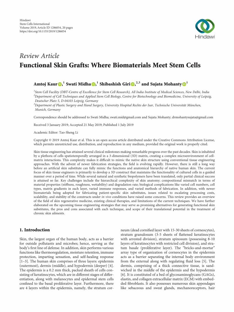
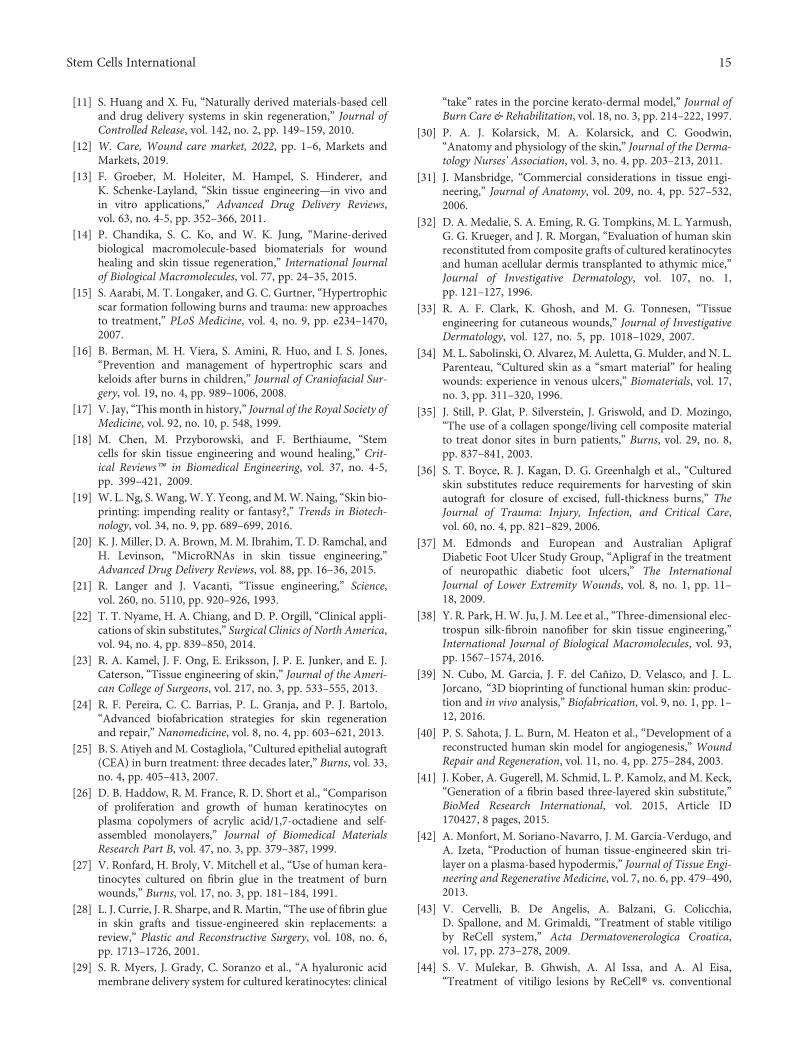
Since their origin in 1874, autologous split-thickness skingrafts (STSG) have been considered as the “gold standards”for treating skin injuries requiring ample amount of healthyskin [15]. STSG aids in the transfer of epidermal stem cellsfrom a healthy site to the wound site. However, this approachfaces drawbacks related to donor site shortage, failure to treatfull-thickness wounds, and scarring at both the donor andrecipient sites [16]. Moreover, the increasing gap betweenthe demand and supply of autologous and allogenic graftshas paved the way for skin tissue engineering (STE). STEtakes advantage of an artificial construct where autologouscells of the individual are isolated, cultured on constructs,and implanted into the wound site to facilitate the healingprocess (Figure 1). From the pioneering work performed byJacques-Louis Reverdin in 1870 by the application of “freshskin” allografts, skin replacement and regenerative therapyhas now come a long way utilizing different biological mate-rials with cultured cells in modern medicine [17]. Today, thebroad realm of STE covers numerous cutting-edge strategiessuch as nanotechnology, 3D bioprinting, stem cells, andmicrofluidics [18–20]. There are some key features that arecrucial for tissue-engineered skin substitutes such as bio-compatibility, nontoxicity, nonimmunogenicity, biodegrad-ability, moisture retention property, optimal elasticity, andporosity with good interconnectivity for a free exchange ofgases and nutrients to induce the growth of neovasculaturefor generating functional skin substitutes. In order toattain commercial relevance, these engineered skin substi-tutes must be cost-effective, scalable, have a prolonged shelflife, and should be available off-the-shelf for large-scaleapplication [21–24].
In this review, we discuss the progress made so far in thedevelopment of artificial skin scaffolds using various innova-tive strategies and biomaterials for the fabrication of skin tis-sue substitutes, along with their clinical applications andfuture perspectives.
Skin injury
Proliferation, migration,matrix degradation, andcell-synthesized ECM viaconsistent growth factordelivery
Autologous cellculture
3D scaffold with cells andgrowth factors
The trilayered construct for deeper wounds
Figure 1: Schematic illustrating the stages of skin tissue engineering using biomaterials and stem cell technology. Briefly, autologous cells areisolated from skin biopsies of patients and expanded in vitro for up to 3 weeks. When optimal cell confluency is achieved, cells in combinationwith growth- and differentiation-inducing factors are seeded on biomimetic scaffolds (with structural resemblance to the skin anatomy) forimplantation into the target site to facilitate repair and regeneration of the damaged skin tissue.
2 Stem Cells International

2. Scaffold Characteristics Specific to Skin
While several tissue-engineered scaffolds are being devel-oped, each material needs to be modulated to be able tomatch the properties of the target tissue. Even after attain-ing considerable clinical success with the currently availablecommercial skin replacements, the search for an ideal,functional skin substitute, which exactly recapitulates thepatient’s original tissue, remains elusive. Some key challengesinclude; (i) selecting the most suitable biofabricationapproach that can simulate the complex anatomical hierar-chy of trilayered skin; (ii) optimizing multimaterial composi-tions with desired properties to aid in cellular guidance anddifferentiation to mimic the different layers; (iii) and deter-mining the type and source of stem cells, seeding modality(single cell suspension, aggregates), and seeding density interms of variability between the different layers.
2.1. Types of Skin Substitutes. Depending upon the depthof the tissue, skin substitutes can be categorized into fourdistinct types.
2.1.1. Epidermal Skin Constructs. The epidermal skin con-structs comprise of keratinocytes cultured on a layer of irra-diated feeder cells of murine fibroblasts. The autologouskeratinocytes isolated from the patient usually take 2-3 weeksin expansion media to develop cell sheets of stratified kerati-nocytes, commonly termed as cultured epithelial autografts(CEAs). CEAs are typically 2 to 8 layers thick. However, theyare not very effective for curing burns and are fragile to han-dle [25]. Petroleum gauze dressings and silicone membraneshave been used to render support to the mechanically inferiorcell sheets. Acid functionalization performed on materialsaids in the easy transfer of keratinocytes, apart from aidingin attachment and proliferation [26]. However, these syn-thetic carriers are nonbiodegradable and need to be removedafter sometime. Therefore, a few studies have documentedthe usage of natural biomaterials such as fibrin [27, 28] andhyaluronic acid (HA) [29] as carriers for cultured keratino-cytes as they provide a conducive microenvironmental nichefor promoting migration, proliferation, matrix degradation,and differentiation of keratinocytes.
2.1.2. Dermal Skin Constructs. Dermis comprises of ECMwith fibroblasts [30] which is further divided into an upper“papillary” and lower “reticular” region. The conformationalorientation of thin, randomly aligned collagen fiber bundles(primarily collagen type III) in the papillary region form anintricate ridge-like arrangement. The reticular dermis, onthe other hand, is composed of a more ordered collagenarrangement of predominantly collagen type I. Most of thecommercial dermal skin replacements are cell free and actas an initial framework for facilitating infiltration of cellsand blood vessels from the host tissue. This is mainly dueto low fabrication cost, easy storage, and low immunogenicresponse [31]. Fibroblasts have shown to repopulate acellulardermal substitutes in vivo, 7 days post implantation [32]. Incontrast, in dermal skin substitutes with allogenic humanneonatal fibroblasts like Apligarf, the cells did not survivebeyond a few weeks post implantation [33].
2.1.3. Epidermal-Dermal Skin Constructs. Currently, theclosest and the most sophisticated skin biomimic availablein the market is an epidermal-dermal skin substitute com-prising of both of the upper layers of the skin. The closeassociation between keratinocytes and fibroblasts in theepidermal-dermal skin grafts triggers a cascade of biologicalmoieties (growth factors, cytokines) to expedite tissue heal-ing [34–36]. Significant enhancement in wound closure hasbeen observed where these epidermal-dermal skin constructshave been used to cure chronic injuries and ulcers [37].Several attempts have been made using different fabricationtechniques like electrospinning and 3D bioprinting to fabri-cate bilayered constructs [38, 39]. These bilayered constructsmeasure about 2.5 mm in thickness, which hinders adequatevascularization subsequently resulting in the early death ofconstructs. Hence, advancement in vascularization strategiesis the prime requisite for developing functional bilayered skinconstructs [40].
2.1.4. Trilayered Skin Construct. Trilayered skin constructsinclude the hypodermal adipose tissue along with the dermisand the epidermis. It can be considered as the closest mimicto the native human skin for full-thickness wounds. Thehypodermal layer consists of fatty connective tissue with pre-dominantly collagen VI ECM and a multicellular organiza-tion (preadipocytes, adipocytes, vascular endothelial cells,and adipose macrophages). A few attempts have been madeto fabricate trilayered skin constructs. Kober et al. fabri-cated a fibrin-based trilayered skin construct by depositingadipose-derived stem cells (ADSCs), fibroblasts, and kerati-nocytes in the fibrin matrix for replicating the hypodermis,dermis, and epidermis, respectively [41]. The fabricated con-struct showed a morphology similar to the native humanskin. Another group used human plasma for a trilayered skinconstruct, engineered using a similar combination of cells[42]. There is a need for more extensive research in hypoder-mal engineering in order to cater to full-thickness woundswith special consideration to be given to zonal ECM variationpresent in the different layers of the skin.
2.2. Pigmentation. Pigmentation is not only an importantcosmetic property of the skin, but melanin in the skin alsoprotects against ultraviolet (UV) radiation. An off-the-shelfproduct, ReCell®, makes use of fresh skin biopsy to preparea spray-on cell suspension comprising of a combinationof autologous keratinocytes, melanocytes, and fibroblastsfor treating vitiligo. In such grafts, repigmentation tookapproximately 3-5 weeks, while contrary reports showeddelayed pigmentation which took as long as 4 months to setin [43, 44]. A limiting factor to the approach is the age depen-dency, as the product had limited efficacy (less than 65%) inpatients >30 years of age [44]. Possible contributing factorscould be the relatively thinner skin in elderly individuals[45]. Also, other factors for elderly patients may include com-promised immune functions, disease condition, lower mela-nogenic capacity, and decreased vascularity with increasingage [46]. Apart from this, a 3D bioprinting approach has alsobeen explored for the construction of pigmented skinconstructs. Ng et al. demonstrated the use of a drop-on-
3Stem Cells International

demand bioprinting technique to bioprint a precise patternof one melanocyte surrounded by 8 keratinocytes in a 3 × 3array. The bioprinted skin appeared uniformly pigmentedas compared to the manually casted construct after 39 daysof in vitro culture [47].
2.3. Vasculature. Wound closure after full-thickness burnsrequires the reestablishment of a stable epidermis as a pre-requisite. The stability of the epidermis depends upon thereformation of the basement membrane and vascularizedconnective tissues to anchor the outer skin to the body [48].In skin constructs, anastomosis with the host vasculature isessential for the diffusion of oxygen, nutrients, and other bio-logical moieties, as the diffusion limit is approximately 0.1-0.2 mm only [49]. Problems arise in the wound area primar-ily due to inadequate graft preparation, infection, and scar-ring of the tissue due to a hindered blood supply as a resultof a long interval between injury and grafting (usually >3days) [50]. Having an established vascular supply is criticalin cases where the affected area is large. Few studies havedeveloped prevascularized grafts and demonstrated the full-thickness healing of dermal wounds in preclinical models[51]. As opposed to this, pedicle flaps have been used clini-cally with the advantage being that they carry their ownblood supply. For this to work, these blood supplies need toanastomose with the adjacent host tissue. But these flapsare much thicker than grafts and usually encounter problemswith kinking of the matrix and delayed anastomosis [52]. Tocircumvent the problems associated with the current tech-niques, various approaches have been proposed to inducevasculature [53]. A blend of cellular [54], biomaterial-based [55–57], and microfabrication approaches [58] couldpossibly circumvent delayed vascularization at the site ofinjury. The addition of endothelial cells or stem/progenitorcells, a common cell-based approach, induces the formationof neovasculature at the injury site. Documented evidencedictates that the incorporation of hydrogels such as fibrin[55] or HA [56] within the bioengineered constructs can pro-mote angiogenesis. In the microfabrication approach, sacrifi-cial or nonsacrificial mini- and microchannels are created forthe rapid diffusion of oxygen, nutrients, and growth factors.Detailed studies on various vascularization strategies havealready been reviewed elsewhere [54, 59, 60].
2.4. Optimal Cell Source. A number of cell sources have beenexplored for STE. Embryonic stem cells have been isolatedand differentiated into keratinocytes [61] and fibroblasts[62]. While embryonic stem cells (ESCs) can give rise to themost suitable differentiated cell population, their usage isrestricted due to ethical concerns and their tendency to formteratomas. As an alternative, the dependency on cell linesincreased considerably due to their robustness and immortal-ization. Different cell lines like the keratinocyte cell line (forexample, HaCaT, immortalized adult skin keratinocyte)[63] and the fibroblast cell line (HFF, human foreskin fibro-blast) [63] have been extensively used for STE. Being robust,the use of cell lines undoubtedly helps in better techniqueoptimization but it fails to fully mimic the biological scenarioas cell lines have altered properties which may lead to a dis-
crepancy in the data and therefore are not very reliable. Also,they considerably differ from primary human cells in termsof high propensity and differential gene expression [64].Autologous differentiated cells like keratinocytes, melano-cytes, and fibroblasts are effective alternatives [39, 47]. Isola-tion of these differentiated autologous cells from varioustissues and organs has been fully standardized [65–67].Although, these cells provide the closest biological image ofin vivo conditions, they are difficult to culture and handle,as they do not possess high proliferative capacity. This alsoleads to a need of high initial seeding density, which is usuallydifficult to get in cases of wounds and burns affecting largeareas of the body. Therefore, several tissue sources are beingexplored to get easy access to primary cells. Bone marrow-derived mesenchymal stem cells (BM-MSCs) are the clini-cally proven cell source which finds wide-scale applicationsacross different organs owing to their multilineage differenti-ation potential, but, their isolation procedure is very invasive.Another source of adult stem cells, called adipose-derivedstem cells (ADSCs), is relatively newer and less invasive witha similar cell differentiation potential. Apart from these twopopular sources, Wharton’s jelly and dental pulp are theother sources that are being investigated at the preclinicallevel before human application could be tested. Induced plu-ripotent stem cells (iPSCs) are another viable option for STEwhich, on one hand, possess a potency similar to ESCs while,on the other hand, have the advantage of being ethicallyproven and being an autologous source like mesenchymalstem cells (MSCs). Bilousova et al. demonstrated the differen-tiation of iPSCs into a functional keratinocyte lineage whichfurther regenerated a fully differentiated epidermis alongwith hair follicles and sebaceous glands in vivo [68]. Gledhillet al. also generated keratinocytes, fibroblasts, and melano-cytes from iPSCs further testing their functionality in human3D skin equivalents [69]. Itoh et al. also generated 3D skinequivalents using iPSC-derived keratinocytes and fibroblasts[70]. However, the transgene technology used to developiPSCs may lead to carcinogenesis and tumor formation;hence, its use is currently restricted. Table 1 summarizesthe pros and cons of each cell source.
Although all these cell sources have their own advan-tages, adult stem cells like MSCs have an edge over the othersdue to their multipotency, wound-healing, and immuno-modulatory properties, which make them suitable for allo-genic use. Furthermore, they can be sourced out from theadult body and can be banked, eventually overcoming theissues of scarcity and cost.
3. Biomaterials for Scaffold Fabrication
Although several novel designs of 3D biological scaffolds toreplace the injured skin based on their anatomy and biome-chanical and biochemical properties have been proposed,there are several key challenges that still need to addressed.The choice of biomaterial remains the most critical elementfor any tissue engineering application. For instance, the typeand composition of the biomaterial used and its associatedproperties such as degradation and biocompatibility decidesthe ultimate fate of the material in vivo. Can the scaffold
4 Stem Cells International
provide the basic structural and biomechanical cues to allowappropriate cellular responses? Can the material propertiesbe easily modulated to support specific cell responses? Canthe material be moldable to various geometries such as vis-coelastic ink for 3D printing and electrospinning? Toaddress these, scientists are on a quest to create materials(both natural and synthetic) in multimaterial combinationsin order to customize each formulation based on their endterm application.
3.1. Basic Scaffold Characteristics. A scaffold is a tempo-rary 3D structure that facilitates guided growth and dif-ferentiation of a functional neotissue by serving as a carrierof cells and other biological factors via cell adhesion,migration, proliferation, ECM synthesis, and differentiation(Figure 1) [24, 71]. As discussed before, a scaffold should pos-sess some basic characteristics for it to accomplish tissuerepair and regeneration.
3.1.1. Biocompatibility. Biocompatibility stands for the abilityto support normal cell activities like cell anchorage, ECMsecretion, and cell proliferation without eliciting any type ofimmunogenic response [72, 73]. For instance, in the case ofallogenic grafts, resident cells and ECM proteins prove tobe immunogenic. In decellularized grafts, cell remnants likeDNA and alpha-gal (a carbohydrate usually found in mam-malian cell membrane) serve as common sources of immu-nogens [74]; however, this issue is prevalent only in thecase of xenografts. It is critical to assess scaffolds at bothin vitro and preclinical levels for screening them against tox-icity (fibroinflammatory responses, carcinogenicity). Subcu-taneous implantation in animal models is a common wayof assessing novel biomaterials for their immunogenicity(validated by the presence of macrophages, neutrophils, andother immune cells) and tissue integration prior to clinicalphase trials [75]. Therefore, FDA-approved natural andsynthetic biomaterials like collagen [76], silk [77], pluronicF-127 [78], and poly(ε-caprolactone) (PCL) [79] are beingfabricated alone or in combination to improve their compat-
ibility with the biological tissue. The biocompatibility of amaterial also depends upon the protein adsorption dynamicson its surface, which influence subsequent cell attachmentand proliferation [80]. However, with the large inflow ofnovel materials, vigorous testing protocols need to be under-taken for evaluating their translational potential.
3.1.2. Biodegradability. Biodegradability refers to the prop-erty of the scaffold to degrade naturally in the biologicalenvironment at an optimal rate without leaving behindany non-biocompatible by-products. A faster rate of degra-dation may lead to unsatisfactory mechanical propertiesand improper tissue regeneration, while slower degradationmay increase the chances of fibrotic encapsulation or toxicity[74]. Smart matrices having a tunable degradation rate helpmediate the deposition of new ECM at a rate proportionalto the neo-tissue formation so that, a fully functional, stabletissue is restored overtime [74]. Optimizing biodegradationis a complex task and is largely influenced by material prop-erties (composition, concentration, geometry, surface area,and processing), method of fabrication, etc. which need tobe carefully evaluated in laboratory settings.
3.1.3. Optimal Mechanical Properties. Mechanical propertiespertaining to linear elasticity and anisotropy are crucial inscaffold designing for the skin. In the native skin, a dermalECM comprises of cross-linked fibers of collagen and elastinproteins, which provide the required mechanical frameworkand elasticity to the tissue [81]. The biomechanical propertiesof the excised skin tissue in tension studied across variousgroups have demonstrated a large variability in the range of2.9-150 MPa [82]. Age is another factor that can significantlycontribute towards the mechanical properties of the skin[83]. Despite being constituted of three different anatomicallayers with each bearing different mechanical properties,the skin is often mistaken as a homogeneous material in bio-medical tests [84], while others have named it as a biphasicsystem comprising of a more elastic epidermis and the visco-elastic dermis [85]. Most commercial skin replacements
Table 1: Advantages and disadvantages of various cell sources used in STE.
Cell source Advantages Disadvantages
Embryonic stem cellsPluripotent
Abundant sourceEthical concerns
Tendency to form teratomas
Induced pluripotent stem cellsPluripotent
Ethically approvedAutologous
Difficult to developCarcinogenic and tumorigenic
tendency
Differentiated primary cellsClosest to in vivo
AutologousDifficult to isolate and maintain
Adult stem cells
(1) Bone marrow-derived mesenchymal stem cellsClinically tested
Can be used in allogenic settingInvasive procedure of isolation
(2) Adipose-derived stem cellsNon-invasive procedure of isolationCan be used in allogenic setting
Clinically unapproved
(3) Skin stem cellsPredisposed to differentiation into
skin cell lineageDifficult to isolate
Cell lines Robust; easy to culture Genetically altered
5Stem Cells International
only target singular layers for restoring the damaged skin.While not much attention has been given towards optimiz-ing the biomechanical properties of these skin substitutes sofar, unsatisfactory mechanotransduction cues to the culturedcells may restrict the proliferative and multilineage poten-tial on these matrices, as documented by various in vitrostudies [85].
3.2. Natural Materials. The use of natural polymers is animportant lead in the fabrication of engineered scaffolds.The natural polymers can be either polysaccharides (likechitosan) or proteins (e.g., silk fibroin, collagen, and fibrin-ogen). Natural materials have the advantage of possessinghigh cellular affinity and do not face any drawbacks interms of chronic inflammation, immunological reactions,or toxicity [86].
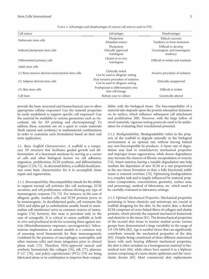
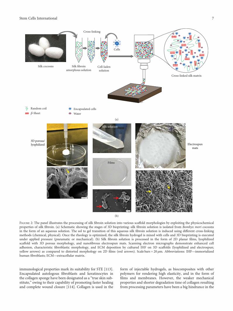
3.2.1. Silk. Silk has long been used as a dressing for woundsdue to its beneficial properties, like good biodegradability,ease of chemical alteration, good oxygen permeability, andthe ability of moisture retention [87, 88]. Silk fibroin (SF) isthe core protein of the silk fiber derived from cocoons ofeither mulberry or nonmulberry origin. It consists of a lightchain (~26 kDa) and a heavy chain (~390 kDa) whichare linked at a ratio of 1 : 1 by a single disulfide bond [89].The molecular weight of SF has been found to affect woundhealing [87]. SF with a narrow range of molecular-weightdistribution accelerates healing with better reepitheliza-tion, reduced scarring, lower infections and immunogenicresponses as compared to SF with a wider molecular-weightrange. However, controlling the biomechanical strength ofSF-derived scaffolds to match the target tissue properties isa challenging task due to the relatively inferior mechanicalproperties of regenerated SF [77]. Therefore, the processingof SF from cocoon shells needs to be carefully optimizedin order to achieve the desired properties in SF-based con-structs. In common lab-based protocols, silk cocoons are dis-solved using the standard lithium bromide (LiBr) approach[90], resulting in an aqueous solution of 6 − 8% w/v(Figure 2). By exploiting the conformational transition prop-erty of silk fibroin from a silk-I to a silk-II structure, therandom coils (predominant in the silk-I solution) are cross-linked to aid the transition of sol to gel (with predominantβ-sheet confirmation), resulting in a hydrogel. This hydrogelform of silk fibroin is heavily exploited in the fabrication of3D bioprinting approaches (Figure 2(a)). Apart from bio-printing, we have also utilized this property of silk fibroinfor the fabrication of constructs in different 3D geometries(Figure 2(b)).
However, several studies have raised concerns with usingsilk alone in scaffold preparation. These may be related to theclogging of needles in the case of 3D bioprinting and electro-spinning (due to the rapid transition into β-sheet structure atthe time of extrusion), inappropriate mechanical propertiesdue to the length of degumming time [91], and the LiBrdissolution process [90] (which often leads to the degrada-tion of protein chains). In order to circumvent this problem,silk-based composites have been developed for potentialapplications in different layers of skin such as 3D porous SF
functionalized with citrus pectin [92], SF/sodium alginatefreeze-dried scaffolds [93], electrospun nanofibers of SF withPLGA [94], SF only [38], collagen-SF [95], and 3D bioprintedSF with keratin [96] and gelatin [97].
3.2.2. Hyaluronic Acid (HA).HA is a common biological con-stituent of connective tissues of the cardiac valves, skin, bone,neuronal tissue and umbilical cord. It is an anionic nonsul-fated GAG possessing various desirable properties likehydrophilicity, optimal viscoelasticity, and lubrication [98–100]. Being an important element of the vertebrate ECM,HA is nonimmunogenic and provides a congenial environ-ment for cellular growth [101]. This has been observed inrecent studies where HA in combination with decellularizedporcine ECM and bFGF showed enhanced healing potentialin rabbit wounds pertaining to both epidermal and dermallayers [102]. Another bilayered artificial skin substitute com-posite of HA-gelatin-chitosan demonstrated appropriatemechanical properties and supported the coculture of kerati-nocytes and fibroblasts for up to 4 weeks in vitro [103]. HAcan stimulate the production of CD44 receptors during skinhealing, which further leads to enzymolysis of HA promotingvascularization and preventing graft contracture. The addi-tion of HA increases the expression of collagens I and III,the primary matrix components of skin [104]. Furthermore,similar to SF, low-molecular-weight HA has been shown toinduce fibrovascular tissue growth better than high-molecular-weight HA [105].
3.2.3. Fibrin Glue. Fibrinogen, a glycoprotein in blood, alsoserves as a potential biomaterial in STE. After its isolationvia precipitation by ammonium sulfate, PEG, or ethanol,the extracted fibrinogen is converted to fibrin glue (FG)via cross-linking by thrombin. The structural and mechan-ical properties of FG can be controlled by changing theextent of cross-linking [106]. FG possesses many advanta-geous properties making it a promising choice for STE.Fibrin glue-encapsulated keratinocytes have demonstratedimproved healing of burn wounds [27] and leg ulcers [107].Compared to other naturally derived materials, FG offers agreater versatility in terms of the customization of the skinsubstitute on the basis of rate of polymerization, geometry,pore size, and fiber thickness by the optimization of somematerial and physiological properties [107]. Some limitationsof FG like high cost, complicated storage conditions, poormechanical properties, long preparation time, gel shrinkageovertime, and the potential risk of disease transmission limitsits applicability in STE applications [28, 108].
3.2.4. Collagen. Collagens are a group of fibrous proteins hav-ing a triple-helical structure comprising of α-chains. Theyform the fundamental components of the ECM of almostall the tissues and play an imperative role by aiding in theregulation of tissue remodeling at the time of tissue repair[109–111]. Collagen possesses target motifs for integrinreceptors of cells thus regulating various properties relatedto adhesion, migration, proliferation, and differentiation[112]. Apart from this, easy isolation and purification;reduced toxic levels; and proven chemical, physical, and
6 Stem Cells International
immunological properties mark its suitability for STE [113].Encapsulated autologous fibroblasts and keratinocytes inthe collagen sponge have been designated as a “true skin sub-stitute,” owing to their capability of promoting faster healingand complete wound closure [114]. Collagen is used in the
form of injectable hydrogels, as biocomposites with otherpolymers for rendering high elasticity, and in the form offilms and membranes. However, the weaker mechanicalproperties and shorter degradation time of collagen resultingfrom processing parameters have been a big hindrance in the
Silk cocoons Silk fibroinamorphous solution
Cell-ladensolution
Cells
Random coil�훽-Sheet
Encapsulated cellsWater
Cross-linked silk matrix
Cross-linking
(a)
3D porous/lyophilized
Silk solution
Films
Electrospunmats
ECM
(b)
Figure 2: The panel illustrates the processing of silk fibroin solution into various scaffold morphologies by exploiting the physicochemicalproperties of silk fibroin. (a) Schematic showing the stages of 3D bioprinting: silk fibroin solution is isolated from Bombyx mori cocoonsin the form of an aqueous solution. The sol to gel transition of this aqueous silk fibroin solution is induced using different cross-linkingmethods (chemical, physical). Once the rheology is optimized, the silk fibroin hydrogel is mixed with cells and 3D bioprinting is executedunder applied pressure (pneumatic or mechanical). (b) Silk fibroin solution is processed in the form of 2D planar films, lyophilizedscaffold with 3D porous morphology, and nanofibrous electrospun mats. Scanning electron micrographs demonstrate enhanced celladhesion, characteristic fibroblastic morphology, and ECM deposition by cultured IHF on 3D scaffolds (lyophilized and electrospun;yellow arrows) as compared to distorted morphology on 2D films (red arrows). Scale bars = 20 μm. Abbreviations: IHF—immortalizedhuman fibroblasts; ECM—extracellular matrix.
7Stem Cells International

application of this protein. Therefore, alternative strategieshave been explored where researchers can incorporatethe biological advantages of collagen while overlooking itsweaker biomechanical aspect. For instance, collagen scaffoldshave been reinforced by combining polymers like PCL whichincrease the overall tensile strength. Collagen has also beencross-linked by various methods like UV polymerization,glutaraldehyde cross-linking, chitosan blending, and enzy-matic treatment to induce various ionic and covalent bondswhich improve its mechanical strength. [115]. For instance,glutaraldehyde cross-linking of collagen scaffolds aided inretaining the structural integrity and delaying the processof degradation in skin substitutes when grafted in athymicmice models [116, 117]. While such fixatives acted as goodcross-linkers, the residual by-components in vivo werefound to be cytotoxic [118]. Depending upon the degreeof cross-linking induced, collagen-based matrices aredegraded by collagenases into peptide fragments and aminoacids usually within the time frame of 3–6 weeks, subse-quently replacing the scaffold with native type I collagen pro-duced by resident fibroblasts [118]. Therefore, nontoxiccross-linkers such as EDC- (carbodiimide-) NHS (N-hydro-xysuccinimide) were applied to collagen structures, whichacted as successful potential dermal substitutes in vivo.However, the take rate of grafts was compromised in thecross-linked scaffolds over unaltered controls indicatingreduced integration of such cross-linked structures. Although,the problem of low take rate was resolved by applying atwo-step grafting procedure, it is not the most ideal processfor the clinical setting [119]. Hence, more effective andbiocompatible methods are being explored such as theuse of amino acids L-arginine, glutamic acid, and lysine forbio-cross-linking of collagen which have shown promisingoutcomes [120].
3.2.5. Decellularized Extracellular Matrix (ECM). Decellular-ized ECM scaffolds are widely used in the fabrication of sev-eral tissue substitutes in which the donor tissue undergoesremoval of cellular components without disturbing theECM comprising of collagen, GAGs, elastins, and growthfactors. Decellularization of the skin allows for completeremoval of the resident cell populations (hence reducingimmune responses) while retaining the collagen framework,which acts as the reservoir for growth factors and proteincomponents within this ECM network. In vivo studies inrat abdominal wall have documented rapid integration ofthese hydrogels with the host tissue and retainment of thestructures for up to 35 days in vivo [121]. With the adventof 3D bioprinting, this complex mixture can be explored asa potential bioink (when mixed with autologous cells) to pro-duce substitutes with adequate biological activity for healingcartilage, adipose, cardiac [122], and liver [123] tissues.Acellular dermal matrix (ADM) and acellular amnioticmembrane (AAM) have also been used in STE. ADM fromthe skin of goats, pigs [124], and fish [125] have been usedsatisfactorily in wound healing. Milan et al. used humandecellularized dermal matrix (DDM) seeded with humanumbilical cord perivascular cells (HUCPVCs) to treatdiabetic wounds in rats [126]. The HUCPVC-loaded DDM
scaffolds demonstrated accelerated wound healing andhigher VEGFR-2 expression and vascular density than thecontrol groups at 7 days post implantation in vivo. How-ever, on one hand as human decellularized ECM haslimited availability and is very costly, the use of xenogenicdecellularized ECM also exhibits the risk of disease trans-mission and immunogenicity. AAM serves as an excellentbiomaterial for curing the wounds as it aids in painreduction and moisture retention. Furthermore, it inhibitsscarring and extends antimicrobial activity and noninflam-matory and antifibroblastic effects [127, 128]. AAMspreserve the tissue ECM properly and have a potential useas a membrane for skin wound healing. TGF-β3 expressingbone marrow stromal cells cultured on AAM as a dermalequivalent led to the deposition of parallel, uniformcollagen bundles with improved cosmetic appearance anddecreased scar formation when transplanted onto full-thickness excisional skin wounds in rats for 85 days [129].The decellularized matrix can serve as ready-to-use tissuemodels with essential ECM molecules like collagen, elastins,GAGs, and growth factors for improved cell attachmentand proliferation.
3.3. Commercial Skin Substitutes. Many commercial skinsubstitutes are available for skin tissue repair and regenera-tion. Commercially available skin substitutes can be classifiedas acellular (e.g., AlloDerm® and Integra®) and cellulargrafts. Cellular skin substitutes can be further classifiedaccording to the skin layer they are targeting: (a) epidermal(CellSpray, MySkin), (b) dermal (Hyalograft 3D, Derma-graft®), or (c) epidermal-dermal composite (PermaDerm™,Apligraf®) [130]. Griffiths et al. proved that Apligraf®behaves only as a carrier dressing for deep-dermal woundsas the allogenic cells did not survive for long within thematrix in in vivo conditions [131]. OrCel™, another cellularskin substitute comprising of fibroblasts laden in bovinecollagen type I matrix as the dermal component and kerati-nocytes seeded at the air-liquid interface as the epidermalcomponent is used commercially for partial-thickness inju-ries; nonetheless, the use of bovine collagen poses a potentialrisk of rejection and disease [132]. Similar substitutes likeDermagraft® and TransCyte® are also used in combinationwith cells. But the long incubation times associated with thesesubstitutes (>6 weeks) may not be favorable for trauma cases[133]. Integra®, the most common commercial skin replace-ment, was developed in the 1980s by Yannas and Burke as anacellular bilayered construct [134]. It is a porous constructfabricated from bovine tendon collagen and shark GAGs(chondroitin-6-sulfate) which serve as a dermal substitute,while the epidermal representative is the semipermeablepolysiloxane (silicone) layer. Most commonly availablecommercial skin substitutes fall in this category such asBiobrane® and AlloDerm. Biobrane® comprises of a nylonmesh with a silicone membrane mimicking the dermis andepidermis, respectively, in porcine collagen. Though theapplication involves a single stage procedure, the substitutecarries risk of contamination with porcine collagen and isfound to be intolerant towards infection sites. While thereported functionality of this substitute is better, the
8 Stem Cells International
procedure involves a two-stage application hiking the costof treatment [130]. Commercially available artificial skinsubstitutes have been widely used in wound-healing stud-ies in combination with both autologous and allogeneicskin cells. A full-thickness skin substitute developed forfoot ulcers is Tiscover™. However, these products beartheir own limitations, and as such there is no ideal skinsubstitute yet. No construct so far has been able to reca-pitulate the 3D geometry, chemistry, and functionality mim-icking the native skin tissue. They face several limitations likenonintegrity, immune rejection, poor take, and reducedmechanical strength [130]. Therefore, a tissue-engineeredconstruct with off-the-shelf availability is urgently neededfor large-scale application.
4. Current Innovative Strategies in Skin TissueEngineering (STE)
Although great progress has been made in reducing mor-bidity and mortality occurring as a result of burn wounds,some of the most exciting advances remain ahead. Tissue-engineered skin substitutes using a combination of scaffoldsand growth factors appear to be a promising alternative.Use of multimaterial strategy to develop composite scaffoldshelps alleviate the limitations associated with individualmaterials such as inferior mechanical properties and biocom-patibility [135]. The ultimate aim is to completely restoreskin anatomy and physiology using (a) advanced nanofunc-tionalized materials for triggering specific responses usingnanotechnology; (b) automated and robotic fabrication ofengineered tissues to increase efficacy, reduce costs, and caterto individual patient needs using 3D bioprinting; and (c)regenerative therapy using stem cell technology for individu-ally targeting pigmentation, wound closure, angiogenesis,and skin sensation.
4.1. Nanotechnology. Nanotechnology has been used in twoways in STE; firstly, in scaffold fabrication and secondly, inloading the scaffolds with growth factors and/or drugs fortargeted delivery to the tissue. Electrospinning is a widelyused technique for scaffold fabrication in which the polymersolution is spun into nanofibers under the force of an electricfield. Advantages of electrospun scaffolds are large surface-to-volume ratio, better pore interconnectivity, easy repro-ducibility, and easy fabrication method. The nanofibrousarchitecture aptly mimics the ultrastructure of native tissueECM; hence, it strengthens the resultant cell-matrix interac-tions in vitro [136]. PLGA electrospun scaffolds have shownto promote fibroblast survival and maintenance in in vitroculture for 5 days [137]. Using this material, researcherscould achieve a pore size of 100-200 μm for the culture offibroblasts towards skin tissue engineering applications[138]. On the contrary, Chen et al. successfully demonstratedinfiltration of human dermal fibroblasts and subsequentcollagen type I synthesis after 7 days on PLGA electrospunscaffolds possessing pore sizes as low as 5-40 μm [139]. Inanother study, electrospun scaffolds made of collagenshowed enhanced cell growth and organization, significantlyreducing wound contraction by 22% in full-thickness wounds
created in murine models as compared to freeze-dried scaf-folds [140]. This is mainly attributed to the increased surfacearea, interconnectivity, and pore size of electrospun scaffoldsover their freeze-dried counterparts. Park et al. used salt(NaCl) crystals within the silk-polyethylene oxide (PEO)electrospun fibers [38]. The resultant scaffolds had pore sizes(250-300 μm) large enough to support the formation of two-tiered skin in vitro. An aqueous-based fabrication strategyallows for easy inclusion of biological moieties at the timeof electrospinning procedure [75]. Arg-Gly-Asp- (RGD-)functionalized and protease-sensitive poly(ethylene glycol)(PEG) hydrogels have been developed [141]. The additionof cell adhesive and protease-sensitive peptides is very usefulas they allow increased cellular attachment, growth, andmigration on the matrix.
Another effective alternative is the incorporation ofnanoparticles into the engineered scaffolds for targeting spe-cific functions, for instance, excessive fluid loss and on-siteinfections experienced by chronic-burn patients. Also, exu-dates from the wounds are a common cause of infectionsresulting in acute inflammation which pose a hindrance inwound healing. To circumvent this, a collagen–chitosan-based scaffold with silver nanoparticles was developed forrepairing the dermal layer of the skin. The silver nanoparti-cles exhibited bactericidal properties with enhanced biocom-patibility of the construct [142]. Another group fabricatedelectrospun collagen-PEO nanofibrous scaffolds incorpo-rated with gold nanoparticles [143]. Gold nanoparticles areknown to enhance biocompatibility and mechanical strengthand provide antioxidation and enzyme-resistance to the con-structs. All these parameters were tested across a range ofconcentrations, with 14.27 ppm marked as the most optimalconcentration of gold nanoparticles for collagen-PEO elec-trospun mats. The scaffolds demonstrated negligible toxicityand sustained the culture of murine 3T3 fibroblasts andkeratinocytes up to 14 days in vitro. The technique of nano-particle formation has been extended to drug delivery inwounds leading to better healing response. Nanoparticleshave several advantages over use of scaffolds. For instance,particles can be injected to the healthy tissue around thewound, preventing any direct manipulations with the woundbed [144]. They can be precisely modified to regulate therequired release profiles in vivo to match the physiologicalbody conditions [145]. PLGA is the most common polymerused to develop nanoparticles [146]. Chereddy et al. devel-oped curcumin-loaded PLGA nanoparticles which, alongwith lactate, showed enhanced angiogenesis and reducedinflammatory response in full-thickness splinted excisionalwounds in mice [147]. In another unique approach, silica,PEG, and chitosan were mixed to develop nanoparticles forsustained release of nitric oxide in wounds accelerating thehealing process in infected, noninfected, and diabetic woundsin mice models [144, 148, 149]. In yet another approach,lyophilized keratinocyte-targeted nanocarriers loaded withlocked nucleic acid- (LNA-) modified anti-microRNAs(miRs) were applied increasing the expression of Dicer whichplays a pivotal role in reestablishing the barrier function ofthe skin [150]. Hence, nanotechnology has a wide-rangedapplication in STE.
9Stem Cells International

4.2. Scaffold-Free Approach. Scaffold-free approach involvesthe use of a carrier-free or matrix-free cell population cul-tured in the form of a transplantable cell sheet for directimplantation onto the site of injury. The technique is usefulin avoiding the immunogenicity associated with most syn-thetic scaffolds. However, the lack of a mechanical supportfails to provide anchorage to the proliferating cells from thehost. Thermoresponsive materials such as poly(N-isopropy-lacrylamide) may provide an effective carrier-free sheet. Itwas observed that this thermosensitive property of the poly-mer could be applied to modulate changes in the scaffold’spore diameter, i.e., the pore size decreased with an increasein temperature [151]. A porous scaffold of poly(N-isopropy-lacrylamide) developed using the sphere-templating tech-nique with a pore diameter of 55 ± 5 μm was cultured withNIH3T3 fibroblasts, and the temperature was subsequentlychanged to 37 °C. The resulting phase transition constrictedthe pore size to 39 μm, which is the optimal diameter forfacilitating angiogenesis and matrix synthesis. These cellswere cultured for 7 days in vitro and showed a characteristicelongated morphology. Liu et al. constructed a scaffold-freebilayered, vascularized tissue-engineered skin by superim-posing 4 layers of dermal fibroblasts and endothelial cellsheets to form the dermal layer followed by seeding andculture of keratinocytes on it. The sustainability of thisin vitro-developed vascularized skin containing epithelialcells, endothelial cells, and fibroblasts was up to 5 weeks inculture [152]. Recently, the scaffold-free approach has alsobeen applied to 3D bioprinting. A novel bioink formula-tion comprising of 10% w/v gelatin, 0 5% w/v alginate,and 2% w/v fibrinogen was 3D bioprinted with humandermal fibroblasts to develop a construct of 1 cm × 1 cm ×0 5 cm dimensions. The study could successfully demon-strate a printable, clinically conformant functional skin tissuecharacterized at the molecular level [153].
4.3. Stem Cell Technology. Recent advances in stem cell-basedtherapeutics have propelled an increasingly high enthusiasmin STE. The essential ingredients for successful STE includethe choice of biomaterials combined with the appropriatecells and growth-inducing factors [154]. Different types ofstem cells have been explored in the field of STE.
4.3.1. Mesenchymal Stem Cells. Mesenchymal stem cells(MSCs) are multipotent cells found in adults in various tissueslike adipose, bone marrow, and dental pulp. BMSCs have beenshown to promote angiogenesis, epithelialization, granulation,and tissue formation in an in vivo setup leading to effectiveskin regeneration [155]. BMSCs cultured on collagen matriceshave shown therapeutic potential across a variety of wounds[156, 157]. Furthermore, BMSC-derived exosomes are beinginvestigated as mediators in wound healing [158].
ADSCs have also been widely used in wound healingstudies. When encapsulated in fibrin-chitosan hydrogelmatrices, they have demonstrated a consistent release ofangiogenic factors to aid in the wound healing process[159]. Conditioned media of ADSCs stimulated collagendeposition and homing of human dermal fibroblasts [160],thus establishing the role of ADSCs in accelerated wound
repair via the secretion of growth factors. Release of angio-genic cytokines by ADSCs augmented the degree of neovas-cularization [161]. A more recent vascularization strategyinvolves the use of ADSC-derived microvascular fragmentsisolated from mice fat pads [162]. These microvascular frag-ments seeded onto collagen-GAG biomatrices implantedinto the dorsal skinfold chambers of C57BL/6 mice showeddense microvascular branching and lymphatic networks after14 days. On the contrary, this angiogenic response of ADSC-derived microvascular fragments was rather diminishedwhen porous scaffolds were precultivated with microvascularfragments for 28 days in vitro and then subjected to thein vivo dorsal chamber in mice [163]. This study highlightedthe importance of freshly isolated microvascular fragmentsfrom mice fat pads for in vivo implantation purposes. Asuccessful application of these ADSC-derived microvascularfragments was also recently documented in a full-thicknessskin wound model by significantly improving the vascularand lymphatic networks when applied together with STSG[51]. Chan et al. showed the effect of different hydrogels onthe multilineage differentiation potential of ADSCs [164].Lin et al. determined that application of ADSC sheets accel-erates the rate of tissue healing by as early as 18 days posttreatment in comparison to the control group [165]. Trottieret al. established that ADSCs could serve as a plausible alter-native for fibroblasts in STE by fabricating a trilayered skinsubstitute using ADSCs only [166]. Gholipourmalekabadiet al. combined stem cells, nanotechnology, and the healingproperties of the amniotic membrane to develop a 3D bilayerscaffold for burn injuries [167]. In the study, silk fibroin wasdirectly electrospun on decellularized amniotic membrane todevelop the bilayered scaffold. Postfabrication, ADSCs werecultured on the scaffold for 15 days which resulted inincreased expression of two proangiogenic factors, vascularendothelial growth factor (VEGF) and bFGF, which are pre-requisites in wound healing and hence determine the effi-ciency of the skin substitute. Another potential source ofstem cells, human dermal papilla, isolated from the dentalpulp have shown promising potential in hair follicle regener-ation by self-organizing into in vitro organoids [168].
It is a challenge to heal larger wounds and ulcers that canonly be treated with surgical grafts so far. Recent research hasshown successful reprogramming of wound-resident mesen-chymal cells in in vivomice models which were able to regen-erate skin epithelial tissue [169]. Cellular reprogrammingin vivo was conducted by injecting viruses to induce theexpression of four specific genes in the cells of nonhealingulcers to transform them into epithelial cells. These micewere able to demonstrate complete healing of the largewounds by a newly formed layer of epithelial cells within 28days. This novel strategy provided an effective solutiontowards surgical skin transplants or artificial skin grafts forhard-to-heal wounds, particularly in aged population anddiabetic people [169].
4.3.2. Skin Stem Cells. Stem cells in the skin mainly reside inthe hair follicles, sweat glands, and stratum basale of the epi-dermis. These alternate sources of stem cells have also beenexplored for STE applications. In one of the approaches,
10 Stem Cells International

human stem cells derived from sweat glands when seededonto Integra® (a commercial skin replacement possessingcollagen fibers cross-linked with GAGs) demonstratedenhanced dermal regeneration by increasing the extent ofvascularization in a bilateral full-skin wound. The stem cellswere homogenously distributed on the scaffold exhibitingsatisfactory cell-substrate interactions [170]. Nestin-positivestem cells have also been known to contribute towardswound healing via the development of intricate microvascu-lature networks [171, 172]. Induction of neovascularizationby nestin-expressing hair follicle cells that successfully anas-tomosed with the host vasculature was demonstrated byAmoh et al. in nude mice models [171]. According to aclinical study, a team of medical practitioners treated a 7-year-old boy suffering from epidermolysis bullosa, a geneticskin disease also called butterfly disease, wherein approxi-mately 80% of the epidermis was damaged. Scientistsattempted to regenerate the damaged skin using trans-plants derived from modified stem cells. Procedurally, theautologous epidermal stem cells were isolated from thepatient and the gene defect was repaired by adding a cor-rected form of the mutated gene. Such patient-derivedcells containing the genetically corrected gene were grownin a laboratory in order to form a sheet of neoskin tissue.Nearly 400 million of such genetically corrected cells weregrafted into the patient, but most of the transplanted cellsdied after the procedure. Of the transplanted cells, the rela-tively stable epidermal stem cells were found to contributetowards the generation of the new skin tissue. After a month,the patient showed successful signs of skin restoration in theaffected area. Furthermore, after 2 years of follow-up, thenewly regenerated skin showedmolecular markers character-istic of morphologically stable and functional skin tissue.Thus, the combined approach of cell and gene therapy couldsuccessfully regenerate approximately 80% of the patient’sskin [173].
4.3.3. Embryo-Related Stem Cells. Embryo-related stem cellsinclude the embryonic stem cells and stem cells from extra-embryonic tissues like the placenta, umbilical cord, andamniotic fluid. Stem cells from these sources have a greaterpotency than adult stem cells. Skardal et al. used laser deposi-tion bioprinting to print amniotic-fluid stem cells on dorsalskin wounds in mice to investigate their effect on wound heal-ing [174]. Amniotic-fluid stem cells were selected owing totheir tremendous proliferative ability, enhanced angiogenicpotential, and nonimmunogenicity. Immunohistochemistryshowed that stem cells left the wound site by day 7 suggestingthat the stem cells contributed towards the early days of heal-ing by migration, drastic release of growth factors, and theirpropensity to maintain an undifferentiated state for extendedperiods leading to increased cell proliferation [174].
Isolating a differentiated population of autologous cellsbecomes complicated in cases where extensive injury orburns are inflicted covering a large body surface area. Hence,stem cells are a rescue in such scenarios as they possess thetremendous potential of self-renewal and multilineage differ-entiation, so that the scientists can start with low initial celldensity [154].
Another critical parameter is the damaged sweat glandsin burn victims as well as in some genetic disorders, whichcan be a life-threatening condition. Such people are unableto exercise properly as it may lead to heat stroke and braindamage. Hence, the creation of sweat glands using skin graft-ing procedures is clinically important for the regeneration ofa viable skin model. Current clinical methods of skin graftingand regenerative therapy are incapable of inducing the devel-opment of functional sweat glands. Recent research in thefield has highlighted the relevance of precise control in thedevelopment of sweat glands at the molecular level. Thesignaling pathways responsible for the development of hairfollicles and sweat glands are similar but express at differenttime frames. The signaling cascade inducing the expressionof hair follicles is triggered first, followed by the expressionof bone morphogenetic proteins (BMPs), which helps to cre-ate sweat glands. This deep understanding into the underly-ing signaling mechanisms can prove to be very beneficial inthe way of improvising on the skin tissue engineeringmethods for effective skin grafting procedures [175]. Forinstance, the biomolecular cues from fibroblast growthfactor-2 (FGF2) and vascular endothelial growth factor-(VEGF-) loaded StarPEG-heparin hydrogel triggered differ-ential cellular behavior of human umbilical vein endothelialcells (HUVECs) indicating proangiogenic conditions [176].Similarly, Zhou et al. showed that downregulating the expres-sion of microRNA-203 at the wound site by the applicationof antimicroRNA-203 can accelerate wound healing andreduce scarring. Downregulation of microRNA-203 increasesthe expression of the gene Hairy/Enhancer of split-1 (Hes1)which is a downstream signaling molecule in the Notch1/-Jagged1 pathway which in turn promotes epidermal stem cellproliferation and inhibits its differentiation [177].
However, for over several decades, human skin cellshave been cultured in combination with animal culture sys-tems, which create potential risk of infections and immunecomplications. To overcome these limitations, researchersdeveloped a fully animal-free, human-based system for thecultivation of skin cells for safer skin grafting [178]. Theresearchers used laminin proteins, specifically LN-511 orLN-421, as animal-free components for providing a robustyet safe in vitro cell culture system for effective skin graft-ing. This may lead to a more progressive research approachby pushing such in vitro standardized systems towardsclinical application.
4.4. 3D Bioprinting. 3D bioprinting is a computer-aidedadvanced technology involving the precise placement of cellsinto predetermined 3D patterns. At the preclinical level, thetechnique has showcased successful replication of naturalskin anatomy [179] as compared to conventional fabricationmethods employing top-seeding of cells [180]. 3D bioprintedscaffolds are fabricated using the following two approaches:(a) top-down approach, in which cells are directly madeavailable to a prefabricated biomimetic scaffold for tissuematuration in a bioreactor and (b) bottom-up approach,which provides only an initial temporary support and dependson the seeded cells for the deposition of ECM [181, 182]. Forbioprinting, the biomaterial processed in the form of “ink”
11Stem Cells International

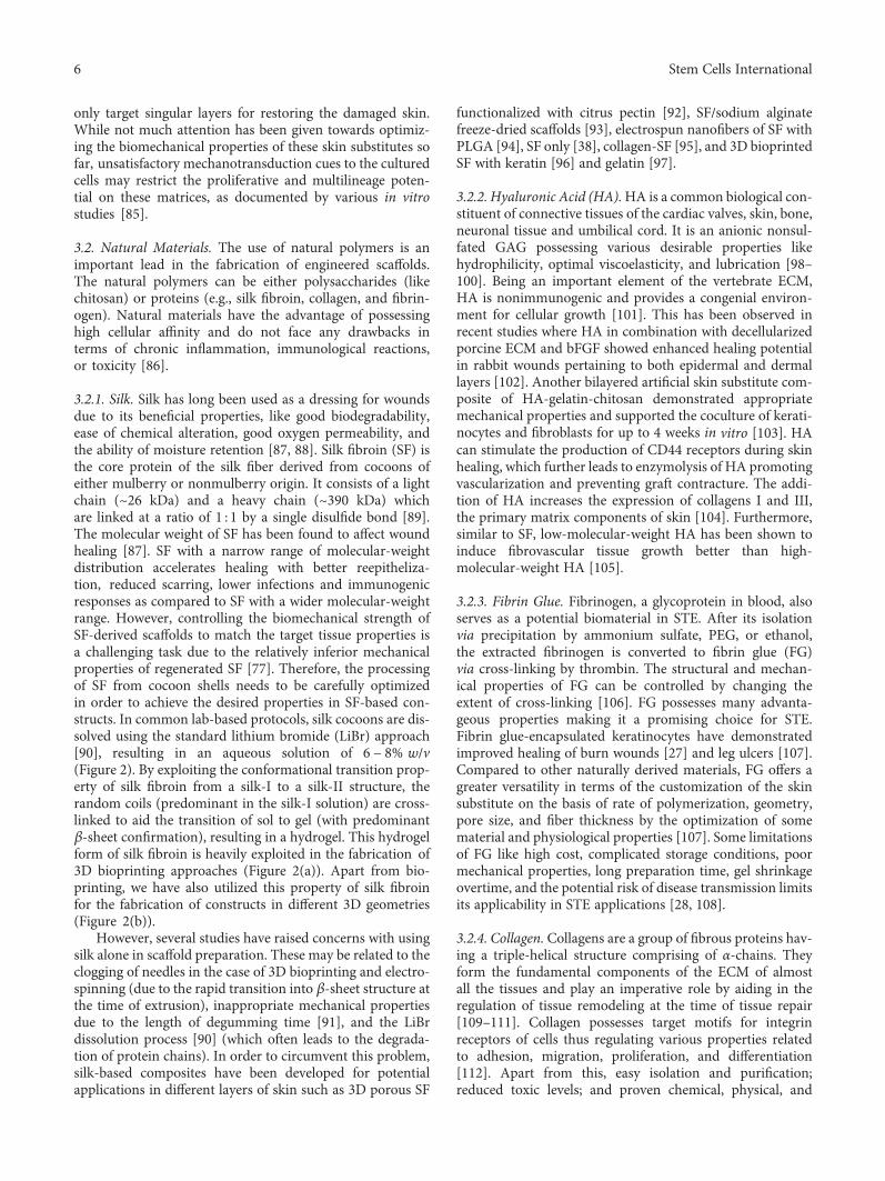
should also be “printable” apart from being biocompatibleand durable. Printability depends on two attributes: rheology(covers aspects like shear thinning and viscoelasticity ofmaterial) and cross-linking methods employed (chemical(enzymatic), physical (sonication), photodependent, tempera-ture-dependent, or ionic mechanism) [183]. The inks are usu-ally made from synthetic polymers, like PCL, PLGA, PEG, andpluronic F-127, or natural polymers, like silk fibroin, collagen,or fibrinogen [63] or an amalgamation of the two types ofmaterials. Apart from the material or ink, the optimal choiceof cells is another critical component in fabricating constructs.The most commonly used cells for skin constructs have beenkeratinocytes and fibroblasts [38]. Using a customized 3D bio-printer built by Alfatek Systems, Kolkata, we have successfullyfabricated custom-made 3D bioprinted constructs made of
pluronic-based bioink mixed with immortalized human fibro-blasts (IHF). The constructs demonstrated optimal viscosityfor extrusion bioprinting and post-printing stability by retain-ing the multilayered stack of the construct. In addition, thegreen fluorescent protein- (GFP) tagged IHF population wasviable for more than 7 days in in vitro culture and showedcharacteristic fibroblastic markers, as shown in Figure 3.
Lee et al. in 2009 were the first to successfully generate a3D bioprinted human skin construct using collagen type Iwith a co-culture of fibroblasts and keratinocytes [63]. Astratified construct was printed by sandwiching the collagenmatrix between fibroblasts and keratinocytes. The printedcells showcased characteristic cellular morphology; however,the proliferation was affected by poor printing resolution,which was far from the optimal resolution of 300 μm. In
3D bioprinting
(a)
(b) (c) (d) (e) (f)
In situ bioprinting
(g)
Figure 3: 3D bioprinting to develop custom-made skin tissue constructs. (a) A customized printer used in our lab (by Alfatek Systems,Kolkata). (b) The CAD image prepared using the inbuilt software in a readable file format for bioprinting. (c) Following the CAD image, apluronic-based bioink mixed with fibroblasts is loaded into the syringe fitted with a nozzle and the printing process is executed underapplied pressure. (d) 3D bioprinted construct. (e) Fluorescence micrograph of GFP-tagged fibroblasts (green) showing cellular distributioninside the filaments of the construct immediately after printing. (f) Protein expression of skin-specific marker vimentin (red) in a 3Dbioprinted pluronic-based construct 3 days postprinting by immunofluorescent staining. Nuclear staining was done by DAPI (blue). (g) Insitu bioprinting strategy schematically depicted on the burnt skin of a patient demonstrating deposition of the bioink directly on theregion of interest. Scale bars = 30μm. Abbreviations: CAD—computer-aided design; IHF—immortalized human fibroblasts. Immortalizedhuman fibroblasts (IHF) were used for 3D bioprinting.
12 Stem Cells International
another landmark study, Binder was the first to demonstratein situ bioprinting of a dorsal skin defect (10 × 10 cm) in aporcine model [184]. The wound dimensions were measuredby laser scanning, and the bioink made from collagen-fibrinogen with keratinocytes and fibroblasts was deliveredusing an ink-jet bioprinter onto the wound. Complete ree-pithelialization occurred by the 8th week, which was signifi-cantly enhanced as compared to all the control groups(allogenic, untreated, and hydrogel matrix only). The con-cept of in situ bioprinting comes from the fact that theinjury/lesion is repaired by bioprinting the cell-materialbiocomposite directly on the target site (Figure 3). Advan-tages include rapid implantation, no delay in surgical time,and reduced risk of on-site infections. Another form of bio-printing called laser-assisted bioprinting, which exploitslaser-induced forward deposition, was used by Koch et al.[185] for STE. The procedure involved depositing the cellsonto a MatriDerm® scaffold (a commercial graft made upof collagen and elastin) using a laser pulse inducing cellknockouts with high precision from a laser-absorbing donorlayer (~60 nm in thickness) directly onto the scaffold.Cadherins, the molecules forming the gap junctions, wereobserved at the basal lamina (Cx43 expression) indicatingdifferentiation and maturation of the printed tissue. Michaelet al. also used laser-assisted bioprinting to fabricate abilayered skin construct made of collagen comprising of theprimary constituent skin cells (fibroblasts and keratinocytes)and demonstrated full-thickness wound healing in mice[179]. Ki67 staining confirmed the presence of the prolifera-tive layer (stratum basale) of the skin. Inadequate tissue dif-ferentiation and vascularization were observed at the site ofimplantation, which may be attributed to the short experi-mental duration of only 11 days.
Recently, Lee et al. used a robotic approach in a solid free-form fabrication system with contactless dispensers capableof achieving a resolution of ~500 μm (approximately 28 nlof the bioink) [63]. A density of 2 3 × 10⁶ cells/ml was usedto print both keratinocytes and fibroblasts. More recently,Cubo et al. printed a biocomposite of skin cells mixed withhuman plasma using an in-house modified extrusion bio-printer having four dispensers [39]. The bioprinted productwas prepared on a P100 plate before grafting it subcutane-ously in an immunodeficient mice for 8 weeks, which is oneof the maximal time periods tested so far in vivo. At theend of 8 weeks, the implanted skin exhibited the generationof two new layers, the basal lamina and the stratum corneum,indicating complete differentiation. The biggest achievementof this study was the ability to bioprint skin constructs in <35minutes highlighting the quick application and translationalpotential of the technology.
Next, advances in 3D bioprinting have led to the develop-ment of constructs with a controlled pore architecture andguided cellular assembly in vitro for the synthesis of sweatgland mimic [186]. The drop-on-demand technology canhelp place the cells or the polymer precisely in a spatiotempo-ral pattern so that the complex structures like pigmentationunit, hair follicle, or sweat gland can be fabricated asrequired. However, the technique needs to be extensivelystandardized in terms of the matrix with an adequate
mechano- and compositional niche for cell proliferation,migration, and morphogenesis for generating phenotypicallystable and complex structures. While it is clear that fibro-blasts, keratinocytes, and stem cells are printable using vari-ous inks, but reproducing the integrity and functionality ofthe human skin for STE applications and inducing sensationhave not been achieved yet. Some of the challenges beingfaced include printing resolution, adequate vascularization,choice of optimal cell type and source, bioink compositionto match the zonal tissue architecture, growth factor gradi-ent, dimensions of clinically conformant constructs, cost ofbioprinted skin, and the complexity of the technique beforeclinical success could be envisaged. Scalability, althoughtoday a challenge, can be overcome in the very near futurewith fastly evolving software and machinery, which requirerelatively lower initial cell densities. This helps in meetingthe demands for developing constructs to cover a large bodysurface; a major limiting factor with the autologous grafts;current gold standards. With the evolving field of 3D bio-printing, scalability can be combined with customization ofthe graft, achieving a major milestone in STE and woundhealing. Smaller 3D skin tissue in vitro models are at a betterposition of being translated for applicability in ex vivo testingof cosmetics and drugs, before this technology reaches thehospitals for reconstructive surgeries [187]. The success ofthe technology in the research field is reflected by cumulativeinvestments of > $44 million in grants made by companieslike Procter & Gamble, L’Oréal, and Poietis into R&D of3D bioprinted functional in vitro skin models [187]. How-ever, abundant work needs to be undertaken for determiningthe underlying molecular mechanisms that regulate tissuedifferentiation in such artificial in vitro model systems forbetter exploitation of the technology.
5. Future Prospectives
The field of STE is maturing and the biomaterials used (suchas collagen) in developing products have benefited patientsover decades. However, even after decades of research inthe field of STE, production of large constructs for victimswith more than 50% skin loss is still a challenging proposi-tion. Despite the availability of a large number of techniquesand commercial skin substitutes, the quest for a functionalartificial skin capable of supporting skin functions like ther-moregulation, sensation, perspiration, and UV radiation pro-tection with normal aesthetic appearance is still on [130].Several biomaterials supplied as commercial skin substitutesare available, albeit with associated limitations. For instance,risk of disease transmission from the use of porcine collagencould be prevented using synthetic collagen-based materialsor recombinantly produced collagen. Next task would be toidentify the cell adhesion and tissue integration propertiesof this synthesized collagen. Also, such cell-free biomaterialsmay act as appropriate carriers/substrates for smaller injurieswhere host cells could migrate into the wound site. Withlarger wounds, the biomaterials alone are not sufficient. Theyneed a prevascularized bed with cells to trigger the regenera-tive process. To achieve these functions, stem cell-based tis-sue engineering therapy has grasped much attention owing
13Stem Cells International
to their self-renewal capacity and multilineage potential,critical for mimicking the physiological hierarchy and func-tions of native tissue. However, finding the optimal cellsource, standardization of processing, and application inclinical settings, as well as precisely regulating the responseof stem cells in situ, is still subjected to consideration. Theother major problem is with scalability of technique. Mostof the research has been carried out on small wounds cre-ated in rodent models; therefore, critical measures need tobe undertaken to escalate their applicability for preparingclinically conformant skin grafts [2]. The robustness andreproducibility of nanotechnology-based techniques makeit suitable for producing off-the-shelf products while, onthe other hand, an advanced technique like 3D bioprintingis capable of developing customized substitutes. Each ofthe approaches has its own lacunae which can be overcomeby permutating and combining the available techniques soas to develop a skin substitute which can meet the needsof the wound care market. Therefore, there is a pressingdemand for more active and high-paced research in orderto cope with the increasing demand for skin tissue grafts.With the advent of cutting-edge technologies, fabricating afunctional patient-specific skin substitute does not appearto be far-fetched.
Abbreviations
3D: Three dimensionalAAM: Acellular amniotic membraneADM: Acellular dermal matrixADSCs: Adipose-derived stem cellsbFGF: Basic fibroblast growth factorBM-MSCs: Bone marrow-derived mesenchymal stem cellsBMSCs: Bone marrow stem cellsCAD: Computer-aided designCEA: Cultured epithelial autograftsCx43: Connexin 43DDM: Decellularized dermal matrixDNA: Deoxyribonucleic acidECM: Extracellular matrixESCs: Embryonic stem cellsFDA: Food and Drug AdministrationGAGs: GlycosaminoglycansGFP: Green fluorescent proteinHA: Hyaluronic acidIHF: Immortalized human fibroblastsiPSCs: Induced pluripotent stem cellsLiBr: Lithium bromideMSCs: Mesenchymal stem cellsNaCl: Sodium chloridePCL: Poly(ε-caprolactone)PEG: Polyethylene glycolPEO: Polyethylene oxidePLGA: Poly(lactic-co-glycolic) acidRGD: Arginine-glycine-aspartic acidSF: Silk fibroinSTE: Skin tissue engineeringSTSG: Split-thickness skin graftsUSD: United States dollar
UV: UltravioletVEGF: Vascular endothelial growth factorWHO: World Health Organization.
Conflicts of Interest
The authors declare that they have no conflicts of interest.
Acknowledgments
I would like to acknowledge the contribution of Mr.Ramchandra C. Pokale (Chief Artist, Centre for CommunityMedicine, AIIMS), Mr. Manish Prajapati, and Mayuri Dutta(Research Trainee, Stem Cell Facility, AIIMS) in preparingthe figures and in proofreading. This work was supportedby the DBT-Centre of Excellence for Stem Cell Research(BT/01/COE/07/03), the Council of Scientific and Indus-trial Research (CSIR) (09/006(0478)/2018-EMR-I), and theDepartment of Science and Technology- (DST) InspireFaculty Grant (DST/INSPIRE/04/2017/000645).
References
[1] P. Zanger, “Staphylococcus aureus positive skin infectionsand international travel,” Wiener klinische Wochenschrift,vol. 122, no. S1, pp. 31–33, 2010.
[2] N. Bhardwaj, D. Chouhan, and B. B. Mandal, “3D functionalscaffolds for skin tissue engineering,” in Functional 3D TissueEngineering Scaffolds, pp. 345–365, Woodhead Publishing,2018.
[3] H. Liu, C. Wang, C. Li et al., “A functional chitosan-basedhydrogel as a wound dressing and drug delivery system inthe treatment of wound healing,” RSC Advances, vol. 8,no. 14, pp. 7533–7549, 2018.
[4] C. A. Brohem, L. B. Da Silva Cardeal, M. Tiago, M. S. Soengas,S. B. De Moraes Barros, and S. S. Maria-Engler, “Artificialskin in perspective: concepts and applications,” Pigment Cell& Melanoma Research, vol. 24, no. 1, pp. 35–50, 2011.
[5] Z. Nemes and P. M. Steinert, “Bricks and mortar of theepidermal barrier,” Experimental & Molecular Medicine,vol. 31, no. 1, pp. 5–19, 1999.
[6] D. M. Supp and S. T. Boyce, “Engineered skin substitutes:practices and potentials,” Clinics in Dermatology, vol. 23,no. 4, pp. 403–412, 2005.
[7] D. Breitkreutz, N. Mirancea, and R. Nischt, “Basementmembranes in skin: unique matrix structures with diversefunctions?,” Histochemistry and Cell Biology, vol. 132, no. 1,pp. 1–10, 2009.
[8] D. Breitkreutz, I. Koxholt, K. Thiemann, and R. Nischt, “Skinbasement membrane: the foundation of epidermal integri-ty—BM functions and diverse roles of bridging moleculesnidogen and perlecan,” BioMed Research International,vol. 2013, Article ID 179784, 16 pages, 2013.
[9] M. P. Rodero and K. Khosrotehrani, “Skin wound healingmodulation by macrophages,” International Journal of Clini-cal and Experimental Pathology, vol. 3, no. 7, pp. 643–653,2010.
[10] V. W. Wong and G. C. Gurtner, “Tissue engineering for themanagement of chronic wounds: current concepts and futureperspectives,” Experimental Dermatology, vol. 21, no. 10,pp. 729–734, 2012.
14 Stem Cells International
[11] S. Huang and X. Fu, “Naturally derived materials-based celland drug delivery systems in skin regeneration,” Journal ofControlled Release, vol. 142, no. 2, pp. 149–159, 2010.
[12] W. Care, Wound care market, 2022, pp. 1–6, Markets andMarkets, 2019.
[13] F. Groeber, M. Holeiter, M. Hampel, S. Hinderer, andK. Schenke-Layland, “Skin tissue engineering—in vivo andin vitro applications,” Advanced Drug Delivery Reviews,vol. 63, no. 4-5, pp. 352–366, 2011.
[14] P. Chandika, S. C. Ko, and W. K. Jung, “Marine-derivedbiological macromolecule-based biomaterials for woundhealing and skin tissue regeneration,” International Journalof Biological Macromolecules, vol. 77, pp. 24–35, 2015.
[15] S. Aarabi, M. T. Longaker, and G. C. Gurtner, “Hypertrophicscar formation following burns and trauma: new approachesto treatment,” PLoS Medicine, vol. 4, no. 9, pp. e234–1470,2007.
[16] B. Berman, M. H. Viera, S. Amini, R. Huo, and I. S. Jones,“Prevention and management of hypertrophic scars andkeloids after burns in children,” Journal of Craniofacial Sur-gery, vol. 19, no. 4, pp. 989–1006, 2008.
[17] V. Jay, “This month in history,” Journal of the Royal Society ofMedicine, vol. 92, no. 10, p. 548, 1999.
[18] M. Chen, M. Przyborowski, and F. Berthiaume, “Stemcells for skin tissue engineering and wound healing,” Crit-ical Reviews™ in Biomedical Engineering, vol. 37, no. 4-5,pp. 399–421, 2009.
[19] W. L. Ng, S.Wang,W. Y. Yeong, andM.W. Naing, “Skin bio-printing: impending reality or fantasy?,” Trends in Biotech-nology, vol. 34, no. 9, pp. 689–699, 2016.
[20] K. J. Miller, D. A. Brown, M. M. Ibrahim, T. D. Ramchal, andH. Levinson, “MicroRNAs in skin tissue engineering,”Advanced Drug Delivery Reviews, vol. 88, pp. 16–36, 2015.
[21] R. Langer and J. Vacanti, “Tissue engineering,” Science,vol. 260, no. 5110, pp. 920–926, 1993.
[22] T. T. Nyame, H. A. Chiang, and D. P. Orgill, “Clinical appli-cations of skin substitutes,” Surgical Clinics of North America,vol. 94, no. 4, pp. 839–850, 2014.
[23] R. A. Kamel, J. F. Ong, E. Eriksson, J. P. E. Junker, and E. J.Caterson, “Tissue engineering of skin,” Journal of the Ameri-can College of Surgeons, vol. 217, no. 3, pp. 533–555, 2013.
[24] R. F. Pereira, C. C. Barrias, P. L. Granja, and P. J. Bartolo,“Advanced biofabrication strategies for skin regenerationand repair,” Nanomedicine, vol. 8, no. 4, pp. 603–621, 2013.
[25] B. S. Atiyeh andM. Costagliola, “Cultured epithelial autograft(CEA) in burn treatment: three decades later,” Burns, vol. 33,no. 4, pp. 405–413, 2007.
[26] D. B. Haddow, R. M. France, R. D. Short et al., “Comparisonof proliferation and growth of human keratinocytes onplasma copolymers of acrylic acid/1,7-octadiene and self-assembled monolayers,” Journal of Biomedical MaterialsResearch Part B, vol. 47, no. 3, pp. 379–387, 1999.
[27] V. Ronfard, H. Broly, V. Mitchell et al., “Use of human kera-tinocytes cultured on fibrin glue in the treatment of burnwounds,” Burns, vol. 17, no. 3, pp. 181–184, 1991.
[28] L. J. Currie, J. R. Sharpe, and R. Martin, “The use of fibrin gluein skin grafts and tissue-engineered skin replacements: areview,” Plastic and Reconstructive Surgery, vol. 108, no. 6,pp. 1713–1726, 2001.
[29] S. R. Myers, J. Grady, C. Soranzo et al., “A hyaluronic acidmembrane delivery system for cultured keratinocytes: clinical
“take” rates in the porcine kerato-dermal model,” Journal ofBurn Care & Rehabilitation, vol. 18, no. 3, pp. 214–222, 1997.
[30] P. A. J. Kolarsick, M. A. Kolarsick, and C. Goodwin,“Anatomy and physiology of the skin,” Journal of the Derma-tology Nurses' Association, vol. 3, no. 4, pp. 203–213, 2011.
[31] J. Mansbridge, “Commercial considerations in tissue engi-neering,” Journal of Anatomy, vol. 209, no. 4, pp. 527–532,2006.
[32] D. A. Medalie, S. A. Eming, R. G. Tompkins, M. L. Yarmush,G. G. Krueger, and J. R. Morgan, “Evaluation of human skinreconstituted from composite grafts of cultured keratinocytesand human acellular dermis transplanted to athymic mice,”Journal of Investigative Dermatology, vol. 107, no. 1,pp. 121–127, 1996.
[33] R. A. F. Clark, K. Ghosh, and M. G. Tonnesen, “Tissueengineering for cutaneous wounds,” Journal of InvestigativeDermatology, vol. 127, no. 5, pp. 1018–1029, 2007.
[34] M. L. Sabolinski, O. Alvarez, M. Auletta, G. Mulder, and N. L.Parenteau, “Cultured skin as a “smart material” for healingwounds: experience in venous ulcers,” Biomaterials, vol. 17,no. 3, pp. 311–320, 1996.
[35] J. Still, P. Glat, P. Silverstein, J. Griswold, and D. Mozingo,“The use of a collagen sponge/living cell composite materialto treat donor sites in burn patients,” Burns, vol. 29, no. 8,pp. 837–841, 2003.
[36] S. T. Boyce, R. J. Kagan, D. G. Greenhalgh et al., “Culturedskin substitutes reduce requirements for harvesting of skinautograft for closure of excised, full-thickness burns,” TheJournal of Trauma: Injury, Infection, and Critical Care,vol. 60, no. 4, pp. 821–829, 2006.
[37] M. Edmonds and European and Australian ApligrafDiabetic Foot Ulcer Study Group, “Apligraf in the treatmentof neuropathic diabetic foot ulcers,” The InternationalJournal of Lower Extremity Wounds, vol. 8, no. 1, pp. 11–18, 2009.
[38] Y. R. Park, H. W. Ju, J. M. Lee et al., “Three-dimensional elec-trospun silk-fibroin nanofiber for skin tissue engineering,”International Journal of Biological Macromolecules, vol. 93,pp. 1567–1574, 2016.
[39] N. Cubo, M. Garcia, J. F. del Cañizo, D. Velasco, and J. L.Jorcano, “3D bioprinting of functional human skin: produc-tion and in vivo analysis,” Biofabrication, vol. 9, no. 1, pp. 1–12, 2016.
[40] P. S. Sahota, J. L. Burn, M. Heaton et al., “Development of areconstructed human skin model for angiogenesis,” WoundRepair and Regeneration, vol. 11, no. 4, pp. 275–284, 2003.
[41] J. Kober, A. Gugerell, M. Schmid, L. P. Kamolz, and M. Keck,“Generation of a fibrin based three-layered skin substitute,”BioMed Research International, vol. 2015, Article ID170427, 8 pages, 2015.
[42] A. Monfort, M. Soriano-Navarro, J. M. García-Verdugo, andA. Izeta, “Production of human tissue-engineered skin tri-layer on a plasma-based hypodermis,” Journal of Tissue Engi-neering and Regenerative Medicine, vol. 7, no. 6, pp. 479–490,2013.
[43] V. Cervelli, B. De Angelis, A. Balzani, G. Colicchia,D. Spallone, and M. Grimaldi, “Treatment of stable vitiligoby ReCell system,” Acta Dermatovenerologica Croatica,vol. 17, pp. 273–278, 2009.
[44] S. V. Mulekar, B. Ghwish, A. Al Issa, and A. Al Eisa,“Treatment of vitiligo lesions by ReCell® vs. conventional
15Stem Cells International
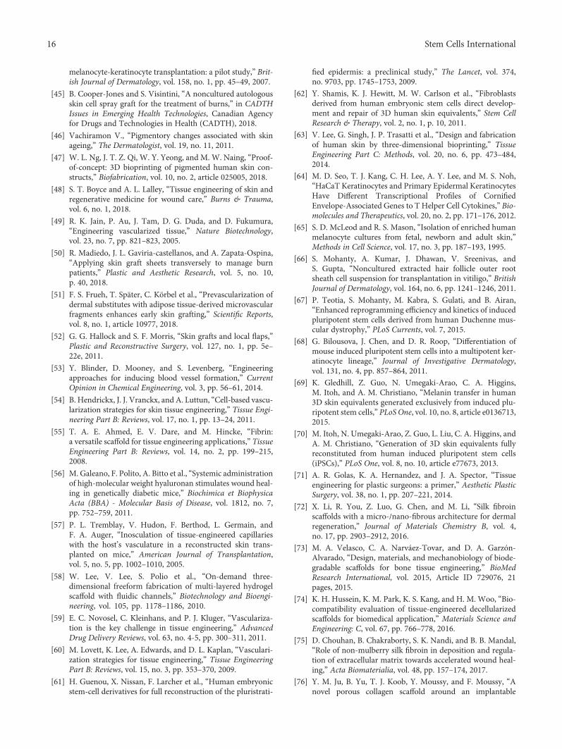
melanocyte-keratinocyte transplantation: a pilot study,” Brit-ish Journal of Dermatology, vol. 158, no. 1, pp. 45–49, 2007.
[45] B. Cooper-Jones and S. Visintini, “A noncultured autologousskin cell spray graft for the treatment of burns,” in CADTHIssues in Emerging Health Technologies, Canadian Agencyfor Drugs and Technologies in Health (CADTH), 2018.
[46] Vachiramon V., “Pigmentory changes associated with skinageing,” The Dermatologist, vol. 19, no. 11, 2011.
[47] W. L. Ng, J. T. Z. Qi, W. Y. Yeong, and M. W. Naing, “Proof-of-concept: 3D bioprinting of pigmented human skin con-structs,” Biofabrication, vol. 10, no. 2, article 025005, 2018.
[48] S. T. Boyce and A. L. Lalley, “Tissue engineering of skin andregenerative medicine for wound care,” Burns & Trauma,vol. 6, no. 1, 2018.
[49] R. K. Jain, P. Au, J. Tam, D. G. Duda, and D. Fukumura,“Engineering vascularized tissue,” Nature Biotechnology,vol. 23, no. 7, pp. 821–823, 2005.
[50] R. Madiedo, J. L. Gaviria-castellanos, and A. Zapata-Ospina,“Applying skin graft sheets transversely to manage burnpatients,” Plastic and Aesthetic Research, vol. 5, no. 10,p. 40, 2018.
[51] F. S. Frueh, T. Später, C. Körbel et al., “Prevascularization ofdermal substitutes with adipose tissue-derived microvascularfragments enhances early skin grafting,” Scientific Reports,vol. 8, no. 1, article 10977, 2018.
[52] G. G. Hallock and S. F. Morris, “Skin grafts and local flaps,”Plastic and Reconstructive Surgery, vol. 127, no. 1, pp. 5e–22e, 2011.
[53] Y. Blinder, D. Mooney, and S. Levenberg, “Engineeringapproaches for inducing blood vessel formation,” CurrentOpinion in Chemical Engineering, vol. 3, pp. 56–61, 2014.
[54] B. Hendrickx, J. J. Vranckx, and A. Luttun, “Cell-based vascu-larization strategies for skin tissue engineering,” Tissue Engi-neering Part B: Reviews, vol. 17, no. 1, pp. 13–24, 2011.
[55] T. A. E. Ahmed, E. V. Dare, and M. Hincke, “Fibrin:a versatile scaffold for tissue engineering applications,” TissueEngineering Part B: Reviews, vol. 14, no. 2, pp. 199–215,2008.
[56] M. Galeano, F. Polito, A. Bitto et al., “Systemic administrationof high-molecular weight hyaluronan stimulates wound heal-ing in genetically diabetic mice,” Biochimica et BiophysicaActa (BBA) - Molecular Basis of Disease, vol. 1812, no. 7,pp. 752–759, 2011.
[57] P. L. Tremblay, V. Hudon, F. Berthod, L. Germain, andF. A. Auger, “Inosculation of tissue-engineered capillarieswith the host’s vasculature in a reconstructed skin trans-planted on mice,” American Journal of Transplantation,vol. 5, no. 5, pp. 1002–1010, 2005.
[58] W. Lee, V. Lee, S. Polio et al., “On-demand three-dimensional freeform fabrication of multi-layered hydrogelscaffold with fluidic channels,” Biotechnology and Bioengi-neering, vol. 105, pp. 1178–1186, 2010.
[59] E. C. Novosel, C. Kleinhans, and P. J. Kluger, “Vasculariza-tion is the key challenge in tissue engineering,” AdvancedDrug Delivery Reviews, vol. 63, no. 4-5, pp. 300–311, 2011.
[60] M. Lovett, K. Lee, A. Edwards, and D. L. Kaplan, “Vasculari-zation strategies for tissue engineering,” Tissue EngineeringPart B: Reviews, vol. 15, no. 3, pp. 353–370, 2009.
[61] H. Guenou, X. Nissan, F. Larcher et al., “Human embryonicstem-cell derivatives for full reconstruction of the pluristrati-
fied epidermis: a preclinical study,” The Lancet, vol. 374,no. 9703, pp. 1745–1753, 2009.
[62] Y. Shamis, K. J. Hewitt, M. W. Carlson et al., “Fibroblastsderived from human embryonic stem cells direct develop-ment and repair of 3D human skin equivalents,” Stem CellResearch & Therapy, vol. 2, no. 1, p. 10, 2011.
[63] V. Lee, G. Singh, J. P. Trasatti et al., “Design and fabricationof human skin by three-dimensional bioprinting,” TissueEngineering Part C: Methods, vol. 20, no. 6, pp. 473–484,2014.
[64] M. D. Seo, T. J. Kang, C. H. Lee, A. Y. Lee, and M. S. Noh,“HaCaT Keratinocytes and Primary Epidermal KeratinocytesHave Different Transcriptional Profiles of CornifiedEnvelope-Associated Genes to T Helper Cell Cytokines,” Bio-molecules and Therapeutics, vol. 20, no. 2, pp. 171–176, 2012.
[65] S. D. McLeod and R. S. Mason, “Isolation of enriched humanmelanocyte cultures from fetal, newborn and adult skin,”Methods in Cell Science, vol. 17, no. 3, pp. 187–193, 1995.
[66] S. Mohanty, A. Kumar, J. Dhawan, V. Sreenivas, andS. Gupta, “Noncultured extracted hair follicle outer rootsheath cell suspension for transplantation in vitiligo,” BritishJournal of Dermatology, vol. 164, no. 6, pp. 1241–1246, 2011.
[67] P. Teotia, S. Mohanty, M. Kabra, S. Gulati, and B. Airan,“Enhanced reprogramming efficiency and kinetics of inducedpluripotent stem cells derived from human Duchenne mus-cular dystrophy,” PLoS Currents, vol. 7, 2015.
[68] G. Bilousova, J. Chen, and D. R. Roop, “Differentiation ofmouse induced pluripotent stem cells into a multipotent ker-atinocyte lineage,” Journal of Investigative Dermatology,vol. 131, no. 4, pp. 857–864, 2011.
[69] K. Gledhill, Z. Guo, N. Umegaki-Arao, C. A. Higgins,M. Itoh, and A. M. Christiano, “Melanin transfer in human3D skin equivalents generated exclusively from induced plu-ripotent stem cells,” PLoS One, vol. 10, no. 8, article e0136713,2015.
[70] M. Itoh, N. Umegaki-Arao, Z. Guo, L. Liu, C. A. Higgins, andA. M. Christiano, “Generation of 3D skin equivalents fullyreconstituted from human induced pluripotent stem cells(iPSCs),” PLoS One, vol. 8, no. 10, article e77673, 2013.
[71] A. R. Golas, K. A. Hernandez, and J. A. Spector, “Tissueengineering for plastic surgeons: a primer,” Aesthetic PlasticSurgery, vol. 38, no. 1, pp. 207–221, 2014.
[72] X. Li, R. You, Z. Luo, G. Chen, and M. Li, “Silk fibroinscaffolds with a micro-/nano-fibrous architecture for dermalregeneration,” Journal of Materials Chemistry B, vol. 4,no. 17, pp. 2903–2912, 2016.
[73] M. A. Velasco, C. A. Narváez-Tovar, and D. A. Garzón-Alvarado, “Design, materials, and mechanobiology of biode-gradable scaffolds for bone tissue engineering,” BioMedResearch International, vol. 2015, Article ID 729076, 21pages, 2015.
[74] K. H. Hussein, K. M. Park, K. S. Kang, and H. M. Woo, “Bio-compatibility evaluation of tissue-engineered decellularizedscaffolds for biomedical application,” Materials Science andEngineering: C, vol. 67, pp. 766–778, 2016.
[75] D. Chouhan, B. Chakraborty, S. K. Nandi, and B. B. Mandal,“Role of non-mulberry silk fibroin in deposition and regula-tion of extracellular matrix towards accelerated wound heal-ing,” Acta Biomaterialia, vol. 48, pp. 157–174, 2017.
[76] Y. M. Ju, B. Yu, T. J. Koob, Y. Moussy, and F. Moussy, “Anovel porous collagen scaffold around an implantable
16 Stem Cells International
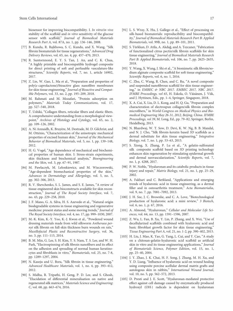
biosensor for improving biocompatibility. I. In vitro/in vivostability of the scaffold and in vitro sensitivity of the glucosesensor with scaffold,” Journal of Biomedical MaterialsResearch Part A, vol. 87A, no. 1, pp. 136–146, 2008.
[77] B. Kundu, R. Rajkhowa, S. C. Kundu, and X. Wang, “Silkfibroin biomaterials for tissue regenerations,” Advanced DrugDelivery Reviews, vol. 65, no. 4, pp. 457–470, 2013.
[78] R. Suntornnond, E. Y. S. Tan, J. An, and C. K. Chua,“A highly printable and biocompatible hydrogel compositefor direct printing of soft and perfusable vasculature-likestructures,” Scientific Reports, vol. 7, no. 1, article 16902,2017.
[79] Z. Lin, W. Gao, L. Ma et al., “Preparation and properties ofpoly(ε-caprolactone)/bioactive glass nanofibre membranesfor skin tissue engineering,” Journal of Bioactive and Compat-ible Polymers, vol. 33, no. 2, pp. 195–209, 2018.
[80] M. Rahmati and M. Mozafari, “Protein adsorption onpolymers,” Materials Today Communications, vol. 17,pp. 527–540, 2018.
[81] T. Ushiki, “Collagen fibers, reticular fibers and elastic fibers.A comprehensive understanding from a morphological view-point,” Archives of Histology and Cytology, vol. 65, no. 2,pp. 109–126, 2002.
[82] A. Ní Annaidh, K. Bruyère, M. Destrade, M. D. Gilchrist, andM. Otténio, “Characterization of the anisotropic mechanicalproperties of excised human skin,” Journal of the MechanicalBehavior of Biomedical Materials, vol. 5, no. 1, pp. 139–148,2012.
[83] H. G. Vogel, “Age dependence of mechanical and biochemi-cal properties of human skin. I: Stress-strain experiments,skin thickness and biochemical analysis,” Bioengineeringand the Skin, vol. 3, pp. 67–91, 1987.
[84] M. Pawlaczyk, M. Lelonkiewicz, and M. Wieczorowski,“Age-dependent biomechanical properties of the skin,”Advances in Dermatology and Allergology, vol. 5, no. 5,pp. 302–306, 2013.
[85] R. V. Shevchenko, S. L. James, and S. E. James, “A review oftissue-engineered skin bioconstructs available for skin recon-struction,” Journal of The Royal Society Interface, vol. 7,no. 43, pp. 229–258, 2010.
[86] J. F. Mano, G. A. Silva, H. S. Azevedo et al., “Natural originbiodegradable systems in tissue engineering and regenerativemedicine: present status and some moving trends,” Journal ofThe Royal Society Interface, vol. 4, no. 17, pp. 999–1030, 2007.
[87] M.-K. Kim, K.-Y. Yoo, K.-J. Kwon et al., “Powdered wounddressing materials made from wild silkworm Antheraea per-nyi silk fibroin on full-skin thickness burn wounds on rats,”Maxillofacial Plastic and Reconstructive Surgery, vol. 36,no. 3, pp. 111–115, 2014.
[88] B. M. Min, G. Lee, S. H. Kim, Y. S. Nam, T. S. Lee, and W. H.Park, “Electrospinning of silk fibroin nanofibers and its effecton the adhesion and spreading of normal human keratino-cytes and fibroblasts in vitro,” Biomaterials, vol. 25, no. 7-8,pp. 1289–1297, 2004.
[89] N. Kasoju and U. Bora, “Silk fibroin in tissue engineering,”Advanced Healthcare Materials, vol. 1, no. 4, pp. 393–412,2012.
[90] S. Midha, R. Tripathi, H. Geng, P. D. Lee, and S. Ghosh,“Elucidation of differential mineralisation on native andregenerated silk matrices,”Materials Science and Engineering:C, vol. 68, pp. 663–674, 2016.
[91] L. S. Wray, X. Hu, J. Gallego et al., “Effect of processing onsilk-based biomaterials: reproducibility and biocompatibil-ity,” Journal of Biomedical Materials Research Part B: AppliedBiomaterials, vol. 99B, no. 1, pp. 89–101, 2011.
[92] S. Türkkan, D. Atila, A. Akdağ, and A. Tezcaner, “Fabricationof functionalized citrus pectin/silk fibroin scaffolds for skintissue engineering,” Journal of Biomedical Materials ResearchPart B: Applied Biomaterials, vol. 106, no. 7, pp. 2625–2635,2018.
[93] Y. Wang, X. Wang, J. Shi et al., “A biomimetic silk fibroin/so-dium alginate composite scaffold for soft tissue engineering,”Scientific Reports, vol. 6, no. 1, 2016.
[94] C. Zhu, C. Wang, R. Chen, and C. Ru, “A novel compositeand suspended nanofibrous scaffold for skin tissue engineer-ing,” in EMBEC & NBC 2017. EMBEC 2017, NBC 2017.IFMBE Proceedings, vol 65, H. Eskola, O. Väisänen, J. Viik,and J. Hyttinen, Eds., pp. 1–4, Springer, Singapore, 2018.
[95] X. A. Cui, X. Liu, D. L. Kong, and H. Q. Gu, “Preparation andcharacteration of electrospun collagen/silk fibroin complexmicrofibers,” in World Congress on Medical Physics and Bio-medical Engineering May 26-31, 2012, Beijing, China. IFMBEProceedings, vol 39, M. Long, Ed., pp. 79–82, Springer, Berlin,Heidelberg, 2013.
[96] N. Bhardwaj, W. T. Sow, D. Devi, K. W. Ng, B. B. Mandal,and N. J. Cho, “Silk fibroin-keratin based 3D scaffolds as adermal substitute for skin tissue engineering,” IntegrativeBiology, vol. 7, no. 1, pp. 53–63, 2015.
[97] S. Xiong, X. Zhang, P. Lu et al., “A gelatin-sulfonatedsilk composite scaffold based on 3D printing technologyenhances skin regeneration by stimulating epidermal growthand dermal neovascularization,” Scientific Reports, vol. 7,no. 1, p. 4288, 2017.
[98] P. W. Noble, “Hyaluronan and its catabolic products in tissueinjury and repair,” Matrix Biology, vol. 21, no. 1, pp. 25–29,2002.
[99] A. Fakhari and C. Berkland, “Applications and emergingtrends of hyaluronic acid in tissue engineering, as a dermalfiller and in osteoarthritis treatment,” Acta Biomaterialia,vol. 9, no. 7, pp. 7081–7092, 2013.
[100] J. H. Sze, J. C. Brownlie, and C. A. Love, “Biotechnologicalproduction of hyaluronic acid: a mini review,” 3 Biotech,vol. 6, no. 1, p. 67, 2016.
[101] A. Almond, “Hyaluronan,” Cellular and Molecular Life Sci-ences, vol. 64, no. 13, pp. 1591–1596, 2007.
[102] Z. Wu, L. Fan, B. Xu, Y. Lin, P. Zhang, and X. Wei, “Use ofdecellularized scaffolds combined with hyaluronic acid andbasic fibroblast growth factor for skin tissue engineering,”Tissue Engineering Part A, vol. 21, no. 1-2, pp. 390–402, 2015.
[103] H. Liu, J. Mao, K. Yao, G. Yang, L. Cui, and Y. Cao, “A studyon a chitosan-gelatin-hyaluronic acid scaffold as artificialskin in vitro and its tissue engineering applications,” Journalof Biomaterials Science, Polymer Edition, vol. 15, no. 1,pp. 25–40, 2004.
[104] J. Y. Zhao, J. K. Chai, H. F. Song, J. Zhang, M. H. Xu, andY. D. Liang, “Influence of hyaluronic acid on wound healingusing composite porcine acellular dermal matrix grafts andautologous skin in rabbits,” International Wound Journal,vol. 10, no. 5, pp. 562–572, 2013.
[105] D. Presti and J. E. Scott, “Hyaluronan-mediated protectiveeffect against cell damage caused by enzymatically producedhydroxyl (OH·) radicals is dependent on hyaluronan
17Stem Cells International
molecular mass,” Cell Biochemistry and Function, vol. 12,no. 4, pp. 281–288, 1994.
[106] J. M. Grasman, R. L. Page, T. Dominko, and G. D. Pins,“Crosslinking strategies facilitate tunable structural proper-ties of fibrin microthreads,” Acta Biomaterialia, vol. 8,no. 11, pp. 4020–4030, 2012.
[107] P. A. Janmey, J. P. Winer, and J. W. Weisel, “Fibrin gels andtheir clinical and bioengineering applications,” Journal of TheRoyal Society Interface, vol. 6, no. 30, pp. 1–10, 2009.
[108] A. Hokugo, T. Takamoto, and Y. Tabata, “Preparation ofhybrid scaffold from fibrin and biodegradable polymer fiber,”Biomaterials, vol. 27, no. 1, pp. 61–67, 2006.
[109] I. Bae, K. Osatomi, A. Yoshida, K. Osako, A. Yamaguchi, andK. Hara, “Biochemical properties of acid-soluble collagensextracted from the skins of underutilised fishes,” Food Chem-istry, vol. 108, no. 1, pp. 49–54, 2008.
[110] J. Myllyharju and K. I. Kivirikko, “Collagens, modifyingenzymes and their mutations in humans, flies and worms,”Trends in Genetics, vol. 20, no. 1, pp. 33–43, 2004.
[111] L. Cen, W. Liu, L. Cui, W. Zhang, and Y. Cao, “Collagentissue engineering: development of novel biomaterials andapplications,” Pediatric Research, vol. 63, no. 5, pp. 492–496, 2008.
[112] P. S. Briquez, J. A. Hubbell, and M. M. Martino, “Extracellu-lar matrix-inspired growth factor delivery systems for skinwound healing,” Advances in Wound Care, vol. 4, no. 8,pp. 479–489, 2015.
[113] A. Gaspar, L. Moldovan, D. Constantin, A. M. Stanciuc, P. M.Sarbu Boeti, and I. C. Efrimescu, “Collagen-based scaffoldsfor skin tissue engineering,” Journal of Medicine and Life,vol. 4, no. 2, pp. 172–177, 2011.
[114] T. Maver, U. Maver, K. S. Kleinschek, I. M. Raščan, and D. M.Smrke, “Advanced therapies of skin injuries,” Wiener kli-nische Wochenschrift, vol. 127, no. S5, pp. 187–198, 2015.
[115] R. Parenteau-bareil, R. Gauvin, and F. Berthod, “Collagen-based biomaterials for tissue engineering applications,”Mate-rials, vol. 3, no. 3, pp. 1863–1887, 2010.
[116] M. D. Harriger, A. P. Supp, G. D. Warden, and S. T. Boyce,“Glutaraldehyde crosslinking of collagen substrates inhibitsdegradation in skin substitutes grafted to athymic mice,”Journal of Biomedical Materials Research, vol. 35, no. 2,pp. 137–145, 1997.
[117] M. Ansari, S. S. Kordestani, S. Nazralizadeh, and H. Eslami,“Biodegradable cell-seeded collagen based polymer scaffoldsfor wound healing and skin reconstruction,” Journal ofMacromolecular Science, Part B, vol. 57, no. 2, pp. 100–109, 2018.
[118] S. Chattopadhyay and R. T. Raines, “Review collagen‐basedbiomaterials for wound healing,” Biopolymers, vol. 101,no. 8, pp. 821–833, 2014.
[119] B. K. H. L. Boekema, M. Vlig, L. Olde Damink et al., “Effectof pore size and cross-linking of a novel collagen-elastindermal substitute on wound healing,” Journal of MaterialsScience: Materials in Medicine, vol. 25, no. 2, pp. 423–433,2014.
[120] S. Udhayakumar, K. G. Shankar, S. Sowndarya, S. Venkatesh,C. Muralidharan, and C. Rose, “l-Arginine intercedes bio-crosslinking of a collagen–chitosan 3D-hybrid scaffold fortissue engineering and regeneration: in silico, in vitro, andin vivo studies,” RSC Advances, vol. 7, no. 40, pp. 25070–25088, 2017.
[121] M. T. Wolf, K. A. Daly, E. P. Brennan-Pierce et al., “A hydro-gel derived from decellularized dermal extracellular matrix,”Biomaterials, vol. 33, no. 29, pp. 7028–7038, 2012.
[122] F. Pati, J. Jang, D. H. Ha et al., “Printing three-dimensionaltissue analogues with decellularized extracellular matrixbioink,” Nature Communications, vol. 5, no. 1, 2014.
[123] H. Lee, W. Han, H. Kim et al., “Development of liver decellu-larized extracellular matrix bioink for three-dimensional cellprinting-based liver tissue engineering,” Biomacromolecules,vol. 18, no. 4, pp. 1229–1237, 2017.
[124] Z. Zhang, L. Lv, M. Mamat, Z. Chen, L. Liu, and Z. Wang,“Xenogenic (porcine) acellular dermal matrix is useful forthe wound healing of severely damaged extremities,” Experi-mental and Therapeutic Medicine, vol. 7, no. 3, pp. 621–624,2014.
[125] B. T. Baldursson, H. Kjartansson, F. Konrádsdóttir,P. Gudnason, G. F. Sigurjonsson, and S. H. Lund, “Healingrate and autoimmune safety of full-thickness wounds treatedwith fish skin acellular dermal matrix versus porcine small-intestine submucosa: a noninferiority study,” The Interna-tional Journal of Lower Extremity Wounds, vol. 14, no. 1,pp. 37–43, 2015.
[126] P. B. Milan, N. Lotfibakhshaiesh, M. T. Joghataie et al.,“Accelerated wound healing in a diabetic rat model usingdecellularized dermal matrix and human umbilical cord peri-vascular cells,” Acta Biomaterialia, vol. 45, pp. 234–246, 2016.
[127] S.-P. Wilshaw, J. Kearney, J. Fisher, and E. Ingham, “Biocom-patibility and potential of acellular human amniotic mem-brane to support the attachment and proliferation ofallogeneic cells,” Tissue Engineering Part A, vol. 14, no. 4,pp. 463–472, 2008.
[128] S.-P. Wilshaw, J. N. Kearney, J. Fisher, and E. Ingham,“Production of an acellular amniotic membrane matrix foruse in tissue engineering,” Tissue Engineering, vol. 12, no. 8,pp. 2117–2129, 2006.
[129] A. Samadikuchaksaraei, A. Mehdipour, M. Habibi Roudke-nar et al., “A dermal equivalent engineered with TGF-β3expressing bone marrow stromal cells and amniotic mem-brane: cosmetic healing of full-thickness skin wounds inrats,” Artificial Organs, vol. 40, no. 12, pp. E266–E279, 2016.
[130] K. Vig, A. Chaudhari, S. Tripathi et al., “Advances in skinregeneration using tissue engineering,” International Journalof Molecular Sciences, vol. 18, no. 4, p. 789, 2017.
[131] M. Griffiths, N. Ojeh, R. Livingstone, R. Price, andH. Navsaria, “Survival of apligraf in acute human wounds,”Tissue Engineering, vol. 10, no. 7-8, pp. 1180–1195, 2004.
[132] A. D. Metcalfe and M. W. J. Ferguson, “Tissue engineering ofreplacement skin: the crossroads of biomaterials, woundhealing, embryonic development, stem cells and regenera-tion,” Journal of The Royal Society Interface, vol. 4, no. 14,pp. 413–437, 2007.
[133] L. Alrubaiy and K. K. Al-rubaiy, “Skin substitutes: a briefreview of types and clinical applications,” Oman MedicalJournal, vol. 24, pp. 6–8, 2009.
[134] E. Constant and J. F. Burke, “Successful use of a physiologi-cally acceptable artificial skin in the treatment of extensiveburn injury,” Plastic and Reconstructive Surgery, vol. 70,no. 6, p. 784, 1982.
[135] L. Yildirimer, N. T. K. Thanh, and A. M. Seifalian, “Skinregeneration scaffolds: a multimodal bottom-up approach,”Trends in Biotechnology, vol. 30, no. 12, pp. 638–648, 2012.
18 Stem Cells International
[136] P. Zarrintaj, A. S. Moghaddam, S. Manouchehri et al., “Canregenerative medicine and nanotechnology combine to healwounds? The search for the ideal wound dressing,” Nanome-dicine, vol. 12, no. 19, pp. 2403–2422, 2017.
[137] X. Zhu, W. Cui, X. Li, and Y. Jin, “Electrospun fibrous matswith high porosity as potential scaffolds for skin tissue engi-neering,” Biomacromolecules, vol. 9, no. 7, pp. 1795–1801,2008.
[138] J. Rnjak-Kovacina and A. S. Weiss, “Increasing the pore sizeof electrospun scaffolds,” Tissue Engineering Part B: Reviews,vol. 17, no. 5, pp. 365–372, 2011.
[139] H. Chen, Y. Peng, S. Wu, and L. Tan, “Electrospun 3D fibrousscaffolds for chronic wound repair,” Materials, vol. 9, no. 4,p. 272, 2016.
[140] H. M. Powell, D. M. Supp, and S. T. Boyce, “Influence of elec-trospun collagen on wound contraction of engineered skinsubstitutes,” Biomaterials, vol. 29, no. 7, pp. 834–843, 2008.
[141] S. C. Rizzi, M. Ehrbar, S. Halstenberg et al., “Recombinantprotein-co-PEG networks as cell-adhesive and proteolyticallydegradable hydrogel matrixes. Part II: Biofunctional charac-teristics,” Biomacromolecules, vol. 7, no. 11, pp. 3019–3029,2006.
[142] C. You, Q. Li, X. Wang et al., “Silver nanoparticle loadedcollagen/chitosan scaffolds promote wound healing via reg-ulating fibroblast migration and macrophage activation,”Scientific Reports, vol. 7, no. 1, article 10489, 2017.
[143] Ö. Aktürk and D. Keskin, “Collagen/PEO/gold nanofibrousmatrices for skin tissue engineering,” Turkish Journal of Biol-ogy, vol. 40, pp. 380–398, 2016.
[144] N. R. Johnson and Y. Wang, “Drug delivery systems forwound healing,” Current Pharmaceutical Biotechnology,vol. 16, no. 7, pp. 621–629, 2015.
[145] A. Kumari, S. K. Yadav, and S. C. Yadav, “Biodegradablepolymeric nanoparticles based drug delivery systems,” Col-loids and surfaces B: biointerfaces, vol. 75, no. 1, pp. 1–18,2010.
[146] E. Sah and H. Sah, “Recent trends in preparation of poly(lac-tide-co-glycolide) nanoparticles by mixing polymeric organicsolution with antisolvent,” Journal of Nanomaterials,vol. 2015, Article ID 794601, 22 pages, 2015.
[147] K. K. Chereddy, R. Coco, P. B. Memvanga et al., “Combinedeffect of PLGA and curcumin on wound healing activity,”Journal of Controlled Release, vol. 171, no. 2, pp. 208–215,2013.
[148] G. Han, L. N. Nguyen, C. Macherla et al., “Nitric oxide–releasing nanoparticles accelerate wound healing by pro-moting fibroblast migration and collagen deposition,” TheAmerican Journal of Pathology, vol. 180, no. 4, pp. 1465–1473, 2012.
[149] K. Blecher, L. R. Martinez, C. Tuckman-vernon et al., “Nitricoxide-releasing nanoparticles accelerate wound healing inNOD-SCID mice,” Nanomedicine: Nanotechnology, Biologyand Medicine, vol. 8, no. 8, pp. 1364–1371, 2012.
[150] M. Mozafari, “Nanotechnology in wound care: one stepcloser to the clinic,” Molecular Therapy, vol. 26, no. 9,pp. 2085-2086, 2018.
[151] A. Galperin, T. J. Long, and B. D. Ratner, “Degradable,Thermo-sensitive poly(N-isopropyl acrylamide)-based scaf-folds with controlled porosity for tissue engineering applica-tions,” Biomacromolecules, vol. 11, no. 10, pp. 2583–2592,2010.
[152] Y. Liu, H. Luo, X. Wang et al., “In vitro construction ofscaffold-free bilayered tissue-engineered skin containing cap-illary networks,” BioMed Research International, vol. 2013,Article ID 561410, 8 pages, 2013.
[153] L. J. Pourchet, A. Thepot, M. Albouy et al., “Human skin 3Dbioprinting using scaffold-free approach,” Advanced Health-care Materials, vol. 6, no. 4, pp. 1–8, 2017.
[154] A. B. M. Hilmi, “Vital roles of stem cells and biomaterials inskin tissue engineering,” World Journal of Stem Cells, vol. 7,no. 2, p. 428, 2015.
[155] A. T. Badillo, R. A. Redden, L. Zhang, E. J. Doolin, and K. W.Liechty, “Treatment of diabetic wounds with fetal murinemesenchymal stromal cells enhances wound closure,” Celland Tissue Research, vol. 329, no. 2, pp. 301–311, 2007.
[156] E. V. Badiavas and V. Falanga, “Treatment of chronicwounds with bone marrow-derived cells,” Archives of Derma-tology, vol. 139, no. 4, pp. 510–516, 2003.
[157] T. Yoshikawa, H. Mitsuno, I. Nonaka et al., “Wound therapyby marrow mesenchymal cell transplantation,” Plastic andReconstructive Surgery, vol. 121, no. 3, pp. 860–877, 2008.
[158] A. Shabbir, A. Cox, L. Rodriguez-Menocal, M. Salgado,and E. Van Badiavas, “Mesenchymal stem cell exosomesinduce proliferation and migration of normal and chronicwound fibroblasts, and enhance angiogenesis in vitro,”Stem Cells and Development, vol. 24, no. 14, pp. 1635–1647,2015.
[159] A. M. Altman, N. Matthias, Y. Yan et al., “Dermal matrix as acarrier for in vivo delivery of human adipose-derived stemcells,” Biomaterials, vol. 29, no. 10, pp. 1431–1442, 2008.
[160] W.-S. Kim, B. S. Park, J. H. Sung et al., “Wound healing effectof adipose-derived stem cells: a critical role of secretory fac-tors on human dermal fibroblasts,” Journal of DermatologicalScience, vol. 48, no. 1, pp. 15–24, 2007.
[161] C. Nie, D. Yang, J. Xu, Z. Si, X. Jin, and J. Zhang, “Locallyadministered adipose-derived stem cells accelerate woundhealing through differentiation and vasculogenesis,” CellTransplantation, vol. 20, no. 2, pp. 205–216, 2011.
[162] F. S. Frueh, T. Später, N. Lindenblatt et al., “Adipose tissue-derived microvascular fragments improve vascularization,lymphangiogenesis, and integration of dermal skin substi-tutes,” Journal of Investigative Dermatology, vol. 137, no. 1,pp. 217–227, 2017.
[163] M. W. Laschke, S. Kleer, C. Scheuer, D. Eglin, M. Alini, andM. D. Menger, “Pre-cultivation of adipose tissue-derivedmicrovascular fragments in porous scaffolds does notimprove their in vivo vascularisation potential,” EuropeanCells and Materials, vol. 29, pp. 190–201, 2015.
[164] R. K. Chan, D. O. Zamora, N. L. Wrice et al., “Developmentof a vascularized skin construct using adipose-derived stemcells from debrided burned skin,” Stem Cells International,vol. 2012, Article ID 841203, 11 pages, 2012.
[165] Y. C. Lin, T. Grahovac, S. J. Oh, M. Ieraci, J. P. Rubin, andK. G. Marra, “Evaluation of a multi-layer adipose-derivedstem cell sheet in a full-thickness wound healing model,”ActaBiomaterialia, vol. 9, no. 2, pp. 5243–5250, 2013.
[166] V. Trottier, G. Marceau-Fortier, L. Germain, C. Vincent, andJ. Fradette, “IFATS collection: using human adipose-derivedstem/stromal cells for the production of new skin substi-tutes,” Stem Cells, vol. 26, no. 10, pp. 2713–2723, 2008.
[167] M. Gholipourmalekabadi, A. Samadikuchaksaraei, A. M.Seifalian et al., “Silk fibroin/amniotic membrane 3D bi-
19Stem Cells International
layered artificial skin,” Biomedical Materials, vol. 13, no. 3,article 035003, 2018.
[168] J. J. Y. Tan and L. Kang, “Engineering the future of hair folli-cle regeneration and delivery,” Therapeutic Delivery, vol. 9,no. 5, pp. 321–324, 2018.
[169] M. Kurita, T. Araoka, T. Hishida et al., “In vivo reprogram-ming of wound-resident cells generates skin epithelial tissue,”Nature, vol. 561, no. 7722, pp. 243–247, 2018.
[170] S. Danner, M. Kremer, A. E. Petschnik et al., “The use ofhuman sweat gland-derived stem cells for enhancing vascu-larization during dermal regeneration,” Journal of Investiga-tive Dermatology, vol. 132, no. 6, pp. 1707–1716, 2012.
[171] Y. Amoh, L. Li, M. Yang et al., “Nascent blood vessels in theskin arise from nestin-expressing hair-follicle cells,” Proceed-ings of the National Academy of Sciences of the United Statesof America, vol. 101, no. 36, pp. 13291–13295, 2004.
[172] R. Aki, Y. Amoh, N. Li, K. Katsuoka, and R. M. Hoffman,“Nestin-expressing interfollicular blood vessel network con-tributes to skin transplant survival and wound healing,” Jour-nal of Cellular Biochemistry, vol. 110, pp. 80–86, 2010.
[173] T. Hirsch, T. Rothoeft, N. Teig et al., “Regeneration of theentire human epidermis using transgenic stem cells,” Nature,vol. 551, no. 7680, pp. 327–332, 2017.
[174] A. Skardal, D. Mack, E. Kapetanovic et al., “Bioprinted amni-otic fluid-derived stem cells accelerate healing of large skinwounds,” Stem Cells Translational Medicine, vol. 1, no. 11,pp. 792–802, 2012.
[175] C. P. Lu, L. Polak, B. E. Keyes, and E. Fuchs, “Spatiotemporalantagonism in mesenchymal-epithelial signaling in sweatversus hair fate decision,” Science, vol. 354, no. 6319, articleaah6102, 2016.
[176] A. Zieris, S. Prokoph, K. R. Levental et al., “FGF-2 and VEGFfunctionalization of starPEG–heparin hydrogels to modulatebiomolecular and physical cues of angiogenesis,” Biomate-rials, vol. 31, no. 31, pp. 7985–7994, 2010.
[177] Z. Zhou, B. Shu, Y. Xu et al., “microRNA-203 modulateswound healing and scar formation via suppressing Hes1expression in epidermal stem cells,” Cellular Physiology andBiochemistry, vol. 49, no. 6, pp. 2333–2347, 2018.
[178] M. S. Tjin, A. W. C. Chua, A. Moreno-Moral et al., “Biologi-cally relevant laminin as chemically defined and fully humanplatform for human epidermal keratinocyte culture,” NatureCommunications, vol. 9, no. 1, article 4432, 2018.
[179] S. Michael, H. Sorg, C. T. Peck et al., “Tissue engineered skinsubstitutes created by laser-assisted bioprinting form skin-like structures in the dorsal skin fold chamber in mice,” PLoSOne, vol. 8, no. 3, article e57741, 2013.
[180] C. Auxenfans, J. Fradette, C. Lequeux et al., “Evolution ofthree dimensional skin equivalent models reconstructedin vitro by tissue engineering,” European Journal of Derma-tology, vol. 19, no. 2, pp. 107–113, 2009.
[181] P. Katakam, B. Dey, F. H. Assaleh et al., “Top-down andbottom-up approaches in 3D printing technologies for drugdelivery challenges,” Critical Reviews™ in Therapeutic DrugCarrier Systems, vol. 32, no. 1, pp. 61–87, 2015.
[182] V. Mironov, T. Boland, T. Trusk, G. Forgacs, and R. R.Markwald, “Organ printing: computer-aided jet-based 3Dtissue engineering,” Trends in Biotechnology, vol. 21, no. 4,pp. 157–161, 2003.
[183] S. Midha and S. Ghosh, “Silk-based bioinks for 3D bio-printing,” in Regenerative Medicine: Laboratory to Clinic,
A. Mukhopadhyay, Ed., pp. 259–276, Springer, Singapore,2017.
[184] K. W. Binder, In situ bioprinting of the skin, Doctoral disser-tation, Wake Forest University, 2011.
[185] L. Koch, A. Deiwick, S. Schlie et al., “Skin tissue generation bylaser cell printing,” Biotechnology and Bioengineering,vol. 109, no. 7, pp. 1855–1863, 2012.
[186] N. Liu, S. Huang, B. Yao, J. Xie, X. Wu, and X. Fu, “3D bio-printing matrices with controlled pore structure and releasefunction guide in vitro self-organization of sweat gland,” Sci-entific Reports, vol. 6, no. 1, 2016.
[187] S. P. Tarassoli, Z. M. Jessop, A. Al-Sabah et al., “Skin tissueengineering using 3D bioprinting: an evolving research field,”Journal of Plastic, Reconstructive & Aesthetic Surgery, vol. 71,no. 5, pp. 615–623, 2018.
20 Stem Cells International

Hindawiwww.hindawi.com
International Journal of
Volume 2018
Zoology
Hindawiwww.hindawi.com Volume 2018
Anatomy Research International
PeptidesInternational Journal of
Hindawiwww.hindawi.com Volume 2018
Hindawiwww.hindawi.com Volume 2018
Journal of Parasitology Research
GenomicsInternational Journal of
Hindawiwww.hindawi.com Volume 2018
Hindawi Publishing Corporation http://www.hindawi.com Volume 2013Hindawiwww.hindawi.com
The Scientific World Journal
Volume 2018
Hindawiwww.hindawi.com Volume 2018
BioinformaticsAdvances in
Marine BiologyJournal of
Hindawiwww.hindawi.com Volume 2018
Hindawiwww.hindawi.com Volume 2018
Neuroscience Journal
Hindawiwww.hindawi.com Volume 2018
BioMed Research International
Cell BiologyInternational Journal of
Hindawiwww.hindawi.com Volume 2018
Hindawiwww.hindawi.com Volume 2018
Biochemistry Research International
ArchaeaHindawiwww.hindawi.com Volume 2018
Hindawiwww.hindawi.com Volume 2018
Genetics Research International
Hindawiwww.hindawi.com Volume 2018
Advances in
Virolog y Stem Cells International
Hindawiwww.hindawi.com Volume 2018
Hindawiwww.hindawi.com Volume 2018
Enzyme Research
Hindawiwww.hindawi.com Volume 2018
International Journal of
MicrobiologyHindawiwww.hindawi.com
Nucleic AcidsJournal of
Volume 2018
Submit your manuscripts atwww.hindawi.com

![Negative pressure wound therapy for skin grafts and ... · [Intervention Review] Negative pressure wound therapy for skin grafts and surgical wounds healing by primary intention Joan](https://img.dokumen.tips/doc/110x75/5eaca82cd857b569f0001932/negative-pressure-wound-therapy-for-skin-grafts-and-intervention-review-negative.jpg)


![Immunological challenges associated with artificial skin …...Transplanting autologous skin grafts [4, 5] is the therapeutic approach of choice that successively reform the skin,](https://img.dokumen.tips/doc/110x75/610d210bde142e0c054be240/immunological-challenges-associated-with-artificial-skin-transplanting-autologous.jpg)