Embed Size (px)
Citation preview

Artificial Skin Grafts in Chronic Wound Care: A Meta-analysis of Clinical Efficacy and a Review of Cost-effectiveness
Technology Report
Issue 52 February 2005
Cite as: Ho C, Tran K, Hux M, Sibbald G, Campbell K. Artificial skin grafts in chronic wound care: a meta-analysis of clinical efficacy and a review of cost-effectiveness [Technology report no 52]. Ottawa: Canadian Coordinating Office for Health Technology Assessment; 2005. Reproduction of this document for non-commercial purposes is permitted provided appropriate credit is given to CCOHTA. CCOHTA is a non-profit organization funded by the federal, provincial and territorial governments. Legal Deposit - 2005 National Library of Canada ISBN: 1-894978-50-1 (print) ISBN: 1-894978-51-X (online) PUBLICATIONS MAIL AGREEMENT NO: 40026386 RETURN UNDELIVERABLE CANADIAN ADDRESSES TO CANADIAN COORDINATING OFFICE FOR HEALTH TECHNOLOGY ASSESSMENT 600-865 CARLING AVENUE OTTAWA ON K1S 5S8
Publications can be requested from:
CCOHTA 600-865 Carling Avenue
Ottawa ON Canada K1S 5S8 Tel. (613) 226-2553 Fax. (613) 226-5392
Email: [email protected]
or download from CCOHTA’s web site: http://www.ccohta.ca

Canadian Coordinating Office for Health Technology Assessment
Artificial Skin Grafts in Chronic Wound Care:
A Meta-analysis of Clinical Efficacy and a Review of Cost-effectiveness
Chuong Ho MD MSc1 Khai Tran MSc PhD1 Margaret Hux MSc2
Gary Sibbald MD FRCPC ABIM.DAAD Med3 Kaitryn Campbell MLIS1
February 2005
1 Canadian Coordinating Office for Health Technology Assessment, Ottawa ON 2 Health Economics and Outcomes Research, Innovus Research Inc., Burlington ON 3 Department of Medicine, University of Toronto; Dermatology Day Care and Wound Healing Clinic, Sunnybrook and Women’s College Health Sciences Centre, Toronto ON

i
Reviewers These individuals kindly provided comments on this report. External Reviewers Brian T. Kunimoto, MD FRCPC Clinical Associate Professor University of British Columbia Vancouver BC
Craig Mitton, PhD Assistant Professor University of British Columbia Vancouver BC
Andrew N. Lin, MD FRCPC Associate Professor University of Alberta Edmonton AB
CCOHTA Scientific Advisory Panel Reviewers David Hailey, PhD Department of Public Health Sciences University of Alberta Edmonton AB
Charles Wright, MD FRCSC FRCSE FRCSEd Consultant in Medical and Academic Affairs, Program Planning and Evaluation Toronto ON
This report is a review of existing public literature, studies, materials and other information and documentation (collectively the “source documentation”) which are available to CCOHTA. The accuracy of the contents of the source documentation on which this report is based is not warranted, assured or represented in any way by CCOHTA and CCOHTA does not assume responsibility for the quality, propriety, inaccuracies or reasonableness of any statements, information or conclusions contained in the source documentation. CCOHTA takes sole responsibility for the final form and content of this report. The statements and conclusions in this report are those of CCOHTA and not of its Panel members or reviewers. Authorship
Dr. Chuong Ho led the protocol development, supervised the literature review, wrote the draft, revised the report and prepared the report for publication. Dr. Khai Tran worked with Dr. Ho to evaluate the articles’ relevance, assess their quality, extract data and complete the report. Dr. Gary Sibbald and Ms. Margaret Hux provided clinical expertise and economic expertise respectively; and contributed to the draft document and its subsequent revisions. Ms. Kaitryn Campbell was responsible for the design and execution of the literature search strategies and for writing the section and associated appendix on literature searching. Acknowledgements The authors are grateful to Ms. Becky Skidmore, CCOHTA information specialist, for verifying and formatting the bibliographic references, and to all CCOHTA staff members who worked as a team to complete this project. Our thanks to Ms. Patricia Coutts, wound care specialist and

ii
clinical trials coordinator at the Wound Healing Clinic in Toronto, for providing information on the costs of artificial skin products. Conflicts of Interest Dr. Gary Sibbald has been a consultant and investigator for Smith & Nephew Inc. (makers of Dermagraft). No conflicts of interests were declared by any of the other authors.
iii
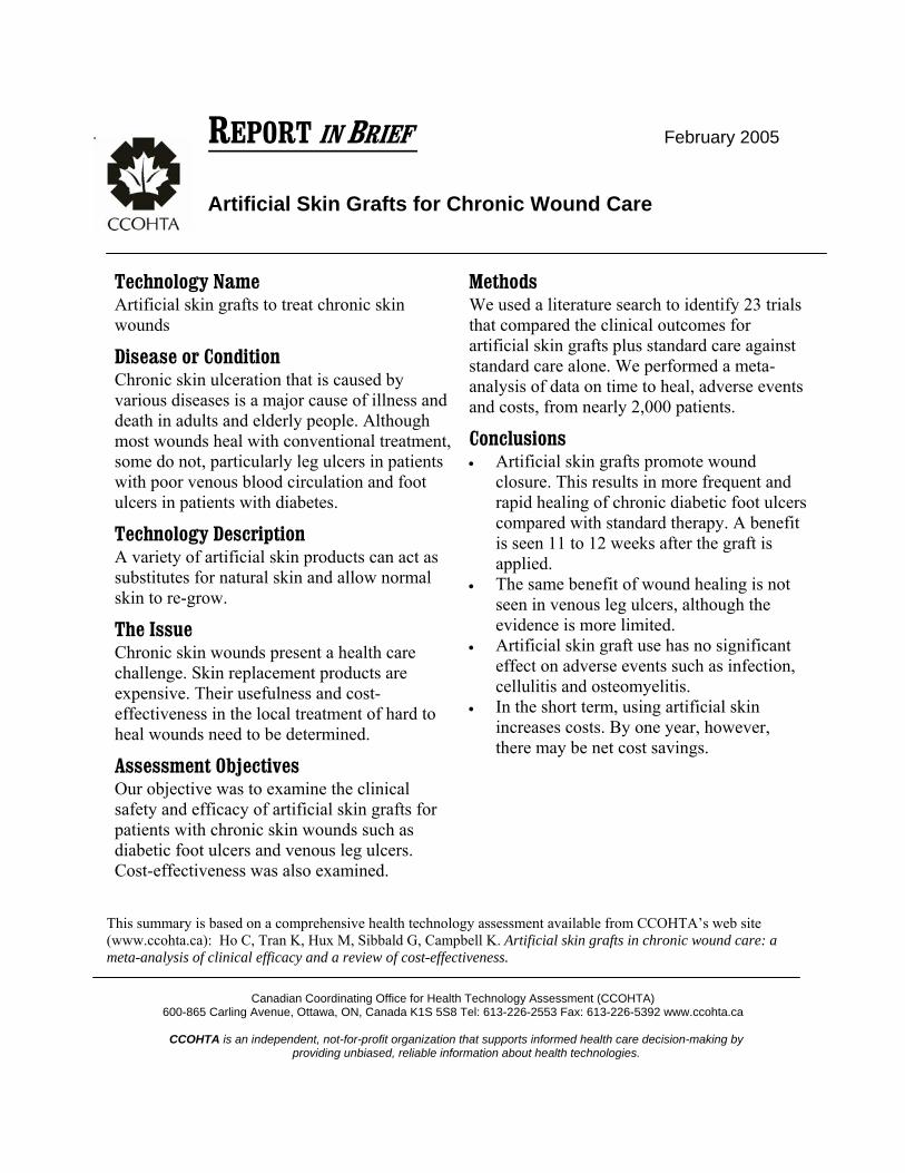
REPORT IN BRIEF February 2005 Artificial Skin Grafts for Chronic Wound Care
Technology Name Artificial skin grafts to treat chronic skin wounds
Disease or Condition Chronic skin ulceration that is caused by various diseases is a major cause of illness and death in adults and elderly people. Although most wounds heal with conventional treatment, some do not, particularly leg ulcers in patients with poor venous blood circulation and foot ulcers in patients with diabetes.
Technology Description A variety of artificial skin products can act as substitutes for natural skin and allow normal skin to re-grow.
The Issue Chronic skin wounds present a health care challenge. Skin replacement products are expensive. Their usefulness and cost-effectiveness in the local treatment of hard to heal wounds need to be determined.
Assessment Objectives Our objective was to examine the clinical safety and efficacy of artificial skin grafts for patients with chronic skin wounds such as diabetic foot ulcers and venous leg ulcers. Cost-effectiveness was also examined.
Methods We used a literature search to identify 23 trials that compared the clinical outcomes for artificial skin grafts plus standard care against standard care alone. We performed a meta-analysis of data on time to heal, adverse events and costs, from nearly 2,000 patients.
Conclusions • Artificial skin grafts promote wound
closure. This results in more frequent and rapid healing of chronic diabetic foot ulcers compared with standard therapy. A benefit is seen 11 to 12 weeks after the graft is applied.
• The same benefit of wound healing is not seen in venous leg ulcers, although the evidence is more limited.
• Artificial skin graft use has no significant effect on adverse events such as infection, cellulitis and osteomyelitis.
• In the short term, using artificial skin increases costs. By one year, however, there may be net cost savings.
This summary is based on a comprehensive health technology assessment available from CCOHTA’s web site (www.ccohta.ca): Ho C, Tran K, Hux M, Sibbald G, Campbell K. Artificial skin grafts in chronic wound care: a meta-analysis of clinical efficacy and a review of cost-effectiveness.
Canadian Coordinating Office for Health Technology Assessment (CCOHTA) 600-865 Carling Avenue, Ottawa, ON, Canada K1S 5S8 Tel: 613-226-2553 Fax: 613-226-5392 www.ccohta.ca
CCOHTA is an independent, not-for-profit organization that supports informed health care decision-making by
providing unbiased, reliable information about health technologies.

iv
EXECUTIVE SUMMARY The Issue Chronic ulceration due to diabetes or vascular insufficiency causes morbidity and mortality in adults and elderly people. Costs, social discomfort and pain are often associated with chronic ulcers. Venous leg ulcers are associated with pain, while diabetic foot ulcers involve functional impairments related to the need to avoid weight bearing. Skin equivalents have been used in the treatment of chronic ulcers. Dermagraft® is a bioengineered human dermis that consists of neonatal dermal fibroblasts. Apligraf® is a dermal layer of human fibroblasts in type 1 bovine collagen with an epidermal layer formed by human keratinocytes. Their clinical efficacy is uncertain, while the large cost of using skin grafts for the treatment of chronic leg ulcers is a health care issue. Several randomized clinical trials have compared the different types of artificial skin grafts to conventional treatment. A systematic review is warranted, because of the large number of randomized controlled trials (RCTs); and the high demand for, high cost of and uncertain efficacy of artificial skin grafts. Objectives Our objective is to examine the scientific evidence of the clinical efficacy, harm and cost-effectiveness of artificial skin grafts that are used for patients with disease-associated chronic skin wounds such as diabetic foot ulcers and venous leg ulcers. We also examine the factors affecting cost-effectiveness. The report is intended to help healthcare decision makers and others who are involved in the delivery of wound care services. Methods Published literature was obtained by searching multiple databases using a defined strategy and by hand searching the bibliographies of selected papers. A meta-analysis of RCTs was performed to compare the clinical outcomes for artificial skin use plus standard care to standard care alone. The proportion of patients who had complete wound healing (CWH) with or without an artificial skin graft was summarized over different time frames (for all types of ulcers); for venous leg ulcers and diabetic foot ulcers separately; and for Dermagraft and Apligraf separately. There were enough studies of these two products for us evaluate them separately. The time to healing and the incidence of adverse events were summarized from clinical trials. The economic consequences of using artificial skin products in venous leg ulcers and diabetic foot ulcers were examined. Results We identified 2,772 abstracts in the original searches of multiple databases. In addition, 11 subsequent alerts were screened up to May 2004. Of these, 117 reports were retrieved. After the elimination of reports that did not satisfy our selection criteria, there were 23 relevant reports describing 17 RCTs and six economic studies.

v
Clinical Review: For CWH outcomes after 12 and 24 weeks, but not at eight weeks, the proportions of patients who underwent treatment with artificial skin grafts, irrespective of the type of ulcers, are significantly higher than those of patients in the groups undergoing conventional treatment. Patients with diabetic foot ulcers have a significant increase in CWH. The increase in CWH in patients with venous leg ulcers is not statistically significant, probably because of the diversity and limited number of studies. The use of Apligraf leads to a higher increase in wound healing than the use of Dermagraft. Treatment with Apligraf produces a lower number needed to treat, compared with Dermagraft. However, this observed difference in efficacy may be due to other factors, such as patient management and baseline risk. Although the groups treated with artificial skin grafts have lower adverse events than the groups undergoing conventional treatment, the differences are not statistically significant as shown by the 95% CI of the relative risks. Economic Review: For venous leg ulcers, two models of cost-effectiveness evaluations were used to compare artificial skin (Apligraf) to standard care.1,2 Over three and six months, the use of artificial skin was associated with 22 and 60 ulcer days averted at an incremental cost of C$14 per day (over three months) and $1.05 per day (over six months). When the clinical benefits of treated ulcers and the costs of untreated ulcers were considered over a year, moderate compression treatment with Apligraf was associated with 2.85 additional ulcer-free months and a cost savings of C$10,089 per patient. For diabetic foot ulcers, two models of evaluations were used to compare the use of artificial skin (Apligraf and Dermagraft) to standard care3,4 over one year. Both evaluations found additional ulcer-free time over the year (two months and 1.3 months). If the cost for seven pieces of Apligraf was included, then there was an incremental cost for the evaluations. The evaluation that included the cost for two pieces of Apligraf was cost saving. In summary, there is evidence of clinical efficacy and cost-effectiveness when artificial skin is used in persons with diabetic foot ulcers. For cost-effective clinical practice, the number of pieces of the skin substitute should be one or two and all other factors need to be optimized. Conclusions The results of clinical trials show that artificial skin grafts promote wound closure, resulting in more frequent and more rapid healing of chronic diabetic foot ulcers, when compared with standard therapy. There is limited evidence of clinical efficacy of artificial skin grafts used for venous leg ulcers. In the short term, the use of artificial skin leads to increased costs. After one year, however, its clinical effects may result in net savings.

vi
TABLE OF CONTENTS EXECUTIVE SUMMARY ......................................................................................................... iv 1 INTRODUCTION................................................................................................................. 1 1.1 Background..................................................................................................................... 1
1.1.1 Burden of disease ................................................................................................ 1 1.2 Technology Overview .................................................................................................... 3 2 THE ISSUE............................................................................................................................ 5 3 OBJECTIVES ....................................................................................................................... 5 4 CLINICAL AND ECONOMIC REVIEW ......................................................................... 6 4.1 Methods .......................................................................................................................... 6
4.1.1 Literature search .................................................................................................. 6 4.1.2 Selection criteria and method .............................................................................. 6 4.1.3 Selection criteria.................................................................................................. 6 4.1.4 Selection method ................................................................................................. 7 4.1.5 Data Extraction Strategy...................................................................................... 7 4.1.6 Strategy for quality assessment ........................................................................... 7 4.1.7 Data analysis method........................................................................................... 7
4.2 Results............................................................................................................................. 8 4.2.1 Quality and quantity of research available .......................................................... 8 4.2.2 Trial characteristics ............................................................................................. 8 4.2.3 Data analysis and synthesis ............................................................................... 10 4.2.4 Results of economic studies .............................................................................. 24
5 DISCUSSION ...................................................................................................................... 28 5.1 Clinical Review ............................................................................................................ 28 5.2 Economic Review......................................................................................................... 29 5.3 Application of Results to Canadian Health Services.................................................... 30 6 CONCLUSION ................................................................................................................... 31 7 REFERENCES.................................................................................................................... 32 Appendix 1: Literature Search Strategies ..................................................................................... 36 Appendix 2: Trial Quality Assessment Form ............................................................................... 44 Appendix 3: Patients’ Characteristics at Baseline for Clinical Studies ........................................ 45 Appendix 4: Inclusion and Exclusion Criteria of Clinical Studies ............................................... 49 Appendix 5: Results of Clinical Studies ....................................................................................... 54

vii
Appendix 6: Cost-effectiveness Studies of Artificial Skin Use in Non-healing
Venous Leg Ulcers ................................................................................................. 56 Appendix 7: Cost-effectiveness Studies of Artificial Skin Use in Non-healing
Diabetic Foot Ulcers .............................................................................................. 58 Appendix 8: Non-comparative Studies of Artificial Skin Use in Venous Leg Ulcers ................. 60 Appendix 9: Offloading Conditions of Apligraf and Dermagraft in Clinical Studies.................. 63

1
1 INTRODUCTION
1.1 Background
The skin consists of three layers (epidermis, dermis and subcutaneous fat). The outer epidermal layer is a barrier against infection and moisture loss. The elasticity and mechanical integrity of the skin reside in the deeper dermal layer, where the blood vessels that nourish the epidermal layer are located. The deepest layer, which is composed of loose connective tissue, contains cushion-like fat pads. Keratinocytes are the predominant cell type in the epidermis. The outer layer of these cells forms a protective barrier between the body and the environment. Stem keratinocyte cells, which are located in the basal layer of the epidermis, differentiate into different subtypes. The extracellular matrix of the dermal layer is formed of proteins that are secreted by fibroblasts, which are connective tissue cells.
Chronic wounds (skin ulcers) can result from venous insufficiency, diabetic neuropathy, peripheral vascular disease, pressure sores, infectious disease or acute surgical wounds (burns or the excision of skin cancer). Skin ulcers have a significant impact on public health through increased disability, morbidity and risk of mortality, which increase the expenditure of health care resources.
This report is a review of the evidence on the use of cultured artificial skin grafts in the treatment of chronic skin ulcers, with an emphasis on venous leg ulcers and diabetic foot ulcers. Studies on burns are excluded.
1.1.1 Burden of disease
Chronic, difficult to heal wounds are common, particularly in the elderly population, in whom underlying disease mechanisms occur with increasing frequency. The prevalence of leg ulcers resulting from venous insufficiency is 1.69% in the elderly population of the UK. The overall incidence rate is 0.76% for men and 1.42% for women.5 Patients with diabetes are at risk for the development of foot ulcers due to neuropathy, which may reduce their ability to sense the trauma that leads to a break in the skin. Poor circulation (vascular disease) and infection in patients with diabetes can be complicating factors or less often, the primary cause for foot ulcers. It is estimated that foot ulcers affect 10% to 15% of patients with diabetes during their lifetimes; and that by 2025, 300 million people will have diabetes.6 Lower extremity amputation is the most feared complication associated with diabetes. Each year, more than 50,000 patients in US require amputation for osteomyelitis. Most of these patients have diabetic foot ulcers.7 Other patients have pressure ulcers. Incidence rates of pressure ulcers in the US range from 0.4% to 38% in acute care, from 2.2% to 23.9% in long-term care and 0% to 17% in home care, between January 1, 1990 and December 31, 2000. A recent comprehensive review found that patients with venous leg ulcers had a significantly poorer quality of life (QOL) compared with healthy people.8 Leg ulcers posed a threat to physical functioning, with a negative impact on psychological functioning and to a lesser degree,

2
on social functioning. Limitations included pain, immobility, sleep disturbance, lack of energy, reduced work and leisure activities, worries, frustrations and a lack of self-esteem.8 Diabetic foot ulcers have a significant impact on QOL. There is a loss of mobility that affects the patients’ ability to perform everyday tasks and to participate in leisure activities. These restrictions affect an individual’s sense of self.9 The consequences of foot ulcers often lead to depression and poor QOL.10 Perceived health and QOL are reduced because of the decreased ability to be active.11 This has a profound impact on QOL and major economic consequences. Skin ulcers are associated with a significant risk of co-morbidity and of mortality.12,13 The costs of healing a venous leg ulcer are related to the severity of the lesion. Most patients with chronic wounds require long-term follow-up, repeat hospitalizations, rehabilitation, social services and home care.14 In a chart review of care for patients with unhealed venous ulcers before a skin graft, Kirsner et al.15 found that the median cost over a 13-week follow-up was $16,860, from a US medical payer’s perspective. Fivenson et al.16 found that the average cost over the three months before a graft was $4,400 from a medical payer’s perspective. The costs of wound care for diabetic foot ulcers are large, with patients requiring ongoing care and costly treatment for the underlying cause. Some patients also require the treatment of infection. Sibbald et al.17 found that over a year’s time, while patients receiving standard care experienced 162 days with an ulcer, the Canadian cost of direct medical care was approximately $11,000. With a societal perspective, including time off from work, the cost was approximately $16,513 per patient. Standard care that follows the current recommended practices can be effective in wound healing.18 The treatment of venous ulcers includes compression to compensate for venous insufficiency; and local dressings for the wound. Recent guidelines recommend high compression instead of the mild compression that was previously used. The recommended treatment for diabetic foot ulcers includes sharp débridement; a regular change of moist wound dressings; the offloading of weight using special footwear or casts; and the control or treatment of infection.18,19 For diabetic foot ulcers, standard care includes treatment at the level of the cause and local care.18,20,21 Vascular insufficiency should be corrected with a bypass or angioplasty. Patients should be followed to ensure proper diabetic control, systemic treatment of infection, proper foot care and local pressure downloading with appropriate orthotic, casting or non-weight bearing regimens. Local ulcer care includes frequent active surgical débridement; this alone increases healing rates22 and provision of a moist wound-healing environment.21 Even with standard care, however, healing can be slow and many ulcers remain unhealed. The prolonged care and associated morbidity often generate a burden to the health care system and to patients. The use of artificial skin grafts plus current standard care provide an optional means in management. Treatments that result in faster healing should be compared not only for clinical safety and effectiveness, but also for economic impact, including the costs of treatments and the costs for health care saved or required. All artificial skin graft products have advantages and disadvantages. The production of newer products may provide further options for the treatment of chronic, difficult to heal wounds.

3
1.2 Technology Overview Most wounds can be healed using conventional treatment, which consists of débridement, moist dressings and pressure relief. Healing is often delayed, however, because of an underlying disease such as diabetes and vascular insufficiency. Some unhealed wounds can persist.12 The grafting of skin from another area of the patient’s body or from a donor has been used to help healing. With some products, the non-living components of skin are applied in a layer to help the formation of new skin. The biologic or synthetic material of skin substitutes mimic some of the most important features of normal skin and allow the normal skin to regenerate. Recently, several products composed of live skin cells have become commercially available for use on chronic wounds. These products have the potential to provide effective and safe treatment for chronic wounds. Biologically based wound-covering skin substitutes can be classified as cultured epidermal grafts, which only replace the surface epidermis; dermal replacements, which replace the lower dermal layer; and composite grafts. There are several products available in each category. This review focuses on those indicated for use in chronic ulcers such as venous leg ulcers and diabetic foot ulcers. Current technologies are summarized in Table 1. Cultured epidermal grafts include cultured keratinocytes, which are based on autologous or allogeneic tissue obtained from biopsy. Autologous keratinocyte sheets (e.g., Epicel, Genzyme Biosurgery, Cambridge MA; EpiDex, EpiSource, Lausanne, Switzerland; Laserskin, Fidia Advanced Biopolymers, Abano Terme, Italy) of bioengineered artificial epidermis are cultured from tissue obtained from the patient who needs treatment. They are not dermis or skin, even though neo-dermis develops at a wound site that is covered with keratinocyte sheets. To produce Epicel, dermal and epidermal tissues from a skin biopsy are separated by trypsin. Keratinocytes are isolated from the epidermis, cultured and grown into sheets over a few weeks. To produce EpiDex, keratinocyte disks of 0.8 cm2 each are cultured from the outer root sheath of a hair plucked from the patient needing artificial skin. These disks are put on the wound to provide approximately 50% coverage. The Laserskin® autograft is an epidermal substitute consisting of autologous keratinocytes cultured on a laser-microperforated membrane of HYAFF®, a biomaterial derived from hyaluronic acid. This dermal substitute can be used for deeper wounds. There are some drawbacks in the use of keratinocyte sheets. A lag of several weeks between the biopsy and the graft is needed to culture the keratinocytes into a sheet ready for use. Other drawbacks include the fragility of the sheets, the short-term stability of the graft, a lack of a dermal component and slow regeneration of the neo-dermis. Using allogeneic keratinocyte sheets produced from a donor other than the patient who needs treatment can reduce the waiting period. Although there is no gross rejection of allogeneic keratinocytes by patients, these sheets do not have a dermal component and suffer from problems of contracture and graft instability.

4
Table 1: Current technologies23
Types Advantages Disadvantages Applications Cost Cultured epidermal autografts: human epidermal keratinocytes cultured using biopsy taken from patient needing treatment
Permanent wound coverage, coverage of large area from skin biopsy
3 weeks for graft cultivation (slow growth usually due to sample from elderly donor), skin biops(y)ies, scar contraction, instability without dermal structural matrix, high cost
Burns, chronic leg ulcers, epidermolysis bullosa, wounds resulting from excision of giant congenital nevi, vitiligo, chronic mastoiditis, congenital spadias, pressure ulcers, corneal replacement
C$29.60/cm2, US$1,089/50 cm2 piece of artificial skin, processing and transport charges also apply (Genzyme Biosurgery)
Cultured epidermal allografts: human epidermal keratinocytes cultured using biopsy taken from donor
Immediate availability, no biopsy necessary, cryopreservation and banking
Possible disease transmission, survival is not permanent
Burns, chronic leg ulcers, donor sites, epidermolysis bullosa, facial dermabrasion wounds
As above
Living allogeneic dermal fibroblasts (e.g., Dermagraft®): human neonatal fibroblasts on polyglactin mesh
Immediate availability, good resistance to tearing, ease of handling, lack of rejection, storage for <6 months
High cost, short shelf life
Diabetic ulcers
C$18.39/cm2, US$689.80/5 cm x 7.5 cm piece of artificial skin (Smith & Nephew)
Living allogeneic bilayered skin construct (e.g., Apligraf® or Graftskin): artificial skin with epidermal layer of cultured human keratinocytes on dermal structure formed from cultured fibroblasts
Immediate availability, easy application, outpatient procedure, avoidance of donor site wound
Short shelf life (must be used within 5 days), high cost
Venous ulcers, excision wounds (skin cancer), epidermolysis bullosa
C$35.56/cm2, US$1,155/7.5 cm diameter piece of artificial skin (Organogenesis)
Artificial skin products that have a dermal matrix include AlloDerm, Integra DRT, Hyalograft 3D, Apligraf and Dermagraft. Dermagraft® (Smith & Nephew, La Jolla CA) is the first single-layer product that contains a metabolically active, living dermal structure.23,24 It consists of cultured fibroblasts bioengineered from neonatal foreskin tissue grown in polyglactin5,25 or polyglycolic acid bioabsorbable mesh. During proliferation, cultured fibroblasts secrete collagen, glycosaminoglycans, fibronectin,

5
growth factors and extracellular matrix proteins. The self-producing dermal matrix circumvents the problems of wound contracture and graft instability. Dermagraft® has been approved by the Food and Drug Administration (FDA) for the treatment of full thickness diabetic foot ulcers. Apligraf® (graftskin; Organogenesis Inc., Canton MA; and Novartis Pharmaceuticals Corporation, East Hanover NJ) is a living skin substitute composed of a lower dermal layer and an upper epidermal layer, with its own dermal matrix and cytokines. It is bioengineered by creating “banks” of fibroblasts and keratinocytes that are obtained from neonatal foreskin. The fibroblasts are mixed with type I bovine collagen in a culture medium. In about a week, the fibroblasts cause the gel to contract, forming a dermal matrix. Over the following two days, keratinocytes cover the dermal matrix. Next, the culture medium is decreased to expose the epidermal layer to air, which allows the maturation of the keratinocyte component by epithelialization. Apligraf has been approved for the treatment of venous ulcers and diabetic foot ulcers in the US and in Canada.25,26 2 THE ISSUE Chronic ulceration caused by diabetes or vascular insufficiency leads to morbidity and mortality in adult and elderly populations. Costs, social discomfort and pain are often associated with chronic ulcers. Skin equivalents, including living allogeneic dermal fibroblasts and a living allogeneic bilayered skin construct, have been used in the treatment of leg ulcers. Their clinical efficacy, however, is uncertain and their cost is a health care issue. Several randomized clinical trials (RCTs) have compared the different types of artificial skin grafts to conventional treatment. Because of the large number of RCTs; and high demand for, the high cost of, and the uncertain efficacy of artificial skin grafts, a systematic review is warranted. 3 OBJECTIVES The objective of this report is to review the evidence of clinical efficacy, harm and cost-effectiveness of artificial skin grafts compared to conventional treatment in patients with chronic skin wounds caused by diabetes and venous insufficiencies. The report is intended to help health care decision makers and others who are involved in the delivery of wound care services. This objective is accomplished by addressing the following questions: • What is the scientific evidence on the clinical efficacy and harm regarding the use of
artificial skin grafts for patients with chronic skin wounds caused by diabetes and venous insufficiencies?
• What is the evidence of cost-effectiveness when artificial skin grafts are used by patients with diabetic foot ulcers and venous leg ulcers?
6
4 CLINICAL AND ECONOMIC REVIEW
4.1 Methods 4.1.1 Literature search
We searched for all publications from 1980 to the present that focused on the use of artificial skin grafts by patients with diabetic foot ulcers and venous leg ulcers. The search was restricted to human studies, with no language restrictions. Update searches were performed at predefined intervals. The list of bibliographic databases that were searched included PubMed, The Cochrane Library (Cochrane Collaboration and Update Software via the Internet), CINAHLdirect® online service via the Internet (1982 to the present), the DIALOG® system, OneSearch® on MEDLINE® (1966 to the present), EMBASE® (1974 to the present), BIOSIS Previews® (1969 to the present), PASCAL and INSPEC (1983 to the present). Duplicate references were automatically removed from DIALOG® searches. Grey literature was identified using the CCOHTA HTA Checklist, which includes the International Network of Agencies for Health Technology Assessment’s (INAHTA) web sites, the University of York NHS CRD databases, practice guideline web sites and trial registries. Conference proceedings and other relevant information were retrieved from specialized databases and the web sites of relevant associations, organizations and societies, including the Canadian Dermatology Association, American Academy of Dermatology, Canadian Society of Plastic Surgeons and American Society of Plastic Surgeons. Google™ was used for Internet searching. The reference lists of relevant studies, review articles and reports were searched by hand to identify relevant articles. Relevant abstracts and proceedings from association conferences and meetings were hand searched to identify additional information. Details about the search strategy are shown in Appendix 1.
4.1.2 Selection criteria and method
a) Selection criteria For this review, the study must be relevant to the objectives of the project. The artificial skin grafts included for review are living grafts (with live skin cells) that are commercially available or approved for use in North America, Europe or Japan for chronic wound care. For the clinical section of this report, included studies must be randomized controlled trials. Letters, editorials, short notes and a second publication of the same study that presents the same results are excluded. The same selection criteria are used for the economic section, except that non-randomized studies are also included.

7
b) Selection method Two reviewers (CH and KT) independently selected the relevant clinical trials. Disagreements were resolved by discussion. Two reviewers (CH and MH) independently selected the relevant economic studies.
4.1.3 Data extraction strategy
After the selection of relevant trials, clinical efficacy and adverse event data were extracted using a form that was designed to capture information on the trial (first author, year of publication, journal, publication status, period and country of study, number of centres, sources of funding, study design, sample size); patient characteristics (age, gender, smoking status, health conditions, prior treatments); intervention (treatment, dosage, concomitant medications); outcomes [number of patients with complete wound healing (CWH), median time for complete healing]; and adverse events (number of general adverse events; number of patients with infection, osteomyelitis, cellulitis, re-ulceration). Data were extracted by two reviewers (CH and KT) and verified for accuracy. For economic data, a form was designed to capture information on the evaluation (first author, year of publication, sponsor, year and country; the evaluation type (methods and design of model, if appropriate); results of the base case analysis; and the sensitivity to changes in the assumptions and parameters of the evaluation. Data were extracted by two reviewers (CH and MH) and verified for accuracy.
4.1.4 Strategy for quality assessment
The quality of the included RCTs was evaluated using the Jadad five-point scale.27 This scale assesses randomization (0 to 2 points), double-blinding (0 to 2 points) and withdrawals or dropouts (0 to 1 point); compared to trials with high scores (>2), trials with low scores (≤2) are associated with exaggerated estimates of benefit. The concealment of allocation was categorized as adequate, inadequate or unclear. The quality was scored using an assessment form (Appendix 2) that was based on the Jadad scale. Economic studies were reviewed using Canadian methodological criteria for the evaluation of published evaluations.28 Non-comparative chart reviews that reported the artificial skin treatment used for these patients in usual practice and the associated clinical outcomes and costs before and after artificial skin use are included separately.
4.1.5 Data analysis method
The outcomes investigated were the numbers of patients with CWH after six to eight weeks, 11 to 12 weeks and 24 weeks; and the median time for CWH. Adverse event endpoints included the number of general adverse events; the number of serious adverse events; and the number of patients with infection, cellulitis, osteomyelitis and re-ulceration. Wound closure is most commonly defined as full epithelialization with the absence of drainage. The adverse events that were most commonly reported were infection, cellulitis and osteomyelitis.

8
All comparisons were between artificial skin grafts (Apligraf, Dermagraft, Hyalograft 3D, EpiDex, epidermal allograft and keratinocyte allograft) plus conventional therapy and conventional therapy alone. Most studies emphasized the importance of recommended clinical care. To compare the outcomes in the different treatment arms, relative risks (RR) with corresponding 95% confidence intervals (CI) were computed and forest plots were generated using Review Manager 4.2.4 software. A value of >1 for a RR indicated that the artificial skin graft treatment was better. A chi-squared test was used to assess the effect size variance among trials, with p<0.10 indicating a significant heterogeneity. The RRs were pooled using the random effects model. To facilitate the interpretation of the clinical significance, visual Rx was used to calculate the numbers needed to treat (NNT) for all statistically significant outcomes. Statistical significance was defined as p<0.05 or 95% CIs of the RR that did not include unity. The NNT of the favourable outcomes was calculated based on an odds ratio (OR), while those of the adverse outcomes were calculated using RR.
4.2 Results 4.2.1 Quality and quantity of research available
We identified 2,772 citations in our original searches of multiple databases. Eleven subsequent citations were screened up to May 2004. From these, 117 reports were retrieved. After the elimination of reports that did not satisfy the selection criteria, there were 23 relevant reports describing 23 unique trials (Figure 1). The median Jadad quality score for RCTs was 2 (range 1 to 4). The most common quality element not met (or not reported adequately) was that of blinding.
4.2.2 Trial characteristics
a) Clinical studies Seventeen RCTs were included in the analysis29-45 (patients with diabetic foot ulcers were involved in nine trials, those with venous leg ulcers in seven trials, those with mixed non-healing foot ulcers in one trial). Appendix 3 outlines the baseline characteristics of trial participants. Appendix 4 outlines the inclusion and exclusion criteria for patients in each trial. No RCTs were conducted exclusively in Canada. Fourteen were multi-centre trials and one of these included patients from Canada. Most of the patients in the included RCTs were from the US.

9
Figure 1: Selected reports
All RCTs were placebo-controlled trials; most of them were not blinded. Dermagraft was evaluated in six trials,34-36,38,39,41 Apligraf in seven trials,30,32,33,40,42,43,45 keratinocyte allograft in two trials,31,37 Hyalograft 3D and Laserskin Autograft in one trial29 and EpiDex (keratinocyte autograft) in one trial.44 Details of the included RCTs are shown in Table 2. b) Economic studies Six economic studies were included in the analysis. Apligraf was evaluated in five trials1-3,15,16 and Dermagraft in one trial.4 Four were modelled economic evaluation studies, two evaluated artificial skin substitutes in venous leg ulcers and two evaluated artificial skin substitutes in diabetic foot ulcers. Two non-comparative chart review studies assessed the clinical and
2,772 citations identified from electronic search and broad screened
3 citations identified from other sources
113 potentially relevant articles retrieved for scrutiny (full text, if available)
2,670 citations excluded
94 references excluded: • abstracts, letters,
conference reports (13) • reviews (62) • duplicate (1) • no control (2) • non-ulcers (3) • outcomes unfitted in
meta-analysis (1) • unclear outcomes (1) • non-randomized
controlled data (11)
11 citations from alert
23 relevant reports describing unique trials • RCTs (17) • economic studies (6)
1 RCT identified from other sources
10
economic outcomes for patients who used Apligraf for venous leg ulcers. Table 3 outlines the characteristics of included economic studies.
4.2.3 Data Analysis and Synthesis
a) Results of clinical studies Patients with complete wound healing For the 17 RCTs summarized in Appendix 5, the outcomes for the patients with CWH being treated with skin grafts plus standard care as compared with the control (standard care alone) are grouped into three categories based on treatment period, type of ulcers and type of skin graft used. The first and second categories do not discriminate between the types of skin grafts used, while the third category covers both types of ulcers. The data from one trial of Apligraf and two trials of keratinocyte allograft are shown in Figure 2. The overall estimates of efficacy of all treatments at 11 to 12 weeks are presented in Figure 3. The data from three trials of Apligraf at 24 weeks are shown in Figure 4. Figure 5 shows the number of patients with venous leg ulcers who had CWH after 11 to 12 weeks. Figure 6 shows the number patients with diabetic foot ulcers who had CWH after 11 to 12 weeks. As there were enough trials that evaluated the efficacy of Apligraf (five trials) and Dermagraft (six trials) compared with standard care alone, a separate meta-analysis was done to assess the use of each product separately. The number of patients with CWH after 11 to 12 weeks of treatment with Apligraf and Dermagraft are shown in Figures 7 and 8 respectively.
Table 2: Characteristics of included clinical studies
Study Setting Design Study Group (number of
patients)
Type of Ulcers
Jadad Score
Sources of Funding
Marston38 US, multi-centre
RCT, single-blinded
Dermagraft (130), control (115)
Diabetic foot
4/5 Smith & Nephew
Caravaggi29 Italy, multi-centre
RCT, no blinding
Hyalograft3D+ Laserskin (43), control (36)
Diabetic foot
2/5 Fidia Advanced Biopolymers
Krishnamoorthy36 UK, Canada, multi-centre
RCT, no blinding
Dermagraft (40), control (13)
Venous leg 3/5 Smith & Nephew
Tausche44 Germany, Switzerland, Multi-centre
RCT, no blinding
Epidex (43), control (34)
Venous leg 2/5 IsoTis
Hanft35 US, multi-centre
RCT, single-blinded
Dermagraft (24), control (22)
Diabetic foot
3/5 Smith & Nephew
Sams43 US, multi-centre
RCT, no blinding
Apligraf (9), control (8)
Diabetic foot
2/5 US Department of Agriculture

11
Study Setting Design Study Group (number of
patients)
Type of Ulcers
Jadad Score
Sources of Funding
Veves45 US, multi-centre
RCT, no blinding
Apligraf (112), control (96)
Diabetic foot
3/5 Organogenesis
Chang30 US RCT, no blinding
Apligraf (21), control (10)
Nonhealing foot
1/5 Not indicated
Falanga33 US, multi-centre
RCT, no blinding
Apligraf (72), control (48)
Hard-to-heal venous leg
1/5 Organogenesis
Pham40 US, multi-centre
RCT, no blinding
Apligraf (16), control (17)
Diabetic foot
2/5 Organogenesis and Novartis
Falanga32 US, multi-centre
RCT, no blinding
Apligraf (146), control (129)
Venous leg 1/5 Organogenesis
Lindgren37 Sweden RCT, no blinding
Keratinocyte allograft (15), control (12)
Venous leg 1/5 Mölnlycke and Nutek
Pollak41 US, multi-centre
RCT, single-blinded
Dermagraft (109), control (126)
Diabetic foot
3/5 Advanced Tissue Sciences
Naughton39 RCT, single-blinded
Dermagraft (139), control (142)
Diabetic foot
3/5 Advanced Tissue Sciences
Gentzkow34 US, multi-centre
RCT, single-blinded
Dermagraft (37), control (13)
Diabetic foot
3/5 Advanced Tissue Sciences
Sabolinski42 US, multi-centre
RCT, no blinding
Apligraf (127), control (106)
Venous 1/5 Organogenesis
Duhra31 UK RCT, double-blinded
Keratinocyte allograft (11), control (11)
Venous 3/5 Not indicated
Table 3: Characteristics of included economic studies
Study Setting Design Study Group
(number of patients) Type of Ulcers
Sources of Funding
Allenet4 France Cost-effectiveness model
Dermagraft, conventional treatment
Diabetic foot Government
Redekop3 Netherlands, Switzerland
Cost-effectiveness model
Apligraf, good wound care
Diabetic foot Novartis
Schonfeld1 US Cost-effectiveness model
Apligraf + low compression, low pressure support boot
Venous leg Novartis
Sibbald2 Canada Cost-effectiveness model
Apligraf + high compression, high compression bandage
Venous leg Novartis
Kirsner15 US Retrospective billing records review
Apligraf (16) Venous leg Novartis
Fivenson16 US Retrospective billing records review
Apligraf (13) Venous leg Novartis
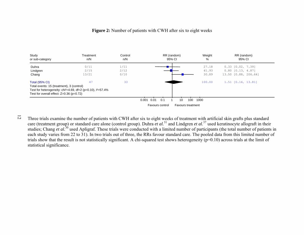
Figure 2: Number of patients with CWH after six to eight weeks
Three trials examine the number of patients with CWH after six to eight weeks of treatment with artificial skin grafts plus standard care (treatment group) or standard care alone (control group). Duhra et al.31 and Lindgren et al.37 used keratinocyte allograft in their studies; Chang et al.30 used Apligraf. These trials were conducted with a limited number of participants (the total number of patients in each study varies from 22 to 31). In two trials out of three, the RRs favour standard care. The pooled data from this limited number of trials show that the result is not statistically significant. A chi-squared test shows heterogeneity (p=0.10) across trials at the limit of statistical significance.
Study Treatment Control RR (random) Weight RR (random)or sub-category n/N n/N 95% CI % 95% CI
Duhra 0/11 1/11 27.18 0.33 [0.02, 7.39] Lindgren 2/15 2/12 41.93 0.80 [0.13, 4.87] Chang 13/21 0/10 30.89 13.50 [0.88, 206.64]
Total (95% CI) 47 33 100.00 1.51 [0.16, 13.81]Total events: 15 (treatment), 3 (control) Test for heterogeneity: chi²=4.69, df=2 (p=0.10), I²=57.4%Test for overall effect: Z=0.36 (p=0.72)
0.001 0.01 0.1 1 10 100 1000
Favours control Favours treatment
12

14
Figure 3: Number of patients with CWH after 11 to 12 weeks
Thirteen trials examine the number of patients with CWH after 11 to 12 weeks of treatment with artificial skin grafts plus standard care (treatment group) or standard care alone (control group). Five trials used Apligraf,30,33,40,43,45 six used Dermagraft,34-36,38,39,41 one used Hyalograft 3D plus Laserskin29 and one used EpiDex.44 All trials, except the study of Tausche et al., have RRs that favour treatment with artificial skin grafts. Only four out of 13 trials, however, have a lower confidence limit >1. The overall estimate shows that a 12-week treatment with an artificial skin graft yields a 44% increase of CWH as compared with standard care alone. This increase is statistically significant with a 95% CI of (1.22, 1.71). A chi-squared test shows homogeneity (p=0.23) across trials.
Study Treatment Control RR (random) Weight RR (random)or sub-category n/N n/N 95% CI % 95% CI
Gentzkow 11/37 1/13 0.73 3.86 [0.55, 27.09] Naughton 54/139 45/142 16.36 1.23 [0.89, 1.69] Pollack 40/109 40/126 14.32 1.16 [0.81, 1.65] Falanga (1999) 29/72 6/48 3.97 3.22 [1.45, 7.17] Pham 12/16 7/17 5.96 1.82 [0.97, 3.44] Chang 18/21 4/10 4.16 2.14 [0.98, 4.67] Veves 63/112 36/96 17.21 1.50 [1.11, 2.04] Hanft 15/24 6/22 4.46 2.29 [1.08, 4.85] Sams 5/9 3/8 2.33 1.48 [0.51, 4.31] Caravaggi 28/43 18/36 12.53 1.30 [0.88, 1.93] Krisnamoorthy 11/40 2/13 1.45 1.79 [0.45, 7.04] Marston 39/130 21/115 9.76 1.64 [1.03, 2.62] Tausche 14/43 14/34 6.76 0.79 [0.44, 1.42]
Total (95% CI) 795 680 100.00 1.44 [1.22, 1.71]Total events: 339 (treatment), 203 (control) Test for heterogeneity: chi²=15.14, df=12 (p=0.23), I²=20.7%Test for overall effect: Z=4.29 (p<0.0001)
0.1 0.2 0.5 1 2 5 10
Favours control Favours treatment
13

15
Figure 4: Number of patients with CWH after 24 weeks
Three trials32,33,42 examine the number of patients with CWH after 24 weeks of treatment with artificial skin grafts plus standard care (treatment group) or standard care alone (control group). All studies used Apligraf and have RRs that favour treatment. The overall estimate shows that a 24-week treatment with Apligraf yields a 45% increase in CWH compared with standard care alone. The increase is statistically significant with a 95% CI of (1.12, 1.86). A chi-squared test shows homogeneity (p=0.14) across trials.
Study Treatment Control RR (random) Weight RR (random)or sub-category n/N n/N 95% CI % 95% CI
Sabolinski 78/127 47/106 40.94 1.39 [1.07, 1.79] Falanga (1998) 92/146 63/129 46.23 1.29 [1.04, 1.60] Falanga (1999) 34/72 9/48 12.82 2.52 [1.33, 4.76]
Total (95% CI) 345 283 100.00 1.45 [1.12, 1.86]Total events: 204 (treatment), 119 (control) Test for heterogeneity: chi²=3.97, df=2 (p=0.14), I²=49.6%Test for overall effect: Z=2.87 (p=0.004)
0.1 0.2 0.5 1 2 5 10
Favours control Favours treatment
14

16
Figure 5: Number of patients with venous leg ulcers patients who showed CWH after 11 to 12 weeks
Three trials examine the effect of artificial skin grafts on the wound healing of patients with venous leg ulcers. Tausche et al.44 studied EpiDex. Falanga et al.33 and Krisnamoorthy,36 using Apligraf and Dermagraft respectively have RRs that favour treatment. The overall estimate shows a 60% increase in the chance of healing after 12 weeks of treatment. The result is not statistically significant for a 95% CI of (0.57, 4.46). A chi-squared test shows heterogeneity (p=0.01) across trials.
Study Treatment Control RR (random) Weight RR (random)or sub-category n/N n/N 95% CI % 95% CI
Falanga (1999) 29/72 6/48 35.53 3.22 [1.45, 7.17] Krisnamoorthy 11/40 2/13 25.04 1.79 [0.45, 7.04] Tausche 14/43 14/34 39.43 0.79 [0.44, 1.42]
Total (95% CI) 155 95 100.00 1.60 [0.57, 4.46]Total events: 54 (treatment), 22 (control) Test for heterogeneity: chi²=8.41, df=2 (p=0.01), I²=76.2%Test for overall effect: Z=0.90 (p=0.37)
0.1 0.2 0.5 1 2 5 10
Favours control Favours treatment
15

17
Figure 6: Number of patients with diabetic foot ulcers who had CWH after 11 to 12 weeks
Nine trials examine the effect of artificial skin grafts on the wound healing of patients with diabetic foot ulcers. Three trials of Apligraf,40,43,45 five of Dermagraft34,35,38,39,41 and one of Hyalograft 3D plus Laserskin.29 All trials have RRs that favour treatment. The overall estimate shows that patients with diabetic foot ulcers who underwent a 12-week treatment with artificial skin grafts have a 40% increase in the chance of CWH. The increase is statistically significant with a 95% CI of (1.21, 1.63). A chi-squared test shows homogeneity (p=0.65) across trials.
Study Treatment Control RR (random) Weight RR (random)or sub-category n/N n/N 95% CI % 95% CI
Gentzkow 11/37 1/13 0.59 3.86 [0.55, 27.09] Naughton 54/139 45/142 21.88 1.23 [0.89, 1.69] Pollack 40/109 40/126 17.63 1.16 [0.81, 1.65] Pham 12/16 7/17 5.53 1.82 [0.97, 3.44] Veves 63/112 36/96 23.86 1.50 [1.11, 2.04] Hanft 15/24 6/22 3.97 2.29 [1.08, 4.85] Sams 5/9 3/8 1.95 1.48 [0.51, 4.31] Caravaggi 28/43 18/36 14.41 1.30 [0.88, 1.93] Marston 39/130 21/115 10.20 1.64 [1.03, 2.62]
Total (95% CI) 619 575 100.00 1.40 [1.21, 1.63]Total events: 267 (treatment), 177 (control) Test for heterogeneity: chi²=5.95, df=8 (p=0.65), I²=0%Test for overall effect: Z=4.43 (p<0.00001)
0.1 0.2 0.5 1 2 5 10
Favours control Favours treatment
16
18
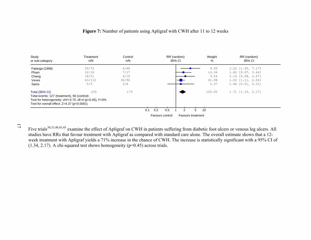
Figure 7: Number of patients using Apligraf with CWH after 11 to 12 weeks
Five trials30,33,40,43,45 examine the effect of Apligraf on CWH in patients suffering from diabetic foot ulcers or venous leg ulcers. All studies have RRs that favour treatment with Apligraf as compared with standard care alone. The overall estimate shows that a 12-week treatment with Apligraf yields a 71% increase in the chance of CWH. The increase is statistically significant with a 95% CI of (1.34, 2.17). A chi-squared test shows homogeneity (p=0.45) across trials.
Study Treatment Control RR (random) Weight RR (random)or sub-category n/N n/N 95% CI % 95% CI
Falanga (1999) 29/72 6/48 9.05 3.22 [1.45, 7.17] Pham 12/16 7/17 14.36 1.82 [0.97, 3.44] Chang 18/21 4/10 9.54 2.14 [0.98, 4.67] Veves 63/112 36/96 61.98 1.50 [1.11, 2.04] Sams 5/9 3/8 5.07 1.48 [0.51, 4.31]
Total (95% CI) 230 179 100.00 1.71 [1.34, 2.17]Total events: 127 (treatment), 56 (control) Test for heterogeneity: chi²=3.70, df=4 (p=0.45), I²=0%Test for overall effect: Z=4.37 (p<0.0001)
0.1 0.2 0.5 1 2 5 10
Favours control Favours treatment
17

19
Figure 8: Number of patients using Dermagraft with CWH after 11 to 12 weeks
Six trials34-36,38,39,41 examine the effect of Dermagraft on CWH in patients suffering from diabetic foot ulcers or venous leg ulcers. All studies have RRs that favour treatment with Dermagraft as compared with standard care alone. The overall estimate shows that an 11- to 12-week treatment with Dermagraft yields a 36% increase in the chance of CWH. The increase is statistically significant with a 95% CI of (1.11, 1.66). A chi-squared test shows homogeneity (p=0.41) across trials.
Study Treatment Control RR (random) Weight RR (random)or sub-category n/N n/N 95% CI % 95% CI
Gentzkow 11/37 1/13 1.07 3.86 [0.55, 27.09] Naughton 54/139 45/142 39.33 1.23 [0.89, 1.69] Pollack 40/109 40/126 31.78 1.16 [0.81, 1.65] Hanft 15/24 6/22 7.21 2.29 [1.08, 4.85] Krisnamoorthy 11/40 2/13 2.16 1.79 [0.45, 7.04] Marston 39/130 21/115 18.46 1.64 [1.03, 2.62]
Total (95% CI) 479 431 100.00 1.36 [1.11, 1.66]Total events: 170 (treatment), 115 (control) Test for heterogeneity: chi²=5.02, df=5 (p=0.41), I²=0.5%Test for overall effect: Z=2.96 (p=0.003)
0.001 0.01 0.1 1 10 100 1000
Favours control Favours treatment18

Time to complete wound healing Nine trials reported a median time for CWH, ranging from 39 to 91 days for treatment groups and from 77 to 196 days for the control groups (Table 4). All trials reported a shorter median time for CWH in patients treated with skin grafts as compared with standard care alone.
Table 4: Time to complete wound healing
Median Time for Complete Ulcer Healing (days) Study Treatment Control
(conventional therapy alone) Caravaggi29 Hyalograft 3D plus Laserskin, 57 77 Veves45 Apligraf, 65 90 Chang30 Apligraf, 49 105 Pham40 Apligraf, 38.5 81 Falanga32 Apligraf, 61 181 Pollack41 Dermagraft, 91 196 Naughton39 Dermagraft, 91 196 Sabolinski42 Apligraf, 57 181 Gentzkow34 Dermagraft*
A: 84 B: >84 C: >84
>84
*Group A: one piece of Dermagraft applied weekly, for a total of eight pieces and eight applications; group B: two pieces of Dermagraft applied every two weeks, for a total of eight pieces and four applications; group C: one piece of Dermagraft applied every two weeks, for a total of four pieces and four applications.
Adverse events Data on adverse events including infection, cellulitis and osteomyelitis, are presented in Figures 9, 10 and 11 respectively. The treatment of chronic ulcers with artificial skin grafts does not show a statistically significant reduction in rates of infection, cellulitis or osteomyelitis compared with standard care alone.
Summary The results from the included RCTs are summarized in Table 5. For CWH outcomes, the proportion of patients achieving complete wound healing in the treatment groups are significantly higher than those of the control groups after 11 to 12 and 24 weeks, but not at six to eight weeks. Patients with diabetic foot ulcers showed a significant increase in CWH, while those with venous leg ulcers did not. Apligraf shows a higher increase in the chance of wound healing than Dermagraft. Treatment with Apligraf exhibits a lower NNT compared with Dermagraft. However, this observed difference in efficacy may be due to other factors, such as patient management and baseline risk. Although the treatment groups show a lower number of specific adverse events than observed in the control groups, the differences are not statistically significant, as shown by the 95% CI of the RR.
19

Figure 9: Infection
Six trials34,35,38,41,44,45 report the rate of infection that occurred during treatment with artificial skin grafts plus standard care or standard care alone. Two trials34,44 out of six have RRs that favour conventional treatment (control), but without statistical significance. The pooled data show that treatment with an artificial skin graft leads to a 20% reduction in the risk of infection. The result is not statistically significant with a 95% CI of (0.60, 1.07). A chi-squared test shows homogeneity (p=0.75) across trials.
Study Treatment Control RR (random) Weight RR (random)or sub-category n/N n/N 95% CI % 95% CI
Gentzkow 9/37 3/13 6.41 1.05 [0.34, 3.31] Pollack 29/139 34/142 43.92 0.87 [0.56, 1.35] Veves 12/112 13/96 15.48 0.79 [0.38, 1.65] Hanft 1/24 2/22 1.54 0.46 [0.04, 4.71] Marston 17/163 27/151 26.24 0.58 [0.33, 1.03] Tausche 7/43 4/34 6.41 1.38 [0.44, 4.34]
Total (95% CI) 518 458 100.00 0.80 [0.60, 1.07]Total events: 75 (treatment), 83 (control) Test for heterogeneity: chi²=2.68, df=5 (p=0.75), I²=0%Test for overall effect: Z=1.53 (p=0.13)
0.1 0.2 0.5 1 2 5 10
Favours treatment Favours control
20

22
Figure 10: Cellulitis
Four trials32,35,38,45 report the rate of cellulitis that occurred during treatment with artificial skin grafts plus standard care or standard care alone. Two trials32,45 out of four have RRs that favour conventional treatment (control), but without statistical significance. The pooled data from this limited number of trials show that treatment with an artificial skin graft leads to a 12% reduction in the risk of cellulitis. The results are not statistically significant with a 95% CI of (0.56, 1.38). A chi-squared test shows homogeneity (p=0.44) across trials.
Study Treatment Control RR (random) Weight RR (random)or sub-category n/N n/N 95% CI % 95% CI
Falanga (1998) 12/146 10/129 31.64 1.06 [0.47, 2.37] Veves 10/112 8/96 25.96 1.07 [0.44, 2.61] Hanft 1/24 5/22 4.80 0.18 [0.02, 1.45] Marston 12/163 14/151 37.60 0.79 [0.38, 1.66]
Total (95% CI) 445 398 100.00 0.88 [0.56, 1.38]Total events: 35 (treatment), 37 (control) Test for heterogeneity: chi²=2.70, df=3 (p=0.44), I²=0%Test for overall effect: Z=0.57 (p=0.57)
0.1 0.2 0.5 1 2 5 10
Favours treatment Favours control
21

23
Figure 11: Osteomyelitis
Three trials35,38,45 report the rate of osteomyelitis that occurred during treatment with artificial skin grafts plus conventional therapy or conventional therapy alone. One trial38 out of three has RR that does not show any difference between the two arms of treatment. The pooled data from this limited number of trials show that treatment with an artificial skin graft leads to a 52% reduction in the risk of osteomyelitis. The results are not statistically significant with a 95% CI of (0.16, 1.42). A chi-squared test shows homogeneity (p=0.11) across trials.
Study Treatment Control RR (random) Weight RR (random)or sub-category n/N n/N 95% CI % 95% CI
Veves 3/112 10/96 33.43 0.26 [0.07, 0.91] Hanft 1/24 4/22 18.31 0.23 [0.03, 1.90] Marston 14/163 13/151 48.26 1.00 [0.48, 2.05]
Total (95% CI) 299 269 100.00 0.48 [0.16, 1.42]Total events: 18 (treatment), 27 (control) Test for heterogeneity: chi²=4.43, df=2 (p=0.11), I²=54.8%Test for overall effect: Z=1.32 (p=0.19)
0.1 0.2 0.5 1 2 5 10
Favours treatment Favours control
22
23
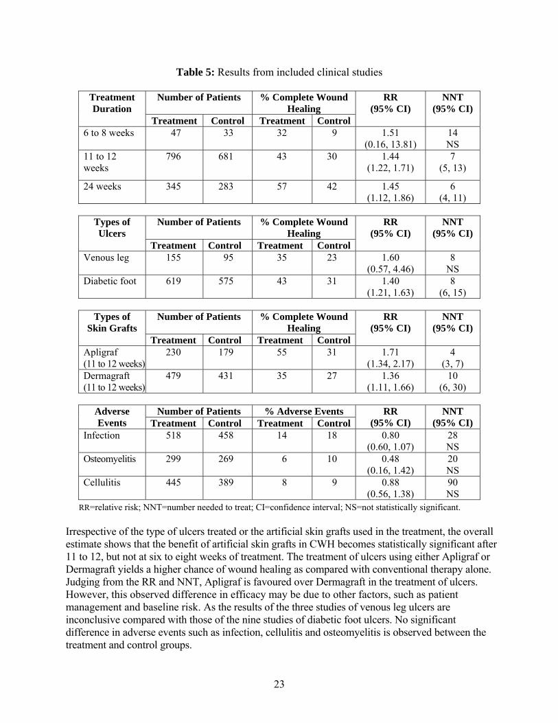
Table 5: Results from included clinical studies
Number of Patients % Complete Wound Healing
Treatment Duration
Treatment Control Treatment Control
RR (95% CI)
NNT (95% CI)
6 to 8 weeks 47 33 32 9 1.51 (0.16, 13.81)
14 NS
11 to 12 weeks
796 681 43 30 1.44 (1.22, 1.71)
7 (5, 13)
24 weeks 345 283 57 42 1.45 (1.12, 1.86)
6 (4, 11)
Number of Patients % Complete Wound
Healing Types of Ulcers
Treatment Control Treatment Control
RR (95% CI)
NNT (95% CI)
Venous leg 155 95 35 23 1.60 (0.57, 4.46)
8 NS
Diabetic foot 619 575 43 31 1.40 (1.21, 1.63)
8 (6, 15)
Number of Patients % Complete Wound
Healing Types of
Skin Grafts Treatment Control Treatment Control
RR (95% CI)
NNT (95% CI)
Apligraf (11 to 12 weeks)
230 179 55 31 1.71 (1.34, 2.17)
4 (3, 7)
Dermagraft (11 to 12 weeks)
479 431 35 27 1.36 (1.11, 1.66)
10 (6, 30)
Number of Patients % Adverse Events Adverse
Events Treatment Control Treatment ControlRR
(95% CI) NNT
(95% CI) Infection 518 458 14 18 0.80
(0.60, 1.07) 28 NS
Osteomyelitis 299 269 6 10 0.48 (0.16, 1.42)
20 NS
Cellulitis 445 389 8 9 0.88 (0.56, 1.38)
90 NS
RR=relative risk; NNT=number needed to treat; CI=confidence interval; NS=not statistically significant. Irrespective of the type of ulcers treated or the artificial skin grafts used in the treatment, the overall estimate shows that the benefit of artificial skin grafts in CWH becomes statistically significant after 11 to 12, but not at six to eight weeks of treatment. The treatment of ulcers using either Apligraf or Dermagraft yields a higher chance of wound healing as compared with conventional therapy alone. Judging from the RR and NNT, Apligraf is favoured over Dermagraft in the treatment of ulcers. However, this observed difference in efficacy may be due to other factors, such as patient management and baseline risk. As the results of the three studies of venous leg ulcers are inconclusive compared with those of the nine studies of diabetic foot ulcers. No significant difference in adverse events such as infection, cellulitis and osteomyelitis is observed between the treatment and control groups.

24
4.2.4 Results of economic studies
A separate review was conducted for studies that evaluated models of cost-effectiveness for artificial skin use in the treatment of venous ulcers and diabetic foot ulcers. Non-comparative chart reviews that evaluated the clinical and economic outcomes associated with artificial skin use in venous leg ulcers were also performed. a) Cost-effectiveness of artificial skin used for venous leg ulcers Two studies used different models to evaluate the cost-effectiveness of artificial skin (Apligraf®) used for hard to heal venous leg ulcers. Their design elements are shown in Table 6 and their results are summarized in Appendix 6. Schonfeld et al.1 conducted an economic evaluation of the use of Apligraf (graftskin) for venous ulcers with low compression dressings compared to low compression using Unna’s boot plus a self-adherent elastic bandage. The latter combination, also called Duke’s boot, provides higher compression. Their evaluation was based on a one-year clinical trial by Falanga et al.,32 who assessed the two treatments over six months with safety follow-up to one year. They found that graftskin showed an improved chance of healing (63% versus 49%, p=0.02) and a shortened healing time (61 days versus 181 days, p=0.003) in all patients with no significant difference in recurrence, rejection, dropout or adverse events. In a subsequent subgroup analysis of hard to heal ulcers, there was an even greater effect; 60% versus 31% of these ulcers healed at one year.33 Schonfeld et al.1 used a Markov model with one-month cycles to evaluate the cost-effectiveness of this treatment in hard to heal ulcers (those present for >1 year) based on these results. They obtained estimates of the resource use associated with treatment of an unhealed ulcer, adverse events and hospitalizations from a survey of dermatologists, vascular surgeons and podiatrists; and applied unit prices from standard costing sources. They assumed the trial average of 3.34 graftskin applications for the initial treatment and for the treatment of recurrent ulcers in patients assigned to the graftskin alternative. Sibbald et al.2 investigated a different strategy based on the clinical judgment that high-pressure compression treatment would be most important for venous ulcers, so that this would be the standard treatment. As a result, artificial skin would be applied with high compression and compared to high compression alone. They chose the four-layer bandage system with or without the use of artificial skin as relevant comparators. Since clinical trial data did not exist directly comparing high compression with or without the use of artificial skin, they presented the best existing evidence from the publication of the Falanga clinical trial and two three-month trials of outcomes with four-layer bandage to a Delphi panel of clinicians, who estimated that over three months, 60% of patients would heal versus 67.5% of patients with Apligraf plus a four-layer bandage. The proportion of patients to experience recurrence and time to recur were equal, although it was assumed that patients who were initially given the skin substitute would heal from recurrence more quickly. An increased proportion of patients to experience moderate and severe infections was assumed with Apligraf, based on the Falanga trial data. One application of Apligraf was assumed. Resource use was estimated by the Delphi panel for the relevant health states and the societal perspective included the cost of time lost from employment.

25
Table 6: Cost-effectiveness studies of artificial skin use in non-healing venous leg ulcers
Schonfeld et al.1 Sibbald et al.2 Study product Graftskin (Apligraf®) with low compression
dressings, up to 8 weekly implants Apligraf® with four-layer bandage system, applied once (assumption)
Comparator Low pressure support (Unna’s boot) High compression alone (4-layer bandage system)
Indication Patients with hard to heal venous leg ulcers Patients with venous leg ulcers Country, perspective, year, currency
US, commercial health plan, 1996, dollars Canada, health care (direct medical cost), 1996-1997, dollars
Study design Markov model monthly cycle Computer-based decision model Analytic horizon One year 3 months, 6 months Data sources for effects
One RCT32 12 months; improved chance of healing, speed of healing and greater effect in hard to heal ulcers; no difference in recurrence, rejection, dropout, adverse events
Delphi panel consensus after review of available evidence (5 dermatologists, 2 GPs); improved chance of healing, speed of healing; higher rate of moderate, severe infections; no difference in recurrence
Data sources for costs
Survey of dermatologists, vascular surgeons, podiatrists conducted to estimate resource use (average used): physician visits, home health visits, compression dressings, laboratory tests, procedures, treatment of adverse events, hospitalizations; 3.3 applications of graftskin (trial average), if recurrence 3.3
Delphi panel; each estimated own resource use (average used): health care professional services, home care, laboratory tests, hospital admissions, emergency room visits, wound care supplies (dressings), patient expenses, time loss from work; one application of Apligraf; included time loss from work in base case
Schonfeld et al. found that the graftskin strategy provided 2.85 additional months in an ulcer-free state over one year and had a cost savings of $7,452 (C$10,089) (currency converted based on historical rates for the year of evaluation obtained at http:\www.xe.com/ict/table.cgi) so it was dominant over the use of Unna’s boot. These results were found to be robust to variations in the healing rates, costs and assumptions that the recurrence and adverse event rates were equal or higher in the graftskin treatment group. The results were found to be sensitive to the usage of hospitalization, so that when hospital costs were doubled or when resource use was assumed to be lower for hospitalization, there was an incremental cost-effectiveness ratio of $800 per additional ulcer-free month (C$1,083), which is equivalent to C$36 per additional ulcer-free day. Sibbald et al. found that the use of Apligraf was associated with 22 fewer ulcer-days over three months at an incremental cost-effectiveness of $14 per ulcer-day averted for the health care system and from a societal perspective. These results were robust to sensitivity analyses with varying parameters, although including the cost of time lost from usual activities in the societal perspective resulted in estimated cost savings. When results were extended to a six-month perspective, there was an estimated 60 to 67 additional ulcer-free days at incremental costs of $1.05 to $4.26 per additional ulcer-free day. b) Cost-effectiveness of artificial skin for diabetic foot ulcers Two studies used different models to evaluate the cost-effectiveness of artificial skin (Apligraf®) use for hard to heal diabetic foot ulcers. The design elements of the studies are shown in Table 7 and their results are summarized in Appendix 7.

26
Table 7: Cost-effectiveness studies of artificial skin use in non-healing diabetic foot ulcers
Allenet et al.4 Redekop et al.3 Study product Dermagraft, up to 8 weekly implants Graftskin (Apligraf®); up to 5 weekly
implants Comparator Sharp débridement, infection control,
moist dressings, weight offloading Debridement, moist dressings, weekly dressing change, weight offloading
Indication Long-standing, full thickness dermal ulcers in French patients with diabetes; no necrosis, no infection, no lower extremity ischemia
Diabetic foot ulcers unhealed for >2 weeks; no necrosis, no infection, no lower extremity ischemia
Country, perspective, year, currency
France, societal (direct costs only), year not stated, French francs
Netherlands, societal (direct costs only), 1999, euros
Study design Markov model cycle of 1 week Markov model cycle of 4 weeks Analytic horizon One year One year Data sources for effects
RCT39 10 week trial, follow-up to 32 weeks; improved weekly chance of healing during first 10 weeks; less recurrence, faster healing of recurring ulcers; same probability of infection while unhealed; same chance of amputation if severe infection
RCT45 12-week trial; improved chance of healing, speed of healing; recurrence not incorporated into model; less infection, amputation
Data sources for costs Expert panel of diabetologists from French centres of excellence; standard national cost sources for unit prices; Dermagraft base case used 7 pieces, sensitivity analysis 8 pieces (maximum)
Literature estimates where possible; amputation cost from 2 studies, one gave frequency of major and minor amputation, another, duration of hospitalization for each; Apligraf base case used 2 pieces; sensitivity analysis 4 pieces (trial average use)
Each evaluation is based on one clinical RCT. In both studies, the use of artificial skin shortened the healing time and resulted in more ulcers healed over the study period. The study of Veves et al.45 (which was used by Redekop et al.) showed fewer infections and amputations with artificial skin. These adverse events without recurrence were incorporated into their model. Allenet et al.4 showed a lower recurrence rate and a faster healing rate associated with artificial skin, with the assumption that both treatments had the same probability of infection and amputation when the ulcer was unhealed. Thus, the benefit for artificial skin is manifested through its reduction of healing time. The patient populations assumed for these evaluations are comparable. Populations consist of patients with unhealed ulcers who do not have necrosis or infection; and who have adequate blood pressure to the affected limb to allow healing. Both groups (with or without artificial skin use) receive similar elements of recommended care, including sharp débridement, moist dressings changed weekly and aids to keep pressure off the ulcer (weight downloading). It is assumed that patients do not use prophylactic antibiotics, unless they are for the treatment of infection.
27
Each evaluation uses a Markov model to combine the transitional probabilities between health states and the cost of health states; and to estimate the total clinical outcomes and costs over a year. Allenet et al.4 base their probabilities of healing and recurrence on the proportion of patients healed at the end of the 10-week trial period and 32-week follow-up. For conventional therapy, they estimated, using regression analysis, a common healing rate (2.8% per week) over the full 32 weeks. For Dermagraft, the healing rate was estimated to be 6.7% for the first 10 weeks and 2.1% weekly thereafter. Based on trial data, ulcer recurrence with Dermagraft was reduced and recurring ulcers were faster to heal, although the same rate of infection and amputation was assumed while the patient had an active ulcer. Redekop et al.3 base the probability of healing on the proportion of patients healed in the trial by 12 weeks and use the same rate (that for good wound care alone) after the first four weeks. They did not include ulcer recurrence in the model, as trial differences (in favour of Apligraf) were not significant. Differences in osteomyelitis and amputation in favour of Apligraf observed in the trial were included in the evaluation model. Allenet et al.4 found that, over a year, the use of Dermagraft provided 21 additional ulcers healed per 100 patients and 8.33 additional weeks per patient in an ulcer-free state at an incremental cost-effectiveness of 38,784 FF (C$10,589) per additional ulcer healed, which is equivalent to C$182 per additional ulcer-free day. Generally, this result was robust to sensitivity analyses, which explored the impact of assuming no weekly cost for the healed state, increasing average usage to eight pieces of Dermagraft (the maximum of any patient in the trial) and varying the duration of rehabilitation for major or minor amputation. The chance of amputation was not varied. Redekop et al.3 found that Apligraf use resulted in 1.3 months additional time in an ulcer-free state over the year at an average savings of €654 (C$1,210), so that Apligraf was found to be dominant (lower cost and better clinical results) compared with standard clinical care without artificial skin graft. This result was robust to varying the costs for uninfected ulcer. If the use of four pieces of Apligraf was assumed (trial average), there would be an incremental cost of €980 (C$1,814) or €641 (C$1,186) per ulcer-free month gained, equivalent to C$60 or C$40 per ulcer-free day. The cost-effectiveness was less and showed an estimated incremental cost per ulcer free month gained up to €2,000 (C$3,702) equivalent to C$124 per ulcer-free day when assumptions around infection and amputation were varied. The other assumptions were that artificial skin use does not lower infection risk beyond reducing the time with an ulcer and a lower cost associated with amputation. c) Non-comparative studies of artificial skin use in venous leg ulcers Two case-series studies15,16 evaluated clinical and economic outcomes in patients with non-healing venous leg ulcers who received treatment with Apligraf in a more ”real” setting (Appendix 8). In the study of Fivenson et al.,16 Apligraf treatment was given to 13 patients whose ulcers had been increasing in size by 2.3% per week over the previous three months. Over the three months after the start of Apligraf treatment (average of 1.5 applications per ulcer), the average ulcer closure rate was 2.9% per week. Cost data derived from five patients indicate that patients’ care cost for three months before and during Apligraf treatment was $4,399 and $4,458 respectively.

28
Kirsner et al.15 identified 16 consecutive patients whose ulcers had been increasing in size by an average of 6% per week since starting treatment at the clinic. Over an average follow-up of 13 weeks after the start of Apligraf treatment (average of 2.25 applications per ulcer), eight patients (13 of 24 ulcers) healed and the average ulcer closure rate was 9.5% per week. The median cost of treatment before and during Apligraf treatment was $16,860 and $15,907 respectively. 5 DISCUSSION A comprehensive review of the literature regarding clinical efficacy, safety and economic outcomes associated with the use of grafts of artificial living skin tissue in patients with chronic skin ulcers identified 17 RCTs of the use of living skin replacement products for the indications of chronic venous leg ulcers (seven trials) and chronic diabetic foot ulcers (nine trials) with one mixed trial. These trials compared the use of artificial skin and standard care to standard care alone.
5.1 Clinical Review The primary efficacy of treatment was evaluated based on the proportion of patients to achieve CWH. No significant benefit was shown six to eight weeks after transplant. By 11 to 12 weeks, however, there was a 44% statistically significant increase in healing that was consistent across the trials. By 24 weeks post-graft, there was a 45% statistically significant increase in the chance of wound healing. In all studies that reported the speed of healing, there was a shorter median time to complete ulcer healing. The preparation of the wound bed and the absence of infection were important in determining the success rate of using an artificial skin graft. Artificial skin cannot replace best conventional care – it should be used with it. Patients with venous leg ulcers had a 60% increase in the chance of CWH compared with standard care after 12 weeks, but the result was not statistically significant. Patients with diabetic foot ulcers had a 40% statistically significant increase in the chance of healing after 12 weeks. The combination of neuropathy, peripheral vascular disease and infection in patients with chronic diabetic skin pathogenesis may affect the rate of wound healing. There were enough trials to evaluate efficacy of Apligraf alone. This product showed a 71% statistically significant increase in the chance of healing compared to standard care without skin substitute by 12 weeks after a graft. Apligraf has an epidermal layer that dies over two to four weeks. If the wound is closed when the epidermal cells die, the wound may open unless the host’s epidermal cell coverage keeps pace with the dieback. For this reason, any wound closure before four weeks after the last application may be doubtful. The use of Dermagraft was associated with a 36% statistically significant increase in the chance of healing by 12 weeks post-graft. The lower efficacy of Dermagraft compared with Apligraf in terms of the chance for CWH may be linked to the fact that five out of six trials with Dermagraft involved patients with diabetic foot ulcers. The pathogenesis of chronic diabetic wounds may have a limiting effect on the use of Dermagraft. Also, in most of the Apligraf studies,30,40,43,45 the

29
patients were in a rigorous non-weight-bearing regime, using crutches or wheelchairs for the first week of the study (Appendix 9). This would give a superior result compared with many Dermagraft studies, where patients received pressure offloading and used casting or special shoes and orthotics.34,38,39,41 Faster healing with the use of an artificial skin substitute would mean that a patient spends less time with an open wound at risk for infection. This is one of many factors that may affect the risk of infection. An infection, however, may be difficult to detect because the wound is covered by a skin substitute. Although trials that reported the rates of different forms of infection did not find statistically significant differences between treatments, an artificial skin graft was associated with an overall 20% reduction in the risk of any infection; a 12% reduction in cellulitis, a form of mild infection; and a 52% reduction in the risk of osteomyelitis, a deep infection reaching to the bone. Controversies in diagnosis and treatment of infection in chronic wounds were discussed by Dow et al.46 Several non-RCTs of wound healing with artificial skin use were identified outside of this study. They were heterogeneous and were limited in quality. These trials lasted from eight to 35 weeks, and provided more information about healing over a longer time. A greater proportion of patients in these studies showed healing than in the RCTs included in the report. More trials are needed to provide enough data to confidently evaluate other skin graft products.
5.2 Economic Review Four models of evaluations examined the costs and outcomes associated with the use of artificial skin products for the treatment of chronic ulcers. Each compared the use of an artificial skin product to standard care without artificial skin. The use of Apligraf in venous leg ulcers was evaluated in two studies over three to six months and over a year.1,2 There is better cost-effectiveness over the long term. Over three months, with a strategy model of high compression with or without Apligraf, it was estimated that 22 ulcer days were averted at an incremental cost of $14 per day to society. Over six months, 60 to 67 ulcer days were averted at an incremental cost of $1.05 to $4.26 per ulcer day averted.2 Over one year, moderate compression treatment with Apligraf was dominant over moderate compression alone, resulting in 2.85 additional ulcer-free months with a savings of C$10,089 per patient.1 The results of both these evaluations were robust with varying assumptions. The use of Dermagraft and Apligraf in diabetic foot ulcers was evaluated over a year in two studies from France and the Netherlands respectively. Each was based on one RCT.3,4 Both evaluations found additional ulcer-free time over the year (at two months and 1.3 months). The use of seven pieces of Dermagraft was associated with an incremental cost of C$182 per ulcer-free day, while the use of two pieces of Apligraf was found to be associated with overall savings. A key cost driver of economic evaluations in both indications was the amount of artificial skin used. With respect to diabetic foot ulcers, the clinical trial on which the Allenet evaluation was based used from one to eight pieces of Dermagraft. The evaluation model used for a base-case
30
estimate, was on average of seven pieces per patient. It assessed, in sensitivity analyses, the impact of the use of eight pieces, the maximum used by any patient. This evaluation found an incremental cost per additional ulcer healed. In the Redekop evaluation, based on data from chart reviews showing healing with a lower average use of artificial skin than in the clinical trial, the base case used an average of two pieces of Apligraf per patient and showed overall cost savings. A sensitivity analysis of four pieces of artificial skin (average use in the trial) found an incremental cost for each additional ulcer healed. Other design elements in the evaluation models involve the effect of artificial skin use on speed of healing, recurrence, infection and amputation. These evaluations differ because there are differences in the underlying trial results. This systematic review has confirmed the main results on which these economic evaluations are based. If confirms that there is an increased probability of CWH over the longer time frame (>12 weeks post graft). Although reductions in infections, cellulitis and osteomyelitis with the use of artificial skin are reasonably consistent across clinical trials, they are not statistically significant. Modelling necessarily involves the use of assumptions and the combination of information from different sources, so that the conclusions can be validated. Evaluation models are often criticized because the intended comparison is with the effectiveness and cost of treatments that are used in “real” practice, whereas the key estimates of efficacy and treatment used to populate the model are often obtained from clinical trials. Non-comparative studies give an assessment of the treatment of patients in a more usual care setting, the effectiveness of treatment and the associated costs. They provide confirmation for the main elements implemented in the economic evaluation models. The use of artificial skin by patients who had failed to heal with conventional care resulted in improved healing rates. Furthermore, the total costs were similar before and after the use of artificial skin, confirming that there were savings due to healing, which helped to offset the acquisition cost of artificial skin. While the results of these economic evaluations seem to be encouraging, they are subject to assumptions and should be considered as preliminary.
5.3 Application of Results to Canadian Health Services Canada was the first country to approve Apligraf for venous leg ulcers in April 1997 and Dermagraft for foot ulcers in persons with diabetes in August 1997. In Canada, patients with these conditions often live in the community, attend specialized clinics or have their wounds managed in home care. The cost to home care is enormous. These patients often require hospital admissions for acute infections or other diabetic complications. Treatments that can speed healing can be an improvement in clinical practice. Several factors must be considered before these products can be used to improve clinical outcomes for patients. Artificial skin products should be considered in patients who are the most likely to benefit. This requires careful patient selection and assessment of co-existing medical and local wound conditions. The average age of patients with venous ulcers in many series is over 70 and

31
recurrences are common. The frequent co-existing illnesses, drugs and co-factors that may delay healing will contribute to the difficulty of translating the RCTs for skin substitutes into cost- effective outcomes for health care systems. Persons with diabetes have an increased incidence of peripheral vascular disease and infection; and deformity from neuropathy. All these factors lead to an increased risk of non-healing ulcers and amputation. In the community, poor blood sugar control and other complications, such as renal disease, will impair the ability of foot ulcers to heal. Many of these poorly controlled patients are excluded from the trials reported in this study. Although artificial skin grafts can stimulate local wound healing, they do not address the cause of the wound, complications contributing to non-healing, patients’ compliance and patients’ concerns about the quality of life and pain. These factors must to be optimized to allow for successful local wound healing. Persons with diabetes and foot ulcers should be assessed by specialized teams for adequate vascular supply, infection and appropriate pressure offloading of the foot. In summary, there is evidence of clinical efficacy and cost-effectiveness when artificial skin is used in persons with diabetic foot ulcers. For cost-effective clinical practice, the number of pieces of skin substitute used should be one or two and all other factors must be optimized. 6 CONCLUSION The results of clinical trials show that the use of an artificial skin graft plus standard therapy promotes wound closure, resulting in more frequent and more rapid healing of chronic diabetic foot ulcers compared with standard therapy alone. The effect on the healing of venous leg ulcers is not statistically significant. The risks of infection, cellulitis and osteomyelitis are not affected by the use of skin grafts. Over a shorter time frame, the use of artificial skin has increased costs beyond those of standard therapy alone. When the clinical effects and costs are considered over a year, however, the cost savings from fewer ulcer days may result in a net savings associated with the use of artificial skin. Factors that determine cost-effectiveness include the number of pieces of artificial skin used; the healing rates; and the rates of recurrence and infection for venous leg ulcers and diabetic foot ulcers. Health care policy decision makers must ensure that expensive artificial skin products are used on patients who are most likely to benefit from their use. This requires careful patient selection and assessment of co-existing medical and local wound conditions. The cost to home care is enormous. More data must be accumulated on the cost-effectiveness of integrating the use of artificial skin products into routine care. The evidence suggests that there may be a role for the use of these products on a small select group of patients.

32
7 REFERENCES
1. Schonfeld WH, Villa KF, Fastenau JM, Mazonson PD, Falanga V. An economic assessment of Apligraf® (Graftskin) for the treatment of hard-to-heal venous leg ulcers. Wound Repair Regen 2000;8(4):251-7.
2. Sibbald RG, Torrance GW, Walker V, Attard C, MacNeil P. Cost-effectiveness of Apligraf™ in the treatment of venous leg ulcers. Ostomy Wound Manage 2001;47(8):36-46.
3. Redekop WK, McDonnell J, Verboom P, Lovas K, Kalo Z. The cost effectiveness of Apligraf® treatment of diabetic foot ulcers. PharmacoEconomics 2003;21(16):1171-83.
4. Allenet B, Parée F, Lebrun T, Carr L, Posnett J, Martini J, et al. Cost-effectiveness modeling of Dermagraft® for the treatment of diabetic foot ulcers in the French context. Diabetes Metab 2000;26(2):125-32.
5. Margolis DJ, Bilker W, Santanna J, Baumgarten M. Venous leg ulcer: incidence and prevalence in the elderly. J Am Acad Dermatol 2002;46(3):381-6.
6. Boulton AJ, Vileikyte L. The diabetic foot: the scope of the problem. J Fam Pract 2000;49(11 Suppl):S3-S8.
7. Kantor J, Margolis DJ. Management of leg ulcers. Semin Cutan Med Surg 2003;22(3):212-21.
8. Persoon A, Heinen MM, van der Vleuten CJ, de Rooij MJ, Van De Kerkhof PC, van Achterberg T. Leg ulcers: a review of their impact on daily life. J Clin Nurs 2004;13(3):341-54.
9. Kinmond K, McGee P, Gough S, Ashford R. 'Loss of self': a psychosocial study of the quality of life of adults with diabetic foot ulceration. J Tissue Viability 2003;13(1):6-16.
10. Ontario drug benefit formulary/comparative drug index: no. 38. Effective September 4, 2003. Toronto: Ministry of Health; 2003. Available: http://www.health.gov.on.ca/english/providers/program/drugs/formulary/ed38_0_bk.pdf.
11. Hjelm K, Nyberg P, Apelqvist J. Gender influences beliefs about health and illness in diabetic subjects with severe foot lesions. J Adv Nurs 2002;40(6):673-84.
12. Nelzén O, Bergqvist D, Lindhagen A. Long-term prognosis for patients with chronic leg ulcers: a prospective cohort study. Eur J Vasc Endovasc Surg 1997;13(5):500-8.
13. Lindholm C, Bergsten A, Berglund E. Chronic wounds and nursing care. J Wound Care 1999;8(1):5-10.
14. Apelqvist J, Ragnarson Tennvall G, Larsson J, Persson U. Long-term costs for foot ulcers in diabetic patients in a multidisciplinary setting. Foot Ankle Int 1995;16(7):388-94.
15. Kirsner RS, Fastenau J, Falabella A, Valencia I, Long R, Eaglstein WH. Clinical and economic outcomes with graftskin for hard-to-heal venous leg ulcers: a single-center experience. Dermatol Surg 2002;28(1):81-2.

33
16. Fivenson D, Scherschun L. Clinical and economic impact of Apligraf® for the treatment of nonhealing venous leg ulcers. Int J Dermatol 2003;42(12):960-5.
17. Sibbald RG, Torrance G, Hux M, Attard C, Milkovich N. Cost-effectiveness of becaplermin for nonhealing neuropathic diabetic foot ulcers. Ostomy Wound Manage 2003;49(11):76-84.
18. Inlow S, Orsted H, Sibbald RG. Best practices for the prevention, diagnosis, and treatment of diabetic foot ulcers. Ostomy Wound Manage 2000;46(11):55-68.
19. Millington JT, Ellenzweig JM. Management and treatment of diabetic foot wounds in the elderly. Ann Long Term Care 2003;11(1):26-32. Available: http://www.mmhc.com/attachments/1054651930-millington.pdf (accessed 2004 Mar 3).
20. Browne AC, Sibbald RG. The diabetic neuropathic ulcer: an overview. Ostomy Wound Manage 1999;45 Suppl 1A:6S-20S.
21. Sibbald RG, Williamson D, Orsted HL, Campbell K, Keast D, Krasner D, et al. Preparing the wound bed--debridement, bacterial balance, and moisture balance. Ostomy Wound Manage 2000;46(11):14-35.
22. Steed DL, Diabetic Ulcer Study Group. Clinical evaluation of recombinant human platelet-derived growth factor for the treatment of lower extremity diabetic ulcers. J Vasc Surg 1995;21(1):71-8.
23. Bello YM, Falabella AF, Eaglstein WH. Tissue-engineered skin. Current status in wound healing. Am J Clin Dermatol 2001;2(5):305-13.
24. Jones I, Currie L, Martin R. A guide to biological skin substitutes. Br J Plast Surg 2002;55(3):185-93.
25. Curran MP, Plosker GL. Bilayered bioengineered skin substitute (Apligraf®): a review of its use in the treatment of venous leg ulcers and diabetic foot ulcers. BioDrugs 2002;16(6):439-55.
26. Shen JT, Falanga V. Innovative therapies in wound healing. J Cutan Med Surg 2003;7(1):217-24.
27. Jadad AR, Moore RA, Carroll D, Jenkinson C, Reynolds DJ, Gavaghan DJ, et al. Assessing the quality of reports of randomized clinical trials: is blinding necessary? Control Clin Trials 1996;17(1):1-12.
28. Drummond MF, O'Brien BJ, Stoddart GL, Torrance GW. Methods for the economic evaluation of health care programmes [Oxford medical publications]. 2nd ed. New York: Oxford University Press; 1997.
29. Caravaggi C, De Giglio R, Pritelli C, Sommaria M, Dalla Noce S, Faglia E, et al. HYAFF 11-based autologous dermal and epidermal grafts in the treatment of noninfected diabetic plantar and dorsal foot ulcers: a prospective, multicenter, controlled, randomized clinical trial. Diabetes Care 2003;26(10):2853-9.
30. Chang DW, Sanchez LA, Veith FJ, Wain RA, Okhi T, Suggs WD. Can a tissue-engineered skin graft improve healing of lower extremity foot wounds after revascularization? Ann Vasc Surg 2000;14(1):44-9.

34
31. Duhra P, Blight A, Mountford E, Cheshire I, Withers A, Ilchyshyn A. A randomized controlled trial of cultured keratinocyte allografts for chronic venous ulcers. J Dermatolog Treat 1992;3:189-91.
32. Falanga V, Margolis D, Alvarez O, Auletta M, Maggiacomo F, Altman M, et al. Rapid healing of venous ulcers and lack of clinical rejection with an allogeneic cultured human skin equivalent. Arch Dermatol 1998;134:293-300.
33. Falanga V, Sabolinski M. A bilayered living skin construct (APLIGRAF®) accelerates complete closure of hard-to-heal venous ulcers. Wound Repair Regen 1999;7(4):201-7.
34. Gentzkow GD, Iwasaki SD, Hershon KS, Mengel M, Prendergast JJ, Ricotta JJ, et al. Use of dermagraft, a cultured human dermis, to treat diabetic foot ulcers. Diabetes Care 1996;19(4):350-4.
35. Hanft JR, Surprenant MS. Healing of chronic foot ulcers in diabetic patients treated with a human fibroblast-derived dermis. J Foot Ankle Surg 2002;41(5):291-9.
36. Krishnamoorthy L, Harding KG, Griffiths D, Moore K, Leaper D, Poskitt K, et al. The clinical and histological effects of Dermagraft® in the healing of chronic venous leg ulcers. Phlebology 2003;18:12-22.
37. Lindgren C, Marcusson JA, Toftgård R. Treatment of venous leg ulcers with cryopreserved cultured allogeneic keratinocytes: a prospective open controlled study. Br J Dermatol 1998;139(2):271-5.
38. Marston WA, Hanft J, Norwood P, Pollak R, for the Dermagraft Diabetic Foot Ulcer Study Group. The efficacy and safety of dermagraft in improving the healing of chronic diabetic foot ulcers: results of a prospective randomized trial. Diabetes Care 2003;26(6):1701-5.
39. Naughton G, Mansbridge J, Gentzkow G. A metabolically active human dermal replacement for the treatment of diabetic foot ulcers. Artif Organs 1997;21(11):1203-10.
40. Pham HT, Rosenblum BI, Lyons TE, Giurini JM, Chrzan JS, Habershaw GM, et al. Evaluation of a human skin equivalent for the treatment of diabetic foot ulcers in a prospective, randomized, clinical trial. Wounds 1999;11(4):79-86.
41. Pollak RA, Edington H, Jensen JL, Kroeker RO, Gentzkow GD, Dermagraft Diabetic Ulcer Study Group. A human dermal replacement for the treatment of diabetic foot ulcers. Wounds 1997;9(1):175-83.
42. Sabolinski ML, Alvarez O, Auletta M, Mulder G, Parenteau NL. Cultured skin as a 'smart material' for healing wounds: experience in venous ulcers. Biomaterials 1996;17(3):311-20.
43. Sams HH, Chen J, King LE. Graftskin treatment of difficult to heal diabetic foot ulcers: one center's experience. Dermatol Surg 2002;28(8):698-703.
44. Tausche AK, Skaria M, Böhlen L, Liebold K, Hafner J, Friedlein H, et al. An autologous epidermal equivalent tissue-engineered from follicular outer root sheath keratinocytes is as effective as split-thickness skin autograft in recalcitrant vascular leg ulcers. Wound Repair Regen 2003;11(4):248-52.

35
45. Veves A, Falanga V, Armstrong DG, Sabolinski ML, for the Apligraf Diabetic Foot Ulcer Study. Graftskin, a human skin equivalent, is effective in the management of noninfected neuropathic diabetic foot ulcers: a prospective randomized multicenter clinical trial. Diabetes Care 2001;24(2):290-5. Available: http://care.diabetesjournals.org/cgi/reprint/24/2/290.pdf (accessed 2003 Aug 26).
46. Dow G, Browne A, Sibbald RG. Infection in chronic wounds: controversies in diagnosis and treatment. Ostomy Wound Manage 1999;45(8):23-40.

36
Appendix 1: Literature Search Strategies Jan. 15, 2004
In Dialog®
de = descriptor, ie. Medical Subject Heading (a controlled vocabulary, or thesaurus term) ti = title (i.e. word has to occur in title field of the bibliographic record) ab = abstract (i.e. word has to occur in abstract field of bibliographic record) ! = explode; picks up narrower terms as well, i.e. terms which are conceptually subsets of
a broader term F1$ = a large MeSH category, e.g. Behavior and behavior mechanisms , which is exploded
to pick up all terms related to behavior and behavior mechanisms, as defined by the National Library of Medicine, i.e. about 400 MeSH terms
() = words must be adjacent (2n) = words a maximum of two words apart in either direction ? = truncation symbol dt = publication type Set 22: Set 23 = Set 22 OR Set 23 In PubMed [MeSH] = Medical Subject Headings (a controlled vocabulary, or thesaurus term) [Title/Abstract] = word must appear in title or abstract of record [All Fields] = word must appear in any field
DATABASE LIMITS KEYWORDS Dialog® OneSearch® (including MEDLINE® (1966-present), EMBASE® (1974-present), BIOSIS Previews® (1969-present), PASCAL, INSPEC (1983-present)
human 1980+
1. s (Skin Transplantation OR Skin()transplantation OR Transplants OR Transplantation OR Transplantation, Homologous! OR Transplantation, Heterologous)/de from 155
2. s Transplantation!/de from 73 3. s (Skin Grafting OR Transplantation OR Organ
Transplantation)/de from 5 4. s (transplant? OR dermatoplasty OR graft? OR
replac? OR autograft? OR allograft?)/ti,ab 5. s s1:s4 6. s ((Artificial Organs OR Bioartificial Organs OR
Prostheses and Implants OR Absorbable Implants OR Implants, Experimental)/de AND (Skin!/de OR skin/ti,ab)) OR ((Tissue Engineering OR Tissue Culture)/de AND (Skin!/de OR skin/ti,ab)) from 155
7. s ((Artificial Organs, Tissues and Cells! OR Endoprosthesis OR Biodegradable Implant)/de AND (Skin!/de OR skin/ti,ab)) OR ((Tissue Engineering OR Tissue Culture!)/de AND (Skin!/de OR skin/ti,ab)) from 73
8. s ((Artificial Organs/de AND (Skin/de OR skin/ti,ab)) OR ((Tissue Engineering/de OR Tissue

37
Culture/de) AND (Skin/de OR skin/ti,ab)) from 5 9. s (artificial? OR bio()artificial OR bioartificial OR
bio()engineer? OR bioengineer? OR cellular()matrix OR culture? OR equivalent OR living()cell()composite? OR man()made OR replac? OR substitute? OR synthetic? OR tissue()engineer?)/ti,ab AND Skin!/de from 155
10. s (artificial? OR bio()artificial OR bioartificial OR bio()engineer? OR bioengineer? OR cellular()matrix OR culture? OR equivalent OR living()cell()composite? OR man()made OR replac? OR substitute? OR synthetic? OR tissue()engineer?)/ti,ab AND Skin!/de from 73
11. s (artificial? OR bio()artificial OR bioartificial OR bio()engineer? OR bioengineer? OR cellular()matrix OR culture? OR equivalent OR living()cell()composite? OR man()made OR replac? OR substitute? OR synthetic? OR tissue()engineer?)/ti,ab AND Skin/de from 5
12. s s6:s11 13. s Skin, Artificial/de from 155 14. s Artificial Skin/de from 73 15. s (Skin Transplantation OR
Skin()transplantation/de) AND (artificial? OR bio()artificial OR bioartificial OR bio()engineer? OR bioengineer? OR cellular()matrix OR culture? OR equivalent OR living()cell()composite? OR man()made OR replac? OR substitute? OR synthetic? OR tissue()engineer?)/ti,ab from 155
16. s (Skin Transplantation! OR Skin Graft!)/de AND (artificial? OR bio()artificial OR bioartificial OR bio()engineer? OR bioengineer? OR cellular()matrix OR culture? OR equivalent OR living()cell()composite? OR man()made OR replac? OR substitute? OR synthetic? OR tissue()engineer?)/ti,ab from 73
17. s Skin Grafting/de AND (artificial? OR bio()artificial OR bioartificial OR bio()engineer? OR bioengineer? OR cellular()matrix OR culture? OR equivalent OR living()cell()composite? OR man()made OR replac? OR substitute? OR synthetic? OR tissue()engineer?)/ti,ab from 5
18. s ((artificial? OR bio()artificial OR bioartificial OR bio()engineer? OR bioengineer? OR cellular()matrix OR culture? OR equivalent OR living()cell()composite? OR man()made OR replac? OR substitute? OR synthetic? OR tissue()engineer?)(3n)(skin OR dermi? OR derma? OR epiderm?))/ti,ab
19. s (Apligraf OR Dermagraft OR Epicel OR Graftskin OR OrCel OR INTEGRA OR TransCyte)/ti,ab from 155, 5, 144, 4
20. s TN=(Apligraf OR Dermagraft OR Epicel OR Graftskin OR OrCel OR INTEGRA OR TransCyte) from 73
21. s s13:s20

38
22. s Single-Blind Method/de OR Double-Blind Method/de OR Meta-Analysis/de OR Random Allocation/de from 155
23. s dt=(Clinical Trial, Phase I OR Clinical Trial, Phase II OR Clinical Trial, Phase III OR Clinical Trial, Phase IV) from 155
24. s dt=(Clinical Trial OR Controlled Clinical Trial OR Meta-Analysis OR Multicenter Study OR Randomized Controlled Trial) from 155
25. s (Controlled Clinical Trials! OR Clinical Trials! OR Epidemiologic Research Design! OR Research Design!)/de from 155
26. s (Comparative Study OR Placebos)/de from 155 27. s Meta Analysis/de OR Randomized Controlled
Trial/de OR Controlled Study!/de from 73 28. s (Clinical Trial OR Multicenter Study)/de from 73 29. s (Phase 1 Clinical Trial OR Phase 2 Clinical Trial
OR Phase 3 Clinical Trial OR Phase 4 Clinical Trial)/de from 73
30. s (Double Blind Procedure OR Major Clinical Study OR Placebo OR Crossover Procedure OR Drug Comparison!)/de from 73
31. s (Single Blind Procedure OR Comparative Study! OR Evidence Based Medicine! OR Clinical Study!)/de from 73
32. s (Clinical Trial OR Comparative Study! OR Meta-Analysis OR Multicenter Study OR Randomized Clinical Trial OR Placebo OR Randomized Controlled Trial OR Randomized Trial)/de from 5
33. s (Clinical Trial OR Randomized Clinical Trial OR Randomized Design OR Double Blind Study OR Metaanalysis OR Multicenter Study OR Randomization OR Placebo)/de from 144
34. s (random? OR RCT? ? OR single()(blind? OR dumm? OR mask?) OR double()(blind? OR dumm? OR mask?))/ti,ab
35. s (triple()(blind? OR dumm? OR mask?) OR treble()(blind? OR dumm? OR mask?))/ti,ab
36. s (placebo? OR meta()analy? OR metaanaly? OR quantitative?()(review? OR overview? OR synthesi?) OR integrative()research OR research()integration)/ti,ab
37. s (systematic?()(review? OR overview?) OR methodologic?()(review? OR overview?))/ti,ab
38. s (clinical()(trial? OR study OR studies) OR multicent?(2n)(trial? OR study OR studies) OR multi()cent?()(trial? OR study OR studies))/ti,ab
39. s (control?()(study OR studies OR trial?) OR crossover()(design OR study OR studies OR trial?)/ti,ab
40. s (comparative()(trial? OR study OR studies))/ti,ab 41. s (head()”to”()head OR off()label? OR
follow()up)/ti,ab 42. s dt=Review from 155 43. s Review/de from 73 44. s (Comparative Study OR Epidemiologic Studies!

39
OR Evaluation Studies! OR Morbidity! OR Mortality! OR Prognosis!)/de from 155
45. s (Cohort Analysis OR Comparative Study! OR Morbidity! OR Mortality! OR Prognosis OR Survival!)/de from 73
46. s (Comparative Study OR Epidemiological Studies OR Morbidity OR Mortality OR Mortality Rate OR Prognosis)/de from 5
47. s (natural()history OR inception()cohort OR predict? OR prognos? OR outcome)/ti,ab
48. s (case()control()(stud? OR trial?))/ti,ab OR (retrospective()(stud? OR trial?))/ti,ab OR (cohort()(stud? OR trial?))/ti,ab
49. s (prospective()(stud? OR trial?))/ti,ab OR (observational()(stud? OR trial?))/ti,ab OR (follow()up()(stud? OR trial?))/ti,ab
50. s s22:s49 51. s ((s5 AND s12) OR s21) AND s50
PubMed
human 1980+
Results will be imported into temporary Reference Manager database; duplicates are automatically detected by Reference Manager duplicate detection program. 1. Skin Transplantation[MeSH] OR
Skin/transplantation[MeSH terms] OR Transplants[MeSH] OR Transplantation[MeSH:noexp] OR Transplantation, Homologous[MeSH] OR Transplantation, Heterologous[MeSH] OR transplant*[title/abstract] OR dermatoplasty[title/abstract] OR graft*[title/abstract] OR replac*[title/abstract] OR autograft*[title/abstract] OR allograft*[title/abstract]
2. ((Artificial Organs[MeSH:noexp] OR Bioartificial Organs[MeSH] OR Prostheses and Implants[MeSH:noexp] OR Absorbable Implants[MeSH] OR Implants, Experimental[MeSH]) AND (Skin[MeSH] OR skin[title/abstract])) OR ((Tissue Engineering[MeSH] OR Tissue Culture[MeSH]) AND (Skin[MeSH] OR skin[title/abstract])) OR ((artificial*[title/abstract] OR bio-artificial[title/abstract] OR bioartificial[title/abstract] OR bio-engineer*[title/abstract] OR bioengineer*[title/abstract] OR culture*[title/abstract] OR equivalent[title/abstract] OR living cell composite*[title/abstract] OR man-made[title/abstract] OR substitut*[title/abstract] OR synthetic*[title/abstract] OR tissue-engineer*[title/abstract] OR cellular matrix[title/abstract]) AND (skin[title/abstract] OR dermi*[title/abstract] OR derma*[title/abstract] OR epiderm*[title/abstract] OR Skin[MeSH]))
3. Skin, Artificial[MeSH] OR artificial skin[title/abstract] OR skin substitute*[title/abstract] OR skin replac*[title/abstract] OR skin

40
equivalent[title/abstract] OR Apligraf[all fields] OR Dermagraft[all fields] OR Epicel[all fields] OR Graftskin[all fields] OR OrCel[all fields] OR INTEGRA[all fields] OR TransCyte[all fields] OR (Skin Transplantation[MeSH] OR Skin/transplantation[MeSH terms]) AND (artificial*[title/abstract] OR bio-artificial[title/abstract] OR bioartificial[title/abstract] OR bio-engineer*[title/abstract] OR bioengineer*[title/abstract] OR culture*[title/abstract] OR equivalent[title/abstract] OR living cell composite*[title/abstract] OR man-made[title/abstract] OR substitut*[title/abstract] OR synthetic*[title/abstract] OR tissue-engineer*[title/abstract] OR cellular matrix[title/abstract])
4. in process[filter] OR publisher[filter] OR clinical trials[MeSH] OR epidemiologic research design[MeSH] OR multicenter study[ptyp] OR randomized controlled trial[ptyp] OR controlled clinical trial[ptyp] OR clinical trial[ptyp] OR Research Design[MeSH] OR random*[Title/Abstract] OR single blind*[Title/Abstract] OR single dumm*[Title/Abstract] OR single mask*[Title/Abstract] OR double blind*[Title/Abstract] OR double dumm*[Title/Abstract] OR double mask*[Title/Abstract] OR triple blind*[Title/Abstract] OR triple dumm*[Title/Abstract] OR triple mask*[Title/Abstract] OR treble blind*[Title/Abstract] OR treble dumm*[Title/Abstract] OR treble mask*[Title/Abstract] OR clinical trial[Title/Abstract] OR clinical trials[Title/Abstract] OR multicent* trial[Title/Abstract] OR multicent* trials[Title/Abstract] OR multicent* study[Title/Abstract] OR multicent* studies[Title/Abstract] OR controlled study[Title/Abstract] OR controlled studies[Title/Abstract] OR controlled trial[Title/Abstract] OR controlled trials[Title/Abstract] OR RCT*[Title/Abstract] OR comparative study[Title/Abstract] OR comparative studies[Title/Abstract] OR crossover design[Title/Abstract] OR crossover study[Title/Abstract] OR crossover studies[Title/Abstract] OR crossover trial*[Title/Abstract] OR head to head[Title/Abstract] OR off label*[Title/Abstract] OR follow up*[Title/Abstract] OR meta-analysis[ptyp] OR Meta-Analysis[MeSH] OR systematic[sb] OR meta analy*[Title/Abstract] OR

41
metaanaly*[Title/Abstract] OR meta-analy*[Title/Abstract] OR quantitative* review*[Title/Abstract] OR quantitative* overview*[Title/Abstract] OR quantitative* synthesi*[Title/Abstract] OR quantitative* syntheses[Title/Abstract] OR integrative research[Title/Abstract] OR research integration[Title/Abstract] OR systematic review*[Title/Abstract] OR systematically review*[Title/Abstract] OR systematic overview*[Title/Abstract] OR systematically overview*[Title/Abstract] OR methodologic review*[Title/Abstract] OR methodologically review*[Title/Abstract] OR methodologic overview*[Title/Abstract] OR methodologically overview*[Title/Abstract] OR review[ptyp] OR Epidemiologic Studies[MeSH] OR Comparative Study[MeSH] OR Evaluation Studies[MeSH] OR Prognosis[MeSH] OR Morbidity[MeSH] OR Mortality[MeSH] OR Morbidity[MeSH Subheading] OR Mortality[MeSH Subheading] OR natural history[title/abstract] OR inception cohort[title/abstract] OR predict*[title/abstract] OR prognos*[title/abstract] OR outcome[title/abstract] OR case control study[Title/Abstract] OR case control studies[Title/Abstract] OR case control trial[Title/Abstract] OR case control trials[Title/Abstract] OR retrospective study[Title/Abstract] OR retrospective studies[Title/Abstract] OR retrospective trial[Title/Abstract] OR cohort study[Title/Abstract] OR cohort studies[Title/Abstract] OR cohort trial[Title/Abstract] OR cohort trials[Title/Abstract] OR prospective study[Title/Abstract] OR prospective studies[Title/Abstract] OR prospective trial[Title/Abstract] OR prospective trials[Title/Abstract] OR observational study[Title/Abstract] OR observational studies[Title/Abstract] OR observational trial*[Title/Abstract] OR follow up study[Title/Abstract] OR follow up studies[Title/Abstract] OR follow up trial*[Title/Abstract] OR followup study[Title/Abstract] OR followup studies[Title/Abstract] OR followup trial[Title/Abstract] OR followup trials[Title/Abstract]
5. ((Set 1 AND Set 2) OR Set 3) AND Set 4
CINAHLdirect® online service
human 1980+
1. Skin Transplantation/de OR Transplantation/de OR Organ Transplantation/de OR Grafts!/de
2. (transplant? OR dermatoplasty OR graft? OR replac? OR autograft? OR allograft?)/ti,ab
3. (Artificial Organs OR Prostheses and Implants OR Tissue Culture)/de AND (Skin!/de OR skin/ti,ab)
4. (artificial? OR bio()artificial OR bioartificial OR

42
bio()engineer? OR bioengineer? OR cellular()matrix OR culture? OR equivalent OR living()cell()composite? OR man()made OR replac? OR substitute? OR synthetic? OR tissue()engineer?)/ti,ab AND Skin!/de
5. Skin, Artificial/de 6. (Skin Transplantation/de) AND (artificial? OR
bio()artificial OR bioartificial OR bio()engineer? OR bioengineer? OR cellular()matrix OR culture? OR equivalent OR living()cell()composite? OR man()made OR replac? OR substitute? OR synthetic? OR tissue()engineer?)/ti,ab
7. ((artificial? OR bio()artificial OR bioartificial OR bio()engineer? OR bioengineer? OR cellular()matrix OR culture? OR equivalent OR living()cell()composite? OR man()made OR replac? OR substitute? OR synthetic? OR tissue()engineer?)(3n)(skin OR dermi? OR derma? OR epiderm?))/ti,ab
8. (Apligraf OR Dermagraft OR Epicel OR Graftskin OR OrCel OR INTEGRA OR TransCyte)/ti,ab
9. ((Set 1:Set 2) AND (Set 3:Set 4)) OR (Set 5:Set 8) 10. Experimental Studies! OR Meta Analysis OR
Clinical Research! 11. Comparative Studies OR Crossover Design OR
Professional Practice, Evidence-Based! OR Nonexperimental Studies!
12. DT=(Clinical trial OR Systematic Review) 13. (random? OR RCT? ? OR single()(blind? OR
dumm? OR mask?) OR double()(blind? OR dumm? OR mask?))/ti,ab
14. (triple()(blind? OR dumm? OR mask?) OR treble()(blind? OR dumm? OR mask?))/ti,ab
15. (placebo? OR meta()analy? OR metaanaly? OR quantitative?()(review? OR overview? OR synthesi?) OR integrative()research OR research()integration)/ti,ab
16. (systematic?()(review? OR overview?) OR methodologic?()(review? OR overview?))/ti,ab
17. (clinical()(trial? OR study OR studies) OR multicent?(2n)(trial? OR study OR studies) OR multi()cent?()(trial? OR study OR studies))/ti,ab
18. (control?()(study OR studies OR trial?) OR crossover()(design OR study OR studies OR trial?) OR head()to()head)/ti,ab
19. (comparative()(trial? OR study OR studies))/ti,ab 20. (head()”to”()head OR off()label? OR
follow()up)/ti,ab 21. DT=Review 22. (Morbidity! OR Mortality! OR Prognosis OR
Treatment Outcomes)de 23. (natural()history OR inception()cohort OR predict?
OR prognos? OR outcome)/ti,ab 24. (case()control()(stud? OR trial?))/ti,ab OR
(retrospective()(stud? OR trial?))/ti,ab OR (cohort()(stud? OR trial?))/ti,ab

43
25. (prospective()(stud? OR trial?))/ti,ab OR (observational()(stud? OR trial?))/ti,ab OR (follow()up()(stud? OR trial?))/ti,ab
26. Set 9 AND (Set 10:Set 25)
The Cochrane Library via Internet
Same strategy as for PubMed i.e. Medical Subject Headings and textwords, using the syntax and system features specific to The Cochrane Library. Results were imported into temporary Reference Manager database; duplicates are automatically detected by Reference Manager duplicate detection program.
CCOHTA HTA Checklist Includes HTA agencies, near-HTA agencies, trial registries, clinical practice guidelines, etc.-Grey literature search, to determine if other projects planned or ongoing, or if there are reviews in this area which have been produced by agencies.
Internet searching Google™ search engine and others, as appropriate. Specialized databases As appropriate.
Appropriate societies/association websites
For conference abstracts.

44
Appendix 2: Trial Quality Assessment Form Reference: Reviewer:
No Category Score 1 Randomization:
Was the study described as randomized (i.e., it included words such as randomly, random, randomization)? A trial reporting that it is "randomized" receives one point. Yes=1, no=0.
Trials describing an appropriate method of randomization (table of random numbers, computer generated) receive an additional point. Appropriate=1, not appropriate=0.
If the report describes the trial as randomized and uses an inappropriate method of randomization (e.g., date of birth, hospital numbers), a point is deducted. Inappropriate= –1.
2 Double-blinding:
Was the study described as double-blinded? A trial reporting that it is "double-blind" receives one point. Yes=1, no =0.
Trials describing an appropriate method of double-blinding (identical placebo: colour, shape, taste) receive an additional point. Yes=1, no=0.
If the report describes a trial as double-blinded and uses an inappropriate method (e.g., comparison of tablets versus injection with no dummy), a point is deducted. Inappropriate= –1.
3 Withdrawals and dropouts:
Was there a description of withdrawals and dropouts? A trial reporting the number and reasons for withdrawals or dropouts receives one point. If there is no description, no point is given. Yes=1, no=0.
Total score (for categories 1 to 3) (0 to 2=low; 3 to 4=moderate, 5=high)
Adequacy level
4 Adequacy of allocation concealment:
Central randomization; numbered or coded bottles or containers; drugs prepared by a pharmacy, serially numbered, opaque, sealed envelopes=adequate
Alternation; reference to case record number or date of birth=inadequate Allocation concealment is not reported or fits neither category=unclear

45
Appendix 3: Patients’ Characteristics at Baseline for Clinical Studies
Study Patient Characteristics Treatment Dermagraft Conventional Marston38 Number of patients Gender (males/females) Age (years) mean Age (years) range Diabetes type 1 Diabetes type 2 Ulcer at forefoot or toe Ulcer at heel Mean ulcer duration (weeks) Ulcer area (cm2) mean Ulcer area (cm2) range
130 90/40 55.8
27 to 83 32 98
112 18 41
2.31 0.75 to 16.7
115 91/24 55.5
31 to 79 27 88
102 13 67
2.53 0.5 to 18.0
Hyalograft 3D + Laserskin
Conventional Caravaggi29
Number of patients Ulcer area (cm2) Ulcer depth (mm) Ulcer duration (months) Ulcer at forefoot (number) Ulcer at midfoot Ulcer at hindfoot Ulcer not specified
43 5.3±6.76 6.1±5.68
4.0 (up to 10.0) 31 7 3 2
36 6.2±7.58 8.0±5.46
4.0 (up to 6.0) 24 7 2 3
Dermagraft Conventional Krishnamoorthy36 Number of patients Gender (males/females) Age (years) mean±SD Ulcer area (cm2) median Ulcer area (cm2) (minimum, maximum) Ulcer duration (days) median Ulcer duration (days) (minimum, maximum)
40 16/24
69.1±13.3 7.0
(3.2, 25.2) 43.3
(9.0, 260.0)
13 6/7
67.3±17.8 9.2
(3.7, 25.0) 73.7
(8.7, 260.0) Epidex Conventional Tausche44 Number of patients Gender (males/females) Age (years) mean±SD Ulcer origin is venous Ulcer origin is arterio-venous Ulcer duration (months) mean Ulcer duration (months) range Ulcer area (cm2) mean±SD Ulcer area (cm2) median Ulcer area (cm2) range
43 20/23
71.6±1.6 35 8
74.2 3 to 588 14.3±1.7
13.8 0.77 to 49.6
34 12/22
66.7±2.1 31 3
68.1 4 to 372 19.9±3.4
12.8 2.08 to 96.6

46
Study Patient Characteristics Treatment Dermagraft Conventional Hanft35 Number of patients Patients with ulcers for >6 weeks Gender (males/females) Age (years) mean±SD Age (years) median Age (years) range Ulcer duration (weeks) mean±SD Ulcer duration (weeks) median Ulcer duration range Ulcer area (cm2) mean±SD Ulcer area (cm2) median Ulcer area (cm2) range Ulcer (number) at forefoot or toe Ulcer (number) at heel
24 14
13/1 54.07±15.62
52.50 27 to 73
21.00±18.20 15.50
7 to 75 1.56±0.83
1.19 0.90 to 3.38
10 4
22 14
13/1 58.21±10.79
56.00 44 to 77
80.79±188.90 16.00
7 to 728 1.54±0.81
1.23 0.84 to 3.77
13 1
Apligraf Conventional Sams43 Number of patients Gender (males/females) Age (years) mean±SD Age (years) median Age (years) range Ulcer duration (months) mean±SD Ulcer duration (months) median Ulcer duration (months) range Ulcer area (cm2) mean±SD Ulcer area (cm2) median Ulcer area (cm2) range
9 7/2
51.9±12.81 55.0
25 to 67 12.8±18.0
5.0 1.0 to 48.0 2.79±2.06
2.38 1.02 to 7.45
8 6/2
55.5±6.19 57.0
46 to 63 23.1±18.6
20.50 3.0 to 60.0 2.42±1.37
1.84 1.00 to 4.22
Apligraf Conventional Veves45 Number of patients Gender (males/females) Age (years) mean±SD Ulcer duration (months) mean±SD Ulcer area (cm2) mean±SD
112 88/24 58±10
11.5±13.3 2.97±3.10
96 74/22 56±10
11.1±12.5 2.83±2.45
Apligraf Conventional Chang30 Number of patients Gender (males/females) Age (years) Ulcer area (cm2) Ulcer (number) at forefoot Ulcer (number) at heel Transmetatarsal amputation
21 16/5 71 4.9 9 2
10
10 8/2 67 4.7 6 0 4
Apligraf Conventional Falanga33 Number of patients Gender (males/females) Age (years) mean±SD Age (years) median Age (years) range Ulcer area (cm2)
74 43/31
58.7±15.7 60
20 to 86 1.82±3.39
48 31/17
57.1±15.2 55
31 to 83 1.62±1.95

47
Study Patient Characteristics Treatment Apligraf Conventional Pham40 Number of patients Gender (males/females) Age (years) median Age (years) range Ulcer area (cm2) median Ulcer area (cm2) range Ulcer duration (months) median Ulcer duration (months) range
16 13/3 58
53 to 60 2.5
1.6 to 3.4 2.5
2.0 to 5.3
17 14/3 56
52 to 61 1.8
1.3 to 5.0 2.0
1.5 to 6.0 Apligraf Conventional Falanga32 Number of patients Gender (males/females) Age (years) mean±SD Age (years) median Age (years) range Ulcer area (cm2) mean±SD Ulcer duration <6 months (number) Ulcer duration 6 months to 1 year (number) Ulcer duration 1 to 2 years (number) Ulcer duration >2 years (number)
146 78/68
60.2±14.7 62.5
28.0 to 84.0 1.33±2.69
43 25 26 52
129 65/64
60.4±15.1 63.0
31.0 to 85.0 1.05±1.61
41 33 12 43
Keratinocyte allograft
Conventional Lindgren37
Number of patients Gender (males/females) Age (years) median Age (years) range Ulcer area (cm2) median Ulcer area (cm2) range Ulcer duration (number/years)
15 3/12 76
62 to 89 7.9
2.3 to 40.2 7/<2, 8/>2
12 6/6 76
56 to 83 4.4
2 to 24.8 5/<2, 7/>2
Dermagraft Conventional Pollack41 Number of patients Gender (males/females) Age (years) Ulcer area (cm2) Ulcer duration (weeks)
109 80/29 55.3 2.9
44.4
126 91/35 55.5 2.8
46.5 Dermagraft Conventional Naughton39 Number of patients 139 142 Dermagraft Conventional Gentzkow34 Number of patients Gender (males/females) Age (years) Ulcer area (cm2) Ulcer duration (weeks)
37 26/11 63.9 2.6
44.8
13 9/4 53.8 1.9
87.0 Apligraf Conventional Sabolinski42 Number of patients Gender (males/females) Age (years) median Ulcer area (cm2) median
127 68/59 62.0 5.15
106 52/54 62.0 3.39

48
Study Patient Characteristics Treatment Keratinocyte
allograft Conventional Duhra31
Number of patients Gender (males/females) Age (years) mean±SE Age (years) range Ulcer area (cm2) mean±SE Ulcer area (cm2) range Ulcer duration (years) mean±SE Ulcer duration (years) range
11 5/6
70.6±4.3 31 to 84 9.1±1.6
1.7 to 24.0 4.8±1.6 0.5 to 20
11 4/7
63.7±5.3 26 to 80 10.7±1.7
2.5 to 24.3 4.5±1.2 0.5 to 20

49
Appendix 4: Inclusion and Exclusion Criteria of Clinical Studies Study Inclusion Criteria Exclusion Criteria
Marston38 ≥18 years old, type I or II diabetes, ulcer present for >2 weeks, foot ulcer on plantar surface of forefoot or heel and ≥1.0 cm2 at day 0; ulcer extends through dermis and into subcutaneous tissue but without exposure of muscle, tendon, bone or joint capsule; wound is free of necrotic debris and appears to be made of healthy vascularized tissue; adequate circulation to foot as evidenced by palpable pulse
Gangrene on any part of affected foot; ulcer over Charcot deformity; ulcer total surface area >20 cm2; ulcer decreased or increased in size by >50% during screening period; severe malnutrition as shown by albumin <2.0; random blood sugar reading >450 mg/dL; urine ketones; non-study ulcer on study foot within 7.0 cm of study ulcer; patients receiving oral or parental corticosteroids, immunosuppressive or cytotoxic agents, coumadin or heparin; history of bleeding disorder; AIDS or HIV-positive; cellulitis, osteomyelitis or other evidence of infection
Caravaggi29 Type 1 or 2 diabetes; ulcer ≥2 cm2 on plantar surface or dorsum of foot without sign of healing for 1 month; Wagner score 1 to 2; transcutaneous oxygen pressure ≥30 mm Hg; ankle brachial pressure index ≥0.5
Ulcers with clinical infection; exposed bone, osteomyelitis diagnosed by radiography; inability to tolerate offloading cast; with poor prognosis diseases
Krishnamoorthy36 Full thickness venous leg ulcer without exposure of muscle, tendon or bone; venous reflux in veins of superficial or deep systems demonstrated by appropriate vascular investigation or proven history of deep vein thrombosis or appearance of leg typical of post-thrombotic limb (i.e., ulcer location, hemosiderosis, interstitial edema, stasis dermatitis); ulcer duration of >2 months but <60 months before screening visit; ulcer 3 cm2 to 25 cm2; ankle brachial pressure index ≥0.7; ulcer healed by <50% from screening visit to day of first application
Other causes of ulceration (e.g., rheumatoid vasculitis, diabetic foot ulcer); severe leg edema not controlled by compression bandaging; soft-tissue infections that interfere with wound healing; impaired mobility; underlying medical condition (e.g., significant peripheral vascular disease, renal disease)

50
Study Inclusion Criteria Exclusion Criteria Tausche44 >40 years old with venous or
combined arterio-venous ulcers between ankle and knee; ulcers lasted >3 months and <100 cm2
Surgically treatable vasculopathy, chronic criteria ischemia (ankle and great toe pressure ≤50 mm Hg and ≤30 mm Hg respectively); inflammatory angiopathy and severe neuropathy in reference leg; fascia, tendon or bone exposure in the reference ulcer; immunodeficiency, abnormal hematologic parameters or severe systemic disease; reduction of ≥30% of ulcer surface area between week 3 and week 6
Hanft35 ≥8 years old with type 1 or type 2 diabetes mellitus; ulcer present for >2 weeks; foot ulcer on plantar surface of forefoot or heel; ulcer area ≥1.0 cm2; ulcers extending to dermis and into subcutaneous tissue, but without exposure of muscle, tendon, bone or joint capsule; wound is free of necrotic debris, no signs of clinical infection; adequate circulation to foot; able to use accepted means of birth control and negative on serum pregnancy test for patients capable of bearing children; willing to participate in clinical study and can comply with follow-up regimen
Gangrene on affected foot; ulcer over Charcot deformity of midfoot or over tarsal bones; ulcer due to non-diabetic etiology; ulcers with tunnels or sinus tracts that cannot be completely débrided; ulcer area ≥20 cm2; ulcer area decreased or increased ≥50% during screening period; additional medical conditions, including renal, hepatic, hematologic, neurologic or immune disease; malignant disease other than facial basal cell carcinoma; serum albumin <2.0 mg/dL; alcohol or drug abuse; random blood sugar >450 mg/dL; urine ketones; non-study ulcer on study foot within 7.0 cm of study ulcer; receiving corticosteroids, immuno-suppressive or cytotoxic agents, Coumadin, heparin; history of bleeding disorder; AIDS or HIV-positive; participated in another study in previous 30 days; elective osseous procedures to study foot in 30 days before screening visit; previously received treatment with Dermagraft; ulcer accompanied by cellulitis, osteomyelitis or other clinical evidence of infection

51
Study Inclusion Criteria Exclusion Criteria Sams43 18 to 80 years old with foot ulcers;
diabetes type 1 or 2; full thickness foot ulcer between 1 cm2 to 16 cm2 after débridement on plantar, medial or lateral foot (extending heel) extending through dermis but without tendon, muscle, capsule or bone exposure; >2 weeks ulcer duration; plantar neuropathy determined by absence of sensation to standard monofilament testing; hemoglobin A1C between 6% and 12%; dorsalis pedis and posterior tibial pulses determined by digital palpation and Doppler ultrasound; ankle brachial index ≥0.65
Infection, sinus tracts or tunnels; active Charcot foot on study extremity; pregnancy or lactation; significant lower extremity ischemia (ankle brachial index <0.65); renal dialysis; active or chronic hepatitis; alcohol or substance abuse; participation in clinical trials for drugs within 3 months or devices within 30 days; exposure to steroids, immunosuppressive agents, radiation or chemotherapy within 1 month; chronic disease with serum albumin <2 mg/dL; alkaline phosphatase or lactate dehydrogenase values >2 times upper limit of normal; renal, hepatic, hematologic, neurologic or immune disease
Veves45 18 to 80 years old; type 1 or 2 diabetes; HbA1c between 6% and 12%; full thickness neuropathic ulcers (excluding dorsum of foot and calcaneus); ulcer duration ≥2 weeks; post-débridement ulcer size between 1 cm2 and 16 cm2; having dorsalis pedis and posterior tibial pulses
Clinical infection at ulcer site; clinically significant lower extremity ischemia (ABI <0.65), active Charcot’s disease; ulcer of non-diabetic pathophysiology (e.g., rheumatoid, radiation-related or vasculitis-related); significant medical conditions that impair wound healing (e.g., liver disease, aplastic anemia, scleroderma, malignancy, treatment with immunosuppressive agents or steroids)
Chang30 Non-healing foot ulcer or required partial foot amputation; ankle brachial index <0.5 before revascularization; underwent bypass or angioplasty within 60 days of inclusion; clean, non-infected granulating wounds débrided (free of necrotic material and exposed tendons at baseline)
ABI <0.7 after revascularization; recent steroid use; chemotherapy; prior radiation treatment; wounds <2.0 cm2 or with exposed cortical bone
Falanga33 Group of patients having hard to heal venous ulcers (ulcer duration >1 year); criteria same as in
Criteria same as in Falanga32

52
Study Inclusion Criteria Exclusion Criteria Falanga32
Pham40 18 to 80 years old; screening period of 7 days; type 1 or 2 diabetes with full thickness ulcers on plantar, medial or lateral aspects of foot; ulcer area between 1 cm2 to 16 cm2; having dorsalis pedis and posterior pulses; HbA1c between 6% to 12%
Ulcer size decreased by 30% during screening period; active Charcot’s disease; clinical infection at study ulcer site; ABI <0.65; ulcer of non-diabetic pathophysiology; medical conditions that impair wound healing
Falanga32 Signs and symptoms of venous ulceration, such as hyperpigmentation of surrounding skin, varicosities and lipodermatosclerosis; absence of arterial insufficiency (ABI >0.65); evidence of venous insufficiency by air or photo plethysmography; free of cellulitis and exudation indicative of heavy bacterial contamination; no containment of eschar or obvious necrotic material
Clinical sign of cellulitis, vasculitis or collagen vascular diseases; pregnancy or lactation; uncontrolled diabetes mellitus; significant medical conditions that impair wound healing (e.g., renal, hepatic, hematologic, neurologic or immunological disease); receiving corticosteroids, immunosuppressive agents, radiation therapy or chemotherapy <1 month before study
Lindgren37 Outpatients with venous ulcers situated at medial distal third of legs
Not reported
Pollack41 Full thickness diabetic ulcers >1 cm2 of plantar surface or heel; ulcers heal <50% and remain >1 cm2 during 2-week screening; wound bed free of necrotic debris and infection at randomization; type 1 or 2 diabetes with adequate glycemic control; adequate circulation of foot; ankle arm index ≥0.7
Not reported
Naughton39 Difficult to heal ulcers; diabetes with neuropathic full thickness plantar surface foot ulcers of forefoot or heel; ulcer area ≥1 cm2
Initial rapid healing in response to standard care during screening period
Gentzkow34 No details No details Sabolinski42 Ulcers secondary to chronic venous
insufficiency (venous filling time <20 seconds, clinical criteria characteristic of venous disease); >1 month history of non-healing; independent review board approved
Ulcer area <1.6 cm2 or >200 cm2; arterial disease (ABI <0.65); vasculitis, rheumatoid arthritis other collagen vascular diseases; medical conditions that impair wound healing; pregnancy or

53
Study Inclusion Criteria Exclusion Criteria informed consent; expected availability 1 year follow-up; 18 to 85 years old
lactation; cellulitis; osteomyelitis; necrotic or avascular wound bed; ulcer with exposed bone, tendon or fascia; uncontrolled diabetes; exposure to corticosteroids, immunosuppressive agents, radiation or chemotherapy; enrolment in other studies within 3 months of trial
Duhra31 Venous ulcers; ulcer area <30 cm2, ulcer duration ≥6 months
Arterial disease (Doppler ankle pressure index <1.0); diabetes, rheumatoid arthritis, scleroderma or systemic lupus erythematosus on clinical, biochemical or serological grounds

54
Appendix 5: Results of Clinical Studies Treatment
Arms Time Points
Number of Patients with
CWH
Infection (%)
Osteomyelitis (%)
Cellulitis (%)
Reulceration (%)
Results from Marston38 Graft 12 weeks 39/130 (30%) 10.4 8.6 7.4 Control 12 weeks 21/115 (18%) 17.9 8.6 9.3
Results from Caravaggi29 Graft 12 weeks 28/43 (65%) Control 12 weeks 18/36 (50%)
Results from Krishnamoorthy36 Graft 12 weeks 11/40 (28%) Control 12 weeks 2/13 (15%)
Results from Tausche44 Graft 12 weeks 14/43 (33%) 16.3 Congrol 12 weeks 14/34 (41%) 11.8
Results from Hanft35 Graft 12 weeks 15/24 (63%) 4.2 4.2 4.2 Control 12 weeks 6/22 (27%) 9 18 22
Results from Sams43 Graft 12 weeks 5/9 (56%) Control 12 weeks 3/8 (38%)
Results from Veves45 Graft 12 weeks 63/112 (56%) 11 3 9 Control 12 weeks 36/96 (38%) 14 10 8
Results from Chang30 Graft 12 weeks 18/21 (86%) Control 12 weeks 4/10 (40%)
Results from Falanga33 Graft 12 weeks 29/72 (40%) Control 12 weeks 6/48 (13%)
Results from Pham40 Graft 12 weeks 12/16 (75%) Control 12 weeks 7/17 (41%)
Results from Falanga32 Graft 24 weeks 92/146 (63%) 8 12 Control 24 weeks 63/129 (49%) 8 16
Results from Lindgren37 Graft 8 weeks 2/15 (13%) Control 8 weeks 2/12 (17%)

55
Treatment Arms
Time Points
Number of Patients with
CWH
Infection (%)
Osteomyelitis (%)
Cellulitis (%)
Reulceration (%)
Results from Pollak41 Graft 12 weeks 40/109 (38.5%) 20.9 Control 12 weeks 40/126 (31.7%) 23.9
Results from Naughton39 Graft 12 weeks 54/139 (39%) Control 12 weeks 45/142 (32%)
Results from Gentzkow34 Graft 12 weeks 11/37 (30%) 24 Control 12 weeks 1/13 (8%) 23
Results from Sabolinski42 Graft 24 weeks 78/127 (61%) Control 24 weeks 47/106 (44%)
Results from Duhra31 Graft 6 weeks 0/11 Control 6 weeks 1/11

Appendix 6: Cost-effectiveness Studies of Artificial Skin Use in Non-healing Venous Leg Ulcers
Schonfeld et al.1 Sibbald et al.2
Study product Graftskin (Apligraf®) with low compression dressings applied at start, up to 8 weekly implants
Apligraf® with 4-layer bandage system applied once (assumption)
Comparator Unna’s boot (low pressure support boot) 4-layer bandage system alone (high compression and moist interactive wound care)
Indication (stated population to generalize to)
Patients with hard to heal venous leg ulcers [skin ulcers (stage 2 or 3) of >1 month duration with inadequate response to conventional ulcer therapy]
Patients with venous leg ulcers
Study population Clinical trial included 240 patients, mean 60 years of age, with full thickness skin loss ulcers (stage 2 or 3) >1 month duration and inadequate response to conventional therapy; ulcer area 1.2+2.5 cm2 (comparatively small)
Typical patient is older, with leg swelling at day’s end; unhealed venous leg ulcer of 3 cm to 4 cm; healthy granulation base; no significant arterial disease
Country, perspective, year, currency
US, commercial health plan, 1996, dollars Canada, health care (direct medical) cost, 1996-1997, dollars
Study design Markov model cycle monthly Computer-based decision model Analytic horizon 1 year 3 months, 6 months Data sources for effects RCT (Falanga et al32) at 12 months showed improved
chance of healing, higher speed of healing in all patients and greater effect in hard to heal ulcers (>6 months duration), no significant difference in recurrence, rejection, dropout, adverse events; model probabilities are: Probability of healing ulcers at different follow-up times: Graftskin Unna’s boot month 1 0.0972 0.0625 month 2 0.3194 0.1042 month 3 0.4028 0.1250 month 12 0.5694 0.3125 Adverse events (month 1 only) 0.087 0.062 Discontinuation (month 1 only) 0.006 0.030 Recurrence (monthly) 0.030 0.037
Delphi panel consensus after review of available evidence (5 dermatologists, 2 GPs) Probabilities at 3 months Apligraf with 4-layer versus 4-layer alone Healing rate 67.5% versus 60% Time to healing 3.7 weeks versus 8 weeks % Recurrence (if healed) 4% (both treatments) Mean time to recur 30 days (both treatments) Weeks to healing if recur 1.25 weeks versus 4 weeks Infection higher rate of moderate, severe infection Model scenarios at 6 months • Apligraf maintained improved effectiveness • Apligraf same effectiveness as 4-layer alone • both groups same reduced effectiveness
56

Schonfeld et al.1 Sibbald et al.2
Data sources for costs Survey of dermatologists, vascular surgeons, podiatrists conducted to estimate resource use (average used): physician visits, home health visits, use of graftskin and Unna’s boot, additional compression dressings, laboratory tests, procedures, treatment of adverse events, hospitalizations; unit prices 1996: Physician Fee and Coding Guide, Red Book, Nationwide Inpatient Sample, Medicare National Limits for Clinical Laboratories; graftskin: assumed 3.34 applications (trial average); if recurrence, also assumed 3.34 applications
Delphi panel each estimated own resource usage (average used); resource usage: health care professional services, home care, laboratory tests, hospital admissions, emergency room visits, wound care supplies (dressings), patient expenses, time loss from work; Apligraf: assumed one application only; included time loss from work in base case
Base case incremental cost- effectiveness ratio results
Graftskin (Apligraf) (1 year) results in 2.85 months of additional time in ulcer-free state, at cost savings of $7,452
3-month analysis • 22 additional days in ulcer-free state • $14 per ulcer day averted (both perspectives) 6-month analysis: 60 (scenario 1) to 67 (scenario 3) additional days ulcer–free; incremental costs range from $1.05 to $4.26 per ulcer day averted (both perspectives)
Sensitivity analyses Results were robust (i.e., graftskin still cost savings) • healing rates equal for graftskin, Unna’s boot • rate of recurrence, adverse event, discontinuation all
equal or higher in graftskin group • cost of graftskin doubled • resource use estimated from subsets of physicians
except for 2 scenarios • when hospital costs doubled, incremental cost-
effectiveness ratio was $1,100 per additional month of healing
• when resource use assumed to be lower for hospitalization, incremental cost-effectiveness ratio was $800 per additional month of healing
Healing rates equal or larger difference; if no increase in infection with Apligraf, small improvement in incremental cost per ulcer day averted; Apligraf price changed to one price that was 15% higher than set price and one that was 15% lower, cost per ulcer day averted range $7 to $20; if time lost from daily activities is included in cost, overall cost savings; for 6-month analysis, no surface area provided
Cost drivers Graftskin 3.4 applications (when cost increased by 50% in sensitivity, still cost savings); for hospitalization cost, graftskin no longer leads to cost savings when cost doubled, but still cost-effective
Healing rates equal or amount of benefit of Apligraf; use 1 sheet only; would become more cost-effective over full year
Conclusions Cost savings after graftskin treatment for patients with hard to heal ulcers over 1 year; results robust to several assumptions regarding efficacy, safety and costs
Results show incremental costs per ulcer-day averted at 3 months; cost per ulcer day reduced at 6 months
57

Appendix 7: Cost-effectiveness Studies of Artificial Skin Use in Non-healing Diabetic Foot Ulcers
Allenet et al.4 Redekop et al.3
Study product Dermagraft applied at start, up to 8 weekly implants Graftskin (Apligraf®) applied at start, up to 5 weekly implants Comparator Conventional treatment: sharp débridement, infection
control, moist dressings, weight offloading Good wound care: débridement, moist dressings, weekly dressing change, weight offloading
Indication (stated population to generalize to)
Long-standing, full thickness dermal ulcers in French patients with diabetes
Diabetic foot ulcers
Study population Diabetic patients with foot ulcers; ulcer size 1 cm2 to 25 cm2, “long-standing” ulcers; no necrosis, no infection, no lower extremity ischemia
Diabetic patients with foot ulcers; ulcer size not stated, >2 weeks unhealed; no necrosis, no infection, no lower extremity ischemia
Country, perspective, year, currency
France, societal (direct costs only), (year not stated), francs Netherlands, societal (direct costs only), 1999, euros
Study design Markov model cycle 1 week Markov model cycle 4 weeks Analytic horizon 1 year 1 year Data sources for effects RCT (Naughton et al.39) 10-week, trial follow-up to 32
weeks, original probabilities unavailable Model probabilities Weekly chance to heal; Derm: 6.7% week 1 to 10, 2.1% week 11 to 52 (by regression) control: 2.8% week 1 to 52 >100% increase in 10 weeks, slight increase at 32 weeks, recurrence 42% versus 46%, healing 78% versus 45%, 2 weeks faster; source was probably trial; infection a weekly probability if unhealed, 1.5% cellulitis, 0.2% osteomyelitis amputation: weekly probability if cellulitis 0.2%
RCT (Veves et al.45) 12 week trial; healing rate 56% versus 38% (47% increase, p<0.01) time to healing 65 days versus 90 days p<0.01 osteomyelitis 2.7% versus 10.4% p=0.04 amputation 6.3% versus 15.6% p=0.03 recurrence 5.9% versus 12,9% (not significant) Model probabilities/outcomes ulcer free 7.8 months versus 6.3 months (difference 1.3 months 24% increase); infected ulcer-free 0.19 month versus 0.58 month (difference 0.39 months); amputation 6.3% versus 17%, difference 12.7%; recurrence not incorporated
58

Allenet et al.4 Redekop et al.3
Data sources for costs Expert panel of French diabetologists from centres of excellence; standard national cost sources for unit prices; ulcer care/week 1,509 FF week 1, 85 FF weekly cellulitis 1,642 FF/week (approximately €1,000/4 weeks); deep infection 40,000 FF/week (approximately €8,500/4 weeks); amputation 264,706 FF one time (approximately €40,300); dermagraft base case 7 pieces sensitivity analysis 8 pieces
Dutch patients with foot ulcers, daily home foot care 20% of patients; amputation cost from 2 studies: one gave frequency of major or minor amputation, another duration of hospitalization for each; €690 for ulcer care/4 weeks or € 172/week; infected ulcer €1,834/4 weeks; gangrene €2,388/4 weeks; amputation minor (toe) €7,569, major (foot) €15,782 (one time); Apligraf base case used 2 pieces (observational data); sensitivity analysis 4 pieces, trial average use of Apligraf
Base case incremental cost- effectiveness ratio results
21 additional ulcers healed (per 100 patients); 8.33 additional weeks per patient in ulcer-free state; 6,967 FF incremental cost (1999 €1,062); 38,784 FF per additional ulcer healed (1999 €5,911).
1.3 months additional time in ulcer free state; €654 savings on average ( €4,656 versus €5,310); Apligraf dominant
Sensitivity analyses No weekly cost for healed state: 44,318 FF/ulcer healed increase to 8 pieces dermagraft (maximum): 53,154 FF/ulcer healed; infection base case rates same while ulcer unhealed; amputation (varied duration of rehabilitation for major or minor); did not change overall magnitude; no sensitivity analysis of overall rate of amputation
4-weekly costs for uninfected ulcer increase and decrease still dominant; increase to 4 pieces Apligraf at a cost of €980 results in €642/ulcer-free month gained; when surface area infection rates same while ulcer unhealed, a €803 euros to €1,044 difference results in €856 to €1,966 per ulcer-free month gained; a lower amputation cost results in €301/ulcer free month
Conclusions Dermagraft is cost-effective as incremental cost-effectiveness ratios stay <57,687 FF per additional ulcer healed (unsure why this level)
Apligraf gives clinical benefits and cost savings over first year
59

Appendix 8: Non-comparative Studies of Artificial Skin Use in Venous Leg Ulcers Fivenson et al.16 Kirsner et al.15
Intervention
Apligraf® plus use of conventional compression therapy applied in clinic (usual practice, not clinical trial setting)
Graftskin (Apligraf®) early clinical experience and cost of therapy (chart review), most common dressing combination with graftskin was non-adherent dressing followed by pressure dressing and multilayered compression wrap applied from toes to knees; graftskin reapplied at physician’s discretion if wound size had not decreased by >20% during follow-up
Comparator(s)
None None
Indication, population, diagnosis
13 patients reporting at Henry Ford Hospital, Detroit MI, January 1998 to December 1999 with non-healing venous leg ulcers and subsequently treated with Apligraf; 54% male, average age 67 years, 85% had previous ulcer, 39% hypertensive; 21 ulcers: 61% ankle, 71% venous stasis; median size 13.5 cm2; 52% recurrent
16 consecutive patients at Cedars Wound Center, Miami; average age 77 years; 24 venous leg ulcers; mean ulcer duration 42 months; all failed multiple therapies
Study design Case series Chart review of 16 patients Analysis type Description of clinical and economic outcomes for case
series Description of clinical and economic outcomes
Industry sponsorship Novartis Pharmaceuticals unrestricted educational grant Novartis Pharmaceuticals unrestricted educational grant Study duration and analytic horizon
Patients followed from baseline (first clinic visit or closest visit to 6 months before Apligraf) to 3 months post Apligraf treatment or ulcer closure (whichever came first)
Pre-graftskin=patient’s first visit to date of first graftskin application; post-graftskin=date of first graftskin application to last available visit date or date of ulcer closure (whichever came first); patients followed for >3 months after first graftskin application; average follow-up of 13 weeks
Data sources for effects Clinical data abstracted from patient charts; patient and ulcer status recorded once weekly for ulcer size (before and after Apligraf) and number of Apligraf treatments
Clinical data abstracted from patient charts; patient and ulcer status before graftskin and at end of follow-up for ulcer size and number of Apligraf treatments
Data sources for costs Medical care utilization costs from wound centre billing records for outpatient wound care centre visits; medical procedures, prescriptions for ulcer care; ulcer dressings, supplies; professional fees related to venous leg ulcers, Apligraf
Medical care utilization costs from wound centre billing records for outpatient wound care centre visits; medical procedures; ulcer dressings, supplies; professional fees related to venous leg ulcers; Apligraf
60

Fivenson et al.16 Kirsner et al.15
Country, year of cost US 1998, 1999 US Cost perspective Payer of direct medical costs Payer of direct medical costs for wound centre budget Discounting No No Other features of model • not a modelled comparison
• patient population would be those who did poorly and needed Apligraf, therefore bias towards improvement after treatment (regression towards mean)
• recurrence excluded in collection
• not a modelled comparison. • patient population would be those who did poorly and needed
Apligraf, therefore bias towards improvement after treatment (regression towards mean)
• recurrence excluded in collection Health-related quality of life Excluded Excluded
61

Outcomes Fivenson et al.16 Kirsner et al. 15
Before Treatment (3 months)
After Treatment
Before Treatment (3 months)
After Treatment
Clinical consequences 5.9% increase in size weekly 16 patients responded to graftskin, mean 9.5% closure weekly
Change in ulcer size per week (cm2)
Proportion of patients with ulcer closure per week
0.72±0.32
−2.29% 8 patients, 13 of 24 ulcers healed
−2.37±1.70
2.90%
8 patients healed, 13 of 24 ulcers healed in mean 13 weeks follow-up
One ulcer
Apligraf treatments Mean 1.5 treatments/ulcer, 14 ulcers with 1 treatment, 5 ulcers
with 2 treatments, 2 ulcers with 4 treatments, no
rejection, good adherence for all
Mean 2.25 graftskins/patient
Cost
Average based on data available for 5 patients is
$4,399.4
Average for all patients including Apligraf is $4,457.6
$16,860
$15,907 Falanga et al.33 used average 3.3 treatments per patient and achieved
63% with complete closure Previous reports indicate there may be a large indirect cost,14,16 which is not considered in this (or other cost-effectiveness evaluations); this is a conservative element of these evaluations
Incremental effectiveness
Faster rate of healing observed (no statistical testing done) 8 patients healed, 13 of 24 ulcers healed in mean 13 weeks follow-up
Base case analysis results
Lower total costs in subgroup Lower total costs observed
Conclusions
Current data suggest Apligraf is associated with faster venous leg ulcer healing than conventional therapies; ulcers no longer expanding but healing after Apligraf use at rate of 2.37 cm2/week, which is predictive of eventual wound closure
Current data suggest Apligraf is likely to be effective therapy in patients with refractory venous leg ulcers; cost data support findings of recent modelled evaluation that found graftskin therapy associated with cost savings compared with Unna’s boot
Comments
Previous reports indicate there may be a large indirect cost,9,11 which is not considered in this (or other cost-effectiveness evaluations); this is a conservative element of these evaluations
Previous reports indicate there may be a large indirect cost,9,11 which is not considered in this (or other cost-effectiveness evaluations); this is a conservative element of these evaluations
62

Appendix 9: Offloading Conditions of Apligraf and Dermagraft in Clinical Studies
Study Study Groups (number of
patients)
Type of Ulcers
Method of Treatment
Marston38 Dermagraft (130), control (115)
Diabetic foot Sharp débridement; to determine influence of Dermagraft on heal rates in “real” treatment environment, study allowed patients to be ambulatory, using extra-depth diabetic foot wear with custom inserts or healing sandals
Krishnamoorthy36 Dermagraft (40), control (13)
Venous leg Offloading not mentioned
Hanft35 Dermagraft (24), control (22)
Diabetic foot Patients received off-weight-bearing instructions; total offloading not mandated
Sams43 Apligraf (9), control (8)
Diabetic foot Aggressive surgical débridement, moist dressing, pressure-relieving footwear, rigourous offloading (use of crutches or wheelchair for first 6 weeks of study)
Veves45 Apligraf (112), control (96)
Diabetic foot Débridement, graftskin, saline-moistened Tegapore, dry gauge, petrolatum gauge; all patients instructed to avoid weight-bearing (i.e., use of crutches or wheelchair for first 6 weeks of study); all patients fitted with customized tridensity sandals at initiation of study
Chang30 Apligraf (21), control (10)
Non-healing foot
Patients encouraged to keep extremity elevated and non-weight-bearing for 4 to 5 days
Falanga33 Apligraf (72), control (48)
Hard to heal venous leg
Offloading not mentioned
Pham40 Apligraf (16), control (17)
Diabetic foot Wounds débrided and irrigated before placement of Apligraf, dry gauge, petrolatum gauge and bandage; non-weight-bearing instructions given
Falanga32 Apligraf (146), control (129)
Venous leg Offloading not mentioned
Pollak41 Dermagraft (109), control (126)
Diabetic foot Three elements in treatment protocols: débridement, moist dressing, pressure relief
Naughton39 Dermagraft (139), control (142)
Diabetic foot Débridement, dressing, standard off-weighting (including special shoes and inserts)
Gentzkow34 Dermagraft (37), control (13)
Diabetic foot Patients received off-weight-bearing instructions and high quality therapeutic shoes (custom-fitted Apex Ambulator)
Sabolinski42 Apligraf (127), control (106)
Venous Offloading not mentioned
63


![NIH Public Access Wound Repair Regen Abstract1] Wound Healing Essentials...growth factors and acceptance of grafts. Chronic ischemic wounds are essentially hypoxic. Primarily based](https://img.dokumen.tips/doc/110x75/5ff5f94a6e7354576d65c101/nih-public-access-wound-repair-regen-1-wound-healing-essentials-growth-factors.jpg)