Embed Size (px)
Citation preview

Hindawi Publishing CorporationAdvances in OrthopedicsVolume 2012, Article ID 726210, 17 pagesdoi:10.1155/2012/726210
Review Article
Biomechanics of Disc Degeneration
V. Palepu, M. Kodigudla, and V. K. Goel
Departments of Bioengineering and Orthopaedic Surgery, Engineering Center for Orthopaedic Research Excellence (E-CORE),Colleges of Engineering and Medicine, University of Toledo, Toledo, 5046 NI, OH 43606, USA
Correspondence should be addressed to V. K. Goel, [email protected]
Received 9 March 2012; Accepted 21 March 2012
Academic Editor: Brian R. Subach
Copyright © 2012 V. Palepu et al. This is an open access article distributed under the Creative Commons Attribution License,which permits unrestricted use, distribution, and reproduction in any medium, provided the original work is properly cited.
Disc degeneration and associated disorders are among the most debated topics in the orthopedic literature over the past fewdecades. These may be attributed to interrelated mechanical, biochemical, and environmental factors. The treatment optionsvary from conservative approaches to surgery, depending on the severity of degeneration and response to conservative therapies.Spinal fusion is considered to be the “gold standard” in surgical methods till date. However, the association of adjacent leveldegeneration has led to the evolution of motion preservation technologies like spinal arthroplasty and posterior dynamicstabilization systems. These new technologies are aimed to address pain and preserve motion while maintaining a proper loadsharing among various spinal elements. This paper provides an elaborative biomechanical review of the technologies aimed toaddress the disc degeneration and reiterates the point that biomechanical efficacy followed by long-term clinical success will allowthese nonfusion technologies as alternatives to fusion, at least in certain patient population.
1. Introduction
Low back pain (LBP) remains the second most commonsymptom for a visit to a physician in the United States[1]. The associated costs may exceed $100 billion per yearand are allied with lost wages and reduced productivity[2]. The pain may arise from any of the spinal structures(discs, facets, ligaments, vertebrae, and muscles), but oneof the leading causes is spinal instability resulting from thedegeneration of inter vertebral disc [3, 4]. Degenerative discdisease (DDD) encompasses disc herniation, spinal stenosis,and degenerative spondylolisthesis, among other changes.DDD becomes a source of chronic pain. Over 90% of spinesurgeries are performed because of the DDD [5].
Intervertebral disc (IVD) is composed of nucleus pulpo-sus in the central region surrounded by annulus fibrosis andcartilaginous end plates [6]. Nucleus is a hydrostatic fluidlike structure, and it has a mixture of water and aggrecan-proteoglycan gel in combination with the collagen type IIand elastin fibers network. Annulus, the other component ofthe disc, forms a structure of 15 to 25 “concentric” lamellaearound the nucleus [6]. Each lamella is composed of collagentype I fibers, which is oriented at ±30◦ to the horizontal
in consecutive layers. The IVD resists compression becauseof the osmotic properties of the proteoglycans [7]. Abilityof the disc to resist anterior and lateral shears along withcompression and flexion makes IVD the most important loadbearing component of spine, beside the facets [8].
DDD is a part of aging, and it can occur due to manyother factors as well. Mechanical factors like heavy liftingleading to abnormal loads, vibrations, immobilization, andtrauma may implicate unfavorable distribution and trans-mission of stresses to adjacent spinal structures resulting instructural failures. Degeneration is attributed to structuralfailures such as annulus tears, disc prolapse, internal discdisruption, end-plate damage, and narrowed disc space [7,9, 10]. Due to poor nutrition supply, water content of thenucleus decreases and the content of proteoglycans alsochanges (biochemical factors). This reduces the hydrostaticpressure and disc height altering the load distribution.The annulus and facet joints are overloaded to meet thisdemand. The annulus becomes “less” flexible in response tothe increased compression, causing annulus fibers to tear.Annulus tears may cause a bulged or herniated disc, whichfurther decreases the disc height. These may pinch the spinalcord or a nerve resulting in radiculopathy. The decreased disc

2 Advances in Orthopedics
height may trigger osteophyte formation across adjoiningvertebrae and/or facet joint arthritis due to the increasedloading on the neural arch by 40% [9]. End-plate damagethat occurs decompresses the nucleus; the nucleus mayprotrude in to the vertebral bodies. The nucleus herniatedthrough the end plate known Schmorl’s node may causeinflammation [7]. Structural or mechanical damages alsodepend on loading history. These damages are irreversiblein older population because of a decrease in the healingpotential with age.
Although the degeneration of the disc takes place as apart of aging and other factors which are interrelated, theunderlying mechanisms for the initiation of disc degenera-tion and its progression are still being pursued [5, 9].
2. Biomechanics of Disc Degeneration
A few of the pertinent biomechanical studies which emulatethe mechanical factors that might affect the intervertebraldisc and the concomitant spinal structures are presented inthe following paragraphs.
Wilke et al. [11, 12] reported that the nucleus pulposusin the early life or in slightly degenerated discs acts like agelatinous mass. A compressive load decreases disc heightdue to a decrease in the volume of gelatinous mass. Thisalso increases the hydrostatic pressure which leads to abulging of outer annulus. During the day, the compressiveload reduces the disc height mainly because of water beingsqueezed out of the disc, and in part due to the creep of theviscoelastic annulus collagen fibers. Both effects are reversiblein healthy discs like unloading of the spine during a night’sbed rest [11]. The longer the load acts on spine, the morethe annulus bulges and the more the facet joints are loaded.Disc degeneration alters the structure and function [13, 14].Finite element (FE) studies showed that the risk of prolapse ishighest in the posterior and posterolateral annulus, especiallyin normal and mildly degenerated discs, while moderate orstrongly degenerated discs have a lower risk for a prolapse[15].
Prolonged sitting results in sustained axial compressiveloading which may alter the viscoelastic properties of thedisc and vertebra [16, 17]. Goel et al. found that an increasein load occurs across the disc at the resonant frequencyof the spine 5 to 8 Hz range [18]. The resonant frequencycan occur during driving and postures that are commonin occupational workplace [19, 20]. This study foundthat at resonating frequency, the corresponding increasein nucleus pressure was about 150% of the static case,which implies that the spine would be exposed to excessiveloads.
Kong et al. conducted an FE study in which muscledysfunction due to quasistatic backlifting conditions wassimulated and found that muscle dysfunction destabilizedthe spine, reduced the role of facet joints in transmitting load,and shifted loads to the discs and ligaments [21].
Wang et al. [22] conducted a study on ten symptomaticpatients with DDD and reported that the discs at the adjacentlevels experienced higher tensile and shear deformations
during end ranges of lumbar motion, compared to thehealthy subjects. The authors also evaluated the effect oflumbar DDD on in vivo motion of the facet joints underfunctional weight bearing activities and concluded thatthe DDD alters the facet joint motion at the degener-ated and adjacent levels. They also observed the hyper-mobility in coupled rotations implying a biomechanicalmechanism leading to further adjacent level degeneration[23].
Disc degeneration at one or multiple levels may affect theother spinal component of that level or other levels [3, 24].Panjabi et al. [24] found that any damage to disc alters thebiomechanics of facet joints by disproportionately sharingthe facet loads.
The relationship between the intervertebral disc degen-eration and nonlinear multidirectional spinal flexibility wasinvestigated by Mimura et al. [25]. They studied 47 lumbardiscs under sagittal, frontal, and transverse plane loadings(pure moments) and found that the range of motion (ROM)decreased in flexion-extension and lateral bending. Theneutral zone-to-range of motion (NZ/ROM) ratio increasedfor all the three rotations, indicating greater joint laxity withdegeneration.
Another study measured the stress distribution in vitroin normal, healthy discs and degenerated discs undercompression [26]. They found that the stress distributionwas uniform, isotropic for normal disc; nonuniform andanisotropic for the degenerated disc.
Shirazi-Adl et al. [27] performed a FE study in whichthey simulated a characteristic of the degenerated disc, thatis, 50% loss of disc pressure than that of normal discand subjected the motion segment to sagittal plane puremoments up to a maximum of 60 Nm. They found thata 50% reduction in intradiscal pressure had a decrease inthe segmental stiffness. Additionally, they reported that theflexion rotation had lower intradiscal pressure in a discwith pressure less than in a normal disc, indicating that theportion of load transmitted through the nucleus decreaseswith degeneration.
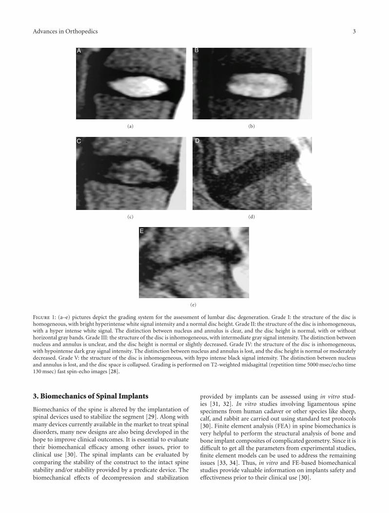
Some authors have devised ways of grading the level ofdisc degeneration (Figure 1; Table 1) in the lumbar spinebased on the MRI images [28].
The level of disc degeneration varies among patientsand so does the type of treatment. The treatment mayrange from conservative treatment such as bed rest andprescription of pain relievers for mild disc degeneration tosurgical intervention in severe chronic degeneration cases.
It is essential to treat DDD to relieve pain andpossibly prevent further degeneration at index level andadjacent levels. Conservative treatment includes chiro-practic adjustments, physical therapy, yoga, acupuncture,and medication. If conservative treatment fails, surgerywould be the next option. Spine surgery involves dif-ferent surgical techniques, using appropriate instrumen-tation to relieve pain. There are many surgical treat-ments such as fusion with or without rigid instrumenta-tion and nonfusion techniques like dynamic stabilization,total disc arthroplasty, and implanting interspinous devices[3].
Advances in Orthopedics 3
(a) (b)
(c) (d)
(e)
Figure 1: (a–e) pictures depict the grading system for the assessment of lumbar disc degeneration. Grade I: the structure of the disc ishomogeneous, with bright hyperintense white signal intensity and a normal disc height. Grade II: the structure of the disc is inhomogeneous,with a hyper intense white signal. The distinction between nucleus and annulus is clear, and the disc height is normal, with or withouthorizontal gray bands. Grade III: the structure of the disc is inhomogeneous, with intermediate gray signal intensity. The distinction betweennucleus and annulus is unclear, and the disc height is normal or slightly decreased. Grade IV: the structure of the disc is inhomogeneous,with hypointense dark gray signal intensity. The distinction between nucleus and annulus is lost, and the disc height is normal or moderatelydecreased. Grade V: the structure of the disc is inhomogeneous, with hypo intense black signal intensity. The distinction between nucleusand annulus is lost, and the disc space is collapsed. Grading is performed on T2-weighted midsagittal (repetition time 5000 msec/echo time130 msec) fast spin-echo images [28].
3. Biomechanics of Spinal Implants
Biomechanics of the spine is altered by the implantation ofspinal devices used to stabilize the segment [29]. Along withmany devices currently available in the market to treat spinaldisorders, many new designs are also being developed in thehope to improve clinical outcomes. It is essential to evaluatetheir biomechanical efficacy among other issues, prior toclinical use [30]. The spinal implants can be evaluated bycomparing the stability of the construct to the intact spinestability and/or stability provided by a predicate device. Thebiomechanical effects of decompression and stabilization
provided by implants can be assessed using in vitro stud-ies [31, 32]. In vitro studies involving ligamentous spinespecimens from human cadaver or other species like sheep,calf, and rabbit are carried out using standard test protocols[30]. Finite element analysis (FEA) in spine biomechanics isvery helpful to perform the structural analysis of bone andbone implant composites of complicated geometry. Since it isdifficult to get all the parameters from experimental studies,finite element models can be used to address the remainingissues [33, 34]. Thus, in vitro and FE-based biomechanicalstudies provide valuable information on implants safety andeffectiveness prior to their clinical use [30].

4 Advances in Orthopedics
Table 1: Table lists the classification of levels of disc degeneration. Adapted from Pfirrmann et al. [28].
Grade StructureDistinction of nucleus
and annulusSignal intensity
Height of intervertebraldisc
I Homogeneous; bright white ClearHyperintense; isointense tocerebrospinal fluid
Normal
IIInhomogeneous with orwithout horizontal bands
ClearHyperintense; isointense tocerebrospinal fluid
Normal
III Inhomogeneous; gray Unclear IntermediateNormal to slightly
decreased
IV Inhomogeneous; gray to black Lost Intermediate to hypointenseNormal to moderately
decreased
V Inhomogeneous; black Lost Hypointense Collapsed disc space
3.1. Fusion Systems. Fusion restricts the motion of involvedsegment. It may reduce progressive degeneration and relievethe patient from back pain. The main clinical indicationsfor fusion are failed conservative treatment, prolonged backpain more than a year, and advanced degenerated disc [3].Fusion surgeries are performed with or without supplementinstrumentation. Segment fusion is achieved through the useof autograft, allograft, bone graft substitute, demineralizedbone matrix (DBM), ceramic-based bone graft, recombi-nant human bone morphogenetic proteins (rhBMP-2), β-tricalcium phosphate (TCP), calcium sulphate (CaS), andhydroxyapatite (HA) [3, 35]. Fusion has been the goldstandard in treating DDD and practiced since the beginningof the 20th century. Fusion without instrumentation hasoften led to nonunion of bone known as pseudoarthrosis.To overcome this complication, many spinal implants havebeen developed which are now used in fusion surgeries. Theusage of spinal instrumentation provides segmental stabilityand facilitates high fusion rates.
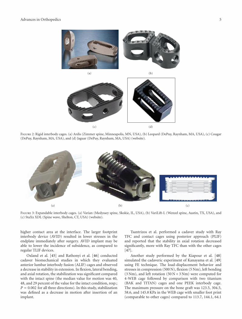
Lumbar interbody fusion (LIF) was introduced byCloward, and currently, it is being used widely [3, 36]. InLIF, cages filled with bone graft are placed in the disc space,supported by instrumentation to stabilize the spine andthereby enhance the fusion process. The bone grafts placedin between the vertebrae experience 80% of compressiveloads, which enhances the fusion process. The grafts in LIFoccupy 90% of bony area in between the vertebrae, whichhas rich vascular supply leading to enhanced fusion [36].The cages were initially designed as rigid systems (Figure 2)in cylindrical, rectangular, and other shapes. However, toovercome the problems associated with these rigid cages [37],expandable cages (Figure 3) have been developed in recenttimes.
There are several implants used as spinal instrumentationin fusion procedures like pedicle screw system and rods,plates (Figure 4), clamps, and wires. Pedicle screw systemis considered to be an effective supportive instrumentationin achieving highest fusion rates [38]. Interspinous fixationsystems (Figure 5) are also currently being developed andare gaining some popularity as their performance is similarto standard pedicle screw system [39, 40]. Interspinousdevices are implanted by minimally invasive procedures inthe posterior region, and they are also used in conjunctionwith interbody fusion procedures.
There are both anterior and posterior approaches forfusion surgery. The posterior approaches include posterolat-eral fusion (PLF), posterior lumbar interbody fusion (PLIF),and transforaminal lumbar interbody fusion (TLIF). Ante-rior approach includes anterior lumbar interbody fusion(ALIF) and extreme lateral inter body fusion (XLIF). XLIF,which is gaining popularity recently, involves lateral accessingof anterior column using sophisticated imaging technologyto avoid neural disruption [41]. This procedure has theadvantage of overcoming the complications associated withPLF, PLIF, TLIF, and ALIF. Combination of both anteriorand posterior approach is called anteroposterior fusion, alsoknown as 360◦ fusion. Depending on the level of surgery,sex of the patient, anatomic variations, and history ofspine surgery, one or combination, of the aforementionedprocedures is selected to treat LBP, and it is surgeon-specific[42].
A biomechanical study was performed by Kiapour et al.[43] using finite element (FE) technique to evaluate the effectof VariLift expandable and BAK cages on biomechanics of thelumbar spine motion segment. The cages were simulated atthe L4-L5 level using PLIF surgical approach. The VariLiftcage depicted comparable biomechanical effects on thelumbar segment with those of BAK cage. The expansionmechanism led to a relatively larger contact area between thecage and the endplate improving the chances of solid fusionto occur after surgery. The expansion of the cage also followsthe lordotic angle of the treated segment ensuring a bettercontact between the cage and endplates.
The footprint size of the interbody fusion device isan important factor that determines the biomechanicalstability afforded by these implants. Moreover, occurrenceof subsidence is also influenced by the cage’s footprint. Afinite element (FE) analysis was conducted by the samegroup [44] to compare the loading and stresses at vertebralendplates following implantation with AVID TLIF cage of alarger foot print compared to regular TLIF cage in differentconfigurations (Figure 6). A follower load of 400 N wasapplied to the spine to simulate compression (at standingposture), and then, a 10 Nm bending moment was applied tothe segment to simulate physiological flexion and extensionloadings. They found that the double TLIF and AVIDcases observed slightly higher normal loads at the endplatescompared to other cases in all loading modes due to their
Advances in Orthopedics 5
(a) (b)
(c) (d)
Figure 2: Rigid interbody cages. (a) Ardis (Zimmer spine, Minneapolis, MN, USA), (b) Leopard (DePuy, Raynham, MA, USA), (c) Cougar(DePuy, Raynham, MA, USA), and (d) Jaguar (DePuy, Raynham, MA, USA) (website).
(a) (b) (c)
Figure 3: Expandable interbody cages. (a) Varian (Medyssey spine, Skokie, IL, USA), (b) VariLift-L (Wenzel spine, Austin, TX, USA), and(c) StaXx XDL (Spine wave, Shelton, CT, USA) (website).
higher contact area at the interface. The larger footprintinterbody device (AVID) resulted in lower stresses in theendplate immediately after surgery. AVID implant may beable to lower the incidence of subsidence, as compared toregular TLIF devices.
Oxland et al. [45] and Rathonyi et al. [46] conductedcadaver biomechanical studies in which they evaluatedanterior lumbar interbody fusion (ALIF) cages and observeda decrease in stability in extension. In flexion, lateral bending,and axial rotation, the stabilization was significant comparedwith the intact spine (the median value for motion was 40,48, and 29 percent of the value for the intact condition, resp.;P = 0.002 for all three directions). In this study, stabilizationwas defined as a decrease in motion after insertion of animplant.
Tsantrizos et al. performed a cadaver study with RayTFC and contact cages using posterior approach (PLIF)and reported that the stability in axial rotation decreasedsignificantly, more with Ray TFC than with the other cages[47].
Another study performed by the Kiapour et al. [48]simulated the cadaveric experiment of Kanayama et al. [49]using FE technique. The load-displacement behavior andstresses in compression (500 N), flexion (5 Nm), left bending(3 Nm), and left rotation (50 N + 3 Nm) were computed for4-WEB cage followed by comparison with two titanium(BAK and TITAN) cages and one PEEK interbody cage.The maximum pressure on the bone graft was 123.5, 304.5,58.6, and 145.8 KPa in the WEB cage with smaller foot print(comparable to other cages) compared to 113.7, 144.1, 64.1

6 Advances in Orthopedics
(a) (b)
(c)
Figure 4: Anterior plate system. (a) Aegis (DePuy, Raynham, MA, USA), (b) Aspida (Alphatec spine, Carlsbad, CA, USA), and (c) Trinica(Zimmer spine, Minneapolis, MN, USA) (website).
(a) (b) (c) (d)
Figure 5: Interspinous fusion devices. (a) CD Horizon spire (Medtronic, Memphis, TN, USA), (b) Aspen (Lanx, Inc., Broomfield, CO, USA),(c) Prima LOK (OsteoMed, Addison, TX, USA), and (d) Axle (X-Spine, Miamisburg, OH, USA) are currently being studied (website).
and 121 KPa in PEEK, 146.4, 132.6, 57.5, and 160 KPa inTITAN, and 30.7, 82.7, 17.7, and 36 KPa in the BAK device.The 4-WEB implanted segment had lesser peak stress at theinterface with bony endplates.
The XLIF surgical procedure was simulated in a FE studyby Kiapour et al. [50] on lumbar-pelvis segment to com-pare the biomechanics of interspinous fixation device withtraditional screw-rod fixation system. Segmental motionand loads on sacroiliac joint (SIJ) and vertebral endplateswere computed for all cases after applying a 400 N ofcompressive load and 10 Nm moment. They reported thatthe placement of fixation constructs leads to a significantdecrease in range of motion of all index levels (L2–L5)in all loadings. At each of implanted levels the motiondecreased by about 95% (Flex), 93% (Ext), 80% (LB), and
90% (LR) in interspinous device implanted model comparedto intact case. The reductions in motion were 97%, 95%,96%, and 94% for screw fixation and 51%, 48%, 68%, and86% for cage alone cases, for same loadings, respectively.Also, the maximum load at SIJ decreased by 4% in Flex andincreased by 8% in Ext, 8% in LB, and 7% in LR for allimplanted cases compared to intact case. In the posteriorplate model, the shear load at endplates of the most superiorimplanted segment increased and decreased in extension andleft bending loadings, respectively compared to other fixationconstructs.
Both cadaver and FE studies evaluated standalone cagesand reported less stabilization of the spine in the literature.Anterior or posterior instrumentation systems along withcages are essential to have proper stability at the implanted

Advances in Orthopedics 7
Double TLIF
Symmetric TLIF
Asymmetric TLIF
AVID
Figure 6: Four configurations of the interbody devices implanted atL4-L5 level in the FE model. (a) double cage TLIF; (b) regular TLIFSymmetrically placed; (c) regular TLIF asymmetrically placed; (d)large footprint TLIF (AVID) [44].
level. However, expandable cages may provide enoughstability without additional instrumentation [43].
The main complication associated with fusion is adjacentsegment degeneration. The reason for this has not yet beenclear and has become the point of debate. Some people arguethat degeneration at adjacent level is part of aging spine,and others argue that it is due to reduced motion resultedfrom fusion. The reasons for adjacent level degeneration canbe hyper mobility, increased disc pressure, increased facetjoint pressure, and alteration in histological properties ofligaments at adjacent level to the index level [51]. Manyin vitro and FEA studies showed the adjacent level hypermobility after fusion [34, 52–56], but there are very fewin vivo studies [51], which showed that the adjacent hypermobility was not significant.
The success of the fusion surgery is defined as achievingarthrodesis across index level to provide stability and relievepain. The modern techniques are successful in achievingfusion in 95% of the cases; however, the pain in the lowback is relieved in less than 70% of the cases [52]. In spiteof its wide application, fusion has varied clinical outcomes[3, 52, 53]. The causes of adjacent segment degeneration werenot clear, though they are attributed to reduced motion atindex level and increased motion at the adjacent level. Inorder to overcome morbidity associated with fusion, motionpreservation devices were developed [52, 57].
The literature review of fusion systems enumerates majordrawbacks like restricted (or) lack of motion, pseudoarthro-sis, adjacent level degeneration, and donor site pain. Theabove shortcomings of fusion have led the researchers todevelop an alternative approach for the treatment of discdegenerative disease.
Many non-fusion techniques have been investigated andhave emerged in recent times to replace the conventionalfusion techniques in treating degenerative discogenic pain.
These techniques include spinal arthroplasty (artificial discand nucleus) and dynamic stabilization systems. Thesesystems aim to provide a more physiologic solution.
3.2. Total Disc Replacement. Disc arthroplasty or total discreplacement is one such option that is being seen asa potential alternative to fusion. As the name suggests,the goal of disc arthroplasty is to completely replace thedegenerated intervertebral disc by an artificial implant whichhas capability not only to treat the pain causing symptomsbut also promises to restore the lumbar motion and createa proper load balance with surrounding tissue withoutcompromising patient safety.
The first human implantation of lumbar artificial discwas performed by Fernstrom in 1966 [58]. He used ametal ball (SKF ball bearing) to reproduce the mechanismof the disc. However, the obtained results were poor, andthe implant was withdrawn. The SB Charite prosthesis, thefirst FDA-approved artificial disc for clinical use in USA,was designed in the former East Germany in the early1980s by Schellnac and Buttner and was first implanted byZippel in 1986 [59]. This event triggered the developmentof several variety of artificial discs aiming on parameterslike restoring natural motion, biocompatibility, corrosionand wear resistance, stability, strength to sustain maximumexpected loads, maintain intervertebral height, preservelordosis, and to restore the energy absorptive qualities of thenative disc. Table 2 lists and Figure 7 depicts some of themajor lumbar artificial disc designs. The present lumbar discdesigns can be classified into four groups:
(i) composite discs: comprise of several articulatingparts; often with different materials (Charite, and-ProDisc);
(ii) hydraulic discs: these are designed for nucleusreplacement and include an expandable fluidenclosed by a woven/porous bag (PDN);
(iii) mechanical discs: which are made of articulatingparts made of single type of material (Maverick,Flexicore, and Kineflex);
(iv) elastic discs: include a deformable cores, usuallymade of elastomers or polymers attached to metallicendplates (Acroflex).
These artificial discs are also classified based on con-straint parameter as constrained, semiconstrained, andunconstrained, respectively. The unconstrained design strat-egy allows for six-degrees-of-freedom segmental motion,with translations and rotations about three independentaxes. Constrained devices typically permit rotation in allplanes and include a fixed center of rotation, which limitssegmental translation under flexion-extension and lateralbending conditions [61].
Biomechanical data from in vitro and mathematicalmodeling are presented. Different biomechanical parameterssuch as segmental motion, instantaneous axis of rotation,intradiscal pressure, facet loads, load/stress distribution atbone-implant interface, and wear at articulating sites, have

8 Advances in Orthopedics
Table 2: Table lists different lumbar artificial discs and respective types of materials and features [60–63] .
Lumbar discsArticulating surfaces
and materialsConstraint Center of rotation Manufacturer
SB Charite Metal-polymer-metal Unconstrained Mobile DePuy Spine, Raynham, MA, USA
Prodisc-L Metal-polymer-metal Semiconstrained Mobile Synthes, West Chester, PA,USA
Maverick Metal-metal Semiconstrained Fixed Medtronic, Minneapolis, MN, USA
Flexicore Metal-metal Fully constrained Fixed Stryker, Kalamazoo, MA, USA
Mobidisc Metal-metal Unconstrained Mobile LDR medical, Troyes, France
Activ-L Metal-polymer-metal Semiconstrained Mobile Aesculap AG Tuttlingen, Germany
Kineflex Metal-metal Semiconstrained Mobile Spinal Motion, South Africa
AcroflexRubber core with
titanium endplates(elastomeric disc)
Unconstrained Mobile DePuy Spine, Raynham, MA, USA
Composite Hydraulic
ProDisc
Mechanical
Maverick Kineflex
Elastic
Charite
Flexicore
PDN (prosthetic disc nucleus)
Acroflex disk
Figure 7: Different lumbar artificial disc concepts: Composite (Charite, Prodisc), Hydraulic (PDN), Mechanical (Maverick, Flexicore,Kineflex), and Elastic (Acroflex) [60].
been analyzed after disc replacements to understand device’sability to mimic the intact disc behavior and predict itsdurability in the long run.
3.2.1. In-Vitro Studies. The in vitro studies enable us tounderstand the effects of total disc arthroplasty (TDA) on thekinematics of the implanted and adjacent levels of the spine.
Hitchon et al. studied the biomechanics of Maverickanterior disc using an in vitro setup with 7 human lumbarspecimens, in which pure moments of 6 Nm were appliedin all planes of rotation after implanting the artificial discat L4-L5 level [64]. They observed that the artificial discdecreased flexibility compared to discectomy, and the motionwas comparable with the intact state.
Rousseau et al. did an in vitro study on twelve humanlumbar spine segments after disc replacement with ProdiscII (6) and Charite III (6) versus intact. They measured thefacet forces and instantaneous axes of rotation (IAR) fordifferent spinal positions under simulated weight-bearing
conditions. They concluded that the degree of constraintaffects postimplantation kinematics and load transfer. Withthe Prodisc (3 DOF), the facets were partially unloaded,though IAR did not match the fixed geometrical center ofthe UHMWPE. The latter observation suggests joint surfaceincongruence is developed during movement. With theCharite disc (5 DOF), the IAR was less variable, yet the facetforces tended to increase, particularly during lateral bending.These results highlight the important role the facets play inguiding movement, and that implant constraint influencesfacet and implant synergy [65].
Ha et al. [62] conducted a study on five L2-S2 spinesin which range of motion, facet strains and intradis-cal pressures were monitored. A 400 N compressive loadand 8 Nm moments in all three planes were applied tocompare the intact, postimplantation of SemiconstrainedActiv-L device at L4-L5 level. They reported that eventhough the device could not restore the normal motionof the intact spine, results of other parameters implicated

Advances in Orthopedics 9
a reduction in the incidence of adjacent segment disease.Those parameters were insignificant decrease of intradiscalpressure at the inferior adjacent disc, and the statisticallysignificant decrease of facet strains at the operative levelduring flexion and strains at the inferior facets in axialrotation.
Goel et al. studied the biomechanics of spine implantedwith Charite disc using a hybrid loading protocol [66].They employed both in vitro experiment and finite elementmodeling. Results indicated that the Charite artificial discplacement slightly increased motion at the implanted level,with a resultant increase in facet loading when compared tothe adjacent segments. The motions and loads were less atthe adjacent levels.
Most of the lumbar artificial discs are of articulatingtype. These have potential for wear, much like the hip andknee arthroplasties. Cyclic loading and relative motion at thebearing surface may increase the risk to surrounding spinalstructures like spinal cord and blood vessels. Therefore,biotribological tests serve as an effective preclinical toolto investigate device wear characteristics. A wear rate of1.1 mg/million cycles [67] has been reported for the Chariteartificial disc. Pare et al. [68] reported a steady state wear rateof 0.33 ± 0.12 mm3/million cycles in flexion-extension and0.43± 0.06 mm3/million cycles in combined motion tests forthe metal-on-metal Maverick disc (constrained).
3.2.2. FE Analyses. A finite element study was conductedby Rohlmann et al. to understand the effects of ProDiscon lumbar spine kinematics. They loaded their modelwith the upper body weight and muscle forces to simulatestanding, 30-degree flexion, 15-degree extension, and 6-degree axial rotation. The disc position was varied by upto 2 mm in both the anterior and posterior direction.Three different disc heights were investigated as well asthe influence of removing different portions of the naturaldisc and resuturing the ALL ligaments. They observed thatimplant position strongly influenced intersegmental rotationfor the loading cases of standing and flexion. Also, theyfound that a disc height 2 mm in excess of the normal discspace increased intersegmental rotation at implanted levelduring standing and extension. The intersegmental rotationswere closer to the intact spine, when lateral portions ofthe annulus were not removed. Finally they concluded thatwhen implanting an artificial disc, great care should betaken in choosing the optimal height and correct positionfor the implant. Lateral portions of the annulus should bepreserved whenever possible. A perfect reconstruction ofthe ALL would help restore the biomechanics to normal[69].
Moumene and Geisler [70] performed a study to evaluatethe loading on the facet joints and stress on the polyethylenecore after implantation of Charite (unconstrained) andProdisc (Semiconstrained) TDA. The unconstrained TDAunloads the facet joints and presents decreased core stress ascompared to the fixed-core Semiconstrained TDA.
In a computational study performed by Dooris et al.[71], the effects of facet load sharing following TDA were
Posterior
Anterior
Single peakloading
Double peakloading
0.514
0.48
0.446
0.411
0.377
0.343
0.309
0.274
0.24
0.206
0.171
0.137
0.103
0.069
0.034
0
(mm
)
Figure 8: Linear wear contour predicted for ProDisc-L using finiteelement technique. Adapted from [62].
examined. Different annular window sizes and varied antero-posterior artificial disc placement was simulated for a ball-on-socket disc design by Medtronic. Findings demonstratedthat an artificial disc can alter spinal bending stiffness inthe sagittal plane. Changes in spinal stiffness were notedto be dependent on the position of the disc and degree ofannular resection. Anterior placement of the device led toincreased facet joint loads in compression and extension.These findings suggest that if the anterior longitudinalligament is preserved and the implant is placed posteriorlywithin the disc, the spinal stiffness will be restored, and facetloads will be maintained at preimplantation levels.
A study performed by Denoziere and Ku [72] to compareTDA and fusion at one level of lumbar spine indicated thatthe level implanted with the artificial disc showed excessiveligament tensions (greater than 500 N), high facet pressures(greater than 3 MPa), and a higher risk of instability.The mobility and the stresses in the level adjacent to thearthroplasty also increased. They concluded that there wasa greater risk of instability and further degeneration forartificial disc implanted model than that predicted for thefused model.
FE models have also been utilized to understand wearcharacteristics of joint replacements in the hip and knee.FE-based wear study was conducted by Rawlinson et al.[73], which depicted a uniformly distributed wear patternas per ISO 18192 which was not observed during theretrieval analysis (Figure 8). This study was validated againstexperimental wear simulation of ProDisc-L implant.
FE technique was also applied in cervical spine byBhattacharya et al. [74] to evaluate wear in a simulated C5-C6 FSU. A predictive FE wear model of the artificial discalone (TDR only) was developed, and it was implanted intoC5-C6 FE model (TDR + FSU). Both of these models weresubjected to a motion profile (rotation about three axes)

10 Advances in Orthopedics
with varying preloads of 50 to150 N at 1 Hz, consistent withISO 18192. A subroutine based on Archard law simulatedabrasive wear on the polymeric core up to 10 millioncycles. The TDR + FSU model was further modified tosimulate facetectomy, sequential addition of ligaments, andcompressive load. They reported more predicted localizedwear in certain regions for TDR + FSU, in contrast to theuniformly distributed wear pattern of the TDR-only model.In addition, the cumulative volumetric wear for the TDR-only model was 10 times that of the TDR + FSU model.The TDR + FSU model also revealed a separation at thearticulating interface during extension and lateral bending.After facetectomy, the wear pattern remained lopsided, butlinear wear increased eight-fold, whereas volumetric wearalmost tripled. This was accompanied by a reduction inobserved liftoff.
Similar kind of studies in the lumbar spine may enablethe scientists to pursue and understand the effects of clinicaland other parameters (like surgical variables, different load-ing profiles, different disc designs, and bone quality) on wearof lumbar artificial discs.
3.2.3. In Vivo Studies. In the in vivo study of Siepe etal. [75], 175 patients with disc replacement with meanfollowup of 29.3 months were investigated. Facet joint pain,predominantly at the index level, was identified in 22 patients(12.6%). The sacroiliac joint was also a frequent cause ofpost-operative pain (n = 21; 12.0%). Pain from bothstructures influenced all outcome parameters negatively (P <0.05). Patients with an early onset of pain ≤ 6 months) were2–5 fold higher at risk of developing persisting complaintsand unsatisfactory outcome at later stages in comparisonto the entire study cohort (P < 0.05). They also observedthat the level of TDR significantly influenced postoperativeoutcome. Best results were achieved for the TDRs at L4/5(incidence of posterior joint pain: 14.8%). Inferior outcomeand a significantly higher incidence of posterior joint painwere observed for TDR at L5/S1 (21.6%) and bisegmentalTDR at L4/5/S1 (33.3%), respectively. Their study was unableto address that TDR will reduce the incidence of posteriorjoint pain, unlike the lumbar fusion procedures.
Zigler [76] did a clinical study on 78 patients with min-imum 6-month followup replacement of ProDisc. Amongthe patients, 54 also had a 1-year followup, enrolled in aprospective randomized FDA study evaluating the safety andefficacy of ProDisc II versus control, a 360-degree lumbarspinal fusion. At 6-month follow-up, there were 55 ProDiscpatients out of which 23 underwent fusion. Both fusion anddisc replacement group had similar clinical outcomes. Also atrend was identified at 6 months in patient satisfaction ratesfavoring ProDisc versus fusion (P = 0.08), which were notsignificant at 1-year follow-up period. Similar clinical studiesand randomized trials have been conducted in the past toevaluate the performance of an artificial disc in terms ofsafety and efficacy [77–83].
Based on the above studies, increased facet joint loading,increased lordosis at the implanted level, hyper mobility, andwear at articulating surfaces are the major issues with TDA
and need further investigations. Even though the short-termresults are promising [76], the long-term complications andbenefits of TDA are yet to be realized, especially in terms ofpreventing adjacent level disc degeneration [84, 85]. Hence,it cannot be concluded that total disc replacement is superiorto spinal fusion in terms of clinical outcome, at least atpresent.
3.3. Dynamic Stabilization Systems. Spinal fusion surgeriesaim at limiting the motion of the segment and restoringthe stability. Anterior lumbar disc replacements are used torestore spinal alignment and kinematics of a degenerated seg-ment. Compared to fusion of the segment, disc replacementsmay prevent adjacent segment degeneration. To resolve someof the deficiencies of anterior lumbar arthroplasty, such asthe approach itself, difficulty of revision, and postoperativefacet pain, 360◦ motion preservation systems based onposterior disc and posterior dynamic stabilization system(PDS) designs are being pursued [86].
Dynamic stabilization systems aim at altering favorablythe movement and load transmission through the spinalmotion segment [87]. The hypothesis behind dynamicstabilization system is that control of abnormal motion andmore physiologic load transmission would relieve pain andprevent adjacent segment degeneration.
The biomechanical action of a dynamic stabilizationsystem is two-fold: (i) permit or restore “normal” motionand (ii) share load with the disc and the facets. The loadsharing should be more or less uniform during the entirerange of motion. This implies that the kinematics of thesegment stabilized with a dynamic system should be similarto the intact spine. This is achieved when the location ofthe instantaneous axis of rotation of the construct lies closeto the intact segment [87]. There are two types of dynamicstabilizations systems currently available: dynamic pediclescrew-based systems and interspinous spacers.
3.3.1. Dynamic Pedicle Screw-Based Systems. Some flexiblestabilization systems consist of pedicle screws threaded intoadjacent segments and a member spanning between theheads of the pedicle screws to limit the movements of thespinal segment.
In 1994 Henri Graf, (Lyon, France) introduced the Grafligament, designed to provide less stressful load sharing.It consists of a nonelastic band as a ligament to connectthe pedicle screws across the segment to be stabilized tolock the segment in full lordosis. The concept was thatabnormal rotatory movement causes instability and lockingthe facets would control the rotation movement. The systemwould allow for limited flexion and no rotatory motion. Theligaments get lax in extension; hence there is no restriction inthe motion [88].
The fulcrum assisted soft stabilization system (FASSsystem) was developed to address the disadvantages of theGraf ligament. In this system, a fulcrum is placed betweenthe pedicle screws in front of the ligament. The fulcrumdistracts the posterior annulus. When the elastic ligament isplaced posterior to the fulcrum to compress the pedicle screw

Advances in Orthopedics 11
heads, the fulcrum transforms this posterior compressionforce into an anterior distraction force, which distracts theanterior annulus. The lordosis is not dependent on thepatient’s ability but is created by the tension in the ligament.Experimental studies have shown that the implant unloadsthe disc, but the flexibility of the segment is lost as greaterunloading of the disc occurs by the adjustment of the tensionin the ligament and the fulcrum [88].
The Dynesys system (dynamic neutralization system) wasdeveloped by Gilles and Muller. Dynesys system comprisesof three components: (i) pedicle screws, (ii) polyethylene-terephthalate (PET) ligaments, and (iii) polycarbonate ure-thane (PCU) spacers. The spacers are bilaterally placedbetween the pedicle screw heads to withstand compressiveloads. The ligaments are run through the hollow core of thespacers. A tensile preload of about 300 N is used to stabilizethe construct [88]. The plastic cylinder between the screwheads limits the degree of lordosis that can be created. Asthe ligament is not elastic, flexion compresses the disc, andthe axis of flexion is the posterior ligament, which is wellposterior to the normal axis of flexion [88]. Active extensionwill open up the anterior annulus without compression ofthe posterior annulus. Theoretically, lordosis can be achievedby the action of the spinal extensor muscles; in extension thecylinder will take increasing load [79]. Thus, the principle ofthe system is its ability to create load sharing and restorationof disc height, not necessarily motion preservation becausethe system is rigid [87].
The Cosmic system is a pedicle screw-based dynamicinstrumentation system (Ulrich, Ulm, Germany) equippedwith a hinge between the screw head and threaded portion.Cosmic is a load sharing system which reduces mechanicalstress on the implants. Thus, protection against implantfailure and loosening is achieved. The hinged screw allowsonly for axial load, due to this, it is important to have a largelyintact anterior column for implantation of this system. WhileDynesys stabilizes by neutralizing motion, Cosmic correctsthe sagittal plane and maintains motion in flexion/extension.
The Isobar TTL is another novice device in this category,comprised of a titanium alloy rod and a dampener element.The dampener element is formed of a series of helical springsthat allow linear and angular motion and serve as a shockabsorber. This instrumentation allows flexion-extension andaxial rotation, while lateral bending is restricted. A lordoticangle is also incorporated into this system. Benefits associ-ated with this device include ease of implantation, motionsegment stabilization, maintenance of lordotic angle, loadsharing, and conformance to the IAR of the motion segment[89]. Other notable devices in this category include theAxient, BioFlex, TalinRod, CD Horizon Agile, and Stabilimaxsystems. Figure 9 depicts some of these implants.
In Vitro Studies. The Dynesys stabilization system has beenwidely studied. Freudiger et al. tested the Dynesys systemon four cadaveric spine specimens on a lumbar spinesimulator, which allowed the simultaneous application ofbending moments, and compressive and shear loads. Theyconcluded that the Dynesys reduces flexion and extensionangles significantly [90].
Aylott et al. investigated the stresses of the intervertebraldiscs at the instrumented and the adjacent segments undercompressive loading (1 kN) in flexion (6◦) and extension(4◦), in an in vitro study. The effects of spacer height onthe intradiscal pressure distribution were also evaluated.They observed that Dynesys eliminated the peak stresses inthe anterior annulus in flexion and in extension. The peakannulus stresses increased with decrease in the spacer height.However, there was no change in the stresses in the adjacentsegment discs [91].
Niosi et al. (2004) conducted an in vitro biomechanicalstudy to investigate the effect of spacer length of Dynesyson the range of motion. The test conditions included intact,injury at L3-L4, and Dynesys at L3-L4 (standard spacer, longspacer, and short spacer). They quantified range of motionand facet contact loads for a pure moment of ±7.5 Nm withand without a preload of 600 N. The trends in motion weresimilar with and without preload. Long spacer reduced themotion more than other two cases, the contact loads of thelong and short spacer were 150% and 64% of the standardspacer, respectively [93].
Wilson et al. investigated 10 cadaveric lumbar spine spec-imens, subjected to pure moments of ±7.5 Nm (axial rota-tion, flexion, and extension) to compare range of motion andfacet loads of intact specimens with those of injured speci-mens stabilized with Dynesys. The facet loads were measuredusing thin film electroresistive pressure sensors. They foundthat the facet loads decreased in axial rotation after implanta-tion of Dynesys. In extension, they were similar to the intactspine, and no significant difference compared to the intactcase. They, however, found that the facet loads were signifi-cantly higher in flexion with the Dynesys due to device com-pression. It was found that the Dynesys system reduced spinalmotion from intact and decreased peak facet loading [94].
In addition to this, Schmoelz et al. [95] comparedDynesys to a rigid fixation system. They concluded thatDynesys provides substantial stability in case of degenerativepathologies and can replace conventional fusion surgery inthese indications, while the motion segment is preserved.
FE Analyses. Rohlmann et al. studied the intersegmentalrotations and intradiscal pressures in a degenerated disc afterimplanting the posterior dynamic implant in a FE-basedstudy [96]. Motion at the implanted level decreased, and itslightly increased at the adjacent level. Intradiscal pressurewas also decreased at the injured level with the implant.There is no much effect on IDP at the adjacent level with theimplant.
In a study performed by Parepalli [97], rigid rod(fusion) system was compared with AXIENT to evaluatethe parameters like range of motion, intradiscal pressure,and facet loads of the implanted and adjacent levels. Theyfound that AXIENT restored kinematics of the degeneratedspine close to normal thanthat with the fusion device (forgrade I and grade II degenerated spine). AXIENT was ableto restore the kinematics of degenerated spine at the adjacentlevels where as fusion increased segmental motion beyondthe intact. Also, stresses in pedicle screws were more for rigid

12 Advances in Orthopedics
(a) (b) (c)
(d) (e) (f)
(g)
Figure 9: Posterior dynamic stabilization systems. (a) Graf system; (b) Dynesys; (c) IsoBar; (d) AccuFlex; (e) Stabilimax; (f) PercuDyn; (g)Transition [92].
system compared to the AXIENT system implicating less riskof screw breakage for AXIENT system. Vishnubhotla et al.[98] performed a study in which FE analysis has been usedto assess the kinematics of a motion segment instrumentedwith (i) Rigid screw rod system (used infusion), (ii) Rigidscrew system with flexible rod (Nitinol; super elastic), (iii)Dynesys (Zimmer, Inc.) a pedicle screw-based dynamicstabilization system, (iv) Cosmic (Ulrich, Ulm. Germany)a pedicle screw based hinged dynamic stabilization system,and (v) Wallis (Spinal Concepts, Inc.) an interspinousbased dynamic stabilization system. They reported that thedynamic stabilization systems are more flexible than rigidsystems but not flexible enough to say that they preservemotion. However, the evaluation of the IAR indicates thatthe Dynesys system achieves kinematics closer to that of theintact spine while restricting motion.
Another study was performed by Goel et al. [99] toevaluate the biomechanical performance of the Dynesys
dynamic stabilization system as a function of graded face-tectomies, including complete bilateral facetectomies. Anexperimentally validated FE model was used to compare thebiomechanics of L3-S1 lumbar spine with graded facetec-tomy (50%, 75%, and total bilateral medial facetectomy) atL4-L5 before and after placement of Dynesys versus intact.A 400 N compressive follower load plus a 10 Nm bendingmoment were applied to all models to simulate physio-logically relevant motions in all planes. Results depictedthe Dynesys dynamic stabilization system constrains themotion of the decompressed segment similar to a rigidsystem. They reported that multiple grades of facetectomyshow minimal effects on the kinematics of the stabilizedsegment in all loading cases, except in axial rotation (AR).In total facetectomy case, increased motion and elevatedpedicle screw stresses were observed in AR as compared tothe intact-stabilized case. Higher screw stresses in AR for50% facetectomies may accelerate screw loosening/failure

Advances in Orthopedics 13
(a) (b) (c) (d)
Figure 10: Interspinous spacers. (a) Wallis system; (b) DIAM system; (c) X-stop; (d) Coflex [92].
especially in combination with other motions like flex-ion/extension during daily activities.
3.3.2. Interspinous Spacers. The interspinous distractiondevices are floating devices, which are not rigidly connectedto the vertebrae. The interspinous spacers are designed to offload the posterior disc and the facet joint, by distracting thespinous processes [98]. There are several interspinous-baseddevices.
The Weiss springs consist of springs anchored to thelamina; the indication for the usage of this system is forfracture and deformity applications [100]. This system wasmodified further to consist of a rod portion attached tothe spinous process using bands; these rods were meant tocontrol rotation. A comparison study with the Harringtondistraction rods concluded that modified Weiss springs oftenmaintain better spinal stability [101].
The X-stop is intended to provide a minimally invasive,nonfusion, alternative to current treatments for degen-erative lumbar spinal stenosis from L2–L5 levels, whichinclude medical management, epidural steroid injections,and decompressive laminectomy with or without fusion. TheX-stop is made of high strength titanium alloy and consistsof two parts. The device is introduced between the spinousprocesses of adjacent level vertebral bodies and is held inplace by the supraspinous ligament keeping the segment ina slightly flexed position. Due to the slightly flexed position,the nerves get decompressed thus providing relief from pain.
French orthopedic surgeon Jean Taylor developedthis device. The device for intervertebral-assisted motion(DIAM) system consists of a polymeric interspinous spacer,with extended wings to act as a posterior shock-absorbingdevice. It consists of a flexible spacer and dual independentligaments, which attach the spacer to the spinous processabove and below, transferring some of the axial load to theposterior elements in flexion and extension (Figure 9). Theflexible spacer is made with an inert medical-grade siliconecore material, and the ligament is made of Graf/Senegas liga-ment. The surgical procedure involved for the DIAM deviceis to distract the spinous process to place the spacer and thento insert each ligament into the adjacent interspinous space.There is minimal wear debris seen in the DIAM, since there
are no articulating surfaces. Other notable devices in this cat-egory include Coflex. Figure 10 depicts some of these spacers.
There are few biomechanical and clinical studies showingthe effectiveness of these kinds of devices. Wilke et al.did a biomechanical study on X-stop, Wallis, Coflex, andDIAM devices to assess the flexibility, stability provided,and the effect on intradiscal pressure after implanting thesedevices. They found that all the devices provided stabilityin extension, but there was no difference for flexion, lateralbending, and axial rotation. The intradiscal pressure droppedin extension and led to no difference in other mentionedloading modes [102].
Six human cadaveric motion segments were subjected tocomplex cyclic loading to determine the risk of interspinousspacer (Superion, VertiFlex Inc, CA, USA) device migrationand to assess damage on the device and specimen underextreme coupled motion [103]. Motion segments withinterspinous spacer were tested for 5-degree extension/10-degree flexion coupled with an axial rotation of±3-degree upto 57600 million cycles. CT images were taken for specimensin neutral, 5-degree extension, and 10-degree flexion beforeand after the implantation of the spacer. Vertebral foramenand canal dimensions were quantified. Results have shownno device migration or subsidence. Specimens did notsustain any significant injury during testing. Canal areawas minimally altered and foramen height, width, and areaincreased in extension and were statistically significant ascompared to intact. It was concluded that interspinous spacereffectively prevents the motion at the implanted level anddoes not change the anatomy significantly.
Kabir et al. conducted a review study to find out theclinical and biomechanical evidences of interspinous devicesafety, effectiveness to suggest the clinical indications forthese kinds of devices. They reviewed articles related tothe aforementioned 4 interspinous spacers. They foundthat most of the studies were conducted related to X-stop, and a few studies, both biomechanical and clinical,were conducted related to other devices. In biomechanicalpoints of view, all the devices have a beneficial effect on thekinematics of spine. The authors found these implants tobe very effective in comparison to conservative treatments.They could not suggest clinical indications for interspinous

14 Advances in Orthopedics
devices because of varied outcomes, and a small numberof studies conducted so far [63, 104]. In spite of the variedresults of these interspinous devices, the author found theseimplants are effective in treating stenosis when comparedto conservative treatment. The authors suggested the needfor randomized controlled studies to evaluate these devicesand to revise clinical indications for these kinds of devices[104].
4. Conclusion
In contrast to the previous paradigms of rigid fixation,new technologies aim to restore and preserve motion whileenabling a proper load sharing. In theory, proper loadsharing and restoration of physiologic motion will reducethe probability of adjacent segment disease. Current focus ofresearch efforts emphasizes long-term evaluation of devicesand validation of theoretical and experimental benefits ina clinical setting. In addition to bench-top testing, well-designed, randomized clinical trials are needed to achievethese goals.
References
[1] M. T. Modic and J. S. Ross, “Lumbar degenerative diskdisease,” Radiology, vol. 245, no. 1, pp. 43–61, 2007.
[2] W. T. Crow and D. R. Willis, “Estimating cost of care forpatients with acute low back pain: a retrospective reviewof patient records,” The Journal of the American OsteopathicAssociation, vol. 109, no. 4, pp. 229–233, 2009.
[3] C. Schizas, G. Kulik, and V. Kosmopoulos, “Disc degenera-tion: current surgical options,” European Cells & Materials,vol. 20, pp. 306–315, 2010.
[4] S. D. Kuslich, C. L. Ulstrom, and C. J. Michael, “The tissueorigin of low back pain and sciatica: a report of pain responseto tissue stimulation during operations on the lumbar spineusing local anesthesia,” Orthopedic Clinics of North America,vol. 22, no. 2, pp. 181–187, 1991.
[5] H. S. An, P. A. Anderson, V. M. Haughton et al., “Introduc-tion. Disc degeneration: summary,” Spine, vol. 29, no. 23, pp.2677–2678, 2004.
[6] V. K. Goel and Y. E. Kim, “Effects of injury on the spinalmotion segment mechanics in the axial compression mode,”Clinical Biomechanics, vol. 4, no. 3, pp. 161–167, 1989.
[7] P. P. Raj, “Inter vertebral disc: anatomy-physiology-pathophysiology-treatment,” Pain Practice, vol. 8, no. 1, pp. 18–44,2008.
[8] K. J. Schnake, M. Putzier, N. P. Haas, and F. Kandziora,“Mechanical concepts for disc regeneration,” European SpineJournal, vol. 15, no. 3, pp. S354–S360, 2006.
[9] M. A. Adams and P. J. Roughley, “What is intervertebral discdegeneration, and what causes it?” Spine, vol. 31, no. 18, pp.2151–2161, 2006.
[10] J. P. G. Urban and S. Roberts, “Degeneration of the interver-tebral disc,” Arthritis Research and Therapy, vol. 5, no. 3, pp.120–130, 2003.
[11] H. J. Wilke, P. Neef, M. Caimi, T. Hoogland, and L. E. Claes,“New in vivo measurements of pressures in the intervertebraldisc in daily life,” Spine, vol. 24, no. 8, pp. 755–762, 1999.
[12] F. Heuer, H. Schmidt, and H. J. Wilke, “The relation betweenintervertebral disc bulging and annular fiber associated
strains for simple and complex loading,” Journal of Biome-chanics, vol. 41, no. 5, pp. 1086–1094, 2008.
[13] M. A. Adams, D. S. McNally, and P. Dolan, “’Stress’distributions inside intervertebral discs. The effects of ageand degeneration,” Journal of Bone and Joint Surgery—SeriesB, vol. 78, no. 6, pp. 965–972, 1996.
[14] H. J. Wilke, F. Rohlmann, C. Neidlinger-Wilke, K. Werner, L.Claes, and A. Kettler, “Validity and interobserver agreementof a new radiographic grading system for intervertebral discdegeneration: part I. Lumbar spine,” European Spine Journal,vol. 15, no. 6, pp. 720–730, 2006.
[15] H. Schmidt, A. Kettler, A. Rohlmann, L. Claes, and H. J.Wilke, “The risk of disc prolapses with complex loadingin different degrees of disc degeneration—a finite elementanalysis,” Clinical Biomechanics, vol. 22, no. 9, pp. 988–998,2007.
[16] V. K. Goel and J. N. Weinstein, Biomechanics of the Spine:clinical and Surgical Perspective, CRC Press, Boca Ranton, Fla,USA, 1990.
[17] H. T. Keller and S. Holm, “The load of the porcine lumbarspine during seated whole body vibration,” in Proceedingsof the 14th International Society for the Study of the LumbarSpine, p. 30, Rome, Italy, 1987.
[18] V. K. Goel, Y. E. Kim, and F. Zhang, “Biomechanical effectsof vibration on the human spine,” in Proceedings of the15th International Society for the Study of the Lumbar Spine,Miami, Fla, USA, 1988.
[19] M. H. Pope, A. M. Kaigle, M. Magnusson, H. Broman,and T. Hansson, “Intervertebral motion during vibration,”Proceedings of the Institution of Mechanical Engineers, Part H,vol. 205, no. 1, pp. 39–44, 1991.
[20] J. L. Kelsey, P. B. Githens, and A. A. White, “An epidemiologicstudy of lifting and twisting on the job and risk for acuteprolapsed lumbar intervertebral disc,” Journal of OrthopaedicResearch, vol. 2, no. 1, pp. 61–66, 1984.
[21] W. Z. Kong, V. K. Goel, L. G. Gilbertson, and J. N. Weinstein,“Effects of muscle dysfunction on lumbar spine mechanics: afinite element study based on a two motion segments model,”Spine, vol. 21, no. 19, pp. 2197–2207, 1996.
[22] S. Wang, Q. Xia, P. Passias, W. Li, K. Wood, and G. Li,“How does lumbar degenerative disc disease affect the discdeformation at the cephalic levels in vivo?” Spine, vol. 36, no.9, pp. E574–E581, 2011.
[23] W. Li, S. Wang, Q. Xia et al., “Lumbar facet joint motionin patients with degenerative disc disease at affected andadjacent levels: an in vivo biomechanical study,” Spine, vol.36, no. 10, pp. E629–E637, 2011.
[24] M. M. Panjabi, M. H. Krag, and T. Q. Chung, “Effects of discinjury on mechanical behavior of the human spine,” Spine,vol. 9, no. 7, pp. 707–713, 1984.
[25] M. Mimura, M. M. Panjabi, T. R. Oxland, J. J. Crisco, I.Yamamoto, and A. Vasavada, “Disc degeneration affects themultidirectional flexibility of the lumbar spine,” Spine, vol.19, no. 12, pp. 1371–1380, 1994.
[26] D. S. McNally and M. A. Adams, “Internal intervertebral discmechanics as revealed by stress profilometry,” Spine, vol. 17,no. 1, pp. 66–73, 1992.
[27] A. Shirazi-Adl, A. M. Ahmed, and S. C. Shrivastava, “A finiteelement study of a lumbar motion segment subjected to puresagittal plane moments,” Journal of Biomechanics, vol. 19, no.4, pp. 331–350, 1986.
[28] C. W. A. Pfirrmann, A. Metzdorf, M. Zanetti, J. Hodler,and N. Boos, “Magnetic resonance classification of lumbar

Advances in Orthopedics 15
intervertebral disc degeneration,” Spine, vol. 26, no. 17, pp.1873–1878, 2001.
[29] V. K. Goel and M. H. Pope, “Biomechanics of fusion andstabilization,” Spine, vol. 20, no. 24, pp. 85S–99S, 1995.
[30] H. J. Wilke, K. Wenger, and L. Claes, “Testing criteria forspinal implants: recommendations for the standardization ofin vitro stability testing of spinal implants,” European SpineJournal, vol. 7, no. 2, pp. 148–154, 1998.
[31] V. K. Goel, N. A. Thomas, C. R. Charles, N. K. Weinstein, andN. James, “A technique to evaluate an internal spinal deviceby use of the selspot system: an application to luque closedloop,” Spine, vol. 12, no. 2, pp. 150–159, 1987.
[32] V. K. Goel, D. G. Wilder, M. H. Pope, W. T. Edwards,R. F. McLain, and S. D. Boden, “Biomechanical testing ofthe spine: load-controlled versus displacement- controlledanalysis,” Spine, vol. 20, no. 21, pp. 2354–2357, 1995.
[33] V. K. Goel, J. N. Grauer, T. C. Patel et al., “Effects of Chariteartificial disc on the implanted and adjacent spinal segmentsmechanics using a hybrid testing protocol,” Spine, vol. 30, no.24, pp. 2755–2764, 2005.
[34] J. N. Grauer, A. Biyani, A. Faizan et al., “Biomechanics of two-level Charite artificial disc placement in comparison to fusionplus single-level disc placement combination,” Spine Journal,vol. 6, no. 6, pp. 659–666, 2006.
[35] K. G. Abdullah, M. P. Steinmetz, E. C. Benzel, and T. E. Mroz,“The state of lumbar fusion extenders,” Spine, vol. 36, no. 20,pp. E1328–E1334, 2011.
[36] J. C. Wang, P. V. Mummaneni, and R. W. Haid, “Currenttreatment strategies for the painful lumbar motion segment:posterolateral fusion versus interbody fusion,” Spine, vol. 30,no. 16, pp. S33–S43, 2005.
[37] N. N. Bhatia, K. H. Lee, C. N. H. Bui, M. Luna, G. M. Wahba,and T. Q. Lee, “Biomechanical evaluation of an expandablecage in single segment posterior lumbar interbody fusion,”Spine, vol. 37, no. 2, pp. E79–E85, 2011.
[38] J. C. Wang, R. W. Haid Jr., J. S. Miller, and J. C. Robinson,“Comparison of CD HORIZON SPIRE spinous process platesystem stabilization and pedicle screw fixation after anteriorlumbar interbodyfusion: invited submission from the JointSection Meeting on Disorders of the Spine and PeripheralNerves,” Journal of Neurosurgery. Spine, vol. 4, pp. 160–164,2006.
[39] J. C. Wu and P. V. Mummaneni, “Using lumbar interspinousanchor with transforaminal lumbar interbody fixation,”World Neurosurgery, vol. 73, no. 5, pp. 471–472, 2010.
[40] D. G. Karahalios, T. Kaibara, R. W. Porter et al., “Biomechan-ics of a lumbar interspinous anchor with anterior lumbarinterbody fusion,” Journal of Neurosurgery. Spine, vol. 12, no.4, pp. 372–380, 2010.
[41] J. A. Youssef, P. C. McAfee, C. A. Patty et al., “Minimallyinvasive surgery: lateral approach interbody fusion: resultsand review,” Spine, vol. 35, pp. S302–S311, 2010.
[42] M. Djurasovic, S. D. Glassman, J. R. Dimar, J. M. Howard, K.R. Bratcher, and L. Y. Carreon, “Does fusion status correlatewith patient outcomes in lumbar spinal fusion?” Spine, vol.36, no. 5, pp. 404–409, 2011.
[43] A. Kiapour, A. M. Kiapour, M. K. Kodigudla, S. Mishra, andV. K. Goel, Biomechanics of a Novel Expandable InterbodyCage Design, Orthopedic Research Society, Poster presenta-tion, San Francisco, Calif, USA, 2012.
[44] A. Faizan, A. Kiapour, A. M. Kiapour, and V. K. Goel,Effect of Interbody Cage’s Footprint on the Endplate StressesImmediatelyafter TLIF Surgery: A Finite Element Based Study,
Orthopedic Research Society, SanFrancisco, Calif, USA,2012.
[45] T. R. Oxland, Z. Hoffer, T. Nydegger, G. C. Rathonyi, andL. P. Nolte, “A comparative biomechanical investigationof anterior lumbar interbody cages: central and bilateralapproaches,” Journal of Bone and Joint Surgery—Series A, vol.82, no. 3, pp. 383–393, 2000.
[46] G. C. Rathonyi, T. R. Oxland, U. Gerich, S. Grassmann, andL. P. Nolte, “The role of supplemental translaminar screws inanterior lumbar interbody fixation: a biomechanical study,”European Spine Journal, vol. 7, no. 5, pp. 400–407, 1998.
[47] A. Tsantrizos, H. G. Baramki, S. Zeidman, and T. Steffen,“Segmental stability and compressive strength of posteriorlumbar interbody fusion implants,” Spine, vol. 25, no. 15, pp.1899–1907, 2000.
[48] A. Kiapour, V. K. Goel, L. Ferrara, and J. Hunt, SubsidenceEvaluation of 4-WEB, a Novel Cross Strut Based, InterbodyCage Design, Poster presentation, International Society forthe Study of the Lumbar Spine (ISSLS), Gothenburg, Sweden,2011.
[49] M. Kanayama, B. W. Cunningham, C. J. Haggerty, K. Abumi,K. Kaneda, and P. C. McAfee, “In vitro biomechanicalinvestigation of the stability and stress-shielding effect oflumbar interbody fusion devices,” Journal of Neurosurgery,vol. 93, no. 2, pp. 259–265, 2000.
[50] A. Kiapour, J. O’Donnell, V. K. Goel, and A. Biyani,Comparison of Biomechanics of Lumbar-Pelvis Segment withPosterior Screw-Rod Versus Interspinous Plate Fixation System,Poster Presentation, ASME, Vancouver, Canada, 2010.
[51] B. Cakir, C. Carazzo, R. Schmidt, T. Mattes, H. Reichel,and W. Kafer, “Adjacent segment mobility after rigid andsemirigid instrumentation of the lumbar spine,” Spine, vol.34, no. 12, pp. 1287–1291, 2009.
[52] M. Panjabi, G. Henderson, C. Abjornson, and J. Yue,“Multidirectional testing of one- and two-level ProDisc-Lversus simulated fusions,” Spine, vol. 32, no. 12, pp. 1311–1319, 2007.
[53] M. Panjabi, G. Malcolmson, E. Teng, Y. Tominaga, G.Henderson, and H. Serhan, “Hybrid testing of lumbarCHARITI discs versus fusions,” Spine, vol. 32, no. 9, pp. 959–966, 2007.
[54] D. H. K. Chow, K. D. K. Luk, J. H. Evans, and J. C. Y.Leong, “Effects of short anterior lumbar interbody fusion onbiomechanics of neighboring unfused segments,” Spine, vol.21, no. 5, pp. 549–555, 1996.
[55] S. I. Esses, B. J. Doherty, M. J. Crawford, and V. Dreyzin,“Kinematic evaluation of lumbar fusion techniques,” Spine,vol. 21, no. 6, pp. 676–684, 1996.
[56] K. Y. Ha, M. J. Schendel, J. L. Lewis, and J. W. Ogilvie, “Effectof immobilization and configuration on lumbar adjacent-segment biomechanics,” Journal of Spinal Disorders, vol. 6,no. 2, pp. 99–105, 1993.
[57] T. S. Videbaek, N. Egund, F. B. Christensen, A. Grethe Jurik,and C. E. Bunger, “Adjacent segment degeneration afterlumbar spinal fusion: the impact of anterior column support:a randomized clinical trial with an eight-to thirteen-yearmagnetic resonance imaging follow-up,” Spine, vol. 35, no.22, pp. 1955–1964, 2010.
[58] U. Fernstrom, “Arthroplasty with intercorporal endoprosthe-sis in herniated disc and in painful disc,” Acta ChirurgicaScandinavica. Supplementum, vol. 357, pp. 154–159, 1966.

16 Advances in Orthopedics
[59] H. Zippel, K. Schellnack, and K. Buttner, “Exchangingintervertebral disks. The concept and clinical experienceusing a cement-free intervertebral disk endoprosthesis of the“Charite Modular SB” type,” Chirurgia Narzadow Ruchu iOrtopedia Polska, vol. 51, no. 4, pp. 245–248, 1986.
[60] A. Kiapour, Investigation into lumbar spine biomechanics of360 motion preservation systems, Dissertation, University ofToledo, Toledo, Ohio, USA, 2010.
[61] B. W. Cunningham, G. L. Lowery, H. A. Serhan et al., “Totaldisc replacement arthroplasty using the Acroflex lumbar disc:a non-human primate model,” European Spine Journal, vol.11, no. 2, pp. S115–S123, 2002.
[62] S. K. Ha, S. H. Kim, D. H. Kim, J. Y. Park, D. J. Lim, and S.K. Lee, “Biomechanical study of lumbar spinal arthroplastywith a semi-constrained artificial disc (Activ L) in the humancadaveric spine,” Journal of Korean Neurosurgical Society, vol.45, no. 3, pp. 169–175, 2009.
[63] P. A. Anderson and J. P. Rouleau, “Intervertebral discarthroplasty,” Spine, vol. 29, no. 23, pp. 2779–2786, 2004.
[64] P. W. Hitchon, K. Eichholz, C. Barry et al., “Biomechanicalstudies of an artificial disc implant in the human cadavericspine,” Journal of Neurosurgery. Spine, vol. 2, no. 3, pp. 339–343, 2005.
[65] M. A. Rousseau, D. S. Bradford, R. Bertagnoli, S. S. Hu, andJ. C. Lotz, “Disc arthroplasty design influences intervertebralkinematics and facet forces,” Spine Journal, vol. 6, no. 3, pp.258–266, 2006.
[66] V. K. Goel, J. N. Grauer, T. C. Patel et al., “Effects of Chariteartificial disc on the implanted and adjacent spinal segmentsmechanics using a hybrid testing protocol,” Spine, vol. 30, no.24, pp. 2755–2764, 2005.
[67] H. A. Serhan, A. P. Dooris, M. L. Parsons, P. J. Ares, and S.M. Gabriel, “In vitro wear assessment of the charite artificialdisc according to ASTM recommendations,” Spine, vol. 31,no. 17, pp. 1900–1910, 2006.
[68] P. E. Pare, F. W. Chan, and M. L. Powell, “Wear character-ization of the A-MAV anterior motion replacement using aspine wear simulator,” Wear, vol. 263, no. 7–12, pp. 1055–1059, 2007.
[69] A. Rohlmann, T. Zander, and G. Bergmann, “Effect of totaldisc replacement with ProDisc on intersegmental rotation ofthe lumbar spine,” Spine, vol. 30, no. 7, pp. 738–743, 2005.
[70] M. Moumene and F. H. Geisler, “Comparison of biomechan-ical function at ideal and varied surgical placement for twolumbar artificial disc implant designs: mobile-core versusfixed-core,” Spine, vol. 32, no. 17, pp. 1840–1851, 2007.
[71] A. P. Dooris, V. K. Goel, N. M. Grosland, L. G. Gilbertson,and D. G. Wilder, “Load-sharing between anterior andposterior elements in a lumbar motion segment implantedwith an artificial disc,” Spine, vol. 26, no. 6, pp. E122–E129,2001.
[72] G. Denoziere and D. N. Ku, “Biomechanical comparisonbetween fusion of two vertebrae and implantation of anartificialintervertebral disc,” Journal of Biomechanics, vol. 39,no. 4, pp. 766–775, 2006.
[73] J. J. Rawlinson, K. P. Punga, K. L. Gunsallus, D. L. Bartel,and T. M. Wright, “Wear simulation of the ProDisc-L discreplacement using adaptive finite element analysis,” Journalof Neurosurgery. Spine, vol. 7, no. 2, pp. 165–173, 2007.
[74] S. Bhattacharya, V. K. Goel, X. Liu, A. Kiapour, and H. A.Serhan, “Models that incorporate spinal structures predictbetter wear performance of cervical artificial discs,” SpineJournal, vol. 11, no. 8, pp. 766–776, 2011.
[75] C. J. Siepe, A. Korge, F. Grochulla, C. Mehren, and H. M.Mayer, “Analysis of post-operative pain patterns followingtotal lumbar disc replacement: results from fluoroscopicallyguided spine infiltrations,” European Spine Journal, vol. 17,no. 1, pp. 44–56, 2008.
[76] J. E. Zigler, “Lumbar spine arthroplasty using the ProDisc II,”Spine Journal, vol. 4, no. 6, supplement, pp. 260S–267S, 2004.
[77] C. Tournier, S. Aunoble, J. C. Le Huec et al., “Total discarthroplasty: consequences for sagittal balance and lumbarspine movement,” European Spine Journal, vol. 16, no. 3, pp.411–421, 2007.
[78] B. Yaszay, J. A. Bendo, J. A. Goldstein, M. Quirno, J. M.Spivak, and T. J. Errico, “Effect of intervertebral disc heighton postoperative motion and outcomes after ProDisc-Llumbar disc replacement,” Spine, vol. 33, no. 5, pp. 508–513,2008.
[79] J. R. Stieber and G. D. Donald, “Early failure of lumbar discreplacement: case report and review of the literature,” Journalof Spinal Disorders and Techniques, vol. 19, no. 1, pp. 55–60,2006.
[80] P. Tropiano, R. C. Huang, F. P. Girardi, F. P. Cammisa, andT. Marnay, “Lumbar total disc replacement: seven to eleven-year follow-up,” Journal of Bone and Joint Surgery—Series A,vol. 87, no. 3, pp. 490–496, 2005.
[81] P. Tropiano, R. C. Huang, F. P. Girardi, F. P. Cammisa,and T. Marnay, “Lumbar total disc replacement. Surgicaltechnique,” Journal of bone and Joint Surgery—Series A, vol.88, pp. 50–64, 2006.
[82] J. E. Zigler, T. A. Burd, E. N. Vialle, B. L. Sachs, R. F.Rashbaum, and D. D. Ohnmeiss, “Lumbar spine arthroplastyearly results using the ProDisc II: a prospective randomizedtrial of arthroplasty versus fusion,” Journal of Spinal Disordersand Techniques, vol. 16, no. 4, pp. 352–361, 2003.
[83] R. C. Sasso, D. M. Foulk, and M. Hahn, “Prospective,randomized trial of metal-on-metal artificial lumbar discreplacement: initial results for treatment of discogenic pain,”Spine, vol. 33, no. 2, pp. 123–131, 2008.
[84] B. J. C. Freeman and J. Davenport, “Total disc replacementin the lumbar spine: a systematic review of the literature,”European Spine Journal, vol. 15, no. 3, pp. S439–S447, 2006.
[85] D. K. Resnick and W. C. Watters, “Lumbar disc arthroplasty:a critical review,” Clinical Neurosurgery, vol. 54, pp. 83–87,2007.
[86] V. K. Goel, A. Kiapour, A. Faizan, M. Krishna, and T. Friesem,“Finite Element Study of Matched Paired Posterior DiscImplant and Dynamic Stabilizer (360◦ Motion PreservationSystem),” SAS Journal, vol. 1, no. 1, pp. 55–62, 2007.
[87] D. K. Sengupta, “Dynamic stabilization devices in thetreatment of low back pain,” Orthopedic Clinics of NorthAmerica, vol. 35, no. 1, pp. 43–56, 2004.
[88] R. C. Mulholland and D. K. Sengupta, “Rationale, principlesand experimental evaluation of the concept of soft stabiliza-tion,” European Spine Journal, vol. 11, no. 2, pp. S198–S205,2002.
[89] D. Kim, F. Cammisa Jr., and D. H. Kim, Dynamic Reconstruc-tion of the Spine, 2006.
[90] S. Freudiger, G. Dubois, and M. Lorrain, “Dynamic neu-tralisation of the lumbar spine confirmed on a new lumbarspine simulator in vitro,” Archives of Orthopaedic and TraumaSurgery, vol. 119, no. 3-4, pp. 127–132, 1999.
[91] C. E. W. Aylott, K. J. McKinlay, B. J. C. Freeman, J. Shepperd,and D. S. McNally, In-Vitro Biomechanical Effects of Dynesys.Conference Abstract, ISSLS, Porto, Portugal, 2004.

Advances in Orthopedics 17
[92] H. Serhan, D. Mhatre, H. Defossez, and C. M. Bono,“Motion-preserving technologies for degenerative lumbarspine: the past, present, and future horizons,” SAS Journal,vol. 5, no. 3, pp. 75–89, 2011.
[93] C. Niosi, Q. Zhu, D. C. Wilson, O. Keynan, D. R. Wilson,and T. Oxland, Does Spacer Length of Dynamic PosteriorStabilization System have an Effect on Range of Motion?Conference Abstract, ISSLS, Porto, Portugal, 2004.
[94] D. C. Wilson, C. Niosi, Q. Zhu et al., How Does Loadingin the Facet Joint a Change with Implantation of a DynamicPosterior Stabilization System? Conference Abstract, ISSLS,Porto, Portugal, 2004.
[95] W. Schmoelz, J. F. Huber, T. Nydegger, Dipl-Ing, L. Claes,and H. J. Wilke, “Dynamic stabilization of the lumbar spineand its effects on adjacent segments: an in vitro experiment,”Journal of Spinal Disorders and Techniques, vol. 16, no. 4, pp.418–423, 2003.
[96] A. Rohlmann, N. K. Burra, T. Zander, and G. Bergmann,“Comparison of the effects of bilateral posterior dynamic andrigid fixation devices on the loads in the lumbar spine: a finiteelement analysis,” European Spine Journal, vol. 16, no. 8, pp.1223–1231, 2007.
[97] B. K. Parepalli, Biomechanical Evaluation of PosteriorDynamic Stabilization Systems in Lumbar Spine,, Matersthesis, University of Toledo, Toledo, Ohio, USA, 2009.
[98] S. Vishnubhotla, V. Goel, J. Walkenhorst, L. Boyd, S.Vadapalli, and M. Shaw, Biomechanical Advantages of UsingDynamic Stabilization Over Rigid Stabilization. Poster Presen-tation, ISSLS, Porto, Portugal, 2005.
[99] A. Kiapour, D. Ambati, R. W. Hoy, and V. K. Goel, “Effectof graded facetectomy on biomechanics of dynesys dynamicstabilization system,” SPINE, vol. 37, no. 10, pp. 581–589,2012.
[100] E. C. Benzel and S. J. Larson, “Functional recovery afterdecompressive operation for thoracic and lumbar spinefractures,” Neurosurgery, vol. 19, no. 5, pp. 772–778, 1986.
[101] D. J. Maiman, A Sances Jr., S. J. Larson et al., “Comparisonof the failure biomechanics of spinal fixation devices,”Neurosurgery, vol. 17, no. 4, pp. 574–580, 1985.
[102] H. J. Wilke, J. Drumm, K. Haussler, C. Mack, W. I. Steudel,and A. Kettler, “Biomechanical effect of different lumbarinterspinous implants on flexibility and intradiscal pressure,”European Spine Journal, vol. 17, no. 8, pp. 1049–1056, 2008.
[103] A. Goyal, V. K. Goel, A. Mehta, D. Dick, S. R. Chinthakunta,and L. Ferrara, “Cyclic loads do not compromise functional-ity of the interspinous spacer or cause damage to the spinalsegment: an in vitro analysis,” Journal of Long-Term Effects ofMedical Implants, vol. 18, no. 4, pp. 289–302, 2008.
[104] S. M. Kabir, S. R. Gupta, and A. T. Casey, “Lumbarinterspinous spacers: a systematic review of clinical andbiomechanical evidence,” Spine, vol. 35, no. 25, pp. E1499–E1506, 2010.