Embed Size (px)
Citation preview

RETINAL PHYSIOLOGY Eye36a (1)
Retinal Physiology Last updated: May 9, 2019
PHOTORECEPTOR MECHANISM .............................................................................................................. 1 Ionic Basis ........................................................................................................................................ 2
PHOTOSENSITIVE COMPOUNDS .............................................................................................. 2
RHODOPSIN ............................................................................................................................................ 2 IODOPSINS .............................................................................................................................................. 2
SYNAPTIC MEDIATORS IN RETINA ........................................................................................................... 2 IMAGE FORMATION ................................................................................................................................. 3
COLOR VISION ......................................................................................................................................... 4
DARK ADAPTATION ................................................................................................................................. 4
in cones, saccules are formed by infoldings of cell
membrane, but in rods, disks are separated from cell
membrane.
fovea contains no rods; each foveal cone has single
midget bipolar cell connecting it to single ganglion
cell (1 foveal cone → 1 fiber in optic nerve).
in extrafoveal retina, rods predominate, and there is
good deal of convergence (flat bipolar cells make
synaptic contact with several cones, and rod bipolar
cells make synaptic contact with several rods).
Source of picture: John Bullock, Joseph Boyle III, Michael B. Wang “NMS Physiology”, 4th ed. (2001); Lippincott Williams &
Wilkins; ISBN-13: 978-0683306033 >>
in each human eye - 6 million cones, 120 million rods; only 1.2 million nerve fibers in each optic
nerve - overall convergence ≈ 105:1.
divergence from this point on - twice as many fibers in geniculocalcarine tracts (as in optic
nerves), and neuron number in visual cortex is 1000 × fiber number in optic nerves.
Cones Rods
Size smaller larger
Threshold high exquisitely low
Distribution ubiquitous (concentrated in
fovea)
peripheral retina only (not in fovea)
Convergence in fovea limited extensive
Illumination photopic (daylight) scotopic (twilight)
Function central-detail-color vision peripheral-poor detail-achromatic
vision
PHOTORECEPTOR MECHANISM
only ganglion cells generate all-or-none action
potentials.
all other cells produce only local, graded potentials:
rods, cones, horizontal cells – HYPERPOLARIZING
potentials.
bipolar cells - either HYPERPOLARIZING or
DEPOLARIZING potentials.
amacrine cells - DEPOLARIZING potentials and spikes
(may act as generator potentials for
propagated spikes produced in ganglion
cells).
cone receptor potential has sharp onset and offset;
rod receptor potential has sharp onset and slow
offset.
rod (R) on left is receiving light flash,
whereas rod on right is receiving steady, low-
intensity illumination. H, horizontal cell; B,
bipolar cells; A, amacrine cell; G, ganglion
cell.
curves relating receptor potential amplitude to stimulus intensity have similar shapes in rods and
cones, but rods are much more sensitive:
– rod responses are proportionate to stimulus intensity at levels of illumination that are
below threshold for cones; rods respond to light entering at any direction (i.e. rods detect
absolute illumination).
– cone responses are proportionate to stimulus intensity at high levels of illumination when
rod responses are maximal and cannot change; cones respond only to light entering
directly along their axis (i.e. cones generate good responses to changes in light intensity
above background but do not represent absolute illumination well).
RETINAL PHYSIOLOGY Eye36a (2)
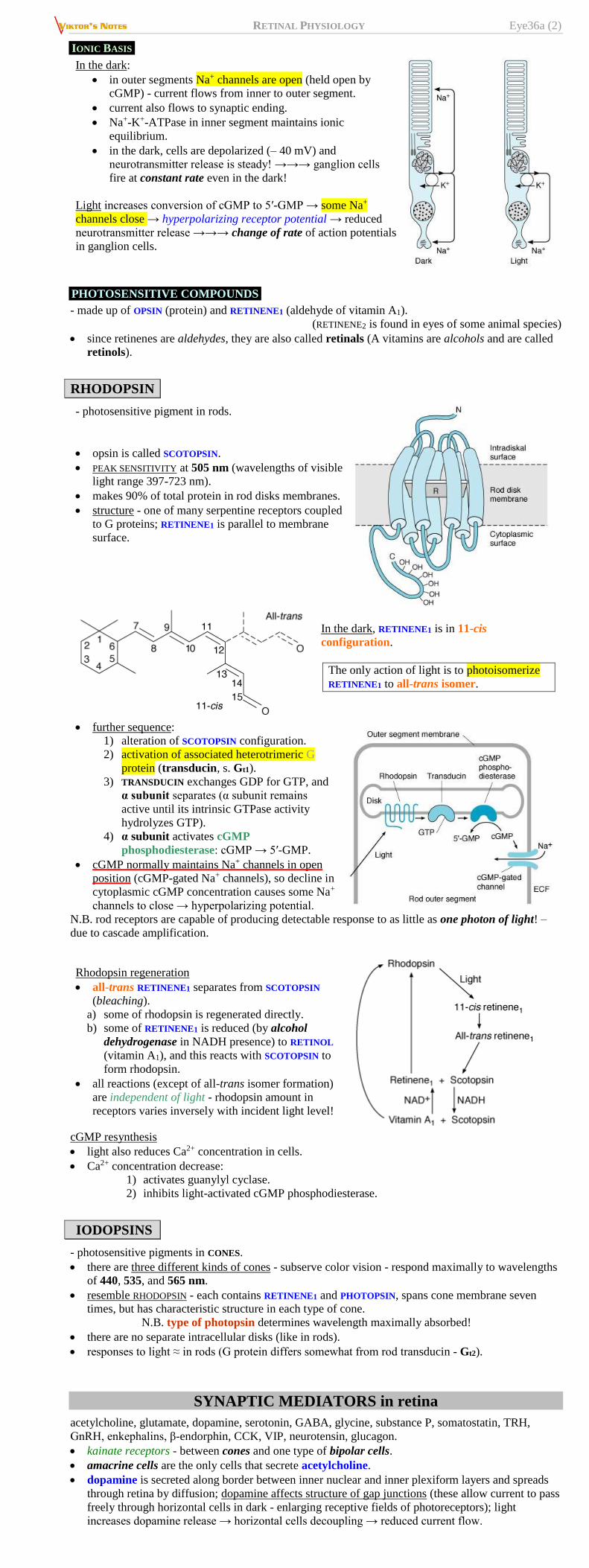
IONIC BASIS
In the dark:
in outer segments Na+ channels are open (held open by
cGMP) - current flows from inner to outer segment.
current also flows to synaptic ending.
Na+-K+-ATPase in inner segment maintains ionic
equilibrium.
in the dark, cells are depolarized (– 40 mV) and
neurotransmitter release is steady! →→→ ganglion cells
fire at constant rate even in the dark!
Light increases conversion of cGMP to 5′-GMP → some Na+
channels close → hyperpolarizing receptor potential → reduced
neurotransmitter release →→→ change of rate of action potentials
in ganglion cells.
PHOTOSENSITIVE COMPOUNDS
- made up of OPSIN (protein) and RETINENE1 (aldehyde of vitamin A1).
(RETINENE2 is found in eyes of some animal species)
since retinenes are aldehydes, they are also called retinals (A vitamins are alcohols and are called
retinols).
RHODOPSIN
- photosensitive pigment in rods.
opsin is called SCOTOPSIN.
PEAK SENSITIVITY at 505 nm (wavelengths of visible
light range 397-723 nm).
makes 90% of total protein in rod disks membranes.
structure - one of many serpentine receptors coupled
to G proteins; RETINENE1 is parallel to membrane
surface.
In the dark, RETINENE1 is in 11-cis
configuration.
The only action of light is to photoisomerize
RETINENE1 to all-trans isomer.
further sequence:
1) alteration of SCOTOPSIN configuration.
2) activation of associated heterotrimeric G
protein (transducin, s. Gt1).
3) TRANSDUCIN exchanges GDP for GTP, and
α subunit separates (α subunit remains
active until its intrinsic GTPase activity
hydrolyzes GTP).
4) α subunit activates cGMP
phosphodiesterase: cGMP → 5′-GMP.
cGMP normally maintains Na+ channels in open
position (cGMP-gated Na+ channels), so decline in
cytoplasmic cGMP concentration causes some Na+
channels to close → hyperpolarizing potential.
N.B. rod receptors are capable of producing detectable response to as little as one photon of light! –
due to cascade amplification.
Rhodopsin regeneration
all-trans RETINENE1 separates from SCOTOPSIN
(bleaching).
a) some of rhodopsin is regenerated directly.
b) some of RETINENE1 is reduced (by alcohol
dehydrogenase in NADH presence) to RETINOL
(vitamin A1), and this reacts with SCOTOPSIN to
form rhodopsin.
all reactions (except of all-trans isomer formation)
are independent of light - rhodopsin amount in
receptors varies inversely with incident light level!
cGMP resynthesis
light also reduces Ca2+ concentration in cells.
Ca2+ concentration decrease:
1) activates guanylyl cyclase.
2) inhibits light-activated cGMP phosphodiesterase.
IODOPSINS
- photosensitive pigments in CONES.
there are three different kinds of cones - subserve color vision - respond maximally to wavelengths
of 440, 535, and 565 nm.
resemble RHODOPSIN - each contains RETINENE1 and PHOTOPSIN, spans cone membrane seven
times, but has characteristic structure in each type of cone.
N.B. type of photopsin determines wavelength maximally absorbed!
there are no separate intracellular disks (like in rods).
responses to light ≈ in rods (G protein differs somewhat from rod transducin - Gt2).
SYNAPTIC MEDIATORS in retina
acetylcholine, glutamate, dopamine, serotonin, GABA, glycine, substance P, somatostatin, TRH,
GnRH, enkephalins, β-endorphin, CCK, VIP, neurotensin, glucagon.
kainate receptors - between cones and one type of bipolar cells.
amacrine cells are the only cells that secrete acetylcholine.
dopamine is secreted along border between inner nuclear and inner plexiform layers and spreads
through retina by diffusion; dopamine affects structure of gap junctions (these allow current to pass
freely through horizontal cells in dark - enlarging receptive fields of photoreceptors); light
increases dopamine release → horizontal cells decoupling → reduced current flow.
RETINAL PHYSIOLOGY Eye36a (3)
IMAGE FORMATION
Processing of visual information in retina involves sequential formation of three images:
First image - formed by light action on photoreceptors.
Second image in bipolar cells.
Third image in ganglion cells.
in formation of second image, signal is altered by horizontal cells, and in formation of third, it is
altered by amacrine cells.
third image reaches occipital cortex (little change in impulse pattern in lateral geniculate bodies).
Inhibitory feedback from one photoreceptor to another mediated via horizontal cells: activation of
photoreceptors triggers horizontal cell hyperpolarization, which in turn inhibits response in nearby
photoreceptors - LATERAL INHIBITION (i.e. activation of particular neural unit is associated with
inhibition of activity of nearby units - general phenomenon in mammalian sensory systems - helps to
sharpen stimulus edges - improves discrimination).
RECEPTIVE FIELD - part of retina whose photoreceptors (rods & cones) pertain to single ganglion
cell (optic nerve fiber).
receptive field is circular; in fovea – only 10 μm Ø; in peripheral retina – up to 1 mm Ø.
cones in center of receptive field (FIELD CENTER) convey information
directly to ganglion cells (via bipolar
cells);
cones at periphery of receptive field (FIELD SURROUND) reach ganglion cell
indirectly – via horizontal cells
(horizontal cells synapse with axons of
field center cones).
receptive fields are organized into
CENTER-SURROUND ANTAGONISTIC
REGIONS.
N.B. rods are not organized into
center-surround receptive fields!
Ganglion cell response to stimulation of its receptive field depends on:
1) type of receptive field (“on” or “off” type)
2) part of field that is illuminated (center or surround).
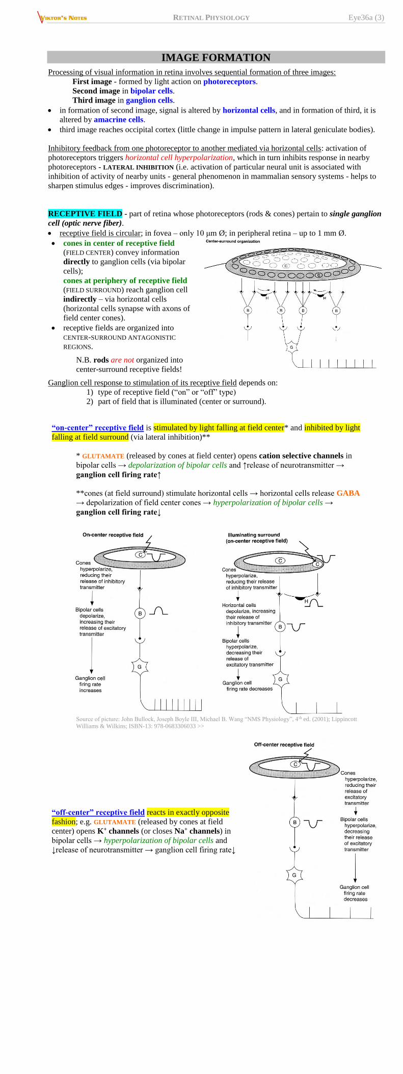
“on-center” receptive field is stimulated by light falling at field center* and inhibited by light
falling at field surround (via lateral inhibition)**
* GLUTAMATE (released by cones at field center) opens cation selective channels in
bipolar cells → depolarization of bipolar cells and ↑release of neurotransmitter →
ganglion cell firing rate↑
**cones (at field surround) stimulate horizontal cells → horizontal cells release GABA
→ depolarization of field center cones → hyperpolarization of bipolar cells →
ganglion cell firing rate↓
Source of picture: John Bullock, Joseph Boyle III, Michael B. Wang “NMS Physiology”, 4 th ed. (2001); Lippincott
Williams & Wilkins; ISBN-13: 978-0683306033 >>
“off-center” receptive field reacts in exactly opposite
fashion; e.g. GLUTAMATE (released by cones at field
center) opens K+ channels (or closes Na+ channels) in
bipolar cells → hyperpolarization of bipolar cells and
↓release of neurotransmitter → ganglion cell firing rate↓

RETINAL PHYSIOLOGY Eye36a (4)
Source of picture: John Bullock, Joseph Boyle III, Michael B. Wang “NMS Physiology”, 4th ed. (2001); Lippincott Williams & Wilkins;
ISBN-13: 978-0683306033 >>
“on” and “off” receptive fields occupy overlapping
regions in retina – light striking any retinal region
activates both types of receptive fields (light
intensity is signaled by difference in firing rates
between “on” and “off” receptive fields).
in either case, net response depends on complex
switching action in retina.
when entire receptive field is equally illuminated, it
has little or no effect on ganglion cell firing:
COLOR VISION
colors have three attributes: hue, intensity, saturation (degree of freedom from dilution with
white).
for any color there is COMPLEMENTARY COLOR that, when properly mixed with it, produces
sensation of white.
black is sensation produced by light absence, but it is positive sensation (blind eye does not "see
black" it "sees nothing").
white or any spectral color can be produced by mixing various proportions of red light (723-647
nm), green light (575-492 nm), and blue light (492-450 nm).
Red, green, and blue are primary colors
color perceived depends in part on background color.
Young-Helmholtz theory - three kinds of cones exist - each containing different photopigment
(maximally sensitive to one of three primary colors).
S pigment (blue-sensitive or short-wave pigment)
absorbs maximally blue-violet light (peak 440
nm).
M pigment (green-sensitive or middle-wave
pigment) absorbs maximally green light (peak
535 nm).
L pigment (red-sensitive or long-wave pigment)
absorbs maximally yellow light (peak 565 nm),
but sensitive enough in red portion of
spectrum.
there is variation in L pigment (62% normal individuals have Ser at site 180, whereas
38% have Ala – different absorption peaks).
RHODOPSIN gene is on chromosome 3.
S pigment gene is on chromosome 7.
M pigment and L pigment genes are arranged in tandem on Xq chromosome (their opsins show
96% homology of amino acid sequences).
many mammals are dichromats (have only two cone pigments), but humans are trichromats.
DARK ADAPTATION
When one passes from brightly lighted to dim
environment, retinas slowly become more sensitive
to light (visual threshold↓) - DARK ADAPTATION.
maximal in 20 minutes (some further decline
possible over longer periods).
persons who need maximal visual sensitivity in
dim light (e.g. radiologists, aircraft pilots) can
avoid having to wait 20 minutes to become dark-
adapted if they wear red glasses when in bright
light (red light stimulates rods to only slight
degree).
When one passes suddenly from near darkness to
bright sunlight (light intensity increases by 10 log
units, i.e. by factor of 10 billion), light seems
uncomfortably bright until eyes adapt (visual
threshold rises) – LIGHT ADAPTATION.
occurs over 5 minutes.
strictly speaking, it is merely disappearance of
dark adaptation.
another mechanism - pupil diameter↓ - when it
reduces from 8 to 2 mm, its area decreases by
factor of 16 and light intensity is reduced by > 1
log unit.

RETINAL PHYSIOLOGY Eye36a (5)
Components to dark adaptation:
1) first drop in visual threshold (rapid but small in
magnitude) - adaptation of CONES.
2) further drop occurs as result of adaptation of RODS
(mainly – rebuilding RHODOPSIN stores).
BIBLIOGRAPHY for ch. “Ophthalmology” → follow this LINK >>
Viktor’s Notes℠ for the Neurosurgery Resident
Please visit website at www.NeurosurgeryResident.net