Embed Size (px)
Citation preview
This is the author manuscript accepted for publication and has undergone full peer review but has
not been through the copyediting, typesetting, pagination and proofreading process, which may
lead to differences between this version and the Version of Record. Please cite this article as doi:
10.1111/1754-9485.12447
This article is protected by copyright. All rights reserved
-
M.G. Jameson1,2,3; J. McNamara4,5; M. Bailey4,5; P.E. Metcalfe1,3; L.C. Holloway1,2,3,6; K. Foo6,7,8; V. Do3,8,9;
L. Mileshkin10,11; C.L. Creutzberg12; P. Khaw
10,11 5
1 Centre for Medical Radiation Physics, University of Wollongong, NSW, Australia
2 Liverpool Cancer Therapy Centre, NSW, Australia
3Ingham Institute for Applied Medical Research, NSW, Australia
4 Illawarra Shoalhaven Cancer & Haematology Network, NSW, Australia 10
5 Illawarra Cancer Care Centre, Wollongong, NSW, Australia
6School of Physics, University of Sydney, Sydney, NSW, Australia
7 Chris O’Brien Lifehouse, Sydney, NSW, Australia
8 Sydney Medical School, University of Sydney, Sydney, NSW, Australia
9Crown Princess Mary Cancer Centre Westmead, Sydney, NSW, Australia 15
10 Peter MacCallum Cancer Centre, VIC, Australia
11 University of Melbourne, VIC, Australia
12
Department of Radiation Oncology, Leiden University Medical Centre, Leiden, The Netherlands
20
clinical trial, benchmarking, dummy run
PORTEC-3 Dummy Run Report
Corresponding author: 25
Michael Jameson
Cancer Therapy Centre & Ingham Institute, Liverpool Hospital
Locked Bag 7103, LIVERPOOL NSW 1871
Australia
Phone: +402852750 30
Auth
or
Manuscript

ANZGOG TROG PORTEC-3 benchmarking report Jameson et al
This article is protected by copyright. All rights reserved
Email: [email protected]
Total number of pages: 9 35
Number of words in manuscript: 2547
Number of words in abstract: 204
Total number of tables: 1
Total number of figures: 3
Auth
or
Manuscript
ANZGOG TROG PORTEC-3 Benchmark case report Jameson et al
This article is protected by copyright. All rights reserved
Received Date : 11-Nov-2015
Accepted Date : 19-Feb-2016
Article type : Radiation Oncology Original Article
5
Abstract
Introduction:
Protocol deviations in Randomised Controlled Trials (RCT) have been found to result in a
significant decrease in survival and local control. In some cases, the magnitude of the detrimental 10
effect can be larger than the anticipated benefits of the interventions involved. The implementation
of appropriate quality assurance of radiotherapy (QART) measures for clinical trials has been found
to result in fewer deviations from protocol. This paper reports on a benchmarking study conducted
in preparation for the PORTEC-3 trial in Australasia.
Method: 15
A benchmarking CT dataset was sent to each of the Australasian investigators, it was requested
they contour and plan the case according to trial protocol using local treatment planning systems.
These data was then sent back to TROG for collation and analysis.
Results:
Thirty three investigators from eighteen institutions across Australia and New Zealand took part in 20
the study. The mean CTV volume was 383.4(228.5–497.8) cm3 and the mean dose to a reference
gold standard CTV was 48.8(46.4-50.3) Gy.
Conclusions:
Although there were some large differences in the contouring of the CTV and its constituent parts,
these did not translate into large variations in dosimetry. Where individual investigators had 25
deviations from the trial contouring protocol, feedback was provided. The results of this study will
be used to compare with the international study QA for the PORTEC-3 trial.
Introduction
Accurate delineation of target volumes and organs at risk (OAR) for radiation therapy planning is 30
required for high quality treatment as it has a direct flow-on effect for the rest of the radiotherapy
chain1. Previous studies have investigated inter-observer contouring variability and the dosimetric
implications on patient treatment1-4. Several methods have been suggested to improve the
Auth
or
Manuscript
ANZGOG TROG PORTEC-3 Benchmark case report Jameson et al
This article is protected by copyright. All rights reserved
consistency of structure delineation. Training through teaching courses2 and progressive feedback
such as implementing a plan-do-check-act cycle5 have been found to result in contouring 35 submissions that are closer to the expert╆s delineated structures. Institutional experience gathered
over the course of a trial6 and the use of atlases7 result in more consistent submissions between
observers.
Protocol deviations in Randomised Controlled Trials (RCT) have been found to result in a 40
significant decrease in survival and local control8. In some cases the magnitude of the detrimental
effect can be larger than the anticipated benefits of the interventions involved8, 9. Implementation of
appropriate quality assurance of radiotherapy (QART) measures for clinical trials has been found to
result in fewer deviations from protocol 10. Facility assessment prior to the trial commencing is an
important aspect of QART11. This may include credentialing, dosimetry audits, patient case studies 45
or a Benchmarking case(s)12. In a benchmarking exercise the investigators are provided with an
anonymized representative dataset and are instructed to contour and plan according to the trial protocol. A prospective evaluation is undertaken prior to trial commencement of the institution╆s ability to comply with the protocol and potential issues can then be fed back to the investigators.
Benchmarking exercises have been used to standardize delineation procedures and have been 50
found to increase the probability of successful completion of subsequent procedures within the
same trial6.
PORTEC-3 is a RCT that compares concurrent chemoradiation and adjuvant chemotherapy with
pelvic radiation alone in high risk and advanced stage endometrial carcinoma13. Volumetric 55
contouring for female pelvic cases was not standard practice in Australasia when PORTEC-3 opened
for recruitment. Therefore, an Australasian study was implemented where investigators were
required to complete credentialing before recruiting patients to the trial. Contouring variation
across contributing Australasian centres is compared to a reference dataset and the impact of this
variation on patient dosimetry is presented here. 60
Materials and methods
Benchmark case procedure
The Trans-Tasman Oncology Group (TROG) conducted a treatment planning benchmarking
exercise for the Australia and New Zealand Gynaecological Oncology Group (ANZGOG) PORTEC-3 65
RCT. This benchmarking exercise, which required clinicians involved in the PORTEC-3 study to
Auth
or
Manuscript

ANZGOG TROG PORTEC-3 Benchmark case report Jameson et al
This article is protected by copyright. All rights reserved
contour a female pelvis according to the PORTEC-3 protocol, was not standard practice in Australia
and New Zealand (ANZ) centres at the time. Thirty three observers from 18 institutions were
supplied with a CD containing the anonymised DICOM images of an eligible patient. Accompanying
the CD were instructions and a description of the clinical case. Once completed, this was then 70
returned to TROG for collation and assessment by the Radiation Oncology Trial Management team.
Participants were asked to contour the case according to the PORTEC-3 protocol on their local
systems. The clinical target volume(CTV) was to be delineated including the upper 50% of the
vagina, the vaginal tissues superior to the vaginal marker, the paravaginal/parametrial soft tissues, 75
and the distal common, external, and internal iliac lymph node regions. Inclusion of the subaortic
pre-sacral nodes was recommended for tumours with involvement of the cervix. A margin of 7-10
mm was to be used from CTV to Planning Target Volume(PTV) with a variable margin in the upper
vaginal region to account for bladder and rectal filling. Participating centres were provided with
guidelines for contouring as described in the trial protocol13. 80
Participating institutions were also asked to plan the benchmarking case on their local treatment
planning systems(TPS). The PORTEC3 protocol specified a total dose of 48.6 Gy in 1.8 Gy fractions,
five times a week. Patients were to be treated on a linear accelerator using 6 MV photons or higher.
The dose was specified at the ICRU reference point and homogeneity requirements were according 85
to ICRU-50 recommendations14. Either a 4-field (4FLD) box, 3-field or multiple field technique could
be used. The treatment plan was computed using the dose grid spacing that was standard to the
local hospital practice. Sites were required to export all relevant data including dose, treatment
plan, DRRs and DVHs. Dose information was to be provided for CTV, PTV, both kidneys and the
spinal cord. 90
Structure analysis
A qualitative analysis was undertaken whereby a local expert panel reviewed the benchmarking
case data using the SWAN15 system provided by TROG. The expert reviewers analysed the plans to
ensure that the target and OAR structures had been contoured per protocol. Results were 95
categorized into acceptable, minor variation, major variation or inevaluable.
Further quantitative geometric and dosimetric analysis was performed on the submitted plans after
review by the expert panel. All evaluable DICOM data was imported into Matlab using CERR16
Auth
or
Manuscript
ANZGOG TROG PORTEC-3 Benchmark case report Jameson et al
This article is protected by copyright. All rights reserved
where in house software1 was used to analyse geometric variation. The geometric parameters 100
analysed included volume, centre of mass(COM), dimension(x,y,z) and DICE similarity coefficient17.
A consensus contour generated by the local expert trial QA group per protocol guidelines was used
as a ╅gold standard╆ reference for comparison when calculating the DICE similarity coefficient.
Dosimetric analysis 105
Dose volume histogram (DVH) data were extracted for the ╅gold standard╆ target and OAR contours
for each observer plan submitted, noting that these ╅gold standard╆ contours were different to those
for which the plan was generated at the local institutions (see above). The Comp Plan18 program
was used to extract dose volume parameters for each of the structures (table 1).
110
Results
Thirty three investigators from 18 institutions submitted plans for review. From these, 495
individual target structures were manually reviewed with 435/495(87.9%) acceptable,
49/495(9.9%) had a minor variation and 11/495(2.2%) with major variations (table 1). The
majority of the minor variations were due to incorrect PTV margin 22/49(44.9%) largely in the 115
required differential PTV for the vaginal vault CTV 18/49(36.7%). Other minor variations included
incorrect contouring of the parametrium CTV with respect to the pelvic sidewall 10/49(20.4%) and
contouring of the pelvic lymph nodes 11/49(24.5%). Major variations related to PTV margin of
greater than 1 cm being used 1/11(9.1%) and incorrect superior CTV boundary 4/11(36.4%) and
lymph node contouring 5/11(45.5%). 120
Of the 33 investigator datasets, 22 were evaluable in DICOM format for quantitative geometric and
dosimetric analysis, see figure 1. The main reasons for the reduced number of datasets were
corruption, duplication, incorrect format (RTOG) and missing data after transfer from review
software for quantitative analysis. The CTV geometric structure analysis results can be seen in 125
figure 2. The range in volumes of contoured CTV volume was 228.5–497.8 cm3 with a mean of 383.4
cm3. The mean CTV x(med/lat), y(ant/post) and z(sup/inf) dimensions were 15.3(13.2-16.9),
11.7(9.4-13.1) and 15.9(10.0-21.0) cm, respectively. The mean difference in COM between the ╅gold standard╆ and the investigator CTVs was 1.3(0.2 – 2.7) cm while the mean DICE coefficient was
0.67(0.54-0.96). 130
Auth
or
Manuscript
ANZGOG TROG PORTEC-3 Benchmark case report Jameson et al
This article is protected by copyright. All rights reserved
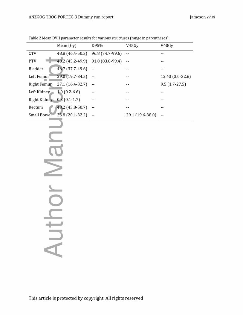
Collated ╅gold standard╆ CTV and small bowel DVHs are displayed in figure 3 while table 2 shows
mean(range) DVH parameter values for various structures. The mean V45 for the small bowel was
29.1±4.5 Gy across all observers. The mean D98 for the CTV and PTV was 87.0±6.9% and
79.1±8.31%, respectively. 135
Discussion
The mean CTV DICE coefficient of 0.67±0.09 was lower in the current study compared to the
reported value from Young et al19 of 0.77±0.03. This may be due to the smaller number of observers
(n=3) in the study by Young et al compared to that of the current study (n=22), as a larger number 140
of observers is likely to have a wider spread of contouring variation. The mean dose to the CTV and
PTV were 48.80±0.78 Gy and 48.20±1.03 Gy respectively, which matches closely with specified
ICRU-50 reference point dose of 48.6 Gy in the trial protocol. There was however some variation in
the CTV mean dose with a range of 46.44–50.30 Gy. The impact of this uncertainty in planned dose
is difficult to ascertain, as dose ranges of 45-50 Gy are generally found acceptable for postoperative 145
radiation therapy in gynaecological cancers. For head and neck cancer it has been suggested that
such dose variation may adversely impact the outcome of a clinical trial8.
Although the dosimetric impact of the contouring variation seen in this study was small it should be
noted that the planning technique used by all centres was a simple 4FLD box. This technique is 150 relatively insensitive to contouring variation within the borders of the ╉box╊ dose distribution.
More conformal intensity modulated techniques would be more sensitive as these allow for
sculpting of the dose distribution around sensitive structures such as the small bowel 20, 21.
The participants in this benchmarking exercise were all based in Australasia and thus the results 155
may be influenced by local treatment practices and not representative of the larger PORTEC-3 trial.
Also, this benchmarking exercise was part of a credentialing activity and as such was completed at
the beginning of the trial, if it were repeated later in the recruitment phase after investigators had
gained additional experience the results may be different. For investigators with violations in their
submitted plans, feedback was provide by the TROG trial management committee prior to enrolling 160
patients. In order to calculate the DICE similarity coefficient, difference in COM and DVH
parameters a ╅gold standard╆ reference contour is required. The ╅gold standard╆ in this study was a
consensus volume delineated by the Australasian trial coordinators. The choice of ╅gold standard╆ is
Auth
or
Manuscript

ANZGOG TROG PORTEC-3 Benchmark case report Jameson et al
This article is protected by copyright. All rights reserved
controversial as there is no widely accepted technique; consensus, average, STAPLE, median and
simple majority have all been used previously1. 165
In conclusion although there were some large differences in the contouring of the CTV and its
constituent parts, these did not translate into large variations in dosimetry. Where individual
investigators had deviations from the trial contouring protocol, feedback was provided to ensure
future compliance and individual case reviews were also performed. The results of this study will 170
be used to compare with the international study QA for the PORTEC-3 trial.
References
[1] Jameson MG, Kumar S, Vinod SK, Metcalfe PE, Holloway LC. Correlation of contouring variation
with modeled outcome for conformal non-small cell lung cancer radiotherapy. Radiotherapy and 175
Oncology. 2014.
[2] Dewas S, Bibault J-E, Blanchard P, et al. Delineation in thoracic oncology: a prospective study of
the effect of training on contour variability and dosimetric consequences. Radiat Oncol. 2011;6:118.
[3] Mitchell DM, Perry L, Smith S, et al. Assessing the effect of a contouring protocol on
postprostatectomy radiotherapy clinical target volumes and interphysician variation. International 180
Journal of Radiation Oncology* Biology* Physics. 2009;75:990-3.
[4] Poortmans PM, Venselaar JL, Struikmans H, et al. The potential impact of treatment variations
on the results of radiotherapy of the internal mammary lymph node chain: a quality-assurance
report on the dummy run of EORTC Phase III randomized trial 22922/10925 in Stage I–III breast
cancer. International Journal of Radiation Oncology* Biology* Physics. 2001;49:1399-408. 185
[5] Breunig J, Hernandez S, Lin J, et al. A system for continual quality improvement of normal tissue
delineation for radiation therapy treatment planning. International Journal of Radiation Oncology*
Biology* Physics. 2012;83:e703-e8.
[6] Fairchild A, Collette L, Hurkmans C, et al. Do results of the EORTC dummy run predict quality of
radiotherapy delivered within multicentre clinical trials? European Journal of Cancer. 190
2012;48:3232-9.
[7] Fuller CD, Nijkamp J, Duppen JC, et al. Prospective randomized double-blind pilot study of site-
specific consensus atlas implementation for rectal cancer target volume delineation in the
Auth
or
Manuscript
ANZGOG TROG PORTEC-3 Benchmark case report Jameson et al
This article is protected by copyright. All rights reserved
cooperative group setting. International Journal of Radiation Oncology* Biology* Physics.
2011;79:481-9. 195
[8] Peters LJ, O'Sullivan B, Giralt J, et al. Critical impact of radiotherapy protocol compliance and
quality in the treatment of advanced head and neck cancer: Results from TROG 02.02. Journal of
Clinical Oncology. 2010;28:2996.
[9] Fairchild A, Straube W, Laurie F, Followill D. Does quality of radiation therapy predict outcomes
of multicenter cooperative group trials? A literature review. International Journal of Radiation 200
Oncology* Biology* Physics. 2013;87:246-60.
[10] Bekelman JE, Deye JA, Vikram B, et al. Redesigning radiotherapy quality assurance:
opportunities to develop an efficient, evidence-based system to support clinical trials—report of
the National Cancer Institute Work Group on Radiotherapy Quality Assurance. International Journal
of Radiation Oncology* Biology* Physics. 2012;83:782-90. 205
[11] Fenton PA, Hurkmans C, Gulyban A, et al. Quality assurance of the EORTC 22043-30041 trial in
post-operative radiotherapy in prostate cancer: Results of the Dummy Run procedure.
Radiotherapy and Oncology. 2013;107:346-51.
[12] Melidis C, Bosch WR, Izewska J, et al. Global harmonization of quality assurance naming
conventions in radiation therapy clinical trials. International Journal of Radiation Oncology* 210
Biology* Physics. 2014;90:1242-9.
[13] http://clinicaltrials.gov/ct2/show/NCT00411138.
[14] ICRU Report 50: Prescribing, Recording and Reporting Photon Beam Therapy. ICRU Publ
Bethesda MD. 1993.
[15] Ebert MA, Haworth A, Kearvell R, et al. Detailed review and analysis of complex radiotherapy 215
clinical trial planning data: evaluation and initial experience with the SWAN software system.
Radiotherapy and Oncology. 2008;86:200-10.
[16] Deasy JO, Blanco AI, Clark VH. CERR: a computational environment for radiotherapy research.
Medical physics. 2003;30:979-85.
[17] Dice LR. Measures of the amount of ecologic association between species. Ecology. 220
1945;26:297-302.
Auth
or
Manuscript
ANZGOG TROG PORTEC-3 Benchmark case report Jameson et al
This article is protected by copyright. All rights reserved
[18] Holloway LC, Miller J-A, Kumar S, Whelan BM, Vinod SK. Comp Plan: A computer program to
generate dose and radiobiological metrics from dose-volume histogram files. Medical Dosimetry.
2012;37:305-9.
[19] Young AV, Wortham A, Wernick I, Evans A, Ennis RD. Atlas-based segmentation improves 225
consistency and decreases time required for contouring postoperative endometrial cancer nodal
volumes. International Journal of Radiation Oncology* Biology* Physics. 2011;79:943-7.
[20] Heron D, Gerszten K, Selvaraj R, et al. Conventional 3D conformal versus intensity-modulated
radiotherapy for the adjuvant treatment of gynecologic malignancies: a comparative dosimetric
study of dose–volume histograms☆. Gynecologic oncology. 2003;91:39-45. 230
[21] Small W, Mell LK, Anderson P, et al. Consensus guidelines for delineation of clinical target
volume for intensity-modulated pelvic radiotherapy in postoperative treatment of endometrial and
cervical cancer. International Journal of Radiation Oncology* Biology* Physics. 2008;71:428-34.
235
Captions:
Fig. 1. Axial, coronal and sagittal views of the benchmarking case CT displaying investigator CTVs
Fig. 2. Boxplots displaying variation in contours with respect to A) volume, B) dimension, C) COM
and D) DICE similarity coefficient
Fig. 3. Collated DVH data for all evaluable datasets for A) CTV and B) small bowel 240
Table 1 Mean DVH parameter results for various structures (range in parentheses)
Auth
or
Manuscript
ANZGOG TROG PORTEC-3 Dummy run report Jameson et al
This article is protected by copyright. All rights reserved
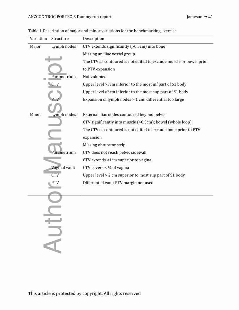
Table 1 Description of major and minor variations for the benchmarking exercise
Variation Structure Description
Major Lymph nodes CTV extends significantly (>0.5cm) into bone
Missing an iliac vessel group
The CTV as contoured is not edited to exclude muscle or bowel prior
to PTV expansion
Parametrium Not volumed
CTV Upper level >3cm inferior to the most inf part of S1 body
Upper level >3cm inferior to the most sup part of S1 body
PTV Expansion of lymph nodes > 1 cm; differential too large
Minor Lymph nodes External iliac nodes contoured beyond pelvis
CTV significantly into muscle (>0.5cm); bowel (whole loop)
The CTV as contoured is not edited to exclude bone prior to PTV
expansion
Missing obturator strip
Parametrium CTV does not reach pelvic sidewall
CTV extends <1cm superior to vagina
Vaginal vault CTV covers < ¼ of vagina
CTV Upper level > 2 cm superior to most sup part of S1 body
PTV Differential vault PTV margin not used
Auth
or
Manuscript
ANZGOG TROG PORTEC-3 Dummy run report Jameson et al
This article is protected by copyright. All rights reserved
Table 2 Mean DVH parameter results for various structures (range in parentheses)
Mean (Gy) D95% V45Gy V40Gy
CTV 48.8 (46.4-50.3) 96.8 (74.7-99.6) -- --
PTV 48.2 (45.2-49.9) 91.8 (83.8-99.4) -- --
Bladder 46.7 (37.7-49.6) -- -- --
Left Femur 29.0 (19.7-34.5) -- -- 12.43 (3.0-32.6)
Right Femur 27.1 (16.4-32.7) -- -- 9.5 (1.7-27.5)
Left Kidney 1.0 (0.2-6.6) -- -- --
Right Kidney 0.3 (0.1-1.7) -- -- --
Rectum 48.2 (43.8-50.7) -- -- --
Small Bowel 23.8 (20.1-32.2) -- 29.1 (19.6-38.0) --
Auth
or
Manuscript

ara_12447_f1.jpg
Thisarticleisprotectedbycopyright.Allrightsreserved
Auth
or
Manuscript

ara_12447_f2.jpg
Thisarticleisprotectedbycopyright.Allrightsreserved
Auth
or
Manuscript

ara_12447_f3.jpg
Thisarticleisprotectedbycopyright.Allrightsreserved
Auth
or
Manuscript
Minerva Access is the Institutional Repository of The University of Melbourne
Author/s:
Jameson, MG; McNamara, J; Bailey, M; Metcalfe, PE; Holloway, LC; Foo, K; Do, V;
Mileshkin, L; Creutzberg, CL; Khaw, P
Title:
Results of the Australasian (Trans-Tasman Oncology Group) radiotherapy benchmarking
exercise in preparation for participation in the PORTEC-3 trial
Date:
2016-08-01
Citation:
Jameson, M. G., McNamara, J., Bailey, M., Metcalfe, P. E., Holloway, L. C., Foo, K., Do, V.,
Mileshkin, L., Creutzberg, C. L. & Khaw, P. (2016). Results of the Australasian (Trans-
Tasman Oncology Group) radiotherapy benchmarking exercise in preparation for
participation in the PORTEC-3 trial. JOURNAL OF MEDICAL IMAGING AND RADIATION
ONCOLOGY, 60 (4), pp.554-559. https://doi.org/10.1111/1754-9485.12447.
Persistent Link:
http://hdl.handle.net/11343/291146
File Description:
Accepted version