Embed Size (px)
Citation preview
UNIVERSIDADE DE LISBOA
FACULDADE DE CIÊNCIAS
DEPARTAMENTO BIOLOGIA VEGETAL
Response to flucytosine in Candida glabrata at the
membrane proteome level: role of the DHA family
and the transcription factor CgPdr1
Carla Sofia Amorim Pires
Mestrado em Microbiologia Aplicada
Dissertação orientada por:
Doutor Miguel Nobre Parreira Cacho Teixeira
Doutora Ana Tenreiro
2018
ii
Acknowledgments
Firstly, I would like to thank my supervisor Professor Miguel Teixeira for its incomparable
patience, encouragement and sympathy as well as the possibility to join his team, at Institute
for Bioengineering and Biosciences, during the last two years, so essential and important for
my academic journey. I would like to acknowledge Professor Isabel Sá-Correia for allowing me
to integrate the Biological Sciences Research Group, which she leads.
I want to thank Catarina Costa in a special way, who was, during these two years, above all, a
great companion. She helped and guided me at the level of the experimental work. Without
her help, everything would have been much more difficult! I also thank you for the
encouragement, dedication and availability. It was a pleasure to meet you! ☺
To the whole BSRG team, thank you for welcoming me and guiding me whenever I needed
throughout the practical work. Without you, it would not be the same! I would like to thank
my colleagues of journey, André Henriques, Guida Camacho, Andreia Ponte and Jonathan
Ribeiro for all the conversations and exchanges of ideas, so essential to practical work and
laboratorial life.
A big thank you for my parents, my sister and my friends, without you I never do and finish
this. A special thank you to Tiago, for being there for me at all times, for listen and encourage
me in the good and difficult moments!
I would like to acknowledge Prof. Hiroji Chibana, from the Chiba University, Japan, and Prof.
Thomas Edlind, Drexel University, Philadelphia, PA, for kindly providing the KUE100 and
66032 strains.
This work was supported by Fundação para a Ciência e Tecnologia (FCT), contracts
PTDC/EBB-BIO/119356/2010 and PTDC/BBB-BIO/4004/2014. Funding received by iBB-Institute
for Bioengineering and Biosciences from FCT-Portuguese Foundation for Science and
Technology (UID/BIO/04565/2013) and from Programa Operacional Regional de Lisboa 2020
(Project N. 007317) is acknowledged.
iii
Response to flucytosine in Candida glabrata at the
membrane proteome level: role of the DHA family
and the transcription factor CgPdr1
Dissertation guided by Prof. Dr. Miguel Cacho Teixeira (IST-UL)
and Prof. Dra. Ana Tenreiro (FCUL)
Carla Sofia Amorim Pires
Master Thesis
2018
This thesis was fully performed at the Biological Sciences Research Group of the
Institute for Bioengineering and Biosciences, Instituto Superior Técnico under the
direct supervision of Prof. Dr. Miguel Cacho Teixeira and Dr. Catarina Costa.
Prof. Dra. Ana Tenreiro was the internal designated supervisor in the scope of the
Master in Applied Microbiology of the Faculty of Sciences of the University of Lisbon.

iv
v
Abstract
Candida glabrata is pathogenic fungi that emerged in the last decades as the second most
common cause of candidiasis worldwide. The infections caused by this yeast are characterized
for a high mortality and morbidity, and the action of drug efflux pumps is impairing treatment
effectiveness. Multidrug resistance has emerged in most organisms and poses a severe clinical
problem for the treatment because the extensive use of antifungal drugs had led to a huge
increase in the number of intrinsically resistant infections with fungal pathogens.
Since resistance often relies on the action of membrane transporters, including drug efflux
pumps from ATP-Binding Cassette (ABC) family or from the Drug:H+ Antiporter (DHA) family, an
iTRAQ-based (isobaric Tags for Relative and Absolute Quantification) membrane proteomics
analysis was performed to identify all the membrane-associated proteins whose abundance
changes in C. glabrata cells exposed to the Fluoropyrimidine drug 5-Flucytosine (5-FC). A total
of 32 proteins were found to display significant expression changes in the membrane fraction
of cells exposed to 5-FC, when compared to cells in the unstressed control conditions, 50 % of
which under the influence of the transcription factor CgPdr1, found to be a determinant of
C. glabrata resistance to 5-FC. These proteins cluster into functional groups associated to cell
wall assembly, lipid metabolism, amino acid/nucleotide metabolism, ribosome components
and translation machinery, mitochondrial function, glucose metabolism and multidrug
resistance transport.
Among the proteins whose concentration was found increased in 5-FC stressed cells, CgFlr1
was elected for further studies. The role of CgFlr1, and of its close homolog CgFlr2, in 5-FC
resistance was assessed. Results obtained demonstrate that both proteins confer 5-FC
resistance. Despite their high degree of homology, CgFlr1 seems to specifically confer
resistance to the fungicide mancozeb, while CgFlr2 appears to confer resistance to azole
antifungal drugs and amphotericin B. CgFlr1 was found to be localized in the plasma
membrane in C. glabrata and when heterologously expressed in Saccharomyces cerevisiae,
complementing the 5-FC susceptibility phenotype exhibited by the S. cerevisiae Δflr1 mutant.
Additionally, the deletion of CgFLR1 and CgFLR2 was found to lead to increased intracellular
accumulation of 5-FC, suggesting that these transporters play a direct role in the resistance to
5-FC by extruding this antifungal drug to the extracellular medium.
KeyWords
Candida glabrata, 5-Flucytosine drug resistance, membrane proteome, drug:H+ antiporters.
vi
Resumo
Candida glabrata é um fungo patogénico que emergiu nas últimas décadas como a segunda
causa mais comum de candidíase em todo o mundo. O género Candida compreende quase
duzentas espécies, a maioria das quais são omnipresentes em numerosos habitats naturais e
artificiais. As espécies patogénicas de Candida são encontradas como comensais no trato oral,
gastrointestinal e genital de hospedeiros saudáveis e têm a propensão de se tornar
patogénicas quando o hospedeiro se torna imunodeprimido. As infeções causadas por esta
levedura são caracterizadas por uma elevada mortalidade e morbilidade. Entre outros fatores
de virulência, as espécies do género Candida exibem, em comparação com espécies não
patogénicas, uma expansão de famílias de transportadores de enzimas extracelulares e
transmembranares, bem como um enriquecimento em transportadores de superfície celular
com o potencial de conferirem resistência aos fármacos por funcionarem como bombas de
efluxo de drogas, prejudicando a eficácia das terapias antifúngicas. C. glabrata é
filogeneticamente mais próxima da levedura modelo - Saccharomyces cerevisiae do que das
outras espécies do género Candida. No entanto, é frequentemente a segunda ou terceira
causa mais comum de infeções fúngicas, sendo que em populações como diabéticos e idosos
chega a ser o fungo patogénico dominante. Em comparação com outras espécies, C. glabrata
fornece um modelo promissor para o estudo da base genética da resistência a múltiplos
antifúngicos.
A resistência a múltiplos fármacos é definida como a aquisição simultânea de resistência a um
grande espectro de substâncias químicas citotóxicas estrutural e funcionalmente não
relacionadas, às quais o organismo nunca tinha sido exposto. Este fenómeno está a tornar-se
um problema clínico crescente, uma vez que compromete a eficácia da terapêutica
antimicrobiana. Para diminuir com sucesso o número de agentes patogénicos resistentes a
múltiplos fármacos, é fundamental adquirir um conhecimento extenso dos mecanismos
moleculares subjacentes à resistência aos fármacos antimicrobianos. Esta resistência a
fármacos pode ser adquirida através de vários mecanismos: (I) alteração dos níveis de
expressão e atividade de proteínas da membrana plasmática ou canais e transportadores de
membrana; (II) degradação enzimática ou inativação dos fármacos; (III) ativação de sistemas
de replicação e reparação de DNA; (IV) impedimento da entrada do fármaco na célula e/ou
extrusão ativa do mesmo catalisada por transportadores transmembranares; (V) sequestro do
fármaco em vesículas intracelulares; (VI) alteração ou modificação do alvo do fármaco. A ação
de transportadores de membrana, incluindo bombas de efluxo da superfamília ATP-Binding
Cassette (ABC) e transportadores da família Drug:H+ Antiporter (DHA), é fundamental para a
vii
resistência a múltiplos fármacos. Os transportadores ABC obtêm energia por hidrólise de ATP,
enquanto que os transportadores DHA utilizam o gradiente de protões através da membrana
plasmática para exportar compostos para fora da célula. O genoma de C. glabrata codifica
18 transportadores ABC, seis meios transportadores e 12 transportadores completos, dos
quais seis pertencem à subfamília PDR (Pleiotropic Drug Resistance) e apenas alguns foram
estudados para resistência a fármacos ou atividades de transporte; codifica ainda
15 transportadores DHA1 que codificam para proteínas DHA com 12 segmentos
transmembranares. Desde a década de 1950 várias classes de drogas antifúngicas foram
desenvolvidas, no entanto, apenas quatro deles são atualmente usados na prática clínica para
tratar infeções por Candida: análogos de pirimidina, polienos, azóis e equinocandinas. A
extensão da resistência aos fármacos antifúngicos varia para as diferentes classes de fármacos.
Em espécies de Candida há resistência bastante limitada aos polienos e equinocandinas,
enquanto que a resistência ao análogo de pirimidina 5-flucitosina e a azóis é mais comum.
Neste trabalho foi estudado o efeito da exposição de células de C. glabrata ao fármaco
antifúngico 5-flucitosina, ao nível do proteoma de membrana. Hoje em dia, novas abordagens
quantitativas que utilizam a Espectrometria de Massa (MS) e a química estável de rotulagem
de isótopos oferecem uma alternativa às técnicas tradicionais que empregam a eletroforese
bidimensional comparativa para estudos de proteómica de expressão. A MS é uma ferramenta
particularmente relevante no caso de proteínas de membrana, que não são detetáveis em géis
bidimensionais, porque são em grande parte insolúveis em tampões de focagem isoelétrica.
Neste trabalho, realizou-se análise proteómica de membrana baseada em iTRAQ-MS (isobaric
Tags for Relative and Absolute Quantification) para identificar e quantificar as proteínas
associadas à membrana em células de C. glabrata cuja concentração se altera após exposição à
5-flucitosina (5-FC). Um total de 32 proteínas apresentaram diferenças de expressão na
membrana depois de expostas a 5-FC, quando comparadas com células em condições
controlo. Estas proteínas estão envolvidas nos seguintes processos biológicos: remodelação da
parede celular, metabolismo lipídico, metabolismo de aminoácidos/nucleotídeos,
componentes ribossomais e maquinaria de tradução, função mitocondrial, metabolismo da
glicose e transportadores de membrana.
Por comparação com as alterações que se verificam nas mesmas condições ambientais no
mutante Δpdr1, com o gene PDR1 eliminado, observou-se que 50 % das proteínas cuja
expressão se altera após exposição a 5-FC estão sob influência do fator de transcrição CgPdr1,
que se verificou ser determinante na resistência à 5-FC por C. glabrata. Este resultado
surpreendente é muito interessante, uma vez que o fator de transcrição CgPdr1 é o principal
viii
determinante de resistência a azóis em C. glabrata. A observação de que também poderá estar
envolvido na aquisição de resistência a 5-FC sugere a existência de um mecanismo comum à
resistência a duas famílias de antifúngicos, o que é preocupante na perspetiva da aquisição de
resistência a agentes antifúngicos, sem necessidade de exposição prévia aos mesmos.
Entre as proteínas encontradas com expressão aumentada encontra-se o transportador da
família DHA - CgFlr1, que foi eleito para estudos posteriores. Foi estudado o papel do CgFlr1, e
do seu homólogo CgFlr2, na resistência à 5-flucitosina. Os resultados obtidos demonstram que
ambas as proteínas conferem resistência a 5-flucitosina. Adicionalmente, e apesar do seu
elevado grau de homologia, CgFlr1 parece conferir resistência especificamente ao fungicida
mancozeb, enquanto CgFlr2 parece conferir resistência a azóis e a anfotericina B. Verificou-se
que a proteína CgFlr1 se localiza na membrana plasmática, quando expresso em C. glabrata,
mas também quando expresso em S. cerevisiae, complementando o fenótipo de
suscetibilidade a 5-FC exibido pelo mutante ∆flr1. Verificou-se que a eliminação dos genes
CgFLR1 e CgFLR2 leva a uma acumulação intracelular de 5-FC maior do que a registada em
células da estirpe selvagem, o que sugere que estes transportadores desempenham um papel
direto na resistência à 5-FC por expulsão desta droga antifúngica para o meio extracelular.
No global, os resultados descritos neste estudo salientam a importância dos transportadores
de múltiplos fármacos da família DHA no fenótipo de resistência a antifúngicos, em particular
no que diz respeito a 5-FC. A caracterização dos transportadores CgFlr1 e CgFlr2 como estando
envolvidos na resistência a 5-FC reforça a noção de que é necessário estudar os restantes
membros desta família em C. glabrata, dado o seu provável impacto clínico. Adicionalmente,
este estudo realça a importância do recurso a abordagens à escala do genoma, em particular
ao nível do proteoma, na identificação e compreensão dos mecanismos de resposta e
resistência a antifúngicos. Os processos biológicos e os seus efetores identificados no presente
estudo representam alvos promissores para o desenvolvimento de novos sensitizadores da
resistência à flucitosina, que poderão permitir a utilização terapêutica de 5-FC em mais baixas
concentrações e sem riscos tão elevados de falência da terapêutica, limitando o
desenvolvimento de resistência a 5-FC em C. glabrata que habitualmente acontece com
elevada rapidez.
Palavras-Chave
Candida glabrata, resistência ao fármaco flucitosina, proteoma de membrana, antiportadores
droga:H+

ix
x
Contents 1 Introduction .........................................................................................................................1
1.1 The genus Candida .......................................................................................................1
1.1.1 Phylogeny .............................................................................................................1
1.1.2 Pathogenic Candida species .................................................................................2
1.1.3 Candida glabrata ..................................................................................................3
1.2 Multidrug Resistance Phenomenon .............................................................................3
1.2.1 Multidrug Resistance Transporters.......................................................................4
1.2.2 The ABC superfamily .............................................................................................5
1.2.2.1 ABC transporters in S. cerevisiae ......................................................................7
1.2.2.2 ABC transporters in C. glabrata ........................................................................9
1.2.3 The Major Facilitator Superfamily ......................................................................10
1.2.3.1 MFS-MDR transporters in S. cerevisiae ...........................................................10
1.2.3.2 MFS-MDR transporters in C. glabrata.............................................................14
1.2.4 The CgPdr1 transcription factor ..........................................................................15
1.3 Antifungal Drugs and Fungicides explored in the course of this study .......................16
1.3.1 Polyene – Amphotericin B ..................................................................................17
1.3.2 Azoles .................................................................................................................18
1.3.3 Fluoropyrimidine – 5-Flucytosine .......................................................................20
1.4 iTRAQ-based Proteomics ............................................................................................21
2 Materials and Methods ......................................................................................................24
2.1 Cell Culture .................................................................................................................24
2.1.1 Strains .................................................................................................................24
2.1.2 Cloning of the C. glabrata CgFLR1_1 gene (ORF CAGL0H06017g). .....................24
2.1.3 Growth media .....................................................................................................25
2.1.4 Antifungal drugs .................................................................................................26
2.2 Susceptibility assays ...................................................................................................26
xi
2.3 CgFlr1 Subcellular Localization Assessment................................................................27
2.4 Extraction and Multiplexed Membrane Protein Quantitation ....................................28
2.4.1 Cell cultivation ....................................................................................................28
2.4.2 Membrane proteome extraction ........................................................................28
2.4.3 Expression proteomic analysis using iTRAQ ........................................................29
2.5 3H-5-Flucytosine accumulation assays ........................................................................29
2.6 Gene expression analysis ............................................................................................30
2.6.1 Total RNA extraction and quantification ............................................................30
2.6.2 Real Time RT-PCR................................................................................................31
3 Results ................................................................................................................................35
3.1 Role of the transcription factor CgPdr1 in the resistance to 5-Flucytosine and other
antifungal drugs .....................................................................................................................35
3.2 Characterization of the C. glabrata membrane proteome .........................................36
3.3 C. glabrata response to stress induced by 5-Flucytosine, at the membrane proteome
level ……………………………………………………………………………………………………………………………….37
3.4 Role of CgPdr1 in the C. glabrata response to 5-Flucytosine ......................................42
3.5 Evaluation of the role of four C. glabrata DHA transporters, CgFlr1, CgFlr2, CgTpo1_1
and CgTpo1_2, in the resistance to 5-Flucytosine and other antifungal drugs.......................43
3.5.1 Characterization of the effect of CgFlr1 and CgFlr2 expression in antifungal drug
resistance ...........................................................................................................................43
3.5.2 CgFlr1_1 is targeted to the plasma membrane in S. cerevisiae, being able to
complement its homolog Flr1 ............................................................................................45
3.5.3 Assessment of the contribution of CgFLR1, CgFLR2, CgTPO1_1 and CgTPO1_2 to
5-Flucytosine accumulation ................................................................................................46
3.5.4 Transcript levels of genes CgFLR1, CgFLR2, CgTPO1_1 and CgTPO1_2 ..............47
4 Discussion ...........................................................................................................................49
5 References..........................................................................................................................52
xii
List of Figures
Figure 1.1 - Phylogenetic tree of sequenced Candida and Saccharomyces clade species. ...........1
Figure 1.2 - Schematic representation of the ABC and MFS transporter ......................................5
Figure 1.3 - Schematic representation of domain arrangements of ABC transporters.................6
Figure 1.4 - Localization of S. cerevisiae ABC transporters ...........................................................7
Figure 1.5 - The PDR network in yeast..........................................................................................9
Figure 1.6 - Domain arrangements of MFS transporters from yeasts species ............................10
Figure 1.7 - Phylogenetic relationship of the DHA1 family and the DHA2 family .......................11
Figure 1.8 - Targets of antifungal agents ....................................................................................16
Figure 1.9 - Salvage pathway for 5-Flucytosine ..........................................................................21
Figure 1.10 - Isobaric Tags for Relative and Absolute Quantification (iTRAQ). ...........................22
Figure 1.11 - Example MS/MS spectrum of peptide TPHPALTEAK .............................................23
Figure 2.1 - Graphical representation of real-time PCR data. .....................................................33
Figure 3.1 - Spot assays comparing the growth susceptibility ....................................................35
Figure 3.2 - Spot assays comparing the growth susceptibility ....................................................36
Figure 3.3 - Categorization of the proteins identified in membrane enriched fractions of
C. glabrata cells ..........................................................................................................................37
Figure 3.4 - Major functional groups found to have significant expression changes in the
membrane-enriched proteome upon exposure to 5-FC in C. glabrata. .....................................41
Figure 3.5 - Spot assays comparing the growth susceptibility ....................................................44
Figure 3.6 - Spot assays comparing the growth susceptibility. ...................................................44
Figure 3.7 – Fluorescence of exponential phase BY4741 S. cerevisiae cells ...............................45
Figure 3.8 – Fluorescence of exponential phase KUE100 C. glabrata cells. ................................45
Figure 3.9 - Spot assays comparing the growth susceptibility ....................................................46
Figure 3.10 - Time-course accumulation of 5-FC in strains .........................................................47
Figure 3.11 - Time-course accumulation of 5-FC in strains .........................................................47
Figure 3.12 – Comparison of the variation of the expression of CgFLR1, CgFLR2, CgTPO1_1 and
CgTPO1_2 by real-time analysis .................................................................................................48
Figure 4.1 - Current model of the mechanisms of resistance to 5-FC in C. glabrata ..................49
xiii
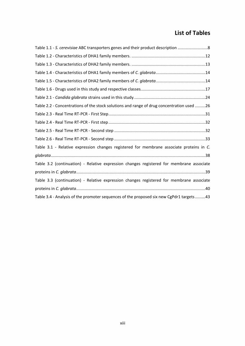
List of Tables
Table 1.1 - S. cerevisiae ABC transporters genes and their product description ..........................8
Table 1.2 - Characteristics of DHA1 family members. ................................................................12
Table 1.3 - Characteristics of DHA2 family members. ................................................................13
Table 1.4 - Characteristics of DHA1 family members of C. glabrata ...........................................14
Table 1.5 - Characteristics of DHA2 family members of C. glabrata. ..........................................14
Table 1.6 - Drugs used in this study and respective classes........................................................17
Table 2.1 - Candida glabrata strains used in this study. .............................................................24
Table 2.2 - Concentrations of the stock solutions and range of drug concentration used .........26
Table 2.3 - Real Time RT-PCR - First Step ....................................................................................31
Table 2.4 - Real Time RT-PCR - First step ....................................................................................32
Table 2.5 - Real Time RT-PCR - Second step ...............................................................................32
Table 2.6 - Real Time RT-PCR - Second step ...............................................................................33
Table 3.1 - Relative expression changes registered for membrane associate proteins in C.
glabrata......................................................................................................................................38
Table 3.2 (continuation) - Relative expression changes registered for membrane associate
proteins in C. glabrata................................................................................................................39
Table 3.3 (continuation) - Relative expression changes registered for membrane associate
proteins in C. glabrata................................................................................................................40
Table 3.4 - Analysis of the promoter sequences of the proposed six new CgPdr1 targets .........43
xiv
Thesis Outline
This dissertation is divided into four chapters.
In Chapter 1, an introduction of the current knowledge of the genus Candida, its phylogeny,
epidemiology and treats, focusing mainly on Candida albicans and Candida glabrata, the two
most prevalent species responsible for human candidemias. The multidrug resistance
phenomenon and the role of the multidrug transporters of the ABC and MFS superfamilies is
discussed, having as background, the far more well-studied yeast Saccharomyces cerevisiae.
The mode of action of the five families of antifungal agents is also discussed, highlighting the
resistance mechanisms known for each antifungal family. Finally, the iTRAQ-based proteomics
is also presented.
Chapter 2 is focused on the experimental methods and approaches used to achieve the goals
of the present study.
The Chapter 3 presents the results obtained in all assays. Begins with the role of the
transcription factor CgPdr1 in the resistance to 5-Flucytosine and other antifungal drugs. After
that, characterization of the C. glabrata membrane proteome and the response to stress
induced by 5-Flucytosine at the membrane proteome level. Assessment of the role of CgPdr1
in C. glabrata response to 5-Flucytosine are also performed. Finally, the evaluation of the role
of four C. glabrata DHA transporters CgFlr1, CgFlr2, CgTpo1_1 and CgTpo1_2 in the resistance
to 5-Flucytosine and other antifungal drugs is tested.
In Chapter 4 the results obtained throughout this work are discussed in light of previous
knowledge and the new facts learned about MDR efflux pumps in antifungal drug resistance in
C. glabrata. Finally, the conclusions of the present work are presented, as well as some
suggestions for further work on this subject.
Most of the results displayed in this thesis were published in Frontiers in Microbiology
[Pais, P.*, Pires, C.*, Costa, C., Okamoto, M., Chibana, H., Teixeira, M.C., Membrane proteomics
analysis of the Candida glabrata response to 5-Flucytosine: Unveiling the role and regulation of
the drug efflux transporters CgFlr1 and CgFlr2, Frontiers in Microbiology.2016. 7:2045.
(*these authors have contributed equally to this work)].
xv
Abbreviations
5-FC – flucytosine or 5-Flucytosine
5-FU – 5-fluorouracil
5-fluoro-UMP – 5-fluorouridine monophospate
4-NQO – 4-Nitroquinoline 1-Oxide
2-DE – Two-dimensional Eletrophoresis
AAA – Amino-Acid Analysis
ABC – ATP Binding Cassette
CID – Collision-induced Dissociation
CYP – Cytochrome P-450
DF – Frequency in Dataset
DHA – Drug:H+ Antiporter
DMSO – Dimethyl Sulfoxide
FDA – Food and Drug Administration
GFP – Green Fluorescent Protein
GOF – Gain-of-function
GRAS – “General Regarded As Safe”
iTRAQ – isobaric Tags for Relative and Absolute Quantification
MCPA – 2-methyl-4-chlorophenoxyacetic acid
MDR – Multidrug Resistance
MFS – Major Facilitator Superfamily
MMB – Minimal Medium for BY4741
MMG – Minimal Growth Medium
MMTS – Methyl Methane Thiosulfonate
MRP – Multidrug Resistance-associated Protein
MS – Mass Spectrometry
NBD – Nucleotide Binding Domain
xvi
OD – Optical Density
OMP – Orotidine-5’-phosphate
PDR – Pleiotropic Drug Resistance
PEMT – Phosphatidylethanolamine Methyltransferase
RF – Frequency Reference Genome
rRNA – Ribossomal RNA
SGD – Saccharomyces Genome Database
TCEP – Tris(2-carboxyethyl) Phosphine
TEAB – Tetraethylammonium Bromide
TMD – Transmembrane Domain
UDP – Uridine Diphosphate
UMP – Uridine Monophosphate
UPRT – Uracil Phosphoribosyl Transferase
WGD – Whole Genome Duplication
YPD – Yeast Peptone Dextrose
1
1 Introduction
1.1 The genus Candida
1.1.1 Phylogeny
The genus Candida comprises almost 200 Candida species 1 and a wide range of
phylogenetically unrelated anamorphic fungi. Yeasts of the polyphyletic, artificial genus
Candida include plant endophytes, insect symbionts and opportunistic human pathogens,
most of which are ubiquitous in numerous natural and artificial habitats 2. The genomes of
Candida species show enormous variations in size and phenotypic outcome, however, the
predicted numbers of protein-coding genes are very similar in all the species 1.
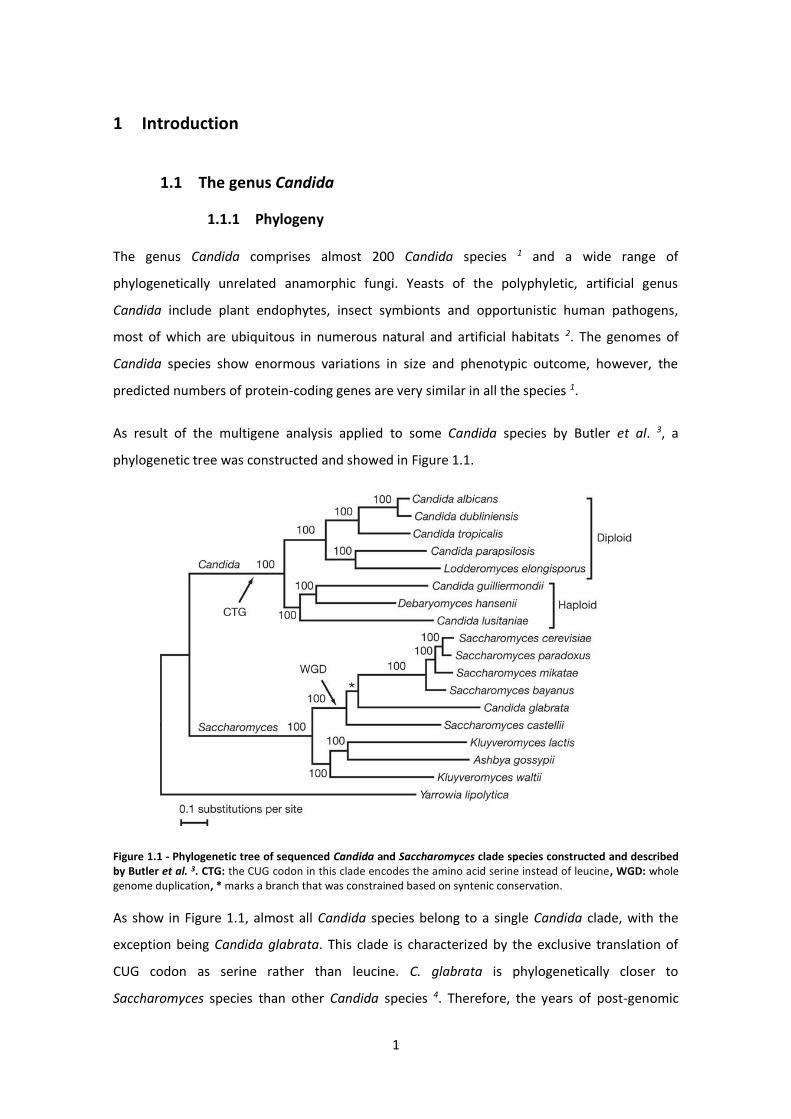
As result of the multigene analysis applied to some Candida species by Butler et al. 3, a
phylogenetic tree was constructed and showed in Figure 1.1.
Figure 1.1 - Phylogenetic tree of sequenced Candida and Saccharomyces clade species constructed and described by Butler et al. 3. CTG: the CUG codon in this clade encodes the amino acid serine instead of leucine, WGD: whole genome duplication, * marks a branch that was constrained based on syntenic conservation.
As show in Figure 1.1, almost all Candida species belong to a single Candida clade, with the
exception being Candida glabrata. This clade is characterized by the exclusive translation of
CUG codon as serine rather than leucine. C. glabrata is phylogenetically closer to
Saccharomyces species than other Candida species 4. Therefore, the years of post-genomic
2
research dedicated to Saccharomyces cerevisiae are expected to provide reliable clues to guide
C. glabrata functional studies 5 and make C. glabrata a good model organism for the study of
fungal pathogenesis and for the identification of antifungal drug targets and drug resistance
mechanisms 5. An additional characteristic of C. glabrata that distinguishes it from other
pathogenic Candida species is its haploid genome 6.
1.1.2 Pathogenic Candida species
The genus Candida includes unicellular fungi that have been used in industrial fermentation
processes, mainly food and beverage industries, and are listed in “Generally Regarded As Safe”
(GRAS) organism by the Food and Drug Administration (FDA) of the USA 2. However, they have
the propensity to become pathogens 7 and are the most common cause of opportunistic fungal
infection worldwide 3 with high morbidity and mortality 8.
Although the genus Candida contains over 150 heterogeneous species only a small part is
implicated in human candidiasis, since there are few it can grow at 37 oC and become
successful pathogens or commensals on humans 6. Pathogenic Candida species are found as
commensals in the gastrointestinal and genital tract of healthy hosts and have the propensity
to become pathogens when the host is immunocompromised 2.
Colonization rates increase with severity of illness and duration of hospitalization 9 and the
currently higher predisposition of human hosts to contract fungal systemic infections are
further related to increased aged population, higher number of surgical interventions and the
widespread and increased use of immunosuppressive therapy and broadspectrum antibiotic
therapy, leaving the human hosts exposed to fungal infections 10, 11.
Besides the host’s predisposition to infections, Candida pathogenicity is also facilitated by
several virulence factors that worsen this problem. These pathogens are able to form biofilms
and adhere to host cells and medical devices 12. Another virulence factor is the production of
extracellular enzymes, namely proteinases and phospholipases and this proteolytic and
lipolytic activity has been linked with tissue invasion 12, 13. Furthermore, Candida albicans can
adapt and switch among different phenotypes including smooth, rough and irregular wrinkle at
high frequency 14. Finally, Candida species show expansions of extracellular enzyme and
transmembrane transporters families as well as an enrichment of cell-surface transporters
with the potential to confer drug resistance 3. These gene families are not found in
3
Saccharomyces species or are present in S. cerevisiae but significantly expanded in pathogens,
explaining the importance of the extracellular activities in virulence and pathogenicity 3.
Although Candida species are the most common cause of fungal infection, only four of these
species account for 90 % of Candida infections: C. albicans, C. glabrata, Candida tropicalis and
Candida parapsilosis 15. C. albicans is the most predominant cause of fungal infections 16, while
C. glabrata, the object of this study, is the second.
1.1.3 Candida glabrata
Initially, this species was classified within the genus Torulopsis, but in 1978 Torulopsis glabrata
was reclassified to the genus Candida, due to its association with human infections 4.
C. glabrata is a non-dimorphic haploid yeast that does not form pseudohyphae at
temperatures above 37 oC. Its blastoconidia are smaller than that of C. albicans and this is a
useful characteristic to distinguish one colonies from the others 17.
The genome of this haploid yeast comprises 13 chromossomes 18 and shows a significally
greater degree of gene loss, compared with S. cerevisiae, resulting from a regressive
evolution 19, 20. C. glabrata can also be distinguished from C. albicans by its small-subunit
ribosomal RNA (rRNA) 20.
Although C. glabrata is phylogenetically closer to S. cerevisiae it is often the second or third
most common cause of invasive fungal infections 4, 21. The colonization by Candida spp. is
almost universal, but in some populations, such as diabetics and the elderly, C. glabrata may
even be the dominant pathogen 22. In comparison with other Candida species, C. glabrata
provides a promising model for studying the genetic basis of multidrug resistance 4.
1.2 Multidrug Resistance Phenomenon
Multidrug resistance (MDR) is defined as the simultaneous acquisition of resistance to a large
spectrum of structurally and functionally unrelated cytotoxic chemicals, to which the organism
had never been exposed before 23. This phenomenon occurs in a diversity of organisms, from
the simplest to the most complex, and is becoming an increasing clinical problem, especially
for immunocompromised patients, and for the control of plant pathogens and weeds 23, 24.
4
The emerge of widespread MDR is a challenge for therapeutics, food preservation and crop
protection 25. On the other hand, MDR can be advantageous from the industrial point of view,
conferring tolerance to chemical stress in microbial strains and cell lines used in
biotechnological processes 24. To successfully decrease the numbers of multidrug resistant
pathogens extensive knowledge of the molecular mechanisms underlying microbial drug
resistance is fundamental 26.
Drug resistance on cells may be acquired through several mechanisms that succeed in
overcoming citotoxicity by: (i) changed levels of expression and activity of plasma membrane
or endomembrane channels and transporters; (ii) enzymatic degradation or inactivation of the
drugs; (iii) DNA replication and repair systems; (iv) preventing the drug from entering the cell
and active extrusion of the drugs through proteins which catalyze transmembrane drug
transport; (v) sequestration of drug in intracellular vesicles; (vi) alteration or modification of
drug target 26-28.
There is a consensus among authors that MDR emergence is mainly due to active membrane
transporters that pump a broad spectrum of chemically distinct, cytotoxic molecules out of
cells 23, 29. Several membrane proteins that mediate active drug extrusion have been identified
and thoroughly studied in different organisms; however, many of the molecular mechanisms
behind the activity of multidrug efflux pumps in MDR remain unknown 20, 23, 26, 30-32.
The eukaryotic model S. cerevisiae has been widely used to study drug resistance phenomenon
(in which the multidrug resistant phenotype is also referred to as pleiotropic drug resistance
or PDR) 31.
1.2.1 Multidrug Resistance Transporters
There are several membrane transport systems involved in drug resistance, both in
prokaryotes and eukaryotes, but efflux-mediated drug tolerance seems to be the major factors
responsible for clinical multidrug resistance 33, 34. The first ever described multidrug efflux
pump was the mammalian P-glycoprotein 34. This is an ATP-driven pump that provides
resistance to a broad range of drugs, including some anticancer chemotherapeutic agents 27, 35.
Multidrug efflux pumps are present in the plasma membrane of multiple species and the
importance of these transporters is evidenced by the fact that genome sequence analysis
revealed that multidrug efflux transporters constitute more than 10 % of the transporters
5
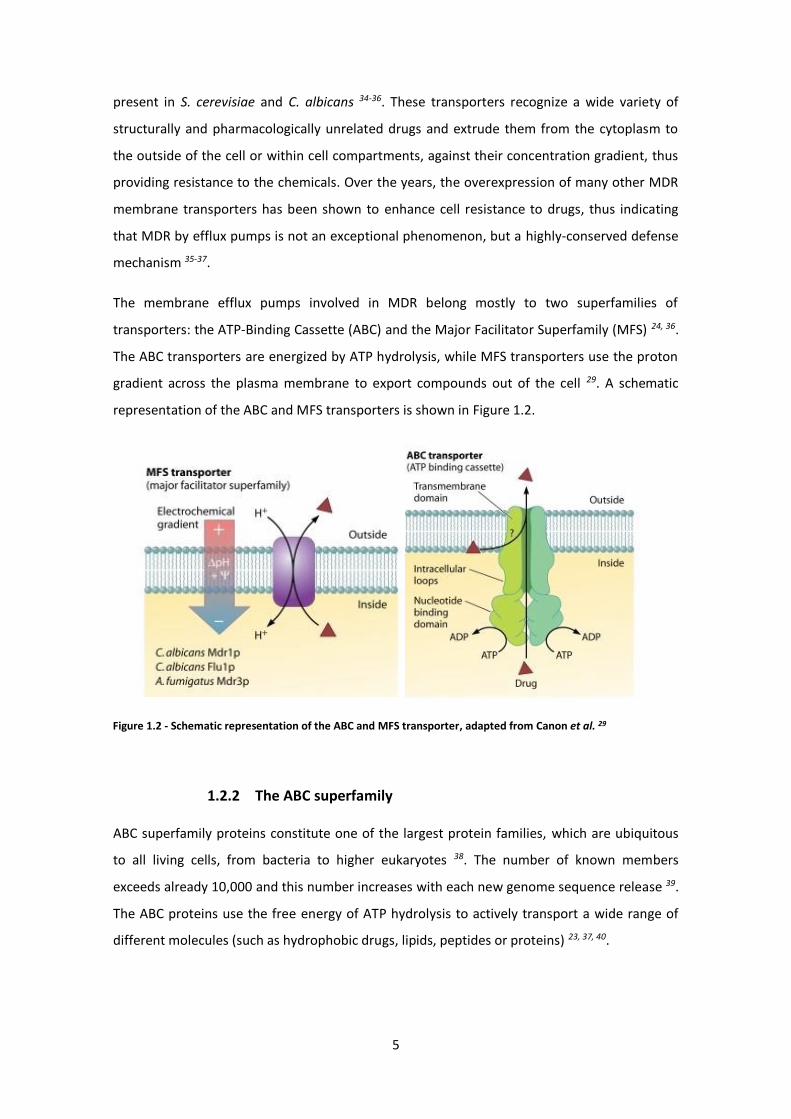
Figure 1.2 - Schematic representation of the ABC and MFS transporter, adapted from Canon et al. 29
present in S. cerevisiae and C. albicans 34-36. These transporters recognize a wide variety of
structurally and pharmacologically unrelated drugs and extrude them from the cytoplasm to
the outside of the cell or within cell compartments, against their concentration gradient, thus
providing resistance to the chemicals. Over the years, the overexpression of many other MDR
membrane transporters has been shown to enhance cell resistance to drugs, thus indicating
that MDR by efflux pumps is not an exceptional phenomenon, but a highly-conserved defense
mechanism 35-37.
The membrane efflux pumps involved in MDR belong mostly to two superfamilies of
transporters: the ATP-Binding Cassette (ABC) and the Major Facilitator Superfamily (MFS) 24, 36.
The ABC transporters are energized by ATP hydrolysis, while MFS transporters use the proton
gradient across the plasma membrane to export compounds out of the cell 29. A schematic
representation of the ABC and MFS transporters is shown in Figure 1.2.
1.2.2 The ABC superfamily
ABC superfamily proteins constitute one of the largest protein families, which are ubiquitous
to all living cells, from bacteria to higher eukaryotes 38. The number of known members
exceeds already 10,000 and this number increases with each new genome sequence release 39.
The ABC proteins use the free energy of ATP hydrolysis to actively transport a wide range of
different molecules (such as hydrophobic drugs, lipids, peptides or proteins) 23, 37, 40.
6
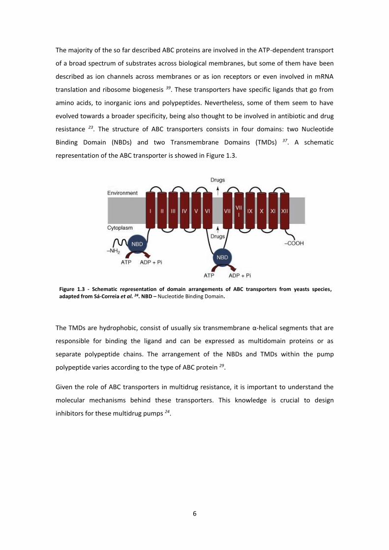
Figure 1.3 - Schematic representation of domain arrangements of ABC transporters from yeasts species, adapted from Sá-Correia et al. 24. NBD – Nucleotide Binding Domain.
The majority of the so far described ABC proteins are involved in the ATP-dependent transport
of a broad spectrum of substrates across biological membranes, but some of them have been
described as ion channels across membranes or as ion receptors or even involved in mRNA
translation and ribosome biogenesis 39. These transporters have specific ligands that go from
amino acids, to inorganic ions and polypeptides. Nevertheless, some of them seem to have
evolved towards a broader specificity, being also thought to be involved in antibiotic and drug
resistance 23. The structure of ABC transporters consists in four domains: two Nucleotide
Binding Domain (NBDs) and two Transmembrane Domains (TMDs) 37. A schematic
representation of the ABC transporter is showed in Figure 1.3.
The TMDs are hydrophobic, consist of usually six transmembrane α-helical segments that are
responsible for binding the ligand and can be expressed as multidomain proteins or as
separate polypeptide chains. The arrangement of the NBDs and TMDs within the pump
polypeptide varies according to the type of ABC protein 29.
Given the role of ABC transporters in multidrug resistance, it is important to understand the
molecular mechanisms behind these transporters. This knowledge is crucial to design
inhibitors for these multidrug pumps 24.
7
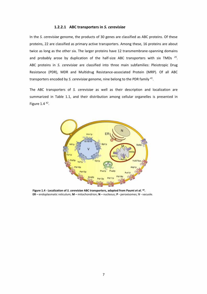
Figure 1.4 - Localization of S. cerevisiae ABC transporters, adapted from Paumi et al. 42. ER – endoplasmatic reticulum; M – mitochondrion; N – nucleous; P - peroxisomes; V - vacuole.
1.2.2.1 ABC transporters in S. cerevisiae
In the S. cerevisiae genome, the products of 30 genes are classified as ABC proteins. Of these
proteins, 22 are classified as primary active transporters. Among these, 16 proteins are about
twice as long as the other six. The larger proteins have 12 transmembrane-spanning domains
and probably arose by duplication of the half-size ABC transporters with six TMDs 25.
ABC proteins in S. cerevisiae are classified into three main subfamilies: Pleiotropic Drug
Resistance (PDR), MDR and Multidrug Resistance-associated Protein (MRP). Of all ABC
transporters encoded by S. cerevisiae genome, nine belong to the PDR family 41.
The ABC transporters of S. cerevisiae as well as their description and localization are
summarized in Table 1.1, and their distribution among cellular organelles is presented in
Figure 1.4 42.
8
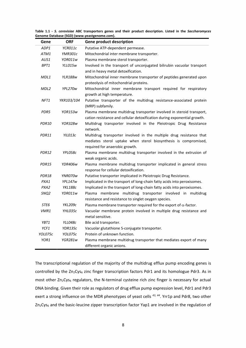
Table 1.1 - S. cerevisiae ABC transporters genes and their product description. Listed in the Saccharomyces Genome Database (SGD) (www.yeastgenome.com).
Gene ORF Gene product description
ADP1 YCR011c Putative ATP-dependent permease.
ATM1 YMR301c Mitochondrial inter membrane transporter.
AUS1 YOR011w Plasma membrane sterol transporter.
BPT1 YLL015w Involved in the transport of unconjugated bilirubin vacuolar transport
and in heavy metal detoxification.
MDL1 YLR188w Mitochondrial inner membrane transporter of peptides generated upon
proteolysis of mitochondrial proteins.
MDL2 YPL270w Mitochondrial inner membrane transport required for respiratory
growth at high temperature.
NFT1 YKR103/104 Putative transporter of the multidrug resistance-associated protein
(MRP) subfamily.
PDR5 YOR153w Plasma membrane multidrug transporter involved in steroid transport,
cation resistance and cellular detoxification during exponential growth.
PDR10 YOR328w Multidrug transporter involved in the Pleiotropic Drug Resistance
network.
PDR11 YIL013c Multidrug transporter involved in the multiple drug resistance that
mediates sterol uptake when sterol biosynthesis is compromised,
required for anaerobic growth.
PDR12 YPL058c Plasma membrane multidrug transporter involved in the extrusion of
weak organic acids.
PDR15 YDR406w Plasma membrane multidrug transporter implicated in general stress
response for cellular detoxification.
PDR18 YNR070w Putative transporter implicated in Pleiotropic Drug Resistance.
PXA1 YPL147w Implicated in the transport of long-chain fatty acids into peroxisomes.
PXA2 YKL188c Implicated in the transport of long-chain fatty acids into peroxisomes.
SNQ2 YDR011w Plasma membrane multidrug transporter involved in multidrug
resistance and resistance to singlet oxygen species.
STE6 YKL209c Plasma membrane transporter required for the export of -factor.
VMR1 YHL035c Vacuolar membrane protein involved in multiple drug resistance and
metal sensitive.
YBT1 YLL048c Bile acid transporter.
YCF1 YDR135c Vacuolar glutathione S-conjugate transporter.
YOL075c YOL075c Protein of unknown function.
YOR1 YGR281w Plasma membrane multidrug transporter that mediates export of many
different organic anions.
The transcriptional regulation of the majority of the multidrug efflux pump encoding genes is
controlled by the Zn2Cys6 zinc finger transcription factors Pdr1 and its homologue Pdr3. As in
most other Zn2Cys6 regulators, the N-terminal cysteine rich zinc finger is necessary for actual
DNA binding. Given their role as regulators of drug efflux pump expression level, Pdr1 and Pdr3
exert a strong influence on the MDR phenotypes of yeast cells 43, 44. Yrr1p and Pdr8, two other
Zn2Cys6 and the basic-leucine zipper transcription factor Yap1 are involved in the regulation of
9
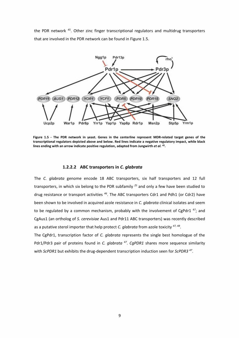
Figure 1.5 - The PDR network in yeast. Genes in the centerline represent MDR-related target genes of the transcriptional regulators depicted above and below. Red lines indicate a negative regulatory impact, while black lines ending with an arrow indicate positive regulation, adapted from Jungwirth et al. 45.
the PDR network 45. Other zinc finger transcriptional regulators and multidrug transporters
that are involved in the PDR network can be found in Figure 1.5.
1.2.2.2 ABC transporters in C. glabrata
The C. glabrata genome encode 18 ABC transporters, six half transporters and 12 full
transporters, in which six belong to the PDR subfamily 25 and only a few have been studied to
drug resistance or transport activities 46. The ABC transporters Cdr1 and Pdh1 (or Cdr2) have
been shown to be involved in acquired azole resistance in C. glabrata clinical isolates and seem
to be regulated by a common mechanism, probably with the involvement of CgPdr1 47; and
CgAus1 (an ortholog of S. cerevisiae Aus1 and Pdr11 ABC transporters) was recently described
as a putative sterol importer that help protect C. glabrata from azole toxicity 47, 48.
The CgPdr1, transcription factor of C. glabrata represents the single best homologue of the
Pdr1/Pdr3 pair of proteins found in C. glabrata 47. CgPDR1 shares more sequence similarity
with ScPDR1 but exhibits the drug-dependent transcription induction seen for ScPDR3 47.
10
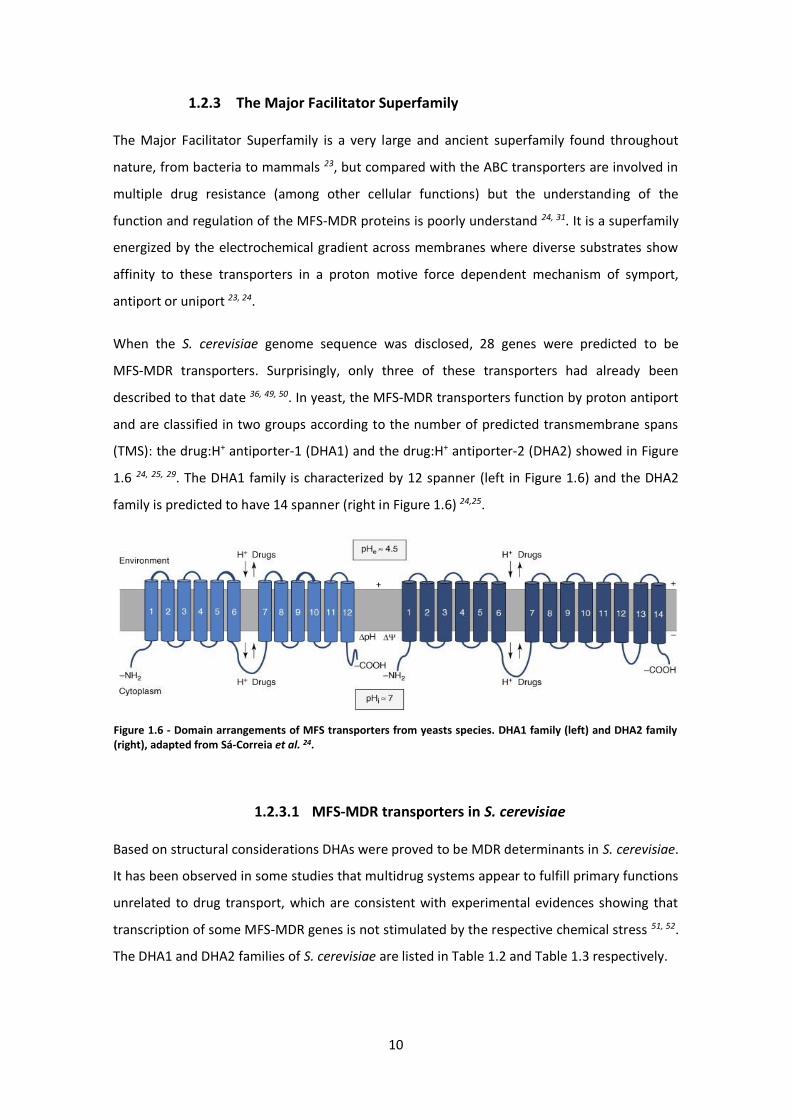
Figure 1.6 - Domain arrangements of MFS transporters from yeasts species. DHA1 family (left) and DHA2 family (right), adapted from Sá-Correia et al. 24.
1.2.3 The Major Facilitator Superfamily
The Major Facilitator Superfamily is a very large and ancient superfamily found throughout
nature, from bacteria to mammals 23, but compared with the ABC transporters are involved in
multiple drug resistance (among other cellular functions) but the understanding of the
function and regulation of the MFS-MDR proteins is poorly understand 24, 31. It is a superfamily
energized by the electrochemical gradient across membranes where diverse substrates show
affinity to these transporters in a proton motive force dependent mechanism of symport,
antiport or uniport 23, 24.
When the S. cerevisiae genome sequence was disclosed, 28 genes were predicted to be
MFS-MDR transporters. Surprisingly, only three of these transporters had already been
described to that date 36, 49, 50. In yeast, the MFS-MDR transporters function by proton antiport
and are classified in two groups according to the number of predicted transmembrane spans
(TMS): the drug:H+ antiporter-1 (DHA1) and the drug:H+ antiporter-2 (DHA2) showed in Figure
1.6 24, 25, 29. The DHA1 family is characterized by 12 spanner (left in Figure 1.6) and the DHA2
family is predicted to have 14 spanner (right in Figure 1.6) 24,25.
1.2.3.1 MFS-MDR transporters in S. cerevisiae
Based on structural considerations DHAs were proved to be MDR determinants in S. cerevisiae.
It has been observed in some studies that multidrug systems appear to fulfill primary functions
unrelated to drug transport, which are consistent with experimental evidences showing that
transcription of some MFS-MDR genes is not stimulated by the respective chemical stress 51, 52.
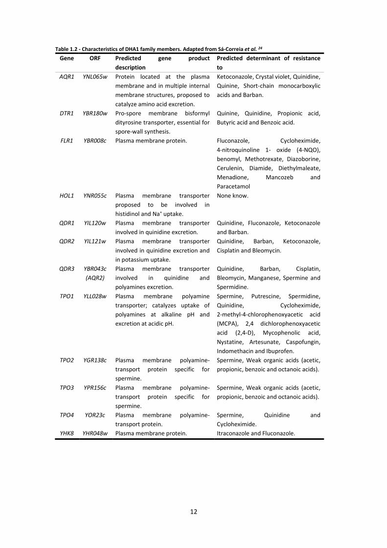
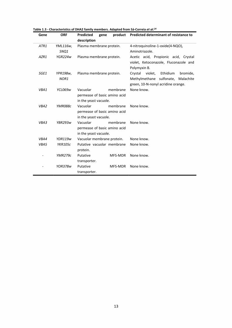
The DHA1 and DHA2 families of S. cerevisiae are listed in Table 1.2 and Table 1.3 respectively.

11
Figure 1.7 - Phylogenetic relationship of the DHA1 family (left) and the DHA2 family (right) in various yeast species. Genes from S. cerevisiae are black and the C. glabrata genes are blue. Adapted from Gbelska et al. 25.
When the genome of S. cerevisiae was analyzed, a classification in clusters of the DHA
transporters was obtained. Members of DHA1 family were segregated in two clusters (plus one
isolated protein – Hol1) and three clusters were discriminated for DHA2 family 49. Figure 1.7
displays the phylogenetic relationship of DHA1 and DHA2 proteins among several yeast species
highlighting the sequence similarities of S. cerevisiae transporters and those predicted to play
a similar role in C. glabrata 25.
12
Table 1.2 - Characteristics of DHA1 family members. Adapted from Sá-Correia et al. 24
Gene ORF Predicted gene product
description
Predicted determinant of resistance
to
AQR1 YNL065w Protein located at the plasma
membrane and in multiple internal
membrane structures, proposed to
catalyze amino acid excretion.
Ketoconazole, Crystal violet, Quinidine,
Quinine, Short-chain monocarboxylic
acids and Barban.
DTR1 YBR180w Pro-spore membrane bisformyl
dityrosine transporter, essential for
spore-wall synthesis.
Quinine, Quinidine, Propionic acid,
Butyric acid and Benzoic acid.
FLR1 YBR008c Plasma membrane protein. Fluconazole, Cycloheximide,
4-nitroquinoline 1- oxide (4-NQO),
benomyl, Methotrexate, Diazoborine,
Cerulenin, Diamide, Diethylmaleate,
Menadione, Mancozeb and
Paracetamol
HOL1 YNR055c Plasma membrane transporter
proposed to be involved in
histidinol and Na+ uptake.
None know.
QDR1 YIL120w Plasma membrane transporter
involved in quinidine excretion.
Quinidine, Fluconazole, Ketoconazole
and Barban.
QDR2 YIL121w Plasma membrane transporter
involved in quinidine excretion and
in potassium uptake.
Quinidine, Barban, Ketoconazole,
Cisplatin and Bleomycin.
QDR3 YBR043c
(AQR2)
Plasma membrane transporter
involved in quinidine and
polyamines excretion.
Quinidine, Barban, Cisplatin,
Bleomycin, Manganese, Spermine and
Spermidine.
TPO1 YLL028w Plasma membrane polyamine
transporter; catalyzes uptake of
polyamines at alkaline pH and
excretion at acidic pH.
Spermine, Putrescine, Spermidine,
Quinidine, Cycloheximide,
2-methyl-4-chlorophenoxyacetic acid
(MCPA), 2,4 dichlorophenoxyacetic
acid (2,4-D), Mycophenolic acid,
Nystatine, Artesunate, Caspofungin,
Indomethacin and Ibuprofen.
TPO2 YGR138c Plasma membrane polyamine-
transport protein specific for
spermine.
Spermine, Weak organic acids (acetic,
propionic, benzoic and octanoic acids).
TPO3 YPR156c Plasma membrane polyamine-
transport protein specific for
spermine.
Spermine, Weak organic acids (acetic,
propionic, benzoic and octanoic acids).
TPO4 YOR23c Plasma membrane polyamine-
transport protein.
Spermine, Quinidine and
Cycloheximide.
YHK8 YHR048w Plasma membrane protein. Itraconazole and Fluconazole.
13
Table 1.3 - Characteristics of DHA2 family members. Adapted from Sá-Correia et al.24
Gene ORF Predicted gene product
description
Predicted determinant of resistance to
ATR1 YML116w,
SNQ1
Plasma membrane protein. 4-nitroquinoline-1-oxide(4-NQO),
Aminotriazole.
AZR1 YGR224w Plasma membrane protein. Acetic acid, Propionic acid, Crystal
violet, Ketoconazole, Fluconazole and
Polymyxin B.
SGE1 YPR198w,
NOR1
Plasma membrane protein. Crystal violet, Ethidium bromide,
Methylmethane sulfonate, Malachite
green, 10-N-nonyl acridine orange.
VBA1 YCL069w Vacuolar membrane
permease of basic amino acid
in the yeast vacuole.
None know.
VBA2 YMR088c Vacuolar membrane
permease of basic amino acid
in the yeast vacuole.
None know.
VBA3 YBR293w Vacuolar membrane
permease of basic amino acid
in the yeast vacuole.
None know.
VBA4 YDR119w Vacuolar membrane protein. None know.
VBA5 YKR105c Putative vacuolar membrane
protein.
None know.
- YMR279c Putative MFS-MDR
transporter.
None know.
- YOR378w Putative MFS-MDR
transporter.
None know.
14
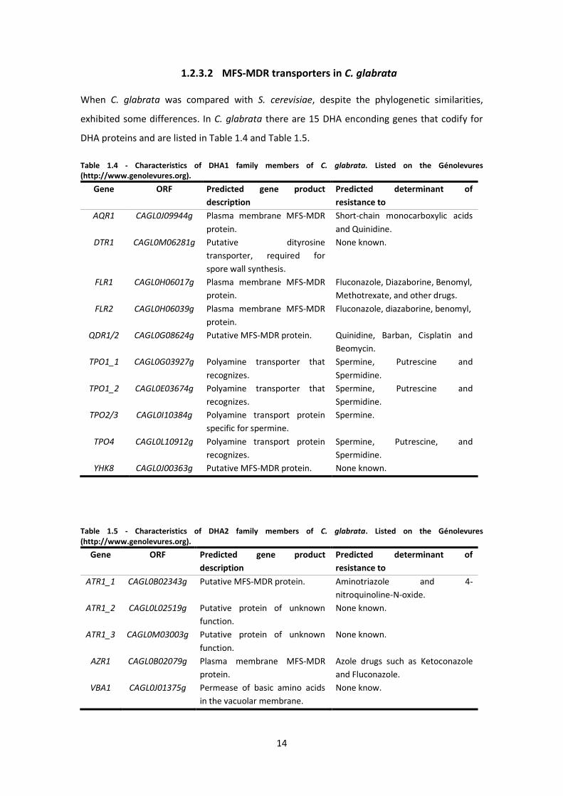
1.2.3.2 MFS-MDR transporters in C. glabrata
When C. glabrata was compared with S. cerevisiae, despite the phylogenetic similarities,
exhibited some differences. In C. glabrata there are 15 DHA enconding genes that codify for
DHA proteins and are listed in Table 1.4 and Table 1.5.
Table 1.4 - Characteristics of DHA1 family members of C. glabrata. Listed on the Génolevures (http://www.genolevures.org).
Gene ORF Predicted gene product
description
Predicted determinant of
resistance to
AQR1 CAGL0J09944g Plasma membrane MFS-MDR
protein.
Short-chain monocarboxylic acids
and Quinidine.
DTR1 CAGL0M06281g Putative dityrosine
transporter, required for
spore wall synthesis.
None known.
FLR1 CAGL0H06017g Plasma membrane MFS-MDR
protein.
Fluconazole, Diazaborine, Benomyl,
Methotrexate, and other drugs.
FLR2 CAGL0H06039g Plasma membrane MFS-MDR
protein.
Fluconazole, diazaborine, benomyl,
QDR1/2 CAGL0G08624g Putative MFS-MDR protein. Quinidine, Barban, Cisplatin and
Beomycin.
TPO1_1 CAGL0G03927g Polyamine transporter that
recognizes.
Spermine, Putrescine and
Spermidine.
TPO1_2 CAGL0E03674g Polyamine transporter that
recognizes.
Spermine, Putrescine and
Spermidine.
TPO2/3 CAGL0I10384g Polyamine transport protein
specific for spermine.
Spermine.
TPO4 CAGL0L10912g Polyamine transport protein
recognizes.
Spermine, Putrescine, and
Spermidine.
YHK8 CAGL0J00363g Putative MFS-MDR protein. None known.
Table 1.5 - Characteristics of DHA2 family members of C. glabrata. Listed on the Génolevures (http://www.genolevures.org).
Gene ORF Predicted gene product
description
Predicted determinant of
resistance to
ATR1_1 CAGL0B02343g Putative MFS-MDR protein. Aminotriazole and 4-
nitroquinoline-N-oxide.
ATR1_2 CAGL0L02519g Putative protein of unknown
function.
None known.
ATR1_3 CAGL0M03003g Putative protein of unknown
function.
None known.
AZR1 CAGL0B02079g Plasma membrane MFS-MDR
protein.
Azole drugs such as Ketoconazole
and Fluconazole.
VBA1 CAGL0J01375g Permease of basic amino acids
in the vacuolar membrane.
None know.
15
There are two predicted TPO1 orthologues and two predicted FLR1 orthologs in the C. glabrata
genome. ScFLR1 homologs in C. glabrata include ORFs CAGL0H06017g (CgFLR1) and
CAGL0H06039g (CgFLR2), with 59 % and 54 % identity between their deduced amino acid
sequences, respectively. On the other hand, ScTPO1 has two TPO1 homologs, CAGL0G03927g
(CgTPO1_1) and CAGL0E03674g (CgTPO1_2), in C. glabrata with 70 % and 73 % identity
between their deduced amino acid sequences, respectively 53.
FLR1 is known as a Yap1 target gene and has three Yap1 response elements (YREs) in its
promoter region at positions -148 (YRE1), -167 (YRE2), and -364 (YRE3) 54. YRE3 is a palindrome
known to function on either strand of many YAP1 target genes and is found upstream of
several homologous genes in C. glabrata including CgFLR1. However, none of the YRE
consensus sequences was found within the 1-kb upstream region of CgFLR2 54.
According to Chen et al. 55 CgFLR1 was considered to be the closest FLR1 homolog in
C. glabrata and the disruption of CgFLR1 did not affect sensitivity to fluconazole,
cycloheximide, 4-NQO, hydrogen peroxide, cerulenin, cadmium or chloramphenicol. The
CgFLR1 deletion led to only very minor increases in sensitivity to benomyl, diamide and
menadione. An important fact CgFlr1 was involved in benomyl resistance in C. glabrata and
CgYap1 regulates the transcription of CgFLR1_1 gene in response to benomyl stress 55.
1.2.4 The CgPdr1 transcription factor
The C. glabrata Pdr1 transcription factor is the single ortholog of two S. cerevisiae zinc cluster
transcription factors ScPdr1 and ScPdr3, which regulates the expression of genes involved in
multidrug resistance 56. Similarly to Pdr1 in S. cerevisiae, CgPdr1 may bind constitutively to the
promoters of a repertoire of genes and activate a subset of them, depending on the
mechanism by which it is activated 56.
CgPdr1 has been linked to the clinical acquisition of azole antifungal drugs resistance 57, since
single point mutations in the functional domains of CgPdr1, called gain-of-function (GOF)
mutations, have been found in numerous azole resistant C. glabrata clinical isolates, these
point mutations resulting in constitutive increased transcription of the drug efflux pump
encoding genes CgCDR1, CgPDH1 and CgSNQ2 58. Interestingly, these GOF mutations in CgPdr1
were further found to be important for pathogen-host interactions, as they are associated with
enhanced virulence 58, 59.
16
1.3 Antifungal Drugs and Fungicides explored in the course of this
study
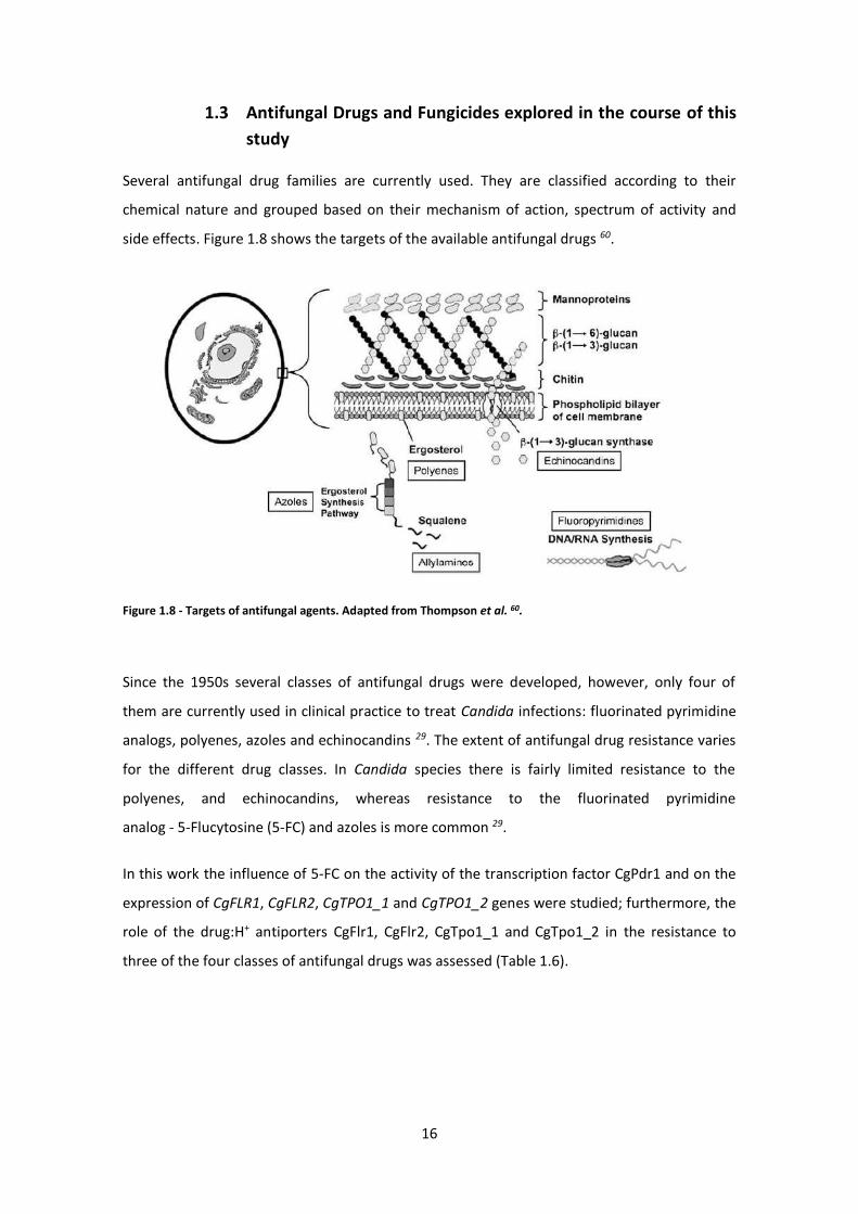
Several antifungal drug families are currently used. They are classified according to their
chemical nature and grouped based on their mechanism of action, spectrum of activity and
side effects. Figure 1.8 shows the targets of the available antifungal drugs 60.
Figure 1.8 - Targets of antifungal agents. Adapted from Thompson et al. 60.
Since the 1950s several classes of antifungal drugs were developed, however, only four of
them are currently used in clinical practice to treat Candida infections: fluorinated pyrimidine
analogs, polyenes, azoles and echinocandins 29. The extent of antifungal drug resistance varies
for the different drug classes. In Candida species there is fairly limited resistance to the
polyenes, and echinocandins, whereas resistance to the fluorinated pyrimidine
analog - 5-Flucytosine (5-FC) and azoles is more common 29.
In this work the influence of 5-FC on the activity of the transcription factor CgPdr1 and on the
expression of CgFLR1, CgFLR2, CgTPO1_1 and CgTPO1_2 genes were studied; furthermore, the
role of the drug:H+ antiporters CgFlr1, CgFlr2, CgTpo1_1 and CgTpo1_2 in the resistance to
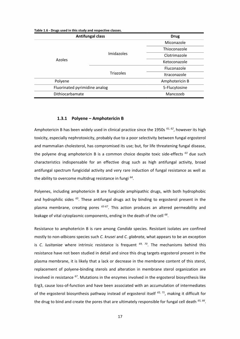
three of the four classes of antifungal drugs was assessed (Table 1.6).
17
Table 1.6 - Drugs used in this study and respective classes.
Antifungal class Drug
Azoles
Imidazoles
Miconazole
Thioconazole
Clotrimazole
Ketoconazole
Triazoles Fluconazole
Itraconazole
Polyene Amphotericin B
Fluorinated pyrimidine analog 5-Flucytosine
Dithiocarbamate Mancozeb
1.3.1 Polyene – Amphotericin B
Amphotericin B has been widely used in clinical practice since the 1950s 61, 62, however its high
toxicity, especially nephrotoxicity, probably due to a poor selectivity between fungal ergosterol
and mammalian cholesterol, has compromised its use; but, for life threatening fungal disease,
the polyene drug amphotericin B is a common choice despite toxic side-effects 63 due such
characteristics indispensable for an effective drug such as high antifungal activity, broad
antifungal spectrum fungicidal activity and very rare induction of fungal resistance as well as
the ability to overcome multidrug resistance in fungi 64.
Polyenes, including amphotericin B are fungicide amphipathic drugs, with both hydrophobic
and hydrophilic sides 65. These antifungal drugs act by binding to ergosterol present in the
plasma membrane, creating pores 65-67. This action produces an altered permeability and
leakage of vital cytoplasmic components, ending in the death of the cell 68.
Resistance to amphotericin B is rare among Candida species. Resistant isolates are confined
mostly to non-albicans species such C. krusei and C. glabrata, what appears to be an exception
is C. lusitaniae where intrinsic resistance is frequent 69, 70. The mechanisms behind this
resistance have not been studied in detail and since this drug targets ergosterol present in the
plasma membrane, it is likely that a lack or decrease in the membrane content of this sterol,
replacement of polyene-binding sterols and alteration in membrane sterol organization are
involved in resistance 67. Mutations in the enzymes involved in the ergosterol biosynthesis like
Erg3, cause loss-of-function and have been associated with an accumulation of intermediates
of the ergosterol biosynthesis pathway instead of ergosterol itself 65, 71, making it difficult for
the drug to bind and create the pores that are ultimately responsible for fungal cell death 65, 66.
18
The ERG6 gene also seems to be involved in the resistance to polyenes since the mutation in
the C. glabrata ERG6 gene was found to result in its poor polyene susceptibility 72.
1.3.2 Azoles
Azoles are five-membered nitrogen heterocyclic ring compounds containing at least one other
non-carbon atom of either nitrogen, sulfur or oxygen and are the most used today when
treating fungal infections, especially, topical imidazoles for mucosal or skin infections and
oral-parenteral triazoles for invasive and refractory mucosal infections 47. The azoles family can
be classified into two groups: the imidazoles (clotrimazole, miconazole, ketoconazole and
thioconazole) and the triazoles (fluconazole, itraconazole, voriconazole and posaconazole) 73
and the big difference between these two groups is the mechanism of inhibition of the
cytochrome P-450 (CYP) dependent lanosterol 14-α-demethylase, while in imidazoles
the N3 compound binds to the heme iron CYP, the N4 of the triazoles bind to the heme group.
This confers to triazoles comparatively to imidazoles higher specificity 47.
Within the subfamily of the imidazole antifungal drugs, clotrimazole is widely used in
treatment of superficial mycoses 74, vaginal infections and oral thrush 75. Discovered in 1969,
cannot be given parenterally because has poor oral absorption 76. Ketoconazole is a synthetic
antifungal drug normally used for life threatening systemic infections 77 and used mostly to
prevent and treat fungal skin infections, especially in immunocompromised patients 78.
Discovered in 1978, this antifungal drug presents good oral absorption and a broad spectrum
of activity 76. Miconazole acts by combination of two mechanisms: ergosterol biosynthesis
inhibition, the direct mechanisms of action of azole antifungal drugs, and direct membrane
damage of the fungal cells 79. Discovered in 1969, has poor bioavailability because of its poor
dissolution and absorption in the gastrointestinal tract, but is a useful topical drug for the
treatment of superficial mycoses; however, it is also given as a systemic antifungal agent when
amphotericin B or ketoconazole are either ineffective or contraindicated 76, 80. Thioconazole is
mostly used in treatment of topical mycoses, in particular women’s yeast vaginal
infections 74, 78.
Within the subfamily of the triazole antifungal drugs, fluconazole is currently the most widely
used antifungal azole drug due to excellent bioavailability, tolerability and low-level side
effects. It is active against most Candida species with the exception of C. glabrata and C. krusei
isolates 60. Fluconazole was formulated in 1981 and it is available in both oral and intravenous

19
formulations, which have identical pharmacokinetics 76, 81. Itraconazole, belonging to the the
same class as fluconazole, is used to treat superficial and systemic fungal infections 60.
Discovered in 1986 it is an antifungal drug with broad spectrum of activity, good availability
but is only available in oral form 60, 76.
The increase of the prophylactic use of azoles in recent years has led to an escalation of azole
drug resistance occurrences 71. The emergence of C. glabrata as one of the most prevalent
pathogens responsible for candidemia parallels the introduction in the early 1990s of triazoles
and of many imidazoles 47.
The mode of action of azole antifungal drug is based on the ergosterol biosynthesis pathway
inhibition. Azoles act by inhibiting the CYP-dependent enzyme lanosterol 14α-demethylase,
encoded by ERG11, necessary for the conversion of lanosterol to ergosterol 73. This leads to the
depletion of the ergosterol membrane content and to the accumulation of ergosterol
precursors, such as toxic 14-α-methylated sterols (lanosterol, 4,14- dimethylzymosterol and
24-methylenedihydrolanosterol) 82. The absence of ergosterol production leads to significant
damage to the cell by increasing the cell membrane permeability, which can cause cell lysis
and death 66.
Three mechanisms of secondary azole resistance have been described in C. albicans: reduced
azole accumulation by active extrusion of the drug, alteration or overexpression of the binding
site (lanosterol 14-α-demethylase, encoded by ERG11) and loss-of-function of enzymes
downstream the ergosterol pathway (defective Δ-5,6-desaturase, encoded by ERG3), allowing
the accumulation of less toxic sterols in the presence of azoles 33.
C. glabrata has emerged as a common cause of fungal infection and it is reported that this
yeast has intrinsically low susceptibility to azole antifungals such as fluconazole 47. In
C. albicans the most prevalent mechanisms of resistance is active efflux depending on the
genes that defined a role in the azole efflux 83. CDR1 and CDR2 code for ABC transporters seem
to reduce accumulation of many azoles and MDR1, encoding an MFS-MDR transporter,
reduces accumulation of fluconazole 33.
Fluconazole is the most used azole antifungal drug due to low toxicity, availability in both oral
and intravenous formulations and excellent activity versus most yeast species. 5-Flucytosine
represents an attractive alternative or complement to azoles due to its excellent activity
against C. glabrata 84.
20
1.3.3 Fluoropyrimidine – 5-Flucytosine
Pyrimidine analogs comprise a unique representative, 5-fluorocytosine (5-FC) 85 or flucytosine
which is a synthetic antimycotic drug, was first synthetized in 1957 but its antifungal properties
discovered in 1964 84. 5-Flucytosine has emerged as an attractive alternative or complement to
azoles because it reveals an excellent activity against C. glabrata isolates 84, but it cannot be
used in monotherapy and should always be combined with another antifungal, usually an azole
or polyene drug 85. It can be administrated both orally and intravenously and it is well tolerated
in moderated doses, but when it is used in high doses it can be toxic 84 and the conversion
of 5-FC to fluorouracil by gut bacteria contributes to its elevated toxicity 86.
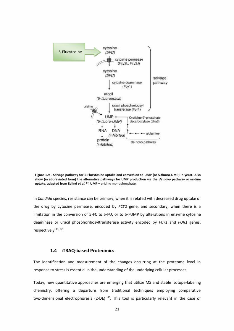
5-Flucytosine is metabolized via the pyrimidine salvage pathway (Figure 1.9 – adapted from
Edlind et al. 84) where it acts as a subversive substrate with the subsequent production of toxic
nucleotides and disruption of DNA and protein synthesis 84, 87. Studies on susceptible fungi, as
S. cerevisiae as a model, shows that 5-FC is taken up into the cell by one or more cytosine
permeases (the most relevant is FCY2 88) where it is converted to 5-fluorouracil (5-FU) by
cytosine deaminase (encoded by FCY1) 89. Subsequent modifications to 5-fluorouridine
monophosphate (5-fluoro-UMP) by uracil phosphoribosyl transferase (UPRT) (encoded
by FUR1) and to 5-fluoro-dUMP ultimately result in the disruption of protein and DNA
synthesis 82, 84, 87.
21
In Candida species, resistance can be primary, when it is related with decreased drug uptake of
the drug by cytosine permease, encoded by FCY2 gene, and secondary, when there is a
limitation in the conversion of 5-FC to 5-FU, or to 5-FUMP by alterations in enzyme cytosine
deaminase or uracil phosphoribosyltransferase activity encoded by FCY1 and FUR1 genes,
respectively 33, 67.
1.4 iTRAQ-based Proteomics
The identification and measurement of the changes occurring at the proteome level in
response to stress is essential in the understanding of the underlying cellular processes.
Today, new quantitative approaches are emerging that utilize MS and stable isotope-labeling
chemistry, offering a departure from traditional techniques employing comparative
two-dimensional electrophoresis (2-DE) 90. This tool is particularly relevant in the case of
5-Flucytosine
Figure 1.9 - Salvage pathway for 5-Flucytosine uptake and conversion to UMP (or 5-fluoro-UMP) in yeast. Also show (in abbreviated form) the alternative pathways for UMP production via the de novo pathway or uridine uptake, adapted from Edlind et al. 84. UMP – uridine monophosphate.
22
membrane proteins, which are not detectable in 2-DE gels, because they are mostly insoluble
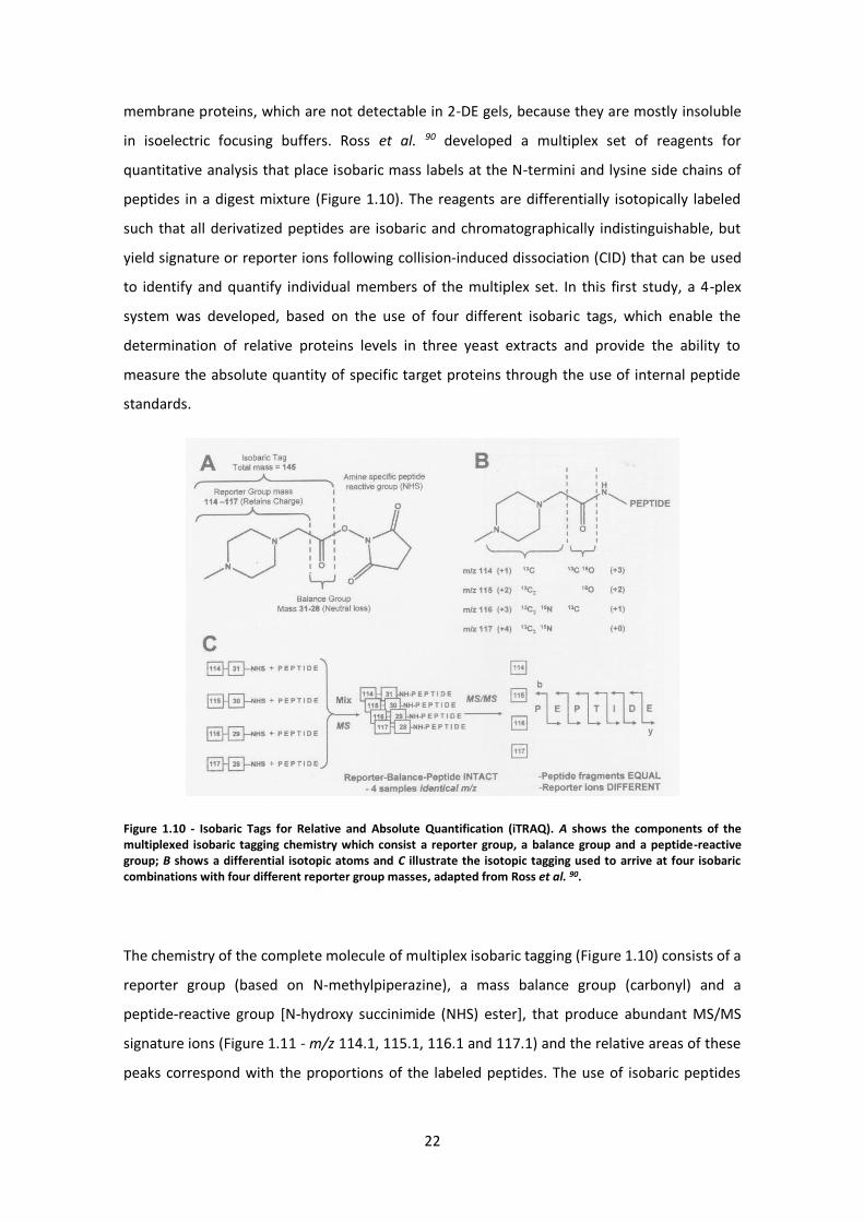
in isoelectric focusing buffers. Ross et al. 90 developed a multiplex set of reagents for
quantitative analysis that place isobaric mass labels at the N-termini and lysine side chains of
peptides in a digest mixture (Figure 1.10). The reagents are differentially isotopically labeled
such that all derivatized peptides are isobaric and chromatographically indistinguishable, but
yield signature or reporter ions following collision-induced dissociation (CID) that can be used
to identify and quantify individual members of the multiplex set. In this first study, a 4-plex
system was developed, based on the use of four different isobaric tags, which enable the
determination of relative proteins levels in three yeast extracts and provide the ability to
measure the absolute quantity of specific target proteins through the use of internal peptide
standards.
Figure 1.10 - Isobaric Tags for Relative and Absolute Quantification (iTRAQ). A shows the components of the multiplexed isobaric tagging chemistry which consist a reporter group, a balance group and a peptide-reactive group; B shows a differential isotopic atoms and C illustrate the isotopic tagging used to arrive at four isobaric combinations with four different reporter group masses, adapted from Ross et al. 90.
The chemistry of the complete molecule of multiplex isobaric tagging (Figure 1.10) consists of a
reporter group (based on N-methylpiperazine), a mass balance group (carbonyl) and a
peptide-reactive group [N-hydroxy succinimide (NHS) ester], that produce abundant MS/MS
signature ions (Figure 1.11 - m/z 114.1, 115.1, 116.1 and 117.1) and the relative areas of these
peaks correspond with the proportions of the labeled peptides. The use of isobaric peptides
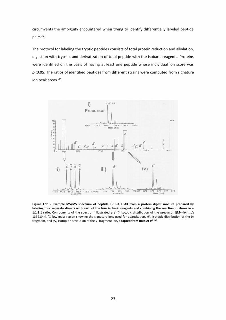
23
circumvents the ambiguity encountered when trying to identify differentially labeled peptide
pairs 90.
The protocol for labeling the tryptic peptides consists of total protein reduction and alkylation,
digestion with trypsin, and derivatization of total peptide with the isobaric reagents. Proteins
were identified on the basis of having at least one peptide whose individual ion score was
p0.05. The ratios of identified peptides from different strains were computed from signature
ion peak areas 90.
Figure 1.11 - Example MS/MS spectrum of peptide TPHPALTEAK from a protein digest mixture prepared by labeling four separate digests with each of the four isobaric reagents and combining the reaction mixtures in a 1:1:1:1 ratio. Components of the spectrum illustrated are (i) isotopic distribution of the precursor [(M+H)+, m/z 1352,84)], (ii) low mass region showing the signature ions used for quantitation, (iii) isotopic distribution of the b6 fragment, and (iv) isotopic distribution of the y7 fragment ion, adapted from Ross et al. 90.
24
2 Materials and Methods
2.1 Cell Culture
2.1.1 Strains
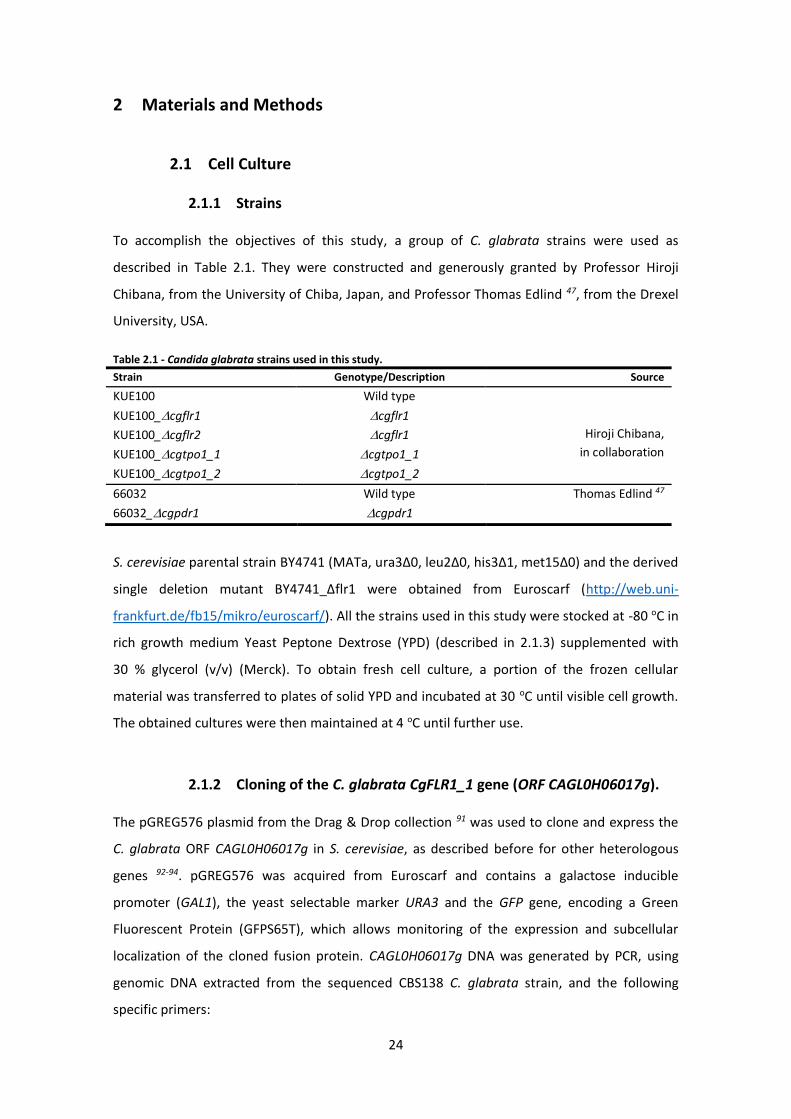
To accomplish the objectives of this study, a group of C. glabrata strains were used as
described in Table 2.1. They were constructed and generously granted by Professor Hiroji
Chibana, from the University of Chiba, Japan, and Professor Thomas Edlind 47, from the Drexel
University, USA.
Table 2.1 - Candida glabrata strains used in this study.
Strain Genotype/Description Source
KUE100 Wild type
Hiroji Chibana,
in collaboration
KUE100_cgflr1 cgflr1
KUE100_cgflr2 cgflr1
KUE100_cgtpo1_1 cgtpo1_1
KUE100_cgtpo1_2 cgtpo1_2
66032 Wild type Thomas Edlind 47
66032_cgpdr1 cgpdr1
S. cerevisiae parental strain BY4741 (MATa, ura3Δ0, leu2Δ0, his3Δ1, met15Δ0) and the derived
single deletion mutant BY4741_Δflr1 were obtained from Euroscarf (http://web.uni-
frankfurt.de/fb15/mikro/euroscarf/). All the strains used in this study were stocked at -80 oC in
rich growth medium Yeast Peptone Dextrose (YPD) (described in 2.1.3) supplemented with
30 % glycerol (v/v) (Merck). To obtain fresh cell culture, a portion of the frozen cellular
material was transferred to plates of solid YPD and incubated at 30 oC until visible cell growth.
The obtained cultures were then maintained at 4 oC until further use.
2.1.2 Cloning of the C. glabrata CgFLR1_1 gene (ORF CAGL0H06017g).
The pGREG576 plasmid from the Drag & Drop collection 91 was used to clone and express the
C. glabrata ORF CAGL0H06017g in S. cerevisiae, as described before for other heterologous
genes 92-94. pGREG576 was acquired from Euroscarf and contains a galactose inducible
promoter (GAL1), the yeast selectable marker URA3 and the GFP gene, encoding a Green
Fluorescent Protein (GFPS65T), which allows monitoring of the expression and subcellular
localization of the cloned fusion protein. CAGL0H06017g DNA was generated by PCR, using
genomic DNA extracted from the sequenced CBS138 C. glabrata strain, and the following
specific primers:

25
5’ – GAATTCGATATCAAGCTTATCGATACCGTCGACAATGTATATCGGTGCATTTCAGGAC - 3’ and
5’ – GCGTGACATAACTAATTACATGACTCGAGGTCGACTCATGAATCTGGACTAAATCTTG - 3’.
The designed primers contain, besides a region with homology to the first 24 and last
23 nucleotides of the CAGL0H06017g coding region (italic), nucleotide sequences with
homology to the cloning site flanking regions of the pGREG576 vector (underlined). The
amplified fragment was co-transformed into the parental S. cerevisiae strain BY4741 with the
pGREG576 vector, previously cut with the restriction enzyme SalI, to obtain the
pGREG576_CgFLR1 plasmid. The recombinant plasmid pGREG576_CgFLR1 was obtained
through homologous recombination in S. cerevisiae and verified by DNA sequencing. The GAL1
promoter present in the pGREG576_CgFLR1 plasmid was then replaced by the copper-induced
MTI C. glabrata promoter, giving rise to the pGREG576_MTI_CgFLR1 plasmid. The MTI
promoter DNA was generated by PCR, using genomic DNA extracted from the sequenced
CBS138 C. glabrata strain, and the following specific primers:
5′-TTAACCCTCACTAAAGGGAACAAAAGCTGGAGCTCTGTACGACACGCATCATGTGGCAATC -3′ and
5′-GAAAAGTTCTTCTCCTTTACTCATACTAGTGCGGCTGTGTTTGTTTTTGTATGTGTTTGTTG -3′. The
recombinant plasmids pGREG576_CgFLR1, and pGREG576_MTI_CgFLR1 were obtained
through homologous recombination in S. cerevisiae and verified by DNA sequencing.
2.1.3 Growth media
The rich medium used for the cultivation of C. glabrata cells was YPD, containing, per litre:
20 g glucose (Merck); 20 g yeast extract (Himedia); 10 g peptone (Difco). C. glabrata cells
minimal growth medium – MMG – containing, per litre: 20 g glucose (Merck); 2.7 g (NH4)2SO4
(Merck); 1.7 g yeast nitrogen base (Difco) without amino acids or (NH4)2SO4. No amino acids
were added to this medium. S. cerevisiae cells were batch-cultured at 30 °C, with orbital
agitation (250 rpm) in MMB (Minimal Medium for BY4741) medium, with the following
composition (per liter): 1.7 g yeast nitrogen base without amino acids or NH4+ (Difco, Franklin
Lakes, NJ), 20 g glucose (Merck, Kenilworth, NJ), and 2.65 g (NH4)2SO4 (Merck), 20 mg / L
methionine, 20 mg / L histidine, 60 mg / L leucine, 20 mg / L uracil (all from Sigma).
All the previous referred growth media were prepared in deionized water and autoclaved for
15 min at 121 oC and 1 atm. Solid media, derived from any of the above, were prepared by
adding 20 g.L-1 agar (Iberagar).
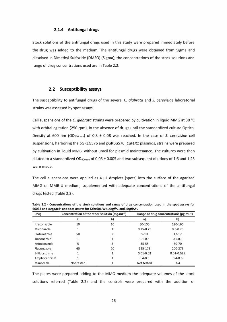
26
2.1.4 Antifungal drugs
Stock solutions of the antifungal drugs used in this study were prepared immediately before
the drug was added to the medium. The antifungal drugs were obtained from Sigma and
dissolved in Dimethyl Sulfoxide (DMSO) (Sigma); the concentrations of the stock solutions and
range of drug concentrations used are in Table 2.2.
2.2 Susceptibility assays
The susceptibility to antifungal drugs of the several C. glabrata and S. cerevisiae laboratorial
strains was assessed by spot assays.
Cell suspensions of the C. glabrata strains were prepared by cultivation in liquid MMG at 30 oC
with orbital agitation (250 rpm), in the absence of drugs until the standardized culture Optical
Density at 600 nm (OD600 nm) of 0.8 ± 0.08 was reached. In the case of S. cerevisiae cell
suspensions, harboring the pGREG576 and pGREG576_CgFLR1 plasmids, strains were prepared
by cultivation in liquid MMB, without uracil for plasmid maintenance. The cultures were then
diluted to a standardized OD600 nm of 0.05 ± 0.005 and two subsequent dilutions of 1:5 and 1:25
were made.
The cell suspensions were applied as 4 µL droplets (spots) into the surface of the agarized
MMG or MMB-U medium, supplemented with adequate concentrations of the antifungal
drugs tested (Table 2.2).
Table 2.2 - Concentrations of the stock solutions and range of drug concentration used in the spot assays for
66032 and cgpdr1a and spot assays for Kchr606 Wt, cgflr1 and cgflr2b.
Drug Concentration of the stock solution (mg.mL-1) Range of drug concentrations (µg.mL-1)
a) b) a) b)
Itraconazole 10 10 60-100 120-160
Miconazole 1 1 0.25-0.75 0.5-0.75
Clotrimazole 50 50 5-10 12-17
Tioconazole 1 1 0.1-0.5 0.5.0.9
Ketoconazole 5 5 35-55 60-70
Fluconazole 60 20 125-175 200-275
5-Flucytosine 1 1 0.01-0.02 0.01-0.025
Amphotericin B 1 1 0.4-0.6 0.4-0.6
Mancozeb Not tested 1 Not tested 3-4
The plates were prepared adding to the MMG medium the adequate volumes of the stock
solutions referred (Table 2.2) and the controls were prepared with the addition of
27
DMSO (Sigma) but without the addition of drugs. After inoculation, the agar plates were
incubated at 30 oC for 2 to 5 days, during such period the growth as visually inspected.
2.3 CgFlr1 Subcellular Localization Assessment
The subcellular localization of the CgFlr1 protein was determined based on the observation of
BY4741 S. cerevisiae or KUE100. C. glabrata cells transformed with pGREG576_CgFLR1. These
cells express the CgFlr1_GFP fusion proteins, whose localization was determined using
fluorescence microscopy. Before that, the plasmids placed into the cell by the following
method: Inoculate 5 mL of MMB-U with a colony 2–3 mm in diameter from a fresh plate and
grow overnight at 30˚C with shaking (16-18 h). Dilute to an OD600 nm of 0.2 ± 0.02 in 100 mL of
fresh MMB-U and continue to grow until the OD600 nm reaches 0.6 ± 0.06. Place cells in 50 ml
tubes and centrifuge (1000 rpm, 5 min). Discard supernatants and thoroughly resuspend the
cell pellets in 25 mL of distilled H2O. Pool and centrifuge again (1000 rpm, 5 min). Decant
supernatant and resuspend the cell pellet in 1 ml of freshly prepared, sterile 100 mM LiAc in
TE. Transfer the cells to a 1.5 mL tube and centrifuge at top speed for 5 s. Remove the
supernatant and add 500 µL of 100 mM LiAc in TE. In a separate 1.5 mL tube add 0.1 µg of
plasmid DNA and 100 µg of salmon sperm DNA. Add 100 µL of yeast competent cells to each
tube and mix well by vortexing. Add 600 µL of sterile freshly made PEG/LiAc (40 % PEG +
100 mM LiAc in 1x TE) solution to each tube and vortex at high speed for 10 s to mix. Incubate
at 30°C for 30 min with shaking at 200 rpm. Add 70 µL of DMSO. Mix well by gentle inversion
whitout vortex. Heat shock for 15 min in a 42°C water bath. Chill cells on ice for 1–2 min.
Centrifuge cells (5 s, 14000 rpm) and remove the supernatant and resuspend the cells in 0.5
mL of sterile 1xTE buffer. Plate 100 µl and incubate plates up side down at 30°C until colonies
appear.
S. cerevisiae cell suspension was prepared by cultivation in MMB-U medium, containing 0.5 %
glucose and 0.1 % galactose, at 30 oC, with orbital shaking (250 rpm) until a standard culture
OD600 nm of 0.4 ± 0.04 was reached. At this point, cells were transferred to the same medium
containing 0.1 % glucose and 1 % galactose, to induce protein expression.
C. glabrata cell suspensions were prepared in MMB-U medium, until a standard culture
OD600 nm of 0.4 ± 0.04 was reached and transferred to the same medium supplemented with
50 µm CuSO4 (Sigma), to induce protein overexpression.
28
After 5 h of incubation, the distribution of CgFlr1_GFP fusion protein in S. cerevisiae or in
C. glabrata living cells was detected by fluorescence microscopy in a Zeiss Axioplan microscope
(Carl Zeiss MicroImaging, Oberkochen, Germany), using excitation and emission wavelength
of 395 and 509 nm, respectively. Fluorescence images were captured using a cooled CCD
camera (Cool SNAPFX, Roper Scientific Photometrics, Sarasota, FL).
2.4 Extraction and Multiplexed Membrane Protein Quantitation
2.4.1 Cell cultivation
Wild-type 66032 and cgpdr1 deletion mutant were cultured in 500 mL liquid MMG at 30 oC
with orbital agitation (250 rpm) in the absence of drugs until the standardized culture
OD600 nm of 0.8 ± 0.08 was reached. Adequate volumes of cellular suspensions were then
harvested by filtration in order to obtain an OD600 nm of 0.4 ± 0.04 for a volume of 1 L of liquid
MMG medium. Cells were then transferred to fresh medium in the absence of stress (control
conditions) or in the presence of 5-Flucytosine 4 µg/mL, and cultured for 1 h at 30 oC with
orbital agitation (250 rpm). Cells were harvested by centrifugation at 8000 rpm for 5 min
at 4 oC. The cell pellet was stocked at -80 oC. This process was repeated to obtain independent
biological triplicates.
2.4.2 Membrane proteome extraction
The cell pellets were resuspend in 2 mL of A Buffer (for each 2 mL of A Buffer must joined
100 µL of ABC protease inhibitors and 20 µL of D protease inhibitor) and an equal volume of
glass beads was added. For 15 min, cycles of 30 s/30 s on ice/vortex were done, followed by
incubation on ice for 5 min. Then, 1 mL of A Buffer was added and 10 min of 30 s/30 s on
ice/vortex cycles were carried out. The mixture was centrifuged (8000 rpm, 5 min, 4 oC) and
the top phase collected and put on ice. The resulting pellet was resuspended in 2 mL
of A Buffer and subjected to cycles of 30 s/30 s on ice/vortex. The mixture was centrifuged
(8000 rpm, 5 min, 4 oC) and this supernatant was put together with previous. A Buffer was
added to this mix to a final volume of 8 mL. This final mix was ultracentrifuged on a Beckman
XL-90 ultracentrifuge at 24000 rpm, 90 min, 4 oC. The supernatant was removed and the pellet
was washed with 8 mL of Na2CO3 (0.1 M) and incubated on ice for 30 min with orbital agitation
(60 rpm). After that, the mixture was ultracentrifuged at 26000 rpm, 60 min, 4 oC and the top
29
phase removed. The pellet was again washed with 8 mL of 50 mM Tetraethylammonium
bromide (TEAB) and ultracentrifuged. This procedure was repeated 2 more times and, finally,
the supernatant was resuspended in 325 µL of TEAB (50 mM) with urea (8 M).
2.4.3 Expression proteomic analysis using iTRAQ
Although this part of the work was carried out as a paid service, the whole procedure is
described here, in detail. Samples were submitted in 8 M urea, 50 mM of TEAB at varying
volumes that were adjusted to all equal 50 µL, adding 8 M urea. Sonicate each 15 s with 0.5 s
pulsing and reduce proteins adding 5 µL of 50 mM of [tris(2-carboxyethyl) phosphine)] (TCEP).
The mixture was incubated at 60 oC, 60 min and then was cooled; after that, was added 2.5 µL
of 200 mM methyl methane thiosulfonate (MMTS) and incubate 10 min at room temperature.
Finally, 140 µL of 500 mM TEAB was added to each sample, followed by 10 µL of 1 mg/mL
Lys-C. After incubation at 37 oC, for 3 h, 10 µL of 1 mg/mL trypsin were added, followed by
overnight incubation at 37 oC. For the peptide clean-up and quantitation, was done a macro-
spin desalt the digests with C18 spin columns and dried the effluents in speed vac, then
dissolve back up in 65 µL of 500 mM TEAB and remove 2 µL each for amino-acid analysis (AAA)
quantitation (also will nanodrop 2 µL).
iTRAQ Labelling based on the AAA quantitation was performed as follows: mutant and wild-
type pools were generated, and equal amounts of each sample for three 8-plex experiments
were labeled. The three iTRAQ experiments were run through the SCX cartridge, after which
the mass spectrometry analysis was performed on a TripleTOF 5600 mass spectrometer.
2.5 3H-5-Flucytosine accumulation assays
To assess whether the role of the transporters under study in 5-Flucytosine resistance is
associated to a reduction in their intracellular accumulation, 3H-5-Flucytosine accumulation
assays were carried out in the C. glabrata wild type strain KUE100 and the derived ∆cgflr1,
∆cgflr2, ∆cgtpo1_1 and ∆cgtpo1_2 deletion mutants.
Cells were grown at 30 oC with orbital agitation (250 rpm) in minimal medium MMG until an
OD600 nm of 0.8 ± 0.08 was reached. Adequate volumes of the cellular suspensions were then
harvested by filtration in order to obtain an OD600 nm of 0.5 ± 0.05 in 2 mL of liquid MMG fresh
medium. After being resuspended, 1.6 mL of cellular suspensions were transferred and 1 µM
30
of 3H-labeled 5-FC (American Radiolabeled Chemicals, USA) and 3 mg/L of cold 5-FC (Sigma)
were added to the cell suspension. Cells were then incubated for 30 min at 30 oC with orbital
agitation (170 rpm). Intracellular and extracellular 3H-5-Flucytosine were monitored in specific
time intervals (1, 5, 10, 20 and 30 min). To access intracellular accumulation
of 3H-5-Flucytosine, 200 µL of cellular suspension were harvested at each time interval by
filtration through pre-wetted glass microfibers (Whatman GF/C), washed twice with 4 mL of ice
cold TM buffer [0.1 M MES hydrate (Sigma)] and immersed in 7 mL of scintillation liquid
(Beckman). At the same time intervals, 70 µL of the cell suspension were centrifuged
at 13000 rpm for 2 min and 50 µL of the supernatant obtained were collected and added
to 7 mL of scintillation liquid (Beckman), to access the extracellular 3H-5-Flucytosine
concentration. The radioactivity was measured in a Beckman LS 5000TD scintillation counter.
2.6 Gene expression analysis
In this work Real Time RT-PCR was used to estimate the expression level of CgFLR1, CgFLR2,
CgTPO1_1 and CgTPO1_2 genes. C. glabrata wild-type 66032 and derived ∆cgpdr1 deletion
mutant cells were used to inoculate fresh MMG medium with and without 5 µg/mL
5-Flucytosine to an initial OD600 nm of 0.2 ± 0.02. Growth curves of these cultures were followed
by measuring culture OD600 nm and cells were harvested after 1 h of incubation. The pellets
obtained from centrifugation (8000 rpm, 4 oC, 5 min) were immediately frozen and
stored -80 oC until further use.
2.6.1 Total RNA extraction and quantification
mRNA extraction from C. glabrata cells, cultivated as above mentioned, was performed using
the hot phenol method 95 and acidic pH. To start, pellets were resuspended in 900 µL of AE
buffer [10 mM Tris-HCl (Sigma), 0.5 mM EDTA (Aldrich)] and transferred into 2 mL eppendorfs.
Then, 90 µL of SDS 10 % (w/v) (Sigma) and 800 µL of phenol (Sigma) were added to the
suspension, followed by a short vortex. Incubation at 65 oC for 4 min was followed by
incubation in dry ice until phenol crystals were visible. The mixture was centrifuged
(15000 rpm, 4 oC, 5 min) and the top phase was collected. This was followed by two successive
extractions with 450 µL of phenol (Sigma) and a 450 µL of a 24:1 chloroform/isoamilic alchool
solution. The top phase was collected and 90 µL of sodium acetate 3 M (Merck, pH 5.3) was
added. mRNA was purified by ethanol 100 % precipitation (1 mL) and kept at -20 oC for about
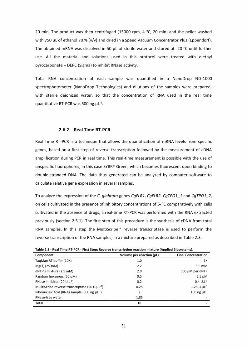
31
20 min. The product was then centrifuged (15000 rpm, 4 oC, 20 min) and the pellet washed
with 750 µL of ethanol 70 % (v/v) and dried in a Speed Vacuum Concentrator Plus (Eppendorf).
The obtained mRNA was dissolved in 50 µL of sterile water and stored at -20 oC until further
use. All the material and solutions used in this protocol were treated with diethyl
pyrocarbonate – DEPC (Sigma) to inhibit RNase activity.
Total RNA concentration of each sample was quantified in a NanoDrop ND-1000
spectrophotometer (NanoDrop Technologies) and dilutions of the samples were prepared,
with sterile deionized water, so that the concentration of RNA used in the real time
quantitative RT-PCR was 500 ng.µL-1.
2.6.2 Real Time RT-PCR
Real Time RT-PCR is a technique that allows the quantification of mRNA levels from specific
genes, based on a first step of reverse transcription followed by the measurement of cDNA
amplification during PCR in real time. This real-time measurement is possible with the use of
unspecific fluorophores, in this case SYBR® Green, which becomes fluorescent upon binding to
double-stranded DNA. The data thus generated can be analyzed by computer software to
calculate relative gene expression in several samples.
To analyze the expression of the C. glabrata genes CgFLR1, CgFLR2, CgTPO1_1 and CgTPO1_2,
on cells cultivated in the presence of inhibitory concentrations of 5-FC comparatively with cells
cultivated in the absence of drugs, a real-time RT-PCR was performed with the RNA extracted
previously (section 2.5.1). The first step of this procedure is the synthesis of cDNA from total
RNA samples. In this step the MultiScribe™ reverse transcriptase is used to perform the
reverse transcription of the RNA samples, in a mixture prepared as described in Table 2.3.
Table 2.3 - Real Time RT-PCR - First Step: Reverse transcription reaction mixture (Applied Biosystems).
Component Volume per reaction (µL) Final Concentration
TaqMan RT buffer (10X) 1.0 1X
MgCl2 (25 mM) 2.2 5.5 mM
dNTP’s mixture (2.5 mM) 2.0 500 µM per dNTP
Random hexamers (50 µM) 0.5 2.5 µM
RNase inhibitor (20 U.L-1) 0.2 0.4 U.L-1
MuiltiScribe reverse transcriptase (50 U.µL-1) 0.25 1.25 U.µL-1
Ribonucleic Acid (RNA) sample (500 ng.µL-1) 2 100 ng.µL-1
RNase-free water 1.85 -
Total 10 -

32
The reverse transcription reaction was executed using a thermal cycler block (7500 Real-Time
PCR System – Applied Biosystems) and accordingly to the parameters described in Table 2.4.
Table 2.4 - Real Time RT-PCR - First step: Thermal cycling parameters (Applied Biosystems).
Step
Incubation Reverse transcription Reverse transcriptase inactivation
Time 10 min 30 min 5 min
Temperature 25 oC 48 oC 95 oC
After finishing the thermal cycling, the cDNA samples were immediately placed on ice and
stored at -20oC until further use. In order to be used in the second step, the cDNA samples
obtained were diluted so that the quantity of cDNA in each reaction was kept around 10 ng.
The second step of real time RT-PCR step was carried out using SYBR® Green reagents and the
mixture used was prepared as described in Table 2.5.
Table 2.5 - Real Time RT-PCR - Second step: reserve transcription reaction mixture (Applied Biosystems).
Component Volume per reaction (µL)
SYBR Green PCR master mix (2x) 12.5
Forward primer (4 pmol.µL-1) 2.5
Reverse primer (4 pmol.µL-1) 2.5
cDNA 2.5
Water 5.0
Total 25
The reaction was carried out using a thermal cycler block (7500 Real-Time PCR System –
Applied Biosystems) following these same conditions: 10 min at 95 oC, then 40 cycles repeating
the set of two steps: 15 s at 95 oC and 1 min at 60 oC.
Five sets of primers were used to analyze each of the cDNA samples obtain in the first step.
The five sets of primers correspond to the four genes under study, CgFLR1, CgFLR2, CgTPO1_1,
CgTPO1_2 and CgACT1, encoding the housekeeping protein actin (Table 2.6). Primers for the
amplification of cDNA were designed using Primer Express Software (Applied Biosystems). The
CgACT1 mRNA expression was used as an internal control accounting for variability in the
initial concentration and quality of the total RNA as well as in the conversion efficiency of the
reverse transcription reaction.
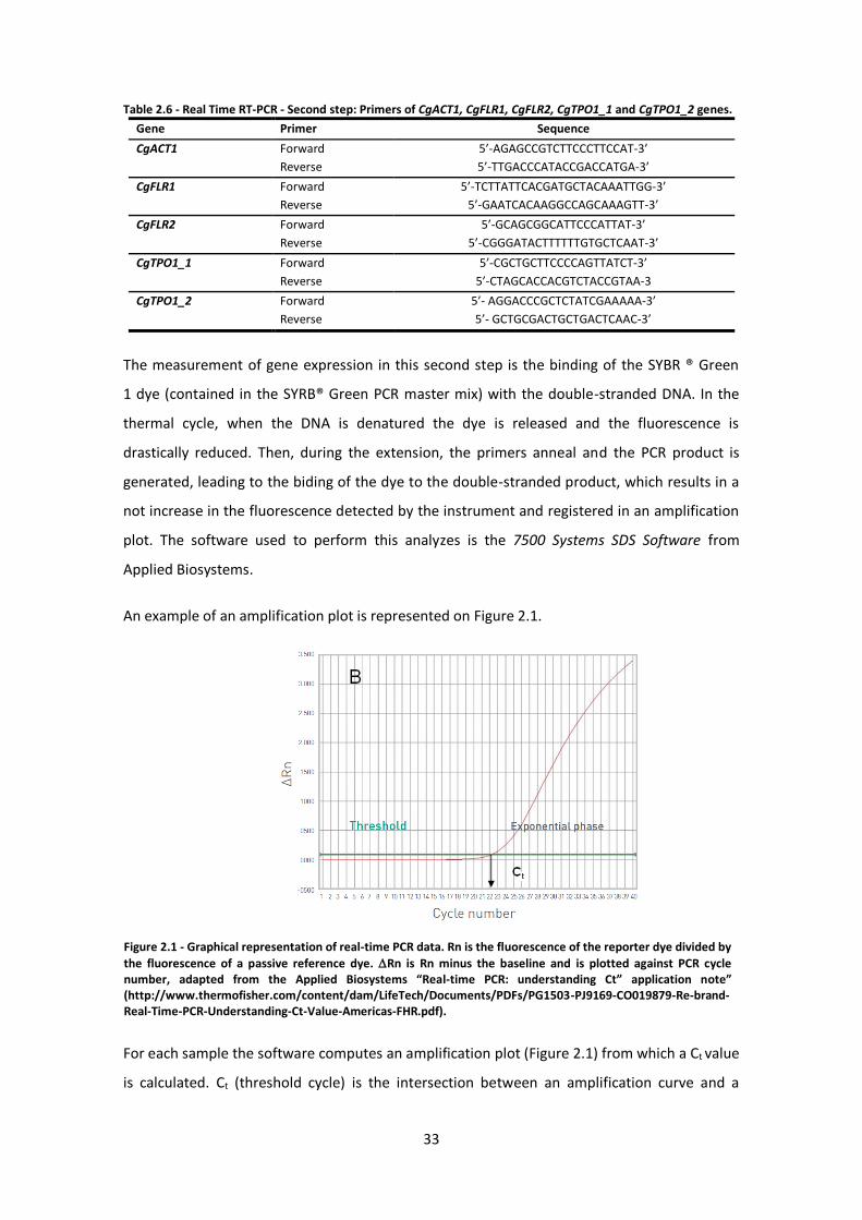
33
Figure 2.1 - Graphical representation of real-time PCR data. Rn is the fluorescence of the reporter dye divided by
the fluorescence of a passive reference dye. Rn is Rn minus the baseline and is plotted against PCR cycle number, adapted from the Applied Biosystems “Real-time PCR: understanding Ct” application note” (http://www.thermofisher.com/content/dam/LifeTech/Documents/PDFs/PG1503-PJ9169-CO019879-Re-brand-Real-Time-PCR-Understanding-Ct-Value-Americas-FHR.pdf).
Table 2.6 - Real Time RT-PCR - Second step: Primers of CgACT1, CgFLR1, CgFLR2, CgTPO1_1 and CgTPO1_2 genes.
Gene Primer Sequence
CgACT1 Forward 5’-AGAGCCGTCTTCCCTTCCAT-3’
Reverse 5’-TTGACCCATACCGACCATGA-3’
CgFLR1 Forward 5’-TCTTATTCACGATGCTACAAATTGG-3’
Reverse 5’-GAATCACAAGGCCAGCAAAGTT-3’
CgFLR2 Forward 5’-GCAGCGGCATTCCCATTAT-3’
Reverse 5’-CGGGATACTTTTTTGTGCTCAAT-3’
CgTPO1_1 Forward 5’-CGCTGCTTCCCCAGTTATCT-3’
Reverse 5’-CTAGCACCACGTCTACCGTAA-3
CgTPO1_2 Forward 5’- AGGACCCGCTCTATCGAAAAA-3’
Reverse 5’- GCTGCGACTGCTGACTCAAC-3’
The measurement of gene expression in this second step is the binding of the SYBR ® Green
1 dye (contained in the SYRB® Green PCR master mix) with the double-stranded DNA. In the
thermal cycle, when the DNA is denatured the dye is released and the fluorescence is
drastically reduced. Then, during the extension, the primers anneal and the PCR product is
generated, leading to the biding of the dye to the double-stranded product, which results in a
not increase in the fluorescence detected by the instrument and registered in an amplification
plot. The software used to perform this analyzes is the 7500 Systems SDS Software from
Applied Biosystems.
An example of an amplification plot is represented on Figure 2.1.
For each sample the software computes an amplification plot (Figure 2.1) from which a Ct value
is calculated. Ct (threshold cycle) is the intersection between an amplification curve and a

34
threshold line (Figure 2.1). It is a measure of the concentration of target in the PCR reaction.
The Ct values obtained for the target genes and the reference gene, CgACT1, and used to
calculate relative expression fold-differences using the following equations:
∆Ct sample = Ct (target)s - Ct (reference)s
∆Ct control = Ct (target)c- Ct (reference)c
∆∆Ct = ∆Ct sample - ∆Ct control
Fold difference = 2^-∆∆Ct
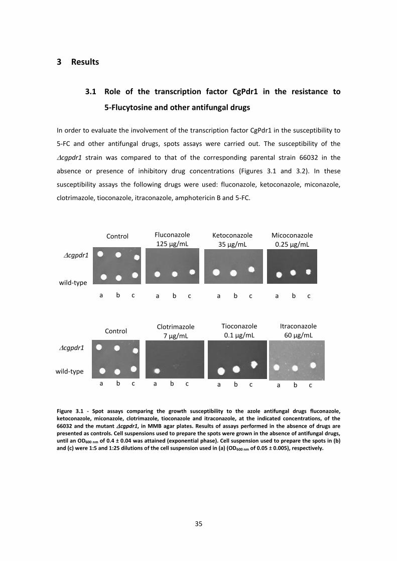
35
3 Results
3.1 Role of the transcription factor CgPdr1 in the resistance to
5-Flucytosine and other antifungal drugs
In order to evaluate the involvement of the transcription factor CgPdr1 in the susceptibility to
5-FC and other antifungal drugs, spots assays were carried out. The susceptibility of the
cgpdr1 strain was compared to that of the corresponding parental strain 66032 in the
absence or presence of inhibitory drug concentrations (Figures 3.1 and 3.2). In these
susceptibility assays the following drugs were used: fluconazole, ketoconazole, miconazole,
clotrimazole, tioconazole, itraconazole, amphotericin B and 5-FC.
Figure 3.1 - Spot assays comparing the growth susceptibility to the azole antifungal drugs fluconazole, ketoconazole, miconazole, clotrimazole, tioconazole and itraconazole, at the indicated concentrations, of the
66032 and the mutant cgpdr1, in MMB agar plates. Results of assays performed in the absence of drugs are presented as controls. Cell suspensions used to prepare the spots were grown in the absence of antifungal drugs, until an OD600 nm of 0.4 ± 0.04 was attained (exponential phase). Cell suspension used to prepare the spots in (b) and (c) were 1:5 and 1:25 dilutions of the cell suspension used in (a) (OD600 nm of 0.05 ± 0.005), respectively.
wild-type
Micoconazole 0.25 µg/mL
cgpdr1
Control
a b c a b c a b c a b c
wild-type
cgpdr1
Control
a b c a b c
Clotrimazole 7 µg/mL
a b c
Tioconazole 0.1 µg/mL
Itraconazole 60 µg/mL
a b c
Ketoconazole 35 µg/mL
Fluconazole 125 µg/mL

36
Figure 3.2 - Spot assays comparing the growth susceptibility to antifungal drugs 5-FC and amphotericin B, at the
indicated concentrations, of the 66032 and the mutant cgpdr1, in MMB agar plates. Results of assays performed in the absence of drugs are presented as controls. Cell suspensions used to prepare the spots were grown in the absence of antifungal drugs, until an OD600 nm of 0.4 ± 0.04 was attained (exponential phase). Cell suspension used to prepare the spots in (b) and (c) were 1:5 and 1:25 dilutions of the cell suspension used in (a) (OD600 nm of 0.05 ± 0.005) respectively.
The transcription factor CgPdr1 was found to be involved in resistance to the azole drugs
fluconazole, ketoconazole, miconazole, clotrimazole, tioconazole and itraconazole, as
previously demonstrated 56, 96, but also to amphotericin B and 5-FC, since the growth of the
cgpdr1 mutant, in the presence of the selected antifungal drugs, is visibly inhibited
comparatively to the growth observed for the wild-type strain (Figure 3.2).
3.2 Characterization of the C. glabrata membrane proteome
The membrane proteome of C. glabrata cells exposed to 5-Flucytosine-induced stress was
compared to that of unstressed cells. The C. glabrata membrane enriched fraction was found
to include 624 detectable proteins, comprising around 10 % of the predicted C. glabrata
proteome of the first membrane proteome-wide analysis carried out in C. glabrata and is an
invaluable repository of information on the functional analysis of these proteins.
Categorization, based on the biological process Gene Ontology, of the proteins identified in
membrane-enriched fractions of C. glabrata cells, using the GoToolBox software
(http://genome.crg.es/GOToolBox/), enabled the identification of the most statistically
significant (p-value<0.01) GO terms enriched in the dataset under analysis (Figure 3.3).
5-Flucytosine 0.015 µg/mL
∆cgpdr1
wild-type
Control
a b c a b c
Amphotericin B 0.05 µg/mL
a b c
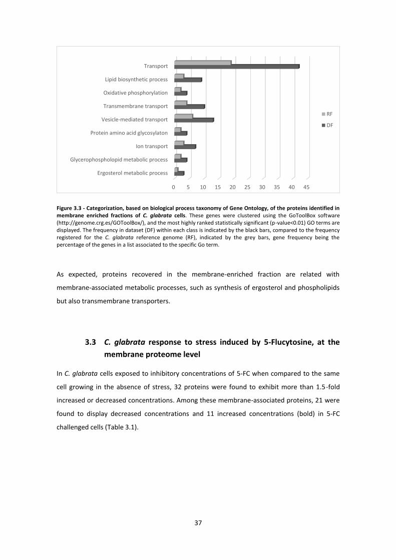
37
Figure 3.3 - Categorization, based on biological process taxonomy of Gene Ontology, of the proteins identified in membrane enriched fractions of C. glabrata cells. These genes were clustered using the GoToolBox software (http://genome.crg.es/GOToolBox/), and the most highly ranked statistically significant (p-value<0.01) GO terms are displayed. The frequency in dataset (DF) within each class is indicated by the black bars, compared to the frequency registered for the C. glabrata reference genome (RF), indicated by the grey bars, gene frequency being the percentage of the genes in a list associated to the specific Go term.
As expected, proteins recovered in the membrane-enriched fraction are related with
membrane-associated metabolic processes, such as synthesis of ergosterol and phospholipids
but also transmembrane transporters.
3.3 C. glabrata response to stress induced by 5-Flucytosine, at the
membrane proteome level
In C. glabrata cells exposed to inhibitory concentrations of 5-FC when compared to the same
cell growing in the absence of stress, 32 proteins were found to exhibit more than 1.5-fold
increased or decreased concentrations. Among these membrane-associated proteins, 21 were
found to display decreased concentrations and 11 increased concentrations (bold) in 5-FC
challenged cells (Table 3.1).
0 5 10 15 20 25 30 35 40 45
Ergosterol metabolic process
Glycerophospholopid metabolic process
Ion transport
Protein amino acid glycosylaton
Vesicle-mediated transport
Transmembrane transport
Oxidative phosphorylation
Lipid biosynthetic process
Transport
RF
DF
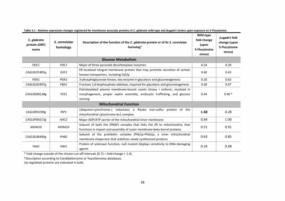
38
Table 3.1 - Relative expression changes registered for membrane associate proteins in C. glabrata wild-type and Δcgpdr1 strains upon exposure to 5-Flucytosine.
C. glabrata
protein (ORF)
name
S. cerevisiae
homologs
Description of the function of the C. glabrata protein or of its S. cerevisiae
homologa
Wild-type
fold change
(upon
5-Flucytosine
stress)
cgpdr1 fold
change (upon
5-Flucytosine
stress)
Glucose Metabolism
PDC1 PDC1 Major of three pyruvate decarboxylase isozymes 0.54 0.39
CAGL0L01485g GSF2 ER localized integral membrane protein that may promote secretion of certain
hexose transporters, including Gal2p 0.60 0.43
PGK1 PGK1 3-phosphoglycerate kinase, key enzyme in glycolysis and gluconeogenesis 0.20 0.63
CAGL0L02497g FBA1 Fructose 1,6-bisphosphate aldolase, required for glycolysis and gluconeogenesis 0.58 0.47
CAGL0G06138g YCK1
Palmitoylated plasma membrane-bound casein kinase I isoform; involved in
morphogenesis, proper septin assembly, endocytic trafficking, and glucose
sensing
0.44 0.90 *
Mitochondrial Function
CAGL0I03190g RIP1 Ubiquinol-cytochrome-c reductase; a Rieske iron-sulfur protein of the
mitochondrial cytochrome bc1 complex 1.68 0.28
CAGL0F04213g AAC2 Major ADP/ATP carrier of the mitochondrial inner membrane 0.64 1.00
MDM10 MDM10 Subunit of both the ERMES complex that links the ER to mitochondria, that
functions in import and assembly of outer membrane beta-barrel proteins 0.51 0.91
CAGL0L06490g PHB2 Subunit of the prohibitin complex (Phb1p-Phb2p), a inner mitochondrial
membrane chaperone that stabilizes newly synthesized proteins 0.63 0.85
YIM1 YIM1 Protein of unknown function; null mutant displays sensitivity to DNA damaging
agents 0.24 0.48
* Fold change outside of the chosen cut-off intervals (0.71 < fold change < 1.4). a Description according to CandidaGenome or YeastGenome databases. Up regulated proteins are indicated in bold.
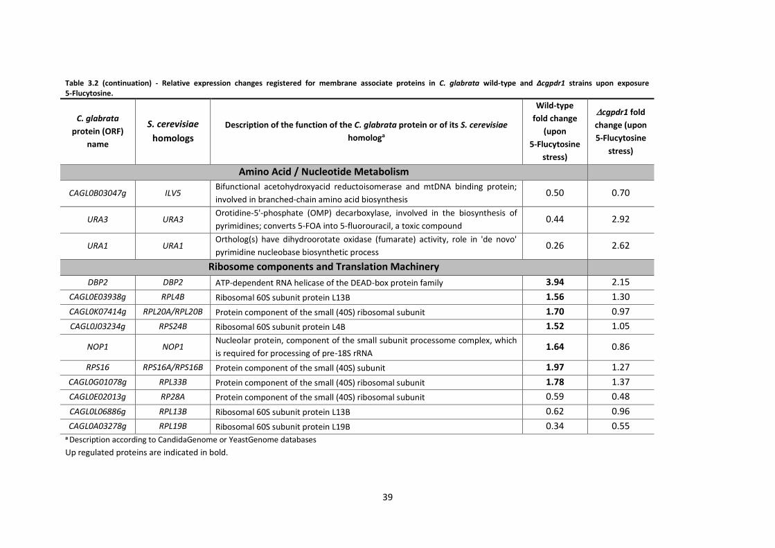
39
Table 3.2 (continuation) - Relative expression changes registered for membrane associate proteins in C. glabrata wild-type and Δcgpdr1 strains upon exposure 5-Flucytosine.
C. glabrata
protein (ORF)
name
S. cerevisiae
homologs
Description of the function of the C. glabrata protein or of its S. cerevisiae
homologa
Wild-type
fold change
(upon
5-Flucytosine
stress)
cgpdr1 fold
change (upon
5-Flucytosine
stress)
Amino Acid / Nucleotide Metabolism
CAGL0B03047g ILV5 Bifunctional acetohydroxyacid reductoisomerase and mtDNA binding protein;
involved in branched-chain amino acid biosynthesis 0.50 0.70
URA3 URA3 Orotidine-5'-phosphate (OMP) decarboxylase, involved in the biosynthesis of
pyrimidines; converts 5-FOA into 5-fluorouracil, a toxic compound 0.44 2.92
URA1 URA1 Ortholog(s) have dihydroorotate oxidase (fumarate) activity, role in 'de novo'
pyrimidine nucleobase biosynthetic process 0.26 2.62
Ribosome components and Translation Machinery
DBP2 DBP2 ATP-dependent RNA helicase of the DEAD-box protein family 3.94 2.15
CAGL0E03938g RPL4B Ribosomal 60S subunit protein L13B 1.56 1.30
CAGL0K07414g RPL20A/RPL20B Protein component of the small (40S) ribosomal subunit 1.70 0.97
CAGL0J03234g RPS24B Ribosomal 60S subunit protein L4B 1.52 1.05
NOP1 NOP1 Nucleolar protein, component of the small subunit processome complex, which
is required for processing of pre-18S rRNA 1.64 0.86
RPS16 RPS16A/RPS16B Protein component of the small (40S) subunit 1.97 1.27
CAGL0G01078g RPL33B Protein component of the small (40S) ribosomal subunit 1.78 1.37
CAGL0E02013g RP28A Protein component of the small (40S) ribosomal subunit 0.59 0.48
CAGL0L06886g RPL13B Ribosomal 60S subunit protein L13B 0.62 0.96
CAGL0A03278g RPL19B Ribosomal 60S subunit protein L19B 0.34 0.55 a Description according to CandidaGenome or YeastGenome databases
Up regulated proteins are indicated in bold.
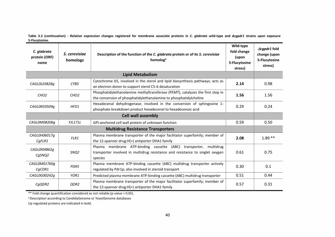
40
Table 3.3 (continuation) - Relative expression changes registered for membrane associate proteins in C. glabrata wild-type and Δcgpdr1 strains upon exposure 5-Flucytosine.
C. glabrata
protein (ORF)
name
S. cerevisiae
homologs
Description of the function of the C. glabrata protein or of its S. cerevisiae
homologa
Wild-type
fold change
(upon
5-Flucytosine
stress)
cgpdr1 fold
change (upon
5-Flucytosine
stress)
Lipid Metabolism
CAGL0L03828g CYB5 Cytochrome b5, involved in the sterol and lipid biosynthesis pathways; acts as
an electron donor to support sterol C5-6 desaturation 2.14 0.98
CHO2 CHO2 Phosphatidylethanolamine methyltransferase (PEMT), catalyzes the first step in
the conversion of phosphatidylethanolamine to phosphatidylcholine 1.56 1.56
CAGL0K03509g HFD1 Hexadecenal dehydrogenase; involved in the conversion of sphingosine 1-
phosphate breakdown product hexadecenal to hexadecenoic acid 0.29 0.24
Cell wall assembly
CAGL0M08206g YJL171c GPI-anchored cell wall protein of unknown function 0.59 0.50
Multidrug Resistance Transporters
CAGL0H06017g
CgFLR1 FLR1
Plasma membrane transporter of the major facilitator superfamily; member of
the 12-spanner drug:H(+) antiporter DHA1 family 2.08 1.89 **
CAGL0I04862g
CgSNQ2 SNQ2
Plasma membrane ATP-binding cassette (ABC) transporter, multidrug
transporter involved in multidrug resistance and resistance to singlet oxygen
species
0.61 0.75
CAGL0M01760g
CgCDR1 PDR5
Plasma membrane ATP-binding cassette (ABC) multidrug transporter actively
regulated by Pdr1p; also involved in steroid transport 0.30 0.1
CAGL0G00242g YOR1 Predicted plasma membrane ATP-binding cassette (ABC) multidrug transporter 0.51 0.44
CgQDR2 QDR2 Plasma membrane transporter of the major facilitator superfamily; member of
the 12-spanner drug:H(+) antiporter DHA1 family 0.57 0.31
** Fold change quantification considered as not reliable (p-value > 0.05). a Description according to CandidaGenome or YeastGenome databases
Up regulated proteins are indicated in bold.
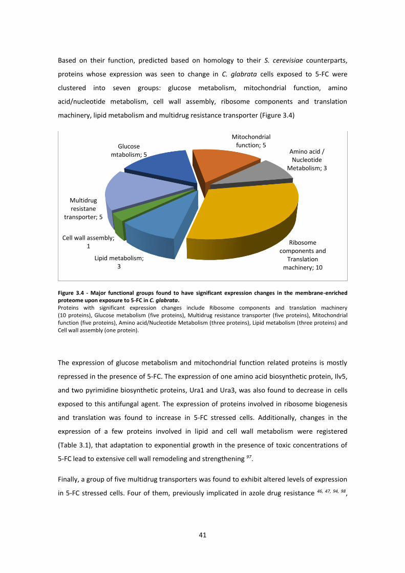
41
Based on their function, predicted based on homology to their S. cerevisiae counterparts,
proteins whose expression was seen to change in C. glabrata cells exposed to 5-FC were
clustered into seven groups: glucose metabolism, mitochondrial function, amino
acid/nucleotide metabolism, cell wall assembly, ribosome components and translation
machinery, lipid metabolism and multidrug resistance transporter (Figure 3.4)
Figure 3.4 - Major functional groups found to have significant expression changes in the membrane-enriched proteome upon exposure to 5-FC in C. glabrata. Proteins with significant expression changes include Ribosome components and translation machinery (10 proteins), Glucose metabolism (five proteins), Multidrug resistance transporter (five proteins), Mitochondrial function (five proteins), Amino acid/Nucleotide Metabolism (three proteins), Lipid metabolism (three proteins) and Cell wall assembly (one protein).
The expression of glucose metabolism and mitochondrial function related proteins is mostly
repressed in the presence of 5-FC. The expression of one amino acid biosynthetic protein, Ilv5,
and two pyrimidine biosynthetic proteins, Ura1 and Ura3, was also found to decrease in cells
exposed to this antifungal agent. The expression of proteins involved in ribosome biogenesis
and translation was found to increase in 5-FC stressed cells. Additionally, changes in the
expression of a few proteins involved in lipid and cell wall metabolism were registered
(Table 3.1), that adaptation to exponential growth in the presence of toxic concentrations of
5-FC lead to extensive cell wall remodeling and strengthening 97.
Finally, a group of five multidrug transporters was found to exhibit altered levels of expression
in 5-FC stressed cells. Four of them, previously implicated in azole drug resistance 46, 47, 94, 98,
Glucose mtabolism; 5
Mitochondrial function; 5
Amino acid / Nucleotide
Metabolism; 3
Ribosome components and
Translation machinery; 10
Lipid metabolism; 3
Cell wall assembly; 1
Multidrug resistane
transporter; 5
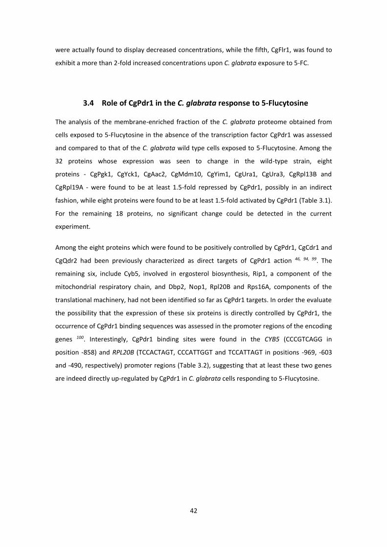
42
were actually found to display decreased concentrations, while the fifth, CgFlr1, was found to
exhibit a more than 2-fold increased concentrations upon C. glabrata exposure to 5-FC.
3.4 Role of CgPdr1 in the C. glabrata response to 5-Flucytosine
The analysis of the membrane-enriched fraction of the C. glabrata proteome obtained from
cells exposed to 5-Flucytosine in the absence of the transcription factor CgPdr1 was assessed
and compared to that of the C. glabrata wild type cells exposed to 5-Flucytosine. Among the
32 proteins whose expression was seen to change in the wild-type strain, eight
proteins - CgPgk1, CgYck1, CgAac2, CgMdm10, CgYim1, CgUra1, CgUra3, CgRpl13B and
CgRpl19A - were found to be at least 1.5-fold repressed by CgPdr1, possibly in an indirect
fashion, while eight proteins were found to be at least 1.5-fold activated by CgPdr1 (Table 3.1).
For the remaining 18 proteins, no significant change could be detected in the current
experiment.
Among the eight proteins which were found to be positively controlled by CgPdr1, CgCdr1 and
CgQdr2 had been previously characterized as direct targets of CgPdr1 action 46, 94, 99. The
remaining six, include Cyb5, involved in ergosterol biosynthesis, Rip1, a component of the
mitochondrial respiratory chain, and Dbp2, Nop1, Rpl20B and Rps16A, components of the
translational machinery, had not been identified so far as CgPdr1 targets. In order the evaluate
the possibility that the expression of these six proteins is directly controlled by CgPdr1, the
occurrence of CgPdr1 binding sequences was assessed in the promoter regions of the encoding
genes 100. Interestingly, CgPdr1 binding sites were found in the CYB5 (CCCGTCAGG in
position -858) and RPL20B (TCCACTAGT, CCCATTGGT and TCCATTAGT in positions -969, -603
and -490, respectively) promoter regions (Table 3.2), suggesting that at least these two genes
are indeed directly up-regulated by CgPdr1 in C. glabrata cells responding to 5-Flucytosine.
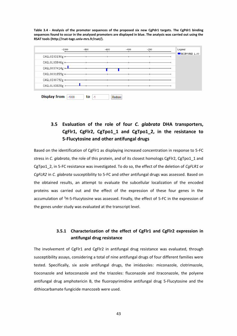
43
Table 3.4 - Analysis of the promoter sequences of the proposed six new CgPdr1 targets. The CgPdr1 binding sequences found to occur in the analysed promoters are displayed in blue. The analysis was carried out using the RSAT tools (http://rsat-tagc.univ-mrs.fr/rsat/).
3.5 Evaluation of the role of four C. glabrata DHA transporters,
CgFlr1, CgFlr2, CgTpo1_1 and CgTpo1_2, in the resistance to
5-Flucytosine and other antifungal drugs
Based on the identification of CgFlr1 as displaying increased concentration in response to 5-FC
stress in C. glabrata, the role of this protein, and of its closest homologs CgFlr2, CgTpo1_1 and
CgTpo1_2, in 5-FC resistance was investigated. To do so, the effect of the deletion of CgFLR1 or
CgFLR2 in C. glabrata susceptibility to 5-FC and other antifungal drugs was assessed. Based on
the obtained results, an attempt to evaluate the subcellular localization of the encoded
proteins was carried out and the effect of the expression of these four genes in the
accumulation of 3H-5-Flucytosine was assessed. Finally, the effect of 5-FC in the expression of
the genes under study was evaluated at the transcript level.
3.5.1 Characterization of the effect of CgFlr1 and CgFlr2 expression in
antifungal drug resistance
The involvement of CgFlr1 and CgFlr2 in antifungal drug resistance was evaluated, through
susceptibility assays, considering a total of nine antifungal drugs of four different families were
tested. Specifically, six azole antifungal drugs, the imidazoles: miconazole, clotrimazole,
tioconazole and ketoconazole and the triazoles: fluconazole and itraconazole, the polyene
antifungal drug amphotericin B, the fluoropyrimidine antifungal drug 5-Flucytosine and the
dithiocarbamate fungicide mancozeb were used.
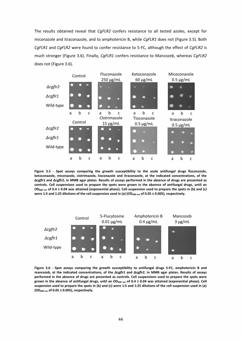
44
The results obtained reveal that CgFLR2 confers resistance to all tested azoles, except for
miconazole and itraconazole, and to amphotericin B, while CgFLR1 does not (Figure 3.5). Both
CgFLR1 and CgFLR2 were found to confer resistance to 5-FC, although the effect of CgFLR2 is
much stronger (Figure 3.6). Finally, CgFLR1 confers resistance to Mancozeb, whereas CgFLR2
does not (Figure 3.6).
Figure 3.5 - Spot assays comparing the growth susceptibility to the azole antifungal drugs fluconazole, ketoconazole, miconazole, clotrimazole, tioconazole and itraconazole, at the indicated concentrations, of the ∆cgflr1 and ∆cgflr2, in MMB agar plates. Results of assays performed in the absence of drugs are presented as controls. Cell suspensions used to prepare the spots were grown in the absence of antifungal drugs, until an OD600 nm of 0.4 ± 0.04 was attained (exponential phase). Cell suspension used to prepare the spots in (b) and (c) were 1:5 and 1:25 dilutions of the cell suspension used in (a) (OD600 nm of 0.05 ± 0.005), respectively.
Figure 3.6 - Spot assays comparing the growth susceptibility to antifungal drugs 5-FC, amphotericin B and mancozeb, at the indicated concentrations, of the ∆cgflr1 and ∆cgflr2, in MMB agar plates. Results of assays performed in the absence of drugs are presented as controls. Cell suspensions used to prepare the spots were grown in the absence of antifungal drugs, until an OD600 nm of 0.4 ± 0.04 was attained (exponential phase). Cell suspension used to prepare the spots in (b) and (c) were 1:5 and 1:25 dilutions of the cell suspension used in (a) (OD600 nm of 0.05 ± 0.005), respectively.
Control Fluconazole 250 µg/mL
Ketoconazole 60 µg/mL
Micoconazole 0.5 µg/mL
Δcgflr2
Δcgflr1
Wild-type
Control
Δcgflr2
Δcgflr1
Wild-type
Clotrimazole 15 µg/mL
Tioconazole 0.5 µg/mL
Itraconazole 0.5 µg/mL
a b c a b c a b c a b c
a b c a b c a b c a b c
Control 5-Flucytosine 0.01 µg/mL
Amphotericin B 0.4 µg/mL
Mancozeb 3 µg/mL
a b c a b c a b c a b c
Δcgflr2
Δcgflr1
Wild-type
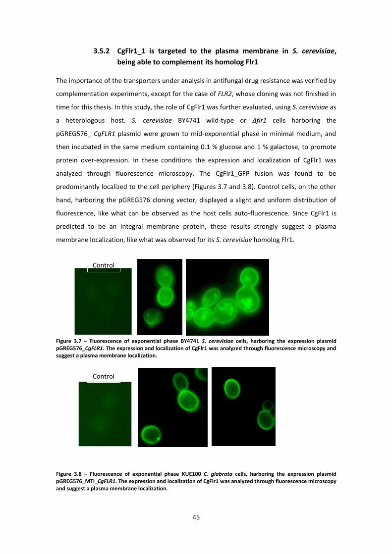
45
3.5.2 CgFlr1_1 is targeted to the plasma membrane in S. cerevisiae,
being able to complement its homolog Flr1
The importance of the transporters under analysis in antifungal drug resistance was verified by
complementation experiments, except for the case of FLR2, whose cloning was not finished in
time for this thesis. In this study, the role of CgFlr1 was further evaluated, using S. cerevisiae as
a heterologous host. S. cerevisiae BY4741 wild-type or Δflr1 cells harboring the
pGREG576_ CgFLR1 plasmid were grown to mid-exponential phase in minimal medium, and
then incubated in the same medium containing 0.1 % glucose and 1 % galactose, to promote
protein over-expression. In these conditions the expression and localization of CgFlr1 was
analyzed through fluorescence microscopy. The CgFlr1_GFP fusion was found to be
predominantly localized to the cell periphery (Figures 3.7 and 3.8). Control cells, on the other
hand, harboring the pGREG576 cloning vector, displayed a slight and uniform distribution of
fluorescence, like what can be observed as the host cells auto-fluorescence. Since CgFlr1 is
predicted to be an integral membrane protein, these results strongly suggest a plasma
membrane localization, like what was observed for its S. cerevisiae homolog Flr1.
Figure 3.7 – Fluorescence of exponential phase BY4741 S. cerevisiae cells, harboring the expression plasmid pGREG576_CgFLR1. The expression and localization of CgFlr1 was analyzed through fluorescence microscopy and suggest a plasma membrane localization.
Figure 3.8 – Fluorescence of exponential phase KUE100 C. glabrata cells, harboring the expression plasmid pGREG576_MTI_CgFLR1. The expression and localization of CgFlr1 was analyzed through fluorescence microscopy and suggest a plasma membrane localization.
Control
Control
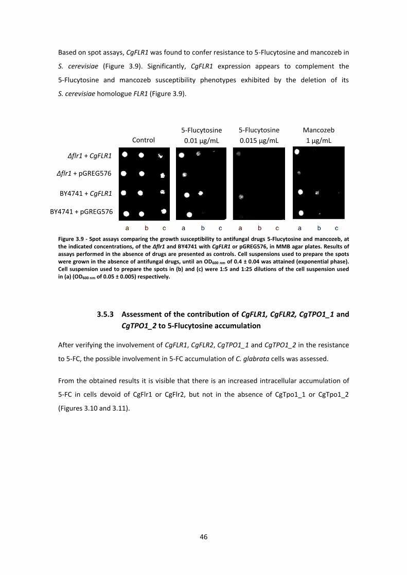
46
Based on spot assays, CgFLR1 was found to confer resistance to 5-Flucytosine and mancozeb in
S. cerevisiae (Figure 3.9). Significantly, CgFLR1 expression appears to complement the
5-Flucytosine and mancozeb susceptibility phenotypes exhibited by the deletion of its
S. cerevisiae homologue FLR1 (Figure 3.9).
Figure 3.9 - Spot assays comparing the growth susceptibility to antifungal drugs 5-Flucytosine and mancozeb, at the indicated concentrations, of the ∆flr1 and BY4741 with CgFLR1 or pGREG576, in MMB agar plates. Results of assays performed in the absence of drugs are presented as controls. Cell suspensions used to prepare the spots were grown in the absence of antifungal drugs, until an OD600 nm of 0.4 ± 0.04 was attained (exponential phase). Cell suspension used to prepare the spots in (b) and (c) were 1:5 and 1:25 dilutions of the cell suspension used in (a) (OD600 nm of 0.05 ± 0.005) respectively.
3.5.3 Assessment of the contribution of CgFLR1, CgFLR2, CgTPO1_1 and
CgTPO1_2 to 5-Flucytosine accumulation
After verifying the involvement of CgFLR1, CgFLR2, CgTPO1_1 and CgTPO1_2 in the resistance
to 5-FC, the possible involvement in 5-FC accumulation of C. glabrata cells was assessed.
From the obtained results it is visible that there is an increased intracellular accumulation of
5-FC in cells devoid of CgFlr1 or CgFlr2, but not in the absence of CgTpo1_1 or CgTpo1_2
(Figures 3.10 and 3.11).
5-Flucytosine
0.01 µg/mL Control
Mancozeb
1 µg/mL
5-Flucytosine
0.015 µg/mL
∆flr1 + CgFLR1
∆flr1 + pGREG576
BY4741 + pGREG576
BY4741 + CgFLR1
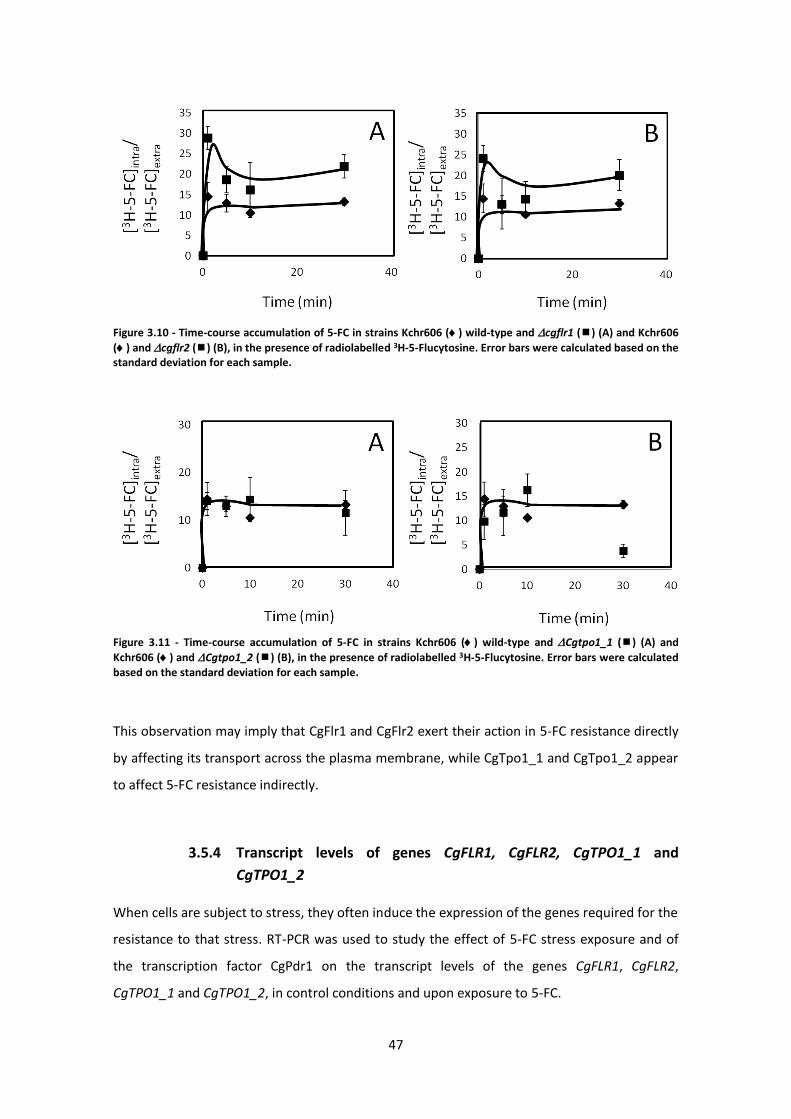
47
Figure 3.10 - Time-course accumulation of 5-FC in strains Kchr606 () wild-type and cgflr1 () (A) and Kchr606
() and cgflr2 () (B), in the presence of radiolabelled 3H-5-Flucytosine. Error bars were calculated based on the standard deviation for each sample.
Figure 3.11 - Time-course accumulation of 5-FC in strains Kchr606 () wild-type and Cgtpo1_1 () (A) and
Kchr606 () and Cgtpo1_2 () (B), in the presence of radiolabelled 3H-5-Flucytosine. Error bars were calculated based on the standard deviation for each sample.
This observation may imply that CgFlr1 and CgFlr2 exert their action in 5-FC resistance directly
by affecting its transport across the plasma membrane, while CgTpo1_1 and CgTpo1_2 appear
to affect 5-FC resistance indirectly.
3.5.4 Transcript levels of genes CgFLR1, CgFLR2, CgTPO1_1 and
CgTPO1_2
When cells are subject to stress, they often induce the expression of the genes required for the
resistance to that stress. RT-PCR was used to study the effect of 5-FC stress exposure and of
the transcription factor CgPdr1 on the transcript levels of the genes CgFLR1, CgFLR2,
CgTPO1_1 and CgTPO1_2, in control conditions and upon exposure to 5-FC.
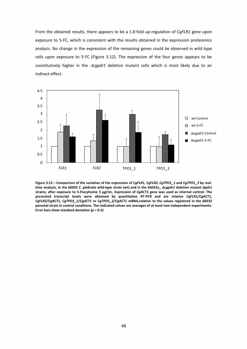
48
0
0,5
1
1,5
2
2,5
3
3,5
4
4,5
FLR1_1 FLR1_2 TPO1_1 TPO1_2
wt-Control
wt-5-FC
Dpdr1-Control
Dpdr1-5-FC
From the obtained results, there appears to be a 1.8-fold up-regulation of CgFLR1 gene upon
exposure to 5-FC, which is consistent with the results obtained in the expression proteomics
analysis. No change in the expression of the remaining genes could be observed in wild-type
cells upon exposure to 5-FC (Figure 3.12). The expression of the four genes appears to be
constitutively higher in the cgpdr1 deletion mutant cells which is most likely due to an
indirect effect.
Figure 3.12 – Comparison of the variation of the expression of CgFLR1, CgFLR2, CgTPO1_1 and CgTPO1_2 by real-time analysis, in the 66032 C. glabrata wild-type strain (wt) and in the 66032u_ Δcgpdr1 deletion mutant ∆pdr1 strains, after exposure to 5-Flucytosine 5 µg/mL. Expression of CgACT1 gene was used as internal control. The presented transcript levels were obtained by quantitative RT-PCR and are relative CgFLR1/CgACT1, CgFLR2/CgACT1, CgTPO1_1/CgACT1 or CgTPO1_2/CgACT1 mRNA,relative to the values registered in the 66032 parental strain in control conditions. The indicated values are averages of at least two independent experiments. Error bars show standard deviation (p < 0.5).
Δcgpdr1-Control
Δcgpdr1-5-FC
wt-Control
wt-5-FC
4.5
4
3.5
3
2.5
2
1.5
1
0.5
0
FLR1
FLR2
TPO1_1
TPO1_2
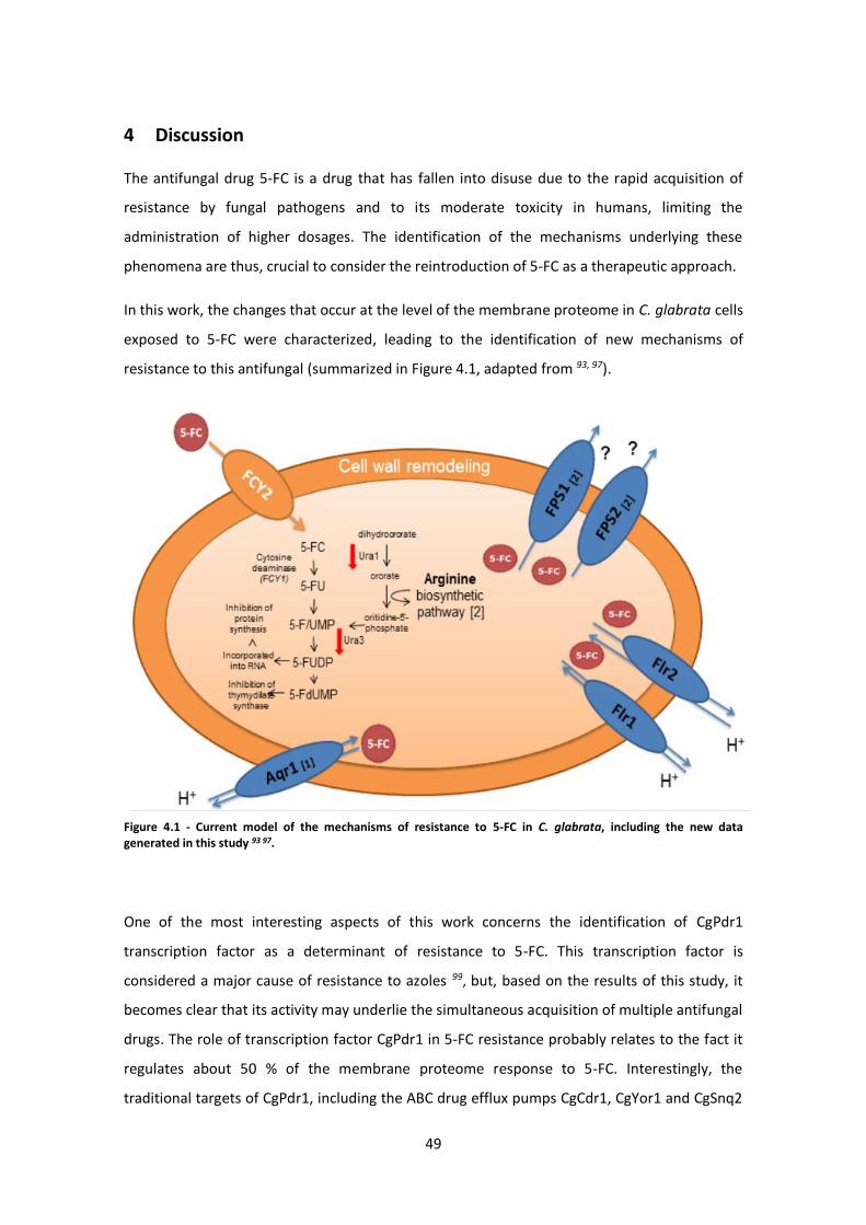
49
4 Discussion
The antifungal drug 5-FC is a drug that has fallen into disuse due to the rapid acquisition of
resistance by fungal pathogens and to its moderate toxicity in humans, limiting the
administration of higher dosages. The identification of the mechanisms underlying these
phenomena are thus, crucial to consider the reintroduction of 5-FC as a therapeutic approach.
In this work, the changes that occur at the level of the membrane proteome in C. glabrata cells
exposed to 5-FC were characterized, leading to the identification of new mechanisms of
resistance to this antifungal (summarized in Figure 4.1, adapted from 93, 97).
Figure 4.1 - Current model of the mechanisms of resistance to 5-FC in C. glabrata, including the new data generated in this study 93 97.
One of the most interesting aspects of this work concerns the identification of CgPdr1
transcription factor as a determinant of resistance to 5-FC. This transcription factor is
considered a major cause of resistance to azoles 99, but, based on the results of this study, it
becomes clear that its activity may underlie the simultaneous acquisition of multiple antifungal
drugs. The role of transcription factor CgPdr1 in 5-FC resistance probably relates to the fact it
regulates about 50 % of the membrane proteome response to 5-FC. Interestingly, the
traditional targets of CgPdr1, including the ABC drug efflux pumps CgCdr1, CgYor1 and CgSnq2
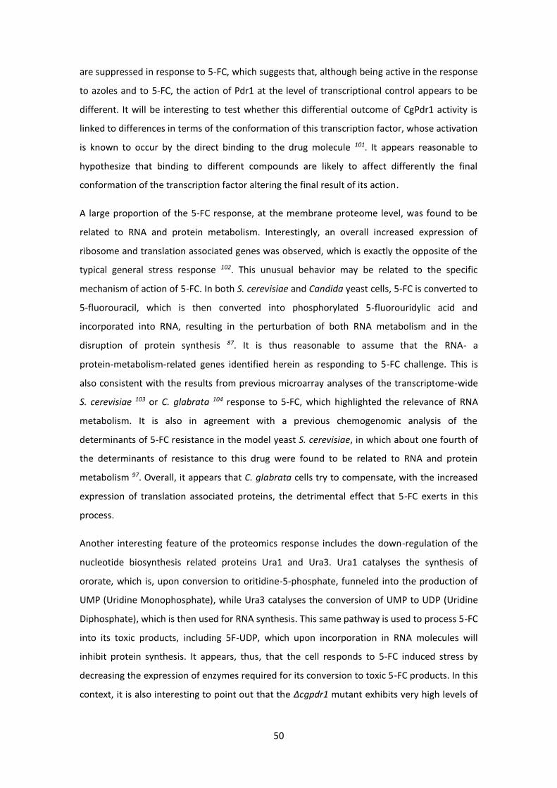
50
are suppressed in response to 5-FC, which suggests that, although being active in the response
to azoles and to 5-FC, the action of Pdr1 at the level of transcriptional control appears to be
different. It will be interesting to test whether this differential outcome of CgPdr1 activity is
linked to differences in terms of the conformation of this transcription factor, whose activation
is known to occur by the direct binding to the drug molecule 101. It appears reasonable to
hypothesize that binding to different compounds are likely to affect differently the final
conformation of the transcription factor altering the final result of its action.
A large proportion of the 5-FC response, at the membrane proteome level, was found to be
related to RNA and protein metabolism. Interestingly, an overall increased expression of
ribosome and translation associated genes was observed, which is exactly the opposite of the
typical general stress response 102. This unusual behavior may be related to the specific
mechanism of action of 5-FC. In both S. cerevisiae and Candida yeast cells, 5-FC is converted to
5-fluorouracil, which is then converted into phosphorylated 5-fluorouridylic acid and
incorporated into RNA, resulting in the perturbation of both RNA metabolism and in the
disruption of protein synthesis 87. It is thus reasonable to assume that the RNA- a
protein-metabolism-related genes identified herein as responding to 5-FC challenge. This is
also consistent with the results from previous microarray analyses of the transcriptome-wide
S. cerevisiae 103 or C. glabrata 104 response to 5-FC, which highlighted the relevance of RNA
metabolism. It is also in agreement with a previous chemogenomic analysis of the
determinants of 5-FC resistance in the model yeast S. cerevisiae, in which about one fourth of
the determinants of resistance to this drug were found to be related to RNA and protein
metabolism 97. Overall, it appears that C. glabrata cells try to compensate, with the increased
expression of translation associated proteins, the detrimental effect that 5-FC exerts in this
process.
Another interesting feature of the proteomics response includes the down-regulation of the
nucleotide biosynthesis related proteins Ura1 and Ura3. Ura1 catalyses the synthesis of
ororate, which is, upon conversion to oritidine-5-phosphate, funneled into the production of
UMP (Uridine Monophosphate), while Ura3 catalyses the conversion of UMP to UDP (Uridine
Diphosphate), which is then used for RNA synthesis. This same pathway is used to process 5-FC
into its toxic products, including 5F-UDP, which upon incorporation in RNA molecules will
inhibit protein synthesis. It appears, thus, that the cell responds to 5-FC induced stress by
decreasing the expression of enzymes required for its conversion to toxic 5-FC products. In this
context, it is also interesting to point out that the Δcgpdr1 mutant exhibits very high levels of
51
Ura1 and Ura3, when compared to the parental strain, which may contribute to the
susceptibility phenotype displayed by this mutant strain.
Finally, among the results obtained from the membrane proteomics analysis, the role of the
MDR transporter CgFlr1, and of its homologs, in 5-FC response was further analysed. Both
CgFlr1 and CgFlr2 were found to confer resistance to 5-FC, apparently due to their role in
controlling the levels of 5-FC accumulation within C. glabrata cells. Interestingly, CgFlr2 was
further found to confer resistance to azoles and amphotericin B placing this transporter at the
intersection of multiple antifungal resistance mechanisms. These two transporters, belonging
to the DHA family in C. glabrata, constitute, thus, two additional players in the antifungal drug
resistance phenomenon. Our group had previously shown that the acquaglyceroporins CgFps1
and CgFps2 97, as well as the DHA transporters Aqr1 93 are determinants of 5-FC resistance as
well, suggesting that 5-FC extrusion is an important mechanism of resistance against this
antifungal drug and showing this phenomenon to be the consequence of the additive
contribution of several players. Surprisingly, in the case of CgTpo1_1 and CgTpo1_2 this study
shows that their contribution does not affect directly the intracellular concentration of the
drug. It will be interesting to see what is the role played by Tpo1_1 and Tpo1_2 transporters,
indirectly leading to the acquisition of resistance to 5-FC.
In conclusion, the results described in this study highlight the importance of MDR transporters
from the MFS in antifungal resistance phenotypes, particularly concerning 5-FC. The
characterization of C. glabrata CgFlr1 and CgFlr2 MDR transporters involved in 5-FC drug
resistance reinforces the need to study the remaining members of this family in this
increasingly relevant pathogenic yeast, given that these transporters are likely to have clinical
impact 32. This work further highlights the importance of genome/proteome-wide approaches
in the identification of new antifungal resistance mechanisms. The newly identified processes
stand out as promising targets for the development of new 5-FC chemosensitizers, which
would expectedly allow for the use of decreased therapeutic dosages of 5-FC, limiting the
development of 5-FC resistance which usually happens very fast and enabling a more extensive
(re)use of this antifungal drug.
52
5 References
[1] Schulze, J., and Sonnenborn, U. (2009) Yeasts in the gut: from commensals to infectious agents, Dtsch Arztebl Int 106, 837-842.
[2] Tsui, C. K., Daniel, H. M., Robert, V., and Meyer, W. (2008) Re-examining the phylogeny of clinically relevant Candida species and allied genera based on multigene analyses, FEMS Yeast Res 8, 651-659.
[3] Butler, G., Rasmussen, M. D., Lin, M. F., Santos, M. A., Sakthikumar, S., Munro, C. A., Rheinbay, E., Grabherr, M., Forche, A., Reedy, J. L., Agrafioti, I., Arnaud, M. B., Bates, S., Brown, A. J., Brunke, S., Costanzo, M. C., Fitzpatrick, D. A., de Groot, P. W., Harris, D., Hoyer, L. L., Hube, B., Klis, F. M., Kodira, C., Lennard, N., Logue, M. E., Martin, R., Neiman, A. M., Nikolaou, E., Quail, M. A., Quinn, J., Santos, M. C., Schmitzberger, F. F., Sherlock, G., Shah, P., Silverstein, K. A., Skrzypek, M. S., Soll, D., Staggs, R., Stansfield, I., Stumpf, M. P., Sudbery, P. E., Srikantha, T., Zeng, Q., Berman, J., Berriman, M., Heitman, J., Gow, N. A., Lorenz, M. C., Birren, B. W., Kellis, M., and Cuomo, C. A. (2009) Evolution of pathogenicity and sexual reproduction in eight Candida genomes, Nature 459, 657-662.
[4] Fidel, P. L., Jr., Vazquez, J. A., and Sobel, J. D. (1999) Candida glabrata: review of epidemiology, pathogenesis, and clinical disease with comparison to C. albicans, Clin Microbiol Rev 12, 80-96.
[5] Ueno, K., Uno, J., Nakayama, H., Sasamoto, K., Mikami, Y., and Chibana, H. (2007) Development of a highly efficient gene targeting system induced by transient repression of YKU80 expression in Candida glabrata, Eukaryot Cell 6, 1239-1247.
[6] Silva, S., Negri, M., Henriques, M., Oliveira, R., Williams, D. W., and Azeredo, J. (2012) Candida glabrata, Candida parapsilosis and Candida tropicalis: biology, epidemiology, pathogenicity and antifungal resistance, FEMS Microbiol Rev 36, 288-305.
[7] M. H. Miceli, J. A. D. A. S. A. L. (2001) Emerging opportunistic yeast infections, Lancet Infect Disease 11, 10.
[8] Ricardo, E., Costa-de-Oliveira, S., Dias, A. S., Guerra, J., Rodrigues, A. G., and Pina-Vaz, C. (2009) Ibuprofen reverts antifungal resistance on Candida albicans showing overexpression of CDR genes, FEMS Yeast Res 9, 618-625.
[9] Williams, D., and Lewis, M. (2011) Pathogenesis and treatment of oral candidosis, J Oral Microbiol 3.
[10] Knoke, M., Schulz, K., and Bernhardt, H. (1997) Dynamics of Candida isolations from humans from 1992-1995 in Greifswald, Germany, Mycoses 40, 105-110.
[11] Komshian, S. V., Uwaydah, A. K., Sobel, J. D., and Crane, L. R. (1989) Fungemia caused by Candida species and Torulopsis glabrata in the hospitalized patient: frequency, characteristics, and evaluation of factors influencing outcome, Rev Infect Dis 11, 379-390.
[12] Hawser, S. P., and Douglas, L. J. (1994) Biofilm formation by Candida species on the surface of catheter materials in vitro, Infect Immun 62, 915-921.
[13] Calderone, R. A., and Fonzi, W. A. (2001) Virulence factors of Candida albicans, Trends Microbiol 9, 327-335.
[14] Soll, D. R. (1992) High-frequency switching in Candida albicans, Clin Microbiol Rev 5, 183-203.
[15] Pfaller, M. A., Diekema, D. J., Gibbs, D. L., Newell, V. A., Ellis, D., Tullio, V., Rodloff, A., Fu, W., Ling, T. A., and Global Antifungal Surveillance, G. (2010) Results from the ARTEMIS DISK Global Antifungal Surveillance Study, 1997 to 2007: a 10.5-year analysis of susceptibilities of Candida Species to fluconazole and voriconazole as determined by CLSI standardized disk diffusion, J Clin Microbiol 48, 1366-1377.
53
[16] Ruhnke, M. (2006) Epidemiology of Candida albicans infections and role of non-Candida-albicans yeasts, Curr Drug Targets 7, 495-504.
[17] Kurtzman, C. P., Fell, J. W. (1998) The Yeasts: A Taxomic Study, Nature. [18] Dujon, B., Sherman, D., Fischer, G., Durrens, P., Casaregola, S., Lafontaine, I., De Montigny,
J., Marck, C., Neuveglise, C., Talla, E., Goffard, N., Frangeul, L., Aigle, M., Anthouard, V., Babour, A., Barbe, V., Barnay, S., Blanchin, S., Beckerich, J. M., Beyne, E., Bleykasten, C., Boisrame, A., Boyer, J., Cattolico, L., Confanioleri, F., De Daruvar, A., Despons, L., Fabre, E., Fairhead, C., Ferry-Dumazet, H., Groppi, A., Hantraye, F., Hennequin, C., Jauniaux, N., Joyet, P., Kachouri, R., Kerrest, A., Koszul, R., Lemaire, M., Lesur, I., Ma, L., Muller, H., Nicaud, J. M., Nikolski, M., Oztas, S., Ozier-Kalogeropoulos, O., Pellenz, S., Potier, S., Richard, G. F., Straub, M. L., Suleau, A., Swennen, D., Tekaia, F., Wesolowski-Louvel, M., Westhof, E., Wirth, B., Zeniou-Meyer, M., Zivanovic, I., Bolotin-Fukuhara, M., Thierry, A., Bouchier, C., Caudron, B., Scarpelli, C., Gaillardin, C., Weissenbach, J., Wincker, P., and Souciet, J. L. (2004) Genome evolution in yeasts, Nature 430, 35-44.
[19] Wong, S., Fares, M. A., Zimmermann, W., Butler, G., and Wolfe, K. H. (2003) Evidence from comparative genomics for a complete sexual cycle in the 'asexual' pathogenic yeast Candida glabrata, Genome Biol 4, R10.
[20] Barns, S. M., Lane, D. J., Sogin, M. L., Bibeau, C., and Weisburg, W. G. (1991) Evolutionary relationships among pathogenic Candida species and relatives, J Bacteriol 173, 2250-2255.
[21] Falagas, M. E., Roussos, N., and Vardakas, K. Z. (2010) Relative frequency of albicans and the various non-albicans Candida spp among candidemia isolates from inpatients in various parts of the world: a systematic review, Int J Infect Dis 14, e954-966.
[22] Pfaller, M. A., and Diekema, D. J. (2007) Epidemiology of invasive candidiasis: a persistent public health problem, Clin Microbiol Rev 20, 133-163.
[23] Higgins, C. F. (2007) Multiple molecular mechanisms for multidrug resistance transporters, Nature 446, 749-757.
[24] Sa-Correia, I., dos Santos, S. C., Teixeira, M. C., Cabrito, T. R., and Mira, N. P. (2009) Drug:H+ antiporters in chemical stress response in yeast, Trends Microbiol 17, 22-31.
[25] Gbelska, Y., Krijger, J. J., and Breunig, K. D. (2006) Evolution of gene families: the multidrug resistance transporter genes in five related yeast species, FEMS Yeast Res 6, 345-355.
[26] van Veen, H. W., and Konings, W. N. (1998) The ABC family of multidrug transporters in microorganisms, Biochim Biophys Acta 1365, 31-36.
[27] Stavrovskaya, A. A. (2000) Cellular mechanisms of multidrug resistance of tumor cells, Biochemistry (Mosc) 65, 95-106.
[28] Simon, S. M., and Schindler, M. (1994) Cell biological mechanisms of multidrug resistance in tumors, Proc Natl Acad Sci U S A 91, 3497-3504.
[29] Cannon, R. D., Lamping, E., Holmes, A. R., Niimi, K., Baret, P. V., Keniya, M. V., Tanabe, K., Niimi, M., Goffeau, A., and Monk, B. C. (2009) Efflux-mediated antifungal drug resistance, Clin Microbiol Rev 22, 291-321, Table of Contents.
[30] Saier, M. H., Jr., and Paulsen, I. T. (2001) Phylogeny of multidrug transporters, Semin Cell Dev Biol 12, 205-213.
[31] Gulshan, K., and Moye-Rowley, W. S. (2007) Multidrug resistance in fungi, Eukaryot Cell 6, 1933-1942.
[32] Costa, C., Dias, P. J., Sa-Correia, I., and Teixeira, M. C. (2014) MFS multidrug transporters in pathogenic fungi: do they have real clinical impact?, Front Physiol 5, 197.
[33] Kontoyiannis, D. P., and Lewis, R. E. (2002) Antifungal drug resistance of pathogenic fungi, Lancet 359, 1135-1144.
[34] White, T. C., Holleman, S., Dy, F., Mirels, L. F., and Stevens, D. A. (2002) Resistance mechanisms in clinical isolates of Candida albicans, Antimicrob Agents Chemother 46, 1704-1713.
54
[35] Paulsen, I. T. (2003) Multidrug efflux pumps and resistance: regulation and evolution, Curr Opin Microbiol 6, 446-451.
[36] Sa-Correia, I., and Tenreiro, S. (2002) The multidrug resistance transporters of the major facilitator superfamily, 6 years after disclosure of Saccharomyces cerevisiae genome sequence, J Biotechnol 98, 215-226.
[37] Seeger, M. A., and van Veen, H. W. (2009) Molecular basis of multidrug transport by ABC transporters, Biochim Biophys Acta 1794, 725-737.
[38] Dassa, E., and Bouige, P. (2001) The ABC of ABCS: a phylogenetic and functional classification of ABC systems in living organisms, Res Microbiol 152, 211-229.
[39] Kovalchuk, A., and Driessen, A. J. (2010) Phylogenetic analysis of fungal ABC transporters, BMC Genomics 11, 177.
[40] Ernst, R., Kueppers, P., Stindt, J., Kuchler, K., and Schmitt, L. (2010) Multidrug efflux pumps: substrate selection in ATP-binding cassette multidrug efflux pumps--first come, first served?, FEBS J 277, 540-549.
[41] Decottignies, A., and Goffeau, A. (1997) Complete inventory of the yeast ABC proteins, Nat Genet 15, 137-145.
[42] Paumi, C. M., Chuk, M., Snider, J., Stagljar, I., and Michaelis, S. (2009) ABC transporters in Saccharomyces cerevisiae and their interactors: new technology advances the biology of the ABCC (MRP) subfamily, Microbiol Mol Biol Rev 73, 577-593.
[43] Mamnun, Y. M., Pandjaitan, R., Mahe, Y., Delahodde, A., and Kuchler, K. (2002) The yeast zinc finger regulators Pdr1p and Pdr3p control pleiotropic drug resistance (PDR) as homo- and heterodimers in vivo, Mol Microbiol 46, 1429-1440.
[44] Morschhauser, J. (2010) Regulation of white-opaque switching in Candida albicans, Med Microbiol Immunol 199, 165-172.
[45] Jungwirth, H., and Kuchler, K. (2006) Yeast ABC transporters-- a tale of sex, stress, drugs and aging, FEBS Lett 580, 1131-1138.
[46] Sanglard, D., Ischer, F., Calabrese, D., Majcherczyk, P. A., and Bille, J. (1999) The ATP binding cassette transporter gene CgCDR1 from Candida glabrata is involved in the resistance of clinical isolates to azole antifungal agents, Antimicrob Agents Chemother 43, 2753-2765.
[47] Vermitsky, J. P., and Edlind, T. D. (2004) Azole resistance in Candida glabrata: coordinate upregulation of multidrug transporters and evidence for a Pdr1-like transcription factor, Antimicrob Agents Chemother 48, 3773-3781.
[48] Nakayama, H., Tanabe, K., Bard, M., Hodgson, W., Wu, S., Takemori, D., Aoyama, T., Kumaraswami, N. S., Metzler, L., Takano, Y., Chibana, H., and Niimi, M. (2007) The Candida glabrata putative sterol transporter gene CgAUS1 protects cells against azoles in the presence of serum, J Antimicrob Chemother 60, 1264-1272.
[49] Nelissen, B., De Wachter, R., and Goffeau, A. (1997) Classification of all putative permeases and other membrane plurispanners of the major facilitator superfamily encoded by the complete genome of Saccharomyces cerevisiae, FEMS Microbiol Rev 21, 113-134.
[50] Dias, P. J., Seret, M. L., Goffeau, A., Correia, I. S., and Baret, P. V. (2010) Evolution of the 12-spanner drug:H+ antiporter DHA1 family in hemiascomycetous yeasts, OMICS 14, 701-710.
[51] Tenreiro, S., Vargas, R. C., Teixeira, M. C., Magnani, C., and Sa-Correia, I. (2005) The yeast multidrug transporter Qdr3 (Ybr043c): localization and role as a determinant of resistance to quinidine, barban, cisplatin, and bleomycin, Biochem Biophys Res Commun 327, 952-959.
[52] Tenreiro, S., Nunes, P. A., Viegas, C. A., Neves, M. S., Teixeira, M. C., Cabral, M. G., and Sa-Correia, I. (2002) AQR1 gene (ORF YNL065w) encodes a plasma membrane transporter of the major facilitator superfamily that confers resistance to short-chain
55
monocarboxylic acids and quinidine in Saccharomyces cerevisiae, Biochem Biophys Res Commun 292, 741-748.
[53] Bialkova, A., and Subik, J. (2006) Biology of the pathogenic yeast Candida glabrata, Folia Microbiol (Praha) 51, 3-20.
[54] Nguyen, D. T., Alarco, A. M., and Raymond, M. (2001) Multiple Yap1p-binding sites mediate induction of the yeast major facilitator FLR1 gene in response to drugs, oxidants, and alkylating agents, J Biol Chem 276, 1138-1145.
[55] Chen, K. H., Miyazaki, T., Tsai, H. F., and Bennett, J. E. (2007) The bZip transcription factor Cgap1p is involved in multidrug resistance and required for activation of multidrug transporter gene CgFLR1 in Candida glabrata, Gene 386, 63-72.
[56] Caudle, K. E., Barker, K. S., Wiederhold, N. P., Xu, L., Homayouni, R., and Rogers, P. D. (2011) Genomewide expression profile analysis of the Candida glabrata Pdr1 regulon, Eukaryot Cell 10, 373-383.
[57] Calabrese, D., Bille, J., and Sanglard, D. (2000) A novel multidrug efflux transporter gene of the major facilitator superfamily from Candida albicans (FLU1) conferring resistance to fluconazole, Microbiology 146 ( Pt 11), 2743-2754.
[58] Ferrari, S., Sanguinetti, M., Torelli, R., Posteraro, B., and Sanglard, D. (2011) Contribution of CgPDR1-regulated genes in enhanced virulence of azole-resistant Candida glabrata, PLoS One 6, e17589.
[59] Vale-Silva, L., Ischer, F., Leibundgut-Landmann, S., and Sanglard, D. (2013) Gain-of-function mutations in PDR1, a regulator of antifungal drug resistance in Candida glabrata, control adherence to host cells, Infect Immun 81, 1709-1720.
[60] Thompson, G. R., 3rd, Cadena, J., and Patterson, T. F. (2009) Overview of antifungal agents, Clin Chest Med 30, 203-215, v.
[61] Cowen, L. E., and Steinbach, W. J. (2008) Stress, drugs, and evolution: the role of cellular signaling in fungal drug resistance, Eukaryot Cell 7, 747-764.
[62] Ostrosky-Zeichner, L., Casadevall, A., Galgiani, J. N., Odds, F. C., and Rex, J. H. (2010) An insight into the antifungal pipeline: selected new molecules and beyond, Nat Rev Drug Discov 9, 719-727.
[63] Ellis, D. (2002) Amphotericin B: spectrum and resistance, J Antimicrob Chemother 49 Suppl 1, 7-10.
[64] Baginski, M., Sternal, K., Czub, J., and Borowski, E. (2005) Molecular modelling of membrane activity of amphotericin B, a polyene macrolide antifungal antibiotic, Acta Biochim Pol 52, 655-658.
[65] Shapiro, R. S., Robbins, N., and Cowen, L. E. (2011) Regulatory circuitry governing fungal development, drug resistance, and disease, Microbiol Mol Biol Rev 75, 213-267.
[66] Arnold, T. M., Dotson, E., Sarosi, G. A., and Hage, C. A. (2010) Traditional and emerging antifungal therapies, Proc Am Thorac Soc 7, 222-228.
[67] Espinel-Ingroff, A. (2008) Mechanisms of resistance to antifungal agents: yeasts and filamentous fungi, Rev Iberoam Micol 25, 101-106.
[68] Sobel, R. (2009) Antifungal targets, mechanisms of action and resistance in Candida albicans, Antimicrobial Drug Resistance, 347-407.
[69] Martins, M. D., and Rex, J. H. (1996) Resistance to antifungal agents in the critical care setting: problems and perspectives, New Horiz 4, 338-344.
[70] Pfaller, M. A., Messer, S. A., and Hollis, R. J. (1994) Strain delineation and antifungal susceptibilities of epidemiologically related and unrelated isolates of Candida lusitaniae, Diagn Microbiol Infect Dis 20, 127-133.
[71] Cowen, L. E. (2008) The evolution of fungal drug resistance: modulating the trajectory from genotype to phenotype, Nat Rev Microbiol 6, 187-198.
[72] Vandeputte, P., Tronchin, G., Berges, T., Hennequin, C., Chabasse, D., and Bouchara, J. P. (2007) Reduced susceptibility to polyenes associated with a missense mutation in the
56
ERG6 gene in a clinical isolate of Candida glabrata with pseudohyphal growth, Antimicrob Agents Chemother 51, 982-990.
[73] Chapman, S. W., Sullivan, D. C., and Cleary, J. D. (2008) In search of the holy grail of antifungal therapy, Trans Am Clin Climatol Assoc 119, 197-215; discussion 215-196.
[74] Sheehan, D. J., Hitchcock, C. A., and Sibley, C. M. (1999) Current and emerging azole antifungal agents, Clin Microbiol Rev 12, 40-79.
[75] Parfitt, N. R., Skinner, M. F., Bon, C., and Kanfer, I. (2011) Bioequivalence of topical clotrimazole formulations: an improved tape stripping method, J Pharm Pharm Sci 14, 347-357.
[76] Andriole, V. T. (1999) The 1998 Garrod lecture. Current and future antifungal therapy: new targets for antifungal agents, J Antimicrob Chemother 44, 151-162.
[77] Sanglard, D., and Odds, F. C. (2002) Resistance of Candida species to antifungal agents: molecular mechanisms and clinical consequences, Lancet Infect Dis 2, 73-85.
[78] Goodman, L. S., et al. (2011) Goodman & Gilman's pharmacological basis of therapeutics, p 2084, New York.
[79] Tenjarla, S., Puranajoti, P., Kasina, R., and Mandal, T. (1998) Preparation, characterization, and evaluation of miconazole-cyclodextrin complexes for improved oral and topical delivery, J Pharm Sci 87, 425-429.
[80] Ribeiro, A., Figueiras, A., Santos, D., and Veiga, F. (2008) Preparation and solid-state characterization of inclusion complexes formed between miconazole and methyl-beta-cyclodextrin, AAPS PharmSciTech 9, 1102-1109.
[81] Alarco, A. M., and Raymond, M. (1999) The bZip transcription factor Cap1p is involved in multidrug resistance and oxidative stress response in Candida albicans, J Bacteriol 181, 700-708.
[82] Ghannoum, M. A., and Rice, L. B. (1999) Antifungal agents: mode of action, mechanisms of resistance, and correlation of these mechanisms with bacterial resistance, Clin Microbiol Rev 12, 501-517.
[83] Perea, S., Lopez-Ribot, J. L., Kirkpatrick, W. R., McAtee, R. K., Santillan, R. A., Martinez, M., Calabrese, D., Sanglard, D., and Patterson, T. F. (2001) Prevalence of molecular mechanisms of resistance to azole antifungal agents in Candida albicans strains displaying high-level fluconazole resistance isolated from human immunodeficiency virus-infected patients, Antimicrob Agents Chemother 45, 2676-2684.
[84] Edlind, T. D., and Katiyar, S. K. (2010) Mutational analysis of flucytosine resistance in Candida glabrata, Antimicrob Agents Chemother 54, 4733-4738.
[85] Vandeputte, P., Ferrari, S., and Coste, A. T. (2012) Antifungal resistance and new strategies to control fungal infections, Int J Microbiol 2012, 713687.
[86] Vermes, A., Guchelaar, H. J., and Dankert, J. (2000) Flucytosine: a review of its pharmacology, clinical indications, pharmacokinetics, toxicity and drug interactions, J Antimicrob Chemother 46, 171-179.
[87] Hope, W. W., Tabernero, L., Denning, D. W., and Anderson, M. J. (2004) Molecular mechanisms of primary resistance to flucytosine in Candida albicans, Antimicrob Agents Chemother 48, 4377-4386.
[88] Paluszynski, J. P., Klassen, R., Rohe, M., and Meinhardt, F. (2006) Various cytosine/adenine permease homologues are involved in the toxicity of 5-fluorocytosine in Saccharomyces cerevisiae, Yeast 23, 707-715.
[89] Erbs, P., Exinger, F., and Jund, R. (1997) Characterization of the Saccharomyces cerevisiae FCY1 gene encoding cytosine deaminase and its homologue FCA1 of Candida albicans, Curr Genet 31, 1-6.
[90] Ross, P. L., Huang, Y. N., Marchese, J. N., Williamson, B., Parker, K., Hattan, S., Khainovski, N., Pillai, S., Dey, S., Daniels, S., Purkayastha, S., Juhasz, P., Martin, S., Bartlet-Jones, M., He, F., Jacobson, A., and Pappin, D. J. (2004) Multiplexed protein quantitation in
57
Saccharomyces cerevisiae using amine-reactive isobaric tagging reagents, Mol Cell Proteomics 3, 1154-1169.
[91] Jansen, G., Wu, C., Schade, B., Thomas, D. Y., and Whiteway, M. (2005) Drag&Drop cloning in yeast, Gene 344, 43-51.
[92] Costa, C., Nunes, J., Henriques, A., Mira, N. P., Nakayama, H., Chibana, H., and Teixeira, M. C. (2014) Candida glabrata drug:H+ antiporter CgTpo3 (ORF CAGL0I10384g): role in azole drug resistance and polyamine homeostasis, J Antimicrob Chemother 69, 1767-1776.
[93] Costa, C., Henriques, A., Pires, C., Nunes, J., Ohno, M., Chibana, H., Sa-Correia, I., and Teixeira, M. C. (2013) The dual role of candida glabrata drug:H+ antiporter CgAqr1 (ORF CAGL0J09944g) in antifungal drug and acetic acid resistance, Front Microbiol 4, 170.
[94] Costa, C., Pires, C., Cabrito, T. R., Renaudin, A., Ohno, M., Chibana, H., Sa-Correia, I., and Teixeira, M. C. (2013) Candida glabrata drug:H+ antiporter CgQdr2 confers imidazole drug resistance, being activated by transcription factor CgPdr1, Antimicrob Agents Chemother 57, 3159-3167.
[95] Kohrer, K., and Domdey, H. (1991) Preparation of high molecular weight RNA, Methods Enzymol 194, 398-405.
[96] Vermitsky, J. P., Earhart, K. D., Smith, W. L., Homayouni, R., Edlind, T. D., and Rogers, P. D. (2006) Pdr1 regulates multidrug resistance in Candida glabrata: gene disruption and genome-wide expression studies, Mol Microbiol 61, 704-722.
[97] Costa, C., Ponte, A., Pais, P., Santos, R., Cavalheiro, M., Yaguchi, T., Chibana, H., and Teixeira, M. C. (2015) New Mechanisms of Flucytosine Resistance in C. glabrata Unveiled by a Chemogenomics Analysis in S. cerevisiae, PLoS One 10, e0135110.
[98] Torelli, R., Posteraro, B., Ferrari, S., La Sorda, M., Fadda, G., Sanglard, D., and Sanguinetti, M. (2008) The ATP-binding cassette transporter-encoding gene CgSNQ2 is contributing to the CgPDR1-dependent azole resistance of Candida glabrata, Mol Microbiol 68, 186-201.
[99] Tsai, H. F., Krol, A. A., Sarti, K. E., and Bennett, J. E. (2006) Candida glabrata PDR1, a transcriptional regulator of a pleiotropic drug resistance network, mediates azole resistance in clinical isolates and petite mutants, Antimicrob Agents Chemother 50, 1384-1392.
[100] Tsai, H. F., Sammons, L. R., Zhang, X., Suffis, S. D., Su, Q., Myers, T. G., Marr, K. A., and Bennett, J. E. (2010) Microarray and molecular analyses of the azole resistance mechanism in Candida glabrata oropharyngeal isolates, Antimicrob Agents Chemother 54, 3308-3317.
[101] Thakur, J. K., Arthanari, H., Yang, F., Pan, S. J., Fan, X., Breger, J., Frueh, D. P., Gulshan, K., Li, D. K., Mylonakis, E., Struhl, K., Moye-Rowley, W. S., Cormack, B. P., Wagner, G., and Naar, A. M. (2008) A nuclear receptor-like pathway regulating multidrug resistance in fungi, Nature 452, 604-609.
[102] Gasch, A. P., Spellman, P. T., Kao, C. M., Carmel-Harel, O., Eisen, M. B., Storz, G., Botstein, D., and Brown, P. O. (2000) Genomic expression programs in the response of yeast cells to environmental changes, Mol Biol Cell 11, 4241-4257.
[103] Zhang, L., Zhang, Y., Zhou, Y., Zhao, Y., Zhou, Y., and Cheng, J. (2002) Expression profiling of the response of Saccharomyces cerevisiae to 5-fluorocytosine using a DNA microarray, Int J Antimicrob Agents 20, 444-450.
[104] Liu, T. T., Lee, R. E., Barker, K. S., Lee, R. E., Wei, L., Homayouni, R., and Rogers, P. D. (2005) Genome-wide expression profiling of the response to azole, polyene, echinocandin, and pyrimidine antifungal agents in Candida albicans, Antimicrob Agents Chemother 49, 2226-2236.