Embed Size (px)
Citation preview

RESPONSE OF MICE TO THE INOCULATIONS OF BOTH CANDIDAALBICANS AND ESCHERICHIA COLI
I. THE ENHANCEMENT PHENOMENON
DAVID GALE AND BENITO SANDOVAL
Clinical Laboratory, Veterans Administration Hospital, Albuquerque, New Mexico
Received for publication November 7, 1956
It has been established that candida in thegastrointestinal tract and on mucous membranesincreases appreciably following antibiotic ther-apy (Huppert et al., 1955). One explanation forthis phenomenon may be that the antibioticinhibits the growth of the normal flora, particu-larly coliforms, in the gastiointestinal tract. Thefew candida which may be present as part of thenormal flora, and which may be held down bythe coliforms, may then multiply to sufficientnumbers to produce the disturbances usuallyfound after such antibiotic therapy. Paine (1952)and Rosebury et al. (1954) independently re-ported their findings that Escherichia coli in-hibited the in vtitro growth of Candida albicans.In the latter work, it was apparent that the con-centration of E. coli was the limiting factor, notthe concentration of C. albicans. Freyschuss andhis co-workers (1955) described the isolation of afungicidal antibiotic, coliformin, produced by a"Bacillus coli type," which was effective in vitroagainst C. albicans and other pathogenic fungi.
This investigation was undertaken to deter-mine whether inhibition of C. albicans by E. colicould also occur in vivo. Two opposing effectswere observed, dependent on the concentrationof E. coli inoculated: (1) a delay in the time ofdeath of mice injected with a lethal concentrationof C. albicans (protection), and (2) a decrease inthe time of death as compared to C. albicanscontrols (enhancement). The influence of thestrain of albino mouse used in demonstration ofthese phenomena will be described elsewhere.This report is concerned with the enhancementphenomenon, and its possible mechanism.The strain of C. albicans was obtained from
the collection of Dr. N. F. Conant of DukeUniversity; the strain of E. coli was isolatedfrom the stool of a patient with diarrhea. Bothorganisms were maintained by subculture atmonthly intervals in brain heart (BH) infusion
broth (Difco). The inocula were saline suspen-sions of 3 X washed organisms grown in BHbroth. Viable counts of the 22-hr cultures weremade by serial dilution in saline, and streakplates of the proper dilutions on 3 to 4 brom-cresol green (BCG) agar plates. BCG agar con-tains heart infusion broth (Difco), 1.0 per centglucose, 2.5 per cent agar, and 0.002 per centBCG. E. coli takes up the dye selectively, andcan be easily differentiated from the whiteC. albicans (Rosebury et al., 1954).
Several strains of inbred albino Swiss micewere used: (1) Taconic Farms (TF) mice and(2) Namru (Garber and Hauth, 1950), which ismaintained as our breeding colony. A commer-cial strain of white mice, which were not inbred,was used only in a preliminary experiment.Female animals, 6 to 8 weeks old (16 to 20 g)were utilized. Intravenous inoculations weredone in the tail veins.Each mouse was tagged with dye, and observed
for the time of death at 1-hr intervals for thefirst 12 hr, at 2-hr intervals for the next 6 days,and daily thereafter. All experiments were re-peated two to three times, and representativedata reported. The mean time of death for eachgroup of mice was calculated, and used forstatistical evaluation. This was done by thecomparison of the various groups of mice by theStudent "t" method for the significance of twomeans in small samples. The standard deviationsin the tables were computed for small samples(Fisher, 1948).A factor which must be considered in these
experiments is that the mean time of death maymask two opposing effects which may occur inthe same group of mice; e. g., the enhancingeffect in 1 mouse, as opposed to a protectiveeffect in 3 to 4 mice. Such a variable is disclosedby the large standard deviations for many ofthe groups.
616
on October 1, 2018 by guest
http://jb.asm.org/
Dow
nloaded from
RESPONSE OF MICE TO C. ALBICANS AND E. COLI
The 0 antigen was extracted from E. coli bythe Boivin technique, using trichloracetic acidand precipitation with 68 per cent ethanol(Kabat and Mayer, 1948). Coli was grown for24 hr in 3 L BH broth, and centrifuged. Thesediment was washed 10 X with saline to removesoluble broth constituents, and dried in vacuo.The yield of dried cells was 1.645 g. After extrac-tion with 0.5 N trichloracetic acid at 0 C, dialysiswith running water to remove the trichloraceticacid, and precipitation of the dialyzed materialwith 68 per cent ethanol, an opalescent materialwas obtained which was dried in vacuo. Theyield was 31.0 mg, which was approximately 2per cent of the original dry cells. This was dis-solved in sufficient sterile saline to give a solutioncontaining 5.0 mg per ml. It was toxic to micein a 500-j,g dose, ip (S dead in 18 hr). The BHbroth supernatant, in which the coli had beengrown, was filtered through a Seitz filter, andconcentrated to h its original volume by evapora-tion in Visking casings. This material, A, wasnot toxic to mice. As a control, 8-fold concen-trated BH broth B, was prepared, which was alsonot toxic to mice.
RESULTS
The interaction of these two organisms wastested in vitro, using both living and heat killedE. coli, respectively, by two methods: (1) growthin liquid media of a mixture of the two organisms,as determined by viable plate counts, comparedto that of the respective organisms alone; and(2) results obtained on solid media.In the first method, 4 tubes were inoculated
with aliquots of 18-hr cultures in BH broth, asfollows:
(a) 0.2 ml coli plus 19.8 ml BH broth.(b) 1.0 ml candida plus 19.0 ml BH broth.(c) 0.2 ml coli and 1.0 ml candida plus 18.8
ml BH broth.(d) 1.0 ml heat killed coli and 1.0 ml candida
plus 18.0 ml BH broth.At 0, 5, 24, and 48 hr after inoculation and
incubation at 37 C, 0.5-ml aliquots were re-moved, respectively. The aliquots were seriallydiluted in saline, and viable counts on 4 BCGplates were done with the following dilutions:
(a) Coli alone: 10-6 and 10-v dilutions.(b) Candida alone: 10-3 and 10-4 dilutions.(c) Mixture of living coli and candida: 10-2
through 10- 7, inclusive.
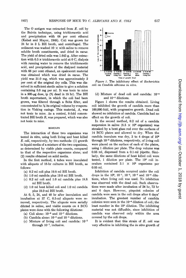
*Candida8- clC ili
0 52448 0 52448 0 52448 0 52448 0 52448Candida Candida + Candida Coli Candidaalone living Coli dead Coli alane living Call
Time af growth (hra.)
Figure 1. The inhibitory effect of Escherichiacoli on Candida albicans in vitro.
(d) Mixture of dead coli and candida: 10-4and 10-5 dilutions.
Figure 1 shows the results obtained. Livingcoli inhibited the growth of candida more than100,000-fold, with progressive growth. Dead colishowed no inhibition of candida. Candida had noeffect on the growth of coli.
In the second method, 0.2 ml of a candidasuspension in saline (6.5 X 10⁶ organisms) wasstreaked by a bent glass rod over the surfaces of14 BCG plates and allowed to dry. When thecandida inoculum was dry, 3 to 4 drops of 100through 10-6 dilutions, respectively, of living coliwere placed on the surface of each of the plates,using 1 dilution per plate. The drop volume was0.05 ml, dispensed from a 0.1-ml pipette. Simi-larly, the same dilutions of heat killed coli weretested, 1 dilution per plate. The 100 coli in-oculum contained 3.1 X 10⁸ organisms per0.05 ml.
Inhibition of candida occurred under the colidrops in the 10°, 10-', 10-2, 10-3 and 10-4 dilu-tions, when living coli was used. No inhibitionwas observed with the dead coli. Such observa-tions were made after incubation of 24 hr, 72 hrand 6 days. However, pinpoint colonies ofcandida were seen in the coli drops after 6 days'incubation. The greatest number of candidacolonies were seen in the 10-4 dilution of coli; theleast number in the 10c dilution. The inhibitingmaterial was not diffusible, since inhibition ofcandida was observed only within the areacovered by the coli drops.
It is evident that this strain of E. coli wasvery effective in inhibiting the in vitro growth of
19571 617
on October 1, 2018 by guest
http://jb.asm.org/
Dow
nloaded from

GALE AND SANDOVAL
C. albicans. The greater the concentration ofcoli, the more effective was the inhibition ofcandida.
The response of mice to the inoculation of bothC. albicans and E. coli. In this experiment, theeffects of the route of inoculation and of thesequence of inoculation were ascertained. Candidawas injected intravenously; coli was injectedeither intravenously or intraperitoneally intogroups of 7 mice (a commercial strain), re-
spectively, in the sequence and at the time inter-vals indicated in table 1.
It is apparent that with 1.8 X 10⁶ candida and3.7 X 107 coli:
(1) Intravenous inoculation of candida alonekilled 80 per cent of the mice in 18 hr, and allthe mice within 24 hr.
(2) Prior inoculation of coli, 30 min before thelethal candida injection, afforded some protec-tion, regardless of the route by which the coliwas injected. The prior intravenous inoculationof coli was more effective than the intraperitonealinoculation, since 70 per cent of the mice were
alive 48 hr after inoculation, as against 30 per
cent for the latter group.(3) Intravenous inoculation of coli after the
candida injection was ineffectual in delaying thetime of death or in affecting the number ofdeaths due to the candida injection.
(4) Some protective activity was produced bythe intraperitoneal inoculation of coli after thecandida, at both time intervals.
The effect of the concentration of intraperi-toneally injected E. coli on the lethal activity of C.albicans. Although prior inoculation of coli was
more effective in protecting mice against a lethalconcentration of candida than the injection ofcoli after the candida inoculation, it was decidedto investigate the effect of intraperitoneal inocula-tion of coli after the candida, for two reasons:
(1) any therapeutic to treat candidiasis would beutilized after candida infection had occurred; and(2) intravenous inoculation of coli showed no
different results than those obtained with candidacontrols.Groups of 7 mice (TF strain), suitably tagged
with dye, were inoculated with 1.8 X 10⁶ candidaintravenously, followed at the various timeintervals and concentrations of coli as shown intable 2 and figure 2. Mice were also injected witha high concentration of coli alone, and withcandida alone, as controls.When 107 coli was injected 30 min after
candida, a statistically significant protective ac-
tion was seen, as compared to the candida con-
trols. Such delay in the mean time of death was
also observed with the 2-hr interval betweeninoculations, but not to the same degree. With a
TABLE 1The interaction of Candida albicans and Escherichia coli in vivo*
TimeGrouip First Inoculation Second Inoculation Interval Resultst SurvivorsNo. ~~~~~~~~~~~~between
Inoculations
min
1 Candida iv None 0 4/7 (8); 6/7 (17); 7/7 (24) 0
2 Coli iv None 0 1/7 (10) 63 Coli ip None 0 0/7 7
4 Candida iv Coli ip 30 2/7 (8); 5/7 (17) 25 Candida iv Coli ip 150 1/7 (9); 4/7 (18); 5/7 (42) 2
6 Candida iv Coli iv 30 6/7 (18); 7/7 (46) 07 Candida iv Coli iv 150 6/7 (10); 7/7 (24) 0
8 Coli iv Candida iv 30 1/7 (24); 2/7 (28); 4/7 (120) 39 Coli ip Candida iv 30 1/7 (24); 2/7 (26); 5/7 (42) 2
* Candida concentration, 1.8 X 10⁶ organisms; coli concentration, 3.7 X 10⁷ organisms. All surviv-ing mice of this commercial strain were sacrificed after 6 days.
t Deaths/total; (time of death, hr).
618 [v'oi.. 73
on October 1, 2018 by guest
http://jb.asm.org/
Dow
nloaded from

RESPONSE OF MICE TO C. ALBICANS AND E. COLI
10-fold decrease in the concentration of coli, pro-tection was apparent at the 2-hr interval, but notat the 30-min interval. A 10-fold increase in coliconcentration (108 organisms) produced a sig-nificant decrease in the mean time of death ascompared to that of candida controls at the30-min and 1-hr intervals. No such effect wasseen with the increasing time intervals.
It therefore appeared that the protective phe-nomenon was exerted by the coli within a narrowrange of concentration. In addition, the timeinterval between inoculations markedly in-fluenced the results obtained. The course of theeffect was determined within 2 hr after the in-oculation of candida.
TABLE 2The effect of intravenously inoculated Candida
albicans and intraperitoneally inoculatedEscherichia coli in mice
TimeIn-
Candida Coli terval Mean Time2 Inoculated Inoculated be- of Death Ptz iv ip tween SDeah*Inoc- :1SD
ula-tions
hr hr
1 1.8 X 10⁶ None 0 333v ±13kx -
2 1.8 X 10⁶ 1.2 X 10⁸ 0.5 13½i±14,% <0.013 1.8 X 10⁶ 1.2 X 10⁸ 1 14zjiL-5%j <0.01
4 1.8 X 10⁶ 1.2 X 10⁸ 2 31Mj-22 0.85 1.8 X 10⁶ 1.2 X 10⁸ 3 39 i24}J 0.5
6 1.8 X 10⁶ 1.2 X 10⁸ 4 36±i27 0.8
7 1.8 X 10⁶ 1.2 X 10⁷ 0.5 96½444% <0.018 1.8 X 10⁶ 1.2 X 10⁷ 2 51 ±21 0.1
9 1.8 X 10⁶ 1.2 X 10⁶ 0.5 27 ±10 0.310 1.8 X 10⁶ 1.2 X 10⁶ 2 58%-±113% <0.01
11 None 1.2 X 10⁸ 0 >144 '
The mice used were the TF (Taconic Farms)strain.
* Standard deviation computed for smallsamples.
t The Student t test for the significance be-tween two means, for small samples. Comparisonof all groups to group I (candida controls). Levelof significance is at P of 0.05.
$ 6 mice, all other groups 7 mice. All survivorswere sacrificed after 6 days.
The effect of heat killed E. coli and C. albicansin vivo. Heat killed coli had no inhibitory effecton candida in vitro (figure 1). What activitymight it have in tivo?Groups of 7 mice each (TF strain) were in-
jected with 6.8 X 10⁶ candida intravenously,and the concentration of living or dead coli de-scribed in figure 3, 30 min later. The results ob-tained were compared to those with candidaalone, and between each pair of the same con-centration.A significant difference was seen between the
effect of the inoculation of living coli and that ofdead coli, with the 107 and 106 concentrations.The mean time of death due to dead coli was
1oo-
90-
s8-
70-
' 60-
o 50-
, 40E I2s0-H Ii
.5 1. 2 3. 4 5 .5 2 hot0 1.2 xiO8 1.2 x 1o7 I.?2 xo6
Conc. of E. coli
Figure 2. The effect of varying concentrationsof Escherichia coli injected intraperitoneally atvarious time intervals on the lethal activity ofCandida albicans in mice.
60,*UvingS.661
50 *DeOd £.colz
'c 40-
'S-E
E20 M | |
0 29x10 29x1 2.9x5 2.9x 2l9xConc. of E. Coli
Figure S. The effect of heat killed Escherichiacoli on the lethal activity of Candida albicans ascompared to that of living E. coli.
19571 619
on October 1, 2018 by guest
http://jb.asm.org/
Dow
nloaded from

GALE AND SANDOVAL
less, for all concentrations of coli, than that dueto candida alone. The time of death due to thelower concentrations of living coli was similar tothat of the candida alone. It is interesting to notethat the dose of 2.9 X 10⁸ dead coli withoutprior inoculation of candida was not lethal, but2.9 X 103 dead coli with prior candida injectionwas markedly lethal.
It may be concluded that dead coli is no more
inhibitory to candida in vivo than it was in vitro.The enhancement of the time of death, however,was seen in the mice injected with dead coli in allconcentrations tested.
Several questions were raised by these findings.What is the enhancement effect? Is the de-creased time of death due to lethal action by thecandida or by the coli? Is the protective activityan antibiotic effect of the coli due to colicines or
coliformin?A working hypothesis to explain the enhance-
ment phenomenon is that the prior inoculation ofcandida may interfere with the defense mech-
anisms of the host. Living coli may then multiplyto the threshold concentration which would belethal because of the production of endotoxin;dead coli would be lethal because of its endo-
n°80e- Onormal'a IZCandida i.vc *Cdi i.p.2fi 70- *Candida iv8 folwed by..' min latere 60-
60
IV50 1
c.EcL
E
cL
e6cE
2 45 60 90 120 150 180
Time in minutes after the first inoculation
Figure 4. The leukocytic response in the periph-eral blood of mice inoculated with a lethal con-
centration of Candida albicans and a nonlethalconcentration of Escherichia coli.
TABLE 3The activity of the 0 antigen of Escherichia coli in the candida-coli interaction in vivlo
Group No. Candida Inoculated Coli or Coli Materials Inoculated Mean + SD Piv ip, 30 Min Later
hour
1 2.2 X 10⁶ None 45 :4 12½
2 2.2 X 10⁶ 0 antigen, 100 'Agt 82 A 22t <0.013 None 0 antigen 100 IAgt
4 2.2 X 10⁶ At 10'2 41 6 <0.015 None At > 144
6 2.2 X 10⁶ Bt 31'2 4 16'2§ 0.27 None Bt > 144
8 2.2 X 10⁶ 1.3 X 10⁸ living 11}j i 7 <0.019 2.2 X 10⁶ 1.3 X 10⁸ dead 11 i61± <0.01
10 2.2 X 10⁶ 1.3 X 10⁹ dead 9 i 5§ <0.01
11 None 1.3 X 10⁹ living 9- 4212 None 1.3 X 10⁹ dead >144
13 None 1.3 X 10⁸ living >14414 None 1.3 X 10⁸ dead >144
* Student t test for the significance of two means, small samples. All groups, where applicable, com-pared to group I. Level of significance at P of 0.05.
t 0 antigen of E. coli, extracted by the Boivin technique. A is the 8-fold concentrated brain heart(BH) broth in which the E. coli was grown. B is the 8-fold concentrated BH broth control.
t 2/5 mice dead in 36 hr, the 3 survivors lived more than 144 hr.§ 6 mice, all other groups 7 mice (TF strain).
620 [VOL. 73
on October 1, 2018 by guest
http://jb.asm.org/
Dow
nloaded from

RESPONSE OF MICE TO C. ALBICANS AND E. COLI
toxin content. The leukocytic response is one ofsuch defense mechanisms of the host.
The leukocytic response in mice injected withcandida intravenously (iv) and a high concentrationof coli, intraperitoneally (ip). Three groups of 10mice (TF strain) were inoculated as follows:
Series I: 5.5 X 10⁶ candida, ivSeries II: 1.0 X 10⁸ co]i, ipSeries III: 5.5 X 10⁶ candida, iv; followed 30
min later by 1.0 x 10⁸ coli, ipAt the time intervals shown in figure 4, 5 mice
in each series were bled from the tail veins, eachmouse being bled for all the time intervals. Dif-ferential smears were prepared and stained withGiemsa stain. The percentage of polymorpho-nuclear leukocytes per 100 cells and counts ofthe number of organisms present in the peripheralblood were obtained simultaneously. The resultsare summarized as follows:
(1) Uninoculated, normal mice have a mean
leukocyte count in their peripheral blood of 31±10 per cent. The inoculation of candida alone(series I) produced a marked leukocytic response,
reaching a plateau at 60 min after inoculation.The coli control mice (series II) showed a leuko-penic response until 120 min after inoculation. Inseries III, the leukocytic response reflected a
possible interaction of candida and coli. It isdifficult to determine whether the coli inocula-tion significantly lowered the leukocytic responseto candida, or whether the prior injection ofcandida significantly increased the leukocyteresponse to coli.
(2) Candida was present to 150 min after in-oculation in series I (candida alone), but indecreased numbers after the first 30 min; coli wasfound in small numbers in the peripheral blood to30 min after inoculation, and sporadically there-after (series II, coli alone). In series III, candidawas cultured in small numbers to 90 min afterthe first inoculation; coli was present in largenumbers to 180 min after the first inoculationand sporadically thereafter. It would seem thatthe prior inoculation of candida effected a colibacteremia.
(3) The mean time of death of the bled micein the three groups was: series I, 24 hr; series II,66y hr; series III, 5 hr.
The effect of the lipopolysaccharide 0 antigen ofE. coli on C. albicans in vivo. If the enhancementphenomenon were due to the endotoxin of coli,then intraperitoneal inoculation of the 0 antigen(endotoxin) derived from coli, 30 min after the
intravenous candida injection should showsimilar enhancing results.Groups of 7 mice each (TF strain) were in-
oculated intravenously with 2.2 X 10⁶ candida,followed in 30 min by the materials and concen-trations of living and dead coli shown in table 3.Injection of 100 ;&g 0 antigen was as enhancingas the high concentration of living or dead coli.The Seitz-filtered, concentrated BH broth inwhich co]i had been grown, A, also showed lethalactivity with the prior inoculation of candida(enhancement), but no such lethal action wheninoculated alone. The BH broth control of sameconcentration, B, exhibited no such effect. It isprobable that the lethal activity of material Awas due to small amounts of 0 antigen. Thereseemed to be no evidence for a diffusible anti-biotic produced by coli during its growth.A determination of the least amount of 0
antigen required to show the enhancement phe-nomenon was done in Namru mice, as shown intable 4. As little as 1.0 ,ug 0 antigen was suf-ficient to show the effect.
TABLE 4The effect of varying concentrations of the 0 antigen
of Escherichia coli on the intravenousinoculation of Candida albicans in mice*
0 AntigenGroup Candida Inocu-No. Inoculated iv lated Mean d SD Pt
30 minLater
Ag hr
1 5.3 X 10⁶ 0 640 _
2 5.3 X 10⁶ 100 3:iA. <0.013 None 100 -t
4 5.3 X 10⁶ 10 449Y <0.015 None 10 > 144
6 5.3 X 10⁶ 1.0 54% <0.017 None 1.0 > 144
8 5.3 X 10⁶ 0.1 6±i2/ 0.79 None 0.1 > 144
10 5.3 X 10⁸ 0.01 8ff-gA2 0.1311 None 0.01 > 144
* Namru strain of white mice.t Student t test. All groups where applicable
compared to group 1. Level of significance at Pof 0.05.
t Y dead in 16 hr, 2 survivors lived after 144 hr.
6211957]
on October 1, 2018 by guest
http://jb.asm.org/
Dow
nloaded from

GALE AND SANDOVAL
Evidence has been presented to show that thedecrease in mean time of death in the mice in-oculated intravenously with a lethal concentra-tion of candida followed by the intraperitonealinoculation of high concentrations of living ordead coli (the enhancement phenomenon) is dueto the toxemia caused by the endotoxin ofE. coli.
DISCUSSION
It is well established that interactions betweenmicroorganisms occur in vivo, best exemplifiedby the interference phenomenon observed withmany viral agents (Schlesinger, 1952). Schle-singer (1952) also points out that virus exalta-tion, the ability of one virus to make a host ab-normally susceptible to the effects of anothervirus, may also be seen.Examples of interactions between different
kinds of organisms in vivo are not as numerous asin the virus-virus interactions. Most of the re-ports are concerned with increased resistance toinfection by one agent, which is influenced bythe route of inoculation and concentration of themodifying agent. Horsfall and McCarty (1947)reported the modifying effect of a nonhemolyticstreptococcus on the course of PVM virus infec-tion (pneumonia virus of mice), both admin-istered intranasally. Scherr (1953a) describedincreased resistance of mice to encephalomyo-carditis virus injected intranasally, by Crypto-coccus neoformans inoculated intraperitoneally, 9days previously. Salvin and Bell (1955) demon-strated inhibition of infection of mice by Rickett-sia typhi or Rickettsia tsutsugamushi, followingthe intraperitoneal inoculation of a suitableconcentration of Histoplasma capsulatum. Nyka(1956) reported enhanced resistance to experi-mental tuberculosis in mice which had been in-fected previously with either virulent or avirulentBrucella abortus. The route of administration ofbrucella influenced the degree of resistance of theanimals.Another aspect of the interaction of micro-
organisms in vivo is the proimmunity (a non-specific, rapid immunity, not related to theformation of antibodies), described by Oerskov(1940), Field et al. (1955), Brandis (1954), andothers. These authors have found that the in-oculation of killed or living organisms, the sameor similar to the challenge organisms, protected
mice against death, if administered 4 to 48 hrbefore the lethal challenge dose of virulentorganisms. Oerskov (1940) reported his findingsthat the proimmunity effect of the pretreatmentof mice with E. coli vaccine inoculated 24 hrbefore a lethal dose of Shigella shiga was causedby a rapid phagocytosis and inhibition of lysisof the Shigella. The proimmunitv had no effecton death due to injected endotoxin, or to killedShigella shiga.Rowley (1955) observed the effect of the in-
jection of cell walls of E. coli either intravenouslyor intraperitoneally, on the subsequent infectionof white mice with E. coli suspended in mucin.Enhanced susceptibility to the infection occurredif the time interval between inoculation was lessthan 2 hr. Increased resistance was demon-strated when the challenge dose of E. coli inmucin was delayed 24 hr after the inoculation ofthe cell walls of E. coli. These findings have beencorrelated with the immediate rapid fall ofserum-properdin levels to 20 per cent of theirnormal value, and the subsequent rapid restora-tion of the properdin levels to greater than 4 Xnormal level, 48 hr after the inoculation of micewith cell walls of E. coli or with "zymosan" (ayeast polysaccharide), as shown by Pillemer andhis associates (1955). Injection of mice withzymosan produced the same results to challengewith E. coli in mucin as the injection of cell wallsof E. coli. It is of interest that Scherr (1953b)showed that the pathogenicity of intraperi-toneally injected C. albicans for mice was en-hanced by either living or formolized washedcells of Saccharomyces cerevisiae injected intra-peritoneally at 48-hr intervals into the candidainoculated animals.
In our experiments, the enhancement phe-nomenon is very similar to the enhanced sus-ceptibility of mice to E. coli within 2 hr after theinoculation of cell walls of E. coli or of zymosan(Rowley, 1955). It is likely that the properdinsystem (Pillemer et al, 1954, 1955) may play apart in the mechanism of this phenomenon, sincecell walls of a Candida species were very effectivein removing properdin from serum in vitro. More-over, infection promoting materials like hoggastric mucin (Gale and Elberg, 1952) andlevans (Hestrin et al., 1954) produced a markedrapid fall in the properdin level in the serum ofmice injected intravenously with these mate-
622 [VOL. 73
on October 1, 2018 by guest
http://jb.asm.org/
Dow
nloaded from
RESPONSE OF MICE TO C. ALBICANS AND E. COLI
rials, within 2 to 3 hr (Pillemer et al., 1954). Theprior inoculation of C. albicans in our experi-ments may also have lowered the properdinlevel in the mice so that the living E. coli, inhigh concentration, could multiply uninter-ruptedly to the threshold level which was lethalto mice. A coli bacteremia was observed in themice inoculated with candida and the high con-centration of coli.However, the properdin system does not pro-
vide the entire answer. It is difficult to see howthis immunity factor can influence the deathdue to the toxemia caused by endotoxin. Thetime of death in the mice inoculated with verysmall numbers of dead E. coli after the priorinjection of C. albicans was very much decreasedfrom that of 108 dead E. coli alone (no deaths).Moreover, the time of death of mice injectedwith 1.0 ,ug 0 antigen (lipopolysaccharide) afterthe prior inoculation of C. albicans was greatlydecreased from that of 1,000 ,ug "O" antigenalone. Other defense mechanisms of the hostmust also be involved.The leukocytic response in the peripheral
blood of the mice following the inoculation ofthe two organisms was significantly differentfrom that of either organism alone, for the first3 hr. Whether the change was due to the influenceof the candida inoculation on the coli response, orto the effect of the coli injection on the candidaleukocytosis, cannot be assessed from the data.However, it is probable that the response ob-served may have resulted from the effect of theprior inoculation of candida on the coli leuko-penia.With regard to the protective effect, no ex-
planation for the mechanism involved can beattempted at the present time. The only informa-tion available from the data above is that theeffect was reproducible both in vitro and in vivo,and that it was not due to a diffusible solubleantibiotic similar to the colicines. There seems tobe no similarity to the increased resistance toinfection to one organism by the modification ofanother unrelated organism, since the concen-tration differential of the coli inoculum is sonarrow. It may be related to the proimmunitystudies described, and may thus be influencedby the properdin system. Experiments are nowin progress in an attempt to elucidate such amechanism.
SUMMARY
Prior inoculation of a nonlethal concentrationof living Escherichia coli either intravenously orintraperitoneally protected mice from a lethalconcentration of Candida albicans, injectedintravenously.The intraperitoneal inoculation of nonlethal
concentrations of E. coli after the intravenousinjection of a lethal concentration of C. albicansproduced two contrary effects dependent on thecoli concentration: (1) a delay in the mean timeof death (protective), and (2) a decrease in themean time of death (enhancing) as compared tocandida alone.Dead coli of any concentration tested showed
only the enhancing effect. As little as 1.0 Mg ofO antigen extracted from E. coli by the Boivintechnique, was enhancing. The enhancementeffect seemed to be caused by the toxemia due tothe endotoxin of E. coli.The possible mechanism of action of the en-
hancement phenomenon was discussed withreference to the properdin system.
REFERENCESBRANDIS, H. 1954 tYber die Promunitat (De-
pressionsimmunitiat). Ergeb. Hyg. Bakteriol.Immunitiitsforsch. u. Exptl. Therap., 28,141-202.
FIELD, T. E., HOWARD, J. G., AND WHITBY, J. L.1955 Studies of the rapid production of non-specific type of immunity to Salmonella typhiinfection in mice. J. Roy. Army Med.Corps, 101, 324-334.
FISHER, R. A. 1948 Statistical methods forresearch workers, 10th ed. Oliver and Boyd,London, W. C., England.
FREYSCHUSS, S. K. L., PEHRSON, S. O., AND STEEN-BERG, B. 1955 Coliformin: production andisolation. Antibiotics & Chemotherapy, 5,218-223.
GALE, D. AND ELBERG, S. S. 1952 Studies onenhancement of bacterial infection by hoggastric mucin. J. Infectious Diseases, 91,50-62.
GARBER, E. D. AND HAUTH, F. C. 1950 A newmutation with asymmetrical expression in themouse. J. Heredity, 41, 122-124.
HESTRIN, S., SHILO, M., AND FEINGOLD, D. S.1954 Promotion of peritoneal infection byintravenous levan. Brit. J. Exptl. Pathol.,35, 107-111.
HORSFALL, F. L., JR. AND MCCARTY, M. J. 1947The modifying effects of certain substances
1957] 623
on October 1, 2018 by guest
http://jb.asm.org/
Dow
nloaded from
GALE AND SANDOVAL
of bacterial origin on the course of infectionwith pneumonia virus of mice (PVM). J.Exptl. Med., 85, 623-646.
HUPPERT, M., CAZIN, J., JR., AND SMITH, H., JR.1955 Pathogenesis of Candida albicans in-fection following antibiotic therapy. III.The effect of antibiotics on the incidence ofC. albicans in the intestinal tract of mice. J.Bacteriol., 70, 440-447.
KABAT, E. A. AND MAYER, M. M. 1948 Experi-mental immunochemistry, 1st ed. CharlesC Thomas, Springfield, Illinois.
NYKA, W. 1956 Enhancement of resistance totuberculosis in mice experimentally infectedwith Brucella abortus. Am. Rev. Tuberc.,73, 251-265.
OERSKOV, J. 1940 Infektionsmechanishe Unter-suchungen uiber unspezifische lokalegesteigerte bzw. herabgesetzte Resistenz(Promunitat bzw. Mucin). Immunitats-forsch., 98, 359-372.
PAINE, T. F. 1952 In vitro experiments withMonilia and Escherichia coli to explainMoniliasis in patients receiving antibiotics.Antibiotics & Chemotherapy, 2, 653-658.
PILLEMER, L., BLUM, L., LEPOW, I. H., Ross,0. A., TODD, E. W., AND WARDLAW, A. C.1954 The properdin system and immunity.I. Demonstration and isolation of a new serum
protein, properdin, and its role in immunephenomena. Science, 120, 279-284.
PILLEMER, L., SCHOENBERG, M. D., BLUM, L.,AND WURZ, L. 1955 Properdin system andimmunity. II. Interaction of the properdinsystem with polysaccharides. Science, 122,545-549.
ROSEBURY, T., GALE, D., AND TAYLOR, D. F.1954 An approach to the study of interactivephenomena among microorganisms indigenousto man. J. Bacteriol., 67, 135-152.
ROWLEY, D. 1955 Stimulation of natural im-munity to Escherichia coli infections. Ob-servations on mice. Lancet, 268, 232-234.
SALVIN, S. B. AND BELL, E. J. 1955 Resistanceof mice with experimental histoplasmosis toinfection with Rickettsia typhi. J. Immunol.,75, 57-62.
SCHERR, G. H. 1953a The susceptibility of miceinfected with Cryptococcus neoformans toencephalomyocarditis virus. J. Bacteriol.,65, 480-481.
SCHERR, G. H. 1953b Enhanced dissemination ofmoniliasis in mice by the injection of yeastcells. J. Creighton Univ. School of Med., 8,20-24.
SCHLESINGER, R. W. 1952 Interference betweenanimal viruses. In Viral and Rickettsial Dis-eases of Man, 2nd ed., pp. 161-171. J. P.Lippincott Co., Philadelphia, Pa.
624 [VOL. 73
on October 1, 2018 by guest
http://jb.asm.org/
Dow
nloaded from