Embed Size (px)
Citation preview

A COMPARATIVE STUDY OF ORGANISMS OF THEFRIEDLANDER AND COLI-AEROGENES GROUPS
I. MORPHOLOGICAL AND CULTURAL CHARACTERISTICS,WITH EMPHASIS ON VARIATION
ELIZABETH OSTERMAN' AND LEO F. RETTGERDepartment of Bacteriology, Yale University
Received for publication March 20, 1941
Our understanding of the demarcation and relationships of theso-called FriedlAnder group of organisms remains today in a con-fused state. A review of the literature2 reveals a remarkable lackof agreement with regard to the exact attributes of its members.However, nearly all authorities agree that it may be definedloosely as a group of gram-negative, non-spore-forming, non-motile encapsulated organisms which do not liquefy gelatin andwhich form a luxuriant, raised, non-pigmented mucoid growthon a solid medium (Small and Julianelle 1923, Julianelle 1926a,1928, 1930, Edwards 1928, 1929, Bamforth 1928, Goslings 1934,1936, Topley and Wilson 1936, Parr 1939, Bergey et al. 1939).Pathogenicity of freshly isolated strains may usually be demon-strated. Although progress has been made in establishing sub-divisions within the group, principally through serological studies,its limits remain undefined.Attempts to establish serological differentiations were unsuc-
cessful until Julianelle (1926a, b, c) applied the immunologicalprinciples governing the pneumococci. His studies disclosed thata type specificity is conferred by the capsular polysaccharidesand a species specificity by proteins in the soma of the cells.
1 This paper covers in part the dissertation submitted to the Graduate Schoolof Yale University by the senior author in candidacy for the degree of Doctor ofPhilosophy.
2 For a full historical resum6 the reader is referred to the doctoral dissertationof the senior author, in the Yale University Library.
699
on Septem
ber 17, 2018 by guesthttp://jb.asm
.org/D
ownloaded from

ELIZABETH OSTERMAN AND LEO F. RETTGER
Three types were distinguished among the pneumobacilli-typesA, B and C-and a heterogeneous group, X. The antigenichomogeneity of the non-encapsulated cells, reported by Julianelle(1926b, c) and Goslings (1936) was not confirmed in Randall'sstudies (1939). Strains isolated from patients having ozena andrhinoscleroma were shown to be serologically distinguishable fromthe pneumobacilli and from each other (Morris and Julianelle1934, Julianelle 1935, 1937b, Prica 1930, Prasek and Prica 1933,Goslings 1936, Kurglowicz 1938).The results of biochemical tests applied in the hope of char-
acterizing the group more definitely were found to be exceedinglyvariable (Clairmont 1902, Perkins 1904, Fitzgerald 1914, Coulter1917, Bamforth 1928, Grant 1931, Hay 1932, Bhatnagar andSingh 1935, Lvy-Bruhl and Cado 1937), and to be of littletaxonomic value. Even strains which were found by serologicaltests to be apparently identical exhibited conspicuous differencesin their biochemical reactions (Small and Julianelle 1923, Edwards1928, 1929, Julianelle 1930).
It is apparent from a consideration of the definition of membersof the Friedlander group that organisms of the species Aerobacteraerogenes, which are commonly encapsulated when freshly iso-lated, would be especially difficult to differentiate. They areordinarily non-motile, do not liquefy gelatin and form a luxuriantmucoid growth on solid media. Their IMViC reactions (indole,M.R., V.P., citrate, see Parr 1939) and sugar fermentations maybe and often are simulated by Friedlander strains (Edwards,1929). Typical A. aerogenes cultures were, in fact, included in thecollections of most of the early investigators (Strong 1899, Clair-mont 1902, Fitzgerald 1914, Coulter 1917, Bamforth 1928).Besson (1924) believed that the pneumobacilli and Aerobacteraerogenes were indistinguishable. Castellani and Chalmers (1920),Castellani (1938), and Perkins (1925) advocated their inclusionin a single genus Encapsulatus.
Hill and associates (1929) stated that there seemed to be noway of differentiating Aerobacter cultures from Voges-Proskauerpositive forms of the Friedlander group, nor any way of differ-entiating Voges-Proskauer negative, lactose-fermenting Fried-
700
on Septem
ber 17, 2018 by guesthttp://jb.asm
.org/D
ownloaded from
COMPARATIVE STUDY OF ORGANISMS
lander organisms from encapsulated Escherichia forms. Randall(1939) called attention to the fact that colonial differences or thepresence of capsules could not be relied upon to separate Fried-lander bacilli from coliforms. Parr (1934, 1936) was impressedwith the possible close relationship of the Friedlander and coli-intermediate-aerogenes group, and in his review of the coliforms(1939) gave reasons for their inclusion in a single genus. Bergeyand associates (1939) have recognized the affinities of these or-ganisms by placing the genus Klebsiella in the family Entero-bacteriaciae, with the genera Escherichia and Aerobacter.A limited number of serological studies have contributed infor-
mation on the question. Among 26 cultures labelled Aerobacteraerogenes, Edwards (1929) found 5 which were agglutinated spe-cifically in type B Friedlander antiserum, and in agglutininabsorption tests caused a complete removal of specific agglutinins.Working with three A. aerogenes strains, Julianelle (1937a) foundthat one of them reciprocally agglutinated with a type B Fried-hinder bacillus. From the evidence of agglutinin absorption andprecipitin tests, he concluded that the reaction depended on thepresence of similar, though not identical, capsular carbohydrates.The three Aerobacter cultures, although serologically differentwhen encapsulated, became antigenically the same when decap-sulated; and they did not cross-react with decapsulated type BFriedlander cells. Julianelle (1937b) advanced the hypothesisthat systematic relationships among these groups could be demon-strated more readily if the serological reactions of the non-encap-sulated variants were studied. Although Goslings (1936) sub-stantiated much of Julianelle's work, his results did not indicatethat the non-encapsulated A. aerogenes cultures were antigenicallyhomogeneous.The comprehensiveness of the study of antigenic relationships
among the coliforms by Stuart and associates (1940) makes it acontribution of special value. They found that A. aerogenes strains(IMViC, cellobiose - - + + +) were antigenically so heterog-enous that of 918 chances (103 cultures tested in 9 differentantisera) not one culture was sufficiently related to any of the9 organisms used for immunizing, to remove the homologous
701
on Septem
ber 17, 2018 by guesthttp://jb.asm
.org/D
ownloaded from

ELIZABETH 01TERMAN AND LEO F. RETTGER
agglutinins, although many of the cultures agglutinated to somedegree in one or more of the antisera. It should be stated thatno mention was made of their culture phase, i.e., whether theorganisms were encapsulated or non-encapsulated. It wouldseem that the establishment of serological differentiations amongthese organisms does not promise to be a simple matter.The work of Rakieten, Eggerth and Rakieten (1940), in which
they employed bacteriophages against mucoid strains of Kleb-siella, Aerobacter and E8cherichia, has revealed interesting pos-sibilities.
Pathogenicity of the Friedlander bacilli and non-pathogenicityof Aerobacter strains are commonly considered to be differentialcharacteristics. However, organisms of the Aerobacter genus maybe observed in pathological processes. Hill and associates (1929)identified 79 of 200 cultures isolated from urinary tract infectionsas belonging to the genus Aerobacter. Mucoid cultures of type Aand B FriedlAnder bacilli have been shown to be virulent for whitemice, while those of type C have been found to be devoid ofvirulence (Julianelle 1926a, b). Information concerning thepathogenicity of group X strains is lacking.The encapsulated cultures of A. aerogenes are by no means the
only forms that it is necessary to distinguish from members ofthe Friedlander group. The work of Smith and Bryant (1927),Hill et al. (1929), Parr (1934), Lovell (1937) and others has shownthat encapsulated mucoid organisms identified by biochemicaltests as belonging to the genus Escherichia may be isolated in largenumbers, especially from pathological conditions of the animalor human body. It is essential that these be considered in theproblem of classification.
In addition to mucoid colony forms, rough and smooth-roughintermediate forms have been described in cultures of Escherichiacoli (Hadley 1927, Dulaney 1928, Nungester and Anderson, 1931,and others). Descriptions of colonial and morphological variantsof the pneumobacilli appeared among the earlier studies of disso-ciative phenomena (Toenniessen 1913, 1914, Baerthlein 1918,Hadley 1927). Julianelle (1928) described smooth (S) encap-sulated, and rough (R) non-encapsulated forms. Dawson (1934)introduced the three-phase concept of mucoid (M), smooth (S),
702
on Septem
ber 17, 2018 by guesthttp://jb.asm
.org/D
ownloaded from
COMPARATIVE STUDY OF ORGANISMS
and rough (R) colonies in his studies of the pneumococci; hepointed out the analogy between these and the Friedlinderorganisms, and urged acceptance of this terminology. Hadley(1937) also advocated use of these designations. Randall (1939)described these three principal phase variants among the Fried-hinder bacilli, but he did not designate them accordingly.The importance of considering variational phenomena in at-
tempts at classification and species description has been empha-sized by Hadley (1937) and others. It seems not unlikely thatmuch of the confusion still surrounding the relationships of theFriedlander and coli-aerogenes organisms is due to a failure torecognize the full significance of variation. The authors havesought clarification of the problem in a systematic study of anumber of freshly isolated gram-negative encapsulated organisms.The present paper deals with the cultural and morphologicalcharacteristics of normal and variant forms, with the occurrenceof variants under laboratory conditions, and with methods ofstabilizing the cultures for further study.
EXPERIMENTAL
Organisms employedOne hundred encapsulated strains were used in the investiga-
tion, the majority having been isolated recently from clinical andpathological material. A few were derived from grains, sewageand soil, and several (Sc, E. F10, Egs, Is, AF1, AS3, AS5, AS6)were from stock culture collections, and were not of recent isola-tion. All of the selected organisms were gram-negative, non-spore-forming, non-motile, encapsulated bacillary forms which did notliquefy gelatin or produce a pigment at room temperature, andwhich formed a raised mucoid growth on solid media. The colo-nies of some were of a pasty consistency; others were syrupy orviscid. No organisms isolated from patients having ozena, rhino-scleroma, or inguinal granuloma were included in the presentstudy.The exclusion of motile strains should be explained. In their
study of forty strains of encapsulated gram-negative organismsL6vy-Bruhl and Cado (1937) included eight which were motile.Furthermore, some of their strains liquefied gelatin. They advo-
703
on Septem
ber 17, 2018 by guesthttp://jb.asm
.org/D
ownloaded from
ELIZABETH OSTERMAN AND LEO F. RETTGER
cated revision of the definition of the Bacillus mucosus (capsulatus)group to include forms having these properties. Unfortunately,serological data were not included in their report. No organismswhich have been identified serologically by other workers asFriedlinder types have exhibited these properties. Strains ofthis nature which were encountered in isolations during the pres-ent study (5 in number) were tested in type-specific Friedlinderantisera, but none were agglutinated.To avoid working with mixed cultures, all strains were replated
after isolation at least four times, with vigorous shaking in a waterblank between platings. A number showed marked dissociativetendencies after primary isolation. Those which could not bestabilized with relative ease in the mucoid encapsulated stateby mouse passage and colony selection were not included.
All of the organisms were tested for agglutination in type-specific Friedlander A, B and C antisera. Twenty agglutinatedin type A, 21 in type B and only one, the original homologousstrain, in type C. Provisional designations were given the re-maining 58 cultures on the basis of fermentation of glucose andlactose, their IMViC reactions, and pathogenicity for mice (seeOsterman and Rettger, 1941). The arbitrary nature of thecriteria was recognized. Eight cultures were allocated to Fried-kinder group X, 46 to the species Aerobacter aerogenes, 2 to Esch-erichia coli, 1 to the intermediate group, and 1 aberrant formremained unidentified.
It was interesting to note that 23 of the 46 "aerogenes" strainswere isolated from pathological processes in the human body(abdominal abscesses, genito-urinary infections, pus from wounds,blood cultures, lung in pure culture at autopsy, etc.). Further-more, the sources of several of the Friedlander type A and Bstrains were similar to these. The implication is clear that theorigin of a gram-negative mucoid culture does not serve as aguide to its identity.
Three sets of stock-cultures were initiated from strains stabi-lized in the fully encapsulated state, one by cultivation on plainagar slants, a second on slanted infusion agar, and a third by freez-ing and desiccation in a modified Flosdorf-Mudd "lyophile" appa-ratus (Flosdorf and Mudd, 1935). Slant cultures of the various
704
on Septem
ber 17, 2018 by guesthttp://jb.asm
.org/D
ownloaded from

COMPARATIVE STUDY OF ORGANISMS
organisms were stored in the dark at room temperature and trans-.planted at monthly intervals; the sealed "lyophile" tubes werestored in the refrigerator.
METHODS
A special infusion medium, used in the study of colonial struc-ture, was made according to the formula of the Rockefeller Insti-tute (called by them "pneumococcus broth"). Ten grams ofBacto-peptone and 0.3 gram of sodium phosphate were dissolvedin 500 ml. of fresh beef infusion, the volume made up to 1 literand the pH adjusted to approximately 7.8. For a solid agarmedium, 15 grams of Bacto-agar were dissolved in the waterbefore adding it to the infusion, and, after standing, the mixturewas filtered through cotton. This constitutes the infusion agarto which reference will hereafter be made. Contrasts in colonialform were more marked and more easily discerned on this thanon plain or glucose agar, or on Endo's or eosin-methylene-blue agar.A modified Hiss capsule stain was developed which gave satis-
factory and consistent results.3 The bodies of the organismsstained a deep, and the capsules a light, purple.The colonial characteristics and occurrence of variants in the
cultures were determined by inoculating sterile water blanks withmaterial from agar slant cultures, shaking vigorously twenty-fivetimes and streaking infusion agar plates in duplicate. The plateswere incubated 48 hours at 37TC. and examined with the aid ofa 3 X hand lens against an artificial light source, and with a lowpower objective. The necessity of obtaining well-isolated coloniescannot be over-emphasized. It was noted by Smith and Bryant(1927), working with mucoid cultures of Escherichia coli, thatcrowding of the colonies prevented the appearance of variationalstructures. This has been observed repeatedly in the presentstudy.
RESULTS
The cultures were plated for observation after the variousstrains had been subcultured for periods varying from two to ten
a Particulars may be found in the doctoral dissertation of the senior author inthe Yale University Library.
705
on Septem
ber 17, 2018 by guesthttp://jb.asm
.org/D
ownloaded from

ELIZABETH OSTEBMAN AND LEO F. RETTGER
months. When first placed in stock on agar slants the colonieswere raised, homogeneous, and viscid or pasty (plate 1, fig. 1).The cells were surrounded by a capsule (plate 2, fig. 1). Fried-hinder colonies of this description were termed smooth (S) byJulianelle (1928), Edwards (1928), Randall (1939), Rakieten(1940) and others. According to Dawson's terminology, whichwill be used in the present study, these are mucoid or M colony
TABLE 1Variational changes in mucoid (M) stock cultures
MAINTAINED ON PLAIN NUTRIENT AGARNUMBER
DESIGNATION OF Pe- Ur-sTRAINS FreeS ihrlstria- bnt Nocolonies retors tons colonate change
Friedlander typeA .................. 20 0 3 12 1 7Friedlinder type B.................. 21 1 19 10 1 1Friedlander typeC .................. 1 0 1 1 0 0"Friedlander group X"............... 8 1 4 2 1 2"Aerogenes".......................... 46 7 44 22 6 2"Coli"............................ 2 0 2 2 0 0Miscellaneous........................ 2 1 2 1 1 0
Total ............................ 100 10 75 50 10 12
MAINTAINED ON INFUSION AGAR
Friedlander type A.................. 20 0 2 12 1 8Friedlander type B.................. 21 1 17 11 1 1FriedlAnder typeC .................. 1 0 1 1 0 0"Friedlinder group X"............... 8 0 4 2 0 3"Aerogenes"........................... 46 4 41 21 2 1"Coli".............................. 2 1 2 2 0 0Miscellaneous........................ 2 0 1 1 1 0
Total .......................... 100 6 68 50 5 13
forms. When maintained on plain nutrient agar slants the major-ity of strains exhibited variational changes (table 1). Smooth,flat, translucent coli-like colonies were found scattered among thenormal mucoid colonies on the plates prepared from 10 per centof the 100 strains examined (plate 1, fig. 1). They were composedof small non-encapsulated bacilli (plate 2, fig. 2). These maybe identified as the smooth (S) colony of FriedhInder bacilli.
706
on Septem
ber 17, 2018 by guesthttp://jb.asm
.org/D
ownloaded from
COMPARATIVE STUDY OF ORGANISMS
They are indistinguishable colonially or morphologically from thenon-encapsulated forms of A. aerogenes and E. coli.Three well-defined colony forms transitional between the mu-
coid (M) and smooth (S) were observed which in each instancewere composed of both encapsulated and non-encapsulated cells.The first of these occurred in 75 per cent of the cultures (seetable 1). The colonies were mucoid; at the periphery wedge-shaped sectors of translucent smooth growth developed. Thesesectors have been described in pneumobacillus cultures by manyinvestigators (Toenniessen 1914, Baerthlein 1918, Hadley 1937,Shinn 1939, and others); in mucoid cultures of E. coli by Smithand Bryant (1927); and in cultures of E. coli and a strain ofA. aerogenes by Lovell (1937). Usually a single sector developedat the periphery, as shown in figure 4 (plate 1), but in cultures inwhich the dissociative tendency was extreme, multiple sectoringwas not uncommon, as illustrated by aerogenes strain Sw2(plate 1, fig. 6) and a type B Friedlander strain, No. 47 (plate 1,fig. 5). The sectors were a convenient source of smooth variantstrains; by replating from them the pure S form could be ob-tained readily.The second transitional colony form resembled the original
mucoid (M) colony in consistency, but was marked with fine orcoarse radial striations, the edge being entire or only slightlyirregular (plate 1, fig. 3). They appeared on plates of 50 per centof the cultures and were particularly pronounced among the typeA Friedldnder strains showing variational changes (table 1). Thethird type of colony intermediate betweenM and S was umbonatein form. It was characterized by a raised, opaque center of encap-sulated cells and a flat translucent periphery consisting of non-encapsulated cells. Examples may be seen in the photograph ofcolonies published in Julianelle's study of variation of Friedhinderbacilli (Julianelle 1928). These forms were observed in 10 percent of the cultures. By replating from the central portion a pureM culture could sometimes be recovered, and by replating fromthe periphery the pure S form was obtained occasionally. How-ever, this colony and the striated type exhibited a strong tendency
707
on Septem
ber 17, 2018 by guesthttp://jb.asm
.org/D
ownloaded from

ELIZABETH OSTERMAN AND LEO F. RETGER
to reproduce the same colony form. By passage of these throughmice the original mucoid (M) form could often be regained.
It is not intended to convey the impression here that all, oreven many, of the colonies on the plates displayed these struc-tures. On most of the plates the majority of the colonies were ofthe normal mucoid appearance, only a few of the variant colonyforms being scattered among them. However, in some of thecultures colonies intermediate in type predominated. Many ofthe transitional forms were mixed. An illustration of this maybe seen in figure 3 (plate 1), in which the colony is both finelystriated and indented with a sector.Twelve of the 100 strains gave no evidence of a dissociative
tendency. Eight of the twelve belonged to Friedlander type A.Throughout the study mucoid type A strains were observed to bemore stable under laboratory conditions than type B Friedlanderor A. aerogenes cultures.
Isolations of the S variant in pure form were made from 80 ofthe 100 cultures, and three sets of stock cultures were initiated,as had been done with the M cultures. Platings were made afterthe different strains had been subcultured for periods varyingfrom one to nine months. The S cultures were found to be rela-tively stable under ordinary conditions of stock-culture transferon plain agar slants. In no instance was a tendency to revertto the M phase noted.
In plates from 8 of the 80 cultures (3 Friedlander type A, 1Friedlinder type B, 3 "aerogenes" and 1 "coli") colonies werefound which were rough in appearance (plate 1, fig. 2). Stainedpreparations revealed that they were composed of long, slenderfilamentous cells (plate 2, fig. 3). No capsules could be demon-strated. The description of the filamentous forms agrees withthat of Randall (1939) for R variants of Friedlhnder cultures.It is not certain whether these represent completely rough (R)colonies. Unsuccessful efforts were made to stabilize them byselective plating (25 times), and by daily serial passage through10 per cent homologous serum broth (10 times). When appar-ently rough colonies were picked and replated at the end of theseseries, they still produced transitional S-R and S colonies, as well
708
on Septem
ber 17, 2018 by guesthttp://jb.asm
.org/D
ownloaded from

COMPARATIVE STUDY OF ORGANISMS
as apparently rough ones. In some of the transitional coloniesthe rough growth occurred in streaks and sectors; in others itformed clusters, and in still others it appeared throughout thesubstance. No differences were noted between the Friedlanderand coli-aerogenes cultures with respect to these R or S-R colonies.Limitations of time prevented further attempts to stabilize theR variants.A duplicate series of platings of both the M and the S strains
maintained on infusion agar slants gave essentially the same re-sults as those from plain agar, although a tendency toward greaterstability was noted (table 1).
It was evident that ordinary stock culture transfer did notserve in all instances to maintain the cultures in either the M orthe S phase without dissociative changes. The method of desic-cating by the "lyophile" process was resorted to in order to avoidworking with mixed culture phases in further studies. Substan-tial evidence has now accumulated to show that organisms maybe kept viable and unchanged by this means over a period of years(see Flosdorf and Kimball 1940). During the course of the pres-ent study platings were made from over 350 "lyophile" tubes keptfor periods varying from a month to a year and a half. In everyinstance the cultural appearance was the same as that of theparent culture. Failure to grow out, due perhaps to technicalfaults, occurred in approximately 1.5 per cent of the total number.Attempts were made by many of the early investigators to dif-
ferentiate Friedldnder and A. aerogenes cultures on the basis of thecharacter of their growth. Clairmont (1902) and Coulter (1917)concluded that colonial characteristics aided in separating them.Colonies of the former were translucent, grey and of a syrupyconsistency; those of the latter tended to be more opaque, ivory-white and pasty in consistency. Other workers could discern nosuch constant differences (Perkins 1904, Fitzgerald 1914). Sometechnicians engaged in the isolation and identification of organismsfrom clinical and pathological material have accepted viscidity ornon-viscidity on solid media as a criterion for differentiating thetwo groups. A brief inquiry was made into the validity of theassumption.
709
on Septem
ber 17, 2018 by guesthttp://jb.asm
.org/D
ownloaded from

710 ELIZABETH OSTERMAN AND LEO F. REITTGER
The 100 cultures in theM phase were streaked on freshly pouredand cooled infusion agar plates, and incubated 48 hours at 370C.The consistency was determined by touching with a wire needle.The results (table 2) show that 37 of the 42 strains identified asFriedlinder types A, B and C produced a definitely viscid orsyrupy growth under the conditions stated, and that, on the otherhand, only 8 of the 46 cultures allocated to the A. aerogenes groupproduced a viscid growth; the other 38 produced growth of a pastyconsistency. Perkins (1904) stressed the variations in consistencyof mucoid cultures which resulted from differences in the watercontent of the media. Such differences have been noted re-peatedly by us when working with stored and freshly poured
TABLE 2Consistency of mucoid (M) colonies on infusion agar plates
DESIGNATIONNUMBER OF VISCID OR
ASTYQUESTION-DESIGNATION ~STRAINS SYRUPY A"LE
Friedlander type A..................... 20 18 1 1Friedlainder type B ..................... 21 19 2Friedhinder type C..................... 1 1"FriedhInder group X".................. 8 7 1"Aerogenes"............... 46 8 38"Coli".............................. 2 1 1Miscellaneous........................... 2 1 1
Total.100 54 41 5
plates. It was observed that some of the "aerogenes" strainswhich formed pasty colonies on infusion agar, produced a viscidgrowth on eosin-methylene-blue agar.
DISCUSSION
The three-phase system of naming pneumococcus variants pro-posed by Dawson (1934) is applicable to the FriedlAnder groupand to members of the coli-aerogenes group of organisms. Thewriters are of the opinion that standardization and use of thisnomenclature is essential to clarity of thought and expression.
In the mucoid (M) phase the cultures are composed of encap-sulated cells, the capsular substance of which possesses polysac-
on Septem
ber 17, 2018 by guesthttp://jb.asm
.org/D
ownloaded from
COMPARATIVE STUDY OF ORGANISMS
charides of immunological significance. That this is no less trueof Aerobacter aerogenes and Escherichia coli than of FriedlAnderbacilli is indicated by the work of Tomcsik (1927), Dorthea Smith(1927), Th. Smith (1928), Edwards (1929), Lovell (1937), andJulianelle (1937a). The encapsulated strains of these specieshave all of the characteristics of organisms in the M phase, andthey should be so termed. The smooth (S) colony form, com-posed of non-encapsulated cells, constitutes a stable phase of eachof these organisms. Indeed, the cultures of E. coli isolated fromnature are usually in the S phase. FriedlAnder bacilli and cul-tures of A. aerogenres are ordinarily in the M phase when isolated.Although the R colony was not obtained in a stable form in thepresent study, it was observed in cultures of each of the threegroups. Emphasis should be given the fact that definite differ-ences could not be established between the colonies or cells ofAerobacter, Escherichia and Friedlander bacilli when these werein either the M, S or R phase.
Observations on the consistency of the mucoid growth of Mcultures indicated that organisms serologically identified as Fried-hinder type A or B will usually produce colonies of a viscid ratherthan of a pasty consistency under certain conditions of cultivation.Organisms allocated to the A. aerogenes group on the basis of bio-chemical and pathogenicity tests will usually produce a pastygrowth under the same conditions. However, exceptions are suf-ficiently numerous to question the placing of reliance on thesecharacteristics as criteria of differentiation of the two groups.One of the two "coli" cultures in the M phase produced a pastyand the other a viscid growth.
Randall (1939) has pointed out that Julianelle did not describethe smooth (non-mucoid) form among the FriedlAnder bacilli. Atypical S colony appears, however, in his photograph of Fried-lander variants (Julianelle, 1928). The "R1" variant picturedthere appears to be an intermediate colony form, and his "R2" atrue rough colony. However, it is not improbable that the "R"strains used by him in his extensive serological work were actuallycultures in the S phase. "R" strains received from Dr. Julianelleproduced pure S colonies.
711
on Septem
ber 17, 2018 by guesthttp://jb.asm
.org/D
ownloaded from

ELIZABETH OSTERMAN AND LEO F. BETTER
It is not easy to establish criteria for the purity of a culturephase. Hadley (1937) stressed the necessity of distinguishingclearly between the pure phases and the intermediates, andpointed out that it was likely that much existing confusion wasdue to the tendency of many bacteriologists to employ culturesof mixed phases. But he recognized the difficulty when he stated,"no culture phase is often found in an absolutely pure state."The truth of this statement was realized from observations madein the present study. Not infrequently, cultures judged by boththe completely homogeneous appearance of their colonies and bytheir serological behavior to be in the pure M phase revealednon-encapsulated cells here and there in stained preparations;and apparently stable and colonially homogeneous cultures in theS phase exhibited occasional long filaments characteristic of Rcultures. There is little difficulty in training the eye to recognizethe forms that are obviously intermediate in character; but thequestion remains as to whether the cultures just described con-stitute a pure phase, and whether there is such a thing as an abso-lutely pure phase. It would seem that answers to these questionsawait a further understanding of the nature and dynamics ofvariational mechanisms.
Shinn (1939) has advanced an interesting hypothesis to explainthe development of variational structures within colonies whichhas a bearing on these problems. It is based on a considerationof relative "growth rates" of the parent and variant forms. Therecent work of Bunting (1940) indicates an approach to variationalproblems that promises to be of fundamental significance.
SUMMARY AND CONCLUSIONS
1. A study was made of the colonial and morphological charac-teristics of normal and variant forms of 100 strains of gram-negative encapsulated organisms. Forty-two of the strainswere identified serologically as type A, B or C FriedlAnder bacilli;8 were provisionally designated as organisms of Friedlandergroup X, 46 as non-motile Aerobacter aerogenes, 2 as non-motileEscherichia coli, 1 as an intermediate, and 1 remained unidentified.
2. Three principal phase variants, mucoid (M), smooth (S),
712
on Septem
ber 17, 2018 by guesthttp://jb.asm
.org/D
ownloaded from

COMPARATIVE STUDY OF ORGANISMS
and rough (R), were identified and described in the FriedlAnder,A. aerogenes and E. coli cultures. Three well-defined transitionalM-S colony types were described also.
3. A tendency towardM -> S variation characterized the major-ity of mucoid strains under ordinary conditions of stock culturetransfer. Type A strains were the least unstable.
4. Eighty S variants maintained by monthly stock culturetransfer were relatively stable; in no instance was a tendency torevert to the M phase noted.
5. No constant cultural or morphological differences were dis-cerned among the organisms of the Friedlander and coli-aerogenesgroups when these were in either the M, S or R phase.
6. Cultures which were preserved by the method of "lyophile"desiccation, for further study in the M and S phase, showed nocultural evidence of dissociative change.
REFERENCESBAERTHLEIN, K. 1918 Ueber bakterielle Variabilitat, insbesondere sogenannte
Bakterienmutationen. Zentr. Bakt. Parasitenk., I, 81, 369-435.BAMFORTH, J. 1928 An investigation of the bacilli of the capsulatus-mucosus
group. J. Hyg., 27, 343-378.BERGEY, D. H., BREED, R. S., AND MURRAY, E. G. D. 1939 Manual of Deter-
minative Bacteriology. Fifth edition, The Williams & Wilkins Com-pany, Baltimore.
BEssON, A. 1924 Technique microbiologique et seroth6rapique. Chapters 32and 33. J.-B. Bailiere et fils. Paris.
BHATNAGAR, S. S., AND SINGH, K. 1935 Bacteriological studies in acute lobarpneumonia due to pneumococcus and B. pneumoniae Friedlander.Indian J. Med. Research, 23, 337-345.
BUNTING, M. I. 1940 The production of stable populations of color variants ofSerratia marcescenm #274 in rapidly growing cultures. J. Bact., 40,69-81.
CASTELLANI, A., AND CHALMERS, A. J. 1920 Sur la classification de certainsgroupes de bacilles a6robies de l'intestin humain. Ann. inst. Pasteur,34, 600-621.
CASTELLANI, A. 1938 The classification of certain groups of intestinal bacteriabelonging to the family Bacillaceae: tribe Ebertheae and tribe En-capsulateae. J. Trop. Med., 41, 325; 344; 362.-
CLAIRMONT, P. 1902 Differentialdiagnostische Untersuchungen uiber Kapsel-bakterien. Z. Hyg. Infektionskrankh., 39, 1-85.
COULTER, C. G. 1917 The biological identity of the Friedlander bacillus. J.Exptl. Med., 26, 763-768.
DAWSON, M. H. 1934 Variation in the pneumococcus. J. Path. Bact., 39, 323-343.
713
on Septem
ber 17, 2018 by guesthttp://jb.asm
.org/D
ownloaded from

ELIZABETH OSTERMAN AND LEO F. RETTGER
DuLANEY, A. D. 1928 Microbic dissociation of B. coli-communis. J. InfectiousDiseases, 42, 575-588.
EDWARDS, P. R. 1928 The relationship of encapsulated bacilli found in metritisin mares to encapsulated bacilli from human sources. J. Bact., 15,245-266.
EDWARDS, P. R. 1929 Relationships of the encapsulated bacilli with specialreference to Bact. aerogenes. J. Bact., 17, 339-353.
FITZGERALD, J. G. 1914 A biometrical study of the Mucosus capsuiatus group.J. Infectious Diseases, 15, 268-278.
FLOSDORF, E. W., AND KIMBALL, A. C. 1940 Studies with H. pertussis. II.Maintenance of cultures in phase I. J. Bact., 39, 255-261.
FLOSDORF, E. W., AND MUDD, S. 1935 Procedure and apparatus for preservationin "lyophile" form of serum and other biological substances. J.Immunol., 29, 389-425.
GOSLINGS, W. R. 0. 1934-35 Untersuchungen uber das Scleroma respiratorium(Sklerom). II. Mitteilung: Die biochemischkulturellen Eigenschaf-ten der Skleromstimme im Vergleich mit den anderen Kapselbakterien.Zentr. Bakt. Parasitenk., I., 133, 33-49.
GOSLINGS, W. R. 0. 1936 Untersuchungen tiber das Scleroma respiratorium(Sklerom). IV. Mitteilung. Die antigene Struktur der Sklerom-stamme im Vergleich mit den anderen Kapselbakterien. Zentr. Bakt.Parasitenk., I, 136, 1-24.
GRANT, C. W. 1931 A Morphological, Cultural, Biochemical and SerologicalStudy of the Mucosus Capsulatus Group of Bacteria. Dissertation,Yale University.
HADLEY, P. 1927 Microbic dissociation. J. Infectious Diseases, 40, 1-312.HADLEY, P. 1937 Further advances in study of microbic dissociation. J.
Infectious Diseases, 60, 129-192.HAY, H. R. 1932 A study of the Bacillus mucosus capsulatus group. J. Hyg.,
32, 240-257.HILL, J. H., SEIDMAN, L. R., STADNICHENKO, A. M. S., AND ELLIS, M. G. 1929
A study of two-hundred cultures of gram-negative bacilli isolated fromcases of genito-urinary infection. J. Bact., 17, 205-246.
JULIANELLE, L. A. 1926a A biological classification of Encapsulatus pneumoniae(Friedlinder's bacillus). J. Exptl. Med., 44, 113-128.
JULIANELLE, L. A. 1926b Tmmunological relationships of encapsulated andcapsule-free strains of Encapsulatus pneumoniae (Friedlinder's ba-cillus). J. Exptl. Med., 44, 683-696.
JULIANELLE, L. A. 1926c Immunological relationships of cell constituents ofEncapsulatus pneumoniae (FriedlAnder's bacillus). J. Exptl. Med.,44, 735-751.
JULIANELLE, L. A. 1928 Bacterial variation in cultures of Friedlinder's bacillus.J. Exptl. Med., 48, 889-902.
JULIANELLE, L. A. 1930 The distribution of Friedlinder's bacilli of differenttypes. J. Exptl. Med., 52, 539-545.
JULIANELLE, L. A. 1935 A biological classification of Klebsiella ozenae. J.Bact., 30, 535-543.
JULIANELLE, L. A. 1937a Immunological specificity of Bacterium aerogenes and
714
on Septem
ber 17, 2018 by guesthttp://jb.asm
.org/D
ownloaded from

COMPARATIVE STUDY OF ORGANISMS 715
its antigenic relation to pneumonoccus, type II, and Friedlainder'sbacillus, type B. J. Immunol., 32, 21-33.
JULIANELLE, L. A. 1937b Immunological relationships of encapsulated gram-negative rods. Proc. Soc. Exptl. Biol. Med., 36, 245-248.
KURGLOWICZ, W. 1938 Die Stellung des Sklerombakteriums in der Gruppe derKapselbakterien. Z. Immunitiits., 93, 457-480.
LikVy-BRUHL, M., AND CADO, Y. 1937 Caracteres g6n6raux et biochimiques dequarante touches de Bacillus muqueux. Ann. inst. Pasteur, 58, 498-530.
LOVELL, R. 1937 Classification of Bacterium coli from diseased calves. J.Path. Bact., 44, 125-139.
NUNGESTER, W. J., AND ANDERSON, S. A. 1931 Variation of a Bacillus coli-likeorganism. J. Infectious Diseases, 49, 455-472.
OSTERMAN, E., AND RETTGER, L. F. 1941 A comparative study of organisms ofthe Friedliinder and coli-aerogenes groups. II. Biochemical reactions,pathogenicity, and serological relationships. J. Bact., 42, 721-743.
PARR, L. W. 1933-34 Mucoid Bacterium coli in feces of normal subject. Proc.Soc. Exptl. Biol. Med., 31, 226-227.
PARR, L. W. 1936 Sanitary significance of the succession of coli-aerogenesorganisms in fresh and stored feces. Am. J. Pub. Health, 26, 39-45.
PARR, L. W. 1939 Coliform bacteria. Bact. Revs., 3, 1-48.PERKINS, W. G. 1904 Bacillus mucosus capsulatus. A study of the group and an
attempt at classification of the varieties described. J. InfectiousDiseases, 1, 241-267.
PERKINS, R. G. 1925 Classification of spore-free, gram-negative, aerobic rodswith special reference to fermentation and proteolysis. J. InfectiousDiseases, 37, 232-255.
PRXSEK, E., AND PRICA, M. 1933 Ueber die kohlenhydratartige Substanz derKapsel des B. rhinoscleromatis, B. ozaenae Abel und B. Friedliinder.Zentr. Bakt. Parasitenk., I., 128, 381--388.
PRICA, M. 1930 Studien fiber Kapselbakterien. Zentr. Bakt. Parasitenk.,I., 115, 334-345.
RAKIETEN, M. L., EGGERTH, A. H., AND RAKIETEN, T. L. 1940 Studies withbacteriophages active against mucoid strains of bacteria. J. Bact.,40, 529-545.
RANDALL, W. A. 1939 Colony and antigenic variation in Klebsiella pneumoniaetypes A, B and C. J. Bact., 38, 461-478.
SHINN, L. E. 1939 Factors governing the development of variational structureswithin bacterial colonies. J. Bact., 38, 5-12.
SMALL, J. C., AND JtTLIANELLE, L. A. 1923 Biologic and serologic studies ofBacillus mucosus group. J. Infectious Diseases, 32, 456-470.
SMITH, DORTHEA E. 1927 Studies on pathogenic B. coli from bovine sources.IV. A biochemical study of the capsular substance. J. Exptl. Med.,46, 155-166.
SMITH, TH., AND BRYANT, G. 1927 Studies on pathogenic B. coli from bovinesources. II. Mutations and their immunological significance. J.Exptl. Med., 46, 133-140.
SMITH, TH. 1928 The relation of the capsular substance of B. coli to antibodyproduction. J. Exptl. Med., 48, 351-361.
on Septem
ber 17, 2018 by guesthttp://jb.asm
.org/D
ownloaded from
ELIZABETH OSTERMAN AND LEO F. RETTGER
STRONG, L. W. 1899 Ueber den Kapselbacillus. Zentr. Bakt. Parasitenk.,I., 25, 49-52.
STUART, C. A., BAKER, M., ZIMMERMAN, A., BROWN, C., AND STONE, C. M. 1940Antigenic relationships of the coliform bacteria. J. Bact., 40, 101-142.
TOENNIESSEN, E. 1913 Ueber Wesen und Ursache der Mutation bei Bakterien.Untersuchungen uber die Morphologie und Variabilitat des FriedlAnder-schen Pneumoniebacillus. Zentr. Bakt. Parasitenk., I., 69, 391411.
TOENNIESSEN, E. 1914 Ueber Vererbung und Variabilitit bei Bakterien mitbesonderer Berucksichtingung der Virulenz. Zentr. Bakt. Parasitenk.,I., 73, 241-277.
TOMCSIK, J. 1927 On carbohydrate-like specific substance in the colon aerogenesgroup. Proc. Soc. Exptl. Biol. Med., 24, 810-812.
TOPLEY, W. W. C., AND WILSON, G. S. 1936 Principles of Bacteriology andImmunity. 2d ed. William Wood & Co., Baltimore.
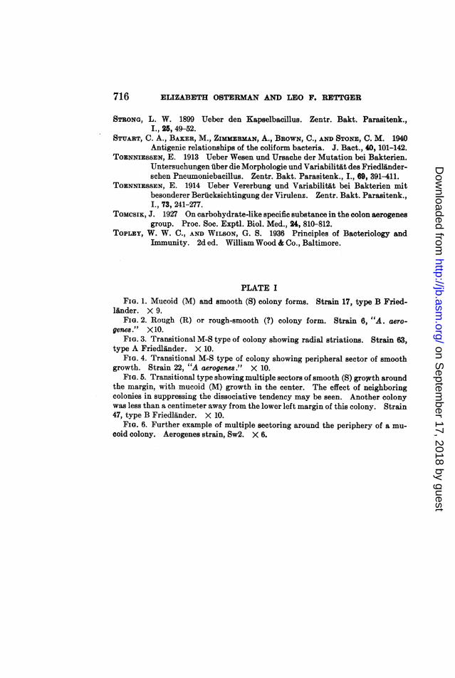
PLATE IFIG. 1. Mucoid (M) and smooth (5) colony forms. Strain 17, type B Fried-
linder. X 9.FIG. 2. Rough (R) or rough-smooth (?) colony form. Strain 6, "A. aero-
genem." X 10.FIG. 3. Transitional M-S type of colony showing radial striations. Strain 63,
type A Friedlinder. X 10.FIG. 4. Transitional M-S type of colony showing peripheral sector of smooth
growth. Strain 22, "A aerogenes." X 10.FIG. 5. Transitional type showing multiple sectors of smooth (5) growth around
the margin, with mucoid (M) growth in the center. The effect of neighboringcolonies in suppressing the dissociative tendency may be seen. Another colonywas less than a centimeter away from the lower left margin of this colony. Strain47, type B Friedlander. X 10.
FIG. 6. Further example of multiple sectoring around the periphery of a mu-coid colony. Aerogenes strain, Sw2. X 6.
716
on Septem
ber 17, 2018 by guesthttp://jb.asm
.org/D
ownloaded from

JOURNAL OF BACTERIOLOGY, VOL. 42 PLATE I
Fi..-3 III..4..... ................
F 5 IO. 6 S
(Elizabeth Osterman and Leo F. Rettger: Comparative study of organisms)
on Septem
ber 17, 2018 by guesthttp://jb.asm
.org/D
ownloaded from

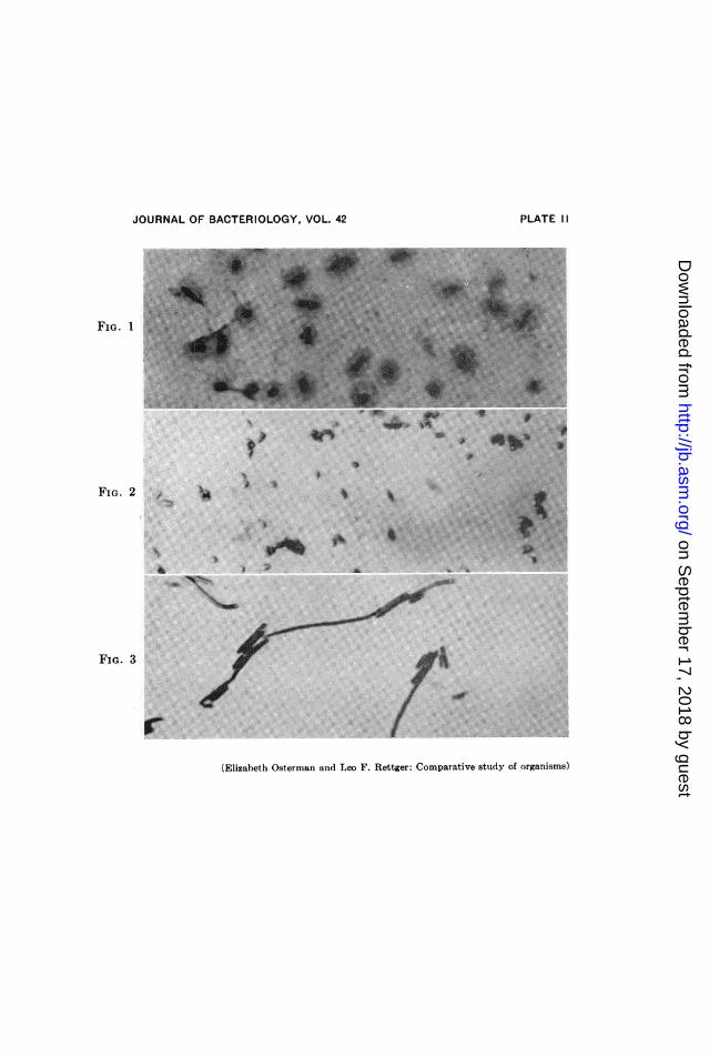
7ELIZABETH OSTERMAN ANI) LEO F. RETTGER
PLATE I I
FIG. 1. Encapsulated organisms from culture in the mucoid (Ml) phase.Aerogenes strain 6. X 1200. Modified Hiss Stain.
FIG. 2. Non-encapsulated cells from culture in smooth (S) phase. Strain 6.X 1200. Gram stain.
FIG. 3. Filamentous cells from culture in rough (It) or rough-smooth (?)phase. Strain 6. X 1200. Gram staiin.
718
on Septem
ber 17, 2018 by guesthttp://jb.asm
.org/D
ownloaded from
JOURNAL OF BACTERIOLOGY, VOL. 42
FIG. 1
Ah. ~ ~ '3 .......
Bu .. : :.:d
FIG. 2
ja
fiA:.
:]....;.ni
'I
*.
*n., 4!W
FIG. 3
r.(Elizabeth Osterman and Leo F. Rettger: Comparative study of organisms)
PLATE II
;,W:
4
on Septem
ber 17, 2018 by guesthttp://jb.asm
.org/D
ownloaded from