Embed Size (px)
Citation preview
Respiratory System
HST I
Includes:
• Nasal cavity
• Sinuses
• Pharynx
• Larynx
• Epiglottis
• Trachea
• Bronchi
• Bronchioles
• Alveoli
• Lungs
• Pleura
• Mediastinum
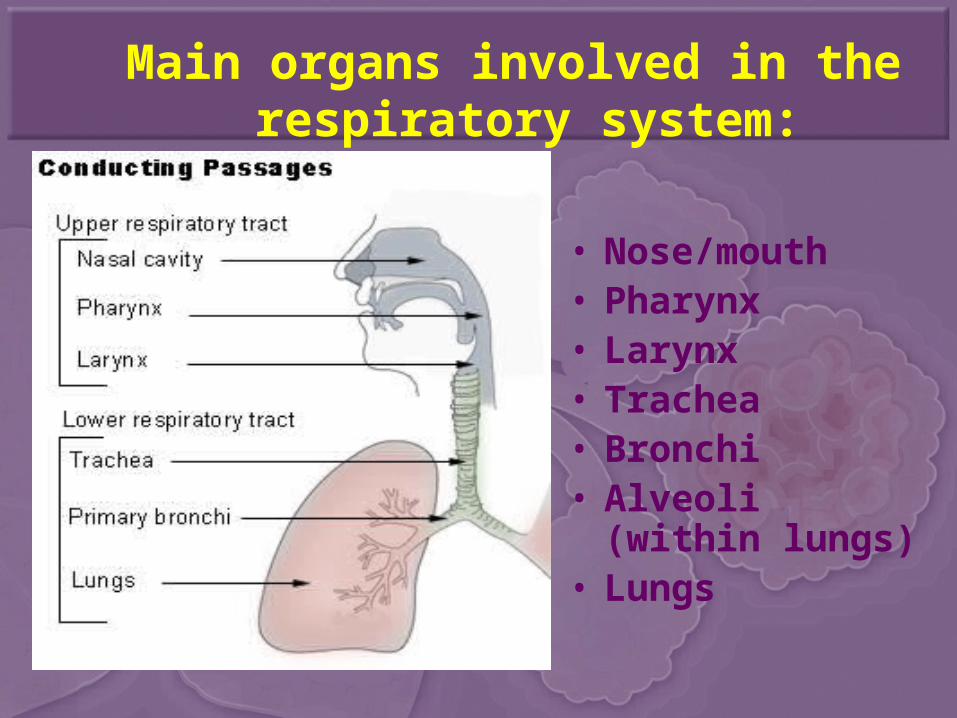
Main organs involved in the respiratory system:
• Nose/mouth • Pharynx• Larynx• Trachea• Bronchi• Alveoli (within
lungs)• Lungs
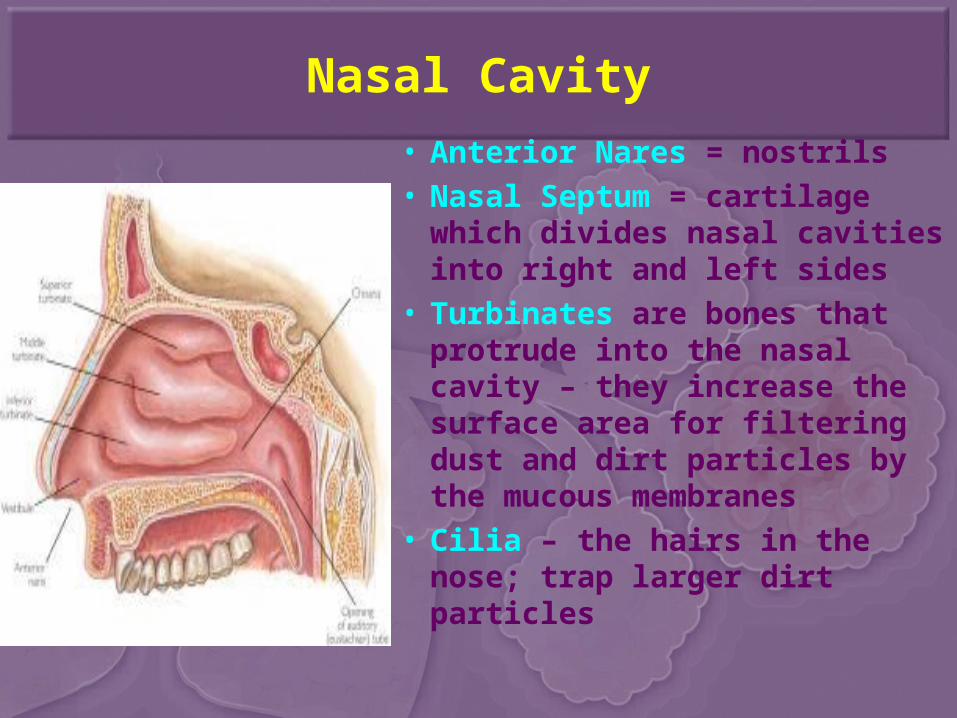
Nasal Cavity
• Anterior Nares = nostrils• Nasal Septum = cartilage
which divides nasal cavities into right and left sides
• Turbinates are bones that protrude into the nasal cavity – they increase the surface area for filtering dust and dirt particles by the mucous membranes
• Cilia – the hairs in the nose; trap larger dirt particles
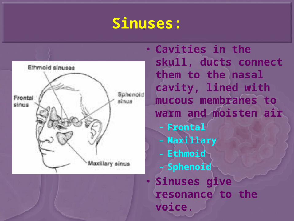
Sinuses:
• Cavities in the skull, ducts connect them to the nasal cavity, lined with mucous membranes to warm and moisten air– Frontal– Maxillary– Ethmoid– Sphenoid
• Sinuses give resonance to the voice.
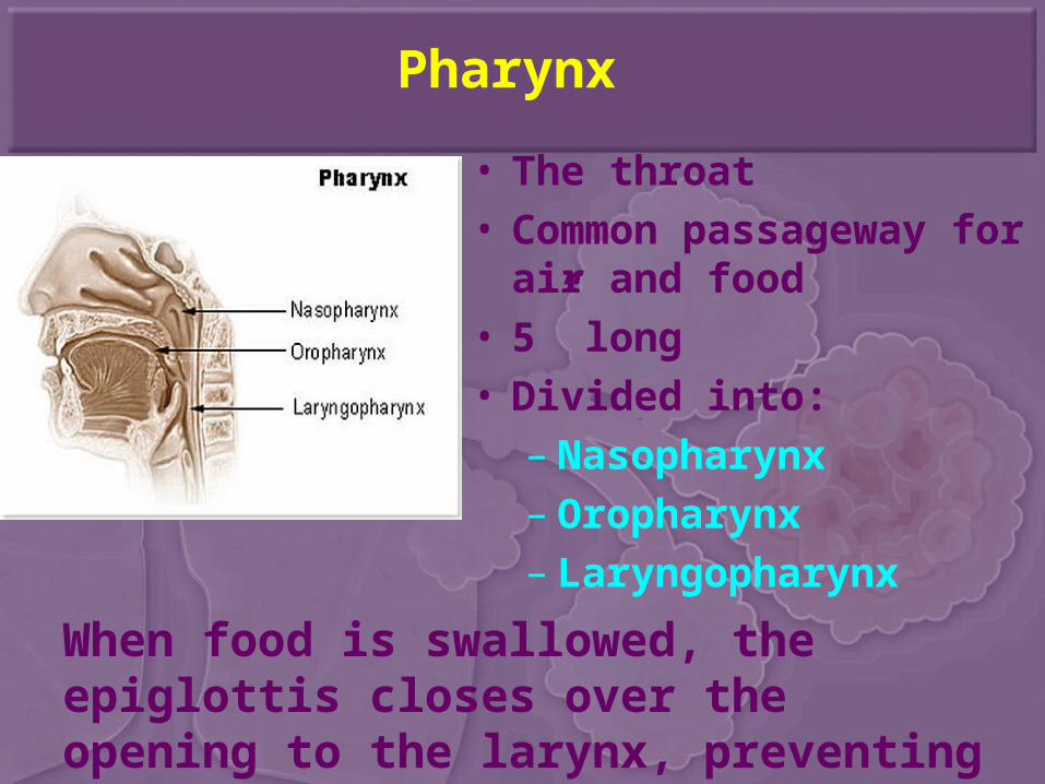
Pharynx
• The throat• Common passageway for
air and food• 5” long• Divided into:
– Nasopharynx– Oropharynx– Laryngopharynx
When food is swallowed, the epiglottis closes over the opening to the larynx, preventing food from entering the lungs.
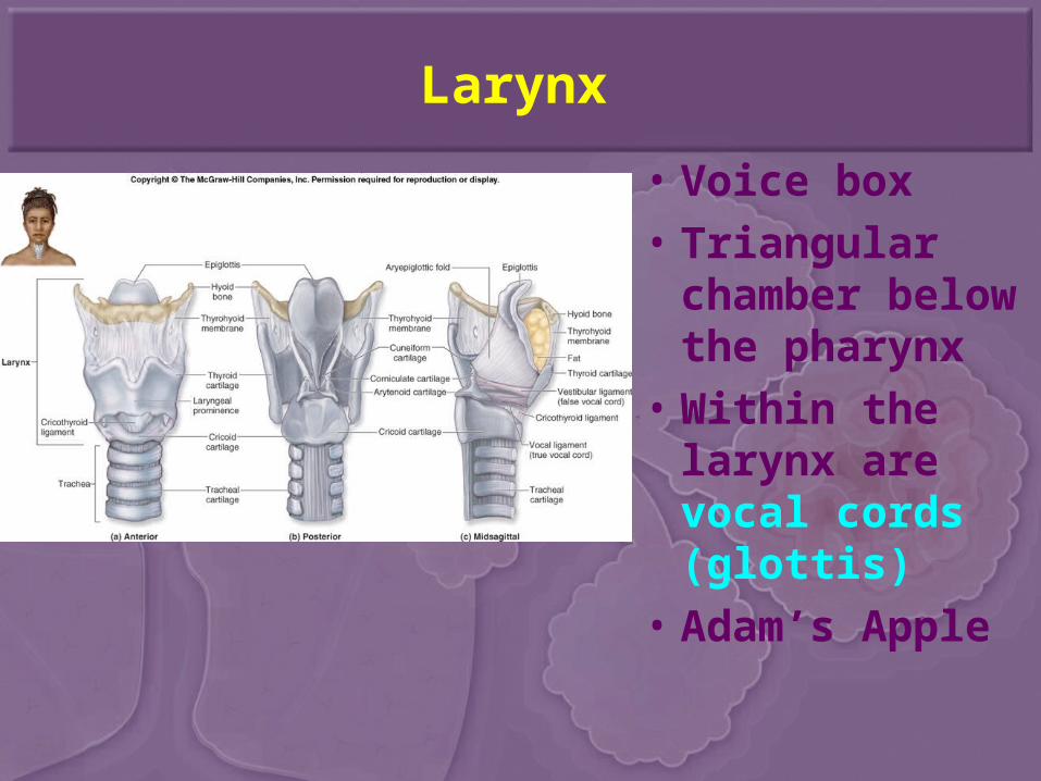
Larynx
• Voice box• Triangular
chamber below the pharynx
• Within the larynx are vocal cords (glottis)
• Adam’s Apple
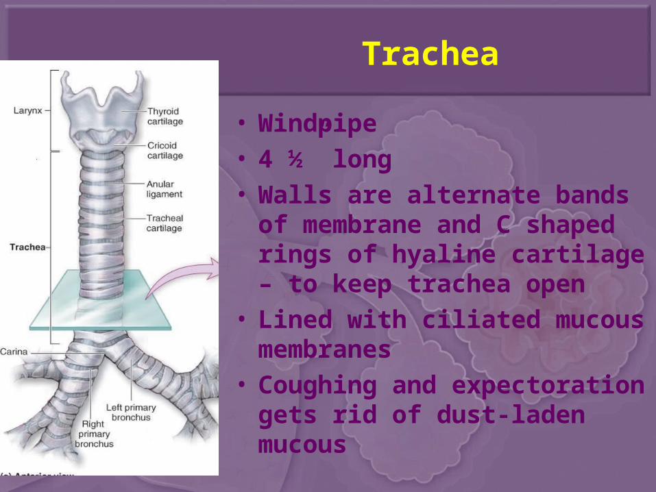
Trachea
• Windpipe• 4 ½” long• Walls are alternate bands of
membrane and C shaped rings of hyaline cartilage – to keep trachea open
• Lined with ciliated mucous membranes
• Coughing and expectoration gets rid of dust-laden mucous
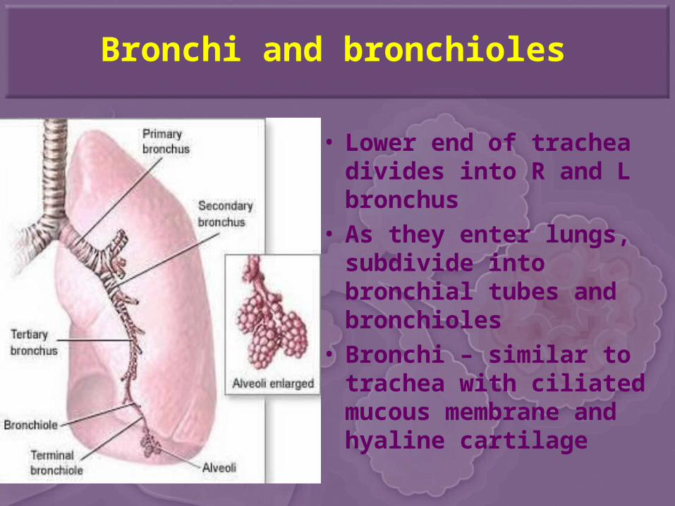
Bronchi and bronchioles
• Lower end of trachea divides into R and L bronchus
• As they enter lungs, subdivide into bronchial tubes and bronchioles
• Bronchi – similar to trachea with ciliated mucous membrane and hyaline cartilage
• Bronchial tubes – cartilaginous plates (instead of C-shaped rings)
• Bronchioles – thinner walls of smooth muscle, lined with ciliated epithelium
• At the end, alveolar ducts and cluster of alveoli
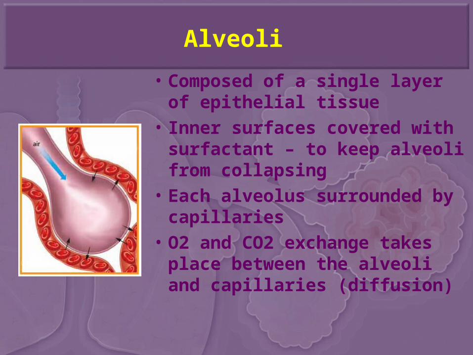
Alveoli
• Composed of a single layer of epithelial tissue
• Inner surfaces covered with surfactant – to keep alveoli from collapsing
• Each alveolus surrounded by capillaries
• O2 and CO2 exchange takes place between the alveoli and capillaries (diffusion)
Lungs
• Fill thoracic cavity
• Separated by mediastinum and heart
• Upper part = apex
• Lower part = base
• Base fits snugly over diaphragm
• Lung tissue porous and spongy – it floats
• R lung = larger and shorter (displaced by the liver) and has 3 lobes
• L lung = smaller (displaced by heart) and has 2 lobes
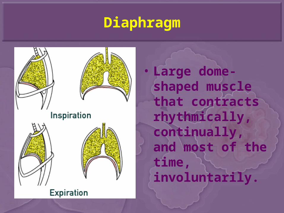
Diaphragm
• Large dome-shaped muscle that contracts rhythmically, continually, and most of the time, involuntarily.
Pleura
• Thin, moist slippery membrane that covers the lungs
• Double-walled sac
• Space is pleural cavity – filled with pleural fluid to prevent friction
Mediastinum
• Interpleural space
• Contains– Thymus gland– Heart (and aorta)– Pulmonary arteries and veins– Superior and inferior vena cava– Esophagus– Trachea– Thoracic duct– Lymph nodes and vessels
Functions of the Respiratory System
1. External, internal, and cellular respiration
2. Production of sound (vocal cords)
• Oxygen is the MOST critical substance needed by the body for survival.– We can only live about 4-6 minutes without
oxygen.
– Arterial blood = 21% O2
– Venous blood = 16% (5% loss per cycle)
• Clinical death – the moment breathing and heartbeat stop
Biological death – when brain cells die, irreversible after 6 – 10 minutes
• Constant removal of carbon dioxide is just as important for survival – maintains homeostasis
Pulmonary Ventilation (Breathing)
• Inspiration
– Intercostal muscles lift ribs outward, sternum rises and the diaphragm contracts and moves downward – this increases the volume of the lungs and air rushes in
• Expiration
– Opposite action takes place
– Exhalation is a passive process
Respiratory Movements
• 1 inspiration + 1 expiration = 1 respiration
• Normal adult = 14 – 20 respirations / min
• Age dependent - newborn = 40 – 60 / min
• Increases with exercise, body temperature, certain diseases
• Sleep = respirations ↓
• Emotion can ↑ or ↓
Lung Capacity and Volume
• Spirometer – device that measures lung capacity
• Tidal Volume – amount of air that moves in and out of lungs with each breath.
– Normal = 500 mL
• Residual Volume – amount of air left in lungs that cannot be voluntarily expelled
• Hyperventilation – – Rapid breathing causes body to lose CO2 too
quickly, blood CO2 decreases which leads to alkalosis
– Symptoms – dizziness and possible fainting– Rx – have person breathe into a paper bag
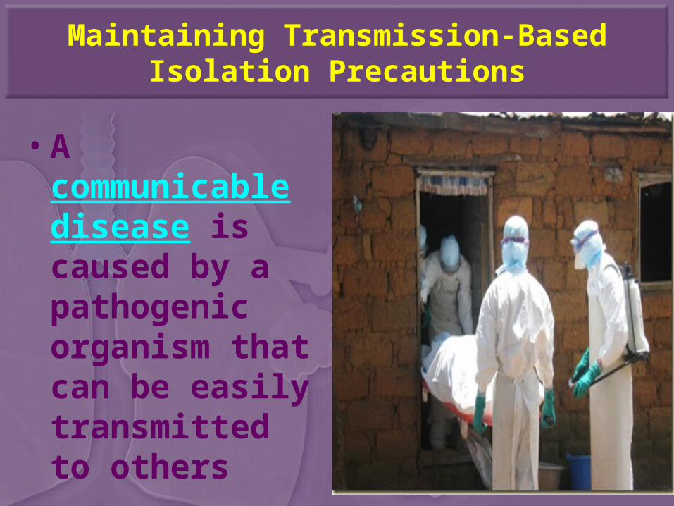
Maintaining Transmission-Based Isolation Precautions
• A communicable disease is caused by a pathogenic organism that can be easily transmitted to others
• An epidemic occurs when the communicable disease spreads rapidly from person to person and affects a large number of people at the same time
• A pandemic exists when the outbreak of disease occurs over a wide geographic area and affects a high proportion of the population
Maintaining Transmission-Based Isolation Precautions
• Transmission-based isolation precautions are methods or techniques of caring for patients who have communicable diseases– Examples of communicable diseases
include:• Tuberculosis• Wound infections• Pertussis (whooping cough)
Transmission-Based Isolation Precautions
• The type of transmission-based isolation depends on the causative organism of the disease, the way the organism is transmitted and whether the pathogen is antibiotic resistant.
• Personal protective equipment (PPE) is used to provide protection from the pathogen. – Some transmission-based isolation require
the use of gowns, gloves, face shields and masks, while others only require the use of a mask.
What’s the difference between standard precautions and isolation precautions?
• Standard precautions are used on all patients, while transmission-based isolation techniques are used to provide extra protection against specific diseases or pathogens to prevent their spread
Vs.
Airborne Precautions
• Used for patients known or suspected to be infected with pathogens transmitted by airborne droplet nuclei, where the droplets contain microorganisms and remain suspended in the air.
• Examples of diseases requiring isolation include rubella (measles), varicella (chicken pox), and tuberculosis.
Airborne Precautions (cont.)
• The patient must be placed in a private room, and the door must be kept closed.
• Air in the room must be discharged to outdoor air or filtered before being circulated to other areas.
• Each person must wear a mask that contains special filter to prevent the entrance of small airborne pathogens.
• If at all possible, the patient should not be moved from the room. If transport is essential, the patient should wear a surgical mask during transport to minimize the release of droplets into the air.
Droplet Precautions
• Must be followed for a patient known or suspected to be infected with pathogens transmitted by large droplets expelled during coughing, sneezing, talking or laughing.
• Examples of diseases requiring these isolation precautions include diphtheria, pertussis, adenovirus, mumps and severe cases of viral influenza, meningitis and pneumonia.
Droplet Precautions (cont.)
• The patient should be placed in a private room. If a private room is not available, the patient can be placed in a room with another patient who has the same infection at least 3 feet away from other patients or visitors.
• Masks must be worn when working within 3 feet of the patient.
• If the patients has to be transported, they must wear a surgical mask.
Contact Precautions
• Must be followed for any patients known or suspected to be infected with epidemiologically microorganisms that can be transmitted by either direct or indirect contact.
• Examples of diseases requiring this type of isolation include any gastrointestinal, respiratory, skin, or wound infections caused by multidrug-resistant organisms; any highly contagious skin infection; and viral or hemorrhagic conjunctivitis or fevers.
Contact Precautions (cont.)
• The patient should be placed in a private room.• Gloves must be worn when entering the room.• Gloves must be changed after having contact with
material that may contain high concentrations of the microorganisms, such as wound drainage or fecal material.
• Gloves must be removed before leaving the room, and the hands must be wash with an antimicrobial agent.
• A gown must be worn in the room if there is any chance of contact with the patient, environmental surfaces or items in the room. The gown must be removed before leaving the room and care must be taken to ensure that clothing is not contaminated after gown removal.
Contact Precautions (cont.)
• Movement and transport of the patient from the room should be for essential purposes only.
• The room and items in it must receive daily cleaning and disinfection as needed.
• If possible, patient-care equipment (bedside commode, stethoscope, thermometer) should be left in the room and used only for this patient. If not, all equipment must be cleaned and disinfected before being used on another patient.
Reverse Isolation Precautions
• Used to protect patients from organisms present in the environment.
• Examples of patients requiring this isolation include patients whose immune systems have been depressed prior to receiving transplants, severely burned patients, patients receiving chemotherapy or radiation treatments, or patients whose immune systems have failed.
Diseases and Abnormal Conditions
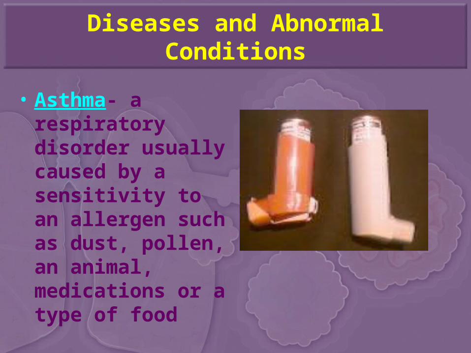
• Asthma- a respiratory disorder usually caused by a sensitivity to an allergen such as dust, pollen, an animal, medications or a type of food
Diseases and Abnormal Conditions
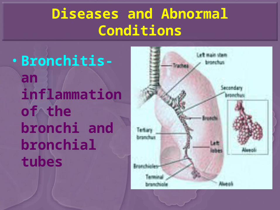
• Bronchitis- an inflammation of the bronchi and bronchial tubes
Diseases and Abnormal Conditions
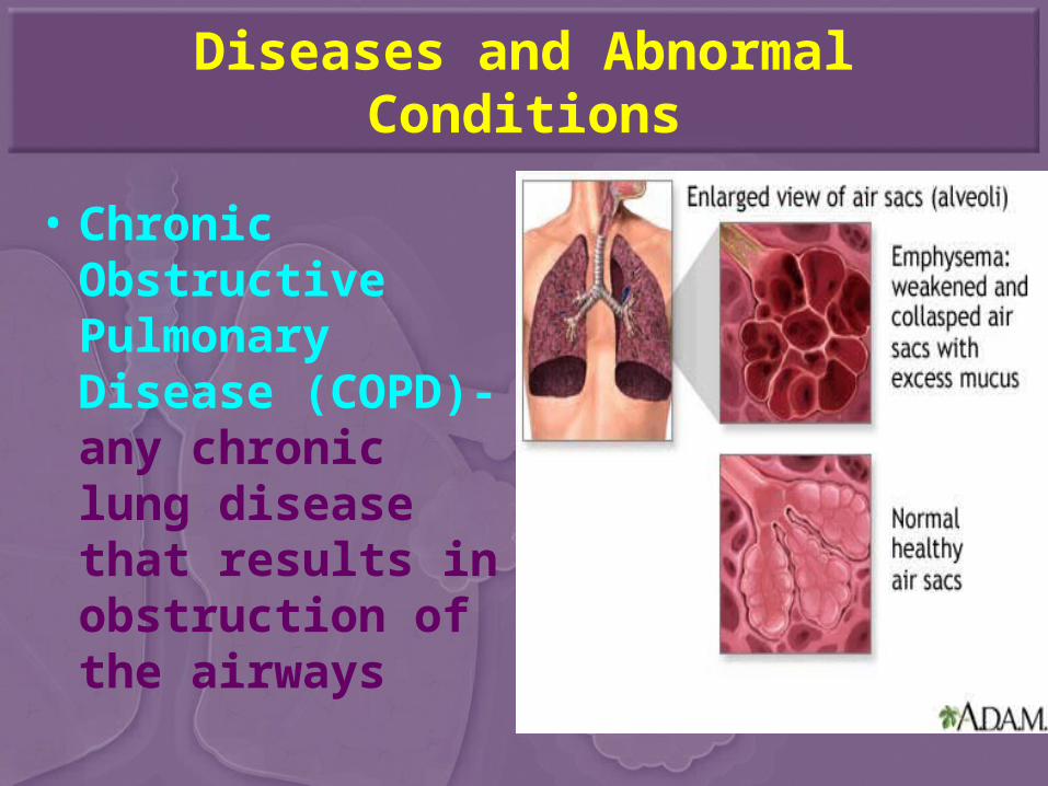
• Chronic Obstructive Pulmonary Disease (COPD)- any chronic lung disease that results in obstruction of the airways
Diseases and Abnormal Conditions
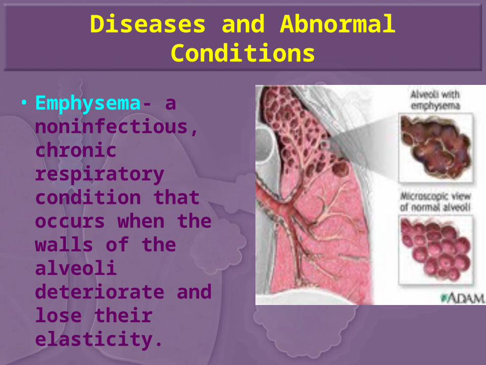
• Emphysema- a noninfectious, chronic respiratory condition that occurs when the walls of the alveoli deteriorate and lose their elasticity.
Diseases and Abnormal Conditions
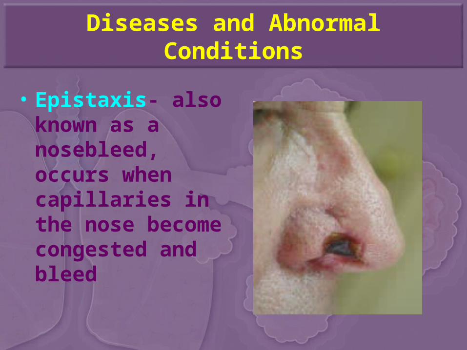
• Epistaxis- also known as a nosebleed, occurs when capillaries in the nose become congested and bleed
Diseases and Abnormal Conditions
• Influenza (flu)- a highly contagious viral infection of the upper respiratory system
Diseases and Abnormal Conditions
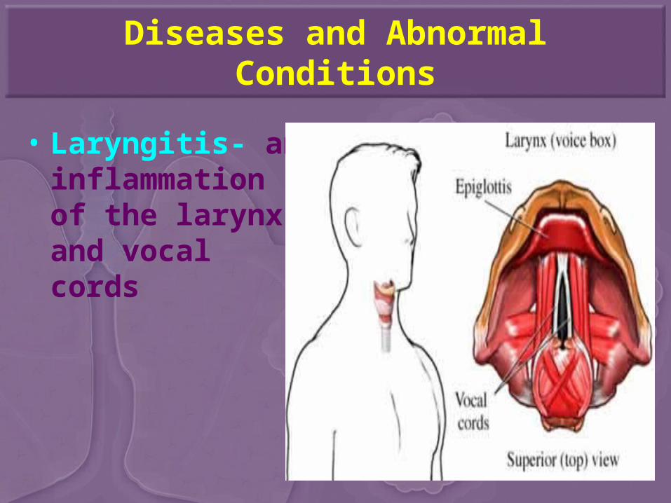
• Laryngitis- an inflammation of the larynx and vocal cords
Diseases and Abnormal Conditions
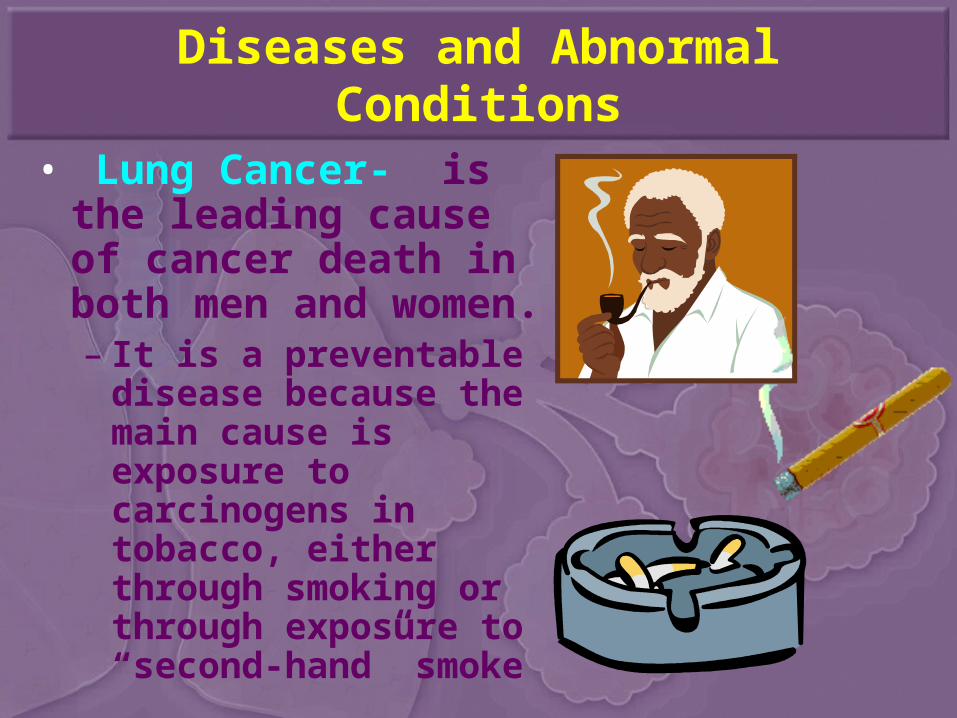
• Lung Cancer- is the leading cause of cancer death in both men and women.– It is a preventable
disease because the main cause is exposure to carcinogens in tobacco, either through smoking or through exposure to “second-hand” smoke
Diseases and Abnormal Conditions
• Pleurisy- an inflammation of the pleura, or membranes, of the lungs– Usually occurs with
pneumonia or other lung infections
Diseases and Abnormal Conditions
• Pneumonia- an inflammation or infection of the lungs characterized by exudate ( a buildup of fluid) in the alveoli.
Diseases and Abnormal Conditions
• Rhinitis- an inflammation of the nasal mucous membrane, resulting in a runny nose, watery eyes, sneezing, soreness and congestion.
Diseases and Abnormal Conditions
• Sinusitis- an inflammation of the mucous membrane lining the sinuses
Diseases and Abnormal Conditions
• Sleep Apnea- a condition in which an individual stops breathing while asleep, causing a measurable decrease in blood oxygen levels– Two types of Sleep Apnea
• Obstructive Sleep Apnea• Central Sleep Apnea
Diseases and Abnormal Conditions
• Tuberculosis (TB)- an infectious lung disease caused by the bacterium Mycobacterium tuberculosis
Diseases and Abnormal Conditions
• Upper Respiratory Infection (URI)- or common cold, is an inflammation of the mucous membrane lining the upper respiratory tract
Related Health Careers
• Internist
• Otolaryngologist
• Perfusionist
• Pulmonologist
• Respiratory Therapist
• Respiratory Therapy Technician
• Thoracic Surgeon
Medical Terminology
• Root Word(s): – Rhin(o)- denotes the nose
• Rhinodynia- pain in the nose• Rhinolith- stone or rock in the nose• Rhinorrhagia- excessive discharge of blood in the
nose• Rhinitis- inflammation of the nose• Rhinomycosis- disease condition of fungus in the
nose
Medical Terminology
• Root Word(s):– Pneumon(o); pneum(ato); pneum(a)- denotes
the lung• Pneumonitis- inflammation in the lung• Pneumonography- to record the lung• Pneumoconiosis- disease condition of dust in the
lung• Pneumothorax- chest cavity in the lung• Pneumocentesis- surgical puncture in the lung
Medical Terminology
• Root Word(s):– Trache(o)- denotes the trachea
• Tracheotomy- surgical removal of the trachea• Tracheoplasty- surgical repair of the trachea• Tracheopathy- disease condition in the trachea• Tracheorrhaphy- to suture the trachea• Tracheitis- inflammation in the trachea
Medical Terminology
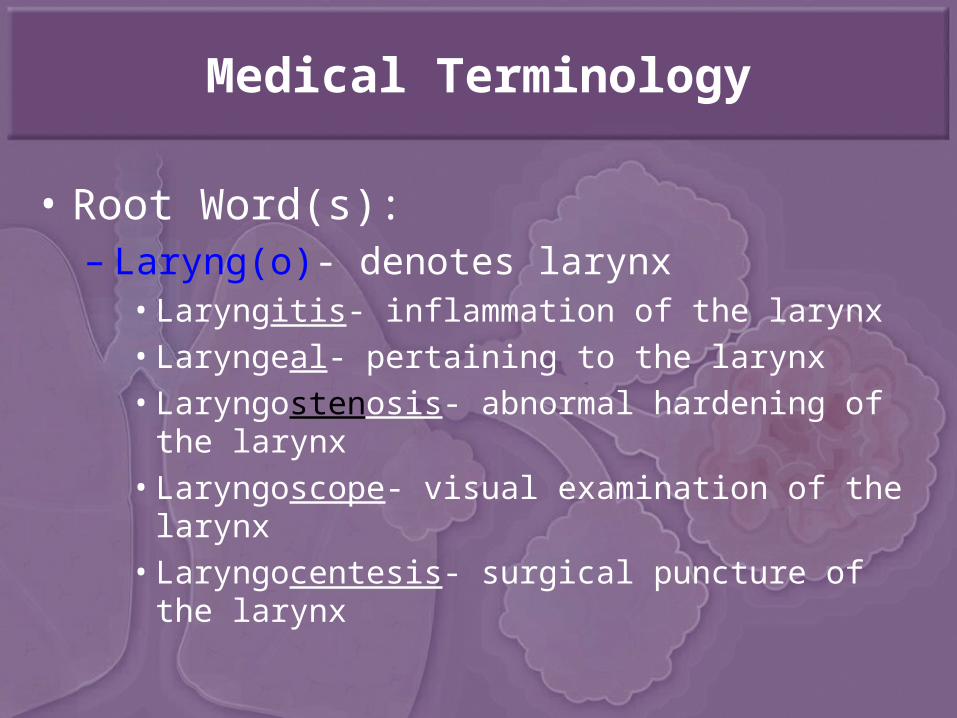
• Root Word(s):– Laryng(o)- denotes larynx
• Laryngitis- inflammation of the larynx• Laryngeal- pertaining to the larynx• Laryngostenosis- abnormal hardening of the larynx• Laryngoscope- visual examination of the larynx• Laryngocentesis- surgical puncture of the larynx
Medical Terminology
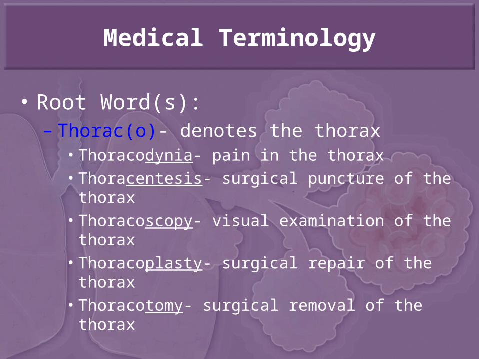
• Root Word(s):– Thorac(o)- denotes the thorax
• Thoracodynia- pain in the thorax• Thoracentesis- surgical puncture of the thorax• Thoracoscopy- visual examination of the thorax• Thoracoplasty- surgical repair of the thorax• Thoracotomy- surgical removal of the thorax
Medical Terminology
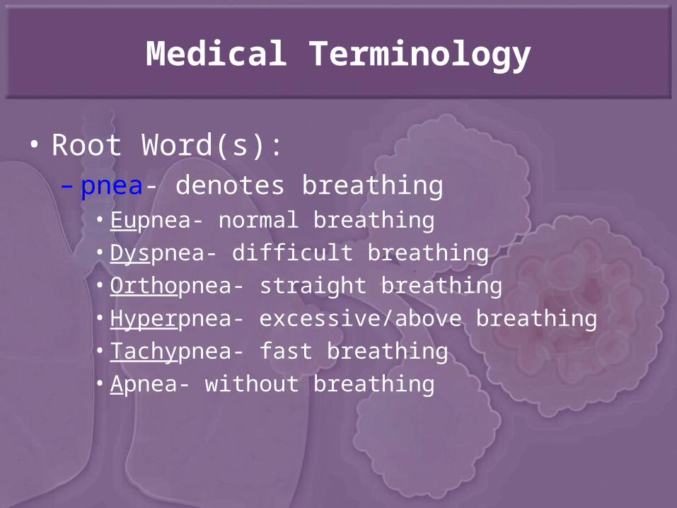
• Root Word(s):– pnea- denotes breathing
• Eupnea- normal breathing• Dyspnea- difficult breathing• Orthopnea- straight breathing• Hyperpnea- excessive/above breathing• Tachypnea- fast breathing• Apnea- without breathing
Medical Terminology
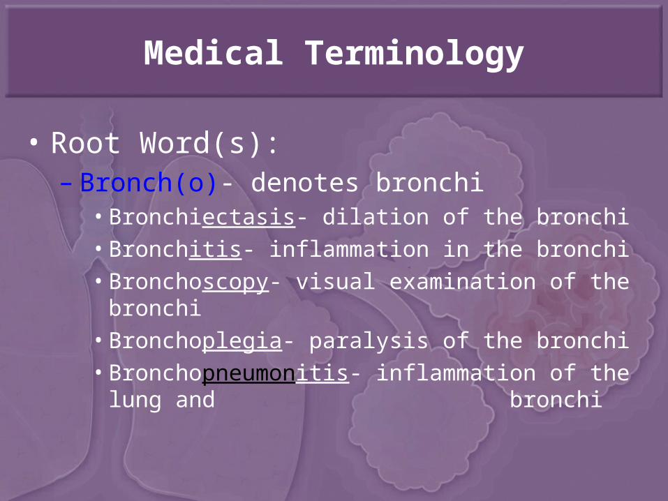
• Root Word(s):– Bronch(o)- denotes bronchi
• Bronchiectasis- dilation of the bronchi• Bronchitis- inflammation in the bronchi• Bronchoscopy- visual examination of the bronchi• Bronchoplegia- paralysis of the bronchi• Bronchopneumonitis- inflammation of the lung and
bronchi
Medical Terminology
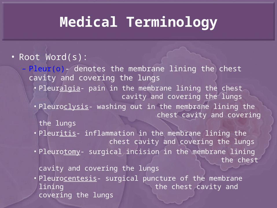
• Root Word(s): – Pleur(o)- denotes the membrane lining the chest cavity
and covering the lungs• Pleuralgia- pain in the membrane lining the chest
cavity and covering the lungs• Pleuroclysis- washing out in the membrane lining the
chest cavity and covering the lungs• Pleuritis- inflammation in the membrane lining the
chest cavity and covering the lungs• Pleurotomy- surgical incision in the membrane lining
the chest cavity and covering the lungs
• Pleurocentesis- surgical puncture of the membrane lining the chest cavity and covering the lungs
Medical Terminology
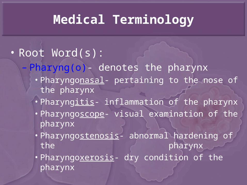
• Root Word(s):– Pharyng(o)- denotes the pharynx
• Pharyngonasal- pertaining to the nose of the pharynx• Pharyngitis- inflammation of the pharynx• Pharyngoscope- visual examination of the pharynx• Pharyngostenosis- abnormal hardening of the
pharynx• Pharyngoxerosis- dry condition of the pharynx

Abbreviations (G-H)
• GA• gal• GB• GC• GI• Gm• gr• GTT• Gyn
• Gastric Analysis• Gallon• Gallbladder• Gonorrhea• Gastrointestinal• Gram• Grain• Glucose Tolerance Test
• Gynecology
Abbreviations (G-H)
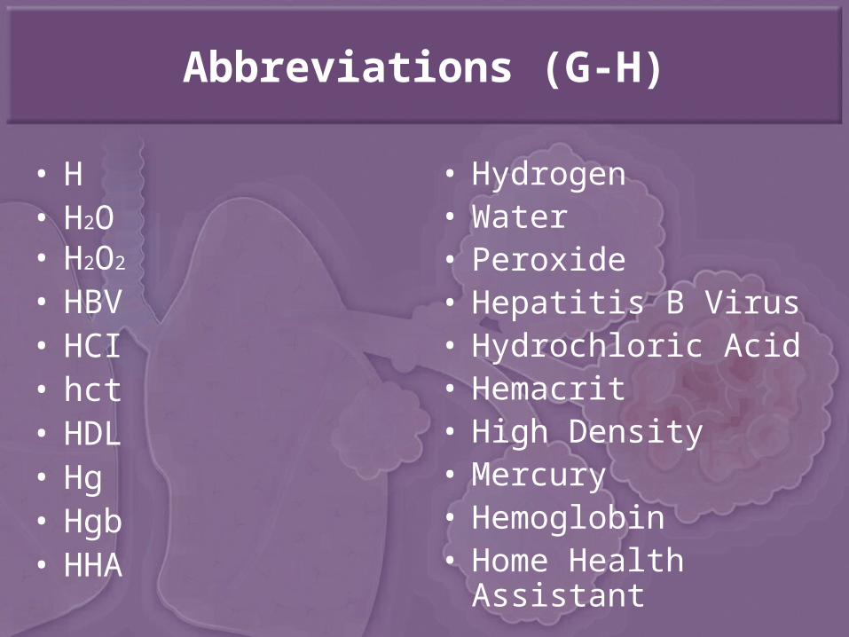
• H• H2O• H2O2
• HBV• HCI• hct• HDL• Hg• Hgb• HHA
• Hydrogen• Water• Peroxide• Hepatitis B Virus• Hydrochloric Acid• Hemacrit• High Density• Mercury• Hemoglobin• Home Health Assistant
Abbreviations (G-H)
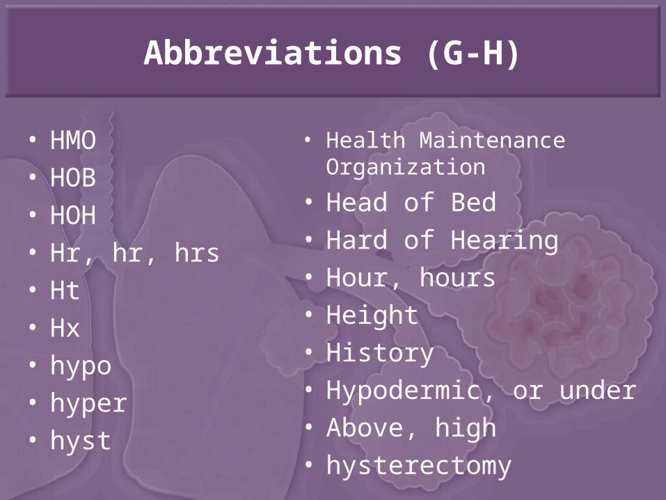
• HMO• HOB• HOH• Hr, hr, hrs• Ht• Hx• hypo• hyper• hyst
• Health Maintenance Organization
• Head of Bed• Hard of Hearing• Hour, hours• Height• History• Hypodermic, or under• Above, high• hysterectomy