Embed Size (px)
Citation preview

Asai-Tajiri et al. Respiratory Research 2014, 15:132http://respiratory-research.com/content/15/1/132
RESEARCH Open Access
Small interfering RNA against CD86 during allergenchallenge blocks experimental allergic asthmaYukari Asai-Tajiri1, Koichiro Matsumoto1*, Satoru Fukuyama1, Keiko Kan-o1, Takako Nakano1, Ken Tonai1,Tatsukuni Ohno2, Miyuki Azuma2, Hiromasa Inoue1,3 and Yoichi Nakanishi1
Abstract
Background: CD86-CD28 interaction has been suggested as the principal costimulatory pathway for the activationand differentiation of naïve T cells in allergic inflammation. However, it remains uncertain whether this pathwayalso has an essential role in the effector phase. We sought to determine the contribution of CD86 on dendriticcells in the reactivation of allergen-specific Th2 cells.
Methods: We investigated the effects of the downregulation of CD86 by short interfering RNAs (siRNAs) onTh2 cytokine production in the effector phase in vitro and on asthma phenotypes in ovalbumin (OVA)-sensitizedand -challenged mice.
Results: Treatment of bone marrow-derived dendritic cells (BMDCs) with CD86 siRNA attenuated LPS-inducedupregulation of CD86. CD86 siRNA treatment impaired BMDCs’ ability to activate OVA-specific Th2 cells. Intratrachealadministration of CD86 siRNA during OVA challenge downregulated CD86 expression in the airway mucosa. CD86siRNA treatment ameliorated OVA-induced airway eosinophilia, airway hyperresponsiveness, and the elevationsof OVA-specific IgE in the sera and IL-5, IL-13, and CCL17 in the bronchoalveolar lavage fluid, but not the gobletcell hyperplasia.
Conclusion: These results suggest that local administration of CD86 siRNA during the effector phase ameliorateslines of asthma phenotypes. Targeting airway dendritic cells with siRNA suppresses airway inflammation andhyperresponsiveness in an experimental model of allergic asthma.
BackgroundInteraction of antigen-presenting cells (APCs) and Tcells is crucial in both the initiation and challenge phasesof allergic asthma and, therefore, has possibility as atarget of anti-asthmatic drugs. Optimal T cell activationand differentiation require not only interaction betweenthe T cell receptor (TCR) and antigen-MHC complexesbut also interaction between costimulatory ligands onAPCs and their putative receptors on T cells. One of thebest-characterized costimulatory molecules is CD28, whichbinds to two costimulatory ligands, B7-1 (CD80) and B7-2(CD86), on APCs. CD28 is constitutively expressed on bothCD4+ and CD8+T cells. By contrast, CD80/86 expressionon dendritic cells and B cells is upregulated after antigenpulse in the process of maturating into APCs. Despite
* Correspondence: [email protected] Institute for Diseases of the Chest, Graduate School of MedicalSciences, Kyushu University, Fukuoka, JapanFull list of author information is available at the end of the article
© 2014 Asai-Tajiri et al.; licensee BioMed CentrCommons Attribution License (http://creativecreproduction in any medium, provided the orDedication waiver (http://creativecommons.orunless otherwise stated.
sharing the same receptor, CD80 and CD86 appear tomediate different mechanisms. CD80 can be more potentthan CD86 in inducing antitumor responses, while CD86preferentially induces Th2-driven allergic responses [1,2].It is generally accepted that during APC/T cell interaction,the B7-CD28 pathway is indispensable for the activationand differentiation of naïve T cells. However, it remainscontroversial whether this pathway has a pivotal role inthe reactivation of primed T cells in the effector phase.It is well known that the dendritic cell (DC) is the most
powerful APC for inducing allergic immune responsesin vivo. The DC network beneath the epithelium of theconducting airways is ideally positioned to perform asurveillance role for inhaled antigens (Ag). By depletingDCs before the inhaled Ag challenge, all the salientfeatures of asthma were diminished, and the effectorcytokine secretion was profoundly reduced [3]. More-over, recent studies suggest that the expression ofCD86, but not CD80, on airway DCs is upregulated
al Ltd. This is an Open Access article distributed under the terms of the Creativeommons.org/licenses/by/4.0), which permits unrestricted use, distribution, andiginal work is properly credited. The Creative Commons Public Domaing/publicdomain/zero/1.0/) applies to the data made available in this article,

Asai-Tajiri et al. Respiratory Research 2014, 15:132 Page 2 of 11http://respiratory-research.com/content/15/1/132
during the effector phase. Ongoing allergic inflammationinduces a specific shift in airway DCs from a CD86-low toa CD86-high phenotype in periphery [4]. Given that lungDCs maturate and upregulate the expression of CD86following the allergen challenge [5], it is important toknow whether the upregulation of CD86 has a role inthe development of asthmatic responses.RNA interference is an endogenous cellular mechanism
in which short interfering RNAs (siRNAs) elicit thesequence-specific degradation of a complementary mRNAtarget. Today, siRNA-mediated gene silencing has becomemore powerful, more specific, and much less toxic thanlow-molecular-weight chemical inhibitors or blockingmAbs for laboratory investigations, particularly in vitro. Itis also characterized by a low-cost and transient effect.Several studies have demonstrated the therapeutic effectsof synthetic siRNA in allergen-induced asthma models[6-8]. These studies targeted STAT6, TRAIL, and PAI-1,and the siRNAs were delivered by intratracheal adminis-tration. Although low transfection efficacy might be apotential hurdle in RNA interference, it is expected thatDCs can be good targets because of their location andtheir prominent particle uptake ability, including theuptake of short-based nucleotides. Here, we examinedthe effects of the downregulation of CD86 by intratrachealadministration of siRNA on Th2 cytokine production inthe effector phase in vitro and on asthma phenotypesin vivo.
MethodsMiceBALB/c mice were purchased from SLC (Hamamatsu,Japan). OVA-specific TCR-expressing DO11.10 transgenic(Tg) mice were provided by Dr. K. Murphy (WashingtonUniversity, St. Louis, MO). All experimental procedureswere approved by the animal research ethics committee ofKyushu University (reference number: A23-048-1).
siRNA preparationThe sequences of the CD86 siRNA were previously deter-mined by our colleague [9]. siRNAs were synthesized usingQiagen 2-for-silencing siRNA duplexes (Qiagen, Valencia,CA). CD86 siRNA has the sequence 5′-CGUUGUGUGUGUUCUGGAAdTdT-3′ (sense) and 5′-UUCCAGAACACACACAACGdTdT (antisense). As a control, we used singlenon-targeting siRNAs.
TransfectionBone marrow-derived DCs (BMDCs) were obtained aspreviously described [10], washed with a serum-freemedium, and cultured in a 24-well tissue culture platewith 2 × 10⁵ cells/500 μl. Transfection reagent (GeneSilencer®, Genlantis) and siRNA were added to the wells.After 4 hours, 500 μl medium with 20% FBS was added.
After 24 hours, lipopolysaccharide (LPS) (1 μg/ml; Sigma,St. Louis, MO, USA) and peptide (p) OVA (C-terminal323–339 epitope) (5 μg/ml; Bachem, Hauptstrasse,Switzerland) were added for 18 hours.
Flow cytometryThe cells were stained with FITC-conjugated anti-CD11cmAb and biotinylated anti-CD86 mAb followed by phyco-erythrin (PE)-conjugated streptavidin (BD Biosciences).FITC-conjugated mouse IgG and PE-conjugated IgG wereused as isotype controls. Flow cytometry was performedon a FACSCalibur flow cytometer equipped with CELL-Quest software (BD Biosciences). Data was assessed bymean fluorescent intensity (MFI) or histogram.
Coculture with Th2 cellsCD4+T cells were isolated from the spleens of naïveDO11.10 mice using magnetic separation (MACS; MiltenyiBiotec, Bergisch Gladbach, Germany). OVA-specific Th2cells were induced as previously described [11] andcultured at a final concentration of 1 × 10⁶ cells/well/500 μl with siRNA-transfected BMDCs (at 2 × 10⁵ cells/well/500 μl). The culture supernatants were collected48 hours later.
Asthma modelBALB/c mice were sensitized with an intraperitonealinjection of 10 μg OVA (GradeV, Sigma-Aldrich, St. Louis,MO) with 0.3 mg of Al(OH)3 (SERVA Electrophoresis) ondays 1 and 14. On days 26–28, animals were anesthetizedand treated with intratracheal CD86 siRNA (12.5 μgsiRNA in 50 μl saline). One hour after each treatment, micewere challenged with aerosolized 1% OVA for 30 minutes.On day 30, mice were assessed for airway hyperrespon-siveness (AHR) followed by blood sampling and broncho-alveolar lavage (BAL) as previously described [12].
HistologyFresh-frozen sections were prepared with Kawamoto’smethod for maintaining a normal structure [13]. Forfluorescence microscopy, cryosections were stained withFITC-conjugated anti-CD11c mAb(clone HL3) or FITC-conjugated anti-CD86 mAb(clone GL1) (BD Biosciences).Images were acquired using an Olympus (Melville, NY)BX61 upright microscope. To assess the localization ofsiRNA, sensitized mice were intratracheally administratedTexas Red-labeled siRNA (40 μM/50 μl/animal, siGLO®)on day 26, followed by the OVA challenge. Twelve hoursafter the challenge, cryosections of the tracheas and lungswere prepared, stained with FITC-labeled anti-CD11cmAb, and viewed on an A1Rsi confocal laser microscope(Nikon, Tokyo, Japan). Micrographs were quantitativelyanalyzed for the presence of anti-CD86 signals with imageanalysis software. For each naïve or control siRNA-treated

Asai-Tajiri et al. Respiratory Research 2014, 15:132 Page 3 of 11http://respiratory-research.com/content/15/1/132
or CD86siRNA-treated sample, 3-4 sections were quanti-fied. To evaluate only CD86-expressing cells, we selectedan area between the airway epithelium and the laminapropria mucosae, and counted only dot signals per unitarea. Goblet cell hyperplasia was assessed by staining withAlcian blue/periodic acid-Schiff (AB/PAS) as describedpreviously [14].
Determination of MUC5AC mRNATotal RNA was isolated and quantitative real-time PCRwas conducted for the detection of MUC5AC as describedpreviously [11].
Measurement of cytokines, chemokine and OVA-specific IgECytokines, chemokine and OVA-specific IgE were quan-tified using ELISA kits (Invitrogen Corporation or R&DSystems, Shibayagi for IgE).
Statistical analysisValues are expressed as the mean (±SEM). Differencesamong groups were analyzed using an ANOVA with aBonferroni analysis. Nonparametric data were analyzedusing the Mann-Whitney U test. A P value of < .05 wasconsidered statistically significant.
ResultssiRNA downregulates CD86 expression on BMDCsWe first investigated the efficacy of CD86 siRNA inBMDCs. Immature BMDCs showed moderate expressionof CD86, which was upregulated by maturation-inducingstimulation with LPS and pOVA for 18 hrs. We comparedthe expression level of CD86 between non-targetingcontrol siRNA-treated and –untreated BMDCs that werestimulated with LPS and pOVA. There was no significantdifference in CD86 expression (control siRNA-treatedBMDCs, MFI 2593 ± 193; untreated BMDCs, MFI 3065 ±191; P =0.106 ). We considered that the transfectionprotocol per se was neutral in the expression status ofCD86. The expression level of CD86 on CD86 siRNA-treated BMDCs was significantly lower than that onnon-targeting control siRNA-treated BMDCs [P =0.0014](Figure 1). The expression of CD80 was unaffected bytreatment with CD86 siRNA in our preliminary study [9].These results suggest that treatment with CD86 siRNAspecifically suppresses the activation-induced upregulationof CD86 on BMDCs in vitro.
CD86 siRNA treatment impairs BMDCs’ ability to activateTh2 cellsAlthough many previous studies have shown an essentialrole of CD86 on APCs in the antigen priming of naïve Tcells and subsequent Th2 commitment, there have been noconsistent results for the role of CD86 in the reactivationof antigen-specific Th2 cells. OVA-specific Th2 cells
were induced by coculture of CD4+T cells purified fromthe spleens of DO11.10 mice with APCs from thespleens of naïve BALB/c mice in the presence of pOVA,recombinant IL-4, neutralizing anti-IL-12 mAb, andagonistic anti-CD28 mAb, and followed by expansionwith recombinant IL-2 (Figure 2A). Th2 specificity wasconfirmed by selective production of IL-4, IL-5, andIL-13, but to a smaller extent, IFN-γ, which is inducedby coculture of the cells with pOVA-loaded and LPS-stimulated BMDCs (referred to as pOVA/LPS-BMDCs).The contents of IL-4, IL-5, and IL-13 in the culturesupernatants from coculture of the cells with CD86siRNA-treated pOVA/LPS-BMDCs were significantlylower than those from coculture of the cells with controlsiRNA-treated pOVA/LPS-BMDCs [IL-4, P =0.0209; IL-5,P = 0.0209; IL-13, P =0.0209] (Figure 2B). When BMDCswere loaded with pOVA but not stimulated with LPS,almost no Th2 cytokine production was detected (datanot shown). These results suggest that the reactivationof murine antigen-specific Th2 cells in vitro is partiallydependent on CD86 on APCs.
Intratracheal administration of CD86 siRNA downregulatesCD86 expression in the airway mucosaIn an attempt to assess how intratracheally administratedsiRNA was distributed to the lung, anesthetized mice wereadministered with Texas Red-labeled non-coding siRNAand then challenged with OVA. One hour and 12 hoursafter the challenge, frozen sections were made and stainedwith FITC-labeled anti-CD11c mAb to detect DCs [15].One hour after the OVA challenge, Red-labeled siRNAwas detected on the airway epithelial layer in a band-likedeposition pattern. Green-labeled DCs were clusteredaround the deposited siRNA (Figure 3A). The immuno-fluorescence of CD11c+cells in proximity of the redsiRNA signal was observed in several sites per onesection, mainly on the airway epithelial layer. Due to itscharacteristic distribution, this was unlikely a sectioning/deparaffinizing/staining artifact. Twelve hours after thechallenge, the red dots of siRNA were distributed beneaththe airway basement membrane and submucosal DCs,indicating that siRNA successfully went through theepithelial barrier (Figure 3B,C). Yellow fluorescencewas observed in several sites, suggestive of siRNA takenup by DCs, although a mere topological stratification offluorescence might not be excluded.Next, for evaluation of the interfering efficacy, sensitized
mice were intratracheally administered with CD86 siRNAor control siRNA and given a single challenge withOVA. Twelve hours after the challenge, the tracheasand lungs were removed and processed for immune-fluorescence study. CD86-positive cells were detected bybiotin-conjugated anti-CD86 mAb followed by streptavidin-conjugated FITC and assessed semi-quantitatively as CD86-

Figure 1 Silencing CD86 expression by transfection with CD86 small interfering RNA (siRNA) on BMDCs. BMDCs were cultured in thepresence of GM-CSF for 7 days. The DCs were transfected with 100nM CD86 siRNA and non-specific (control) siRNA by GeneSilencer®. Twenty-fourhours after gene silencing, DCs were activated with LPS for 18 hours. Cells were stained with FITC-conjugated anti-CD11c mAb and biotinylatedanti-CD86 mAb followed by phycoerithrin (PE)-conjugated streptavidin. CD86 expression on CD11c-positive cells was assessed by flow cytometry.Histograms represent the number of cells at various fluorescent intensities. MFI data was expressed as the mean ± SEM of 4 mice. * indicates statisticallysignificant (P <0.05) difference between the indicated two groups.
Asai-Tajiri et al. Respiratory Research 2014, 15:132 Page 4 of 11http://respiratory-research.com/content/15/1/132
positive areas by image analysis. As shown in Figure 4, inthe sections of naïve mice, there were scarce CD86-positivecells in the airway mucosa, although the area of CD86-positive cells was markedly increased in control siRNA-treated and OVA-challenged mice. CD86-expressing cellswere detected in dense clusters beneath the airwayepithelium. The upregulation of CD86-positive cells wassignificantly suppressed in the CD86 siRNA-treatedmice [P =0.0036]. These results demonstrate that localadministration of siRNA ameliorates the allergen-inducedupregulation of CD86 expression on the airway DCs.
CD86 siRNA ameliorates allergen-induced asthmaphenotypesTo examine the effects of pulmonary CD86 inhibitionon asthma phenotypes, CD86 siRNA was administratedone hour before each OVA challenge (Figures 4 and 5).Thirty-six hours after the last OVA challenge, airwayhyperresponsiveness to acetylcholine aerosol was mea-sured. Allergen-induced airway hyperresponsiveness inCD86 siRNA-treated mice was significantly lower thanthat in control siRNA-treated mice [P =0.0432]. Thenumber of eosinophils in BAL fluid was significantly

Figure 2 Th2 cytokine levels in culture supernatant were analyzed by ELISA. (A) DO11.10 spleen cells were primed with pOVA and APCsin the presence of rIL-4, anti-IL-12 antibody, CD28, and IL-2. Seven days later, CD4-positive cells were purified using MACS and used as effectorT cells. CD86 siRNA-transfected BMDCs were pulsed with pOVA and stimulated with LPS and then cultured with effector T cells. (B) Coculturesupernatants were collected 48 hours later, and cytokine levels were measured by ELISA (n = 4; mean ± SEM). * indicates statistically significant(P <0.05) difference between the indicated two groups.
Asai-Tajiri et al. Respiratory Research 2014, 15:132 Page 5 of 11http://respiratory-research.com/content/15/1/132
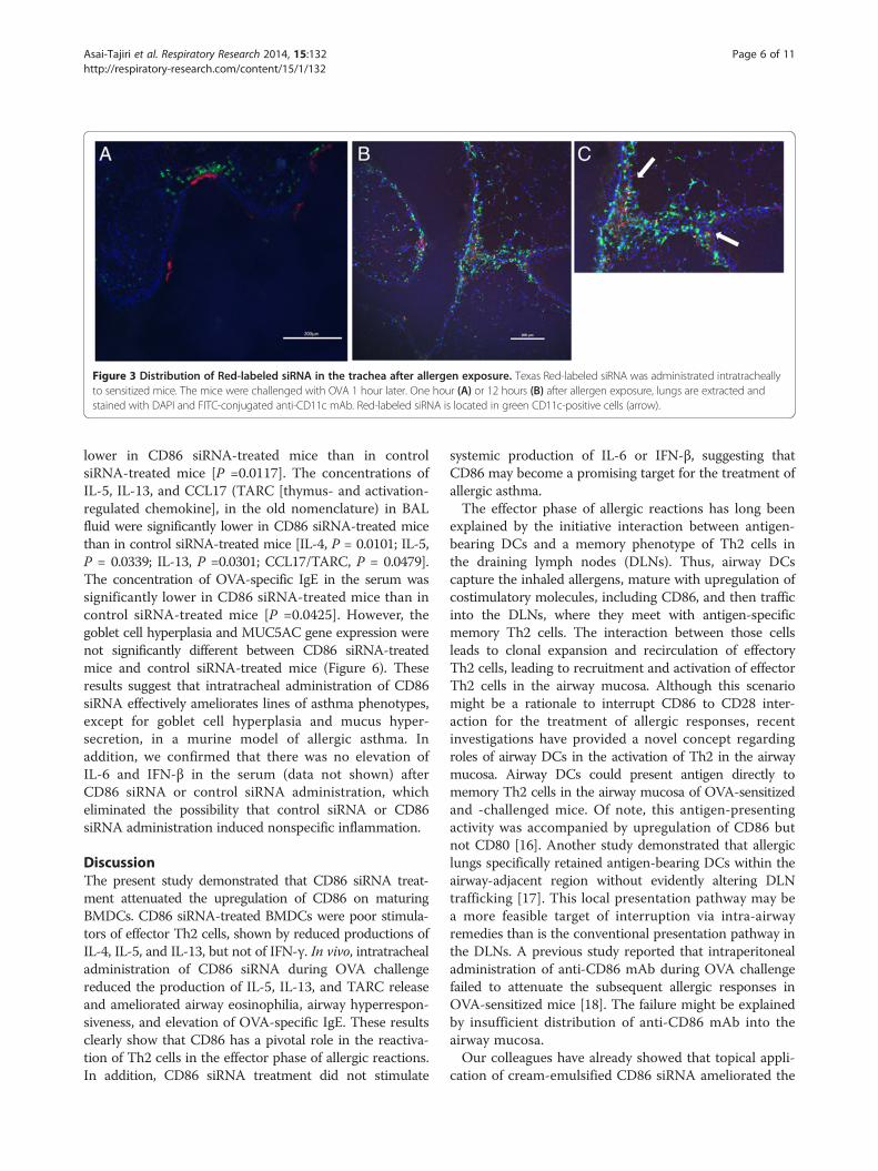
Figure 3 Distribution of Red-labeled siRNA in the trachea after allergen exposure. Texas Red-labeled siRNA was administrated intratracheallyto sensitized mice. The mice were challenged with OVA 1 hour later. One hour (A) or 12 hours (B) after allergen exposure, lungs are extracted andstained with DAPI and FITC-conjugated anti-CD11c mAb. Red-labeled siRNA is located in green CD11c-positive cells (arrow).
Asai-Tajiri et al. Respiratory Research 2014, 15:132 Page 6 of 11http://respiratory-research.com/content/15/1/132
lower in CD86 siRNA-treated mice than in controlsiRNA-treated mice [P =0.0117]. The concentrations ofIL-5, IL-13, and CCL17 (TARC [thymus- and activation-regulated chemokine], in the old nomenclature) in BALfluid were significantly lower in CD86 siRNA-treated micethan in control siRNA-treated mice [IL-4, P = 0.0101; IL-5,P = 0.0339; IL-13, P =0.0301; CCL17/TARC, P = 0.0479].The concentration of OVA-specific IgE in the serum wassignificantly lower in CD86 siRNA-treated mice than incontrol siRNA-treated mice [P =0.0425]. However, thegoblet cell hyperplasia and MUC5AC gene expression werenot significantly different between CD86 siRNA-treatedmice and control siRNA-treated mice (Figure 6). Theseresults suggest that intratracheal administration of CD86siRNA effectively ameliorates lines of asthma phenotypes,except for goblet cell hyperplasia and mucus hyper-secretion, in a murine model of allergic asthma. Inaddition, we confirmed that there was no elevation ofIL-6 and IFN-β in the serum (data not shown) afterCD86 siRNA or control siRNA administration, whicheliminated the possibility that control siRNA or CD86siRNA administration induced nonspecific inflammation.
DiscussionThe present study demonstrated that CD86 siRNA treat-ment attenuated the upregulation of CD86 on maturingBMDCs. CD86 siRNA-treated BMDCs were poor stimula-tors of effector Th2 cells, shown by reduced productions ofIL-4, IL-5, and IL-13, but not of IFN-γ. In vivo, intratrachealadministration of CD86 siRNA during OVA challengereduced the production of IL-5, IL-13, and TARC releaseand ameliorated airway eosinophilia, airway hyperrespon-siveness, and elevation of OVA-specific IgE. These resultsclearly show that CD86 has a pivotal role in the reactiva-tion of Th2 cells in the effector phase of allergic reactions.In addition, CD86 siRNA treatment did not stimulate
systemic production of IL-6 or IFN-β, suggesting thatCD86 may become a promising target for the treatment ofallergic asthma.The effector phase of allergic reactions has long been
explained by the initiative interaction between antigen-bearing DCs and a memory phenotype of Th2 cells inthe draining lymph nodes (DLNs). Thus, airway DCscapture the inhaled allergens, mature with upregulation ofcostimulatory molecules, including CD86, and then trafficinto the DLNs, where they meet with antigen-specificmemory Th2 cells. The interaction between those cellsleads to clonal expansion and recirculation of effectoryTh2 cells, leading to recruitment and activation of effectorTh2 cells in the airway mucosa. Although this scenariomight be a rationale to interrupt CD86 to CD28 inter-action for the treatment of allergic responses, recentinvestigations have provided a novel concept regardingroles of airway DCs in the activation of Th2 in the airwaymucosa. Airway DCs could present antigen directly tomemory Th2 cells in the airway mucosa of OVA-sensitizedand -challenged mice. Of note, this antigen-presentingactivity was accompanied by upregulation of CD86 butnot CD80 [16]. Another study demonstrated that allergiclungs specifically retained antigen-bearing DCs within theairway-adjacent region without evidently altering DLNtrafficking [17]. This local presentation pathway may bea more feasible target of interruption via intra-airwayremedies than is the conventional presentation pathway inthe DLNs. A previous study reported that intraperitonealadministration of anti-CD86 mAb during OVA challengefailed to attenuate the subsequent allergic responses inOVA-sensitized mice [18]. The failure might be explainedby insufficient distribution of anti-CD86 mAb into theairway mucosa.Our colleagues have already showed that topical appli-
cation of cream-emulsified CD86 siRNA ameliorated the

Figure 4 Effect of siRNA treatment on CD86 positive cells around airway. Sensitized mice were intratracheally administered with CD86 siRNAor control siRNA and given a single challenge with OVA. Twelve hours after the challenge, the tracheas and lungs were removed and processed forimmune-fluorescence study. Naïve mice served as negative control. (A) Lung sections from naïve, CD86 siRNA-treated or control siRNA-treated micestained with FITC-conjugated anti-CD86 mAb. Bar, 200 μm for upper panel; 100 μm for lower panel. The upper panel and lower panel were obtainedfrom different mice (B) Micrographs were quantitatively analyzed for the presence of FITC-anti-CD86 signals per unit area with image analysis software.All data mean ± SEM of 3–4 mice per group. * indicates statistically significant (P <0.05) difference between the indicated two groups.
Asai-Tajiri et al. Respiratory Research 2014, 15:132 Page 7 of 11http://respiratory-research.com/content/15/1/132
clinical manifestations of murine contact hypersensitivityand atopic dermatitis-like disease [9]. It also afford col-lateral evidence that CD86 has a role in effector phase.But another study reported that intratracheal injectionof OVA-pulsed DCs from CD80/CD86 double-deficientmice in OVA-sensitized mice led to the reactivation ofTh2 effector responses the same as OVA-pulsed DCsfrom wild-type mice [19]. siRNA theoretically causesubiquitous gene silencing, leaving a possibility thatCD86 siRNA affects not only airway DCs but also othercell types. CD86 on alveolar macrophages, eosinophils,and B cells was reported to play a role in the develop-ment of allergic airway reactions [20,21]. CD86 on Bcells stimulates CD28 on T cells and transduces positive
signals into B cells that increase IgG1 and IgE produc-tion. This pathway may be particularly important formemory B cells in which CD86 is upregulated [22,23].The reduction of OVA-specific IgE in the serum ofCD86 siRNA-treated mice in the present study is highlyconsistent with this pathway.TARC is the CC chemokine that selectively attracts
Th2 lymphocytes toward APCs [24]. A previous studyshowed that TARC expression was highly concentratedin purified lung DCs [25]. In the present study, themechanism remains unclear as to why the concentrationof TARC in BAL fluid was reduced by CD86 siRNAtreatment. We confirmed that CD86 siRNA treatmentper se did not alter the ability to produce TARC in

Figure 5 Effect of treatment with CD86 siRNA on allergic asthmatic response. (A) CD86 siRNA or control siRNA was administrated intratracheally1 hour before each OVA challenge. (B) Airway hyperresponsiveness to inhaled acetylcholine (ACh) was measured 36 hours after the last OVAchallenge. (C) Cell counts in BAL fluids were performed. Mo = macrophage; Neut = neutrophil; Ly = lymphocyte; Eo = eosinophil. (D) SerumOVA-IgE levels were analyzed. (E) Cytokine levels and TARC levels in BAL fluids were analyzed. All data mean ± SEM of 8–12 mice per group.* and † indicate statistically significant (P <0.05) difference between the indicated two groups.
Asai-Tajiri et al. Respiratory Research 2014, 15:132 Page 8 of 11http://respiratory-research.com/content/15/1/132
BMDCs in vitro (unpublished observation). A possibleexplanation is that CD86 siRNA treatment reduces recruit-ment of DCs into the airways by currently unknownmechanism(s), which results in the reduction of TARCin the lungs. The quantitative and qualitative assessmentsof airway DCs await further investigations.Contrasting with the effects on lines of asthma pheno-
types, CD86 siRNA treatment failed to ameliorate the gob-let cell hyperplasia and MUC5AC gene expression, cardinalfeatures of airway remodeling in asthma. A majority ofasthma phenotypes, including airway eosinophilia, airway
hyperresponsiveness, and airway remodeling, are attribut-able to the pluripotent effects of IL-13 and its downstreammolecules. Those IL-13-mediated phenotypes vary insensitivity to therapeutic interventions. Airway eosinophiliainduced by intratracheal IL-13 was feasibly suppressed bysystemic treatment with glucocorticosteroid, while airwayhyperresponsiveness and remodeling were resistant toglucocorticosteroid [12]. Similar results were obtained fromour recent study that examined the effect of an inhibitorof Janus kinase (JAK), a kinase family mediating multiplecytokine signalings, on OVA-sensitized/challenged mice

Figure 6 Lack of effect of treatment with CD86 siRNA on mucus production in OVA-challenged mice. (A) Lungs were obtained 36 hoursafter the last OVA challenge and inflated in formalin. Sections were stained with AB/PAS to identify mucus-containing cells. Representative sectionsfrom naïve, control siRNA, and CD86 siRNA-treated mice are shown. (B) Semi-quantitative analysis of the abundance of PAS-positive cells. The numericscores for the abundance of PAS-positive, mucus-containing cells in each airway were determined to be as follows: 0, <5% PAS-positive cells; 1, 5–25%;2, 25–50%; 3, 50–75%; 4, >75%. (C) MUC5AC expression in mouse whole lung was analyzed by real-time PCR. The relative levels of the MUC5ACtranscripts were presented as fold increase over baseline values. β-actin was taken as a house-keeping gene. All data mean ± SEM of 6–10mice per group. n.s. means statistically not significant.
Asai-Tajiri et al. Respiratory Research 2014, 15:132 Page 9 of 11http://respiratory-research.com/content/15/1/132
[14]. In the present study, however, treatment with CD86siRNA abolished the OVA-challenge-induced elevationof IL-13 in BAL fluid, which makes it difficult to explainthe differences in the effects of siRNA via variant sensitiv-ities of IL-13-mediated asthma phenotypes to therapeuticinterventions. IL-13-independent mechanisms of airwayremodeling must still be elucidated.To our knowledge, this study is the first report target-
ing airway DCs for treatment with siRNA. The efficientdelivery of siRNA to the target cells has been a challengefor therapeutic application of siRNA since a majority oftissue-constructing cells hardly intake a sufficient amountof siRNA for gene silencing. Given the significant effectof naked siRNA against CD86 in the present study, vig-orous phagocytic activity of DCs would be meaningful.Additional carefully designed studies are required to
improve pharmacokinetics and facilitate cellular uptakeof siRNA [26].CD86 shares its ligand, CD28, with CD80. In our col-
leagues’ study, freshly isolated murine CD4+T cells wereincubated with murine mastocytoma P815 cells transfec-tants expressing a similar level of either CD80 or CD86in the presence of anti-CD3 mAb [27]. Both CD80 andCD86 costimulated the proliferation of CD4+T cells atcomparable time-kinetics and magnitude, but CD86alone was able to costimulate IL-4 production in CD4+Tcells. Regarding in vivo models, a previous study showedthat intranasal administration of anti-CD86 mAb markedlyreduced AHR, IgE production, and airway eosinophilia inOVA-sensitized/challenged mice whereas the treatmentwith anti-CD80 reduced airway eosinophilia alone [28].On the other hand, another study supported a role for

Asai-Tajiri et al. Respiratory Research 2014, 15:132 Page 10 of 11http://respiratory-research.com/content/15/1/132
both CD80- and CD86-mediated costimulation in allergen-induced AHR, IgE production, and airway eosinophilia[29]. In the present study, we selected CD86 as a targetbased on the study indicating that T cell activation duringthe late-phase airway allergic response is associated withtracheal DC upregulation of CD86 but not CD80 [16].Concomitant silencing of CD80 and CD86 is subject tofuture investigation.The limitation of this animal study around interpretation
and translatability to humans may be the choice of OVAas the allergen compared an allergen more relevant tohumans, and which has intrinsic activity, such as housedust mite or fungal allergens. Another limitation of thisstudy to the translatability to human asthma may be thequestion of steroids. Inhaled steroid therapy is used asthe mainstay for the treatment of asthma. If steroidseffectively down-regulate CD86, the benefit of CD86siRNA would be minimal. A previous study showed thatglucocorticoids inhibited the maturation of human DCsinduced by LPS. Thus, glucocorticoids down-regulatedthe expression of CD86 following LPS stimulation in vitro[30]. On the other hand, glucocorticoid insensitivity insome Th2 clones was reversed by blockade of CD86 toCD28 signaling in vitro (Dr. Mori A, National SagamiharaHospital, Japan, unpublished observation). The relevanceof CD86-targeted approach in various asthma phenotypesawaits further investigations.
ConclusionWe have shown that local administration of CD86 siRNAduring the effector phase ameliorates lines of asthmaphenotypes. Our study also revealed that reduction ofTARC level in BALF associated in this mechanism. Target-ing airway dendritic cells with siRNA suppresses airwayinflammation and hyperresponsiveness in an experimentalmodel of allergic asthma.
AbbreviationssiRNAs: Short interfering RNAs; OVA: Ovalbumin; BMDCs: Bone marrow-deriveddendritic cells; APCs: Antigen-presenting cells; TCR: T cell receptor; DC: Dendriticcell; Ag: Antigens; Tg: Transgenic; LPS: Lipopolysaccharide; AHR: Airwayhyperresponsiveness; BAL: Bronchoalveolar lavage; AB/PAS: Alcian blue/periodicacid-Schiff; TARC: Thymus- and activation-regulated chemokine; DLNs: Draininglymph nodes.
Competing interestsThis work was supported by JSPS KAKENHI Grant Number 21590967, Grant-in-aid from the Ministry of Health, Labor and Welfare (MHLW), Japan, and bythe National Institute of Biomedical Innovation, Japan. All authors declare noconflict of interests.
Authors’ contributionsYA-T and KM designed all the experiments, conducted the experiments,compiled the results, conducted the statistical analysis, and wrote theinitial drafts of the manuscript. SF helped conceive the study. KK and KTaided with in vitro experiments. TN aided with mouse model experimentsand histological analysis. TO and MA designed and provided siRNA andsupervised the work. HI and YN advised the designing of experiments andhelped with writing the manuscript. All authors read and approved thefinal manuscript.
AcknowledgmentsWe appreciate the technical support from the Research Support Center,Graduate School of Medical Sciences, Kyushu University. The authors alsothank Ms. Misae Awane, and Ms. Mayu Matsuo for their technical assistance.
Author details1Research Institute for Diseases of the Chest, Graduate School of MedicalSciences, Kyushu University, Fukuoka, Japan. 2Department of MolecularImmunology, Graduate School, Tokyo Medical and Dental University, Tokyo,Japan. 3Department of Pulmonary Medicine, Graduate School of Medical andDental Sciences, Kagoshima University, Kagoshima, Japan.
Received: 10 July 2014 Accepted: 16 October 2014
References1. Kuchroo VK, Das MP, Brown JA, Ranger AM, Zamvil SS, Sobel RA, Weiner HL,
Nabavi N, Glimcher LH: B7-1 and B7-2 costimulatory molecules activatedifferentially the Th1/Th2 developmental pathways: application toautoimmune disease therapy. Cell 1995, 80:707–718.
2. Lenschow DJ, Ho SC, Sattar H, Rhee L, Gray G, Nabavi N, Herold KC,Bluestone JA: Differential effects of anti-B7-1 and anti-B7-2 monoclonalantibody treatment on the development of diabetes in the nonobesediabetic mouse. J Exp Med 1995, 181:1145–1155.
3. van Rijt LS, Jung S, Kleinjan A, Vos N, Willart M, Duez C, Hoogsteden HC,Lambrecht BN: In vivo depletion of lung CD11c + dendritic cells duringallergen challenge abrogates the characteristic features of asthma. J ExpMed 2005, 201:981–991.
4. Vermaelen K, Pauwels R: Accelerated airway dendritic cell maturation,trafficking, and elimination in a mouse model of asthma. Am J Respir CellMol Biol 2003, 29:405–409.
5. Gajewska BU, Swirski FK, Alvarez D, Ritz SA, Goncharova S, Cundall M, SniderDP, Coyle AJ, Gutierrez-Ramos JC, Stämpfli MR, Jordana M: Temporal-spatialanalysis of the immune response in a murine model of ovalbumin-induced airways inflammation. Am J Respir Cell Mol Biol 2001, 25:326–334.
6. Darcan-Nicolaisen Y, Meinicke H, Fels G, Hegend O, Haberland A, Kuhl A,Loddenkemper C, Witzenrath M, Kube S, Henke W, Hamelmann E: Smallinterfering RNA against transcription factor STAT6 inhibits allergic airwayinflammation and hyperreactivity in mice. J Immunol 2009, 182:7501–7508.
7. Weckmann M, Collison A, Simpson JL, Kopp MV, Wark PA, Smyth MJ, YagitaH, Matthaei KI, Hansbro N, Whitehead B, Gibson PG, Foster PS, Mattes J:Critical link between TRAIL and CCL20 for the activation of TH2 cells andthe expression of allergic airway disease. Nat Med 2007, 13:1308–1315.
8. Miyamoto S, Hattori N, Senoo T, Onari Y, Iwamoto H, Kanehara M, Ishikawa N,Fujitaka K, Haruta Y, Murai H, Yokoyama A, Kohno N: Intra-airway administrationof small interfering RNA targeting plasminogen activator inhibitor-1attenuates allergic asthma in mice. Am J Physiol Lung Cell Mol Physiol 2011,301:L908–L916.
9. Ritprajak P, Hashiguchi M, Azuma M: Topical application of cream-emulsifiedCD86 siRNA ameliorates allergic skin disease by targeting cutaneousdendritic cells. Mol Ther 2008, 16:1323–1330.
10. Gu X, Xiang J, Yao Y, Chen Z: Effects of RNA interference on CD80 andCD86 expression in bone marrow-derived murine dendritic cells. Scand JImmunol 2006, 64:588–594.
11. Moriwaki A, Inoue H, Nakano T, Matsunaga Y, Matsuno Y, Matsumoto T,Fukuyama S, Kan-O K, Matsumoto K, Tsuda-Eguchi M, Nagakubo D, Yoshie O,Yoshimura A, Kubo M, Nakanishi Y: T cell treatment with small interfering RNAfor suppressor of cytokine signaling 3 modulates allergic airway responses ina murine model of asthma. Am J Respir Cell Mol Biol 2011, 44:448–455.
12. Kibe A, Inoue H, Fukuyama S, Machida K, Matsumoto K, Koto H, Ikegami T,Aizawa H, Hara N: Differential regulation by glucocorticoid of interleukin-13-induced eosinophilia, hyperresponsiveness, and goblet cell hyperplasia inmouse airways. Am J Respir Crit Care Med 2003, 167:50–56.
13. Kawamoto T: Use of a new adhesive film for the preparation ofmulti-purpose fresh-frozen sections from hard tissues, whole-animals,insects and plants. Arch Histol Cytol 2003, 66:123–143.
14. Matsunaga Y, Inoue H, Fukuyama S, Yoshida H, Moriwaki A, Matsumoto T,Asai Y, Kubo M, Yoshimura A, Nakanishi Y: Effects of a Janus kinaseinhibitor, pyridone 6, on airway responses in a murine model of asthma.Biochem Biophys Res Commun 2011, 404:261–267.

Asai-Tajiri et al. Respiratory Research 2014, 15:132 Page 11 of 11http://respiratory-research.com/content/15/1/132
15. von Garnier C, Filgueira L, Wikstrom M, Smith M, Thomas JA, Strickland DH,Holt PG, Stumbles PA: Anatomical location determines the distributionand function of dendritic cells and other APCs in the respiratory tract.J Immunol 2005, 175:1609–1618.
16. Huh JC, Strickland DH, Jahnsen FL, Turner DJ, Thomas JA, Napoli S, Tobagus I,Stumbles PA, Sly PD, Holt PG: Bidirectional interactions betweenantigen-bearing respiratory tract dendritic cells (DCs) and T cellsprecede the late phase reaction in experimental asthma: DC activationoccurs in the airway mucosa but not in the lung parenchyma. J Exp Med2003, 198:19–30.
17. Thornton EE, Looney MR, Bose O, Sen D, Sheppard D, Locksley R, Huang X,Krummel MF: Spatiotemporally separated antigen uptake by alveolardendritic cells and airway presentation to T cells in the lung. J Exp Med2012, 209:1183–1199.
18. Haczku A, Takeda K, Redai I, Hamelmann E, Cieslewicz G, Joetham A, Loader J,Lee JJ, Irvin C, Gelfand EW: Anti-CD86 (B7.2) treatment abolishes allergicairway hyperresponsiveness in mice. Am J Respir Crit Care Med 1999,159:1638–1643.
19. van Rijt LS, Vos N, Willart M, Kleinjan A, Coyle AJ, Hoogsteden HC,Lambrecht BN: Essential role of dendritic cell CD80/CD86 costimulationin the induction, but not reactivation, of TH2 effector responses in amouse model of asthma. J Allergy Clin Immunol 2004, 114:166–173.
20. Balbo P, Silvestri M, Rossi GA, Crimi E, Burastero SE: Differential role ofCD80 and CD86 on alveolar macrophages in the presentation ofallergen to T lymphocytes in asthma. Clin Exp Allergy 2001, 31:625–636.
21. Shi HZ, Xiao CQ, Li CQ, Mo XY, Yang QL, Leng J, Chen YQ: Endobronchialeosinophils preferentially stimulate T helper cell type 2 responses. Allergy2004, 59:428–435.
22. Podojil JR, Sanders VM: Selective regulation of mature IgG1 transcriptionby CD86 and beta 2-adrenergic receptor stimulation. J Immunol 2003,170:5143–5151.
23. Podojil JR, Kin NW, Sanders VM: CD86 and beta2-adrenergic receptorsignaling pathways, respectively, increase Oct-2 and OCA-B Expressionand binding to the 3′-IgH enhancer in B cells. J Biol Chem 2004,279:23394–23404.
24. Imai T, Nagira M, Takagi S, Kakizaki M, Nishimura M, Wang J, Gray PW,Matsushima K, Yoshie O: Selective recruitment of CCR4-bearing Th2 cellstoward antigen-presenting cells by the CC chemokines thymus andactivation-regulated chemokine and macrophage-derived chemokine. IntImmunol 1999, 11:81–88.
25. Vermaelen KY, Cataldo D, Tournoy K, Maes T, Dhulst A, Louis R, Foidart JM,Noël A, Pauwels R: Matrix metalloproteinase-9-mediated dendritic cellrecruitment into the airways is a critical step in a mouse model ofasthma. J Immunol 2003, 171:1016–1022.
26. Corey DR: Chemical modification: the key to clinical application of RNAinterference? J Clin Invest 2007, 117:3615–3622.
27. Nakajima A, Watanabe N, Yoshino S, Yagita H, Okumura K, Azuma M:Requirement of CD28-CD86 co-stimulation in the interaction betweenantigen-primed T helper type 2 and B cells. Int Immunol 1997, 9:637–644.
28. Tsuyuki S, Tsuyuki J, Einsle K, Kopf M, Coyle AJ: Costimulation through B7-2(CD86) is required for the induction of a lung mucosal T helper (TH2)immune response and altered airway responsiveness. J Exp Med 1997,185:1671–1679.
29. Mark DA, Donvan CE, De Sanctis GT, Krinzman SJ, Kbzik L, Linsley PS, Sayegh MH,Lederer J, Perkins DL, Finn PW: Both CD80 and CD86 co-stimulatorymolecules regulate allergic pulmonary inflammation. Int Immunol1998, 10:1647–1655.
30. Larange A, Antonios D, Pallardy M, Kerdine-Romer S: Glucocorticoids inhibitdendritic cell maturation induced by Toll-like receptor 7 and Toll-likereceptor 8. J Leukoc Biol 2012, 91:105–117.
doi:10.1186/s12931-014-0132-zCite this article as: Asai-Tajiri et al.: Small interfering RNA against CD86during allergen challenge blocks experimental allergic asthma. RespiratoryResearch 2014 15:132.
Submit your next manuscript to BioMed Centraland take full advantage of:
• Convenient online submission
• Thorough peer review
• No space constraints or color figure charges
• Immediate publication on acceptance
• Inclusion in PubMed, CAS, Scopus and Google Scholar
• Research which is freely available for redistribution
Submit your manuscript at www.biomedcentral.com/submit


![Carbon Dots for Efficient Small Interfering RNA DeliveryBreakthrough Technologies Carbon Dots for Efficient Small Interfering RNA Delivery and Gene Silencing in Plants[OPEN] Steven](https://img.dokumen.tips/doc/110x75/6097ea46a6cadd37c2441661/carbon-dots-for-eficient-small-interfering-rna-breakthrough-technologies-carbon.jpg)