Embed Size (px)
Citation preview

Suzuki et al. Epigenetics & Chromatin 2013, 6:14http://www.epigeneticsandchromatin.com/content/6/1/14
RESEARCH Open Access
Postnatal epigenetic reprogramming in thegermline of a marsupial, the tammar wallabyShunsuke Suzuki1,2*, Geoffrey Shaw1 and Marilyn B Renfree1
Abstract
Background: Epigenetic reprogramming is essential to restore totipotency and to reset genomic imprints duringmammalian germ cell development and gamete formation. The dynamic DNA methylation change at DMRs(differentially methylated regions) within imprinted domains and of retrotransposons is characteristic of this process.Both marsupials and eutherian mammals have genomic imprinting but these two subgroups have been evolvingseparately for up to 160 million years. Marsupials have a unique reproductive strategy and deliver tiny, altricialyoung that complete their development within their mother's pouch. Germ cell proliferation in the genital ridgecontinues after birth in the tammar wallaby (Macropus eugenii), and it is only after 25 days postpartum that femalegerm cells begin to enter meiosis and male germ cells begin to enter mitotic arrest. At least two marsupialimprinted loci (PEG10 and H19) also have DMRs. To investigate the evolution of epigenetic reprogramming in themarsupial germline, here we collected germ cells from male pouch young of the tammar wallaby and analysed themethylation status of PEG10 and H19 DMR, an LTR (long terminal repeat) and a non-LTR retrotransposons.
Results: Demethylation of the H19 DMR was almost completed by 14 days postpartum and de-novo methylationstarted from 34 days postpartum. These stages correspond to 14 days after the completion of primordial germ cellmigration into genital ridge (demethylation) and 9 days after the first detection of mitotic arrest (re-methylation) inthe male germ cells. Interestingly, the PEG10 DMR was already unmethylated at 7 days postpartum, suggesting thatthe timing of epigenetic reprogramming is not the same at all genomic loci. Retrotransposon methylation was notcompletely removed after the demethylation event in the germ cells, similar to the situation in the mouse.
Conclusions: Thus, despite the postnatal occurrence of epigenetic reprogramming and the persistence ofgenome-wide undermethylation for 20 days in the postnatal tammar, the relative timing and mechanism of germcell reprogramming are conserved between marsupials and eutherians. We suggest that the basic mechanism ofepigenetic reprogramming had already been established before the marsupial-eutherian split and has beenfaithfully maintained for at least 160 million years and may reflect the timing of the onset of mitotic arrest in themale germline.
Keywords: Epigenetic reprogramming, Genomic imprinting, Marsupial germ cells, Germ cell methylation
BackgroundGenome-wide dynamic changes of epigenetic states du-ring mammalian germ cell development, called epige-netic reprogramming, are essential to restore totipotencyand to renew parental imprinting in the male and femalegerm cells [1-4]. In mice, loss of DNA methylation andhistone H3 lysine 9 dimethylation (H3K9me2) followed
* Correspondence: [email protected] of Zoology, The University of Melbourne, Victoria 3010,Australia2Epigenomics Division, Frontier Agriscience and Technology Center, Facultyof Agriculture, Shinshu University, Nagano 399-4598, Japan
© 2013 Suzuki et al.; licensee BioMed CentralCommons Attribution License (http://creativecreproduction in any medium, provided the or
by the gain of H3K27me3 are the first gross epigeneticchanges observed in migrating primordial germ cells(PGCs) between E7.5 and E9.5 [5,6]. Then, the secondwave of DNA demethylation which is associated withthe erasure of parental imprinting, promoter methyla-tion of germline genes and with the reduction of retro-transposon methylation takes place around E11.5, justafter PGCs have entered into the genital ridges [7-10].From E14.5, de-novo DNA methylation dependent onthe actions of the DNMT3 family re-establishes paternalimprints and methylation of retrotransposons in G1-
Ltd. This is an Open Access article distributed under the terms of the Creativeommons.org/licenses/by/2.0), which permits unrestricted use, distribution, andiginal work is properly cited.

Suzuki et al. Epigenetics & Chromatin 2013, 6:14 Page 2 of 9http://www.epigeneticsandchromatin.com/content/6/1/14
arrested male germ cells, known as prospermatogonia ormale gonocytes [11-18].In higher vertebrates, genomic imprinting has been
identified in eutherian and marsupial mammals [19-24].However, of the 16 or so eutherian imprinted genesexamined so far in marsupials, only six are imprinted[23-35]. Furthermore, there are only two DMRs, associ-ated with PEG10 and H19, that have been discovered sofar, in marsupials, both in the tammar wallaby [24,30].The tammar H19 DMR was identified as a germlineDMR because it was fully methylated in adult testes[30]. However, the precise timing of epigenetic repro-gramming in the developing germ cells of marsupialshas never been established. Eutherians and marsupialshave been evolving separately for up to 160 million years[36]. Marsupials have a unique reproductive strategy anddeliver tiny, altricial young that complete their develop-ment within their mother’s pouch [37]. In the tammar,most PGCs complete their migration to the genitalridges just before birth [38]. Post-migratory PGCs con-tinue to proliferate after birth, and it is only after 25 dayspostpartum that female germ cells begin to enter meiosiswhile male germ cells enter into G1-phase mitotic arrest[39,40]. To compare the evolution of epigenetic repro-gramming between this distantly related mammal andthe mouse, we analysed the methylation dynamics of theH19 DMR, which is the only paternal DMR discoveredin marsupials so far, an LTR and a non-LTR retrotrans-posons in the male germline of the tammar wallabyduring the postnatal proliferation and early mitoticarrest stages.
Results and discussionIsolation of germ cells from the tammar pouch young testesTo obtain genomic DNA derived from germ cells, weseparated germ cells from the single cell suspension ofpouch young testes by FACS (fluorescence activated cellsorting). For the labeling of germ cells, we used the anti-bodies against mouse DDX4 (DEAD (Asp-Glu-Ala-Asp)box polypeptide 4)/VASA and SSEA1 (stage specificembryonic antigen 1) that we have previously evaluatedthe specific reactivity for tammar DDX4/VASA andSSEA1 orthologues and the specific staining of tammargerm cells [41,42]. The DDX4/VASA antibody was usedfor cells isolated from animals older than 14 days post-partum and the SSEA1 antibody was used for experi-ments before 14 days postpartum. Using the DDX4/VASA antibody, we confirmed that a distinct subpopula-tion of cells clearly showed brighter fluorescence thanthe rest of population in which the subtle fluorescence isstill detectable (Figure 1A). The cells with brighter fluo-rescence were successfully separated by FACS and weconfirmed that most of the collected cells were stronglyfluorescent (Figure 1B, C). To confirm if the collected
cells were predominantly germ cells, we checked theDNA methylation level of the PEG10 DMR by COBRA(combined bisulphite restriction analysis) using genomicDNA extracted from the collected cells. Because thePEG10 DMR is a maternally methylated DMR, it shouldbe unmethylated in male germ cells (Figure 1D left). Theresult of COBRA using the collected cells showed theonly faint cut band which appears when the AciI recog-nition site is methylated while the control somatic tissue(kidney) showed similar intensities of the cut and uncutbands as expected, suggesting that the somatic cell con-tamination in the collected cells was limited even if allthe cut band was derived from somatic cells (Figure 1Dright). Hence we used genomic DNA prepared by thisway to the following analyses.
DNA demethylation in the germline of tammar malepouch youngTo investigate how DNA methylation levels changeduring male germ cell development in the tammar, wefirst determined when the demethylation occurs intammar PGC development by analysis of the PEG10 andH19 DMR, an LTR of KERV-1 and the 5′ region ofLINE1 as an example of LTR and non-LTR retrotrans-posons, respectively. The monoclonal mouse SSEA1antibody was used to label early germ cells before thestart of DDX4/VASA expression (which does not beginuntil around day 10 postpartum [41]). The SSEA1 anti-body gave a clear strong fluorescence of cells withoutany detectable non-specific reactivity to other cell popu-lations (Figure 2A), consistent with previous obser-vations of specific staining to tammar PGCs using thisSSEA1 antibody [42]. The DNA samples extracted fromthese cells were used for the methylation analysis. Whilethe H19 DMR was still differentially methylated at 1 dayand 5 days postpartum, it had an intermediate level ofmethylation at 10 days postpartum and was nearly fullydemethylated by 14 days postpartum (Figure 2B, C). Thebisulphite conversion method used in this study wouldnot provide the information about 5-hydroxymethyl-cytosine [43-45]. Recently, Hackett et al. reported thaterasure of CpG methylation in mouse PGCs occurs viaconversion to 5-hydroxymethylcytosine [46]. Therefore,it will be important to confirm whether the dynamics ofhydroxymethylation are also conserved between marsu-pials and eutherians in future studies. The methylationlevel of both LTR and non-LTR retrotransposons at 14days postpartum was clearly lower than in the adulttestis, but they were not completely unmethylated,unlike the H19 DMR (Figures 2B and 3A). This is similarto the observations in the mouse that some degree ofmethylation was retained in the IAP, Line1 and SineB1loci in murine germ cells in the undermethylatedstate [7,8,15]. To confirm these results using a more

Figure 1 Evaluation of the purity of the presumed germ cells isolated by FACS. (A) Single cell suspension of the D28 tammar pouch youngtestes labeled by the DDX4/VASA antibody. The white arrows indicate some of presumed germ cells showing the stronger fluorescence. (B) Aresult of FACS showing that the presumed germ cells with the stronger fluorescence are separable from the rest of cell populations in whichsome low level fluorescence was detectable. (C) The content of the presumed germ cell fraction after FACS showing the strong fluorescence inthe most abundant cells. (D) Left: Illustration of the virtual DNA methylation pattern of the PEG10 DMR (a known maternally methylated DMR) insomatic cells and gonocytes (which should not be methylated in male germline). The white and black circles represent unmethylated andmethylated CpG sites, respectively. Right: Result of COBRA (combined bisulphite and restriction analysis) at the PEG10 DMR. U and C bands arethe uncut and cut bands indicating the amount of unmethylated and methylated alleles, respectively. Lane M, size marker. Lane 1, COBRA usingthe genomic DNA extracted from kidney (control somatic tissue). Lane 2, COBRA using the genomic DNA extracted from the presumed germcells collected by FACS. Bottom: The vertical bar represent a single CpG site in the SGCE-PEG10 CpG island and the location of CpG site used forCOBRA is indicated by the arrow head.
Suzuki et al. Epigenetics & Chromatin 2013, 6:14 Page 3 of 9http://www.epigeneticsandchromatin.com/content/6/1/14
quantitative method, we performed a COBRA assay forthese three genomic loci and the maternally methylatedPEG10 DMR using independently prepared samples andusing primers amplifying different region in the H19DMR (Figure 4). Interestingly, the PEG10 DMR wasalready unmethylated at 7 days postpartum, suggestingthe timing of epigenetic reprogramming is not the sameat all genomic loci (Figure 4A). The undermethylatedstate of PEG10 DMR at these stages also confirmed thatthe purity of germ cells was reasonably consistent and ata similar level as the older age shown in Figure 1D, evenwhen the smaller volumes of tissues from the youngeranimals were used. The complete conversion afterbisulphite treatment was shown by the complete MluCIdigestion (digests AATT sites that are created only afterthe bisulphite conversion in the amplified PEG10 DMRfragment) and by the almost undetectable cut band inthe adult sperm sample (Figure 4A). The H19 DMR wasstill methylated at 12 days postpartum, but methylationwas clearly reduced by 14 days postpartum (Figure 4B).These results are consistent with the data of bisulphitesequencing shown in Figure 2, excluding the possibility
that there was a huge cloning bias in the bisulphitesequence data. The methylation levels of the retrotrans-posons were clearly lower in 12 days postpartum germcells comparing the somatic cells and adult sperm(Figure 4C, D). Although we were not able to determinethe precise timing of the demethylation of the PEG10DMR, both the sequencing and COBRA data suggestthat the demethylation event at the H19 DMR startsaround 10 days postpartum and is almost completed by14 days postpartum.
De-novo DNA methylation in the germline of tammarmale pouch youngWe next determined when de-novo methylation tookplace at the H19 DMR and the retrotransposons. At 20,28, 32 and 33 days postpartum, the H19 DMR was stillnearly fully unmethylated, suggesting that the underme-thylated states observed at 14 days postpartum hadpersisted at least until these stages (Figure 3A). At thesame time, these data demonstrate that the effect ofsomatic cell contamination during germ cell separationto the results of methylation analyses was negligible, so

Figure 2 DNA methylation status of the H19 DMR, KERV-1 LTRand LINE1 5′ region in male pouch young germ cells at 1, 5, 10and 14 days postpartum. (A) Single cell suspension of the D5tammar pouch young gonads labeled by the SSEA1 antibody. Thewhite arrows indicate some of presumed germ cells labeled veryclearly. (B) The black and white circles represent methylated andunmethylated CpG sites, respectively. The KERV-1 and LINE1 dataonly show the number of methylated CpG in each sequence as CpGsites are not always conserved among clones because of theheterogeneous amplification of these repetitive sequences. No datafor ‘n.d.’. (C) The vertical axis for the graph of H19 DMR methylationrepresents the percentage of methylated CpG sites based on thedata shown in Figure 2B. In the graphs of retrotransposonmethylation, the vertical axes represent total number of methylatedCpG sites in each sequence instead of percentage. Blue horizontalbars indicate average of the data.
Suzuki et al. Epigenetics & Chromatin 2013, 6:14 Page 4 of 9http://www.epigeneticsandchromatin.com/content/6/1/14
we assume the faint cut bands in the PEG10 DMRCOBRA in Figures 1 and 4 may not be a simple reflec-tion of somatic cell contamination. Also the methylationlevel of both LTR and non-LTR retrotransposons at 20and 28 days postpartum was similar to that at 14 dayspostpartum (Figures 2B and C and 3A and B). On theother hand, we detected de-novo DNA methylation ofthe H19 DMR in three different animals at 34 days post-partum, indicating that 34 days postpartum is the criticalstage for the acquisition of de-novo methylation and thatit occurs rapidly. The increase of methylation in theretrotransposons was detected at 32 days postpartum,two days earlier than de-novo methylation of the H19DMR (Figure 3A, B). It is possible that the methylationmachinery responds more quickly to the retrotrans-posons retaining some degree of methylation than thefully unmethylated H19 DMR. Alternatively, the methy-lation machinery might be slightly differently recruitedto the H19 DMR and to the retrotransposons. The G1-phase entry into mitotic arrest begins only after 25 dayspostpartum in the tammar male germline and is notcomplete until after day 50. Considering that germ celldevelopment in the tammar wallaby takes much longerthan in mouse and occurs postpartum, the relativetiming and pattern of de-novo DNA methylation in themale germ cell development as well as the timing ofdemethylation is remarkably similar in both species. Inmouse male germ cells undergoing mitotic arrest,NANOS2 maintains their arrested state and inducesmale-type germ cell differentiation including the ex-pression of DNMT3L, an essential factor for the estab-lishment of paternal imprinting and retrotransposonmethylation [47]. The orthologue of NANOS2 is foundin the tammar genome (Hickford and Renfree, un-published). Although the precise molecular pathway bet-ween NANOS2 and DNMT3L expression is still largelyunknown, the similar relative timing of de-novo DNAmethylation in the male germline of tammar and mouse,which starts shortly after the entry into mitotic arrest in

Figure 3 DNA methylation status of the H19 DMR, KERV-1 LTR and LINE1 5′ region in male pouch young germ cells at 20, 28, 32, 33and 34 days postpartum and in adult testis. (A) The black and white circles represent methylated and unmethylated CpG sites, respectively.The KERV-1 and LINE1 data only show the number of methylated CpG in each sequence as CpG sites are not always conserved among clonesbecause of the heterogeneous amplification of these repetitive sequences. No data for ‘n.d.’. (B) The vertical axis for the graph of H19 DMRmethylation represents the percentage of methylated CpG sites based on the data shown in Figure 3A. In the graphs of retrotransposonmethylation, the vertical axes represent total number of methylated CpG sites in each sequence instead of percentage. Blue horizontal barsindicate average of the data.
Suzuki et al. Epigenetics & Chromatin 2013, 6:14 Page 5 of 9http://www.epigeneticsandchromatin.com/content/6/1/14
both species, suggests that the molecular basis con-necting these events has been conserved between mar-supials and eutherians. The orthologues of the factorsessential for paternal imprinting establishment in themouse germline, such as DNMT3A, DNMT3L andBORIS/CTCFL, are also present in marsupials [48,49].These orthologues most likely play the same critical roleto establish the methylation imprint in the marsupialH19 DMR, which occurs at a similar relative time in themale germ cell development as in that of the mouse.According to the timing of demethylation and de-novo
methylation in the tammar germline, which occurredaround 14 and 34 days postpartum, respectively, it isclear that tammar germ cells are exposed to theundermethylated state for about 20 days (Figure 5). Inthe mouse germline, some aspects of the piRNA (Piwi-
interacting RNA) pathway related to post-transcriptionalsilencing such as mRNA cleavage, is one candidate toplay a crucial role in retrotransposon inactivation fromthe onset of the undermethylated state until re-methyla-tion occurs [50,51]. Miwi, Miwi2, Mili and Ddx4/Vasaare essential components in the mouse piRNA pathway[52-56]. Because piRNAs exist in the tammar testes [57]and all the orthologues of these four genes are found inthe tammar genome (Hickford et al., 2011; S. Suzuki,unpublished), marsupials also possibly use the piRNApathway to inactivate retrotransposons during the periodwhen germ cells are undermethylated. For anothercandidate mechanism involves Tex19.1 which regulatesactivity of a class of endogenous retroviruses by a post-transcriptional mechanism distinct from the piRNApathway in the mouse male germline [51,58]. Unless the

Figure 4 DNA methylation analysis by COBRA for the PEG10 and H19 DMRs, KERV-1 LTR and LINE1 5′ region in male pouch younggerm and somatic cells at 7, 9, 12 and 14 days postpartum and in adult sperm. The gel pictures show the cut and uncut bands afterdigestion by the restriction enzymes indicated in each panel. The fluorescence positive and negative samples represent positively sortedpresumed germ cells and negatively sorted somatic control cells. Adult sperm samples were labeled as ‘Sp’. The vertical axes of bar graphsrepresent ratio of the intensity of the cut bands reflecting the methylation level of each sample. In C and D, the regions and bands subjected tocut/uncut intensity calculation were labeled by U for uncut and C for cut, respectively. (A) PEG10 DMR, (B) H19 DMR, (C) KERV-1 LTR, (D) LINE15′ region.
Suzuki et al. Epigenetics & Chromatin 2013, 6:14 Page 6 of 9http://www.epigeneticsandchromatin.com/content/6/1/14
partial DNA methylation remaining while they are in theundermethylated state is enough to repress retrotrans-posons, any of these DNA methylation-independentmechanisms must be stable enough to inactivate retro-
transposons for at least 20 days. It is likely that themarsupial orthologues of these factors are expressed ingerm cells during this time but this awaits futureconfirmation.
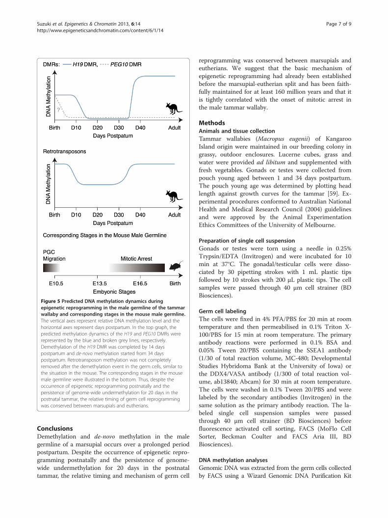
Figure 5 Predicted DNA methylation dynamics duringepigenetic reprogramming in the male germline of the tammarwallaby and corresponding stages in the mouse male germline.The vertical axes represent relative DNA methylation level and thehorizontal axes represent days postpartum. In the top graph, thepredicted methylation dynamics of the H19 and PEG10 DMRs wererepresented by the blue and broken grey lines, respectively.Demethylation of the H19 DMR was completed by 14 dayspostpartum and de-novo methylation started from 34 dayspostpartum. Retrotransposon methylation was not completelyremoved after the demethylation event in the germ cells, similar tothe situation in the mouse. The corresponding stages in the mousemale germline were illustrated in the bottom. Thus, despite theoccurrence of epigenetic reprogramming postnatally and thepersistence of genome-wide undermethylation for 20 days in thepostnatal tammar, the relative timing of germ cell reprogrammingwas conserved between marsupials and eutherians.
Suzuki et al. Epigenetics & Chromatin 2013, 6:14 Page 7 of 9http://www.epigeneticsandchromatin.com/content/6/1/14
ConclusionsDemethylation and de-novo methylation in the malegermline of a marsupial occurs over a prolonged periodpostpartum. Despite the occurrence of epigenetic repro-gramming postnatally and the persistence of genome-wide undermethylation for 20 days in the postnataltammar, the relative timing and mechanism of germ cell
reprogramming was conserved between marsupials andeutherians. We suggest that the basic mechanism ofepigenetic reprogramming had already been establishedbefore the marsupial-eutherian split and has been faith-fully maintained for at least 160 million years and that itis tightly correlated with the onset of mitotic arrest inthe male tammar wallaby.
MethodsAnimals and tissue collectionTammar wallabies (Macropus eugenii) of KangarooIsland origin were maintained in our breeding colony ingrassy, outdoor enclosures. Lucerne cubes, grass andwater were provided ad libitum and supplemented withfresh vegetables. Gonads or testes were collected frompouch young aged between 1 and 34 days postpartum.The pouch young age was determined by plotting headlength against growth curves for the tammar [59]. Ex-perimental procedures conformed to Australian NationalHealth and Medical Research Council (2004) guidelinesand were approved by the Animal ExperimentationEthics Committees of the University of Melbourne.
Preparation of single cell suspensionGonads or testes were torn using a needle in 0.25%Trypsin/EDTA (Invitrogen) and were incubated for 10min at 37°C. The gonadal/testicular cells were disso-ciated by 30 pipetting strokes with 1 mL plastic tipsfollowed by 10 strokes with 200 μL plastic tips. The cellsamples were passed through 40 μm cell strainer (BDBiosciences).
Germ cell labelingThe cells were fixed in 4% PFA/PBS for 20 min at roomtemperature and then permeabilised in 0.1% Triton X-100/PBS for 15 min at room temperature. The primaryantibody reactions were performed in 0.1% BSA and0.05% Tween 20/PBS containing the SSEA1 antibody(1/30 of total reaction volume, MC-480; DevelopmentalStudies Hybridoma Bank at the University of Iowa) orthe DDX4/VASA antibody (1/300 of total reaction vol-ume, ab13840; Abcam) for 30 min at room temperature.The cells were washed in 0.1% Tween 20/PBS and werelabeled by the secondary antibodies (Invitrogen) in thesame solution as the primary antibody reaction. The la-beled single cell suspension samples were passedthrough 40 μm cell strainer (BD Biosciences) beforefluorescence activated cell sorting, FACS (MoFlo CellSorter, Beckman Coulter and FACS Aria III, BDBiosciences).
DNA methylation analysesGenomic DNA was extracted from the germ cells collectedby FACS using a Wizard Genomic DNA Purification Kit

Suzuki et al. Epigenetics & Chromatin 2013, 6:14 Page 8 of 9http://www.epigeneticsandchromatin.com/content/6/1/14
(Promega). Purified genomic DNA was treated with a so-dium bisulphite solution as described previously [60,61].After the bisulphite treatment for the genomic DNA, 30to 38 cycles of PCR with the genomic DNA templates cor-responding to 100 to 5,000 cells were carried out usingthe following primer pairs.PEG10 DMR Forward: 5′- CCTCCCATTAACTTTAA
AATCACC -3′PEG10 DMR Reverse: 5′- ATTGTAGTAATGGGGTA
GGTTATG -3′H19 DMR Forward: 5′- GAATGGGTTAGATGAGGG
TAGTATAG -3′H19 DMR Reverse: 5′- TATCAAACACCAAAACCAC
AAATAA -3′H19 COBRA Forward: 5′- TTATTTTGGAGAAAATT
TGAAGATAAGTAG -3′H19 COBRA Reverse: 5′- TATCCTAAAACATCAA
AACCTAAATTAAAC -3′KERV-1 LTR Forward: 5′- TAAACTCAATTCCAT
ATAAACAATCTC -3′KERV-1 LTR Reverse: 5′- TTTTTGTTTTGTAAGGG
TTTTTTAG -3′LINE1 Forward: 5′- GGAGATTTTTGTTTTAGAGA
GATTTGTAAA -3′LINE1 Reverse: 5′- TATAAAAACACCCCACTCCCC
TCTC -3′The PCR products for COBRA (combined bisulphite
and restriction analysis) were digested with 1 to 10 unitsof MluCI, AciI, TaqI (New England Biolabs) or HinfI(TaKaRa) restriction enzymes for 2-3 h at 37°C or 65°Cfor TaqI. The intensity of the cut and uncut bands wasquantified by ATTO CS Analyzer 3 software (ATTO). ThePCR products for H19 DMR and retrotransposons werecloned, and the clones were sequenced. The sequence datawere analysed by QUMA (quantification tool for methyla-tion analysis; http://quma.cdb.riken.jp) [62].
Competing interestsThe authors declare that they have no competing interests.
Authors’ contributionsSS carried out the molecular and cellular studies, participated in theexperimental design and data analysis, and drafted the manuscript. GS andMBR conceived of the study, and participated in its design and coordinationand helped to draft the manuscript. All authors read and approved the finalmanuscript.
AcknowledgementsWe thank Alison Bradfield and Scott Brownlees for assistance with theanimals, Helen Clark and Bonnie Dopheide for technical assistance and Drs.Hongshi Yu and Danielle Hickford for help in collecting tissue. Fluorescenceactivated cell sorting was operated by Dr. Matt Burton at Murdoch Children’sResearch Institute, Royal Children's Hospital and Susumu Ito at ResearchCenter for Human and Environmental Sciences, Shinshu University.
Received: 11 January 2013 Accepted: 8 May 2013Published: 3 June 2013
References1. Hackett JA, Zylicz JJ, Surani MA: Parallel mechanisms of epigenetic
reprogramming in the germline. Trends Genet 2012, 28:164–174.2. Saitou M, Kagiwada S, Kurimoto K: Epigenetic reprogramming in mouse
pre-implantation development and primordial germ cells. Development2012, 139:15–31.
3. Sasaki H, Matsui Y: Epigenetic events in mammalian germ-cell development:reprogramming and beyond. Nat Rev Genet 2008, 9:129–140.
4. Reik W: Stability and flexibility of epigenetic gene regulation inmammalian development. Nature 2007, 447:425–432.
5. Seki Y, Hayashi K, Itoh K, Mizugaki M, Saitou M, Matsui Y: Extensive andorderly reprogramming of genome-wide chromatin modificationsassociated with specification and early development of germ cells inmice. Dev Biol 2005, 278:440–458.
6. Seki Y, Yamaji M, Yabuta Y, Sano M, Shigeta M, Matsui Y, Saga Y, TachibanaM, Shinkai Y, Saitou M: Cellular dynamics associated with the genome-wide epigenetic reprogramming in migrating primordial germ cells inmice. Development 2007, 134:2627–2638.
7. Hajkova P, Erhardt S, Lane N, Haaf T, El-Maarri O, Reik W, Walter J, SuraniMA: Epigenetic reprogramming in mouse primordial germ cells.Mech Dev 2002, 117:15–23.
8. Lane N, Dean W, Erhardt S, Hajkova P, Surani A, Walter J, Reik W: Resistanceof IAPs to methylation reprogramming may provide a mechanism forepigenetic inheritance in the mouse. Genesis 2003, 35:88–93.
9. Lee J, Inoue K, Ono R, Ogonuki N, Kohda T, Kaneko-Ishino T, Ogura A,Ishino F: Erasing genomic imprinting memory in mouse clone embryosproduced from day 11.5 primordial germ cells. Development 2002,129:1807–1817.
10. Maatouk DM, Kellam LD, Mann MR, Lei H, Li E, Bartolomei MS, Resnick JL:DNA methylation is a primary mechanism for silencing postmigratoryprimordial germ cell genes in both germ cell and somatic cell lineages.Development 2006, 133:3411–3418.
11. Bourc'his D, Bestor TH: Meiotic catastrophe and retrotransposonreactivation in male germ cells lacking Dnmt3L. Nature 2004, 431:96–99.
12. Davis TL, Trasler JM, Moss SB, Yang GJ, Bartolomei MS: Acquisition of theH19 methylation imprint occurs differentially on the parental allelesduring spermatogenesis. Genomics 1999, 58:18–28.
13. Davis TL, Yang GJ, McCarrey JR, Bartolomei MS: The H19 methylationimprint is erased and re-established differentially on the parental allelesduring male germ cell development. Hum Mol Genet 2000, 9:2885–2894.
14. Kaneda M, Okano M, Hata K, Sado T, Tsujimoto N, Li E, Sasaki H: Essentialrole for de novo DNA methyltransferase Dnmt3a in paternal andmaternal imprinting. Nature 2004, 429:900–903.
15. Kato Y, Kaneda M, Hata K, Kumaki K, Hisano M, Kohara Y, Okano M, Li E,Nozaki M, Sasaki H: Role of the Dnmt3 family in de novo methylation ofimprinted and repetitive sequences during male germ cell developmentin the mouse. Hum Mol Genet 2007, 16:2272–2280.
16. Li JY, Lees-Murdock DJ, Xu GL, Walsh CP: Timing of establishment ofpaternal methylation imprints in the mouse. Genomics 2004, 84:952–960.
17. Webster KE, O’Bryan MK, Fletcher S, Crewther PE, Aapola U, Craig J, HarrisonDK, Aung H, Phutikanit N, Lyle R, Meachem SJ, Antonarakis SE, de KretserDM, Hedger MP, Peterson P, Carroll BJ, Scott HS: Meiotic and epigeneticdefects in Dnmt3L-knockout mouse spermatogenesis. Proc Natl Acad SciU S A 2005, 102:4068–4073.
18. Ueda T, Abe K, Miura A, Yuzuriha M, Zubair M, Noguchi M, Niwa K, KawaseY, Kono T, Matsuda Y, Fujimoto H, Shibata H, Hayashizaki Y, Sasaki H: Thepaternal methylation imprint of the mouse H19 locus is acquired in thegonocyte stage during foetal testis development. Genes Cells 2000,5:649–659.
19. Hore TA, Rapkins RW, Graves JA: Construction and evolution of imprintedloci in mammals. Trends Genet 2007, 23:440–448.
20. Renfree MB, Ager EI, Shaw G, Pask AJ: Genomic imprinting in marsupialplacentation. Reproduction 2008, 136:523–531.
21. Renfree MB, Hore TA, Shaw G, Graves JA, Pask AJ: Evolution of genomicimprinting: insights from marsupials and monotremes. Annu RevGenomics Hum Genet 2009, 10:241–262.
22. Reik W, Lewis A: Co-evolution of X-chromosome inactivation andimprinting in mammals. Nat Rev Genet 2005, 6:403–410.
23. Suzuki S, Renfree MB, Pask AJ, Shaw G, Kobayashi S, Kohda T, Kaneko-Ishino T,Ishino F: Genomic imprinting of IGF2, p57(KIP2) and PEG1/MEST in amarsupial, the tammar wallaby. Mech Dev 2005, 122:213–222.
Suzuki et al. Epigenetics & Chromatin 2013, 6:14 Page 9 of 9http://www.epigeneticsandchromatin.com/content/6/1/14
24. Suzuki S, Ono R, Narita T, Pask AJ, Shaw G, Wang C, Kohda T, Alsop AE,Marshall Graves JA, Kohara Y, Ishino F, Renfree MB, Kaneko-Ishino T:Retrotransposon silencing by DNA methylation can drive mammaliangenomic imprinting. PLoS Genet 2007, 3:e55.
25. Killian JK, Byrd JC, Jirtle JV, Munday BL, Stoskopf MK, MacDonald RG,Jirtle RL: M6P/IGF2R imprinting evolution in mammals. Mol Cell 2000,5:707–716.
26. O'Neill MJ, Ingram RS, Vrana PB, Tilghman SM: Allelic expression of IGF2 inmarsupials and birds. Dev Genes Evol 2000, 210:18–20.
27. Rapkins RW, Hore T, Smithwick M, Ager E, Pask AJ, Renfree MB, Kohn M,Hameister H, Nicholls RD, Deakin JE, Graves JA: Recent assembly of animprinted domain from non-imprinted components. PLoS Genet 2006,2:e182.
28. Ager E, Suzuki S, Pask A, Shaw G, Ishino F, Renfree MB: Insulin is imprintedin the placenta of the marsupial. Macropus eugenii. Dev Biol 2007,309:317–328.
29. Edwards CA, Mungall AJ, Matthews L, Ryder E, Gray DJ, Pask AJ, Shaw G,Graves JA, Rogers J, Dunham I, Renfree MB, Ferguson-Smith AC, SAVOIRconsortium: The evolution of the DLK1-DIO3 imprinted domain inmammals. PLoS Biol 2008, 6:e135.
30. Smits G, Mungall AJ, Griffiths-Jones S, Smith P, Beury D, Matthews L,Rogers J, Pask AJ, Shaw G, VandeBerg JL, McCarrey JR, SAVOIR consortium,Renfree MB, Reik W, Dunham I: Conservation of the H19 noncoding RNAand H19-IGF2 imprinting mechanism in therians. Nat Genet 2008,40:971–976.
31. Suzuki S, Shaw G, Kaneko-Ishino T, Ishino F, Renfree MB: Characterisation ofmarsupial PHLDA2 reveals eutherian specific acquisition of imprinting.BMC Evol Biol 2011, 11:244.
32. Suzuki S, Shaw G, Kaneko-Ishino T, Ishino F, Renfree MB: The evolution ofmammalian genomic imprinting was accompanied by the acquisition ofnovel CpG islands. Genome Biol Evol 2011, 3:1276–1283.
33. Stringer JM, Suzuki S, Pask AJ, Shaw G, Renfree MB: GRB10 imprinting iseutherian mammal specific. Mol Biol Evol 2012, 29:3711–3719.
34. Stringer JM, Suzuki S, Pask AJ, Shaw G, Renfree MB: Promoter-specificexpression and imprint status of marsupial IGF2. PLoS One 2012,7:e41690.
35. Stringer JM, Suzuki S, Pask AJ, Shaw G, Renfree MB: Selected imprinting ofINS in the marsupial. Epigenetics Chromatin 2012, 5:14.
36. Luo ZX, Yuan CX, Meng QJ, Ji Q: A Jurassic eutherian mammal anddivergence of marsupials and placentals. Nature 2011, 476:442–445.
37. Tyndale-Biscoe CH, Renfree MB: Monographs on Marsupial Biology:Reproductive physiology of marsupials. Cambridge: Cambridge UniversityPress; 1987.
38. Ullmann SL, Shaw G, Alcorn GT, Renfree MB: Migration of primordial germcells to the developing gonadal ridges in the tammar wallaby Macropuseugenii. J Reprod Fertil 1997, 110:135–143.
39. Alcorn GT, Robinson ES: Germ cell development in female pouch young ofthe tammar wallaby (Macropus eugenii). J Reprod Fertil 1983, 67:319–325.
40. Renfree MBOWS, Short RV, Shaw G: Sexual differentiation of the urogenitalsystem of the fetal and neonatal tammar wallaby, Macropus eugenii.Anat Embryol (Berl) 1996, 194:111–134.
41. Hickford DE, Frankenberg S, Pask AJ, Shaw G, Renfree MB: DDX4 (VASA) isconserved in germ cell development in marsupials and monotremes.Biol Reprod 2011, 85:733–743.
42. Hickford D, Frankenberg S, Renfree MB: Immunohistochemical staining ofsectioned tammar wallaby (Macropus eugenii) tissue. Cold Spring HarbProtoc 2009, 2009:pdb.prot5338.
43. Huang Y, Pastor WA, Shen Y, Tahiliani M, Liu DR, Rao A: The behaviour of5-hydroxymethylcytosine in bisulfite sequencing. PLoS One 2010, 5:e8888.
44. Jin SG, Kadam S, Pfeifer GP: Examination of the specificity of DNAmethylation profiling techniques towards 5-methylcytosine and 5-hydroxymethylcytosine. Nucleic Acids Res 2010, 38:e125.
45. Nestor C, Ruzov A, Meehan R, Dunican D: Enzymatic approaches andbisulfite sequencing cannot distinguish between 5-methylcytosine and5-hydroxymethylcytosine in DNA. Biotechniques 2010, 48:317–319.
46. Hackett JA, Sengupta R, Zylicz JJ, Murakami K, Lee C, Down TA, Surani MA:Germline DNA demethylation dynamics and imprint erasure through5-hydroxymethylcytosine. Science 2013, 339:448–452.
47. Saga Y: Sexual development of mouse germ cells: Nanos2 promotes themale germ cell fate by suppressing the female pathway. Dev GrowthDiffer 2008, Suppl 1:S141–S147.
48. Yokomine T, Hata K, Tsudzuki M, Sasaki H: Evolution of the vertebrateDNMT3 gene family: a possible link between existence of DNMT3L andgenomic imprinting. Cytogenet Genome Res 2006, 113:75–80.
49. Hore TA, Deakin JE, Marshall Graves JA: The evolution of epigeneticregulators CTCF and BORIS/CTCFL in amniotes. PLoS Genet 2008,4:e1000169.
50. Pillai RS, Chuma S: piRNAs and their involvement in male germlinedevelopment in mice. Dev Growth Differ 2012, 54:78–92.
51. Hackett JA, Reddington JP, Nestor CE, Dunican DS, Branco MR, Reichmann J,Reik W, Surani MA, Adams IR, Meehan RR: Promoter DNA methylationcouples genome-defence mechanisms to epigenetic reprogramming inthe mouse germline. Development 2012, 139:3623–3632.
52. Aravin A, Gaidatzis D, Pfeffer S, Lagos-Quintana M, Landgraf P, Iovino N,Morris P, Brownstein MJ, Kuramochi-Miyagawa S, Nakano T, Chien M,Russo JJ, Ju J, Sheridan R, Sander C, Zavolan M, Tushl T: A novel class ofsmall RNAs bind to MILI protein in mouse testes. Nature 2006,442:203–207.
53. Girard A, Sachidanandam R, Hannon GJ, Carmell MA: A germline-specificclass of small RNAs binds mammalian Piwi proteins. Nature 2006,442:199–202.
54. Aravin AA, Sachidanandam R, Girard A, Fejes-Toth K, Hannon GJ:Developmentally regulated piRNA clusters implicate MILI in transposoncontrol. Science 2007, 316:744–747.
55. Kuramochi-Miyagawa S, Watanabe T, Gotoh K, Totoki Y, Toyoda A, Ikawa M,Asada N, Kojima K, Yamaguchi Y, Ijiri TW, Hata K, Li E, Matsuda Y, Kimura T,Okabe M, Sakaki Y, Sasaki H, Nakano T: DNA methylation ofretrotransposon genes is regulated by Piwi family members MILI andMIWI2 in murine fetal testes. Genes Dev 2008, 22:908–917.
56. Kuramochi-Miyagawa S, Watanabe T, Gotoh K, Takamatsu K, Chuma S,Kojima-Kita K, Shiromoto Y, Asada N, Toyoda A, Fujiyama A, Totoki Y,Shibata T, Kimura T, Nakatsuji N, Noce T, Sasaki H, Nakano T: MVH in piRNAprocessing and gene silencing of retrotransposons. Genes Dev 2010,24:887–892.
57. Renfree MB, Papenfuss AT, Deakin JE, Lindsay J, Heider T, Belov K, Rens W,Waters PD, Pharo EA, Shaw G, Wong ES, Lefevre CM, Nicholas KR, Kuroki Y,Wakefield MJ, Zenger KR, Wang C, Ferguson-Smith M, Nicholas FW, HickfordD, Yu H, Short KR, Siddle HV, Frankenberg SR, Chew KY, Menzies BR, StringerJM, Suzuki S, Hore TA, Delbridge ML, et al: Genome sequence of anAustralian kangaroo, Macropus eugenii, provides insight into theevolution of mammalian reproduction and development. Genome Biol2011, 12:R81.
58. Ollinger R, Childs AJ, Burgess HM, Speed RM, Lundegaard PR, Reynolds N,Gray NK, Cooke HJ, Adams IR: Deletion of the pluripotency-associatedTex19.1 gene causes activation of endogenous retroviruses anddefective spermatogenesis in mice. PLoS Genet 2008, 4:e1000199.
59. Poole WE, Simms NG, Wood JT, Lubulwa M: Tables for age determination ofthe Kangaroo Island wallaby (Tammar), Macropus eugenii, from bodymeasurements. Canberra: Australia Division of Wildlife and Ecology; 1991.
60. Frommer M, McDonald LE, Millar DS, Collis CM, Watt F, Grigg GW, MolloyPL, Paul CL: A genomic sequencing protocol that yields a positive displayof 5-methylcytosine residues in individual DNA strands. Proc Natl Acad SciU S A 1992, 89:1827–1831.
61. Raizis AM, Schmitt F, Jost JP: A bisulfite method of 5-methylcytosinemapping that minimizes template degradation. Anal Biochem 1995,226:161–166.
62. Kumaki Y, Oda M, Okano M: QUMA: quantification tool for methylationanalysis. Nucleic Acids Res 2008, 36(Web Server issue):170–175.
doi:10.1186/1756-8935-6-14Cite this article as: Suzuki et al.: Postnatal epigenetic reprogramming inthe germline of a marsupial, the tammar wallaby. Epigenetics &Chromatin 2013 6:14.


![RESEARCH Open Access Epigenetic reprogramming of breast ...mouse blastocyst resulting in normal tissue derived from tumour cells in chimeric mice [9]. Tumorigenicity of metastatic](https://img.dokumen.tips/doc/110x75/5f8a9cf5f9b6054e73143744/research-open-access-epigenetic-reprogramming-of-breast-mouse-blastocyst-resulting.jpg)


![Cell Culture-Induced Gradual and Frequent Epigenetic … · Cell Culture-Induced Gradual and Frequent Epigenetic Reprogramming of Invertedly Repeated Tobacco Transgene Epialleles1[W]](https://img.dokumen.tips/doc/110x75/603cb1840f25594069133327/cell-culture-induced-gradual-and-frequent-epigenetic-cell-culture-induced-gradual.jpg)