Embed Size (px)
Citation preview
Demirbas-Uzel et al. BMC Microbiology 2018, 18(Suppl 1):161https://doi.org/10.1186/s12866-018-1276-7
RESEARCH Open Access
Impact of Glossina pallidipes salivary glandhypertrophy virus (GpSGHV) on aheterologous tsetse fly host, Glossinafuscipes fuscipes
Güler Demirbas-Uzel1,2, Andrew G. Parker1, Marc J. B. Vreysen1, Robert L. Mach2, Jeremy Bouyer1, Peter Takac3,4and Adly M. M. Abd-Alla1*
Abstract
Background: Tsetse flies (Diptera: Glossinidae) are the vectors of African trypanosomosis, the causal agent ofsleeping sickness in humans and nagana in animals. Glossina fuscipes fuscipes is one of the most importanttsetse vectors of sleeping sickness, particularly in Central Africa. Due to the development of resistance of thetrypanosomes to the commonly used trypanocidal drugs and the lack of effective vaccines, vector controlapproaches remain the most effective strategies for sustainable management of those diseases. The SterileInsect Technique (SIT) is an effective, environment-friendly method for the management of tsetse flies in thecontext of area-wide integrated pest management programs (AW-IPM). This technique relies on the mass-production of the target insect, its sterilization with ionizing radiation and the release of sterile males in thetarget area where they will mate with wild females and induce sterility in the native population. It has beenshown that Glossina pallidipes salivary gland hypertrophy virus (GpSGHV) infection causes a decrease infecundity and fertility hampering the maintenance of colonies of the tsetse fly G. pallidipes. This virus has alsobeen detected in different species of tsetse files. In this study, we evaluated the impact of GpSGHV on theperformance of a colony of the heterologous host G. f. fuscipes, including the flies’ productivity, mortality,survival, flight propensity and mating ability and insemination rates.
Results: Even though GpSGHV infection did not induce SGH symptoms, it significantly reduced all examinedparameters, except adult flight propensity and insemination rate.
Conclusion: These results emphasize the important role of GpSGHV management strategy in the maintenanceof G. f. fuscipes colonies and the urgent need to implement measures to avoid virus infection, to ensure theoptimal mass production of this tsetse species for use in AW-IPM programs with an SIT component.
Keywords: Glossinidae, Hytrosaviridae, Longevity, Insemination, Mating ability, Flight propensity
* Correspondence: [email protected] Pest Control Laboratory, Joint FAO/IAEA Division of NuclearTechniques in Food & Agriculture, International Atomic Energy Agency,Vienna International Centre, P.O. Box 100, 1400 Vienna, AustriaFull list of author information is available at the end of the article
© International Atomic Energy Agency; licensee BioMed. 2018 This is an open access article distributed under the terms of theCreative Commons Attribution IGO License (https://creativecommons.org/licenses/by/3.0/igo/) which permits unrestricted use,distribution, and reproduction in any medium, provided appropriate credit to the original author(s) and the source is given.
Demirbas-Uzel et al. BMC Microbiology 2018, 18(Suppl 1):161 Page 246 of 292
BackgroundTsetse flies (Diptera: Glossinidae) are the only cyclicalvectors of the pathogenic African trypanosomes thatcause human African trypanosomosis (HAT) or sleepingsickness and African animal trypanosomosis (AAT) ornagana in sub-Saharan Africa [1]. There are 33 speciesand subspecies of tsetse flies that all belong to the genusGlossina, divided into the Morsitans, Fusca, and Palpalisgroups [2]. Although all tsetse species can transferpathogenic trypanosomes, members of the Palpalis andMorsitans groups are the primary trypanosome vectors[3]. For instance, G. f. fuscipes is a significant vector oftrypanosomes in central Africa [4], particularly inUganda and Western Kenya [5]. In the absence of effect-ive vaccines and drugs against HAT and AAT [6] vectorcontrol represents the most efficient strategy to managethese diseases in mainly rural areas [7, 8]. Currently ac-cepted tsetse control tactics are the sequential aerosoltechnique [9], stationary bait methods (traps and targets)[10], the live bait technology [11] and the sterile insecttechnique (SIT). The SIT is based on the mass-rearingand sterilization of males with ionizing radiation (e.g.,gamma irradiation), and the sequential release of ad-equate numbers of sterile male insects in the target area[12]. Mating between sterile males and wild females willresult in non-viable embryos, leading to the gradual re-duction of the target insect population [13]. The SIT hasproven to be a powerful control tactic for use againsttsetse flies and other Diptera as part of area-wide inte-grated pest management (AW-IPM) approaches [14].The implementation of AW-IPM programs with an
SIT component against tsetse flies poses significant chal-lenges with respect to colonization and mass-rearing ofthe target species. Many factors, such as infections withpathogens when the insects are reared continuously orunder suboptimal rearing conditions [15], might lead tofailures in establishing and maintaining large tsetse col-onies and, as a consequence, fail to produce insects ofadequate quality.Infections of tsetse flies derived from natural popula-
tions and laboratory colonies with the pathogenic saliv-ary gland hypertrophy virus (SGHV) [16–18], a memberof the Glossina Hytrosavirus genus and Hytrosaviridaefamily have been frequently observed [19]. SGHV is arod-shaped enveloped virus (100 × 700–1000 nm) con-taining a large double-stranded DNA genome of 190 kb[19]. The virus infection is mostly asymptomatic in tse-tse flies, but in some cases it can lead to the develop-ment of salivary gland hypertrophy (SGH) symptoms,which has been associated with a reduction in the flies’productivity and eventually loss of the colony [20–22].SGH prevalence of this virus in natural tsetse popula-tions vary across tsetse species and their locations, butare usually low (prevalence of 0.3 to 7%) [23] However,
under mass-rearing conditions of Glossina pallidipes,high prevalence rates have been observed that were as-sociated with the use of the in vitro membrane feedingtechnique that favors horizontal transmission of thevirus. In G. pallidipes, a species that is considered an ef-ficient vector of trypanosomes [24], SGH symptomswere associated with abnormalities of the ovaries andtesticular degeneration, leading to reduced productivityin both male and female flies [15, 23, 25, 26]. Data avail-able on prevalence rates of the virus in colonies of G.pallidipes showed that colony decline and eventual col-lapse could not be averted when the SGH infection ratein the colony reached 70% (Abd-Alla et al., 2016). Tomitigate the negative effects of the virus on colony per-formance, several virus management strategies were de-veloped that have proven to be effective [27–29].Although SGH symptoms have been detected in natural
populations of other tsetse species such as Glossina austeni,G. morsitans morsitans, G. nigrofusca nigrofusca and G. pal-licera pallicera [16, 30, 31], no SGH symptoms have beenobserved in G. f. fuscipes but asymptomatic infection wasdetected [32]. However, in laboratory colony, intra-hemocoelic injections of GpSGHV into five heterologoustsetse species (G. brevipalpis, G. m. morsitans, G. m. centra-lis, G. f. fuscipes and G. p. gambiensis) showed a significantincrease in the titer of viral DNA, demonstrating the abilityto replicate in these heterologous species [33].The Government of Ethiopia has embarked on an
AW-IPM program with an SIT component to eradicatea G. f. fuscipes population in the Deme river valley ofSouthern Ethiopia [34–36]. The campaign required theestablishment and expansion of a colony of the targetspecies in the mass-rearing facility in Kality on the out-skirts of Addis Ababa. The colony was initiated withseed material from a colony maintained at the SlovakAcademy of Sciences (SAS), Slovakia. Although colonygrowth was acceptable in the initial stages subsequentlow productivity and high mortality resulted in a drasticreduction in colony size. Similar observations weremade at the SAS, where the colony was lost. It is worthnoting that more than one tsetse species is being main-tained in both facilities, including G. pallidipes, a spe-cies known to be harbor GpSGHV, a situation that mayfacilitate the transmission of GpSGHV from one tsetsespecies to another especially if both species were fedusing the same membrane as was the case in the SAScolonies. It should be noted that SGHV was detectedby PCR in natural populations of G. f. fuscipes with aprevalence of 25–40% [32] and an increase in virus titerin GpSGHV injected flies has recently been demon-strated [33].This study was undertaken as part of efforts to under-
stand the possible causes of the poor colony perform-ance. In this study, we report on the impact of GpSGHV

Demirbas-Uzel et al. BMC Microbiology 2018, 18(Suppl 1):161 Page 247 of 292
on the performance of G. f. fuscipes flies using standardquality control parameters, such as adult longevity, fe-male productivity and mortality, flight propensity, mat-ing ability, and insemination rate.
MethodsTsetse fliesThe G. f. fuscipes flies used in this study originated from acolony that was established from wild collected material inthe Central African Republic (CAR) and maintained since1986 at the Insect Pest Control Laboratory (IPCL) of theJoint FAO/IAEA Division of Nuclear Techniques in Foodand Agriculture, Seibersdorf, Austria. Experimental flies werefed for 15–20 min, three times per week with defibrinatedbovine blood using an artificial (in vitro) membrane feedingsystem [37]. The adult flies were held in medium size cages(11 cm diameter × 5 cm high) at a ratio of 1:3 male to femaleunder standard tsetse colony rearing conditions (24 ± 0.5 °Cand 75 ± 5% relative humidity (RH)) [38]. The SGHV is notdetectable in this colony by PCR.
Preparation of virus inoculum and intra-hemocoelicinjectionThe GpSGHV inoculum was prepared from intacthypertrophied salivary glands dissected from a 10-day-old male G. pallidipes showing overt SGH symptoms[39]. Briefly, the hypertrophied glands were homoge-nized in phosphate buffered saline (PBS) at a concentra-tion of one pair of glands/ml and the homogenate wascentrifuged at 400 x g for 2 min at room temperature.The supernatant was transferred to a new sterile tubeand used immediately after preparation of the inoculum.Using a 1 ml Myjector U-40 Insulin type syringe (Teruma,
Leuven, Belgium) either 2 μl of filter-sterilized PBS (control)or the virus suspension was injected into the thoracic cavityof prechilled adult flies. For each treatment, newly emergedteneral (male and female) flies were injected and placed intostandard holding cages (20 cm diameter × 5 cm high) at therequired mating ratio and each experiment was replicated 3times. Non-injected and PBS-injected flies were used asnon-injected controls to evaluate the impact of injection andthe virus infection on the flies’ performance.
Prevalence of GpSGHV infection in G. f. fuscipes injectedfliesThe tsetse genomic DNA was extracted from individualnon-injected, PBS- and virus-injected flies using theDNeasy Blood & Tissue kit (QIAGEN Inc., Valencia, CA)following the manufacturer’s instructions. The titer ofGpSGHV was determined in G. f. fuscipes injected malesand females on 0, 9 and 18 days post injection by polymer-ase chain reaction (PCR) using the method previously de-scribed by Abd-Alla et al. [20]. Equal volume of individual
DNA sample was pooled (n = 6 for females and n = 2 formales) and measured to determine the DNA concentra-tion by spectrophotometry (Nanodrop-Synergy H1 Multi-Mode Reader, BioTek, Instruments, Inc., USA), DNAsamples were diluted to final concentration of 4 ng/μl and5 μl was used as template for the qPCR reaction. TheqPCR was performed with odv-e66 (GpSGHV ORF5)gene using the method previously described [20, 39, 40]and the tsetse β-tubulin gene was used as a housekeepinggene to normalize the qPCR reactions.
Impact of GpSGHV infection on survival and productivityof G. f. fuscipesTo evaluate the impact of GpSGHV challenge on G. f.fuscipes, their productivity and longevity under both normalfeeding (blood meal offered three times per week) and star-vation stress (no blood feeding) conditions was monitored innon-, PBS- and virus-injected flies. For each treatment, sevenmales and twenty-one females were kept in standard holdingcages and each treatment was replicated 3 times. The prod-uctivity data is presented as total pupae over the experimen-tal period per initial female (PPIF).
Impact of GpSGHV infection on the flight propensity of G.f. fuscipesThe flight propensity of virus injected flies, non-injectedand PBS-injected flies (the latter two as negative controls)was assessed at 7, 14, 21, 28, 35 and 42 days post injectionunder normal feeding conditions. Flight tests were carriedout in netted cubic mating cages (45 × 45 × 45 cm) thatcontain a black Polyvinyl Chloride (PVC) tube (8.9 cmdiameter, 3 mm thick wall, 10 cm high). The PVC tubeallowed light entering only from the top and the walls werecoated with unscented talcum powder to prevent the fliesfrom walking out the tube [41]. Standard FAO/IAEA/USDA protocols (FAO/IAEA/USDA, 2014 http://www-na-web.iaea.org/nafa/ipc/public/QualityControl.pdf) were usedwith a few modifications i.e. rather than using pupae, theadult flies were chilled at 4 °C for 5 min prior to the test, toenable the transfer to the tube. For each test, seven chilledmales and twenty-one chilled females were put in a plasticPetri dish (90 mm diameter) with the base covered by blackporous paper, and the number of flies that had escapedfrom the tube “flier” was recorded during one hour [42].The light intensity at the top of the tubes was 500 lx. Sixreplicates were conducted for each treatment.
Impact of GpSGHV infection on the mating ability andinsemination rate of G. f. fuscipesThe mating ability and the insemination rate of untreated(normal colony) G. f. fuscipes males of different ages (3-, 6-,9- and 12-days post emergence) were assessed to determinethe optimal mating age [43]. Forty (40) teneral males werereleased in mating cages mating cages (45 × 45 × 45 cm),

Demirbas-Uzel et al. BMC Microbiology 2018, 18(Suppl 1):161 Page 248 of 292
followed 15 min later by an equal number of 9 - day old vir-gin females for mating. Mating events were observed understandard tsetse rearing conditions from 9:30 to 12:30 h tocover the morning mating activity peak [44]. The optimalmating age test was replicated 3 times and mating tests ofvirus-challenged flies were repeated 9 times. All flies wereoffered a blood meal 24 h before mating to increase themating rate, and non-fed flies were removed and replaced.The propensity for mating ratio was calculated accordingto the proportion of females that mated for each treat-ment [44]. After determining the optimal male matingage, 6 to 9-day old non-injected, PBS-injected, andvirus-injected virgin males (40 males) were tested as de-scribed above to determine mating ability and insemin-ation rate of experimental flies.Mating pairs were transferred to small cages (4 cm
diameter × 6 cm high) and kept for 24 h, after whichthe males were removed and the females dissectedunder a binocular microscope to determine insemin-ation rate. Mated female flies were dissected in PBSunder a binocular microscope and the insemination rateand spermathecal contents were assessed subjectively at ×100 magnification using a Carl Zeiss compound micro-scope [45]. The spermathecal fill and insemination ratewere obtained by assessing the content of the spermathecaepairs. Spermathecal fill was scored to the nearest quarterfor each spermathecae separately as empty (0), quarter full(0.25), half-full (0.50), three-quarter-full (0.75) and full (1.0),For the statistical analysis, quarter full (0.25), half-full(0.50), three-quarter-full (0.75) were considered as partialfill. The amount of sperm transferred was then computedas the mean spermathecal filling values of the spermathecaepairs [46].
Statistical analysisThe significance of the virus injections on the variousparameters was assessed by an ANOVA test [47].Pairwise comparisons between group means (PBS vs.virus injections, non-injected vs. virus injections andnon-injected vs. PBS injections effect on flies) was thandetermined by Tukey’s honestly significant difference(HSD) test. The analyses were performed in R [48, 49]using RStudio version 3.4.1. [50] The data was trans-formed using the Box-Cox procedure from the packages.ggplot2 [51], lattice v0.20–35 [52] and MASS v7.3. [53].All survival analyses were performed using Graph
Pad Prism version 5.0 for Windows (GraphPad Soft-ware, San Diego California, USA; graphpad.com). Theeffect of the treatments on fly longevity was analysedusing a Log-rank (Mantel-Cox) test. Differences be-tween treatments pairs were tested using the Bonfer-roni method. Mean longevity (or age in days atdeath) was calculated from the sum of the number oflive flies on each day until the death of the last fly,
divided by the number of flies in the group at thestart of the experiment. The level of significance was0.05 for all statistical analyses.
ResultsDetection of GpSGHV infection in injected fliesThe GpSGHV titer in virus-injected flies was assessed byqPCR at various times post injection to investigatewhether the virus could infect and replicate in injectedflies. The qPCR results indicate a significant increase inthe virus titer over time (F = 1.34, df = 1, 34, P < 0.001).The results indicate that the virus replication was ratherslow as no significant increase in the virus titer between 0time and 9 dpi was observed, but later the virus titer in-crease by 5. 22 fold change at 18 dpi (Fig. 1). In addition,results demonstrated a significant difference in the virustiter between the virus injected flies and negative controls(PBS- injected flies (F = 21.51, df = 1, 68, P < 0.001).
Impact of GpSGHV infection on G. f. fuscipes productivityand survivalVirus challenge reduced the productivity of the flies sig-nificantly (F = 52.05, df = 2,6, P < 0.0001) (Fig. 2). This re-duction was significant when compared with PBS-injected (P < 0.001) and non-injected (P < 0.0005) flies.The injection process had no impact on their productiv-ity as no significant difference (P = 0.079) was observedbetween non-injected and PBS-injected flies.Adult survival was evaluated under normal feeding and
starvation stress conditions. The daily survival rate of the fedvirus-injected flies (males and females) was significantlylower than the non-injected and PBS-injected fed flies (Log--rank X2 = 61.31, df = 2, P < 0.0001) (Fig. 3). The mortality rateof the virus-injected flies was higher (100%) than PBS- (75%)and non-injected flies (70%) when measure at 80 days postinjection. The survival rate of injected flies variedsignificantly between males and females (Log-rank X2= 86.26, df = 3, P < 0.0001) (Fig. 3). Under normal col-ony conditions, the survival of GpSGHV-infectedfemales was significantly reduced as compared withPBS-injected females (Log-rank X2 = 48.3; df = 1, P < 0.0001)and non-injected females (Log-rank X2 = 58.3, df = 1,P < 0.0001) (Fig. 3a), however, no significant difference(Log-rank X2 = 0.50; df = 2, P > 0.05) in survival was observedbetween virus-injected and non-injected males (Fig. 3c).Under starvation stress, the survival rate of the flies
was significantly lower than the survival under normalcondition regardless of treatment. However, the virus-and PBS-injected females showed a lower survival (Log-rankX2 = 87.02, df = 2, P < 0.001, less than 10 days) as comparedwith the non-injected females (Fig. 3b). Similar to femaleflies, the virus- and PBS-injected males lived a significantlyshorter time (Log-rank X2 = 8.741; df = 2, P < 0.001) (less

Fig. 1 Detection of GpSGHV infection in injected G. f. fuscipes. Quantification of GpSGHV titer in virus and PBS- injected flies over 18 daypost injection
Demirbas-Uzel et al. BMC Microbiology 2018, 18(Suppl 1):161 Page 249 of 292
than 10 days, similar to female survival) as compared withthe non-injected males (Fig. 3d).
Flight propensity of GpSGHV injected G. f. fuscipesGpSGHV infection had no significant impact (F = 1.4; df =2, 42; P = 0.25) on the flight propensity of G. f. fuscipesmales and females as compared with the PBS-injected andnon-injected flies (Fig. 4). The average percentage of fliersfor different treatments was evaluated at different timespost emergence (7, 14, 21, 28, 25, 42 days). No significantdifference in flight propensity was recorded at differenttimes regardless of treatment (F = 0.08; df = 1, 52; P = 0.91).
Impact of GpSGHV infection on G. f. fuscipes flies matingabilityIn order to assess the impact of the GpSGHV infectionof the flies’ mating ability, it was essential to determinethe optimal mating age of untreated flies. Mating pro-pensity of 3, 6, 9 and 12 day-old males differed signifi-cantly (F = 3.07, df = 3, 8, P < 0.001) with 3 day-old maleshaving a significantly lower mating success as comparedwith older males (P < 0.001). However, no significant
difference in the mating propensity of 6, 9 and 12 day-oldmales (P > 0.05) was observed (Fig. 5a).Therefore, 6–9 day-old males were used to assess the
impact of GpSGHV infection on the mating ability ofmale flies. Most of the mating pairs were formed in thefirst hour after introduction of the females into the mat-ing cages, and mating gradually reduced during theremaining 2 h of the test. In general, mating propensityof non-, PBS- and virus-injection flies was significantlydifferent (F = 4.89, df = 2, 24, P = 0.016). The mating pro-pensity of virus-injected males was significantly reducedas compared with non-injected males (P = 0.014)(Fig. 5b), while, no significant difference was observedbetween PBS-injected and non-injected males (P = 0. 59)or between the PBS and virus-injected males (P = 0.11).
Impact of GpSGHV infection on insemination rateFemales mated with untreated males of different agesshowed variable insemination rates (Fig. 6a). The pro-portion of females with empty spermathecae decreasedas male age increased (F = 17.89, df = 1, 6, P = 0.005).The percentage of females with partially and fully filledspermathecae increased slightly but not significantly (F

Fig. 2 Impact of GpSGHV infection on G. f. fuscipes fly productivity and survival. Teneral females were injected with GpSGHV suspension or PBSwith non-injected controls. Pupal production per initial female (PPIF) were monitored weekly for 110 days
Demirbas-Uzel et al. BMC Microbiology 2018, 18(Suppl 1):161 Page 250 of 292
= 2.6, df = 1, 6, P = 0.15) while the percentage of femaleswith fully filled spermathecae increased significantly (F= 6. 74, df. = 1, 6; P = 0.04) with increasing age of themales (Fig. 6a).The GpSGHV injection of the males did not affect the
insemination rate for empty, partially and fully filledspermathecae values or empty spermathecal values (F =0.19, df = 2, 24, P = 0.8261) (Fig. 6b).
Fig. 3 Survival of G. f. fuscipes species infected with GpSGHV. a and c: adulrespectively. b and d adult survival under starvation stress conditions for fe
DiscussionThe challenge in establishing large colonies of tsetse flies inmass-rearing facilities for the implementation of the SITcomponent in AW-IPM programs has always been a strongdriver to explore and identify the key factor(s) affecting tsetsebiology. The collapse of colonies of G. pallidipes at the IPCLand in Ethiopia prompted a decade of research on the prod-uctivity problems in these colonies and its association with
t survival under normal feeding condition for females and malesmales and males respectively
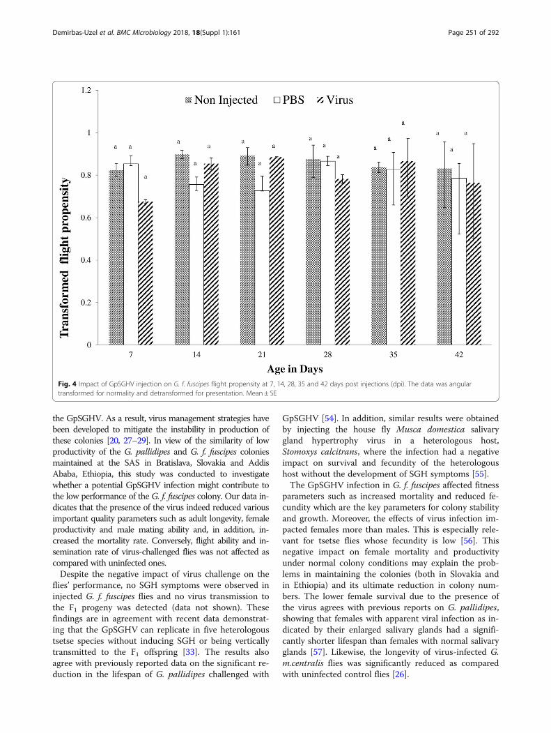
Fig. 4 Impact of GpSGHV injection on G. f. fuscipes flight propensity at 7, 14, 28, 35 and 42 days post injections (dpi). The data was angulartransformed for normality and detransformed for presentation. Mean ± SE
Demirbas-Uzel et al. BMC Microbiology 2018, 18(Suppl 1):161 Page 251 of 292
the GpSGHV. As a result, virus management strategies havebeen developed to mitigate the instability in production ofthese colonies [20, 27–29]. In view of the similarity of lowproductivity of the G. pallidipes and G. f. fuscipes coloniesmaintained at the SAS in Bratislava, Slovakia and AddisAbaba, Ethiopia, this study was conducted to investigatewhether a potential GpSGHV infection might contribute tothe low performance of the G. f. fuscipes colony. Our data in-dicates that the presence of the virus indeed reduced variousimportant quality parameters such as adult longevity, femaleproductivity and male mating ability and, in addition, in-creased the mortality rate. Conversely, flight ability and in-semination rate of virus-challenged flies was not affected ascompared with uninfected ones.Despite the negative impact of virus challenge on the
flies’ performance, no SGH symptoms were observed ininjected G. f. fuscipes flies and no virus transmission tothe F1 progeny was detected (data not shown). Thesefindings are in agreement with recent data demonstrat-ing that the GpSGHV can replicate in five heterologoustsetse species without inducing SGH or being verticallytransmitted to the F1 offspring [33]. The results alsoagree with previously reported data on the significant re-duction in the lifespan of G. pallidipes challenged with
GpSGHV [54]. In addition, similar results were obtainedby injecting the house fly Musca domestica salivarygland hypertrophy virus in a heterologous host,Stomoxys calcitrans, where the infection had a negativeimpact on survival and fecundity of the heterologoushost without the development of SGH symptoms [55].The GpSGHV infection in G. f. fuscipes affected fitness
parameters such as increased mortality and reduced fe-cundity which are the key parameters for colony stabilityand growth. Moreover, the effects of virus infection im-pacted females more than males. This is especially rele-vant for tsetse flies whose fecundity is low [56]. Thisnegative impact on female mortality and productivityunder normal colony conditions may explain the prob-lems in maintaining the colonies (both in Slovakia andin Ethiopia) and its ultimate reduction in colony num-bers. The lower female survival due to the presence ofthe virus agrees with previous reports on G. pallidipes,showing that females with apparent viral infection as in-dicated by their enlarged salivary glands had a signifi-cantly shorter lifespan than females with normal salivaryglands [57]. Likewise, the longevity of virus-infected G.m.centralis flies was significantly reduced as comparedwith uninfected control flies [26].

Fig. 5 Impact of GpSGHV injection on G. f. fuscipes mating ability. a: mating propensity of 3, 6, 9 and 12 day old untreated G. f. fuscipes males; b:Nine day old virgin males from different treatments mated with 9 day old females
Fig. 6 Impact of GpSGHV infection on G. f. fuscipes female insemination rate when mated with a: 3, 6, 9 or 12 day old untreated males;Proportion of empty spermathecae reduced significantly with male age (y = − 0.02165*x + 0.3207, P = 0.005); proportion partially filled (y = −0.01257*x + 0.4139, P = 0.1577) and completely filled increased (y = 0.01040 * x + 0.25526, P = 0.04 between 3 and 12 days post emergence. b: 9-days old virgin males from different treatments
Demirbas-Uzel et al. BMC Microbiology 2018, 18(Suppl 1):161 Page 252 of 292

Demirbas-Uzel et al. BMC Microbiology 2018, 18(Suppl 1):161 Page 253 of 292
Our observed positive correlation between male ageand mating success was in agreement with previously re-ported that in field cages, males younger than 8 daysshowed a significant lower mating ability [43]. Our re-sults showed that 3 day-old males were less successful inmating than older males, but no further significant dif-ference was observed between 6-day old or older males.Similar observations were reported with other species,i.e. 3-day old male G. brevipalpis and G. austeni wereless successful in mating as compared with older males[58]. In other studies, 6–8 day old-males G. p. gambien-sis were used for mating studies [59] and older G.pallidipes males copulated more often than young males[44, 60].The significant reduction of the mating ability of
GpSGHV-challenged male G. f. fuscipes flies is an add-itional negative impact of the presence of the virus. Theobserved reduction in mating success as measured insmall mating cages that mimic well the situation instandard tsetse holding cages, might partly explain thereduction in the females’ fecundity as almost half of thefemales were not inseminated when offered a mating op-portunity with virus-injected males. These results are inagreement with previous studies on the mating perform-ance of G. pallidipes in small laboratory cages [57] or inwalk-in field cages [61]. Our data are also in agreementwith results of Helicoverpa zea males infected with theHz-V2 virus, that were slower in approaching healthy fe-males for mating as compared with non-infected males[62, 63]. This reduction in mating propensity might be aresult of reduced flying and searching activity for femalesor possibly a negative selection by females against in-fected males [64].Our data to imply a different outcome when compared
with the results of Odindo [65] who reported no signifi-cant difference in mating performance between symp-tomatically infected and asymptomatic G. pallidipesflies. In addition, in contrast to our study, Jura andDavies-Cole (1992) speculated that SGHV-infected, andhence sterile, G. pallidipes males showed increased mat-ing competitiveness and concluded that these malescould be used for SIT applications [66]. Although ourand the experiments of Odindo [65] and Jura andDavies-Cole [66] were conducted in similar settings(small laboratory cage), the different results are mostlikely due to the different tsetse species (G. pallidipesversus G. f. fuscipes) populations or strains used in thestudy. However, no data are so far available on the im-pact of the virus in males on the potential selection offemales for mating partners. Further studies on the pres-ence of the virus and its impact on the biological mecha-nisms of mating are necessary.The virus injection has no significant impact on flight
propensity and insemination rate of infected flies. The
absence of a negative impact on the adult flight propen-sity (males and females) observed in this study contra-dicts the finding of Odindo [64] who speculated that thepresence of the virus resulted in reduced physical maleactivity in G. pallidipes. It also contradicts the observa-tion of Burand and Tan [63] who observed that the Hz-1virus makes the H. zea male lazier and slower to move.The reduction in the mating propensity of virus-infectedmales might be due to reduced physical male activity.This might indicate that the physical activity requiredfor the flight propensity test is much less than that re-quired for successful mating and therefore the infectedmales had the propensity to fly but lost the ability toconduct normal mating activity.The absence of any significant impact of the virus in-
fection on insemination rate might be due to the com-pletion of sperm development during the pupal stage inGlossina species prior to the virus challenge of the adultstage. The results contradict earlier data indicating thatvirus infected G. pallidipes males with SGH were unableto successfully inseminate females after mating [21, 61].The difference between our current data and these pub-lished earlier might be due to a different level of virusinfection (virus infected G. f. fuscipes showed no sign ofSGH versus G. pallidipes males with SGH indicating ahigher density of virus particle per flies (> 106) and a dif-ferent tsetse species.
ConclusionsThe data presented in this paper directly demonstratesthe negative impact of GpSGHV infection on the estab-lishment and maintenance of G. f. fuscipes colonies,which will be crucial for the production of sufficientmale flies of adequate biological quality for the applica-tion of the SIT programmes. The combination of in-creased female fly mortality and the reduction in matingpropensity of the virus-infected males will shorten theproduction period and therefore will necessitate an in-crease in colony size to compensate for the loss in pro-duction. Finally, virus-infected males might have a lowercompetitiveness under field conditions, which will re-quire increased release rates. These combined effects ofthe presence of the virus in G. f. fuscipes colonies willimpose serious challenges to mass-rear and produce suf-ficient sterile males of adequate biological quality andwill make the SIT component more expensive and lesscompetitive with other control tactics [67]. Managementstrategies to mitigate the negative effects of virus pres-ence that were based on the use of a clean feeding sys-tem (each fly receives a clean blood meal) and themixing of the blood meals with the antiviral drug valacy-clovir were recently developed for G. pallidipes colonies.However, the implementation of these strategies has sofar been restricted to G. pallidipes colonies where flies

Demirbas-Uzel et al. BMC Microbiology 2018, 18(Suppl 1):161 Page 254 of 292
showed clear SGH symptoms [27–29]. So far, the ab-sence of SGH symptoms in many tsetse species includ-ing G. f. fuscipes has excluded the virus-infection as apossible cause for the poor performance of certain col-onies and consequently no virus management strategieswere implemented. The data presented in this manu-script strongly indicates that colonies that performpoorly should be screened for the presence of the viruswith PCR and in confirmed cases, virus managementstrategies should be implemented even when no SGHsymptoms are observed. Special caution is required inthose tsetse mass-rearing facilities where G. pallidipescolonies are maintained with colonies of other tsetsespecies.
AbbreviationsAAT: animal African trypanosomosis; AW-IPC: area-wide integrated pestmanagement programs; GpSGHV: Glossina pallidipes salivary glandhypertrophy virus; HAT: Human African trypanosomosis; IPCL: Insect PestControl Laboratory; PBS: Phosphate Buffer Saline; qPCR: quantitativepolymerase chain reaction; SAS: Slovak Academy of Sciences; SGHV: salivarygland hypertrophy virus; SIT: Sterile Insect Technique
AcknowledgmentsThe authors acknowledge Carmen Marin, Abdul Hasim Mohamed, EdgardoLapiz and Henry Adun of the Joint FAO/IAEA Programme, Seibersdorf,Austria, for rearing the experimental flies used in this study.
FundingThis work and the publication fees were funded by the Joint FAO/IAEADivision of Nuclear Techniques in Food and Agriculture, IAEA (CRP No.:D4.20.15) Vienna, Austria. This research was partially supported also by theSlovak Research and Development Agency under the contract No. APVV-15-0604 entitled ‘Reduction of fecundity and trypanosomiasis control of tsetseflies by the application of sterile insect techniques and molecular methods’.
Availability of data and materialsMaterial described in the manuscript, including all relevant raw data aredeposited in the following link. https://dataverse.harvard.edu/dataset.xhtml?persistentId=doi:10.7910/DVN/1OVHVD).
About this supplementThis article has been published as part of BMC Microbiology Volume 18Supplement 1, 2018: Enhancing Vector Refractoriness to Trypanosome Infection.The full contents of the supplement are available online at https://bmcmicrobiol.biomedcentral.com/articles/supplements/volume-18-supplement-1.
Authors’ contributionsAMMA, AGP, RLM: designed, supervised the research and writing of themanuscript. GDU, AGP, AMMA: conducted the experiments, collected andanalyzed data and prepared the figs. PT, AGP: Provided live material forexperiments. GDU, AGP, MJBV, JB, PT, RLM: participated in the writing of themanuscript. All authors have read and agreed on the final version on themanuscript.
Ethics approval and consent to participateNot applicable
Consent for publicationNot applicable
Competing interestsThe authors declare that they have no competing interests.
Publisher’s NoteSpringer Nature remains neutral with regard to jurisdictional claims inpublished maps and institutional affiliations.
Author details1Insect Pest Control Laboratory, Joint FAO/IAEA Division of NuclearTechniques in Food & Agriculture, International Atomic Energy Agency,Vienna International Centre, P.O. Box 100, 1400 Vienna, Austria. 2Institute ofChemical, Environmental and Biological Engineering, Research AreaBiochemical Technology, Vienna University of Technology, GumpendorferStraße 1a, 1060 Vienna, Austria. 3Section of Molecular and Applied Zoology,Institute of Zoology, Slovak Academy of Sciences, 845 06 Bratislava, SR,Slovakia. 4Scientica, Ltd., Hybešova 33, 831 06 Bratislava, Slovakia.
Published: 23 November 2018
References1. Hotez PJ, Kamath A. Neglected tropical diseases in sub-saharan Africa:
review of their prevalence, distribution, and disease burden. PLoS Negl TropDis. 2009;3:e412.
2. Gooding RH, Feldmann U, Robinson AS. Care and maintenance of tsetsecolonies. In: Crampton JM, Beard CB, Louis C, editors. The molecular biologyof insect disease vectors: a methods manual. London: Chapman & Hall Ltd;1997. p. 41–55.
3. Aksoy S, Maudlin I, Dale C, Robinson AS, O'Neill SL. Prospects for control ofAfrican trypanosomiasis by tsetse vector manipulation. Trends Parasitol.2001;17:29–35.
4. Rogers DJ, Robinson TP. Tsetse distribution. The trypanosomiases. 2004;2004:139–80.
5. Waiswa C, Picozzi K, Katunguka-Rwakishaya E, Olaho-Mukani W, Musoke RA,Welburn SC. Glossina fuscipes fuscipes in the trypanosomiasis endemic areasof south eastern Uganda: apparent density, trypanosome infection rates andhost feeding preferences. Acta Trop. 2006;99:23–9.
6. Aksoy S, Gibson WC, Lehane MJ. Interactions between tsetse andtrypanosomes with implications for the control of trypanosomiasis. AdvParasitol. 2003;53:1–83.
7. Tirados I, Esterhuizen J, Kovacic V, Mangwiro TC, Vale GA, Hastings I, SolanoP, Lehane MJ, Torr SJ. Tsetse control and Gambian sleeping sickness;implications for control strategy. PLoS Negl Trop Dis. 2015;9:e0003822.
8. Abd-Alla AMM, Bergoin M, Parker A, Maniania NK, Vlak JM, Bourtzis K,Boucias DG, Aksoy S. Improving sterile insect technique (SIT) for tsetse fliesthrough research on their symbionts and pathogens. J Invertebr Pathol.2013;112:S2–S10.
9. Jordan AM. Recent developments in the ecology and methods of control oftsetse flies (Glossina spp.) (Dipt., Glossinidae) - a review. Bull Entomol Res.1974;63:361–99.
10. Green CH. Bait methods for tsetse fly control. Adv Parasitol. 1994;34:229–91.11. Thompson JW, Mitchell M, Rees RB, Shereni W, Schoenfeld AH, Wilson A.
Studies on the efficacy of deltamethrin applied to cattle for the controlof tsetse flies (Glossina spp.) in southern Africa. Trop Anim Health Prod.1991;23:221–6.
12. Vreysen MJB. Principles of area-wide integrated tsetse fly control using thesterile insect technique. Med Trop. 2001;61:397–411.
13. Knipling EF. Potential role of the sterility principle for tsetse eradication.WHO/Vector Control/27. 1963;27:1–17.
14. Hendrichs JP, Kenmore P, Robinson AS, Vreysen MJB. Area-wideintegrated pest management (AW-IPM): principles, practice and prospects.In: Vreysen MJB, Robinson AS, Dordrecht HJ, editors. Area-wide control ofinsect pests: from research to field implementation. The Netherlands:Springer; 2007. p. 3–33.
15. Abd-Alla AMM, Parker AG, Vreysen MJB, Bergoin M. Tsetse salivary glandhypertrophy virus: Hope or hindrance for tsetse control? PLoS Negl TropDis. 2011;5:e1220.
16. Gouteux J-P. Prevalence of enlarged salivary glands in Glossina palpalis, Gpallicera, and G nigrofusca (Diptera: Glossinidae) from the Vavoua area, IvoryCoast. J Med Entomol. 1987;24:268.
17. Jaenson TG. Virus-like rods associated with salivary gland hyperplasia intsetse, Glossina pallidipes. Trans R Soc Trop Med Hyg. 1978;72:234–8.
18. Sang RC, Jura WGZO, Otieno LH, Mwangi RW, Ogaja P. The effects of atsetse DNA virus infection on the functions of the male accessory

Demirbas-Uzel et al. BMC Microbiology 2018, 18(Suppl 1):161 Page 255 of 292
reproductive gland in the host fly Glossina morsitans centralis (Diptera;Glossinidae). Curr Microbiol. 1999;38:349–54.
19. Abd-Alla AMM, Vlak JM, Bergoin M, Maruniak JE, Parker AG, Burand JP, JehleJA, Boucias DG. Hytrosaviridae: a proposal for classification andnomenclature of a new insect virus family. Arch Virol. 2009;154:909–18.
20. Abd-Alla A, Bossin H, Cousserans F, Parker A, Bergoin M, Robinson A.Development of a non-destructive PCR method for detection of the salivarygland hypertrophy virus (SGHV) in tsetse flies. J Virol Methods. 2007;139:143–9.
21. Abd-Alla AMM, Kariithi HM, Parker AG, Robinson AS, Kiflom M, Bergoin M,Vreysen MJB. Dynamics of the salivary gland hypertrophy virus in laboratorycolonies of Glossina pallidipes (Diptera: Glossinidae). Virus Res. 2010;150:103–10.
22. Kariithi HM, Ahmadi M, Parker AG, Franz G, Ros VID, Haq I, Elashry AM, VlakJM, Bergoin M, Vreysen MJB, Abd-Alla AMM. Prevalence and geneticvariation of salivary gland hypertrophy virus in wild populations of thetsetse fly Glossina pallidipes from southern and eastern Africa. J InvertebrPathol. 2013;112:S123–32.
23. Lietze VU, Abd-Alla AMM, Vreysen MJB, Geden CJ, Boucias DG. Salivarygland hypertrophy viruses: a novel group of insect pathogenic viruses.Annu Rev Entomol. 2010;56:63–80.
24. Moloo SK, Zweygarth E, Sabwa CL. Virulence of Trypanosoma simiae in pigsinfected by Glossina brevipalpis, G. pallidipes or G. morsitans centralis. AnnTrop Med Parasitol. 1992;86:681–3.
25. Jura WGZO, Odhiambo TR, Otieno LH, Tabu NO. Gonadal lesions in virus-infected male and female tsetse, Glossina pallidipes (Diptera: Glossinidae). JInvertebr Pathol. 1988;52:1–8.
26. Sang RC, Jura WGZO, Otieno LH, Tukei PM, Mwangi RW. Effects of tsetseDNA virus infection on the survival of a host fly Glossina morsitans centralis(Diptera: Glossinidae). J Invertebr Pathol. 1997;69:253–60.
27. Abd-Alla AMM, Adun H, Parker AG, Vreysen MJB, Bergoin M. The antiviraldrug valacyclovir successfully suppresses salivary gland hypertrophy virus(SGHV) in laboratory colonies of Glossina pallidipes. PLoS One. 2012;7:e38417.
28. Abd-Alla AMM, Kariithi HM, Mohamed AH, Lapiz E, Parker AG, Vreysen MJB.Managing hytrosavirus infections in Glossina pallidipes colonies: feedingregime affects the prevalence of salivary gland hypertrophy syndrome. PLoSOne. 2013;8:e61875.
29. Abd-Alla AMM, Marin C, Parker A, Vreysen M. Antiviral drug valacyclovirtreatment combined with a clean feeding system enhances the suppressionof salivary gland hypertrophy in laboratory colonies of Glossina pallidipes.Parasit Vectors. 2014;7:214.
30. Burtt E. Hypertrophied salivary glands in Glossina: evidence that G. pallidipeswith this abnormality is particularly suited to trypanosome infection. AnnTrop Med Parasitol. 1945;39:11–3.
31. Ellis DS, Maudlin I. Salivary gland hyperplasia in wild caught tsetse fromZimbabwe. Entomologia Experimentalis et Applicata. 1987;45:167–73.
32. Meki IK, Kariithi HM, Ahmadi M, Parker AG, Vreysen MJB, Vlak JM, van Oers MM,Abd-Alla AMM: Hytrosavirus genetic diversity and eco-regional spread inGlossina species. BMC Microbiol. https://doi.org/10.1186/s12866-018-1297-2.
33. Demirbas-Uzel G, Kariithi HM, Parker AG, Vreysen MJB, Mach RL, Abd-AllaAMM. Susceptibility of Tsetse Species to Glossina pallidipes Salivary GlandHypertrophy Virus (GpSGHV). Front Microbiol. 2018;9. https://doi.org/10.3389/fmicb.2018.00701.
34. Alemu T, Kapitano B, Mekonnen S, Aboset G, Kiflom M, Bancha B, WoldeyesG, Bekele K, Feldmann U. Area-wide control of tsetse and trypanosomosis:Ethiopian experience in the southern Rift Valley. In: Vreysen MJB, RobinsonAS, Dordrecht HJ, editors. Area-wide control of insect pests: from researchto field implementation. The Netherlands: Springer; 2007. p. 325–35.
35. Feldmann U, Dyck VA, Mattioli RC, Jannin J. Potential impact of tsetse flycontrol involving the sterile insect technique. In: Dyck VA, Hendrichs J,Dordrecht RAS, editors. Sterile insect technique. Principles and practicein area-wide integrated Pest management. The Netherlands: Springer;2005. p. 701–23.
36. Vreysen MJB, Mebrate A, Menjeta M, Bancha B, Woldeyes G, Musie K, BekeleK, Aboset G. The distribution and relative abundance of tsetse flies in thesouthern Rift Valley of Ethiopia: preliminary survey results. In: Proceedingsof the 25th meeting of the international scientific Council forTrypanosomiasis Research and Control, Mombasa, Kenya, 27 September - 1October 1999. Nairobi: OAU/IBAR; 1999. p. 202–13.
37. Bauer B, Wetzel H. A new membrane for feeding Glossina morsitans Westw.(Diptera: Glossinidae). Bull Entomol Res. 1976;65:563–5.
38. Feldmann U. Guidelines for the rearing of tsetse flies using the membranefeeding technique. In: JPR O’-O, editor. Techniques of insect rearing for the
development of integrated pest and vector management strategies.Nairobi: ICIPE Science Press; 1994. p. 449–71.
39. Boucias DG, Kariithi HM, Bourtzis K, Schneider DI, Kelley K, Miller WJ, ParkerAG, Abd-Alla AMM. Transgenerational transmission of the Glossina pallidipeshytrosavirus depends on the presence of a functional symbiome. PLoS One.2013;8:e61150.
40. Abd-Alla AMM, Cousserans F, Parker A, Bergoin M, Chiraz J, Robinson A.Quantitative PCR analysis of the salivary gland hypertrophy virus (GpSGHV)in a laboratory colony of Glossina pallidipes. Virus Res. 2009;139:48–53.
41. Pagabeleguem S, Seck MT, Sall B, Vreysen MJB, Gimonneau G, Fall AG,Bassene M, Sidibé I, Rayaissé JB, Belem AMG, Bouyer J. Long distancetransport of irradiated male Glossina palpalis gambiensis pupae and itsimpact on sterile male yield. Parasit Vectors. 2015;8:259.
42. Hooper GHS. Application of quality control procedures to large scale rearingof the Mediterranean fruit fly. Entomol Exp Appl. 1987;44:161–7.
43. Abila PP, Kiendrebeogo M, Mutika GN, Parker AG, Robinson AS. The effect ofage on the mating competitiveness of male Glossina fuscipes fuscipes and Gpalpalis palpalis. J Insect Sci. 2003;3:13.
44. Mutika GN, Opiyo E, Robinson AS. Assessing mating performance of maleGlossina pallidipes (Diptera: Glossinidae) using a walk-in field cage. BullEntomol Res. 2001;91:281–7.
45. Pollock JN. Training manual for tsetse control personnel. Vol.1: tsetsebiology, systematics and distribution, techniques. Rome: Food andagricultural Organization of the United Nations; 1982.
46. Nash TAM. The fertilisation of Glossina palpalis in captivity. Bull Entomol Res.1955;46:357–68.
47. Sokal RR, Rohlf FJ. Biometry: the principles and practice of statistics inbiological research. 3rd ed. New York: Freeman and Company; 1995.
48. Core Team R. R Core team (2017). R: a language and environment forstatistical computing. Vienna: R Foundation for Statistical Computing; 2017.
49. Development Core Team. R: a language and environment for statisticalcomputing. Vienna: R Foundation for Statistical Computing; 2008.
50. RStudio. RStudio: Integrated development environment for R (Version 0.96.122) [Computer software]. BostonRetrieved May 20, 2012: RStudio; 2012.
51. Wickham H. ggplot2: elegant graphics for data analysis. New York: Springer; 2009.52. Sarkar D. Lattice: multivariate data visualization with R. New York: Springer;
2008.53. Venables WN, Ripley BD. Modern applied statistics with S. 4th ed. New York:
Springer; 2002.54. Jaenson TGT. Sex ratio distortion and reduced lifespan of Glossina pallidipes
infected with the virus causing salivary gland hyperplasia. Entomol ExpAppl. 1986;41:256–71.
55. Geden C, Garcia-Maruniak A, Lietze VU, Maruniak J, Boucias DG. Impact ofhouse fly salivary gland hyperthrophy virus (MdSGHV) on a heterologoushost, Stomoxys calcitrans. J Med Entomol. 2011;48:1128–35.
56. Leak SGA. Tsetse biology and ecology: their role in the epidemiology andcontrol of trypanosomosis. Wallingford: CABI Publishing; 1998.
57. Jaenson TGT. Mating behaviour of Glossina pallidipes Austen (Diptera,Glossinidae): genetic differences in copulation time between allopatricpopulations. Entomol Exp Appl. 1978;24:100–8.
58. de Beer CJ, Venter GJ, Vreysen MJB. Determination of the optimal matingage of colonised Glossina brevipalpis and Glossina austeni using walk-in fieldcages in South Africa. Parasit Vectors. 2015;8:467.
59. Mutika GN, Kabore I, Seck MT, Sall B, Bouyer J, Parker AG, Vreysen MJB.Mating performance of Glossina palpalis gambiensis strains from BurkinaFaso, Mali, and Senegal. Entomol Exp Appl. 2013;146:177–85.
60. Alphey L, Andreasen M. Dominant lethality and insect population control.Mol Biochem Parasitol. 2002;121:173–8.
61. Mutika GN, Marin C, Parker AG, Vreysen MJB, Boucias DG, Abd-Alla AMM.Impact of salivary gland hypertrophy virus infection on the mating successof male Glossina pallidipes: consequences for the sterile insect technique.PLoS One. 2012;7:e42188.
62. Burand JP, Tan W, Kim W, Nojima S, Roelofs W. Infection with the insectvirus Hz-2v alters mating behavior and pheromone production in femaleHelicoverpa zea moths. J Insect Sci. 2005;5:6.
63. Burand JP, Tan W. Male preference and mating behaviour of maleHelicoverpa zea (Lepidoptera: Noctuidae) infected with the sexuallytransmitted insect virus Hz-2V. Ann Entomol Soc Am. 2006;99:969–73.
64. Odindo MO. Incidence of salivary gland hypertrophy in field populations ofthe tsetse Glossina pallidipes on the South Kenya coast. Insect Sci Appl.1982;3:59–64.

Demirbas-Uzel et al. BMC Microbiology 2018, 18(Suppl 1):161 Page 256 of 292
65. Odindo MO. Glossina pallidipes virus: its potential for use in biologicalcontrol of tsetse. Insect Sci Appl. 1988;9:399–403.
66. Jura WGZO, Davies-Cole JOA. Some aspects of mating behavior ofGlossina morsitans morsitans males infected with a DNA virus. BiolControl. 1992;2:188–92.
67. Hendrichs JP, Vreysen MJB, Enkerlin WR, Cayol JP. Strategic options in usingsterile insects for area-wide integrated pest management. In: Dyck VA,Hendrichs J, Robinson AS, editors. Dordrecht: Springer; 2005. p. 563–600.