Embed Size (px)
Citation preview

ORIGINAL RESEARCH ARTICLEpublished: 17 November 2014
doi: 10.3389/fmicb.2014.00620
Comparative gene expression of Wigglesworthiainhabiting non-infected and Trypanosoma bruceigambiense-infected Glossina palpalis gambiensis fliesIlliassou Hamidou Soumana1, Bernadette Tchicaya1, Gustave Simo2 and Anne Geiger1*
1 UMR 177, Institut de Recherche pour le Développement-CIRAD, Montpellier, France2 Department of Biochemistry, Faculty of Science, University of Dschang, Dschang, Cameroon
Edited by:
Kevin Bradley Clark, Veterans AffairsGreater Los Angeles HealthcareSystem, USA
Reviewed by:
Suleyman Yildirim, Istanbul MedipolUniversity, TurkeyJingwen Wang, Yale University, USA
*Correspondence:
Anne Geiger, UMR 177, IRD-CIRAD,CIRAD TA A-17/G, CampusInternational de Baillarguet,34398 Montpellier, Francee-mail: [email protected]
Tsetse flies (Glossina sp.) that transmit trypanosomes causing human (and animal)African trypanosomiasis (HAT and AAT, respectively) harbor symbiotic microorganisms,including the obligate primary symbiont Wigglesworthia glossinidia. A relationshipbetween Wigglesworthia and tsetse fly infection by trypanosomes has been suggested,as removal of the symbiont results in a higher susceptibility to midgut infection in adultflies. To investigate this relationship and to decipher the role of W. glossinidia in thefly’s susceptibility to trypanosome infection, we challenged flies with trypanosomesand subsequently analyzed and compared the transcriptomes of W. glossinidia fromsusceptible and refractory tsetse flies at three time points (3, 10, and 20 days). More than200 W. glossinidia genes were found to be differentially expressed between susceptibleand refractory flies. The high specificity of these differentially expressed genes makesit possible to distinguish Wigglesworthia inhabiting these two distinct groups of flies.Furthermore, gene expression patterns were observed to evolve during the infectiontime course, such that very few differentially expressed genes were found in commonin Wigglesworthia from the 3-, 10- and 20-day post-feeding fly samples. The overall resultsclearly demonstrate that the taking up of trypanosomes by flies, regardless of whetherflies proceed with the developmental program of Trypanosoma brucei gambiense, stronglyalters gene expression in Wigglesworthia. These results therefore provide a novelframework for studies that aim to decrease or even abolish tsetse fly vector competence.
Keywords: Wigglesworthia, tsetse fly, trypanosomes, tripartite interactions, transcriptome
INTRODUCTIONHuman African trypanosomiasis (HAT), or sleeping sickness,is a neglected vector-borne parasitic disease caused by proto-zoa belonging to the Trypanosoma genus. Two of the subspecies,Trypanosoma brucei gambiense and T. b. rhodesiense, are respon-sible for the chronic form of HAT in western and central Africa,and the acute form of the disease in eastern Africa, respectively(Kennedy, 2008).
Although there are drugs to treat this disease, they have lim-ited effectiveness and produce harmful side effects (Cattand et al.,2001; Barrett, 2006). The repeated use of some drugs has led tothe emergence of resistant forms of different trypanosome species(De Koning, 2001; Matovu et al., 2001). Consequently, the explo-ration for novel strategies must continue, including alternativevector-based strategies (Rio et al., 2004).
To be transmitted, the parasite must first establish itself withinthe insect midgut following an infectious blood meal, and thenmature in the salivary glands or mouthparts (depending onthe trypanosome species) (Vickerman et al., 1988; Van denAbbeele et al., 1999). Tsetse flies are normally refractory to try-panosome infections, and typically exhibit an infection rate ofless than 50%, even under ideal laboratory conditions (Ravel
et al., 2003), while field infection rates rarely exceed 10% of thefly population (Moloo et al., 1986; Frézil and Cuisance, 1994;Maudlin and Welburn, 1994). Furthermore, many infected fliesfail to produce mature parasites and therefore never becomeinfective (Moloo et al., 1986; Dukes et al., 1989; Frézil andCuisance, 1994; Maudlin and Welburn, 1994; Jamonneau et al.,2004). This ability to acquire the parasite, facilitate its matura-tion, and transmit it to a mammalian host is known as vectorcompetence.
Tsetse flies harbor 3 different symbiotic microorganisms(Aksoy, 2000), including Sodalis glossinidius (Cheng and Aksoy,1999; Dale and Maudlin, 1999) and the obligate primary sym-biont Wigglesworthia glossinidia (Aksoy, 1995a; Aksoy et al.,1995). Both are members of the Enterobacteriaceae fam-ily, inhabit the fly’s gut, and are vertically transmitted tothe intrauterine-developing larvae (Cheng and Aksoy, 1999).Wigglesworthia is localized intracellularly in specialized host cellsof the anterior midgut and extracellularly in the female milkglands (Balmand et al., 2013). Sodalis has a broad tissular tropismand can also be found in the hemolymph, salivary glands,etc. (Cheng and Aksoy, 1999). The third symbiont, Wolbachia(O’Neill et al., 1993), belongs to the Rickettsiaceae family and
www.frontiersin.org November 2014 | Volume 5 | Article 620 | 1

Hamidou Soumana et al. Wigglesworthia gene expression profiles
causes cytoplasmic incompatibility in tsetse flies (Alam et al.,2011).
S. glossinidius has been suspected of being involved in thetrypanosome establishment process (Maudlin and Ellis, 1985;Welburn et al., 1993). Epidemiological investigations conductedin several HAT foci in Cameroon have demonstrated an associa-tion between the presence of this symbiont and the establishmentof trypanosome species and subspecies in the fly midgut (Farikouet al., 2010).
Wigglesworthia has coevolved with Glossina species, anddevelops in the fly’s bacteriocytes where it encodes a plethoraof vitamins and biosynthetic products that may promote hostreproduction as well as fly nutrition throughout its develop-ment (Akman et al., 2002; Rio et al., 2012). In the absence ofWigglesworthia, vitamin supplementation of the blood meal canpartially restore host fertility (Nogge, 1976, 1982). The pres-ence of Wigglesworthia is necessary during the larval stages forproper immune development (Weiss et al., 2011, 2012). The pres-ence of larval microbiota also contributes to the development ofthe adult peritrophic matrix (a structure that separates epithelialcells from the lumen content), which regulates immune induc-tion following the trypanosome challenge to the fly (Weiss et al.,2013).
The role of Wigglesworthia in tsetse fly susceptibility to try-panosome infection remains largely unknown. A relationshipbetween Wigglesworthia and trypanosome infection has beensuggested, since removal of the symbiont results in higher suscep-tibility to midgut infection in older, non-teneral flies (Pais et al.,2008; Wang et al., 2009; Snyder and Rio, 2013). It was also previ-ously reported that G. morsitans morsitans (Gmm) demonstratesmuch higher vector competence than G. brevipalpis (Gb) (Harley,1971; Moloo and Kutuza, 1988; Moloo et al., 1994). Recently,comparison of the Wigglesworthia spp. genomes from Gmm andGb revealed metabolomic differences between the primary sym-biont strains harbored by these two fly species (Rio et al., 2012;Snyder and Rio, 2013). One such differences involves the choris-mate, phenylalanine, and folate biosynthetic pathways, which arepresent in Wigglesworthia from Gmm but not Gb. Interestingly,African trypanosomes are auxotrophic for phenylalanine andfolate; their genome, however, encodes the chorismate and folatetransporters, allowing the parasite to absorb these molecules fromtheir environment (Berriman et al., 2005; Jackson et al., 2014).This may explain why Gmm, which harbors a Wigglesworthiastrain capable of providing the trypanosomes with the differ-ent metabolites they are unable to synthesize, is more susceptibleto parasite infection than Gb (Rio et al., 2012; Snyder and Rio,2013).
The mechanisms controlling tsetse fly infection by try-panosomes thus appear to be very complex. Nevertheless,sequencing and annotation of the Wigglesworthia genome fromthe tsetse fly (Akman et al., 2002) has enabled analysis of thegene expression profiles of these bacteria, allowing the iden-tification of genes or pathways involved in specific traits. Inthis context, we have challenged a group of tsetse flies withtrypanosomes and subsequently analyzed the W. glossinidia tran-scriptome from susceptible and refractory tsetse flies at threetime points, in order to identify the differentially expressed genes
between bacteria from these different tsetse fly groups. In addi-tion, we were willing to perform the experiment under strictlythe same conditions as those used to identify the differentiallyexpressed genes of Sodalis (Hamidou Soumana et al., 2014). Theinvestigations on Wigglesworthia transcriptome were, thus, per-formed on the samples that were previously used to analyse theSodalis transcriptome.
MATERIALS AND METHODSETHICS STATEMENTThe experimental protocols involving animals were approvedby the Ethics Committee and the Veterinary Department ofthe Centre International de Recherche Agronomique pour leDéveloppement (CIRAD), Montpellier, France (specific approvalnumbers 12TRYP03, 12TRYP04, and 12TRYP06). The experi-ments were conducted according to internationally recognizedguidelines (French regulation).
T. b. GAMBIENSE STRAINThe S7/2/2 T. b. gambiense strain used in this study was isolatedin 2002 from HAT patients in the Bonon sleeping sickness focus,located in Ivory Coast (Ravel et al., 2006).
EXPERIMENTAL AND SAMPLING PROCEDURESWigglesworthia transcriptome experiments were performed onthe same samples as those previously used to identify the dif-ferential expressed genes of Sodalis (Hamidou Soumana et al.,2014). The main experimental steps are summarized in Figure 1,and described below. All the flies under experiment were fed oninfected or non-infected balb/cj mice.
A set of 100 teneral (less than 32 h old) female G. p. gambiensisflies from the laboratory colony were fed on non-infected mice,and a second set of 900 teneral (less than 32 h old) female fliesfrom the same colony were fed on trypanosome-infected mice.Subsequently, non-gorged flies were removed from the experi-ment. After their blood meal, both gorged flies and control flies,were maintained by feeding on uninfected rabbit, 3 days per week,until the end of each experimental condition (e.g., 3, 10, and 20days post-feeding). Sampling times were chosen according to apreviously determined time course of susceptible fly infection bytrypanosomes (Ravel et al., 2003), so as to compare the expressionprofile of Wigglesworthia at the different stages of the infectionprocess. The 3-day and 10-day sampling times were selectedto target differentially expressed genes involved in early eventsassociated with trypanosome entry into the midgut and withthe establishment of infection, respectively. The 20-day samplingtime point was chosen to target the expression of genes involvedin events occurring relatively late in trypanosome infection timecourse. The first set of flies (uninfected flies) was composed offour groups (four biological replicates), each of seven flies ran-domly chosen. Three days after their blood meal on uninfectedmice, all flies from each group were dissected and the correspond-ing 7 midguts were pooled, yielding 4 pools of 7 midguts. These4 biological replicates were then analyzed separately, by RNAextraction, for their transcriptome. These biological replicates arereferred to as “NS 3 day samples” (NS for “non-stimulated” fliesor flies not exposed to trypanosome infections).
Frontiers in Microbiology | Evolutionary and Genomic Microbiology November 2014 | Volume 5 | Article 620 | 2

Hamidou Soumana et al. Wigglesworthia gene expression profiles
FIGURE 1 | Design depicting experimental infection, sampling procedures, and microarray data analysis.
A stabilate of T. b. gambiense S7/2/2 was thawed at room tem-perature and 0.2 ml was injected intraperitoneally into balb/cjmice, as described in Hamidou Soumana et al. (2014), in orderto feed the second set of flies an infected blood meal. To moni-tor parasitemia, tail blood was examined using a phase-contrastmicroscope at 400× magnification. Teneral (less than 32 h old)female flies were fed on T. b. gambiense-infected mice displayingparasitemia levels ranging from 16 to 64 × 106 parasites/ml. Thisset of flies was then separated into three groups. Three days afteringesting the infected blood meal, the first group was subdividedinto 4 subgroups of 7 flies. All flies from each subgroup were dis-sected separately, and the 7 corresponding midguts were pooled,yielding 4 pools of 7 midguts (i.e., 4 biological replicates), referredto as “S 3 day samples” (S for “stimulated” flies). Following dis-section, the presence of trypanosomes in the blood meal of theseflies was confirmed by microscopy.
The second and third groups consist of flies that were dissected10 and 20 days after feeding on the infected blood meal, respec-tively. Before dissection, the status of each fly (e.g., trypanosomeinfected or non-infected) was controlled by collecting anal dropsfrom each fly, either at 10 (second group) or 20 days (third group)following their blood meal on infected mice. DNA was extractedfrom these anal drops using the Chelex method, as described byRavel et al. (2003), and the presence of trypanosomes was inves-tigated by PCR using the TBR1 and TBR2 primers (Moser et al.,1989). The identification of trypanosomes in anal drops indicates
midgut infections, and flies were considered uninfected if the PCRassays were negative for the anal drops.
Based on these PCR results, tsetse flies from the second andthird groups were subdivided by infection status into a subgroupof flies with trypanosome-infected midguts (e.g., a positive PCRresult for the anal drop test), and a subgroup of flies withoutmidgut infection (e.g., a negative PCR result for the anal droptest). The prevalence of infection in the second group was lessthan 5%, whereas it was greater than 10% in the third group. Fourbiological replicates were processed for each subgroup of the sec-ond and third groups, as described above for “S 3 day samples.”Samples collected 10 days after the blood meal are referred to as“I 10-day samples” and “NI 10-day samples,” and samples col-lected after 20 days are labeled “I 20-day samples” and “NI 20-daysamples” (I and NI signify infected and non-infected flies, respec-tively) (Figure 1). Finally, the experiment included a total of 24samples, consisting of 6 sets of 4 replicates each of 7 flies/7 pooledmidguts; exceptionally, “I 10-day samples” and “NI 10-day sam-ples” only grouped 3 flies, owing to the weak infection prevalenceat the corresponding sampling time.
RNA EXTRACTIONRNA was extracted from the pooled midguts of each biologicalreplicate using TRIzol reagent (Gibco-BRL, France), according tothe manufacturer’s protocol. One previous study has suggestedthat TRIzol extraction may degrade some bacterial RNA samples
www.frontiersin.org November 2014 | Volume 5 | Article 620 | 3

Hamidou Soumana et al. Wigglesworthia gene expression profiles
(Jahn et al., 2008). The study further indicates that reproducibletranscript profiling requires an investigation of RNA integritybefore use, and that RNA Integrity Number values acceptable forsome biological systems may not work well in other biologicalsystems. Thus, the experiments reported here were performed inquadruplicate, and the integrity of each RNA sample (as well asthe absence of contaminating DNA) was checked on an AgilentRNA 6000 Bioanalyzer. The quantification of each RNA samplewas performed using the Agilent RNA 6000 Nano kit (AgilentTechnologies, France).
OLIGONUCLEOTIDE MICROARRAY DESIGNA genome-wide Wigglesworthia transcriptome analysis fromG. p. gambiensis was performed using the Agilent Technologiesoligonucleotide microarray format (www.agilent.com). Thecustom-made density array (8 × 15 K format; AMADID 050087,Agilent Technologies) was designed with 60-mer oligos specific tothe 673 genes of the W. glossinidia chromosome from G. morsitansmorsitans (NCBI Reference Sequence: NC_016893.1) (Akmanet al., 2002). For each gene, 10 different unique probes designedby Hybrigenics (Clermont-Ferrand, France) were used. TheAgilent design uses the uniqueness of probe sequences as one ofthe criteria for probe selection, to avoid cross-hybridization withnon-target genes.
The details of the array design and sample description areavailable at the Gene Expression Omnibus (GEO) under theaccession number GPL18427. The details of the expression dataare available at GEO under the accession number GSE55931.
cDNA PREPARATION AND MICROARRAY HYBRIDIZATIONCy3 dCTP direct cDNA labeling was performed with 100 ng oftotal RNA using the Low Input Quick Amp Labeling Kit One-Color (Agilent Technologies, France). After CyDye-labeled cDNAproduction, samples were carefully matched and hybridizedonto the custom-made whole W. glossinidia Genome OligoMicroarrays. Hybridization was performed at 65◦C for 17 hat 60 rpm. Microarrays were then scanned using an AgilentTechnologies scanner.
MICROARRAY DATA ANALYSIS, GENE ONTOLOGY ANALYSIS ANDDOWNSTREAM ANNOTATIONThe microarray approach has been used to analyse W. glossini-dia gene expression, as its reliability had been validated by qPCRin our previous investigation on Sodalis differential gene expres-sion (Hamidou Soumana et al., 2014). All microarray data areMIAME-compliant, and the raw and normalized data have beendeposited in the MIAME-compliant GEO database (Edgar et al.,2002) (GEO accession number GSE55931). Individual microar-ray quality was evaluated based on the QC report, pair-wiseMA plots, and box plots. The results were extracted using theFeature Extraction software 11.0.1 (Agilent Technologies). Theraw data files were imported to GeneSpring GX (version 12.0,Agilent Technologies), which was used for background adjust-ment, quantile normalization of data (Bolstad et al., 2003; Smythet al., 2005), log-transformation, and gene clustering for the 24microarrays.
Gene ontology was performed only on genes with signif-icant differential expression, using the GeneSpring database.
An unpaired t-test was used for statistical analysis (Qin et al.,2012). A p-value below 0.05 was considered as statistically sig-nificant and indicates significant differences between groups(e.g., between NS 3-day samples/S 3-day samples, between I10-day samples/NI 10-day samples, and between I 20-day sam-ples/NI 20-day samples). The representation of each differentiallyexpressed gene within each ontology category (including cate-gories for molecular function, cellular component, and biologicalprocesses) was measured. A corrected p-value (Benjamini-Yekutelli correction) lower than 0.5 indicates over-representationof the differentially expressed genes within that particularcategory.
Wigglesworthia gene expression data were further analyzed viatwo-dimensional hierarchical clustering, while three-dimensionalclustering of samples was analyzed by principle component anal-ysis (PCA). Hierarchical clustering of differentially expressedgenes was performed using average linkage and the Pearson-centered distance metrics (Claverie, 1999). Gene trees werecreated to group similar genes and to improve visualizationof the data (Butte, 2002). The relationship between genesis represented by a tree in this type of clustering, wherebranch length reflects the degree of similarity between genes.PCA was performed within GeneSpring on infected (or stim-ulated) vs. non-infected (or non-stimulated) conditions atthe different experimental infection times using the list ofgenes.
RESULTSPRINCIPAL COMPONENT AND HIERARCHICAL CLUSTERING ANALYSISOF THE WIGGLESWORTHIA TRANSCRIPTION PROFILESPCA of the differentially expressed Wigglesworthia genes evi-denced two types of results: (a) the ability to discriminate infected(or stimulated) from non-infected (or non-stimulated) flies; and(b) the existence of variability between the replicates (Figure 2).The quality of the discrimination between infected and non-infected flies however depends on the extent of the diversitybetween the repeats. Our results show that PCA can correctly dis-criminate stimulated from non-stimulated flies (3-day sampling;Figure 2A) and infected from non-infected flies at the 20-daysampling (Figure 2C), even though in each case one repeat wasslightly different from the other three. The repeats were even moredispersed at the 10-day sampling, such that the infected sam-ples could not be perfectly discriminated from the non-infectedsamples (Figure 2B).
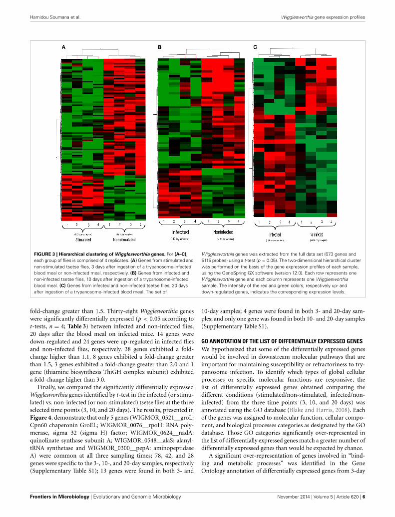
Hierarchical clustering analysis of the transcription profilesdisplayed by Wigglesworthia from stimulated and non-stimulatedflies at 3 days (Figure 3A), and from infected and non-infectedflies at 10 and 20 days (Figures 3B,C, respectively), clearlymade Wigglesworthia expression profiles unique for each group.Clustering of the Wigglesworthia samples was based on the expres-sion levels of: 103 genes with significant differential expressionin stimulated vs. non-stimulated tsetse flies; 61 genes with sig-nificant differential expression in infected vs. non-infected tsetseflies, 10 days after ingesting the trypanosome-infected bloodmeal; and 38 genes with significant differential expression ininfected vs. non-infected tsetse flies, 20 days after ingesting thetrypanosome-infected blood meal.
Frontiers in Microbiology | Evolutionary and Genomic Microbiology November 2014 | Volume 5 | Article 620 | 4

Hamidou Soumana et al. Wigglesworthia gene expression profiles
FIGURE 2 | Principle component analysis (PCA) performed on
differentially expressed Wigglesworthia genes. Two groups containing 4replicates each were compared: samples from infected or stimulated flies(red squares), and from non-infected or non-stimulated flies (blue squares).(A) PCA on differentially expressed Wiggleswortia genes from stimulatedand non-stimulated flies, sampled at day 3 after fly feeding. Stimulated andnon-stimulated flies, were fed on trypanosome-infected and non-infectedmice, respectively; (B) PCA on differentially expressed Wiggleswortia
genes from infected and non-infected flies sampled at day 10 after flyfeeding. (C) PCA on differentially expressed Wiggleswortia genes frominfected and non-infected flies sampled at day 20 after fly feeding. For(B,C), both groups of flies were fed on infected mice; the infected groupcomprised flies in which anal drops were trypanosome-positive, whereasthe non-infected group contained flies in which anal drops weretrypanosome-negative (even though they had ingested an infected bloodmeal).
GENE EXPRESSION PROFILING OF WIGGLESWORTHIA IN TSETSE FLIESTHAT ARE REFRACTORY AND SUSCEPTIBLE TO TRYPANOSOMESMicroarray analysis of the whole Wigglesworthia genomeexpression comparing stimulated (3 days) flies to non-stimulatedflies identified a total of 103 genes with significant differentialexpression (p < 0.05 according to t-tests, n = 4; Table 1). Ofthese genes, 73 were down-regulated and 30 were up-regulatedin stimulated flies and non-stimulated flies, respectively. 79 genes
exhibited a fold-change greater than 1.1 and 2 genes exhibited afold-change greater than 1.5. 61 genes with significant differentialexpression (p < 0.05 according to t-tests, n = 4; Table 2), wereidentified by comparing gene expression profiles of infected andnon-infected flies, 10 days after the blood meal on infected mice.42 genes were down-regulated and 19 genes were up-regulatedin infected flies and non-infected flies, respectively. 54 genesexhibited a fold-change greater than 1.1 and 6 genes exhibited a
www.frontiersin.org November 2014 | Volume 5 | Article 620 | 5
Hamidou Soumana et al. Wigglesworthia gene expression profiles
FIGURE 3 | Hierarchical clustering of Wigglesworthia genes. For (A–C),each group of flies is comprised of 4 replicates. (A) Genes from stimulated andnon-stimulated tsetse flies, 3 days after ingestion of a trypanosome-infectedblood meal or non-infected meal, respectively. (B) Genes from infected andnon-infected tsetse flies, 10 days after ingestion of a trypanosome-infectedblood meal. (C) Genes from infected and non-infected tsetse flies, 20 daysafter ingestion of a trypanosome-infected blood meal. The set of
Wigglesworthia genes was extracted from the full data set (673 genes and5115 probes) using a t-test (p < 0.05). The two-dimensional hierarchical clusterwas performed on the basis of the gene expression profiles of each sample,using the GeneSpring GX software (version 12.0). Each row represents oneWigglesworthia gene and each column represents one Wigglesworthiasample. The intensity of the red and green colors, respectively up- anddown-regulated genes, indicates the corresponding expression levels.
fold-change greater than 1.5. Thirty-eight Wigglesworthia geneswere significantly differentially expressed (p < 0.05 according tot-tests, n = 4; Table 3) between infected and non-infected flies,20 days after the blood meal on infected mice. 14 genes weredown-regulated and 24 genes were up-regulated in infected fliesand non-infected flies, respectively. 38 genes exhibited a fold-change higher than 1.1, 8 genes exhibited a fold-change greaterthan 1.5, 3 genes exhibited a fold-change greater than 2.0 and 1gene (thiamine biosynthesis ThiGH complex subunit) exhibiteda fold-change higher than 3.0.
Finally, we compared the significantly differentially expressedWigglesworthia genes identified by t-test in the infected (or stimu-lated) vs. non-infected (or non-stimulated) tsetse flies at the threeselected time points (3, 10, and 20 days). The results, presented inFigure 4, demonstrate that only 5 genes (WIGMOR_0521__groL:Cpn60 chaperonin GroEL; WIGMOR_0076__rpoH: RNA poly-merase, sigma 32 (sigma H) factor; WIGMOR_0624__nadA:quinolinate synthase subunit A; WIGMOR_0548__alaS: alanyl-tRNA synthetase and WIGMOR_0300__pepA: aminopeptidaseA) were common at all three sampling times; 78, 42, and 28genes were specific to the 3-, 10-, and 20-day samples, respectively(Supplementary Table S1); 13 genes were found in both 3- and
10-day samples; 4 genes were found in both 3- and 20-day sam-ples; and only one gene was found in both 10- and 20-day samples(Supplementary Table S1).
GO ANNOTATION OF THE LIST OF DIFFERENTIALLY EXPRESSED GENESWe hypothesized that some of the differentially expressed geneswould be involved in downstream molecular pathways that areimportant for maintaining susceptibility or refractoriness to try-panosome infection. To identify which types of global cellularprocesses or specific molecular functions are responsive, thelist of differentially expressed genes obtained comparing thedifferent conditions (stimulated/non-stimulated, infected/non-infected) from the three time points (3, 10, and 20 days) wasannotated using the GO database (Blake and Harris, 2008). Eachof the genes was assigned to molecular function, cellular compo-nent, and biological processes categories as designated by the GOdatabase. Those GO categories significantly over-represented inthe list of differentially expressed genes match a greater number ofdifferentially expressed genes than would be expected by chance.
A significant over-representation of genes involved in “bind-ing and metabolic processes” was identified in the GeneOntology annotation of differentially expressed genes from 3-day
Frontiers in Microbiology | Evolutionary and Genomic Microbiology November 2014 | Volume 5 | Article 620 | 6

Hamidou Soumana et al. Wigglesworthia gene expression profiles
Table 1 | Wigglesworthia genes displaying significant differential
expression in trypanosome stimulated vs. non-stimulated flies, 3
days post-blood meal uptake.
Gene Expression GenBank Name
symbols
cspE Down WIGMOR_0606__cspE DNA-binding transcriptionalrepressor
secY Down WIGMOR_0186__secY Preprotein translocasemembrane subunit
groL Down WIGMOR_0521__groL Cpn60 chaperonin GroEL,large subunit of GroESL
Down WIGMOR_0357 MarR family transcriptionalregulator
sucC Down WIGMOR_0345__sucC Succinyl-CoA synthetase,beta subunit
aspS Down WIGMOR_0121__aspS Aspartyl-tRNA synthetase
ycfH Down WIGMOR_0102__ycfH Putative metallodependenthydrolase
rpoC Down WIGMOR_0228__rpoC RNA polymerase, betaprime subunit
motA Down WIGMOR_0035__motA Proton conductorcomponent of flagellamotor
ybgF Down WIGMOR_0352__ybgF SecB-dependent secretoryprotein
flhC Down WIGMOR_0034__flhC Family flagellartranscriptional activator
accC Down WIGMOR_0138__accC Acetyl-CoA carboxylase,biotin carboxylase subunit
yccA Down WIGMOR_0220__yccA Inner membrane protein
lpxA Down WIGMOR_0385__lpxA UDP-N-acetylglucosamineacyltransferase
grpE Down WIGMOR_0116__grpE Pseudo
rpoZ Down WIGMOR_0359__rpoZ RNA polymerase subunitomega
alaS Down WIGMOR_0548__alaS Alanyl-tRNA synthetase
fusA Down WIGMOR_0032__fusA GTP-binding protein chainelongation factor EF-G
Down WIGMOR_0040 Family flagellar basal-bodyP-ring formation protein
mltC Down WIGMOR_0079__mltC Membrane-bound lyticmurein transglycosylase C
thiL Down WIGMOR_0293__thiL Thiamin-monophosphatekinase
znuA Down WIGMOR_0125__znuA Periplasmic component ofa high-affinity zinc uptakeSystem
mrcB Down WIGMOR_0148__mrcB Penicillin-binding protein1B (PBP1B)
gntY Down WIGMOR_0476__gntY Putative gluconatetransport associate protein
tmk Down WIGMOR_0100__tmk Thymidylate kinase
secA Down WIGMOR_0585__secA Preprotein translocasesubunit, ATPase
gyrA Down WIGMOR_0315__gyrA DNA gyrase subunit A
hemF Down WIGMOR_0163__hemF Coproporphyrinogen IIIoxidase
rplP Down WIGMOR_0199__rplP 50S ribosomal protein L16
rpsG Down WIGMOR_0031__rpsG 30S ribosomal protein S7
(Continued)
Table 1 | Continued
Gene Expression GenBank Name
symbols
ftsA Down WIGMOR_0582__ftsA Cell division protein
groS Down WIGMOR_0520__groS Cpn10 chaperonin GroES,small subunit of GroESL
def Down WIGMOR_0262__def Peptide deformylase
Down WIGMOR_0349 TolA family protein
putA Down WIGMOR_0327__putA Transcriptionalrepressor/prolinedehydrogenase/delta-1-pyrroline-5-carboxylatedehydrogenase
serS Down WIGMOR_0269__serS Seryl-tRNA synthetase
ispG Down WIGMOR_0176__ispG 1-hydroxy-2-methyl-2-(E)-butenyl 4-diphosphateSynthase
hslU Down WIGMOR_0502__hslU Molecular chaperone andATPase component ofHslUV protease
thiE Down WIGMOR_0249__thiE Thiamin phosphatesynthase
Down WIGMOR_0310 ncRNA
rpsR Down WIGMOR_0517__rpsR 30S ribosomal protein S18
sucA Down WIGMOR_0343__sucA Thiamin-requiring2-oxoglutaratedecarboxylase
fliN Down WIGMOR_0056__fliN Flagellar motor switchingand energizing component
kdsA Down WIGMOR_0424__kdsA 3-deoxy-D-manno-octulosonate 8-phosphatesynthase
bioC Down WIGMOR_0304__bioC Malonyl-CoAmethyltransferase
fliF Down WIGMOR_0064__fliF Flagellar basal-bodyMS-ring and collar protein
Down WIGMOR_0083 Pyrroline-5-carboxylatereductase
holE Down WIGMOR_0570__holE DNA polymerase IIIsubunit theta
pyrI Down WIGMOR_0154__pyrI Aspartatecarbamoyltransferase,regulatory subunit
tyrS Down WIGMOR_0429__tyrS Tyrosyl-tRNA synthetase
dapF Down WIGMOR_0508__dapF Diaminopimelateepimerase
Down WIGMOR_0443 YhbN family protein
dapA Down WIGMOR_0437__dapA Dihydrodipicolinatesynthase
ydjM Down WIGMOR_0156__ydjM Putative inner membraneprotein
fmt Down WIGMOR_0263__fmt 10-formyltetrahydrofolate:L-methionyl- tRNA(fMet)N-formyltransferase
gpsA Down WIGMOR_0210__gpsA Glycerol-3-phosphatedehydrogenase
recB Down WIGMOR_0512__recB Exonuclease V subunitbeta
pepA Down WIGMOR_0300__pepA Aminopeptidase A
(Continued)
www.frontiersin.org November 2014 | Volume 5 | Article 620 | 7

Hamidou Soumana et al. Wigglesworthia gene expression profiles
Table 1 | Continued
Gene Expression GenBank Name
symbols
folD Down WIGMOR_0539__folD Bifunctional 5,10-methylene-tetrahydrofolatedehydrogenase/5,10-methylene-tetrahydrofolatecyclohydrolase
rpsK Down WIGMOR_0183__rpsK 30S ribosomal protein S11
ileS Down WIGMOR_0483__ileS Isoleucyl-tRNA synthetase
motB Down WIGMOR_0036__motB Family proton-channelcomplex protein
ycfH Down WIGMOR_0102__ycfH Putative metallodependenthydrolase
Down WIGMOR_0595 GltJ familyglutamate/aspartatetransport systempermease protein
rpsI Down WIGMOR_0645__rpsI 30S ribosomal protein S9
rpoH Down WIGMOR_0076__rpoH RNA polymerase, sigma 32(sigma H) factor
Down WIGMOR_0325 tRNA
ribB Down WIGMOR_0545__ribB 3,4-dihydroxy-2-butanone-4-phosphatesynthase
acpP Down WIGMOR_0097__acpP Acyl carrier protein (ACP)
nadA Down WIGMOR_0624__nadA Quinolinate synthasesubunit A
pal Up WIGMOR_0351__pal Peptidoglycan-associatedouter membranelipoprotein
Up WIGMOR_0666 tRNA
yebA Up WIGMOR_0126__yebA Putative peptidase
rplX Up WIGMOR_0195__rplX 50S ribosomal protein L24
yggV Up WIGMOR_0082__yggV dITP/XTP pyrophosphatase
guaB Up WIGMOR_0672__guaB IMP dehydrogenase
Up WIGMOR_0558 tRNA
rplB Up WIGMOR_0203__rplB 50S ribosomal protein L2
hflC Up WIGMOR_0597__hflC Modulator for HflBprotease specific for phagelambda cII repressor
rnhA Up WIGMOR_0072__rnhA Ribonuclease HI
Up WIGMOR_0662 tRNA
sucD Up WIGMOR_0346__sucD Succinyl-CoA synthetasesubunit alpha
purD Up WIGMOR_0245__purD Phosphoribosylglycinamidesynthetasephosphoribosylamine-glycine ligase
metG Up WIGMOR_0214__metG Methionyl-tRNAsynthetase
rpsC Up WIGMOR_0200__rpsC 30S ribosomal protein S3
flgE Up WIGMOR_0044__flgE Flagellar hook protein
zapA Up WIGMOR_0328__zapA Protein that localizes to thecytokinetic ring
fumC Up WIGMOR_0426__fumC Fumarate hydratase
Up WIGMOR_0128 tRNA
Up WIGMOR_0135 tRNA
metK Up WIGMOR_0454__metK Methionineadenosyltransferase 1
(Continued)
Table 1 | Continued
Gene Expression GenBank Name
symbols
infC Up WIGMOR_0086__infC Protein chain initiationfactor IF-3
sucB Up WIGMOR_0344__sucB Dihydrolipoyltranssuccinase
ftsH Up WIGMOR_0553__ftsH Subunit of integralmembrane ATP-dependentzinc metallopeptidase
Up WIGMOR_0134 16S ribosomal RNA
htpX Up WIGMOR_0111__htpX Putative endopeptidase
Up WIGMOR_0237 tRNA
rnhA Up WIGMOR_0072__rnhA Ribonuclease HI
mreB Up WIGMOR_0141__mreB Family actin-like cell wallcomponent
ksgA Up WIGMOR_0022__ksgA S-adenosylmethionine-6-N′,N′-adenosyl (rRNA)dimethyltransferase
Wigglesworthia genes with significant differential expression, p < 0.05.
samples (Table 4). This includes genes that encode proteinsinvolved in protein synthesis as well as chaperonins (Cpn60and Cpn10), which were all down-regulated in stimulated flies.The categories “developmental processes,” “morphogenesis,” and“cellular processes” displayed the greatest proportion of differ-entially expressed genes in the 10-day samples (Table 5), amongwhich both chaperonins (Cpn60 and Cpn10) were up-regulatedin infected flies. Surprisingly, no significant enrichment wasobtained from differentially expressed Wigglesworthia genes in the20-day fly samples.
DISCUSSIONW. glossinidia, the obligate symbiont of the tsetse fly, is involvedin a large portion of physiological events in tsetse, including flysusceptibility or refractoriness to trypanosome infection. Severalmechanisms have previously been reported, such as the modifi-cation of tsetse fly immunity or the supply of different nutrients(Pais et al., 2008; Wang et al., 2009; Rio et al., 2012; Snyder andRio, 2013).
Presently, very few is known on the involvement ofWigglesworthia in tsetse flies vector competence, thus the reasonof our study, a global transcriptomic approach which is the firstone on the W. glossinidia gene differential expression. The analy-ses were performed at 3, 10, and 20 days post-feeding, in orderto target differentially expressed genes involved in early eventsassociated with trypanosome entry into the midgut (3 days sam-pling), with the establishment of infection (10 days sampling),and with events occurring relatively late in trypanosome infectiontime course, respectively.
Among the 673 Wigglesworthia genes, we identified (over thethree sampling points) approximately 200 genes with significantexpression changes in stimulated vs. non-stimulated flies (3-daysampling) and in infected vs. refractory flies (10- and 20-daysamplings). The differences in the levels of gene expression wereusually relatively weak (1.1–1.7-fold over- or under-expression),they nevertheless were statistically significant, suggestive of abiologically meaningful variation.
Frontiers in Microbiology | Evolutionary and Genomic Microbiology November 2014 | Volume 5 | Article 620 | 8

Hamidou Soumana et al. Wigglesworthia gene expression profiles
Table 2 | Wigglesworthia genes displaying significant differential
expression in trypanosome infected vs. non-infected flies, 10 days
post-infected blood meal uptake.
Gene Expression GenBank Name
symbols
mreB Down WIGMOR_0141__mreB Family actin-like cell wallcomponent
lolE Down WIGMOR_0108__lolE Membrane component ofan ABC superfamily outermembrane-specificlipoprotein transporter
Down WIGMOR_0238 tRNA
Down WIGMOR_0236 Translation elongationfactor Tu
yadG Down WIGMOR_0498__yadG Putative ATP-bindingcomponent of an ABCsuperfamily transporter
Down WIGMOR_0160 tRNA
htpX Down WIGMOR_0111__htpX Putative endopeptidase
accD Down WIGMOR_0463__accD Acetyl-CoA carboxylasecarboxyl transferase, betasubunit
kdsA Down WIGMOR_0424__kdsA 3-deoxy-D-manno-octulosonate 8-phosphatesynthase
rplP Down WIGMOR_0199__rplP 50S ribosomal protein L16
clpX Down WIGMOR_0629__clpX ATPase and specificitysubunit of ClpX-ClpPATP-dependent serineprotease
secA Down WIGMOR_0585__secA Preprotein translocasesubunit, ATPase
hslV Down WIGMOR_0501__hslV Peptidase component ofthe HslUV protease
Down WIGMOR_0369 tRNA
Down WIGMOR_0435 tRNA
rpsC Down WIGMOR_0200__rpsC 30S ribosomal protein S3
flgE Down WIGMOR_0044__flgE Flagellar hook protein
gmk Down WIGMOR_0358__gmk Guanylate kinase
nusG Down WIGMOR_0234__nusG Transcription terminationfactor
yadG Down WIGMOR_0498__yadG Putative ATP-bindingcomponent of an ABCsuperfamily transporter
sucB Down WIGMOR_0344__sucB Dihydrolipoyltranssuccinase
pgk Down WIGMOR_0469__pgk Phosphoglycerate kinase
mraW Down WIGMOR_0572__mraW S-adenosyl-dependentmethyltransferase
mreB Down WIGMOR_0141__mreB Family actin-like cell wallcomponent
acpP Down WIGMOR_0097__acpP Acyl carrier protein (ACP)
mdlB Down WIGMOR_0567__mdlB Putative ATP-bindingcomponent of multidrugABC transporter
cysS Down WIGMOR_0538__cysS Cysteinyl-tRNA synthetase
leuS Down WIGMOR_0615__leuS Leucyl-tRNA synthetase
nusG Down WIGMOR_0234__nusG Transcription terminationfactor
mreB Down WIGMOR_0141__mreB Family actin-like cell wallcomponent
(Continued)
Table 2 | Continued
Gene Expression GenBank Name
symbols
atpA Down WIGMOR_0006__atpA F1 sector ofmembrane-bound ATPsynthase, alpha subunit
nadA Down WIGMOR_0624__nadA Quinolinate synthasesubunit A
bioA Down WIGMOR_0307__bioA Adenosylmethionine-8-amino-7-oxononanoateaminotransferase
Down WIGMOR_0549 tRNA
Down WIGMOR_0558 tRNA
ftsA Down WIGMOR_0582__ftsA Cell division protein
fbaA Down WIGMOR_0468__fbaA Fructose-bisphosphatealdolase
atpD Down WIGMOR_0008__atpD F1 sector ofmembrane-bound ATPsynthase, beta subunit
flhC Down WIGMOR_0034__flhC Family flagellartranscriptional activator
alaS Down WIGMOR_0548__alaS Alanyl-tRNA synthetase
Down WIGMOR_0128 tRNA
proS Down WIGMOR_0145__proS Prolyl-tRNA synthetase
rpsQ Down WIGMOR_0197__rpsQ 30S ribosomal protein S17
Down WIGMOR_0458 tRNA
Down WIGMOR_0134 16S ribosomal RNA
Up WIGMOR_0310 ncRNA
ribB Up WIGMOR_0545__ribB 3,4-dihydroxy-2-butanone-4-phosphatesynthase
Up WIGMOR_0310 ncRNA
pnp Up WIGMOR_0564__pnp Polynucleotidephosphorylase/polyadenylase
gyrB Up WIGMOR_0018__gyrB DNA gyrase subunit B
rpsK Up WIGMOR_0183__rpsK 30S ribosomal protein S11
pepA Up WIGMOR_0300__pepA Aminopeptidase A
flhD Up WIGMOR_0033__flhD Family flagellartranscriptional activator
skp Up WIGMOR_0382__skp Periplasmic chaperone
rpoH Up WIGMOR_0076__rpoH RNA polymerase, sigma 32(sigma H) factor
hemL Up WIGMOR_0077__hemL Glutamate-1-semialdehydeaminotransferase
panB Up WIGMOR_0311__panB 3-methyl-2-oxobutanoatehydroxymethyltransferase
groS Up WIGMOR_0520__groS Cpn10 chaperonin GroES,small subunit of GroESL
groL Up WIGMOR_0521__groL Cpn60 chaperonin GroEL,large subunit of GroESL
truA Up WIGMOR_0464__truA tRNA pseudouridinesynthase A
sufS Up WIGMOR_0410__sufS PLP-dependentselenocysteine lyase
prlC Up WIGMOR_0367__prlC Oligopeptidase A
rlmB Up WIGMOR_0605__rlmB 23S rRNAGm2251-methyltransferase
pykA Up WIGMOR_0648__pykA Pyruvate kinase II
Wigglesworthia genes with significant differential expression, p < 0.05.
www.frontiersin.org November 2014 | Volume 5 | Article 620 | 9

Hamidou Soumana et al. Wigglesworthia gene expression profiles
Table 3 | Wigglesworthia genes displaying significant differential
expression in trypanosome-infected vs. non-infected flies, 20 days
post-infected blood meal uptake.
Gene Expression GenBank Name
symbols
cls Down WIGMOR_0400__cls Cardiolipin synthase 1
rpoC Down WIGMOR_0228__rpoC RNA polymerase, betaprime subunit
aroA Down WIGMOR_0272__aroA 3-phosphoshikimate1-Carboxyvinyltransferase
flgC Down WIGMOR_0042__flgC Flagellar component ofcell-proximal portion ofbasal-body rod
yoaE Down WIGMOR_0112__yoaE Hypothetical protein
alaS Down WIGMOR_0548__alaS Alanyl-tRNA synthetase
asnS Down WIGMOR_0515__asnS Asparaginyl tRNAsynthetase
purF Down WIGMOR_0460__purF Amidophosphoribosyl-transferase
thiE Down WIGMOR_0249__thiE Thiamin phosphatesynthase
yadG Down WIGMOR_0498__yadG Putative ATP-bindingcomponent of an ABCsuperfamily transporter
fusA Down WIGMOR_0032__fusA GTP-binding protein chainelongation factor EF-G
groL Down WIGMOR_0521__groL Cpn60 chaperonin GroEL,large subunit of GroESL
metG Down WIGMOR_0214__metG Methionyl-tRNAsynthetase
gpmA Down WIGMOR_0568__gpmA Phosphoglyceromutase
nadA Up WIGMOR_0624__nadA Quinolinate synthasesubunit A
thiH Up WIGMOR_0253__thiH Thiamin biosynthesisThiGH complex subunit
pgi Up WIGMOR_0144__pgi Glucosephosphateisomerase
Up WIGMOR_0290 tRNA
Up WIGMOR_0147 tRNA
rpsB Up WIGMOR_0374__rpsB 30S ribosomal protein S2
pepA Up WIGMOR_0300__pepA Aminopeptidase A
pheS Up WIGMOR_0089__pheS Phenylalanine tRNAsynthetase subunit alpha
hns Up WIGMOR_0399__hns Global DNA-bindingtranscriptional dualregulator H-NS
sufA Up WIGMOR_0406__sufA Fe-S cluster assemblyprotein
Up WIGMOR_0092 Ribonuclease E
ahpC Up WIGMOR_0270__ahpC Alkyl hydroperoxidereductase C22 protein
leuS Up WIGMOR_0615__leuS Leucyl-tRNA synthetase
rnt Up WIGMOR_0432__rnt Ribonuclease T (RNase T)
sufA Up WIGMOR_0406__sufA Fe-S cluster assemblyprotein
pyrG Up WIGMOR_0413__pyrG CTP synthetase
yidC Up WIGMOR_0013__yidC Membrane insertionprotein
rpoH Up WIGMOR_0076__rpoH RNA polymerase, sigma 32(sigma H) Factor
(Continued)
Table 3 | Continued
Gene Expression GenBank Name
symbols
ruvC Up WIGMOR_0122__ruvC Endonuclease componentof RuvABC resolvasome
nrdF Up WIGMOR_0676__nrdF Ferritin-like ribonucleoside-diphosphate reductase 2,beta subunit
bamA Up WIGMOR_0381__bamA Outer membranebeta-barrel proteinassembly factor
sufC Up WIGMOR_0408__sufC Fe-S cluster assemblytransport protein
skp Up WIGMOR_0382__skp Periplasmic chaperone
gshA Up WIGMOR_0534__gshA Gamma-glutamate-cysteine ligase
ftsY Up WIGMOR_0075__ftsY Signal recognition particleprotein
Wigglesworthia genes with significantl differential expression, p < 0.05.
Only five genes were found in common when the list of genesshowing modified expression in Wigglesworthia from 3-day sam-ples was compared to those from 10- and 20-day samples. Thisresult suggests the occurrence of highly complex and specificinteractions that evolve during the time spent following the fly’schallenge with trypanosomes.
PCA of the differentially expressed Wigglesworthia genesappears as a rather poor powerful tool for distinguishingstimulated from non-stimulated flies, as well as infected fromnon-infected flies. In contrast, clustering analysis of the geneexpression data from the samples resulted in a hierarchical treethat discriminates stimulated from non-stimulated flies (3 dayspost-infected blood meal), and infected from non-infected flies(10 and 20 days post-infected blood meal). This clearly indicatesthat the expression of the Wigglesworthia genes not only evolveswith time spent after the fly’s infected blood meal (e.g., 3, 10, or20 days), but that it also depends on the fly’s status (stimulatedor non-stimulated; infected or self-cured). These results are inline with those of Hamidou Soumana et al. (2014), who notedthat trypanosome-responsive Sodalis glossinidius genes interact inwell-defined patterns during the infection time course, making itpossible to distinguish flies that are susceptible from those thatare refractory to parasite infection.
Many of the Wigglesworthia genes that are down-regulatedin trypanosome-stimulated and infected flies are involved inprotein synthesis, such as the genes encoding ribosomal pro-teins, polymerases, elongation factors, etc. This was a some-what surprising finding, as some of these proteins may beinvolved in pathogen survival (Nandan et al., 2002). Strikingly,we also observed that the expression of Cpn60 chaperonin GroEL,Cpn10 chaperonin GroES, and ncRNA (as well as genes impli-cated in the transport of bacterial toxins and in the synthe-sis of thiamine) were down-regulated in Wigglesworthia fromstimulated flies (3 days post-infected blood meal), whereasthe expression of the same three genes was up-regulated inWigglesworthia from infected flies (10 days post-infected bloodmeal).
Frontiers in Microbiology | Evolutionary and Genomic Microbiology November 2014 | Volume 5 | Article 620 | 10

Hamidou Soumana et al. Wigglesworthia gene expression profiles
FIGURE 4 | Schematic representation showing the number of
significant differentially expressed Wigglesworthia genes. Samples arefrom day 3 (stimulated/non-stimulated flies) and days 10 and 20(infected/non-infected flies). The number of differentially expressed genes(specific to the sampling time point) is displayed, as well as the number ofdifferentially expressed genes shared by two or more samples.
Interestingly, it has been reported that acute infection of mam-malian cells with several types of viruses often results in theinduction of heat-shock protein expression (Santoro, 1994), asobserved for the up-regulation of chaperone proteins 24 h afterinfection with DENV (Chen et al., 2011). These studies supportour findings for infected tsetse flies (10 days), in which two up-regulated genes (Cpn60 and Cpn10) from Wigglesworthia encodeproteins involved in protein folding. Along these lines, the expres-sion of a 60 kDa chaperone in the fly midgut was first described byAksoy (1995b) and was reported once again by other researchersin 2002 (Haines et al., 2002). Several hypotheses have been madeby Haines et al. (2002) which could explain the overexpression ofgenes encoding these chaperones:
(i) Chaperones may be required for bacterial survival in thehostile environment of the tsetse midgut.
(ii) In other obligate endosymbionts, non-chaperonin activitieshave been reported for chaperones among which preven-tion of disassembly of invading microbes (Filichkin et al.,1997), protection from proteolytic degradation (Evans et al.,1992). In this context, Wigglesworthia may secrete chaper-ones that could bind proteins produced by the tsetse fly, thusprotecting the trypanosome from the fly immune system.
Enterobacter aerogenes, for example, produces a chaperone thatfunctions as an insect toxin, which contributes to paralyzing theant-lion’s insect prey (Yoshida et al., 2001; Haines et al., 2002).This chaperone contains four key residues (Val 100, Asn 101, Asp338, and Ala 471) that are crucial for toxicity. The Wigglesworthiachaperone possesses three (Val 100, Asn 101, Ala 471) out offour of these crucial residues. This observation suggests that the
W. glossinidia chaperones (Haines et al., 2002) (which have 86%homology with the E. aerogenes chaperone) could also functionas toxins involved in the attrition process of trypanosomes dur-ing the early steps of their developmental cycle (occuring after thefirst 3 days following the infected blood meal).
Down-regulation of chaperone genes 3 days post-infectedblood meal could allow further development of infection. Thisdemonstrates the intricacy of regulations by chaperones, whichdepends on the time spent since the infected blood meal.
The development of trypanosome infection is complex, andseveral different molecules could be involved. For example, wehave found that �(1)-pyrroline-5-carboxylate (P5C) dehydro-genase was down-expressed in tsetse flies stimulated by try-panosomes (3 days after an infected blood meal). The two-stepoxidation of proline is catalyzed by proline oxidase and P5Cdehydrogenase, to produce P5C and glutamate. When exoge-nous proline is supplied by the activities of proline oxidase andP5C reductase (conversion of P5C to proline), then impair-ment of P5C dehydrogenase activity can causes the P5C-prolinecyclization. This proline is oxidized by the proline oxidase-FADcomplex that delivers electrons to the electron transport chainand to O2, leading to mitochondrial reactive oxygen species(ROS) over-production. Coupled activity of proline oxidase andP5C dehydrogenase is therefore important for maintaining ROShomeostasis. In Trypanosoma cruzi, proline is involved in avariety of biological processes that are essential for pathogene-sis (Contreras et al., 1985; Homsy et al., 1989; Martins et al.,2009).
In the case of tsetse flies that were fed on an infected bloodmeal, down-regulation of their P5C dehydrogenase could be dueto a restriction in proline production at day 3 upon trypanosomeingestion. Furthermore, this may decrease the production ofproline-rich proteins like tsetse EP (Haines et al., 2010) (involvedin immunity).
The TolA (Gaspar et al., 2000) and UDP-N-Acetyl-glucosamine Acyltransferase are enzymes involved in thebiosynthetic pathway of the lipopolysaccharide membrane,which acts as a barrier to the entry of antibacterial compounds(Nikaido, 1989). The Glossina scavenger peptidoglycan receptorPGRP-LB, plays a role in the detection and elimination of midguttrypanosomes (Wang et al., 2009). Furthermore, the virulenceof some bacteria was shown to depend on secreted and cell wallpeptidoglycan-associated virulence factors (Zawadzka-Skomialet al., 2006). At 3 days post-infected blood meal, the TolAand UDP-N-Acetyl-glucosamine Acyltransferase genes weredown-regulated in Wigglesworthia from trypanosome-stimulatedflies. This could favor a decrease in the stimulation of the tsetseimmune response and, in turn, promote further establishment oftrypanosomes.
Other proteins have been shown to be down-regulated inflies fed on infected blood meal, such as glycolytic enzymes. Anumber of these proteins, including glyceraldehyde-3-phosphatedehydrogenases (GAPDH), have been found to exhibit non-glycolytic functions which contribute to the ability of severalbacterial pathogens to invade tissues (Boyle and Lottenberg,1997; Tunio et al., 2010). In fact, viral infection usuallycauses the stimulation of glycolytic enzyme production in the
www.frontiersin.org November 2014 | Volume 5 | Article 620 | 11

Hamidou Soumana et al. Wigglesworthia gene expression profiles
Table 4 | Enriched Gene Ontology terms of differentially expressed genesa in trypanosome-stimulated vs. non-stimulated flies, 3 days
post-blood meal uptake.
GO accession GO Termb p-Value Corrected p-valuec Wigglesworthia genes
GO:0044267 Cellular protein metabolic process 6.505E-4 0.2539 rpsK, rplP, rpsC, rplB, fmt, rpsG, fusA, rpsR, groS, groL,rpsI, infC
GO:0030554 Adenyl nucleotide binding 7.432E-4 0.2539 groS, groL, ftsH, secA
GO:0032559 Adenyl ribonucleotide binding 7.432E-4 0.2539 groS, groL, ftsH, secA
GO:0043170 Macromolecule metabolic process 2.837E-4 0.2539 rpsK, rplP, rpsC, rplB, ksgA, rpoC, fmt, rpsG,
GO:004328 fusA, rpoZ, recB, groS, groL, ftsH, rpsI, rnhA, rpoH,infC, rpsR
GO:0005524 ATP binding 7.432E-4 0.2539 groS, groL, ftsH, secA
GO:0044238 Primary metabolic process 6.480E-4 0.25 pyrI, rpsK, rplP, rpsC, rplB, ksgA, rpoC, fmt, gyrA, rpsG,fusA, rpoZ, recB, rpsR, groS, groL, ftsH, nadA, rpsI,rnhA, rpoH, infC, tmk
GO:0005515 Protein binding 3.805E-4 0.25 groL, secA
|GO:0045308
GO:0019538 Protein metabolic process 6.859E-4 0.25 rpsK, rplP, rpsC, rplB, fmt, rpsG, fusA, rpsR,
|GO:0006411 groS, groL, ftsH, rpsI, infC
GO:0071704 Organic substance metabolic process 8.857E-4 0.2689 pyrI, rpsK, rplP, rpsC, rplB, ksgA, rpoC, fmt, gyrA, rpsG,fusA, rpoZ, recB, rpsR, groS, groL, ribB, ftsH, nadA,rpsI, rnhA, rpoH, infC, tmk
GO:0044260 Cellular macromolecule metabolic 0.0012 0.3325 rpsK, rplP, rpsC, rplB, ksgA, rpoC, fmt,
|GO:0034960 Process rpsG, fusA, rpoZ, rpsR, groS, groL, rpsI, rpoH, infC
GO:0001883 Purine nucleoside binding 0.0030 0.4902 fusA, groS, groL, ftsH, secA
aThis set of genes was extracted from the full data set (673 genes and 5115 probes) using a t-test (p < 0.05).bGO terms that were overrepresented in the set of differentially expressed genes.cP-value after application of the Benjamini-Yekutelli correction (P < 0.5).
Table 5 | Enriched Gene Ontology terms of differentially expressed genesa in trypanosome-infected vs. non-infected flies, 10 days post-infected
blood meal uptake.
GO accession GO Termb p-Value Corrected p-valuec Wigglesworthia genes
GO:0048856 Anatomical structure development 1.508E-4 0.0555 mreB
GO:0048869 Cellular developmental process 1.508E-4 0.0555 mreB
GO:0032989 Cellular component morphogenesis 1.508E-4 0.0555 mreB
GO:0009653 Anatomical structure morphogenesis 1.508E-4 0.0555 mreB
GO:0044767 Single-organism developmental process 1.508E-4 0.0555 mreB
GO:0000902 Cell morphogenesis 1.508E-4 0.0555 mreB
|GO:0007148
|GO:0045790
|GO:0045791
GO:0009987 Cellular process 0.001 0.3449 atpA, atpD, mreB, gyrB, rpsK,
|GO:0008151 rpsQ, rplP, rpsC, gmk, flgE,
|GO:0050875 truA, pgk, hslV, groS, groL, ribB, ftsA,nadA, clpX, pykA, rpoH
GO:0032502 Developmental process 0.001 0.3656 mreB
aThis set of genes was extracted from the full data set (673 genes and 5115 probes) using a t-test (p < 0.05).bGO terms that were overrepresented in the set of differentially expressed genes.cP-value after application of the Benjamini-Yekutelli correction (P < 0.5).
midguts of Aedes aegypti infected with chikungunya and dengue-2 viruses (Tchankouo-Nguetcheu et al., 2010). The glycolyticenzyme GAPDH is also involved in the energetic metabolism ofbloodstream trypanosomes, and can be considered as a virulence
factor (Cronín et al., 1989). However, all of these processes do notlikely occur in trypanosome-stimulated flies, since the glycolyticenzymes were shown to be down-regulated 3 days after flies werechallenged with trypanosomes.
Frontiers in Microbiology | Evolutionary and Genomic Microbiology November 2014 | Volume 5 | Article 620 | 12

Hamidou Soumana et al. Wigglesworthia gene expression profiles
Therefore, our results show that trypanosome ingestionstrongly alters gene expression of Wigglesworthia, even thoughthe flies do not proceed with the developmental program of T. b.gambiense.
Ten days following an infected blood meal, the expression ofseveral genes, such as quinolinate synthase, were down-regulatedin Wigglesworthia from infected flies (as compared to non-infected flies). This enzyme allows the synthesis of quinolinicacid, a toxic molecule that has been detected in the central ner-vous system of patients displaying AIDS and meningitis (Eadset al., 1997).
In previous investigations, Weiss et al. (2011) showed thatWigglesworthia must be present during the immature larval stagesfor the tsetse fly immune system to develop and function prop-erly at the adult stage. The artificial elimination of Wigglesworthiafrom larvae will compromise the fly immune system developmentthroughout the development of the flies and the adult tsetse flieswill display an immature immune system. In turn susceptibility togut trypanosome infection in adult flies will be increased; in con-trast, adult flies carrying Wigglesworthia will be highly resistant(Wang et al., 2009).
In addition, it has been suggested that differences in the sus-ceptibility between Gmm and Gb are in turn caused by differencesin the shikimate biosynthetic capabilities (phenylanine, folate andchorismate) of the Wigglesworthia strain they harbor (Rio et al.,2012). Our analyses failed to reveal such a role of the correspond-ing pathway in the vector competence of G. p. gambiensis forT. b. gambiense. Indeed, genes involved in this pathway were notfound to be differentially expressed in stimulated flies at 3 daysor in infected flies at 10 days, whereas some genes were foundto be down-expressed (such as aro A) in infected flies at 20 dayspost-feeding.
Our study showed that, in trypanosomes stimulated flies(3 days post-infected bloodmeal), the expression of someWigglesworthia genes are downregulated. This observation meansthat, despite its crucial role in fly resistance to trypanosome (asdemonstrated by Wang et al., 2009; Weiss et al., 2011), the resis-tance level can be modulated by external factors which could favorfurther establishment of trypanosome. In the case of infected fliesone of these external factors could be the trypanosome itself,against which the symbiont was unable to protect its host fly.
So the involvement of Wigglesworthia symbionts and flyimmune system in tsetse fly infection by trypanosomes seemsto be very complex; it is necessary but not sufficient to ensurefly resistance as demonstrated by the success of fly (carry-ing Wigglesworthia) infections performed out by number ofinvestigators.
At the 3-day sampling time, we compared the gene expres-sion of Wigglesworthia harbored within flies that ingested anon-infected blood meal to that of the symbiont of flies thatingested an infected blood meal. The blood of mice infected bytrypanosomes may be quite different from non-infected mouseblood, as it transports a high concentration of trypanosomesbut also may contain a range of other molecules (trypanosome-secreted proteins and other metabolites, mice reactive or degrada-tion products, etc.). Thus, the question: besides the trypanosomeitself, do these molecules, if any, that are not present in the
non-infected blood meal ingested by control flies, contribute toalter the gene expression of the symbiont from flies having gotthe infected meal? In which extend? There is no response tothis question. However, and whatever it may be, the altered geneexpression of the symbiont from flies fed upon an infected mealis directly caused by the trypanosome presence, and possibly viamolecules that it produced (or induced) during its developmentwithin the mammalian host. This question does not raise regard-ing the 10- and 20-day samples, as both groups of flies undercomparison ingested the same first infected blood meal. Despitethis similarity, the gene expressions differed once the flies ofone group became trypanosome-infected (susceptible flies) andthat of the second group self-cured the ingested trypanosomes(refractory flies).
Surprisingly, even genes encoding ribosomal proteins were dif-ferentially expressed; more surprisingly was that some of thesegenes were up-regulated whereas others were down-regulated. Forexample, in stimulated flies (S3) vs. non-stimulated flies (NS3),50S ribosomal protein L2 and L24, and 30S ribosomal proteinS3 were over-expressed whereas 50S ribosomal protein L16, and30S ribosomal protein S7, S9, S11, and S18 were down regu-lated. Similar results were recorded especially in infected flies vs.refractory flies in day 10 samples (I10 vs. NI10). Similar resultshave been reported by Wang et al. (2013) on roots of Arabidopsiseither phosphate- or iron-deficient vs. control roots. The authorsconsidered the resulting alteration in ribosome composition as“a mechanism by which plants adapt to changing environmen-tal conditions.” In our “model,” the stress is caused by the flyingested trypanosomes, and results, among others, in differencesin expression of genes encoding ribosomal proteins. Are thesemodifications involved in W. glosssinidia adaptation to changingenvironment caused by trypanosome invasion?
Finally, our results demonstrate that some genes considered ashouse-keeping genes are differentially expressed although theirexpression usually showed little variation. Fly infection cannotbe compared with a classical physiological progression whichcould possibly account for the induction of unusual variations,even in the expression of such genes. Some of these genes alsoencode proteins displaying alternative functions (GAPDH, forexample); alternative gene expression regulation may thereforeoccur regarding these genes.
The final objective of the global investigation we have under-taken is to decipher the molecular cross-talk between the tsetse fly,its symbionts, Sodalis and Wigglesworthia, and the trypanosome,in order to identify genes controlling crucial steps of fly infectionby the parasite. The present study is the second step of this globalinvestigation we have begun with the differential expression ofSodalis genes performed on the same biological samples. The lev-els of Wiggleworthia differentially expressed genes were lower thanthose recorded for Sodalis differentially expressed genes. Possiblythe “flexibility” of gene expression is lesser in Wigglesworthiathen in other bacteria; as an obligate symbiont, its fundamen-tal physiological characteristics cannot be drastically modifiedotherwise it would no more fulfill its crucial role allowing flysurvival. Nevertheless, differential expression of Wigglesworthiagenes has been recorded, the corresponding genes identified andGO annotation performed. The results clearly demonstrate fly
www.frontiersin.org November 2014 | Volume 5 | Article 620 | 13

Hamidou Soumana et al. Wigglesworthia gene expression profiles
infection by trypanosomes to affect the symbiont transcriptomicmachinery. Further investigations are necessary to assess theinvolvement of some of these differentially expressed genes in theparasite development process, and to evaluate their significance inthe frame of an anti-vector competence strategy for combattingsleeping sickness.
ACKNOWLEDGMENTSThe authors thank the “Région Languedoc-Roussillon—Appeld’Offre Chercheur d’Avenir 2011,” the “Service de Coopération etd’Action Culturelle de l’Ambassade de France au Niger,” and the“Institut de Recherche pour le Développement” for their finan-cial support. I. Hamidou Soumana is a PhD student supportedby the Niger French Embassy, Service de Coopération et d’ActionCulturelle (SCAC).
SUPPLEMENTARY MATERIALThe Supplementary Material for this article can be foundonline at: http://www.frontiersin.org/journal/10.3389/fmicb.2014.00620/abstract
REFERENCESAkman, L., Yamashita, A., Watanabe, H., Oshima, K., Shiba, T., Hattori,
M., et al. (2002). Genome sequence of the endocellular obligate symbiontof tsetse, Wigglesworthia glossinidia. Nat. Genet. 32, 402–407. doi: 10.1038/ng986
Aksoy, S. (1995a). Wigglesworthia gen. nov. and Wigglesworthia glossinidia sp.nov., taxa consisting of the mycetocyte-associated, primary endosymbiontsof tsetse flies. Int. J. Syst. Bacteriol. 45, 848–851. doi: 10.1099/00207713-45-4-848
Aksoy, S. (1995b). Molecular analysis of the endosymbionts of tsetse flies: 16SrDNA locus and over-expression of a chaperonin. Insect Mol. Biol. 4, 23–29. doi:10.1111/j.1365-2583.1995.tb00004.x
Aksoy, S. (2000). Tsetse-a haven for microorganisms. Parasitol. Today 16, 114–118.doi: 10.1016/S0169-4758(99)01606-3
Aksoy, S., Pourhosseini, A. A., and Chow, A. (1995). Mycetome endosymbionts oftsetse flies constitute a distinct lineage related to Enterobacteriaceae. Insect Mol.Biol. 4, 15–22. doi: 10.1111/j.1365-2583.1995.tb00003.x
Alam, U., Medlock, J., Brelsfoard, C., Pais, R., Lohs, C., Balmand, S., et al. (2011).Wolbachia symbiont infections induce strong cytoplasmic incompatibility inthe tsetse fly Glossina morsitans. PLoS Pathog. 7:e1002415. doi: 10.1371/jour-nal.ppat.1002415
Balmand, S., Lohs, C., Aksoy, S., and Heddi, A. (2013). Tissue distributionand transmission routes for the tsetse fly endosymbionts. J. Invertebr. Pathol.112(Suppl.), S116–S122. doi: 10.1016/j.jip.2012.04.002
Barrett, M. P. (2006). The rise and fall of sleeping sickness. Lancet 367, 1377–1378.doi: 10.1016/S0140-6736(06)68591-7
Berriman, M., Ghedin, E., Hertz-Fowler, C., Blandin, G., Renauld, H.,Bartholomeu, D. C., et al. (2005). The genome of the African trypanosomeTrypanosoma brucei. Science 309, 416–422. doi: 10.1126/science.1112642
Blake, J. A., and Harris, M. A. (2008). The Gene Ontology (GO) project:structured vocabularies for molecular biology and their application togenome and expression analysis. Curr. Protoc. Bioinformatics 7:7.2. doi:10.1002/0471250953.bi0702s23
Bolstad, B. M., Irizarry, R. A., Astrand, M., and Speed, T. P. (2003). A compari-son of normalization methods for high density oligonucleotide array data basedon variance and bias. Bioinformatics 19, 185–193. doi: 10.1093/bioinformat-ics/19.2.185
Boyle, M. D., and Lottenberg, R. (1997). Plasminogen activation by invasive humanpathogens. Thromb. Haemost. 77, 1–10.
Butte, A. (2002). The use and analysis of microarray data. Nat. Rev. Drug Discov. 1,951–960. doi: 10.1038/nrd961
Cattand, P., Jannin, J., and Lucas, P. (2001). Sleeping sickness surveillance: anessential step towards elimination. Trop. Med. Int. Health 6, 348–361. doi:10.1046/j.1365-3156.2001.00669.x
Chen, T. H., Tang, P., Yang, C. F., Kao, L. H., Lo, Y. P., Chuang, C. K., et al. (2011).Antioxidant defense is one of the mechanisms by which mosquito cells survivedengue 2 viral infection. Virology 410, 410–417. doi: 10.1016/j.virol.2010.12.013
Cheng, Q., and Aksoy, S. (1999). Tissue tropism, transmission and expression offoreign genes in vivo in midgut symbionts of tsetse flies. Insect Mol. Biol. 8,125–132. doi: 10.1046/j.1365-2583.1999.810125.x
Claverie, J. M. (1999). Computational methods for the identification of differ-ential and coordinated gene expression. Hum. Mol. Genet. 8, 1821–1832. doi:10.1093/hmg/8.10.1821
Contreras, V. T., Morel, C. M., and Goldenberg, S. (1985). Stage specific geneexpression precedes morphological changes during Trypanosoma cruzi meta-cyclogenesis. Mol. Biochem. Parasitol. 4, 83–96. doi: 10.1016/0166-6851(85)90108-2
Cronín, C. N., Nolan, D. P., and Voorheis, H. P. (1989). The enzymes of theclassical pentose phosphate pathway display differential activities in procyclicand bloodstream forms of Trypanosoma brucei. FEBS Lett. 244, 26–30. doi:10.1016/0014-5793(89)81154-8
Dale, C., and Maudlin, I. (1999). Sodalis gen. nov. and Sodalis glossinidius sp.nov., a microaerophilic secondary endosymbiont of the tsetse fly Glossina mor-sitans morsitans. Int. J. Syst. Bacteriol. 49, 267–275. doi: 10.1099/00207713-49-1-267
De Koning, H. P. (2001). Transporters in African trypanosomes: role in drugaction and resistance. Int. J. Parasitol. 31, 512–522. doi: 10.1016/S0020-7519(01)00167-9
Dukes, P., Kaukas, A., Hudson, K. M., Asonganyi, T., and Gashumba, J. K. (1989).A new method for isolating Trypanosoma brucei gambiense from sleepingsickness patients. Trans. R. Soc. Trop. Med. Hyg. 83, 636–639. doi: 10.1016/0035-9203(89)90379-9
Eads, J. C., Ozturk, D., Wexler, T. B., Grubmeyer, C., and Sacchettini, J. C. (1997).A new function for a common fold: the crystal structure of quinolinic acidphosphoribosyltransferase. Structure 5, 47–58. doi: 10.1016/S0969-2126(97)00165-2
Edgar, R., Domrachev, M., and Lash, A. E. (2002). Gene Expression Omnibus:NCBI gene expression and hybridization array data repository. Nucleic AcidsRes. 30, 207–210. doi: 10.1093/nar/30.1.207
Evans, D. J. Jr., Evans, D. G., Engstrand, L., and Graham, D. Y. (1992).Urease-associated heat shock protein of Helicobacter pylori. Infect. Immun. 60,2125–2127
Farikou, O., Njiokou, F., Mbida Mbida, J. A., Njitchouang, G. R., Nana Djeunga,H., Asonganyi, T., et al. (2010). Tripartite interactions between tsetse flies,Sodalis glossinidius and trypanosomes-an epidemiological approach in two his-torical human African trypanosomiasis foci in Cameroon. Infect. Genet. Evol.10, 115–121. doi: 10.1016/j.meegid.2009.10.008
Filichkin, S. A., Brumfield, S., Filichkin, T. P., and Young, M. J. (1997). In vitrointeractions of the aphid endosymbiotic SymL chaperonin with barley yellowdwarf virus. J. Virol. 71, 569–577.
Frézil, J. L., and Cuisance, D. (1994). Trypanosomiasis, diseases with future:prospects and uncertainty. Bull. Soc. Pathol. Exot. 87, 391–393.
Gaspar, J. A., Thomas, J. A., Marolda, C. L., and Valvano, M. A. (2000). Surfaceexpression of O-specific lipopolysaccharide in Escherichia coli requires thefunction of the TolA protein. Mol. Microbiol. 38, 262–275. doi: 10.1046/j.1365-2958.2000.02094.x
Haines, L. R., Haddow, J. D., Aksoy, S., Gooding, R. H., and Pearson, T. W.(2002). The major protein in the midgut of teneral Glossina morsitans morsitansis a molecular chaperone from the endosymbiotic bacterium Wigglesworthiaglossinidia. Insect Biochem. Mol. Biol. 32, 1429–1438. doi: 10.1016/S0965-1748(02)00063-2
Haines, L. R., Lehane, S. M., Pearson, T. W., and Lehane, M. J. (2010). Tsetse EPprotein protects the fly midgut from trypanosome establishment. PLoS Pathog.6:e1000793. doi: 10.1371/journal.ppat.1000793
Hamidou Soumana, I., Loriod, B., Ravel, S., Tchicaya, B., Simo, G., Rihet, P., et al.(2014). The transcriptional signatures of Sodalis glossinidius in the Glossinapalpalis gambiensis flies negative for Trypanosoma brucei gambiense contrastwith those of this symbiont in tsetse flies positive for the parasite: possibleinvolvement of a Sodalis-hosted prophage in fly Trypanosoma refractoriness?Infect. Genet. Evol. 24, 41–56. doi: 10.1016/j.meegid.2014.03.005
Harley, J. M. (1971). Comparison of the susceptibility of infection withTrypanosoma rhodesiense of Glossina pallidipes, G. morsitans, G. fuscipes and G.brevipalpis. Ann. Trop. Med. Parasitol. 65, 185–189.
Frontiers in Microbiology | Evolutionary and Genomic Microbiology November 2014 | Volume 5 | Article 620 | 14

Hamidou Soumana et al. Wigglesworthia gene expression profiles
Homsy, J. J., Granger, B., and Krassner, S. M. (1989). Some factors inducing for-mation of metacyclic stages of Trypanosoma cruzi. J. Protozool. 36, 150–153. doi:10.1111/j.1550-7408.1989.tb01063.x
Jackson, A. P., Sanders, M., Berry, A., McQuillan, J., Aslett, M. A., Quail, M. A.,et al. (2014). The genome sequence of Trypanosoma brucei gambiense, causativeagent of chronic human african trypanosomiasis. PLoS Negl. Trop. Dis. 4:e658.doi: 10.1371/journal.pntd.0000658
Jahn, C. E., Charkowski, A. O., and Willis, D. K. (2008). Evaluationof isolation methods and RNA integrity for bacterial RNA quanti-tation. J. Microbiol. Methods. 75, 318–324. doi: 10.1016/j.mimet.2008.07.004
Jamonneau, V., Ravel, S., Koffi, M., Kaba, D., Zeze, D. G., Ndri, L., et al. (2004).Mixed infections of trypanosomes in tsetse and pigs and their epidemiologi-cal significance in a sleeping sickness focus of Cote d’Ivoire. Parasitology 129,693–702. doi: 10.1017/S0031182004005876
Kennedy, P. G. (2008). The continuing problem of human African trypanoso-miasis (sleeping sickness). Ann. Neurol. 64, 116–126. doi: 10.1002/ana.21429
Martins, R. M., Covarrubias, C., Rojas, R. G., Silber, A. M., and Yoshida, N.(2009). Use of L- proline and ATP production by Trypanosoma cruzi metacyclicforms as requirements for host cell invasion. Infect. Immun. 77, 3023–3032. doi:10.1128/IAI.00138-09
Matovu, E., Seebeck, T., Enyaru, J. C. K., and Kaminsky, R. (2001). Drug resis-tance in Trypanosoma brucei spp., the causative agents of sleeping sickness inman and nagana in cattle. Microbes Infect. 3, 763–770. doi: 10.1016/S1286-4579(01)01432-0
Maudlin, I., and Ellis, D. S. (1985). Association between intracellular Rickettsia-like infections of midgut cells and susceptibility to trypanosome infec-tion in Glossina spp. Z. Parasitenkd. 71, 683–687. doi: 10.1007/BF00925601
Maudlin, I., and Welburn, S. C. (1994). Maturation of trypanosome infections intsetse. Exp. Parasitol. 79, 202–205. doi: 10.1006/expr.1994.1081
Moloo, S. K., Asonganyi, T., and Jenni, L. (1986). Cyclical development ofTrypanosoma brucei gambiense from cattle and goats in Glossina. Acta Trop. 43,407–408.
Moloo, S. K., Kabata, J. M., and Sabwa, C. L. (1994). A study on the mat-uration of procyclic Trypanosoma brucei brucei in Glossina morsitans cen-tralis and G. brevipalpis. Med. Vet. Entomol. 8, 369–374. doi: 10.1111/j.1365-2915.1994.tb00100.x
Moloo, S. K., and Kutuza, S. B. (1988). Comparative study on the susceptibilityof different Glossina species to Trypanosoma brucei brucei infection. Trop. Med.Parasitol. 39, 211–213.
Moser, D. R., Cook, G. A., Ochs, D. E., Bailey, C. P., McKane, M. R.,and Donelson, J. E. (1989). Detection of Trypanosoma congolense andTrypanosoma brucei subspecies by DNA amplification using the poly-merase chain reaction. Parasitology 99, 57–66. doi: 10.1017/S0031182000061023
Nandan, D., Yi, T., Lopez, M., Lai, C., and Reiner, N. E. (2002). Leishmania EF-1alpha activates the Src homology 2 domain containing tyrosine phosphataseSHP-1 leading to macrophage deactivation. J. Biol. Chem. 277, 50190–50197.doi: 10.1074/jbc.M209210200
Nikaido, H. (1989). Outer membrane barrier as a mechanism of antimi-crobial resistance. Antimicrob. Agents Chemother. 33, 1831–1836. doi:10.1128/AAC.33.11.1831
Nogge, G. (1976). Sterility in tsetse flies (Glossina morsitans Westwood)caused by loss of symbionts. Experentia 32, 995–996. doi: 10.1007/BF01933932
Nogge, G. (1982). Significance of symbionts for the maintenance of an optionalnutritional state for successful reproduction in hematophagous arthropods.Parasitology 82, 299–304.
O’Neill, S. L., Gooding, R. H., and Aksoy, S. (1993). Phylogenetically distant sym-biotic microorganisms reside in Glossina midgut and ovary tissues. Med. Vet.Entomol. 7, 377–383.
Pais, R., Lohs, C., Wu, Y. N., Wang, J. W., and Aksoy, S. (2008). The obligate mutual-ist Wigglesworthia glossinidia influences reproduction, digestion, and immunityprocesses of its host, the tsetse fly. Appl. Environ. Microbiol. 74, 5965–5974. doi:10.1128/AEM.00741-08
Qin, X. Y., Kojima, Y., Mizuno, K., Ueoka, K., Muroya, K., Miyado, M., et al.(2012). Identification of novel low-dose bisphenol a targets in human foreskin
fibroblast cells derived from hypospadias patients. PLoS ONE 7:e36711. doi:10.1371/journal.pone.0036711
Ravel, S., Grébaut, P., Cuisance, D., and Cuny, G. (2003). Monitoring the develop-mental status of Trypanosoma brucei gambiense in the tsetse fly by means of PCRanalysis of anal and saliva drops. Acta Trop. 88, 161–165. doi: 10.1016/S0001-706X(03)00191-8
Ravel, S., Patrel, D., Koffi, M., Jamonneau, V., and Cuny, G. (2006). Cyclicaltransmission of Trypanosoma brucei gambiense in Glossina palpalis gambien-sis displays great differences among field isolates. Acta Trop. 100, 151–155. doi:10.1016/j.actatropica.2006.09.011
Rio, R. V., Hu, Y., and Aksoy, S. (2004). Strategies of the home-team: symbiosesexploited for vector-borne disease control. Trends Microbiol. 12, 325–336. doi:10.1016/j.tim.2004.05.001
Rio, R. V. M., Symula, R. E., Wang, J., Lohs, C., Wu, Y.-N., Snyder,A. K., et al. (2012). Insight into the transmission biology and species-specific functional capabilities of tsetse (Diptera: Glossinidae) obligatesymbiont Wigglesworthia. MBio 3, e00240–e00211. doi: 10.1128/mBio.00240-11
Santoro, M. G. (1994). Heat shock proteins and virus replication: hsp70s as medi-ators of the antiviral effects of prostaglandins. Experientia 50, 1039–1047. doi:10.1007/BF01923459
Smyth, G. K., Michaud, J., and Scott, H. S. (2005). Use of within-array replicate spots for assessing differential expression in microarrayexperiments. Bioinformatics 21, 2067–2075. doi: 10.1093/bioinformatics/bti270
Snyder, A. K., and Rio, R. V. (2013). Interwoven biology of the tsetse holobiont.J. Bacteriol. 195, 4322–4330. doi: 10.1128/JB.00487-13
Tchankouo-Nguetcheu, S., Khun, H., Pincet, L., Roux, P., Bahut, M., Huerre,M., et al. (2010). Differential protein modulation in midguts of Aedes aegyptiinfected with chikungunya and dengue 2 viruses. PLoS ONE 5:10. doi:10.1371/journal.pone.0013149
Tunio, S. A., Oldfield, N. J., Berry, A., Ala’Aldeen, D. A., Wooldridge, K. G., andTurner, D. P. (2010). The moonlighting protein fructose-1, 6-bisphosphatealdolase of Neisseria meningitidis: surface localization and role in hostcell adhesion. Mol. Microbiol. 76, 605–615. doi: 10.1111/j.1365-2958.2010.07098.x
Van den Abbeele, J., Claes, Y., Bockstaele, D., Ray, D., and Coosemans, M. (1999).Trypanosoma brucei spp. development in the tsetse fly: characterization of thepost-mesocyclic stages in the foregut and proboscis. Parasitology 118, 469–478.doi: 10.1017/S0031182099004217
Vickerman, K., Tetley, L., Hendry, A., and Turner, C. M. (1988). Biology ofAfrican trypanosomes in the tsetse fly. Biol. Cell 64, 109–119. doi: 10.1016/0248-4900(88)90070-6
Wang, J., Lan, P., Gao, H., Zheng, L., Li, W., and Schmidt, W. (2013).Expression changes of ribosomal proteins in phosphate- and iron-deficient Arabidopsis roots predict stress-specific alterations inribosome composition. BMC Genomics. 14:783. doi: 10.1186/1471-2164-14-783
Wang, J., Wu, Y., Yang, G., and Aksoy, S. (2009). Interactions between mutualistWigglesworthia and tsetse peptidoglycan recognition protein (PGRP-LB) influ-ence trypanosome transmission. Proc. Natl. Acad. Sci. U.S.A. 106, 12133–12138.doi: 10.1073/pnas.0901226106
Weiss, B. L., Maltz, M., and Aksoy, S. (2012). Obligate symbionts activateimmune system development in the tsetse fly. J. Immunol. 188, 3395–3403. doi:10.4049/jimmunol.1103691
Weiss, B. L., Wang, J., Maltz, M. A., Wu, Y., and Aksoy, S. (2013). Trypanosomeinfection establishment in the tsetse fly gut is influenced by microbiome-regulated host immune barriers. PLoS Pathog. 9:e1003318. doi: 10.1371/jour-nal.ppat.1003318
Weiss, B. L., Wang, J. W., and Aksoy, S. (2011). Tsetse immune system maturationrequires the presence of obligate symbionts in larvae. PLoS Biol. 9:e1000619. doi:10.1371/journal.pbio.1000619
Welburn, S. C., Arnold, K., Maudlin, I., and Gooday, G. W. (1993).Rickettsia-like organisms and chitinase production in relation to trans-mission of trypanosomes by tsetse flies. Parasitology 107, 141–145. doi:10.1017/S003118200006724X
Yoshida, N., Oeda, K., Watanabe, E., Mikami, T., Fukita, Y., Nishimura, K.,et al. (2001). Chaperonin turned insect toxin. Nature 411:44. doi: 10.1038/35075148
www.frontiersin.org November 2014 | Volume 5 | Article 620 | 15

Hamidou Soumana et al. Wigglesworthia gene expression profiles
Zawadzka-Skomial, J., Markiewicz, Z., Nguyen-Distèche, M., Devreese, B.,Frère, J. M., and Terrak, M. (2006). Characterization of the bifunctionalglycosyltransferase/acyltransferase penicillin-binding protein 4 of Listeriamonocytogenes. J. Bacteriol. 188, 1875–1881. doi: 10.1128/JB.188.5.1875-1881.2006
Conflict of Interest Statement: The authors declare that the research was con-ducted in the absence of any commercial or financial relationships that could beconstrued as a potential conflict of interest.
Received: 30 August 2014; accepted: 30 October 2014; published online: 17 November2014.
Citation: Hamidou Soumana I, Tchicaya B, Simo G and Geiger A (2014) Comparativegene expression of Wigglesworthia inhabiting non-infected and Trypanosoma bruceigambiense-infected Glossina palpalis gambiensis flies. Front. Microbiol. 5:620. doi:10.3389/fmicb.2014.00620This article was submitted to Evolutionary and Genomic Microbiology, a section of thejournal Frontiers in Microbiology.Copyright © 2014 Hamidou Soumana, Tchicaya, Simo and Geiger. This is an open-access article distributed under the terms of the Creative Commons Attribution License(CC BY). The use, distribution or reproduction in other forums is permitted, providedthe original author(s) or licensor are credited and that the original publication in thisjournal is cited, in accordance with accepted academic practice. No use, distribution orreproduction is permitted which does not comply with these terms.
Frontiers in Microbiology | Evolutionary and Genomic Microbiology November 2014 | Volume 5 | Article 620 | 16