Embed Size (px)
Citation preview
1491
INTRODUCTIONSelenium is an essential micronutrient for all eukaryotes (Schwarzand Foltz, 1957). From an indispensable constituent ofselenoproteins (Kryukov et al., 2003) to its role in cancer prevention(Rayman, 2005), selenium is proving to have diverse biologicalfunctions. At present, the physiological roles of many selenoproteinsremain unknown. However, selenium can be toxic when itsconcentration marginally exceeds its nutritional requirement.Therefore, the maintenance of selenium homeostasis is of crucialphysiological importance from both nutritional and toxicologicalperspectives.
Current knowledge on the mechanisms of cellular seleniumtransport systems is limited and much of the information comesfrom mammalian studies. Like its functionality, the chemicalspeciation of selenium (e.g. inorganic and organic forms in differentoxidation states) in biological systems is also diverse. This adds tothe complexity in understanding the cellular transport of selenium.The diverse speciation of selenium suggests that the cellular uptakeof selenium is likely to occur via multiple membrane transporters,depending on its discrete chemical forms. Early work by McConnelland Cho suggested a passive transport of inorganic selenium(selenite) in the intestine of golden hamster (McConnell and Cho,1965). Further investigation with human lymphocytes also indicatedthat the transport of selenite is a passive process and sensitive to
sulfhydryl inhibitors (Porter et al., 1979). In human erythrocytes,anion exchanger 1 (AE1) has been suggested to be involved inselenite transport (Galanter et al., 1993; Haratake et al., 2009).However, in addition to selenite, AE1 can transport multiple othersubstrates (phosphate, sulfate and its target substrate bicarbonate)with high efficiency (Galanter et al., 1993). This raises an alternativepossibility that phosphate, sulfate and other bicarbonate transportersmay also transport selenite. A recent study suggested another typeof molecular mimicry where the monocarboxylate transporter Jen1Pcan transport selenite in yeast (McDermott et al., 2010). In general,these findings indicate the possible involvement of multipletransporters in the cellular transport of selenite. Interestingly, it hasbeen reported that selenite exhibits a lower transport rate than itsreduced form(s) in yeast (Tarze et al., 2007) and in transformedkeratinocytes (Ganyc and Self, 2008). Selenite can be effectivelyreduced by bioactive thiols (e.g. glutathione, cysteine) into severalintermediates (e.g. selenotrisulfides, selenopersulfides and hydrogenselenide) (Ganther, 1971), which are also probably transported acrossthe plasma membrane. However, it should be noted here that thespecific transporters involved in the transport of these reducedintermediates of selenite are yet to be uncovered.
Currently, the mechanisms of selenite transport in fish,particularly at the cellular level, are largely unknown. It is interestingto study selenium transport in fish as their selenium requirement is
The Journal of Experimental Biology 215, 1491-1501© 2012. Published by The Company of Biologists Ltddoi:10.1242/jeb.062307
RESEARCH ARTICLE
Transport of selenium across the plasma membrane of primary hepatocytes andenterocytes of rainbow trout
Sougat Misra1, Raymond W. M. Kwong2 and Som Niyogi1,*1Department of Biology, 112 Science Place, University of Saskatchewan, Saskatoon, Saskatchewan, Canada, S7N 5E2 and
2Toxicology Centre, 44 Campus Drive, University of Saskatchewan, Saskatoon, Saskatchewan, Canada, S7N 5B3*Author for correspondence ([email protected])
Accepted 9 January 2012
SUMMARYTransport of essential solutes across biological membranes is one of the fundamental characteristics of living cells. Althoughselenium is an essential micronutrient, little is known about the cellular mechanisms of chemical species-specific seleniumtransport in fish. We report here the kinetic and pharmacological transport characteristics of selenite and its thiol (glutathione andL-cysteine) derivatives in primary cultures of hepatocytes and isolated enterocytes of rainbow trout. Findings from the currentstudy suggest an apparent low-affinity linear transport system for selenite in both cell types. However, we recorded high-affinityHill kinetics (Kd3.61±0.28mmoll–1) in enterocytes exposed to selenite in the presence of glutathione. The uptake of selenite in thepresence of thiols was severalfold higher than uptake of selenite alone (at equimolar concentration) in both hepatocytes andenterocytes. Cellular accumulation of selenium was found to be energy independent. Interestingly, we observed a decrease inselenite transport with increasing pH, whereas selenite uptake increased with increasing pH in the presence glutathione in bothcell types. The cellular uptake of selenite demonstrated a pronounced competitive interaction with a structurally similarcompound, sulfite. The uptake of selenite as well as its thiol derivatives was found to be sensitive to the anion transport blockerDIDS, irrespective of the cell type. Inorganic mercury (Hg2+) elicited an inhibition of selenite transport in both cell types, butaugmented the transport of reduced forms of selenite in hepatocytes. Based on the substrate choice and comparablepharmacological properties, we advocate that multiple anion transport systems are probably involved in the cellular transport ofselenite in fish.
Supplementary material available online at http://jeb.biologists.org/cgi/content/full/215/9/1491/DC1
Key words: selenite, glutathione, L-cysteine, anion transporters, mercury, pH.
THE JOURNAL OF EXPERIMENTAL BIOLOGYTHE JOURNAL OF EXPERIMENTAL BIOLOGY

1492
much higher (5–25mgday–1kg–1 body mass) (Janz, 2011) than thatof humans (0.67mgday–1kg–1 body mass) (Brown and Arthur, 2001).This suggests that fish need to acquire and harness seleniumefficiently to fulfill the requirement. Diet is the primary source ofselenium in fish, and selenomethionine is considered to be the majorchemical form in the natural fish diet (Maher et al., 2010). It hasbeen suggested recently that the uptake of selenomethionine in fishintestine occurs via the methionine transporter(s) (Bakke et al.,2010). Interestingly, inorganic forms of selenium such as seleniteconstitute a substantial fraction (up to 15%) of the total seleniumin the natural aquatic invertebrate prey species (Andrahennadi etal., 2007). In addition, detritivorous fish may absorb a significantamount of selenite as its concentration in the sediment can exceedover 20% of total selenium depending on the redox state of thesystem (Martin et al., 2011). Selenite can also be used as asupplement in commercial fish diet with no differences in growthand muscle selenium content when compared with its organiccounterpart selenomethionine (Cotter et al., 2008). Followingabsorption in the intestine, selenite can be transported to the liver(the primary organ of selenium metabolism) via the hepatic portalcirculation with or without first pass metabolism by enterocytes.The cellular systems involved in the transport of selenite and itsinteractions with other dietary/physiological components (e.g. thiols)need to be examined in fish. This will allow us to understand whetherthe selenium transport mechanisms in fish resemble those ofmammals. Moreover, fundamental knowledge on the physiology ofselenium transport in fish has important environmental implicationsas selenium has been categorized as one of the priority pollutantsin aquatic ecosystems, and selenite is known to be the most toxicinorganic form of selenium to aquatic life (Hamilton, 2004). Thus,the present study was designed to investigate the kinetic andpharmacological properties of the transport of selenite and itsreduced forms in isolated hepatocytes and enterocytes of a modelteleost, rainbow trout (Oncorhynchus mykiss).
MATERIALS AND METHODSFish
Rainbow trout (O. mykiss, Walbaum 1792) weighing 200–300g wereobtained from the Saskatchewan Government Fish Farm, LuckyLake, Saskatchewan. Fish were maintained in 1000l flow-throughaquaria receiving dechlorinated Saskatoon City water at a rate of2lmin–1 under constant aeration. A photoperiod of 16h light: 8hdark and a water temperature of 15±1°C were maintained throughoutthe experimental period. The fish were fed once a day with Martin’scommercial diet (Martin Mills Inc., Elmira, ON, Canada) at a rationof 2% of body mass. All fish were acclimated for at least 2weeksprior to their use in the experiments. Fish used in an experimentwere not fed on the day of the experiment. The measuredconcentration of selenium in the diet was 0.63±0.02mg Seg–1 drymass (N3). The experimental protocol was in accordance with theCanadian Council for Animal Care Guidelines and was approvedby the animal research ethics board at the University ofSaskatchewan.
Chemicals75Se as selenous acid was purchased from the University of MissouriResearch Reactor (UMRR), USA. Selenite, GSH (reducedglutathione), cysteine, collagenase, cell dissociation solution, 4,4�-diisothiocyanostilbene-2,2�-disulfonic acid (DIDS) anddicyclohexylcarbodiimide (DCCD) were obtained from Sigma-Aldrich (Oakville, ON, Canada). High purity propionic acid,phloretin and acetazolamide were obtained from Acros Organics
(Thermo Fisher Scientific, Ottawa, ON, Canada). Leibovitz’s L-15medium was purchased from Invitrogen (Hamilton, ON, Canada).All other chemicals used were of analytical grade and purchasedfrom VWR (Mississauga, ON, Canada), unless mentioned otherwise.
Cell cultureHepatocytes were isolated and cultured using standardmethodology as outlined elsewhere (Misra et al., 2010). Cells wereplated on 100mm BD PrimariaTM plates (BD Biosciences,Mississauga, ON, Canada) to form a monolayer. All of thetransport studies were carried out after 24h of cell isolation. Weused non-enzymatic cell dissociation solution (Sigma-Aldrich, StLouis, MO, USA) for harvesting cells to avoid cellular injuryfollowing the manufacturer’s instructions. Cells were then washedtwice in freshly prepared L-15 medium and kept at 15°C untilused for experimentation.
Enterocytes were isolated following a previously establishedprotocol in our laboratory (Kwong et al., 2010). Isolated cells weresuspended in modified Cortland saline (mmoll–1: NaCl 133.0, KCl5.0, CaCl2 1.0, MgSO4 1.9, NaHCO3 1.9, NaH2PO4 2.9, glucose5.5 and Hepes 10.0, pH7.4). All the experiments with enterocyteswere conducted within 4h of isolation.
For both cell types, cell viability was measured by the TrypanBlue exclusion method using a Countess Automated Cell Counter(Invitrogen), and only cell suspensions showing ≥90% viability wereused for the experiment. Cell viability was measured before andafter each experimental treatment in order to ensure that there wasno unintended cell death during the course of the experiment, andthe results were discarded when >5% cell death was observed duringany experimental treatment.
Kinetics experimentsThe uptake of selenite as well as its reaction products with GSH orL-cysteine (both within the physiologically relevant concentrationrange) was examined. All flux experiments with hepatocytes werecarried out either in L-15 medium or modified Hanks’ medium(mmoll–1: NaCl 136.9, KCl 5.4, MgSO4�7H2O 0.8, Na2HPO4�7H2O0.33, KH2PO4 0.44, Hepes 5.0, Na-Hepes 5.0 and CaCl2 1.5,pH7.63), whereas the experiments with enterocytes were performedin modified Cortland saline. Both sets of experiments wereconducted at 15°C using a thermostatically controlled water bath(Linberg Blue M, Thermo Fisher Scientific). A 400ml sample ofcell suspension containing 1�106 to 1.2�106cellsml–1 was used ineach experiment. In experiments where cells were transferred to adifferent medium, cells were first washed with the new mediumthen resuspended. Radiolabeled stock solution of selenite wasprepared by adding 75Se to cold selenite stock and dilutedaccordingly to achieve the target selenium exposure concentrationsand specific activity (final activity: 14.9mCiml–1 for hepatocytesand 16.25mCiml–1 for enterocytes).
A synopsis of the different experimental treatments performedwith each cell type is provided in Table1. For hepatocytes, theconcentration-dependent (0.0125–5mmoll–1) accumulation of[75Se]selenite was examined in both the absence and presence ofGSH (30mmoll–1) over an exposure period of 2–60min. Similarly,we evaluated the accumulation of [75Se]selenite (0.0125mmoll–1)in the absence and presence of L-cysteine (25–100mmoll–1) over30min of exposure. For enterocytes, we also examined theconcentration-dependent (0.5–20mmoll–1) uptake of [75Se]selenitein both the absence (for 30min) and presence (for 10min) of GSH(30mmoll–1). In addition, the time-dependent accumulation of[75Se]selenite (3.5mmoll–1) was evaluated with (for 5–30min) or
S. Misra, R. W. M. Kwong and S. Niyogi
THE JOURNAL OF EXPERIMENTAL BIOLOGYTHE JOURNAL OF EXPERIMENTAL BIOLOGY

1493Membrane transport of selenium
without (for 5–60min) GSH (30mmoll–1) in the exposure media.Similarly, we recorded the accumulation of [75Se]selenite (0.5 and3.5mmoll–1) in enterocytes in either the absence or presence of L-cysteine (30mmoll–1) over 30min. For both cell types, the effect ofGSH and L-cysteine on [75Se]selenite influx was assessed followingincubation of these thiol compounds with radiolabeled selenite for10min at 15°C. Furthermore, to examine whether the reducingequivalent or sulfhydryl group (GSH or L-cysteine) facilitatesreduced selenium uptake, we examined the effect of NADPH(30mmoll–1, comparable to GSH and L-cysteine concentrations used)on the cellular [75Se]selenite accumulation in hepatocytes underidentical experimental conditions.
Each experimental treatment was run at least in triplicate, usingcells isolated from individual fish, and the sample size (N) for thedata corresponds to the number of individual fish used for eachtreatment. The uptake experiment was initiated by adding therequired 75Se stock volume at time zero. At the end of the exposureperiod, uptake was terminated using the respective ice-cold mediumcontaining 100mmoll–1 of selenite, and the cells were washed 3 times(centrifuged at 1000g for 1min each) at 4°C to remove anyadsorbed 75Se. The radioactivity of the cell pellet was counted bya Wallac 1480 Wizard 3� Gamma Counter (Perkin Elmer, Waltham,MA, USA). The protein content of a reference cell pellet (equal cellnumber and treated alike except no radiolabelled 75Se was added)was measured by the Bradford method (Bradford, 1976) using acommercial kit (Sigma-Aldrich) and bovine serum albumin (BSA)as the standard. The cellular accumulation (pmolmg–1 protein) ofselenium was normalized with the protein content of the cell pelletand calculated as follows:
=×
Cellular accumulationCount from the cell pellet
Specific activity protein content, (1)
where the count from the cell pellet is in c.p.m., the specific activityof selenium in the exposure medium is in c.p.m.pmol–1 and theprotein content of the cell pellet is in mg. The rate of cellularselenium uptake (pmolmg–1 protein/time) was determined bydividing the cellular accumulation by exposure time.
Pharmacological experimentsWe investigated the effects of several inhibitors or antagonists onthe cellular accumulation of selenite, in both the presence andabsence of GSH (30mmoll–1). The final [75Se]selenite concentrationin the exposure medium was 0.0125mmoll–1 for hepatocytes(physiologically relevant) (Lorentzen et al., 1994) and 3.5mmoll–1
for enterocytes in all experiments. In these experiments, cells werepre-incubated with the antagonists for 20min at 15°C andsubsequently exposed to [75Se]selenite for 30min (without GSH)or 10min (with GSH). All of the antagonists used in this study (seebelow for details) were freshly prepared immediately prior to theiruse in the experiment. As most of the inhibitors are toxic, parallelquality control experiments were conducted to ensure that cellviability was not compromised during the course of the experiments.In each experiment, a control treatment was performed concurrentlyfor a better comparison. Some of the experiments were carried outonly with hepatocytes because of the much lower relative yield ofenterocytes isolated from the trout intestine (see Table1 for asummary of the various pharmacological treatments conducted witheach cell type).
Effects of anion transport inhibitorBased on a previous report that selenite may be transported via anionexchangers (Galanter et al., 1993), we examined the effects of DIDS(0.0125–62.5mmoll–1 for hepatocytes and 50–100mmoll–1 forenterocytes) on the accumulation of selenite and its reaction by-
Table 1. Summary of the different experimental conditions under which [75Se]selenite influx was evaluated in rainbow trout hepatocytes andenterocytes
[75Se]selenite(hepatocytes: nmoll–1) GSH Influx period Treatment
Cell type Treatment (enterocytes: mmoll–1) (mmoll–1) (min) effect results
Hepatocytes [75Se]selenite uptake kinetics 12.5–5000 ±30 2–60a,b Fig. 1A,B; Fig. 2A,BL-Cysteine (25–100mmoll–1) 12.5 – 30 Fig. 3A,BNADPH (30mmoll–1) 12.5 – 30 Fig. S1DIDS (0.0125–62.5mmoll–1) 12.5 ±30 10a/30b Fig. 4ASulfite (0.0125–125mmoll–1) 12.5 – 30 Fig. 4BDCDD (0.0125–125mmoll–1) 12.5 ±30 10a/30b Fig. S2AOrthovanadate (0.0125–125mmoll–1) 12.5 ±30 10a/30b Fig. S2BpH (6.5–8.5) 12.5 ±30 10a/30b Fig. 5A,BHg2+ (5–10mmoll–1) 12.5 ±30 10a/30b Fig. 6A,B,E,FDIDS (62.5mmoll–1) ± Hg2+ (5mmoll–1) 12.5 ±30 10a/30b Fig. 6A,BPhloretin (100–200mmoll–1) 12.5 – 30 Fig. 7AAcetazolamide (100–200mmoll–1) 12.5 – 30 Fig. 7ASodium pyruvate (1–5mmoll–1) 12.5 – 30 Fig. S3APropionic acid (1–5mmoll–1) 12.5 – 30 Fig. S3B
Enterocytes [75Se]selenite uptake kinetics 0.5–20 ±30 10a/30b Fig. 1C,D[75Se]selenite uptake kinetics 3.5 ±30 5–30a/5–60b Fig. 2C,DL-Cysteine (30mmoll–1) 0.5–3.5 – 30 Fig. 3C,DDIDS (50–100mmoll–1) 3.5 ±30 10a/30b Fig. 4C,DOrthovanadate (50–100mmoll–1) 3.5 ±30 10a/30b Fig. S2C,DpH (6.5–8.5) 3.5 ±30 10a/30b Fig. 5C,DHg2+ (5–10mmoll–1) 0.5–3.5 ±30 10a/30b Fig. 6C,DPhloretin (50–100mmoll–1) 3.5 ±30 10a/30b Fig. 7B,CSodium pyruvate (1–5mmoll–1) 3.5 – 30 Fig. S3A
Note: a and b indicate the [75Se]selenite influx period with and without 30mmoll–1 of GSH, respectively.
THE JOURNAL OF EXPERIMENTAL BIOLOGYTHE JOURNAL OF EXPERIMENTAL BIOLOGY

1494
products with GSH, as DIDS is a common inhibitor of cellular aniontransporters.
Effects of an inhibitor of ATPase and a uncoupler of ATP synthesisThe majority of cellular anion transport processes are energyindependent. Thus, we hypothesized that the selenite transportprocess is also energy independent, in both the presence and absenceof GSH. Orthovanadate (a P-type ATPase inhibitor) and DCCD (anuncoupling agent of ATP synthesis) were used to examine whetherthe transport process is energy independent. The concentration oforthovanadate and DCCD in the exposure media ranged from 0.0125to 125mmoll–1 for hepatocytes and 50 to 100mmoll–1 for enterocytes.
Effects of sulfite and monocarboxylates on selenite transportWe examined the inhibitory effect of a structurally similar compoundsulfite (0.0125–125mmoll–1) on selenite transport in hepatocytes.To evaluate the possible role of a monocarboxylate transport systemin selenite transport, we investigated the inhibitory effects ofsodium pyruvate and propionic acid (each at 1–5mmoll–1) onselenite uptake in hepatocytes. The effect of sodium pyruvate(1–5mmoll–1) on selenite uptake in enterocytes was also examined.Experiments with propionic acid indicated a possible influence ofpH on the cellular uptake of selenite (see Results for details).Therefore, we also studied the effects of pH (6.5–8.5) on selenitetransport, in both the presence and absence of GSH. The pH of theexposure media was adjusted with 1moll–1 HCl or NaOH. It is alsoimportant to note here that pH is known to influence the speciationof inorganic selenium (Galanter et al., 1993) and the function ofmany solute transporters (e.g. monocarboxylate transporters)(Halestrap and Meredith, 2004).
Effects of mercury and aquaporin inhibitorsIt is well known that mercury (Hg2+) is a potential inhibitor of severalcarrier-mediated transport systems (Pritchard and Renfro, 1983). Thus,we studied the effects of the possible extracellular complexation ofHg2+ and selenium, and the interaction of Hg2+ with the transporter(s)involved in selenium transport, as well as a combination of both. Forthese experiments, cells were pre-incubated either with Hg2+ or withradiolabeled selenite stock spiked with Hg2+, prior to the initiation ofselenite influx studies with or without GSH. The concentration ofHg2+ in the exposure media ranged from 5 to 10mmoll–1 (added asHgCl2). We also used DIDS (62.5mmoll–1) along with Hg2+ toinvestigate whether a component of selenite uptake is DIDSinsensitive.
As we recorded a pronounced inhibitory effect of Hg2+ on cellular[75Se]selenite accumulation (see Results for details), the possibleinvolvement of aquaporin in [75Se]selenite transport washypothesized, given that Hg2+ is a classic inhibitor of aquaporins.The capacity of aquaporin to transport other related metalloids suchas arsenite, antimonite and silicon is in line with this possibility(Haddoub et al., 2009). Therefore, we examined the effects of twodifferent aquaporin inhibitors, phloretin (100–200mmoll–1 forhepatocytes and 50–100mmoll–1 for enterocytes) and acetazolamide(100 and 200mmoll–1, used with hepatocytes only), in addition topropionic acid (described above).
Data analysisAll the statistical analyses and curve fittings were performed usingSigmaPlot® (Version 11.0; Systat Software, Inc., Point Richmond,CA, USA). To determine the kinetic parameters of selenite transport,we used models that provided the best fit. To analyze theconcentration-dependent uptake kinetics of reduced forms of
selenium in enterocytes, the data were fitted with the following three-parameter sigmoid Hill equation:
where Bmax is the maximum rate of uptake, Kd is the substrateconcentration at which half the maximum velocity is attained andh is the Hill coefficient.
To analyze the inhibition kinetics, data were fitted with a ligand-binding model, with either one-site or two-site competition. For theone-site competition model, the following equation was used:
f min + (max – min) / [1 + 10(x – logEC50)] , (3)
and for two-site competition model, the following equation was used:
Comparison among the different treatment groups was carriedout using one-way ANOVA followed by Tukey’s post hoc test orby a pair-wise t-test, as appropriate. All data are presented as means± s.e.m. (N). P≤0.05 was considered to be significant whencomparing different treatments.
RESULTSEffects of the different chemical forms of selenium on the
transport processA direct comparison of the transport kinetics of [75Se]selenite and itsreduced forms between hepatocytes and enterocytes was not alwayspossible as the range of exposure concentrations was different foreach cell type. Data in Fig.1A,B depict the dose dependency of[75Se]selenite accumulation by hepatocytes in the absence andpresence of GSH. Cellular accumulation showed a linear pattern inboth cases. The linear transport profile was persistent up to a50mmoll–1 of [75Se]selenite exposure dose (data not shown). Inenterocytes, the rate of [75Se]selenite uptake was linear over theconcentration range tested (0.5–20mmoll–1) (Fig.1C). However, theuptake followed saturable high-affinity Hill kinetics in the presenceof GSH (Fig.1D), and the maximum rate of uptake (Bmax) was foundto be 6.661±0.279pmolmg–1 proteinmin–1. The kinetic analysis of uptake suggested a two-site substrate interaction (Hillcoefficient1.911±0.23), with a substrate concentration correspondingto the half-maximal uptake rate (Kd) of 3.612±0.283mmoll–1.
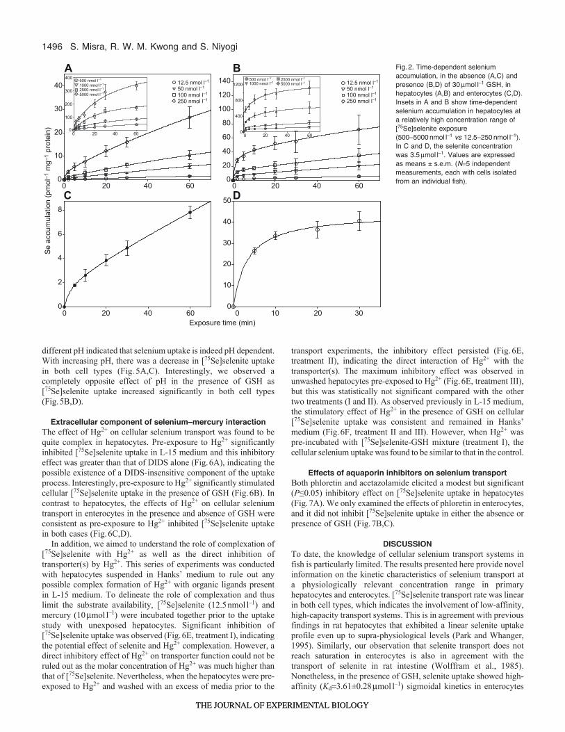
The time-dependent selenium accumulation profile in hepatocytesreached saturation within 30min in the absence of GSH (Fig.2A),whereas saturation occurred at 10min in the presence of GSH(Fig.2B). The initial rate (first 2min) of selenite uptake in thepresence of GSH was about 8 times (5.66±0.66pmolmin–1mg–1
protein) higher than that of selenite alone (0.69±0.13pmolmin–1mg–1
protein). In enterocytes, [75Se]selenite transport (3.5mmoll–1<Kd)did not attain saturation over the exposure period of 60min (Fig.2C).However, the time-dependent uptake profile indicated a saturableuptake over an exposure period of 10min in the presence of GSH(Fig.2D). One of the important findings of the present study wasthat cellular selenium accumulation, in both the absence andpresence of GSH, was ~20 times higher in hepatocytes than inenterocytes at an equimolar selenite exposure concentration
( )
( )( )
( )= + − ×
+
⎧
⎨⎪
⎩⎪
⎫
⎬⎪
⎭⎪+
−
+
⎧
⎨⎪
⎩⎪
⎫
⎬⎪
⎭⎪
⎡
⎣
⎢⎢⎢⎢⎢⎢⎢⎢⎢
⎤
⎦
⎥⎥⎥⎥⎥⎥⎥⎥⎥
( )
( )
⎡⎣ ⎤⎦
⎡⎣ ⎤⎦
f max min
F
F min
1 10
1
1 10
. (4)
x
x
1
– logEC
1
– logEC
50 1
50 2
= ×+
f B XK X[ ]
, (2)h
h hmax
d
S. Misra, R. W. M. Kwong and S. Niyogi
THE JOURNAL OF EXPERIMENTAL BIOLOGYTHE JOURNAL OF EXPERIMENTAL BIOLOGY

1495Membrane transport of selenium
(0.5mmoll–1) for a specific exposure period (30min in the absenceof GSH and 10min in the presence of GSH).
In the subsequent experiments, the effect of L-cysteine, anotherreducing agent, on [75Se]selenite uptake was investigated. As L-15medium is supplemented with amino acids, we conducted transportexperiments in Hanks’ medium as well as in L-15 medium toelucidate the effect of L-cysteine on [75Se]selenite transport. Thecontrol experiment showed a higher uptake of [75Se]selenite in L-15 medium than in Hanks’ medium. However, when both exposuremedia were spiked with equimolar concentrations of L-cysteine,cellular selenium accumulation was comparable in the two mediaand significantly (P≤0.001) higher relative to that in the media notsupplemented with L-cysteine (Fig.3A,B). We also observed thatselenium accumulation in hepatocytes (exposed in Hanks’ medium)increased by ~10-fold in the presence of L-cysteine relative to thecontrol, whereas the increase in accumulation was only ~5-fold inthe presence of GSH (at a concentration comparable to L-cysteine).Similarly, selenium accumulation in enterocytes was ~18-foldhigher in the presence of L-cysteine compared with the ~9-foldincrease at an equimolar concentration of GSH (Fig.3C,D), althoughit should to be noted that the selenite exposure concentration was40- to 280-fold higher for enterocytes relative to that for hepatocytes.
In order to address whether it was the reducing equivalent or thesulfhydryl group (both GSH and L-cysteine contain a free –SHgroup) that facilitated uptake of reduced forms of selenium, wecompared the effect of an equimolar concentration of NADPH withthat of GSH and L-cysteine under identical experimental conditions.No effect of NADPH on [75Se]selenite transport was observed inhepatocytes (see supplementary material Fig.S1), reinforcing therole of the interacting free thiol (–SH) group in efficient selenitereduction and the subsequent transport of reduced selenium species.
Energy independence of selenium transportDCCD was found to be ineffective in blocking selenium transportin hepatocytes (see supplementary material Fig.S2A). Similarly, we
also observed that orthovanadate did not inhibit the uptake of[75Se]selenite or reduced forms of selenium in both hepatocytes andenterocytes (see supplementary material Fig.S2B–D). Together,these observations demonstrate the energy independence ofinorganic selenium transport in both cell types.
Involvement of anion transport systems in selenium uptakeDIDS inhibited the accumulation of [75Se]selenite in hepatocytes,in both the absence and presence of GSH, and the kineticcharacterization indicated a single site interaction with DIDS(Fig.4A). It has been proposed previously that anions such as sulfateand sulfite may be transported via a similar transport pathway toselenite in rat liver mitochondria (Crompton et al., 1974). We chosesulfite over sulfate in this study because of its much closer structuralresemblance to selenite. Our results demonstrated a marked decreaseof [75Se]selenite accumulation in the presence of sulfite, and thekinetic characterization (ligand binding, two-site competition)suggested a competitive interaction between selenite and sulfite attwo transport sites (Fig.4B). In enterocytes, DIDS also inhibitedthe cellular accumulation of [75Se]selenite, in both the absence andpresence of GSH (Fig.4C,D).
Differential effects of pH on selenium transportNo inhibition of [75Se]selenite accumulation was recorded with upto 5mmoll–1 pyruvate (sodium salt) in both hepatocytes andenterocytes (see supplementary material Fig.S3A). Similarly,5mmoll–1 propionic acid did not inhibit [75Se]selenite accumulationin hepatocytes (supplementary material Fig.S3B) in pH-adjustedmedia (pH7.63). However, we observed that the addition ofpropionic acid reduced the pH of L-15 medium, which was easilydiscernible from the obvious change of color (L-15 medium containsPhenol Red). In this pH-unadjusted medium, selenite transport wassignificantly (P≤0.001) higher than in the control (data not shown).This observation led us to deduce that pH probably plays animportant role in selenite transport. Subsequent experiments at
[Selenite] (nmol l–1)0 1000 2000 3000 4000 5000 6000
Se
accu
mul
atio
n (p
mol
mg–
1 pr
otei
n)
0
100
200
300
400
2 min5 min10 min20 min30 min60 min
0 1000 2000 3000 4000 5000 60000
200
400
600
800
1000
12002 min5 min10 min20 min30 min60 min
10 15 20 25
Rat
e of
Se
upta
ke
(pm
ol m
g–1
prot
ein
min
–1)
0
0.2
0.4
0.6
0.8
1.0
1.2
[Selenite] (µmol l–1)10 15 20 25
0
2
4
6
8
A
DC
B
0 5 50
Fig.1. (A,B)Concentration-dependentselenium accumulation in isolatedhepatocytes, in either the absence (A)or presence (B) of 30mmoll–1 GSH(reduced glutathione). Exposure timesare as shown. (C,D)Kinetics of[75Se]selenite transport in isolatedenterocytes in the absence (C) andpresence (D) of 30mmoll–1 GSH.Exposure time for selenite andselenite+GSH was 30 and 10min (for Cand D), respectively. The values areexpressed as means ± s.e.m. (N5–6independent measurements, each withcells isolated from an individual fish).
THE JOURNAL OF EXPERIMENTAL BIOLOGYTHE JOURNAL OF EXPERIMENTAL BIOLOGY
1496
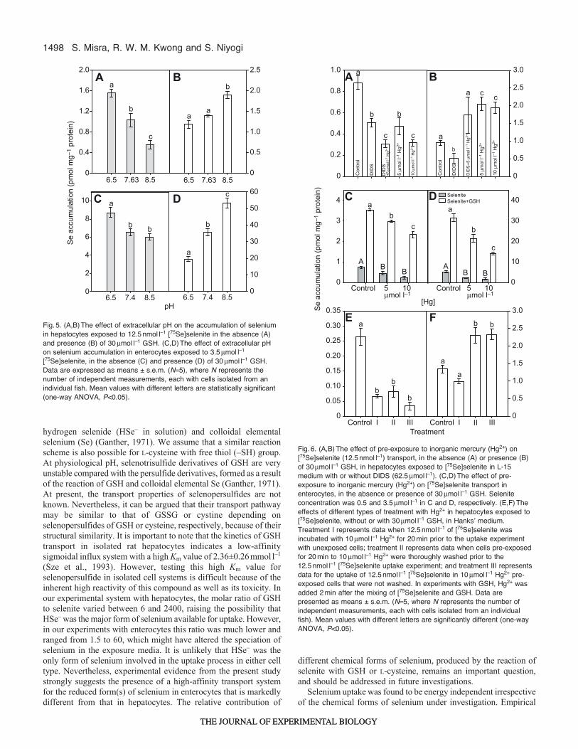
different pH indicated that selenium uptake is indeed pH dependent.With increasing pH, there was a decrease in [75Se]selenite uptakein both cell types (Fig.5A,C). Interestingly, we observed acompletely opposite effect of pH in the presence of GSH as[75Se]selenite uptake increased significantly in both cell types(Fig.5B,D).
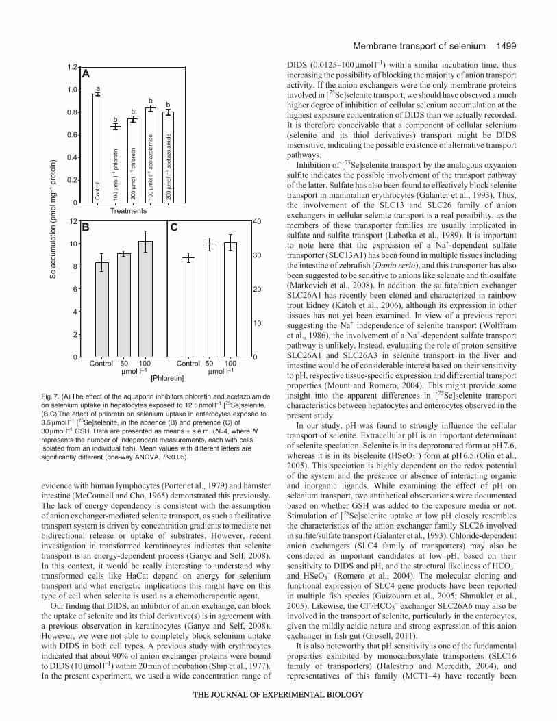
Extracellular component of selenium–mercury interactionThe effect of Hg2+ on cellular selenium transport was found to bequite complex in hepatocytes. Pre-exposure to Hg2+ significantlyinhibited [75Se]selenite uptake in L-15 medium and this inhibitoryeffect was greater than that of DIDS alone (Fig.6A), indicating thepossible existence of a DIDS-insensitive component of the uptakeprocess. Interestingly, pre-exposure to Hg2+ significantly stimulatedcellular [75Se]selenite uptake in the presence of GSH (Fig.6B). Incontrast to hepatocytes, the effects of Hg2+ on cellular seleniumtransport in enterocytes in the presence and absence of GSH wereconsistent as pre-exposure to Hg2+ inhibited [75Se]selenite uptakein both cases (Fig.6C,D).
In addition, we aimed to understand the role of complexation of[75Se]selenite with Hg2+ as well as the direct inhibition oftransporter(s) by Hg2+. This series of experiments was conductedwith hepatocytes suspended in Hanks’ medium to rule out anypossible complex formation of Hg2+ with organic ligands presentin L-15 medium. To delineate the role of complexation and thuslimit the substrate availability, [75Se]selenite (12.5nmoll–1) andmercury (10mmoll–1) were incubated together prior to the uptakestudy with unexposed hepatocytes. Significant inhibition of[75Se]selenite uptake was observed (Fig.6E, treatment I), indicatingthe potential effect of selenite and Hg2+ complexation. However, adirect inhibitory effect of Hg2+ on transporter function could not beruled out as the molar concentration of Hg2+ was much higher thanthat of [75Se]selenite. Nevertheless, when the hepatocytes were pre-exposed to Hg2+ and washed with an excess of media prior to the
transport experiments, the inhibitory effect persisted (Fig.6E,treatment II), indicating the direct interaction of Hg2+ with thetransporter(s). The maximum inhibitory effect was observed inunwashed hepatocytes pre-exposed to Hg2+ (Fig.6E, treatment III),but this was statistically not significant compared with the othertwo treatments (I and II). As observed previously in L-15 medium,the stimulatory effect of Hg2+ in the presence of GSH on cellular[75Se]selenite uptake was consistent and remained in Hanks’medium (Fig.6F, treatment II and III). However, when Hg2+ waspre-incubated with [75Se]selenite-GSH mixture (treatment I), thecellular selenium uptake was found to be similar to that in the control.
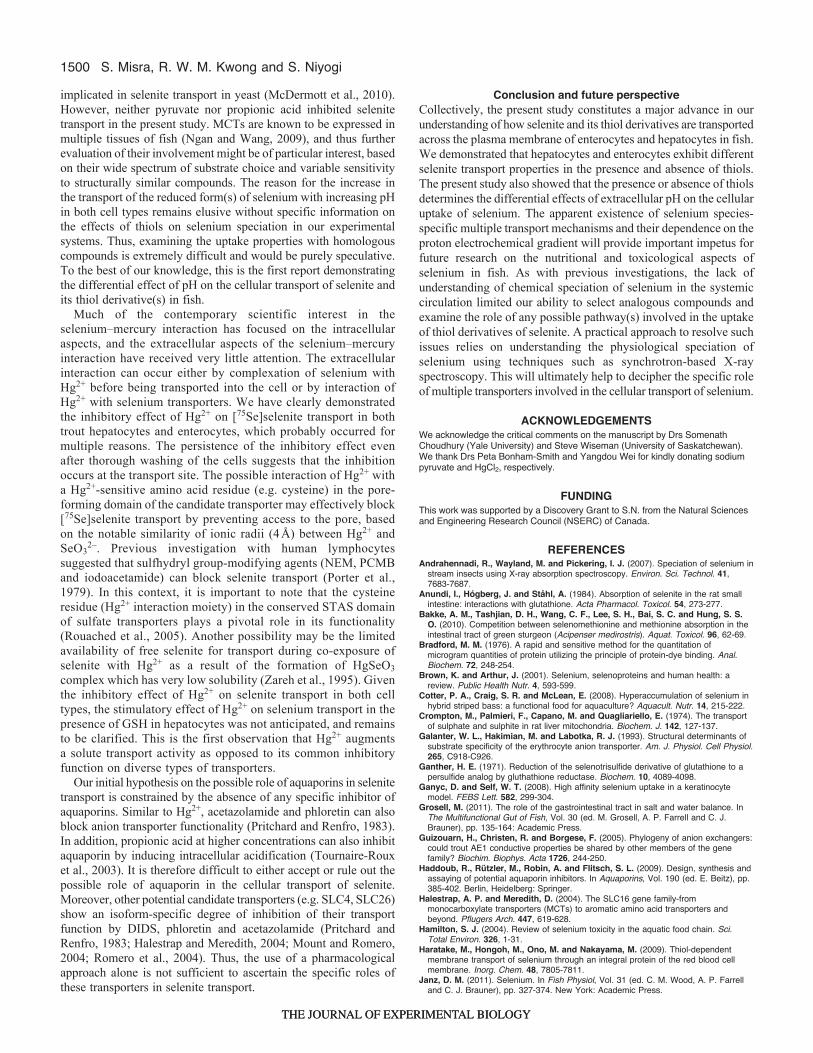
Effects of aquaporin inhibitors on selenium transportBoth phloretin and acetazolamide elicited a modest but significant(P≤0.05) inhibitory effect on [75Se]selenite uptake in hepatocytes(Fig.7A). We only examined the effects of phloretin in enterocytes,and it did not inhibit [75Se]selenite uptake in either the absence orpresence of GSH (Fig.7B,C).
DISCUSSIONTo date, the knowledge of cellular selenium transport systems infish is particularly limited. The results presented here provide novelinformation on the kinetic characteristics of selenium transport ata physiologically relevant concentration range in primaryhepatocytes and enterocytes. [75Se]selenite transport rate was linearin both cell types, which indicates the involvement of low-affinity,high-capacity transport systems. This is in agreement with previousfindings in rat hepatocytes that exhibited a linear selenite uptakeprofile even up to supra-physiological levels (Park and Whanger,1995). Similarly, our observation that selenite transport does notreach saturation in enterocytes is also in agreement with thetransport of selenite in rat intestine (Wolffram et al., 1985).Nonetheless, in the presence of GSH, selenite uptake showed high-affinity (Kd3.61±0.28mmoll–1) sigmoidal kinetics in enterocytes
S. Misra, R. W. M. Kwong and S. Niyogi
Exposure time (min)
Se
accu
mul
atio
n (p
mol
–1 m
g–1
prot
ein)
0
10
20
30
40 12.5 nmol l–1
50 nmol l–1
100 nmol l–1
250 nmol l–1
0
100
200
300
400 500 nmol l–1
1000 nmol l–1 2500 nmol l–1
5000 nmol l–1
20 60400
20 60400
0
20
40
60
80
100
120
140 12.5 nmol l–1
50 nmol l–1
100 nmol l–1
250 nmol l–1
0
400
800
1200500 nmol l–1 1000 nmol l–1
2500 nmol l–1
5000 nmol l–1
20 60400
20 60400
0
2
4
6
8
20 604000
10
20
30
40
50
10 30200
A
DC
B Fig.2. Time-dependent seleniumaccumulation, in the absence (A,C) andpresence (B,D) of 30mmoll–1 GSH, inhepatocytes (A,B) and enterocytes (C,D).Insets in A and B show time-dependentselenium accumulation in hepatocytes ata relatively high concentration range of[75Se]selenite exposure(500–5000nmoll–1 vs 12.5–250nmoll–1).In C and D, the selenite concentrationwas 3.5mmoll–1. Values are expressedas means ± s.e.m. (N5 independentmeasurements, each with cells isolatedfrom an individual fish).
THE JOURNAL OF EXPERIMENTAL BIOLOGYTHE JOURNAL OF EXPERIMENTAL BIOLOGY

1497Membrane transport of selenium
as opposed to the linear uptake rate in hepatocytes. Sigmoidaltransport kinetics indicates positive co-operativity at transport sitesin enterocytes. The Hill coefficient derived from the kinetics datawas almost 2, signifying an interaction of the substrate with at leasttwo transport sites. In such an allosterically regulated transportpathway, binding of the substrate to one site increases the bindingaffinity of the other. However, it should be pointed out that thisderivation is based on fitting of the experimental data and remainsinferential in the absence of direct evidence of multiple carriersystems.
We observed that incubation of selenite with GSH increased totalselenium accumulation in trout hepatocytes and enterocytes, aphenomenon previously reported in isolated rat enterocytes (Anundiet al., 1984). In addition, we found that, like GSH, L-cysteinestimulated selenium uptake, but with a greater magnitude. Scharreret al. previously observed similar effects of L-cysteine on seleniumtransport in rat intestine (Scharrer et al., 1992). Such an apparentincrease in selenium uptake in the presence of different thiols seemsto be attributable to either a differential reducing potential of thethiol moiety of the reducing agents or differences in transportproperties of the intermediates generated as a result of the reaction
of thiols with selenite. Examining the reaction of selenite with GSHand L-cysteine can partially explain such a dramatic increase inuptake. Under physiological pH, the reaction of selenite with GSHcan form selenotrisulfide (GSSeSG), selenopersulfide (GSSeH),
[L-Cysteine] (µmol l–1)Control 25 50 100
Se
accu
mul
atio
n (p
mol
mg–
1 pr
otei
n)
0
0.5
1.0
1.5
2.0
2.5
3.0
3.5
Control 25 50 100
a
a
ba,b
bb
b
b
0
2
4
6
8
10
0
20
40
60
80
100
a
bb
a
Control L-Cysteine Control L-CysteineTreatment
A
DC
B
Fig.3. (A,B)The effect of L-cysteine (25–100mmoll–1) on seleniumaccumulation in hepatocytes exposed to 12.5nmoll–1 [75Se]selenite for30min in L-15 medium (A) and Hanksʼ medium (B). (C,D)The effect of L-cysteine (30mmoll–1) on selenium accumulation in enterocytes exposed to0.5 (C) and 3.5mmoll–1 (D) [75Se]selenite for 30min. Data are presented asmeans ± s.e.m. (N5 independent measurements, each with cells isolatedfrom an individual fish). Mean values with different letters are significantlydifferent (P<0.05). The statistical significance for data presented in A and Bwas evaluated by one-way ANOVA, whereas a pair-wise t-test was usedfor the data presented in C and D.
log10 [DIDS]–2 –1
Se
accu
mul
atio
n (p
mol
mg–
1 pr
otei
n)
0.4
0.6
0.8
1.0
1.2
1.4
SeleniteSelenite + GSH
20 1
log10 [Sulfite]
0.7
0.8
0.9
1.0
1.1
1.2
1.3
–2 –1 20 1
[DIDS]
Control0
2
4
6
8
10
Control0
5
10
15
20
25
5010050 100
A
DC
B
a
b
b
a
bb
µmol l–1 µmol l–1
Fig.4. (A)The inhibitory effect of DIDS on [75Se]selenite (12.5nmoll–1) transportin hepatocytes, in either the absence or presence of 30mmoll–1 GSH (N5–8).The best fit of the inhibition data was achieved with the ligand-binding, one-sitecompetition model (R20.63 without GSH and R20.38 with GSH, both valuesderived using the raw data). Dashed line indicates accumulation with selenitealone, solid line indicates accumulation with selenite+GSH. (B)The inhibitoryeffect of sulfite on [75Se]selenite transport (12.5nmoll–1; N5). The best fit ofthe inhibition data was obtained with the ligand-binding, two-site competitionmodel (R20.59, based on the raw data). Solid line indicates selenite uptake inthe absence of sulfite. (C,D)The inhibitory effect of DIDS on [75Se]selenite(3.5mmoll–1) transport in enterocytes, in the absence (C) and presence (D) of30mmoll–1 GSH (N5–6). Data are presented as means ± s.e.m. (N representsthe number of independent measurements, each with cells isolated from anindividual fish). Mean values with different letters are significantly different(one-way ANOVA, P<0.05).
THE JOURNAL OF EXPERIMENTAL BIOLOGYTHE JOURNAL OF EXPERIMENTAL BIOLOGY
1498
hydrogen selenide (HSe– in solution) and colloidal elementalselenium (Se) (Ganther, 1971). We assume that a similar reactionscheme is also possible for L-cysteine with free thiol (–SH) group.At physiological pH, selenotrisulfide derivatives of GSH are veryunstable compared with the persulfide derivatives, formed as a resultof the reaction of GSH and colloidal elemental Se (Ganther, 1971).At present, the transport properties of selenopersulfides are notknown. Nevertheless, it can be argued that their transport pathwaymay be similar to that of GSSG or cystine depending onselenopersulfides of GSH or cysteine, respectively, because of theirstructural similarity. It is important to note that the kinetics of GSHtransport in isolated rat hepatocytes indicates a low-affinitysigmoidal influx system with a high Km value of 2.36±0.26mmoll–1
(Sze et al., 1993). However, testing this high Km value forselenopersulfide in isolated cell systems is difficult because of theinherent high reactivity of this compound as well as its toxicity. Inour experimental system with hepatocytes, the molar ratio of GSHto selenite varied between 6 and 2400, raising the possibility thatHSe– was the major form of selenium available for uptake. However,in our experiments with enterocytes this ratio was much lower andranged from 1.5 to 60, which might have altered the speciation ofselenium in the exposure media. It is unlikely that HSe– was theonly form of selenium involved in the uptake process in either celltype. Nevertheless, experimental evidence from the present studystrongly suggests the presence of a high-affinity transport systemfor the reduced form(s) of selenium in enterocytes that is markedlydifferent from that in hepatocytes. The relative contribution of
different chemical forms of selenium, produced by the reaction ofselenite with GSH or L-cysteine, remains an important question,and should be addressed in future investigations.
Selenium uptake was found to be energy independent irrespectiveof the chemical forms of selenium under investigation. Empirical
S. Misra, R. W. M. Kwong and S. Niyogi
6.5 7.63 8.5
Se
accu
mul
atio
n (p
mol
mg–
1 pr
otei
n)
0
0.4
0.8
1.2
1.6
2.0
6.5 7.63 8.50
0.5
1.0
1.5
2.0
2.5
a
b
c
aa
b
6.5 7.4 8.50
2
4
6
8
10
pH6.5 7.4 8.5
0
10
20
30
40
50
60a
a
b b b
c
A
DC
B
Fig.5. (A,B)The effect of extracellular pH on the accumulation of seleniumin hepatocytes exposed to 12.5nmoll–1 [75Se]selenite in the absence (A)and presence (B) of 30mmoll–1 GSH. (C,D)The effect of extracellular pHon selenium accumulation in enterocytes exposed to 3.5mmoll–1
[75Se]selenite, in the absence (C) and presence (D) of 30mmoll–1 GSH.Data are expressed as means ± s.e.m. (N5), where N represents thenumber of independent measurements, each with cells isolated from anindividual fish. Mean values with different letters are statistically significant(one-way ANOVA, P<0.05).
0
0.5
1.0
1.5
2.0
2.5
3.0
Se
accu
mul
atio
n (p
mol
mg–
1 pr
otei
n)
0
0.2
0.4
0.6
0.8
1.0
Con
trol
DID
S
DID
S+5
µm
ol l–1
Hg2+
10 µ
mol
l–1 H
g2+
5 µm
ol l–1
Hg2+
a
b
c
b
c ab
a c c
Con
trol
DID
S
DID
S+5
µm
ol l–1
Hg2+
10 µ
mol
l–1 H
g2+
5 µm
ol l–1
Hg2+
TreatmentControl I II III
0
0.05
0.10
0.15
0.20
0.25
0.30
0.35
Control I II III0
0.5
1.0
1.5
2.0
2.5
3.0
a
bb
b
b b
a
a
0
10
20
30
40
0
1
2
3
4
A ABB
B B
bc
a
b
c
SeleniteSelenite+GSH
Control 105 Control 105
[Hg]
A
DC
B
E F
a
µmol l–1 µmol l–1
Fig.6. (A,B)The effect of pre-exposure to inorganic mercury (Hg2+) on[75Se]selenite (12.5nmoll–1) transport, in the absence (A) or presence (B)of 30mmoll–1 GSH, in hepatocytes exposed to [75Se]selenite in L-15medium with or without DIDS (62.5mmoll–1). (C,D)The effect of pre-exposure to inorganic mercury (Hg2+) on [75Se]selenite transport inenterocytes, in the absence or presence of 30mmoll–1 GSH. Seleniteconcentration was 0.5 and 3.5mmoll–1 in C and D, respectively. (E,F)Theeffects of different types of treatment with Hg2+ in hepatocytes exposed to[75Se]selenite, without or with 30mmoll–1 GSH, in Hanksʼ medium.Treatment I represents data when 12.5nmoll–1 of [75Se]selenite wasincubated with 10mmoll–1 Hg2+ for 20min prior to the uptake experimentwith unexposed cells; treatment II represents data when cells pre-exposedfor 20min to 10mmoll–1 Hg2+ were thoroughly washed prior to the12.5nmoll–1 [75Se]selenite uptake experiment; and treatment III representsdata for the uptake of 12.5nmoll–1 [75Se]selenite in 10mmoll–1 Hg2+ pre-exposed cells that were not washed. In experiments with GSH, Hg2+ wasadded 2min after the mixing of [75Se]selenite and GSH. Data arepresented as means ± s.e.m. (N5, where N represents the number ofindependent measurements, each with cells isolated from an individualfish). Mean values with different letters are significantly different (one-wayANOVA, P<0.05).
THE JOURNAL OF EXPERIMENTAL BIOLOGYTHE JOURNAL OF EXPERIMENTAL BIOLOGY
1499Membrane transport of selenium
evidence with human lymphocytes (Porter et al., 1979) and hamsterintestine (McConnell and Cho, 1965) demonstrated this previously.The lack of energy dependency is consistent with the assumptionof anion exchanger-mediated selenite transport, as such a facilitativetransport system is driven by concentration gradients to mediate netbidirectional release or uptake of substrates. However, recentinvestigation in transformed keratinocytes indicates that selenitetransport is an energy-dependent process (Ganyc and Self, 2008).In this context, it would be really interesting to understand whytransformed cells like HaCat depend on energy for seleniumtransport and what energetic implications this might have on thistype of cell when selenite is used as a chemotherapeutic agent.
Our finding that DIDS, an inhibitor of anion exchange, can blockthe uptake of selenite and its thiol derivative(s) is in agreement witha previous observation in keratinocytes (Ganyc and Self, 2008).However, we were not able to completely block selenium uptakewith DIDS in both cell types. A previous study with erythrocytesindicated that about 90% of anion exchanger proteins were boundto DIDS (10mmoll–1) within 20min of incubation (Ship et al., 1977).In the present experiment, we used a wide concentration range of
DIDS (0.0125–100mmoll–1) with a similar incubation time, thusincreasing the possibility of blocking the majority of anion transportactivity. If the anion exchangers were the only membrane proteinsinvolved in [75Se]selenite transport, we should have observed a muchhigher degree of inhibition of cellular selenium accumulation at thehighest exposure concentration of DIDS than we actually recorded.It is therefore conceivable that a component of cellular selenium(selenite and its thiol derivatives) transport might be DIDSinsensitive, indicating the possible existence of alternative transportpathways.
Inhibition of [75Se]selenite transport by the analogous oxyanionsulfite indicates the possible involvement of the transport pathwayof the latter. Sulfate has also been found to effectively block selenitetransport in mammalian erythrocytes (Galanter et al., 1993). Thus,the involvement of the SLC13 and SLC26 family of anionexchangers in cellular selenite transport is a real possibility, as themembers of these transporter families are usually implicated insulfate and sulfite transport (Labotka et al., 1989). It is importantto note here that the expression of a Na+-dependent sulfatetransporter (SLC13A1) has been found in multiple tissues includingthe intestine of zebrafish (Danio rerio), and this transporter has alsobeen suggested to be sensitive to anions like selenate and thiosulfate(Markovich et al., 2008). In addition, the sulfate/anion exchangerSLC26A1 has recently been cloned and characterized in rainbowtrout kidney (Katoh et al., 2006), although its expression in othertissues has not yet been examined. In view of a previous reportsuggesting the Na+ independence of selenite transport (Wolfframet al., 1986), the involvement of a Na+-dependent sulfate transportpathway is unlikely. Instead, evaluating the role of proton-sensitiveSLC26A1 and SLC26A3 in selenite transport in the liver andintestine would be of considerable interest based on their sensitivityto pH, respective tissue-specific expression and differential transportproperties (Mount and Romero, 2004). This might provide someinsight into the apparent differences in [75Se]selenite transportcharacteristics between hepatocytes and enterocytes observed in thepresent study.
In our study, pH was found to strongly influence the cellulartransport of selenite. Extracellular pH is an important determinantof selenite speciation. Selenite is in its deprotonated form at pH7.6,whereas it is in its biselenite (HSeO3
–) form at pH6.5 (Olin et al.,2005). This speciation is highly dependent on the redox potentialof the system and the presence or absence of interacting organicand inorganic ligands. While examining the effect of pH onselenium transport, two antithetical observations were documentedbased on whether GSH was added to the exposure media or not.Stimulation of [75Se]selenite uptake at low pH closely resemblesthe characteristics of the anion exchanger family SLC26 involvedin sulfite/sulfate transport (Galanter et al., 1993). Chloride-dependentanion exchangers (SLC4 family of transporters) may also beconsidered as important candidates at low pH, based on theirsensitivity to DIDS and pH, and the structural likeliness of HCO3
–
and HSeO3– (Romero et al., 2004). The molecular cloning and
functional expression of SLC4 gene products have been reportedin multiple fish species (Guizouarn et al., 2005; Shmukler et al.,2005). Likewise, the Cl–/HCO3
– exchanger SLC26A6 may also beinvolved in the transport of selenite, particularly in the enterocytes,given the mildly acidic nature and strong expression of this anionexchanger in fish gut (Grosell, 2011).
It is also noteworthy that pH sensitivity is one of the fundamentalproperties exhibited by monocarboxylate transporters (SLC16family of transporters) (Halestrap and Meredith, 2004), andrepresentatives of this family (MCT1–4) have recently been
Treatments
Se
accu
mul
atio
n (p
mol
mg–
1 pr
otei
n)
0
0.2
0.4
0.6
0.8
1.0
1.2
Con
trol
a
bb
b b10
0 µm
ol l–1
phl
oret
in
100 µm
ol l–1
ace
tazo
lam
ide
200 µm
ol l–1
ace
tazo
lam
ide
200 µm
ol l–1
phl
oret
in
[Phloretin]
0
2
4
6
8
10
12
0
10
20
30
40
Control 10050 Control 10050
A
B C
µmol l–1 µmol l–1
Fig.7. (A)The effect of the aquaporin inhibitors phloretin and acetazolamideon selenium uptake in hepatocytes exposed to 12.5nmoll–1 [75Se]selenite.(B,C)The effect of phloretin on selenium uptake in enterocytes exposed to3.5mmoll–1 [75Se]selenite, in the absence (B) and presence (C) of30mmoll–1 GSH. Data are presented as means ± s.e.m. (N4, where Nrepresents the number of independent measurements, each with cellsisolated from an individual fish). Mean values with different letters aresignificantly different (one-way ANOVA, P<0.05).
THE JOURNAL OF EXPERIMENTAL BIOLOGYTHE JOURNAL OF EXPERIMENTAL BIOLOGY
1500
implicated in selenite transport in yeast (McDermott et al., 2010).However, neither pyruvate nor propionic acid inhibited selenitetransport in the present study. MCTs are known to be expressed inmultiple tissues of fish (Ngan and Wang, 2009), and thus furtherevaluation of their involvement might be of particular interest, basedon their wide spectrum of substrate choice and variable sensitivityto structurally similar compounds. The reason for the increase inthe transport of the reduced form(s) of selenium with increasing pHin both cell types remains elusive without specific information onthe effects of thiols on selenium speciation in our experimentalsystems. Thus, examining the uptake properties with homologouscompounds is extremely difficult and would be purely speculative.To the best of our knowledge, this is the first report demonstratingthe differential effect of pH on the cellular transport of selenite andits thiol derivative(s) in fish.
Much of the contemporary scientific interest in theselenium–mercury interaction has focused on the intracellularaspects, and the extracellular aspects of the selenium–mercuryinteraction have received very little attention. The extracellularinteraction can occur either by complexation of selenium withHg2+ before being transported into the cell or by interaction ofHg2+ with selenium transporters. We have clearly demonstratedthe inhibitory effect of Hg2+ on [75Se]selenite transport in bothtrout hepatocytes and enterocytes, which probably occurred formultiple reasons. The persistence of the inhibitory effect evenafter thorough washing of the cells suggests that the inhibitionoccurs at the transport site. The possible interaction of Hg2+ witha Hg2+-sensitive amino acid residue (e.g. cysteine) in the pore-forming domain of the candidate transporter may effectively block[75Se]selenite transport by preventing access to the pore, basedon the notable similarity of ionic radii (4Å) between Hg2+ andSeO3
2–. Previous investigation with human lymphocytessuggested that sulfhydryl group-modifying agents (NEM, PCMBand iodoacetamide) can block selenite transport (Porter et al.,1979). In this context, it is important to note that the cysteineresidue (Hg2+ interaction moiety) in the conserved STAS domainof sulfate transporters plays a pivotal role in its functionality(Rouached et al., 2005). Another possibility may be the limitedavailability of free selenite for transport during co-exposure ofselenite with Hg2+ as a result of the formation of HgSeO3
complex which has very low solubility (Zareh et al., 1995). Giventhe inhibitory effect of Hg2+ on selenite transport in both celltypes, the stimulatory effect of Hg2+ on selenium transport in thepresence of GSH in hepatocytes was not anticipated, and remainsto be clarified. This is the first observation that Hg2+ augmentsa solute transport activity as opposed to its common inhibitoryfunction on diverse types of transporters.
Our initial hypothesis on the possible role of aquaporins in selenitetransport is constrained by the absence of any specific inhibitor ofaquaporins. Similar to Hg2+, acetazolamide and phloretin can alsoblock anion transporter functionality (Pritchard and Renfro, 1983).In addition, propionic acid at higher concentrations can also inhibitaquaporin by inducing intracellular acidification (Tournaire-Rouxet al., 2003). It is therefore difficult to either accept or rule out thepossible role of aquaporin in the cellular transport of selenite.Moreover, other potential candidate transporters (e.g. SLC4, SLC26)show an isoform-specific degree of inhibition of their transportfunction by DIDS, phloretin and acetazolamide (Pritchard andRenfro, 1983; Halestrap and Meredith, 2004; Mount and Romero,2004; Romero et al., 2004). Thus, the use of a pharmacologicalapproach alone is not sufficient to ascertain the specific roles ofthese transporters in selenite transport.
Conclusion and future perspectiveCollectively, the present study constitutes a major advance in ourunderstanding of how selenite and its thiol derivatives are transportedacross the plasma membrane of enterocytes and hepatocytes in fish.We demonstrated that hepatocytes and enterocytes exhibit differentselenite transport properties in the presence and absence of thiols.The present study also showed that the presence or absence of thiolsdetermines the differential effects of extracellular pH on the cellularuptake of selenium. The apparent existence of selenium species-specific multiple transport mechanisms and their dependence on theproton electrochemical gradient will provide important impetus forfuture research on the nutritional and toxicological aspects ofselenium in fish. As with previous investigations, the lack ofunderstanding of chemical speciation of selenium in the systemiccirculation limited our ability to select analogous compounds andexamine the role of any possible pathway(s) involved in the uptakeof thiol derivatives of selenite. A practical approach to resolve suchissues relies on understanding the physiological speciation ofselenium using techniques such as synchrotron-based X-rayspectroscopy. This will ultimately help to decipher the specific roleof multiple transporters involved in the cellular transport of selenium.
ACKNOWLEDGEMENTSWe acknowledge the critical comments on the manuscript by Drs SomenathChoudhury (Yale University) and Steve Wiseman (University of Saskatchewan).We thank Drs Peta Bonham-Smith and Yangdou Wei for kindly donating sodiumpyruvate and HgCl2, respectively.
FUNDINGThis work was supported by a Discovery Grant to S.N. from the Natural Sciencesand Engineering Research Council (NSERC) of Canada.
REFERENCESAndrahennadi, R., Wayland, M. and Pickering, I. J. (2007). Speciation of selenium in
stream insects using X-ray absorption spectroscopy. Environ. Sci. Technol. 41,7683-7687.
Anundi, I., Hógberg, J. and Ståhl, A. (1984). Absorption of selenite in the rat smallintestine: interactions with glutathione. Acta Pharmacol. Toxicol. 54, 273-277.
Bakke, A. M., Tashjian, D. H., Wang, C. F., Lee, S. H., Bai, S. C. and Hung, S. S.O. (2010). Competition between selenomethionine and methionine absorption in theintestinal tract of green sturgeon (Acipenser medirostris). Aquat. Toxicol. 96, 62-69.
Bradford, M. M. (1976). A rapid and sensitive method for the quantitation ofmicrogram quantities of protein utilizing the principle of protein-dye binding. Anal.Biochem. 72, 248-254.
Brown, K. and Arthur, J. (2001). Selenium, selenoproteins and human health: areview. Public Health Nutr. 4, 593-599.
Cotter, P. A., Craig, S. R. and McLean, E. (2008). Hyperaccumulation of selenium inhybrid striped bass: a functional food for aquaculture? Aquacult. Nutr. 14, 215-222.
Crompton, M., Palmieri, F., Capano, M. and Quagliariello, E. (1974). The transportof sulphate and sulphite in rat liver mitochondria. Biochem. J. 142, 127-137.
Galanter, W. L., Hakimian, M. and Labotka, R. J. (1993). Structural determinants ofsubstrate specificity of the erythrocyte anion transporter. Am. J. Physiol. Cell Physiol.265, C918-C926.
Ganther, H. E. (1971). Reduction of the selenotrisulfide derivative of glutathione to apersulfide analog by gluthathione reductase. Biochem. 10, 4089-4098.
Ganyc, D. and Self, W. T. (2008). High affinity selenium uptake in a keratinocytemodel. FEBS Lett. 582, 299-304.
Grosell, M. (2011). The role of the gastrointestinal tract in salt and water balance. InThe Multifunctional Gut of Fish, Vol. 30 (ed. M. Grosell, A. P. Farrell and C. J.Brauner), pp. 135-164: Academic Press.
Guizouarn, H., Christen, R. and Borgese, F. (2005). Phylogeny of anion exchangers:could trout AE1 conductive properties be shared by other members of the genefamily? Biochim. Biophys. Acta 1726, 244-250.
Haddoub, R., Rützler, M., Robin, A. and Flitsch, S. L. (2009). Design, synthesis andassaying of potential aquaporin inhibitors. In Aquaporins, Vol. 190 (ed. E. Beitz), pp.385-402. Berlin, Heidelberg: Springer.
Halestrap, A. P. and Meredith, D. (2004). The SLC16 gene family-frommonocarboxylate transporters (MCTs) to aromatic amino acid transporters andbeyond. Pflugers Arch. 447, 619-628.
Hamilton, S. J. (2004). Review of selenium toxicity in the aquatic food chain. Sci.Total Environ. 326, 1-31.
Haratake, M., Hongoh, M., Ono, M. and Nakayama, M. (2009). Thiol-dependentmembrane transport of selenium through an integral protein of the red blood cellmembrane. Inorg. Chem. 48, 7805-7811.
Janz, D. M. (2011). Selenium. In Fish Physiol, Vol. 31 (ed. C. M. Wood, A. P. Farrelland C. J. Brauner), pp. 327-374. New York: Academic Press.
S. Misra, R. W. M. Kwong and S. Niyogi
THE JOURNAL OF EXPERIMENTAL BIOLOGYTHE JOURNAL OF EXPERIMENTAL BIOLOGY

1501Membrane transport of selenium
Katoh, F., Tresguerres, M., Lee, K. M., Kaneko, T., Aida, K. and Goss, G. G.(2006). Cloning of rainbow trout SLC26A1: involvement in renal sulfate secretion.Am. J. Physiol. Regul. Integr. Comp. Physiol. 290, R1468-R1478.
Kryukov, G. V., Castellano, S., Novoselov, S. V., Lobanov, A. V., Zehtab, O.,Guigo, R. and Gladyshev, V. N. (2003). Characterization of mammalianselenoproteomes. Science 300, 1439-1443.
Kwong, R. W., Andres, J. A. and Niyogi, S. (2010). Molecular evidence andphysiological characterization of iron absorption in isolated enterocytes of rainbowtrout (Oncorhynchus mykiss): implications for dietary cadmium and lead absorption.Aquat. Toxicol. 99, 343-350.
Labotka, R. J., Galanter, W. and Misiewicz, V. M. (1989). Erythrocyte bisulfitetransport. Biochim. Biophys. Acta 981, 358-362.
Lorentzen, M., Maage, A. and Julshamn, K. (1994). Effects of dietary selenite orselenomethionine on tissue selenium levels of Atlantic salmon (Salmo salar).Aquaculture 121, 359-367.
Maher, W., Roach, A., Doblin, M., Fan, T., Foster, S., Garrett, R., Möller, G., Oram,L. and Wallschläger, D. (2010). Environmental sources, speciation, and partitioningof selenium. In Ecological Assessment of Selenium in the Aquatic Environment (ed.P. M. Chapman, W. J. Adams, M. Brooks, C. G. Delos, S. N. Luoma, W. A. Maher,H. M. Ohlendorf, T. S. Presser and D. P. Shaw), pp. 47-92. Boca Raton, FL, USA:CRC Press.
Markovich, D., Romano, A., Storelli, C. and Verri, T. (2008). Functional andstructural characterization of the zebrafish Na+-sulfate cotransporter 1 (NaS1) cDNAand gene (slc13a1). Physiol. Genomics 34, 256-264.
Martin, A. J., Simpson, S., Fawcett, S., Wiramanaden, C. I. E., Pickering, I. J.,Belzile, N., Chen, Y. W., London, J. and Wallschläger, D. (2011). Biogeochemicalmechanisms of selenium exchange between water and sediments in two contrastinglentic environments. Environ. Sci. Technol. 45, 2605-2612.
McConnell, K. P. and Cho, G. J. (1965). Transmucosal movement of selenium. Am.J. Physiol. 208, 1191-1195.
McDermott, J. R., Rosen, B. P. and Liu, Z. J. (2010). Jen1p: a high affinity selenitetransporter in yeast. Mol. Biol. Cell 21, 3934-3941.
Misra, S., Peak, D. and Niyogi, S. (2010). Application of XANES spectroscopy inunderstanding the metabolism of selenium in isolated rainbow trout hepatocytes:insights into selenium toxicity. Metallomics 2, 710-717.
Mount, D. B. and Romero, M. F. (2004). The SLC26 gene family of multifunctionalanion exchangers. Pflugers Arch. 447, 710-721.
Ngan, A. K. and Wang, Y. S. (2009). Tissue-specific transcriptional regulation ofmonocarboxylate transporters (MCTs) during short-term hypoxia in zebrafish (Daniorerio). Comp. Biochem. Physiol. 154B, 396-405.
Olin, Å., Noläng, B., Osadchii, E. G., Öhman, L.-O. and Rosén, E. (2005). ChemicalThermodynamics of Selenium. Amsterdam: Elsevier B.V.
Park, Y. C. and Whanger, P. D. (1995). Toxicity, metabolism and absorption ofselenite by isolated rat hepatocytes. Toxicology 100, 151-162.
Porter, E. K., Karle, J. A. and Shrift, A. (1979). Uptake of selenium-75 by humanlymphocytes in vitro. J. Nutr. 109, 1901-1908.
Pritchard, J. B. and Renfro, J. L. (1983). Renal sulfate transport at the basolateralmembrane is mediated by anion exchange. Proc. Natl. Acad. Sci. USA 80, 2603-2607.
Rayman, M. P. (2005). Selenium in cancer prevention: a review of the evidence andmechanism of action. Proc. Nutr. Soc. 64, 527-542.
Romero, M. F., Fulton, C. M. and Boron, W. F. (2004). The SLC4 family of HCO3–
transporters. Pflugers Arch. 447, 495-509.Rouached, H., Berthomieu, P., El Kassis, E., Cathala, N., Catherinot, V., Labesse,
G., Davidian, J.-C. and Fourcroy, P. (2005). Structural and functional analysis ofthe C-terminal STAS (sulfate transporter and anti-sigma antagonist) domain of theArabidopsis thaliana sulfate transporter SULTR1.2. J. Biol. Chem. 280, 15976-15983.
Scharrer, E., Senn, E. and Wolffram, S. (1992). Stimulation of mucosal uptake ofselenium from selenite by some thiols at various sites of rat intestine. Biol. TraceElem. Res. 33, 109-120.
Schwarz, K. and Foltz, C. M. (1957). Selenium as an integral part of factor 3 againstdietary necrotic liver degeneration. J. Am. Chem. Soc. 79, 3292-3293.
Ship, S., Shami, Y., Breuer, W. and Rothstein, A. (1977). Synthesis of tritiated 4,4�-diisothiocyano-2,2�-stilbene disulfonic acid ([3H]DIDS) and its covalent reaction withsites related to anion transport in human red blood cells. J. Membr. Biol. 33, 311-323.
Shmukler, B. E., Kurschat, C. E., Ackermann, G. E., Jiang, L., Zhou, Y., Barut, B.,Stuart-Tilley, A. K., Zhao, J., Zon, L. I., Drummond, I. A. et al. (2005). Zebrafishslc4a2/ae2 anion exchanger: cDNA cloning, mapping, functional characterization,and localization. Am. J. Physiol. Renal Physiol. 289, F835-F849.
Sze, G., Kaplowitz, N., Ookhtens, M. and Lu, S. C. (1993). Bidirectional membranetransport of intact glutathione in Hep G2 cells. Am. J. Physiol. Gastrointest. LiverPhysiol. 265, G1128-G1134.
Tarze, A., Dauplais, M., Grigoras, I., Lazard, M., Ha-Duong, N.-T., Barbier, F.,Blanquet, S. and Plateau, P. (2007). Extracellular production of hydrogen selenideaccounts for thiol-assisted toxicity of selenite against Saccharomyces cerevisiae. J.Biol. Chem. 282, 8759-8767.
Tournaire-Roux, C., Sutka, M., Javot, H., Gout, E., Gerbeau, P., Luu, D.-T., Bligny,R. and Maurel, C. (2003). Cytosolic pH regulates root water transport during anoxicstress through gating of aquaporins. Nature 425, 393-397.
Wolffram, S., Arduser, F. and Scharrer, E. (1985). In vivo intestinal absorption ofselenate and selenite by rats. J. Nutr. 115, 454-459.
Wolffram, S., Anliker, E. and Scharrer, E. (1986). Uptake of selenate and selenite byisolated intestinal brush border membrane vesicles from pig, sheep, and rat. Biol.Trace Elem. Res. 10, 293-306.
Zareh, M. M., Amin, A. S. and Abdel-Aziz, M. (1995). New polycrystalline solid stateresponsive electrodes for the determination of the selenite ion. Electroanalysis 7,587-590.
THE JOURNAL OF EXPERIMENTAL BIOLOGYTHE JOURNAL OF EXPERIMENTAL BIOLOGY


![[320] Web 3: Selenium · for Selenium Java module for Selenium Ruby module for Selenium JavaScript mod for Selenium Chrome Driver Firefox Driver Edge Driver. Examples. Starter Code](https://img.dokumen.tips/doc/110x75/5eadce82cc4f0d7405687f01/320-web-3-selenium-for-selenium-java-module-for-selenium-ruby-module-for-selenium.jpg)