Embed Size (px)
Citation preview

Research ArticlePreliminary Evaluation of Probiotic Properties of LactobacillusStrains Isolated from Sardinian Dairy Products
Maria Barbara Pisano,1 Silvia Viale,1 Stefania Conti,1 Maria Elisabetta Fadda,1
Maura Deplano,1 Maria Paola Melis,2 Monica Deiana,2 and Sofia Cosentino1
1 Department of Public Health, Clinical and Molecular Medicine, University of Cagliari, Cittadella Universitaria,S.S. 554, Km 4,5, 09042 Monserrato, Italy
2 Department of Biomedical Sciences, Unit of Experimental Pathology, University of Cagliari, Cittadella Universitaria,S.S. 554, Km 4,5, 09042 Monserrato, Italy
Correspondence should be addressed to Sofia Cosentino; [email protected]
Received 25 February 2014; Revised 29 May 2014; Accepted 11 June 2014; Published 26 June 2014
Academic Editor: Himanshu Garg
Copyright © 2014 Maria Barbara Pisano et al. This is an open access article distributed under the Creative Commons AttributionLicense, which permits unrestricted use, distribution, and reproduction in any medium, provided the original work is properlycited.
Twenty-three Lactobacillus strains of dairy originwere evaluated for some functional properties relevant to their use as probiotics. Apreliminary subtractive screening based on the abilities to inhibit the growth of microbial pathogens and hydrolyze conjugated bilesalts was applied, and six strains were selected for further characterization including survival under gastrointestinal environmentalconditions, adhesion to gut epithelial tissue, enzymatic activity, and some safety properties. All selected strains maintained elevatedcell numbers under conditions simulating passage through the human gastrointestinal tract, well comparable to the values obtainedfor the probiotic strain Lactobacillus rhamnosusGG, and were able to adhere to Caco-2 cells to various extents (from 3 to 20%). Allstrains exhibited high aminopeptidase, and absent or very lowproteolytic and strong𝛽-galactosidase activities; nonewas found to behaemolytic or to produce biogenic amines and all were susceptible to tetracycline, chloramphenicol, erythromycin, ampicillin, andamoxicillin/clavulanic acid. Our results indicate that the Lactobacillus strains analyzed could be considered appropriate probioticcandidates, due to resistance to GIT simulated conditions, antimicrobial activity, adhesion to Caco-2 cell-line, and absence ofundesirable properties. They could be used as adjunct cultures for contributing to the quality and health related functionalproperties of dairy products.
1. Introduction
Lactic acid bacteria (LAB) are widespread in nature andare one of the major microbial groups involved in thefermentation of different types of food. They represent thedominant microorganisms in milk and milk products wherethey play important roles during both manufacture andripening. LAB possess a large number ofmetabolic propertiesthat are responsible for the organoleptic characteristics ofthe final product as well as its preservation and microbialsafety [1]. They are also known for their potential health andnutritional benefits and therefore are considered “probiotics”or “live microorganisms which upon ingestion in adequateamounts confer health benefits to the host” [2].
As a result of increasing awareness of the close inter-relationship between health and diet, special attention ispresently given to functional properties of LAB associatedwith traditional fermented products. These foods couldrepresent alternative sources of novel probiotic candidateswith physiological and functional properties for potentialbiotechnological use in the manufacturing of functionalproducts in which the probiotic cultures are more active andprotected from the gastrointestinal stress.
Among food ecosystem, cheese has some intrinsic chem-ical and physical characteristics (high pH, buffering capac-ity, solid consistency, high fat content, and high nutrientavailability) that make it suitable for delivery of viableprobiotic microorganisms into the human intestine [3]. Raw
Hindawi Publishing CorporationBioMed Research InternationalVolume 2014, Article ID 286390, 9 pageshttp://dx.doi.org/10.1155/2014/286390
2 BioMed Research International
milk and traditional cheeses are rich environments with avaried and complex autochthonous microbiota composedof diverse group of microorganisms, including LAB, whichcontribute to the biopreservation and the development oforganoleptic properties of the final product. LAB may playdifferent roles in cheese-making; some species designated asstarter LAB (SLAB) participate in the fermentation process,whereas some others indicated as nonstarter LAB (NSLAB)are implicated in the maturation process [4].
Among LAB, Lactobacillus strains have been extensivelyexploited for their probiotic properties [5] and are applied asadjunct cultures in various types of food products [6].
Lactobacilli are generally regarded as safe (GRAS) dueto their long history of safe use and their presence in theintestinal microbiota of humans, and several species havereceived the qualified presumption of safety (QPS) status[7, 8].
Recently an increasing interest was paid to the possibilitythat strains belonging to species such as Lactobacillus plan-tarum, L. paracasei, and L. brevis,which constitute themajor-ity of NSLAB found in most ripened cheese varieties, mayplay a role as health promoters, beyond their technologicalfunction [9].
To perform their probiotic action these bacteria mustarrive at the intestinal tract alive. This requires their survivalduring food processing, product maturation, and shelf-life and, after consumption, their resistance to the acidicconditions of the stomach as well as to bile salts in the smallintestine [10].
As a part of a larger study involving the selection andcharacterization of new probiotic candidates, 23 autoch-thonous Lactobacillus strains of dairy origin were investi-gated for some functional properties relevant to their useas probiotic cultures. Antagonism toward microbial path-ogens, ability to deconjugate bile salts, survival in thegastrointestinal tract, adhesion capability to intestinal epi-thelial cells, enzymatic activities, and some safety featuressuch as hemolytic activity, antimicrobial resistance, andproduction of biogenic amines were in vitro analyzed.
2. Materials and Methods
2.1. Bacterial Strains and Growth Conditions. A total of 23Lactobacillus strains (10 L. paracasei, 9 L. plantarum, and 4L. brevis) isolated from raw milk and artisanal ewes’ cheeseswere included in this study.The strains were identified on thebasis of phenotypic tests and genetic analysis based on poly-merase chain reaction amplification using species-specificprimers derived from 16S rRNA sequences, as previouslyreported [11]. The strains were maintained at −20∘C in MRSbroth (Microbiol, Cagliari, Italy) with 15% (v/v) glycerol andpropagated three times in MRS broth for activation priorto experimental use. Listeria monocytogenes ATCC 7644,Escherichia coli ATCC 35150, Enterococcus faecalis ATCC29212, SalmonellaTyphimuriumATCC 14028, Staphylococcusaureus ATCC 25923, and Candida albicans ATCC 10231were used as indicators. All indicator strains were storedon nutrient broth (Microbiol) plus 20% (v/v) glycerol at
−20∘C. Before use, theywere subcultured twice in appropriatemedium.
2.2. Antimicrobial Activity. The strains were screened forantimicrobial compounds production against the indicatorstrains using an agar spot method [12]. Overnight cultures ofLactobacilli were spotted onto the surface of MRS agar (1.2%(w/v) agar—0.2% (w/v) glucose) plates, which were thenincubated anaerobically for 24 h at 37∘C.The indicator strainswere inoculated into 7mL of soft agar medium (nutrientbroth containing 0.7% w/v agar) to a final concentration ofapproximately 107 colony forming unit (cfu)/mL; then the softmedia were poured on the plates. After 24 h of incubationat the optimal growth temperature and atmosphere for theindicator strains, inhibition halos were measured. The widthof the clear zone (𝑅) was calculated as follows: 𝑅 = (𝑑Inhib −𝑑Spot)/2, where 𝑑Inhib is the diameter of the zone withoutpathogen growth and 𝑑Spot is the diameter of the spot.
2.3. Bile Salt Hydrolase Activity. Bile salt hydrolase (BSH)activity was screened by spotting in duplicate 10 𝜇L ofcultures grown overnight in MRS broth on the surface ofMRS agar plates supplemented with 0.5% (w/v) sodium salttaurodeoxycholic acid (TDCA, Sigma, Milano, Italy) or 0.2%(w/v) glycodeoxycholic acid (GDCA, Sigma) and 0.37 g/L ofCaCl2[13]. Plates were incubated in anaerobic conditions at
37∘C for 72 h.The presence of halos around colonies (inMRS-GDCA) or white opaque colonies (in MRS-TDCA) indicatedBSH activity. MRS agar plates without supplementation wereused as negative controls. Enterococcus faecalis ATCC 14433was used as BSH-positive strain.
2.4. In Vitro Resistance to Gastrointestinal Conditions. Transittolerance in the upper GIT was assessed using an in vitromodel simulating gastric and pancreatic juices (simulatedstomach-duodenum passage, SSDP), as reported by VizosoPinto et al. [14]. The strains were inoculated to a finalconcentration of approximately 2.5 × 10⁸ cfu/mL in 10mL ofsimulated gastric juice (6.2 g/L NaCl, 2.2 g/L KCl, 0.22 g/LCaCl2, 1.2 g/L NaHCO
3, 0.3% pepsin, and pH 3.0) and
incubated at 37∘C in a shaking water bath (Dubnoff 750, Asal,Milano, Italy) to simulate peristalsis. After 90min, 17.5mL ofsynthetic duodenum juice (6.4 g/L NaHCO
3, 0.239 g/L KCl,
1.28 g/L NaCl, and 0.1% pancreatin), adjusted to pH 7.4 with5M HCl, and 4mL of 10% (w/v) oxgall (Sigma) were addedto the cell suspensions to simulate passage into the upperintestinal tract [15]. After 0, 90, and 180min of incubation, thesurvival rate was determined by the plate method using MRSincubated anaerobically at 37∘C for 48 h. The experimentswere repeated twice and results are expressed as the mean logcfu/mL.
2.5. Adhesion Properties to Human Cell Line. The humancolonic carcinoma cell line Caco-2 (ECACC, Salisbury,UK) was routinely cultured in Dulbecco’s modified Eagle’sminimal essential medium DMEM (Sigma), supplementedwith 10% (w/v) foetal bovine serum (FBS, Sigma), 1% (w/v)nonessential amino acids solution (Sigma), and antibiotic

BioMed Research International 3
solution (100U/mL penicillin, 100 𝜇g/mL streptomycin).Cells were maintained in T-75 culture flasks at 37∘C in a 5%CO2atmosphere. For adhesion assay, the Caco-2 cells were
seeded at a concentration of 105 cells/well in 6-well tissueculture plates (Falcon) to obtain confluence and culturedfor 20 days prior to use in adhesion assay. The cell culturemedium was changed on alternate days and replaced byfresh DMEM supplemented with 2% (w/v) FBS and withoutantibiotic at least 1 h before the adhesion assay. A 1mLaliquot of Lactobacillus suspension (108 ufc/mL in phosphatebuffered saline, PBS) was added to each well of the tissueculture plate and incubated at 37∘C in 5%CO
2atmosphere for
3 h. Afterwards, the cells were washed three times with 1mLof PBS in order to remove nonadherent bacteria and lysedby addition of Triton X 100 (0.05% solution) for 10min; thenappropriate dilutions were plated onMRS agar. Adhesionwasexpressed as the percentage of bacteria adhered to Caco-2cells compared to the initial amount of bacteria.
2.6. Enzymatic Activities. Enzymatic activities of Lactobacil-lus strains were evaluated by using the API ZYM galleries(BioMerieux, Itay) as described by the manufacturer. Theresults were graded from 0 to 5 by comparing the colourdeveloped within 5min with the API-ZYM colour reactionchart.The results were expressed in nanomoles of hydrolysedsubstrate, from the intensity of the reactions obtained, in therange 0 (no activity) to 5 (40 or more nanomoles liberated),following the manufacturer’s instructions.
2.7. Safety Assessment. The method of Bover-Cid and Holz-apfel [16] was used to screen Lactobacillus strains for theproduction of biogenic amines. Briefly, the test strains weresubcultured twice at 24 h intervals in MRS broth containing1% of each precursor amino acid: tyrosine disodium salt, l-histidine monohydrochloride, l-ornithine monohydrochlo-ride, and lysine monohydrochloride (Sigma), and 0.005%pyridoxal-5-phosphate (Sigma) as a codecarboxylase factor.All strains were then streaked in duplicate on decarboxylasemedium plates each containing only one of the abovemen-tioned amino acids and bromocresol purple as pH indicatorand incubated for 4 days in anaerobic conditions at 37∘C.Decarboxylase medium without amino acids was used ascontrol. A colour change from brown to purple in themedium indicated an increase in pH and was considered apositive result.
Antibiotic susceptibilitytesting was carried out by discdiffusion method according to the Clinical and Labora-tory Standards Institute (CLSI) guidelines [17] but Mueller-Hinton agar was replaced by MRS agar (Microbiol). Thefollowing antibiotics (Oxoid or BBL) were tested: ampicillin(AM; 10 𝜇g), amoxicillin/clavulanic acid (AmC; 30 𝜇g), van-comycin (VA; 30 𝜇g), teicoplanin (TEC; 30𝜇g) (inhibitors ofcell wall synthesis), tetracycline (TE; 30 𝜇g), streptomycin (S;10 𝜇g), kanamycin (K; 30 𝜇g), gentamicin (GM; 10 𝜇g), chlo-ramphenicol (C; 30𝜇g), erythromycin (E; 15𝜇g) (inhibitors ofprotein synthesis), ciprofloxacin (CIP; 5𝜇g), and rifampicin(RA; 30 𝜇g) (inhibitors of nucleic acids). A suspension fromfresh overnight cultures with a density of McFarland 0.5 in
buffered saline was plated onMRS agar plates; then antibioticdiscs were dispensed onto the plates. After incubation at37∘C for 24 h in anaerobiosis, the diameters of the bacterialfree-zone were measured and results expressed in terms ofresistance according to the interpretative criteria issued by theCLSI [18].
Haemolytic activity was determined by streaking thestrains on Columbia Blood (Microbiol) agar plates sup-plemented with 5% defibrinated sheep blood after 48 h ofincubation at 37∘C.The haemolytic reaction was recorded byobservation of a clear zone of hydrolysis around the colonies(𝛽-haemolysis), a partial hydrolysis and greenish zone (𝛼-haemolysis), or no reaction (𝛾-haemolysis).
2.8. Statistical Analysis. Statistical analysis of datawas carriedout usingGraphPad Prism Statistics software package version3.00 (GraphPad Prism Software Inc., San Diego, CA, USA).One-way ANOVA followed by Bonferroni test was used todetermine significant differences of viability of the Lactobacil-lus strains during SSDP treatment and with respect to theadhesion ability. Data were analysed at the significance levelof 𝑃 < 0.05.
3. Results and Discussion
A preliminary subtractive screening based on the abilitiesto inhibit the growth of microbial pathogens and hydrolyseconjugated bile salts was applied to the strains. As shownin Table 1, most strains were able to inhibit the growth ofL. monocytogenes, S. aureus, and E. coli O157:H7 with clearinhibition zones of more than 6mm in agar-spot plates. Alower activity was detected against E. faecalis, while onestrain showed an inhibition zone of 4mm against C. albicansATCC 10231. Such broad antagonistic activity of Lactobacillustowards different pathogens has been reported [19, 20];however, 27 strains of L. plantarum isolated from cheeseshowed no activity against selected indicator pathogens [21].
LAB are known to produce many different substanceswith antimicrobial activity, including the major metabolicend products such as organic acids, hydrogen peroxide,ethanol, and bacteriocins [22].When the strains with the bestinhibitory activity in the agar spot-test were further testedusing the well-diffusion assay, to investigate the presenceof bacteriocin-like compounds, no inhibitory activity wasobserved (data not shown), presumably indicating that theproduction of organic acids was responsible for the observedantimicrobial effect. The inhibitory effect of hydrogen perox-ide was excluded due to incubation in anaerobic conditions.
BSH activity is a relevant property for probiotic strains tosurvive the toxicity of conjugated bile salts in the duodenum[23]. In our study, while all strains were able to grow inthe presence of conjugated bile salts after 72 h of incubation,only five (L. plantarum 11/20966, 4/16868, 19/20711 andL. paracasei 62LP39, 1A6M) demonstrated also the abilityto hydrolyze both sodium glycodeoxycholate (GDCA) andsodium taurodeoxycholate (TDCA), and four were able todeconjugate only GDCA, as indicated by the BSH test onagar plates. Some authors have shown that in Lactobacilli

4 BioMed Research International
Table 1: Antimicrobial activity against themicrobial pathogens tested, bile salt hydrolase (BSH) activity, and raffinose fermentation of selectedpotential probiotic Lactobacillus strains isolated from Sardinian dairy products.
Strains Target strains Hydrolysis ofS. aureus
ATCC 25923L. monocytogenes
ATCC 7644E. coli
ATCC 35150E. faecalisATCC 29212
S. TyphimuriumATCC 14028
C. albicansATCC 10231 TDCA GDCA Raffinose∗
L. paracasei 62LP39 7.5 9 6.5 1.5 >10 2 + + +L. paracasei 8/18710 5 9 7.5 2 >10 1.5 − − −
L. paracasei 1A6M 6 7.5 6 2 >10 1 + + +L. paracasei 3A1M 3 6 5 1 7.5 0 − − −
L. paracasei 5/22019 6.5 8.5 5 0.5 7.5 0 − + −
L. paracasei 2C6M 6 5 6 0.5 7.5 0 − − −
L. paracasei 60LP37 7.5 9 9 1 >10 0.5 − − −
L. paracasei 2A11 6.5 9 5 0.5 >10 0 − − −
L. paracasei 6B1M 5 7.5 6 1 >10 0.5 − − −
L. paracasei 2B1M 5 9 6 1.5 >10 1 − − −
L. plantarum 11/20966 7 9 7.5 1 >10 2.5 + + +L. plantarum 19/20711 7.5 9 7.5 2 >10 2 + + +L. plantarum 4/16868 10 10 9 2.5 >10 2 + + +L. plantarum 8C1M 5 5 6 1.5 >10 0 − + −
L. plantarum 9FS15 6 1.5 5 1.5 >10 1.5 − − −
L. plantarum 28SP1 4.5 2.5 3 1 >10 1.5 − − −
L. plantarum 2AFS11 6.5 9 5 1 >10 0.5 − − −
L. plantarum 5FS12 6 5 6 0.5 7.5 0 − − −
L. plantarum 12LP13 5 7.5 6 1 >10 0 − − −
L. brevis 1C3M 6.5 7.5 6 0.5 >10 4 − + −
L. brevis 9/11B 5 9 5 0.5 >10 0.5 − + −
L. brevis 21FS1B 5 7 5 1 >10 0.5 − − −
L. brevis 9A3M 4 5 5 0 >10 0.5 − − −∗Determined by API-50 CHL galleries (bioMerieux).
the resistance to bile salts toxicity is not related to hydrolaseactivity [21, 24–26]. Other mechanisms, alternative to BSH,could be involved in counteract bile damage, as suggested byNoriega et al. [27].
BSH activity is frequently observed in specific groups ofbacteria like Lactobacillus, Bifidobacterium, and Enterococcusisolated from the gastrointestinal tract (GIT): it has beenreported for L. plantarum [14, 28] but not for L. paracaseistrains [26, 29]. Presently its role is controversial since it hasbeen reported to act either positively in lowering of serumcholesterol [30] or negatively in increasing the level of unde-sirable deconjugated bile salts [31]. However, the bacterialgenera most frequently used as probiotics (Bifidobacteria andLactobacilli) are not capable of dehydroxylating deconjugatedbile salts [32, 33] and so the majority of the breakdownproducts may be precipitated and excreted with feces [34].On the other hand, BSH activity by a probiotic bacteriummay be desirable since it increases the intestinal survival andpersistence of producing strains, which in turn increases theoverall beneficial effects associated with the strain [35].
Among the BSH positive strains, five showing the abilityto ferment the trisaccharide raffinose, known for its prebioticactivity, and one with a high antagonistic activity against
C. albicans ATCC 10231 were selected for further functionalcharacterization including in vitro tests to assess their survivalunder gastrointestinal environmental conditions and adhe-sion to gut epithelial tissue and to determine the enzymaticactivity and some safety properties.
The survival responses of the strains, after 90 and180min of exposure to different gastrointestinal conditions,are shown in Table 2. The probiotic strain L. rhamnosus GGwas included in the study for comparison purpose. All strainsretained high viability during simulated gastric juice (pH 3.0)transit for 90min, and no significant reduction (𝑃 > 0.05)was found for any of the strains after exposure to artificialduodenum juice (pH 8.0). The strains L. paracasei 62LP39and L. plantarum 11/20966 and 4/16868 presented the bestsurvival rates values with more than 98% surviving cells.
Exposure to gastric and intestinal fluids is the main envi-ronmental stress that decreases viability of ingested probiotics[36]. All selected strains maintained elevated cell numbersunder conditions simulating passage through the humangastrointestinal tract, well comparable to the values obtainedfor the probiotic strain L. rhamnosus GG. The survival rateswere above 90% after 3 h despite the high bile concentration(more than 1%) used in the test. These results are consistent

BioMed Research International 5
Table 2: Resistance of Lactobacillus strains to simulated stomach duodenum-passage (SSDP). The values are reported as log cfu/mL (mean± E.S.).
Species Initial mean count Survival after90min at pH 3.0
Survival after 180min ofwhich 90min in 5% oxgall
StrainL. paracasei
62LP39 8.33 ± 0.03 8.67 ± 0.11 8.66 ± 0.841A6M 8.49 ± 0.07 8.08 ± 0.02 7.47 ± 1.39
L. plantarum11/20966 8.66 ± 0.15 9.70 ± 0.91 8.62 ± 0.1719/207111 8.58 ± 0.13 8.58 ± 0.3 7.76 ± 0.354/16868 8.21 ± 0.03 8.06 ± 0.2 8.47 ± 0.2
L. brevis1C3M 8.59 ± 0.23 8.75 ± 0.05 8.39 ± 0.14
L. rhamnosus GG 8.59 ± 0.02 9.03 ± 0.18 8.23 ± 0.59
with those previously reported for other potentially probioticstrains belonging to L. paracasei [26] and L. plantarum [21]and correlate well with those obtained for BSH activity.
The adhesion ability of Lactobacillus strains is reportedin Figure 1. All strains were able to adhere to Caco-2 cellsto various extents (ranging from 3 to 20%), confirmingthat adhesion is a strain-specific property. Three strainsshowed significantly higher adherence to Caco-2 cells thanL. rhamnosus GG (𝑃 < 0.05). L. paracasei 62LP39 was themost adhesive strain since approximately 20% of the addedbacteria were bound to Caco-2 cells, followed by L. brevis1C3M (14%) and L. plantarum 11/20966 (10.5%). A loweradhesion rate was observed for the strain L. paracasei 1A6M(3%).Thepercentage of adhesion ofL. rhamnosusGG, used aspositive control, was similar to those obtained with the samestrain in other studies [37, 38] but lower than the values foundby Xu et al. [39]. L. plantarum strains have been shown toadhere more efficiently to Caco-2 cells than L. paracasei or L.brevis [29, 38], althoughToumola and Salminen [37] obtainedan adhesion percentage at the level of 6.7%. Other authorsobserved that L. paracasei strains were effective in reducingadhesion of Salmonella to Caco-2 cells in competitive assays,in spite of their low adherence ability [40–42].
Several in vitro studies suggested that probiotic adhesionmay interfere with the adherence of pathogens, exerting abarrier against pathogen colonization through competitiveexclusion mechanisms [39, 40].
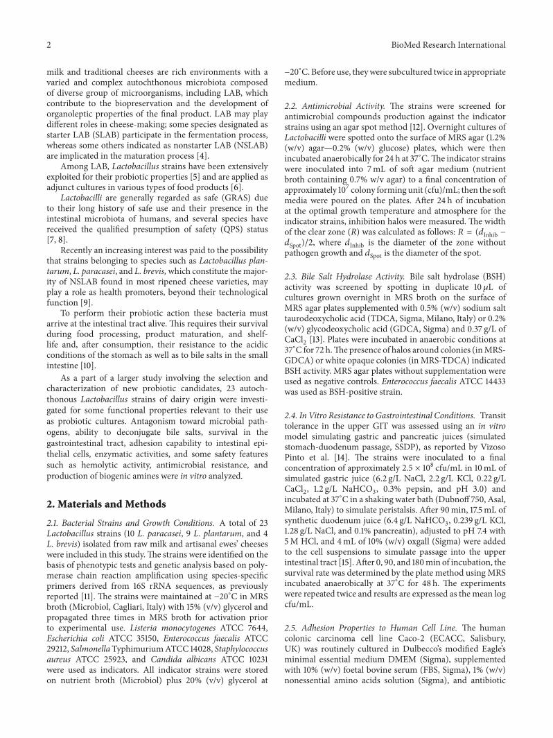
Enzymatic characterization of the selected strains wascarried out in order to evaluate their potential for using asadjunct cultures in the manufacturing of probiotic cheeseand is reported in Table 3. All strains exhibited high amino-peptidase activity (leucine and valine arylamidase) whileesterase and lipase activities were generally moderate to low.The proteolytic and N-acetyl-𝛽-glucosaminidase activitieswere absent or very low. All strains exhibited strong 𝛽-galactosidase and moderate-to-high 𝛼-galactosidase activ-ities, which are responsible for the hydrolysis of lactoseand raffinose, respectively. None of the strains showed 𝛽-glucuronidase or 𝛼-mannosidase activity.
a a
bb
c
ab ab
50
45
40
35
30
25
20
15
10
5
0
LGG
1C3
M
1A6
M
11
/20966
62
LP39
19
/20711
4/16868
Adhe
sion
(%)
Lactobacillus strains
Figure 1: Adhesion ability to Caco-2 human colon cell lines of dairyLactobacillus strain (mean±E.S. of three independent experiments).Mean values with different superscript letters were significantlydifferent at 𝑃 < 0.05.
Overall, the enzymatic profiles of our strains are similarto those reported by other authors particularly for the speciesL. plantarum [42–44]. Mathara et al. [43] could not detectany 𝛽-galactosidase in 14 L. paracasei isolates and Herreroset al. [42] found a certain degree of activity in two strainsof L. brevis. Strains with low proteinase, high peptidase,and low esterase-lipase activities may be useful in reducingbitterness and improving body and texture defects [43, 44].𝛽-galactosidase activity is an important feature for strains to beused as probiotics, since it may be useful in improving lactosetolerance in the gut [45].The lack of 𝛽-glucuronidase activityis an important trait, as well, since its negative role has beensuggested in increasing the risk of carcinogenesis [46].
In order for a strain to be used as probiotic culture, itshould be evaluated for the presence of virulence determi-nants to determine what potential risks might be involvedin its use. Biogenic amines are produced by LAB during the
6 BioMed Research International
Table 3: Enzymatic profiles of selected dairy Lactobacillus strains assayed by the API-ZYM system.
Enzyme testedStrains
L. paracasei L. plantarum L. brevis62LP39 1A6M 11/20966 19/20711 4/116868 1C3M
Alkaline phosphatase 20 10 10 5 10 5Esterase (C4) 10 5 5 0 5 20Esterase lipase (C8) 10 10 5 10 10 10Lipase (C14) 10 20 10 10 20 20Leucine arylamidase >40 >40 >40 30 >40 >40Valine arylamidase >40 >40 30 10 30 >40Cystine arylamidase 20 20 10 0 20 20Trypsin 5 5 0 0 5 0𝛼-Chymotrypsin 0 0 5 5 5 5Acid phosphatase 20 30 20 20 20 30Napthol-AS-BI-phosphohydrolase 30 20 10 10 10 10𝛼-Galactosidase 20 10 10 0 0 30𝛽-Galactosidase >40 30 >40 10 20 >40𝛽-Glucuronidase 0 0 0 0 0 0𝛼-Glucosidase 30 30 20 0 20 30𝛽-Glucosidase 30 10 30 20 30 30N-Acetyl-glucosaminidase 5 0 0 5 0 0𝛼-Mannosidase 0 0 0 0 0 0𝛼-Fucosidase 0 5 0 0 0 0Enzyme activity is expressed as the approximate nanomoles of hydrolysed substrate after 4 h of incubation at 37∘C.
Table 4: Antibiotic susceptibility of selected Lactobacillus strains.
Species Strain Antibiotic tested∗
AM10 AmC30 VA30 TEC30 TE30 S10 K30 GM10 C30 E15 CIP30 RA30
L. paracasei 62LP39 S S R R S R R MS S S R S1A6M S S R R S R R MS S S R S
L. plantarum11/20966 S S R R S R R MS S S R S19/20711 S S R R S R R MS S S R S4/16868 S S R R S R R MS S S R S
L. brevis 1C3M S S R R S R R MS S S R S∗Antibiotics: (AM) ampicillin; (AmC) amoxicillin/clavulanic acid; (VA) vancomycin; (TEC) teicoplanin; (TE) tetracycline; (S) streptomycin; (K) kanamycin;(GM) gentamicin; (C) chloramphenicol; (E) erythromycin; (Cip) ciprofloxacin; (RA) rifampicin. R: resistant; S: sensitive; MS: moderately susceptible.
process of fermentation of foods and beverages by aminoacid decarboxylation. Bover-Cid andHolzapfel [16] suggestedthat the capability to produce biogenic amines in a syntheticmediummight be strain dependent rather than being relatedto specific species. In our screening, none of the strains testedwas found to decarboxylate lysine, histidine, ornithine, ortyrosine (data not shown), in agreement with other findings[29, 47].
The Lactobacillus strains were assayed for their resistanceto 12 antibiotics using a disc diffusion method on MRSagar plates under anaerobic conditions, and the results arereported in Table 4. Within the group of antimicrobial agentsthat inhibit the cell wall synthesis, all strains were resistantto vancomycin and teicoplanin and susceptible to ampicillinand amoxicillin/clavulanic acid. All strains were suscep-tible to tetracycline, chloramphenicol, and erythromycin,
moderately susceptible to gentamycin, and resistant to strep-tomycin and kanamycin. As for the antibiotics that inhibitthe nucleic acids synthesis, all strains were resistant tociprofloxacin and susceptible to rifampicin.
Our results are in substantial agreement with thosereported for L. plantarum and L. paracasei strains [26, 29],although other authors observed a certain variability instrains’ reaction to tetracycline [19, 48].
The resistance observed against some antibiotics testedsuggests that our strains would not be affected by therapiesusing these antibiotics and might help maintain the naturalbalance of intestinal microflora during antibiotic treatments[49]. Resistance to some antibiotics such as aminoglycosides,quinolones, and glycopeptides appears to be intrinsic forLactobacilli [50, 51]. In particular, vancomycin resistance iswell documented in Lactobacilli and it has been attributed to
BioMed Research International 7
the synthesis of modified cell wall peptidoglycan precursorsthat end in a depsipeptide d-alanine-d-lactate instead ofthe dipeptide d-alanine-d-alanine, the target for vancomycinactivity [52]. Moreover, Klein et al. [50] showed that theglycopeptide resistance in Lactobacillus strains is not of thetransmissible type.The strains tested in our work do not seemto represent a source for transfer of genes encoding resistance,since they were phenotypically susceptible to tetracycline,erythromycin, and chloramphenicol, but the absence ofthe genes needs to be confirmed genotypically. Comunianet al. [48] recently found that tetracycline and erythromycinresistance is not usually detected in strains originating fromcheeses produced in geographical areas (such as Sardiniaisland) where no systematic use of antibiotics as growth pro-moters was carried out over the years in animal husbandry.
Finally, none of the strains was found to be haemolytic(data not shown), in agreement with previous studies [53].
4. Conclusions
In conclusion, our results indicate that some Lactobacil-lus strains (particularly L. paracasei 62LP39, L. plantarum11/20966 and 4/16868, and L. brevis 1C3M) could be con-sidered appropriate probiotic candidates, due to resistance toGIT simulated conditions, antimicrobial properties, adhesionto human intestinal epithelial cell-line (Caco-2), and absenceof undesirable properties. They could be used as adjunctcultures for contributing to the quality and health relatedfunctional properties of dairy products; however, additionalstudies are required to confirm in vivo this findings as well asto assess the strains stability to manufacturing processes.
Conflict of Interests
The authors declare that there is no conflict of interestsregarding the publication of this paper.
Acknowledgments
This work has been supported by Regione AutonomaSardegna (RAS, L.R. 7/2007, CRP 17617). Silvia Viale wassupported by a Grant from P.O. Sardegna FSE 2007-2013.
References
[1] S. Rodgers, “Novel applications of live bacteria in food services:probiotics and protective cultures,” Trends in Food Science andTechnology, vol. 19, no. 4, pp. 188–197, 2008.
[2] FAO/WHO, Probiotics in Food: Health and Nutritional Prop-erties and Guidelines for Evaluation, FAO Food and NutritionPaper no. 85, FAO/WHO, Rome, Italy, 2006.
[3] R. Karimi, A. M. Mortazavian, and A. G. Da Cruz, “Viabilityof probiotic microorganisms in cheese during production andstorage: a review,” Dairy Science and Technology, vol. 91, no. 3,pp. 283–308, 2011.
[4] S. Menendez, J. A. Centeno, R. Godınez, and J. L. Rodrıguez-Otero, “Effects of Lactobacillus strains on the ripening andorganoleptic characteristics of Arzua-Ulloa cheese,” Intena-tional Journal of Food Microbiology, vol. 59, pp. 37–46, 2000.
[5] D. Jonkers, J. Penders, A. Masclee, andM. Pierik, “Probiotics inthe management of inflammatory bowel disease: a systematicreview of intervention studies in adult patients,” Drugs, vol. 72,no. 6, pp. 803–823, 2012.
[6] F. Leroy and L. de Vuyst, “Lactic acid bacteria as functionalstarter cultures for the food fermentation industry,” Trends inFood Science and Technology, vol. 15, no. 2, pp. 67–78, 2004.
[7] European Food Safety Authority, “QPS: qualified presumptionof safety of microorganisms in food and feed,” EFSA ScientificColloquium Summary Report, European Food Safety Author-ity, Brussels, Belgium, 2004.
[8] European Food Safety Authority, “Scientific opinion on themaintenance of the list of QPS biological agents intentionallyadded to food and feed (2013 update),” The EFSA Journal, vol.11, no. 11, pp. 1–108, 2013.
[9] L. Settanni and G. Moschetti, “Non-starter lactic acid bacteriaused to improve cheese quality and provide health benefits,”Food Microbiology, vol. 27, no. 6, pp. 691–697, 2010.
[10] M. B. Ugarte, D. Guglielmotti, G. Giraffa, J. Reinheimer, and E.Hynes, “Nonstarter lactobacilli isolated from soft and semihardArgentinean cheeses: genetic characterization and resistance tobiological barriers,” Journal of Food Protection, vol. 69, no. 12,pp. 2983–2991, 2006.
[11] M. B. Pisano,M. E. Fadda,M. Deplano, A. Corda, and S. Cosen-tino, “Microbiological and chemical characterization of FioreSardo, a traditional Sardinian cheese made from ewe’s milk,”International Journal of Dairy Technology, vol. 59, no. 3, pp. 171–179, 2006.
[12] U. Schillinger and F. K. Lucke, “Antibacterial activity of Lac-tobacillus sake isolated from meat,” Applied and EnvironmentalMicrobiology, vol. 55, no. 8, pp. 1901–1906, 1989.
[13] M. P. Dashkevicz and S. D. Feighner, “Development of a differ-ential medium for bile salt hydrolase-active Lactobacillus spp.,”Applied and EnvironmentalMicrobiology, vol. 55, no. 1, pp. 11–16,1989.
[14] M. G. Vizoso Pinto, C. M. A. P. Franz, U. Schillinger, and W.H. Holzapfel, “Lactobacillus spp. with in vitro probiotic prop-erties from human faeces and traditional fermented products,”International Journal of Food Microbiology, vol. 109, no. 3, pp.205–214, 2006.
[15] M. F. Fernandez, S. Boris, and C. Barbes, “Probiotic propertiesof human lactobacilli strains to be used in the gastrointestinaltract,” Journal of Applied Microbiology, vol. 94, no. 3, pp. 449–455, 2003.
[16] S. Bover-Cid and W. H. Holzapfel, “Improved screening pro-cedure for biogenic amine production by lactic acid bacteria,”International Journal of FoodMicrobiology, vol. 53, no. 1, pp. 33–41, 1999.
[17] Clinical and Laboratory Standard Institute, Performance Stan-dards for Antimicrobial Disk Susceptibility Tests; Approved Stan-dard, Clinical and Laboratory Standard Institute, Wayne, Pa,USA, 10th edition, 2009.
[18] Clinical and Laboratory Standard Institute, Performance Stan-dards for Antimicrobial Disk Susceptibility Tests; Twentieth Infor-mational Supplement, (CLSI Document M100-S20), Clinicaland Laboratory Standard Institute, Wayne, Pa, USA, 2010.
[19] R. N. Georgieva, I. N. Iliev, V. A. Chipeva, S. P. Dimitonova,J. Samelis, and S. T. Danova, “Identification and in vitro char-acterisation of Lactobacillus plantarum strains from artisanalBulgarian white brined cheeses,” Journal of Basic Microbiology,vol. 48, no. 4, pp. 234–244, 2008.
8 BioMed Research International
[20] F. Bendali, A. Durand, M. Hebraud, and D. Sadoun, “Lac-tobacillus paracasei subsp. paracasei: an Algerian isolate withantibacterial activity against enteric pathogens and probioticfitness,” Journal of Food and Nutrition Research, vol. 50, no. 3,pp. 139–149, 2011.
[21] M. Zago, M. E. Fornasari, D. Carminati et al., “Characterizationand probiotic potential of Lactobacillus plantarum strains iso-lated from cheeses,” Food Microbiology, vol. 28, no. 5, pp. 1033–1040, 2011.
[22] A. L. Servin, “Antagonistic activities of lactobacilli and bifi-dobacteria against microbial pathogens,” FEMS MicrobiologyReviews, vol. 28, no. 4, pp. 405–440, 2004.
[23] I. de Smet, L. van Hoorde, M. vande Woestyne, H. Christiaens,and W. Verstraete, “Significance of bile salt hydrolytic activitiesof lactobacilli,” Journal of Applied Bacteriology, vol. 79, no. 3, pp.292–301, 1995.
[24] S. A. Moser and D. C. Savage, “Bile salt hydrolase activity andresistance to toxicity of conjugated bile salts are unrelated prop-erties in lactobacilli,” Applied and Environmental Microbiology,vol. 67, no. 8, pp. 3476–3480, 2001.
[25] E. BertazzoniMinelli, A. Benini,M.Marzotto et al., “Assessmentof novel probiotic Lactobacillus casei strains for the productionof functional dairy foods,” International Dairy Journal, vol. 14,no. 8, pp. 723–736, 2004.
[26] L. Solieri, A. Bianchi, G. Mottolese, F. Lemmetti, and P. Giudici,“Tailoring the probiotic potential of non-starter Lactobacillusstrains from ripened Parmigiano Reggiano cheese by in vitroscreening and principal component analysis,” Food Microbiol-ogy, vol. 38, pp. 240–249, 2014.
[27] L. Noriega, I. Cuevas, A. Margolles, and C. G. de los Reyes-Gavilan, “Deconjugation and bile salts hydrolase activity byBifidobacterium strains with acquired resistance to bile,” Inter-national Dairy Journal, vol. 16, no. 8, pp. 850–855, 2006.
[28] A. Belicova, M. Mikulasova, and R. Dusinsky, “Probioticpotential and safety properties of Lactobacillus plantarum fromSlovak Bryndza cheese,” BioMed Research International, vol.2013, Article ID 760298, 8 pages, 2013.
[29] L. Lavilla-Lerma, R. Perez-Pulido, M. Martınez-Bueno, M.Maqueda, and E. Valdivia, “Characterization of functional,safety, and gut survival related characteristics of Lactobacillusstrains isolated from farmhouse goat’s milk cheeses,” Interna-tional Journal of FoodMicrobiology, vol. 163, no. 2-3, pp. 136–145,2013.
[30] T. D. T. Nguyen, J. H. Kang, and M. S. Lee, “Characterization ofLactobacillus plantarum PH04, a potential probiotic bacteriumwith cholesterol-lowering effects,” International Journal of FoodMicrobiology, vol. 113, no. 3, pp. 358–361, 2007.
[31] G. Corzo and S. E. Gilliland, “Bile salt hydrolase activity of threestrains of Lactobacillus acidophilus,” Journal of Dairy Science,vol. 82, no. 3, pp. 472–480, 1999.
[32] T. Takahashi and M. Morotomi, “Absence of cholic acid 7𝛼-dehydroxylase activity in the strains of Lactobacillus and Bifi-dobacterium,” Journal of Dairy Science, vol. 77, no. 11, pp. 3275–3286, 1994.
[33] Y. T. Ahn, G. B. Kim, K. S. Lim, Y. J. Baek, and H. U. Kim,“Deconjugation of bile salts by Lactobacillus acidophilus iso-lates,” International Dairy Journal, vol. 13, no. 4, pp. 303–311,2003.
[34] L. A. Thomas, M. J. Veysey, T. Bathgate et al., “Mechanismfor the transit-induced increase in colonic deoxycholic acidformation in cholesterol cholelithiasis,” Gastroenterology, vol.119, no. 3, pp. 806–815, 2000.
[35] M. Begley, C. Hill, and C. G. M. Gahan, “Bile salt hydrolaseactivity in probiotics,”Applied and Environmental Microbiology,vol. 72, no. 3, pp. 1729–1738, 2006.
[36] U. Schillinger, C. Guigas, and W. H. Holzapfel, “In vitroadherence and other properties of lactobacilli used in probioticyoghurt-like products,” International Dairy Journal, vol. 15, no.12, pp. 1289–1297, 2005.
[37] E. M. Tuomola and S. J. Salminen, “Adhesion of some probioticand dairy Lactobacillus strains to Caco-2 cell cultures,” Interna-tional Journal of FoodMicrobiology, vol. 41, no. 1, pp. 45–51, 1998.
[38] C. L. Ramos, L. Thorsen, R. F. Schwan, and L. Jespersen,“Strain-specific probiotics properties of Lactobacillus fermen-tum, Lactobacillus plantarum and Lactobacillus brevis isolatesfrom Brazilian food products,” Food Microbiology, vol. 36, no. 1,pp. 22–29, 2013.
[39] H. Xu, H. S. Jeong, H. Y. Lee, and J. Ahn, “Assessment of cellsurface properties and adhesion potential of selected probioticstrains,” Letters in Applied Microbiology, vol. 49, no. 4, pp. 434–442, 2009.
[40] E. Grzesiuk, A. Jankowska, D. Laubitz, H. Antushevich, andR. Zabielski, “Competition of Lactobacillus paracasei withSalmonella enterica for adhesion to Caco-2 cells,” Journal ofBiomedicine and Biotechnology, vol. 2008, Article ID 357964, 6pages, 2008.
[41] S. M. Lim and D. H. Ahn, “Factors affecting adhesion of lacticacid bacteria to Caco-2 cells and inhibitory effect on infec-tion of Salmonella Typhimurium,” Journal of Microbiology andBiotechnology, vol. 22, no. 12, pp. 1731–1739, 2012.
[42] M. A. Herreros, J. M. Fresno, M. J. Gonzalez Prieto, and M.E. Tornadijo, “Technological characterization of lactic acidbacteria isolated from Armada cheese (a Spanish goats’ milkcheese),” International Dairy Journal, vol. 13, no. 6, pp. 469–479,2003.
[43] J. M. Mathara, U. Schillinger, P. M. Kutima, S. K. Mbugua, andW. H. Holzapfel, “Isolation, identification and characterisationof the dominant microorganisms of kule naoto: the Maasaitraditional fermented milk in Kenya,” International Journal ofFood Microbiology, vol. 94, no. 3, pp. 269–278, 2004.
[44] R. Georgieva, I. Iliev, T. Haertle, J. Chobert, I. Ivanova, andS. Danova, “Technological properties of candidate probioticLactobacillus plantarum strains,” International Dairy Journal,vol. 19, no. 11, pp. 696–702, 2009.
[45] M. de Vrese, A. Stegelmann, B. Richter, S. Fenselau, C. Laue,and J. Schrezenmeir, “Probiotics—compensation for lactaseinsufficiency,” The American Journal of Clinical Nutrition, vol.73, no. 2, pp. 421s–429s, 2001.
[46] P. M. Heavey and I. R. Rowland, “Microbial-gut interactionin health and disease. Gastrointestinal cancer,” Best Practice &Research: Clinical Gastroenterology, vol. 18, no. 2, pp. 323–336,2004.
[47] Y. Ji, H. Kim, H. Park et al., “Functionality and safety of lacticbacterial strains from Korean kimchi,” Food Control, vol. 31, no.2, pp. 467–473, 2013.
[48] R. Comunian, E. Daga, I. Dupre et al., “Susceptibility totetracycline and erythromycin of Lactobacillus paracasei strainsisolated from traditional Italian fermented foods,” InternationalJournal of Food Microbiology, vol. 138, no. 1-2, pp. 151–156, 2010.
[49] S. Salminen, A. Von Wright, L. Morelli et al., “Demonstrationof safety of probiotics—a review,” International Journal of FoodMicrobiology, vol. 44, no. 1-2, pp. 93–106, 1998.
[50] G. Klein, C. Hallmann, I. A. Casas, J. Abad, J. Louwers, and G.Reuter, “Exclusion of vanA, vanB and vanC type glycopeptide
BioMed Research International 9
resistance in strains of Lactobacillus reuteri and Lactobacillusrhamnosus used as probiotics by polymerase chain reaction andhybridizationmethods,” Journal of AppliedMicrobiology, vol. 89,no. 5, pp. 815–824, 2000.
[51] M. Danielsen and A. Wind, “Susceptibility of Lactobacillus spp.to antimicrobial agents,” International Journal of Food Micro-biology, vol. 82, no. 1, pp. 1–11, 2003.
[52] S. Handwerger, M. J. Pucci, K. J. Volk, J. Liu, and M. S. Lee,“Vancomycin-resistant Leuconostoc mesenteroides and Lacto-bacillus casei synthesize cytoplasmic peptidoglycan precursorsthat terminate in lactate,” Journal of Bacteriology, vol. 176, no. 1,pp. 260–264, 1994.
[53] G. Zoumpopoulou, B. Foligne, K. Christodoulou, C. Grangette,B. Pot, and E. Tsakalidou, “Lactobacillus fermentum ACA-DC 179 displays probiotic potential in vitro and protectsagainst trinitrobenzene sulfonic acid (TNBS)-induced colitisand Salmonella infection in murine models,” InternationalJournal of Food Microbiology, vol. 121, no. 1, pp. 18–26, 2008.

Submit your manuscripts athttp://www.hindawi.com
Hindawi Publishing Corporationhttp://www.hindawi.com Volume 2014
Anatomy Research International
PeptidesInternational Journal of
Hindawi Publishing Corporationhttp://www.hindawi.com Volume 2014
Hindawi Publishing Corporation http://www.hindawi.com
International Journal of
Volume 2014
Zoology
Hindawi Publishing Corporationhttp://www.hindawi.com Volume 2014
Molecular Biology International
GenomicsInternational Journal of
Hindawi Publishing Corporationhttp://www.hindawi.com Volume 2014
The Scientific World JournalHindawi Publishing Corporation http://www.hindawi.com Volume 2014
Hindawi Publishing Corporationhttp://www.hindawi.com Volume 2014
BioinformaticsAdvances in
Marine BiologyJournal of
Hindawi Publishing Corporationhttp://www.hindawi.com Volume 2014
Hindawi Publishing Corporationhttp://www.hindawi.com Volume 2014
Signal TransductionJournal of
Hindawi Publishing Corporationhttp://www.hindawi.com Volume 2014
BioMed Research International
Evolutionary BiologyInternational Journal of
Hindawi Publishing Corporationhttp://www.hindawi.com Volume 2014
Hindawi Publishing Corporationhttp://www.hindawi.com Volume 2014
Biochemistry Research International
ArchaeaHindawi Publishing Corporationhttp://www.hindawi.com Volume 2014
Hindawi Publishing Corporationhttp://www.hindawi.com Volume 2014
Genetics Research International
Hindawi Publishing Corporationhttp://www.hindawi.com Volume 2014
Advances in
Virolog y
Hindawi Publishing Corporationhttp://www.hindawi.com
Nucleic AcidsJournal of
Volume 2014
Stem CellsInternational
Hindawi Publishing Corporationhttp://www.hindawi.com Volume 2014
Hindawi Publishing Corporationhttp://www.hindawi.com Volume 2014
Enzyme Research
Hindawi Publishing Corporationhttp://www.hindawi.com Volume 2014
International Journal of
Microbiology