Embed Size (px)
Citation preview
RESEARCH ARTICLE Open Access
Transforming growth factor b receptor 1 is a newcandidate prognostic biomarker after acutemyocardial infarctionYvan Devaux1*, Melanie Bousquenaud1, Sophie Rodius1, Pierre-Yves Marie2, Fatiha Maskali2, Lu Zhang1,Francisco Azuaje1 and Daniel R Wagner1,3
Abstract
Background: Prediction of left ventricular (LV) remodeling after acute myocardial infarction (MI) is clinicallyimportant and would benefit from the discovery of new biomarkers.
Methods: Blood samples were obtained upon admission in patients with acute ST-elevation MI who underwentprimary percutaneous coronary intervention. Messenger RNA was extracted from whole blood cells. LV functionwas evaluated by echocardiography at 4-months.
Results: In a test cohort of 32 MI patients, integrated analysis of microarrays with a network of protein-proteininteractions identified subgroups of genes which predicted LV dysfunction (ejection fraction ≤ 40%) with areasunder the receiver operating characteristic curve (AUC) above 0.80. Candidate genes included transforming growthfactor beta receptor 1 (TGFBR1). In a validation cohort of 115 MI patients, TGBFR1 was up-regulated in patientswith LV dysfunction (P < 0.001) and was associated with LV function at 4-months (P = 0.003). TGFBR1 predicted LVfunction with an AUC of 0.72, while peak levels of troponin T (TnT) provided an AUC of 0.64. Adding TGFBR1 tothe prediction of TnT resulted in a net reclassification index of 8.2%. When added to a mixed clinical modelincluding age, gender and time to reperfusion, TGFBR1 reclassified 17.7% of misclassified patients. TGFB1, theligand of TGFBR1, was also up-regulated in patients with LV dysfunction (P = 0.004), was associated with LVfunction (P = 0.006), and provided an AUC of 0.66. In the rat MI model induced by permanent coronary ligation,the TGFB1-TGFBR1 axis was activated in the heart and correlated with the extent of remodeling at 2 months.
Conclusions: We identified TGFBR1 as a new candidate prognostic biomarker after acute MI.
BackgroundLeft ventricular (LV) remodeling after acute myocardialinfarction (MI) sets the stage for the development ofheart failure (HF). In spite of modern reperfusion thera-pies, morbidity and mortality of HF post MI remain ele-vated, with a 5-year prevalence of 63 to 76% [1,2]. Arapid and accurate prediction of the development of HFafter MI would be a major breakthrough since HF ispotentially preventable [3].Several factors determine the magnitude of LV remo-
deling and dysfunction, including infarct size and other
clinical variables such as age, gender and time to reper-fusion. These factors have conventionally been used topredict remodeling after acute MI [4]. Circulating bio-markers such as troponins and natriuretic peptides havethe potential to improve this prediction and to selectpatients for new biological or mechanical therapies.However, existing biomarkers are not accurate prognos-tic indicators of the development of LV remodeling andHF after acute MI.In recent studies, we have implemented integrated
strategies based on the concepts of systems biology toidentify new prognostic biomarkers of LV remodeling[5-8]. Approaching LV remodeling with systems-basedtechnologies is a prerequisite to address the complexityof LV remodeling. Some of these studies relied on the
* Correspondence: [email protected] of Cardiovascular Research Centre de Recherche Public-Santé,Luxembourg, L-1150, LuxembourgFull list of author information is available at the end of the article
Devaux et al. BMC Medical Genomics 2011, 4:83http://www.biomedcentral.com/1755-8794/4/83
© 2011 Devaux et al; licensee BioMed Central Ltd. This is an Open Access article distributed under the terms of the Creative CommonsAttribution License (http://creativecommons.org/licenses/by/2.0), which permits unrestricted use, distribution, and reproduction inany medium, provided the original work is properly cited.

assumption that angiogenesis may beneficially affect LVremodeling and participate in cardiac repair. Indeed,intracoronary myocardial contrast echocardiography andmagnetic resonance imaging have shown that microvas-cular perfusion greatly affects LV remodeling [9-11].However, angiogenesis is certainly not the only regulatorof LV remodeling. A transcriptomic profile of angio-genic factors has been revealed [12] and we havereported the capacity of transcriptional networks inblood cells to characterize LV remodeling [8,13].In the present study, we implemented a combined
analysis of transcriptomic profiles of blood cells fromMI patients and protein interaction networks of angio-genic proteins to identify new biomarkers of LVremodeling.
ResultsPatient selection and characteristics of the test cohortPatients presenting with acute ST-elevation MI, treatedwith primary percutaneous revascularization, wereenrolled in this study. Blood samples were obtained atthe time of mechanical reperfusion. A test cohort of two
groups of 16 patients selected based on their EF 4months after MI (Table 1) was used for transcriptomicanalyses. One group of patients had a preserved LV sys-tolic function with high EF after MI (> 40%, median63%, range 45-73), and the other group impaired LVfunction with low EF (≤ 40%, median 35%, range 20-40).Demographic features of these 2 groups were similar,except for infarct size as indicated by higher levels ofTnT and CPK in the low EF group.
Transcriptomic analysis of blood cellsGene expression profiles of whole-blood cells wereobtained using 25,000 genes microarrays [14]. 525 geneswere found differentially expressed between high andlow EF patients with a 1.3-fold change threshold. Ofnote, the 5% threshold for statistical significance wasreached for 54% of these genes. 226 genes were up-regu-lated in the high EF group and 299 were up-regulated inthe low EF group. Using gene set enrichment analysis(GSEA), we retrieved the 50 genes most significantlyassociated with one or the other group of patients. Theheat-map drawn with the expression data of these 50
Table 1 Clinical characteristics.
Test cohort Validation cohort
High EF Low EF P value
(n = 16) (n = 16) (high vs low EF) (n = 115)
4-months EF, % (median-range) 63 45-73 35 20-40 0.0003 40 15-86
Age, y (median-range) 56 43-84 68 38-83 0.57 56 32-90
Sex (male, n, %) 13 81% 13 81% 1 98 85%
Body Mass Index (median-range) 28 23-35 26 20-38 0.25 27 19-43
Serum markers (median-range)
Troponin T (ng/mL) 0.78 0.06-10.69 11.64 1.46-26.1 0.007 4.76 0.03-26.1
CPK (units/L) 762 603-3798 4860 1015-9383 < 0.001 1978 602-9383
Cardiovascular history, n (%)
Prior MI 2 13% 3 19% 0.64 7 6%
CABG 0 0% 1 6% 0.33 3 3%
PTCA 1 6% 2 13% 0.56 11 10%
Diabetes 4 25% 6 38% 0.46 22 19%
Hypertension 6 38% 7 44% 0.73 47 41%
Hypercholesterolemia 7 44% 9 56% 0.50 45 39%
Tobacco 4 25% 5 31% 0.71 53 46%
Medications, n (%)
Beta-blockers 16 100% 14 88% 0.16 98 85%
Calcium antagonists 3 19% 0 0% 0.08 0 0%
Nitrates 6 38% 6 38% 1 31 27%
ACE inhibitors 10 63% 10 63% 1 51 44%
Statins 14 88% 14 88% 1 89 77%
Angiotensin receptor inhibitors 2 13% 0 0% 0.16 1 1%
All MI patients had successful mechanical reperfusion and stenting of the infarct artery within 12 hours of chest pain onset. All patients received Aspirin,Clopidogrel, Heparin and Abciximab in the presence of a large thrombus.
ACE: Angiotensin Converting Enzyme; CABG: Coronary Artery Bypass Grafting; EF: Ejection Fraction; PTCA: Percutaneous Transluminal Coronary Angioplasty; MI:Myocardial Infarction.
Devaux et al. BMC Medical Genomics 2011, 4:83http://www.biomedcentral.com/1755-8794/4/83
Page 2 of 13

genes shows that LV function assessed 4-months afterMI is associated with a specific biosignature of bloodcells obtained the day of MI (Figure 1A).
Angiogenic genes associated with clinical outcome afterMIThere is ample evidence that angiogenesis plays a signif-icant role in LV remodeling after MI. We thereforeaimed to identify among the 525 genes differentiallyexpressed between high and low EF patients the genesrelated to angiogenesis. For this purpose, we retrievedfrom the Entrez Gene database a list of 494 genesknown to be related to angiogenesis in humans with thefollowing query: “angiogenesis” AND “homo sapiens”.We assigned to these 494 genes the expression values ofthe test cohort obtained by microarrays. 28 genes were
found differentially expressed: 20 genes were up-regu-lated in the low EF group and 8 genes were up-regu-lated in the high EF group (Figure 1B).
Building of a network of protein-protein interactionsWe implemented an interaction network of proteinsknow to be involved in angiogenesis, a process involvedin LV remodeling. For this purpose, we retrieved fromthe Human Protein Reference Database all known inter-actions between the 28 proteins encoded by the 28genes differentially expressed between the 2 groups ofpatients from the test cohort (Figure 1B). The networkbuilt with these interactions is represented in Figure 2A,and contains 441 nodes (proteins) and 458 edges (inter-actions). It has to be noted that the proteins PBEF1 andPFKFB3 did not have known protein-protein interaction.
Figure 1 Gene Set Enrichment Analysis (GSEA) of expression profiles of blood cells from MI patients. Gene expression profiles of bloodcells from two groups of 16 MI patients having either high EF (> 40%) or low EF (≤ 40%) were obtained by 25,000 genes oligonucleotidemicroarrays. After normalization and filtering steps, expression data were analyzed by GSEA and are visualized by heat-maps. Colors (red, pink,light blue, dark blue) show the range of expression values (high, moderate, low, lowest, respectively). Black boxes denote missing values. (A)Expression ranges of the 50 most differentially expressed genes showing distinct transcriptomic biosignatures between patients with high andlow EF. (B) Among the 525 differentially expressed genes between high and low EF patients, 28 genes were involved in angiogenesis accordingto the Entrez Gene database resource: 20 genes were up regulated in the low EF group and 8 genes were up regulated in the high EF group. Aheat-map representing the expression values of these 28 genes is shown.
Devaux et al. BMC Medical Genomics 2011, 4:83http://www.biomedcentral.com/1755-8794/4/83
Page 3 of 13

Then we used MCODE algorithm to identify clusters ofinterconnected proteins. This was based on the assump-tion that highly interacting proteins may have importantbiological functions. Nine clusters from 9 to 35 proteinswere identified (Table 2).
Prognostic performances of clusters of angiogenic genesTo determine the prognostic value of the genes of theclusters identified by network analysis, we used theexpression values obtained by microarrays in the two
groups of MI patients of the test cohort. To limit ourinvestigations to a reasonable number of genes, wefocused on the 16 genes of cluster 6 (Table 2). Of thesegenes, TGFBR2 and SUMO4 were not detected bymicroarrays. The sub-network formed by the 16 genes isdisplayed in Figure 2B. Logistic regression models attrib-uted the best predictive value to a group of 3 genes:TGFBR1, PTK2 and ITK (AUC = 0.89). These 3 genesclassified patients into low EF and high EF groups with87% sensitivity (Table 3).
Figure 2 Protein-protein interaction network. A global network (A) was built from 441 proteins (nodes) and 458 interactions (edges). Greennodes represents the 16 proteins of cluster 6 used to built the subnetwork (B).
Table 2 Subsets of angiogenic genes associated with LV function.
Cluster Proteinnumber
Interactionnumber
MCODEscore*
Genes
1 3 4 1 TGFBR1, TGFBR2, CLU
2 5 7 1 TGFBR1, TGFBR2, CLU, MAP3K7, FOS
3 10 16 1.3 TGFBR1, TGFBR2, CLU, MAP3K7, FOS, SUMO4, STAT1, PTK2, BMX, CAV1
4 12 20 1.42 TGFBR1, TGFBR2, CLU, MAP3K7, FOS, SUMO4, STAT1, PTK2, BMX, CAV1, ITK, PAK1,
5 13 22 1.38 TGFBR1, TGFBR2, CLU, MAP3K7, FOS, SUMO4, STAT1, PTK2, BMX, CAV1, ITK, PAK1, PTGS2
6 16 27 1.37 TGFBR1, TGFBR2, CLU, MAP3K7, FOS, SUMO4, STAT1, PTK2, BMX, CAV1, ITK, PAK1, PTGS2, FYN, LCK,CD55
7 21 34 1.38 TGFBR1, TGFBR2, CLU, MAP3K7, FOS, SUMO4, STAT1, PTK2, BMX, CAV1, ITK, PAK1, PTGS2, FYN, LCK,CD55, ATP5B, SRC, MYOC, ANXA2, PLG
8 28 44 1.32 TGFBR1, TGFBR2, CLU, MAP3K7, FOS, SUMO4, STAT1, PTK2, BMX, CAV1, ITK, PAK1, PTGS2, FYN, LCK,CD55, ATP5B, SRC, MYOC, ANXA2, PLG, SOD1, BCL2, MAPK14, SMAD7, SHC1, MME, ADM
9 35 55 1.34 TGFBR1, TGFBR2, CLU, MAP3K7, FOS, SUMO4, STAT1, PTK2, BMX, CAV1, ITK, PAK1, PTGS2, FYN, LCK,CD55, ATP5B, SRC, MYOC, ANXA2, PLG, SOD1, BCL2, MAPK14, SMAD7, SHC1, MME, ADM, CFH,THBS1, KNG1, PLAUR, PRKCA, RAB25
*MCODE cluster score = density * number of nodes.
Devaux et al. BMC Medical Genomics 2011, 4:83http://www.biomedcentral.com/1755-8794/4/83
Page 4 of 13

Independent validationTo confirm the results obtained from the test cohort, wemeasured the expression of TGFBR1, PTK2 and ITK byquantitative PCR in blood cells from a validation cohortof 115 acute MI patients. Demographic features of thesepatients are shown in Table 1. As for the test cohort,blood samples were obtained at presentation and follow-up was performed at 4-months by echocardiography. Ofthe 3 candidate genes, TGFBR1 was the most robustlyassociated with LV function. PTK2 and ITK were notsignificantly correlated with the EF (data not shown).TGFBR1 expression was inversely correlated with the EF(r = -0.28, P = 0.003) and was higher in patients withLV dysfunction (Figure 3A). Linear regression analysesattested that TGFBR1 was associated with LV dysfunc-tion at 4-months (P = 0.003). The serum markers crea-tine phosphokinase and troponin T predicted TGFBR1expression (P = 0.03 and P = 0.003 by linear regression,respectively), suggesting that TGFBR1 is responsive toinfarct size. All together, these results show thatTGFBR1 expression is associated with LV function in anindependent cohort.
Prognostic performance of TGFBR1The prognostic value of TGFBR1 in the cohort of 115acute MI patients was first evaluated using ROC curveanalysis, which reported an AUC of 0.72 (Figure 3B). Asa comparison, peak levels of TnT and CPK provided anAUC of 0.64 (Figure 3B). CPK or troponin T did notimprove the prognostic value of TGFBR1 (not shown),suggesting that this value is independent of infarct size.Reclassification analyses reported a NRI of 8.2% whenTGFBR1 was added to TnT prediction (Table 4).In the validation cohort, 7 patients (6%) had a pre-
vious MI (Table 1). In the 108 patients who presentedwith a first MI, the predictive value of TGFBR1 had anAUC of 0.72, identical to the AUC reported for thewhole validation cohort (Figure 3B). Therefore, althoughit is expected that a population of patients with firstinfarcts would better reflect post-MI LV remodeling, thepresence of few patients with a second infarct did notaffect the predictive value of TGFBR1.
We next investigated the additive value of TGFBR1 toa mixed clinical model including age, gender and timeto reperfusion, all known to determine the extent of LVremodeling. We observed significant correlationsbetween the EF measured at 4-months and each ofthese parameters, patients of older age (R = -0.35, P =0.0002), females (R = 0.20, P = 0.02), and presenting lateafter chest pain onset (R = -0.26, P = 0.007) having thehighest risk of impaired EF (EF ≤ 40%). By multiplelogistic regression, however, only age was a significantpredictor of impaired EF (P < 0.001). The clinical modelpredicted LV function with an AUC of 0.73 (Figure 3C),comparable to the AUC provided by TGFBR1 alone(AUC = 0.72, Figure 3B). Adding TGFBR1 to the clinicalmodel increased its AUC to 0.76 (Figure 3C). Reclassifi-cation analyses attested that TGFBR1 was able to reclas-sify 17.7% of patients misclassified by the clinical model(Table 5).Together, these data confer a significant prognostic
value to TGFBR1.
Prognostic performance of TGFB1We then evaluated the ability of TGFB1, the ligand ofTGFBR1, to predict LV dysfunction. TGFB1 was mea-sured in the plasma obtained at presentation in the 115patients of the validation cohort. As observed forTGFBR1, TGFB1 was inversely correlated with EF (r =-0.20, P = 0.04) and was over-expressed in patients withLV dysfunction (Figure 3D). Linear regression revealed asignificant association between TGFB1 and LV dysfunc-tion (P = 0.006) and ROC curves reported a modestprognostic value with an AUC of 0.66 (Figure 3E). Add-ing TGFB1 to TGFBR1 did not result in a statisticallydetectable improvement in prediction (not shown).Therefore, TGFB1 is also associated with LV functionbut does not provide an additive value to TGFBR1.
The TGFB1-TGFBR1 axis in the heartTo investigate the physiological relevance of our find-ings, we subjected rats to LAD-occlusion, a standardanimal model of MI. The amount of total TGFB1 in theheart was increased 2 months after MI (Figure 4A).
Table 3 Prognostic performances of subsets of angiogenic genes.
Proteins AUC True positive rate (%) False positive rate (%)
TGFBR1, CLU, MAP3K7, FOS, STAT1, PTK2, BMX, CAV1, ITK, PAK1, PTGS2, FYN, LCK, CD55 0.67 62 38
CLU, FOS, ITK, PTGS2, LCK, CD55 0.83 69 31
FOS, LCK, CLU 0.84 69 31
ITK, LCK, CD55 0.82 72 28
ITK, CD55 0.84 75 25
TGFBR1, PTK2, ITK 0.89 87 13
Logistic regression models were used for these analyses. AUC: area under the receiver operating characteristic (ROC) curve. True positive rate (= sensitivity)indicates the percentage of patients correctly classified in the EF group (EF ≤ 40% or EF > 40%). False positive rate (= 1-specificity) indicates the percentage ofpatients misclassified in the EF group. Optimal classification models are shown; all other combination of genes provided lower classification performances.
Devaux et al. BMC Medical Genomics 2011, 4:83http://www.biomedcentral.com/1755-8794/4/83
Page 5 of 13

0
0.2
0.4
0.6
0.8P<0.001
TG
FBR
1mR
NA
EF<=40%EF>40%
A
1-Specificity
Sens
itivi
ty
0
0.2
0.4
0.6
0.8
1.0
00.20.40.60.81.0
TGFBR1TnTCPK
B
C D
020406080
100120140
TG
FB1(
ng/m
L) P=0.004
1-Specificity
Sens
itivi
ty
0
0.2
0.4
0.6
0.8
1.0
00.20.40.60.81.0
TGFB1AUC=0.66
EF<=40%EF>40%
AUC0.720.640.64
1-Specificity
Sens
itivi
ty
0
0.2
0.4
0.6
0.8
1.0
00.20.40.60.81.0
ClinmodClinmod+TGFBR1
E
AUC0.730.76
Figure 3 Expression levels and prognostic values of TGFBR1 and TGFB1. Blood samples from 115 MI patients obtained upon admissionwere used to measure TGFR1 mRNA expression in blood cells by quantitative PCR and TGFB1 plasma level by ELISA. TGFBR1 mRNA expressionwas normalized to SF3A1. (A and D) Box-plots showing that TGFBR1 and TGFB1 are over-expressed in patients with LV dysfunction (EF ≤ 40%)compared to patients with preserved LV function (EF > 40%) assessed at 4-months follow-up. The lower boundary of the box indicates the 25thpercentile, the line within the box marks the median, and the upper boundary of the box indicates the 75th percentile. Whiskers (error bars)above and below the box indicate the 90th and 10th percentiles. (B-C-E) ROC curve analysis showing the performance of TGFBR1 and TGFB1measured at presentation, peak levels of TnT and CPK, and a mixed clinical model (Clin mod; including age, gender and time to reperfusion) topredict LV dysfunction. AUCs are indicated.
Devaux et al. BMC Medical Genomics 2011, 4:83http://www.biomedcentral.com/1755-8794/4/83
Page 6 of 13
However, the amount of activated TGFB1 was notincreased (not shown), suggesting that the up-regulationof TGFB1 expression in the heart of MI animals reflectsan increase of the latent form. Total TGFB1 expressiondid not significantly correlate with LV function, asassessed by the EF at 2-months (Figure 4B). Interest-ingly, there was a positive correlation between TGBF1and LV remodeling, as assessed by the variation of end-diastolic and end-systolic volumes between 48 hoursand 2 months after MI. Rats with the largest degree ofremodeling (increased LV volumes) had the highestlevels of TGFB1 (Figure 4C-D).Faintly expressed in the heart of healthy animals
(sham), TGFBR1 was up-regulated in the border zonebut not in the remote zone 2-months after MI (Figure4E). TGFBR1 was highly expressed 2 weeks after MI,but not after 2 days (Figure 4F). Trichrome Massonstaining showed the presence of collagen deposition andfibrosis in the necrotic part of the heart after 2 months(Figure 4G). Therefore, the TGFB1-TGFBR1 axis is acti-vated in cardiac tissue after experimental MI in rats andcorrelates with LV remodeling.
DiscussionBased on the concepts of systems biology, this study wasdesigned to identify new prognostic biomarkers afteracute MI. First, using a test cohort of 32 patients, weobserved that LV dysfunction after MI (4-months EF <40%) had a biosignature in blood cells in the acutephase. Among the genes associated with LV dysfunction,28 were linked to angiogenesis, which is known to playa key role in cardiac repair. Then, using clustering ana-lysis of a network of protein-protein interactions builtwith these 28 angiogenic genes, we isolated a group of 3genes -TGFBR1, PTK2, ITK- which predicted LV func-tion with and AUC of 0.89. In an independent validationcohort of 115 MI patients, TGFBR1 was found to have a
prognostic value which may become clinically useful.Finally, we showed that the TGFB1-TGFBR1 is activatedafter MI in rats and correlates with the extent ofremodeling.Left ventricular remodeling is a highly complex phe-
nomenon involving diverse biological processes, such asinflammation, regulation of extracellular matrix turn-over, fibrosis, cell death, and angiogenesis. Such com-plex diseases can be approached with systems-basedconcepts. We chose angiogenesis to filter the relativelyhigh number of genes (525) found to be differentiallyexpressed between patients with or without LV dysfunc-tion. It is clear that other family of genes involved inremodeling may also be worth studying. It is expectedthat genes involved in multiple biological pathways orsharing many interactions with other genes may bemore susceptible to play significant roles in remodelingand have a prognostic value. Accordingly, TGFBR1 wasa node in the network of 16 angiogenic proteins shownin Figure 2B and was connected to 6 other proteins,suggesting a central role in angiogenesis. A more globalapproach, which could be used for future studies, wouldbe to widen the search for biomarkers to all pathwaysknown to play a role in LV remodeling, and not torestrain to a specific pathway such as angiogenesis.Transcriptional profiling has emerged as an interesting
tool to study cardiovascular diseases and ultimately topersonalize therapeutic strategies [15]. A key feature ofour study is the use of readily available blood cells,rather than cardiac biopsies. While it may be arguedthat transcriptomic analysis of cardiac tissues wouldmore accurately reflect the myocardial response to MI,it is accepted that several cardiovascular conditionsincluding coronary artery disease [16] and chronic HF[17] are characterized by specific transcriptomic bio-signatures in blood cells. Our study shows for the firsttime that TGFBR1 expression level in blood cells
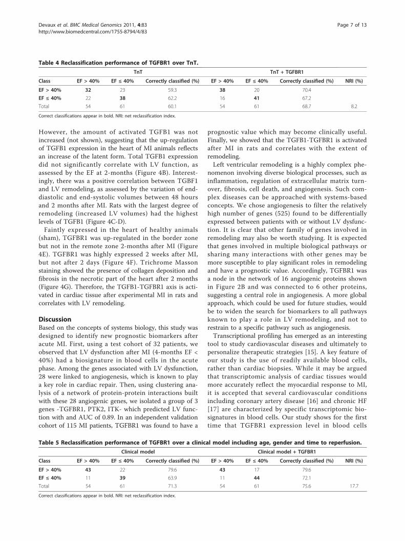
Table 4 Reclassification performance of TGFBR1 over TnT.
TnT TnT + TGFBR1
Class EF > 40% EF ≤ 40% Correctly classified (%) EF > 40% EF ≤ 40% Correctly classified (%) NRI (%)
EF > 40% 32 23 59.3 38 20 70.4
EF ≤ 40% 22 38 62.2 16 41 67.2
Total 54 61 60.1 54 61 68.7 8.2
Correct classifications appear in bold. NRI: net reclassification index.
Table 5 Reclassification performance of TGFBR1 over a clinical model including age, gender and time to reperfusion.
Clinical model Clinical model + TGFBR1
Class EF > 40% EF ≤ 40% Correctly classified (%) EF > 40% EF ≤ 40% Correctly classified (%) NRI (%)
EF > 40% 43 22 79.6 43 17 79.6
EF ≤ 40% 11 39 63.9 11 44 72.1
Total 54 61 71.3 54 61 75.6 17.7
Correct classifications appear in bold. NRI: net reclassification index.
Devaux et al. BMC Medical Genomics 2011, 4:83http://www.biomedcentral.com/1755-8794/4/83
Page 7 of 13

A
Technicalcontrol
Remotezone
Car
diac
TGFB
1
B
ShamMI
C EF2m(%)
EDV2m-EDV48h( μL)
D
0
1
2
3P=0.04
Car
diac
TGFB
1
0
1
2
3
0200400600
Car
diac
TG
FB1
0
1
2
3
203040506070
ESV2m-EDV48h( μL)0200400 600
Car
diac
TGFB
10
1
2
3r=0.61P=0.02
r=0.54P=0.04
E F
r=-0.46P=0.06
Remotezone Necroticzone
G
2 d a y s 2 w e e k s
Phasecontrast
DAPI
TGFBR1
Figure 4 The TGFB1-TGFBR1 axis in rats after MI. 17 rats were subjected to MI through permanent ligation of the LAD coronary artery. 4 ratswere sham-operated. Rats were assessed by PET to determine LV volumes and EF 48 hours and 2 months after ligation. Rats were sacrificed after2 days (n = 3), 2 weeks (n = 3) or 2 months (n = 11). At sacrifice, hearts were harvested. (A) Total TGFB1 measured by ELISA in cardiac samplesis up-regulated 2 months after MI. Data are expressed in pg of TGFB1 per μg of total cardiac proteins. Shown are mean ± 95% CI (n = 4 forsham, n = 11 for MI). (B) TGFB1 expression in the heart mildly correlates with 2-months EF. (C-D) TGFB1 expression correlates with the variationsof LV end-diastolic and end-systolic volumes between 48 hours and 2 months. Correlation coefficients and P values are indicated. (E)Immunohistochemical analysis of TGFBR1 expression in cardiac slices. The technical control without anti TGFBR1 antibody attests for thespecificity of the detection. TGFBR1 staining appears in red colour and nuclei appear in blue. TGFBR1 expression is up-regulated in the borderzone of MI rats. Representative pictures of sham-operated rats (upper panels) and MI rats (lower panels) are shown. (F) TGFBR1 expression in theborder zone 2 days and 2 weeks after MI. TGFBR1 expression is up-regulated in the border zone 2 weeks after MI. Representative pictures areshown. (G) Masson trichrome staining showing collagen deposition and fibrosis in the necrotic zone of the heart 2 months after MI.Representative pictures from a MI rat are shown.
Devaux et al. BMC Medical Genomics 2011, 4:83http://www.biomedcentral.com/1755-8794/4/83
Page 8 of 13
obtained at presentation in acute MI patients has prog-nostic value for long-term LV remodeling.In addition, we have observed an association between
TGFB1, which has prognostic values for remodeling andhypertrophy in patients with hypertension and aorticstenosis [18,19], and LV function. This association wasnot as robust as TGFBR1, and the prognostic value ofTGFBR1 was not found to be improved by TGFB1determination. Interestingly, TGFBR1 added some valueto the prediction of TnT, a marker of infarct size knownto affect LV remodeling. The reclassification of 8.2% ofpatients misclassified by TnT is clinically relevant. Simi-larly, the reclassification of 17.7% of patients misclassi-fied by a standard risk factor model based on age,gender and time to reperfusion, is pertinent.The integration of multiple biomarkers is thought to
improve the estimation of the course of cardiovasculardiseases [20], although this assumption has been ques-tioned [21]. Heidecker and coworkers reported that agroup of 45 genes identified from endomyocardial biop-sies predicted 5-year outcome of new onset HF patientswith a sensitivity of 74% and a specificity of 90% [22].More recently, a panel of 5 plasma proteins involved inextracellular matrix turnover was demonstrated to be amore accurate predictor of LV hypertrophy than anysingle biomarker [23]. Alternatively, combining biomar-kers representing diverse disease mechanisms has alsobeen shown to add incremental risk stratification valuein patients with non ST-elevation acute coronary syn-drome [24]. Our data showing that TGFBR1 has anadditive value to traditional markers are consistent withthe concept that multiplication of biomarkers may beclinically useful.More than 80% of the patients enrolled in this study
were males. The low number of females enrolled (6 inthe test cohort and 17 in the validation cohort) pre-vented us from investigating whether gender contributesto the prognostic value of TGFBR1. However, TGFBR1expression was not different between women and men(not shown), suggesting that gender may not influencethe prognostic value of TGFBR1.Activation of the TGFB1-TGFBR1 pathway is mainly
associated with stimulation of fibrosis, although it alsoaffects LV hypertrophy, matrix metabolism, inflamma-tion, and angiogenesis [25-27]. After MI, this pathwaytriggers the switch from inflammation to fibrosis[28-30]. TGFBR1 expression has been reported to behigher in the infarcted region of pig hearts after perma-nent coronary ligation compared to the remote region[31]. Our data in rats confirm and extend these observa-tions. It is believed that biomarkers should reflect thepathophysiology, and our experiments in rats providevaluable proof for this. Inhibition of TGFBR1 activity byorally active specific inhibitors [28,32] or competitive
inhibition of TGFB1 by a soluble form of TGFBR2 [33]dampens cardiac remodeling after MI. Other strategiesto block TGFB, which have been recently reviewed [34],may also be tested to limit cardiac remodelingEndothelial progenitor cells participate in cardiac
revascularization and healing after acute MI. However,considering their extremely low frequency in the circula-tion, these cells are certainly not the main source ofTGBFR1 signal measured by microarrays. Circulatingmonocytes-macrophages may most probably account forthe majority of TGFBR1 expression. Interestingly, thesecells secrete many cytokines, growth factors and fibroticfactors which regulate LV remodeling and cardiac heal-ing. The higher expression of TGFBR1 in patients withlow EF is consistent with the more robust activation ofinflammation observed in these patients, as attested byhigher white blood cells counts (not shown). Wholeblood cells profiling can be affected by shifts in leuko-cyte populations. However, the proportions of circulat-ing monocytes were comparable between patients withlow EF and patients with high EF, suggesting that thisconfounding factor did not importantly affect ourresults. It would be interesting to accurately determinewhich cell type(s) express TGFBR1 in the heart, andwhether TGFBR1 expression is regulated in cellsinvolved in cardiac healing. The animal model of coron-ary artery ligation used in the animal study is a valuabletool to answer these questions.The main limitation of the present study relies in the
small number of patients enrolled. This is particularlyrelevant for the test cohort used in microarray experi-ments. Nevertheless, using a different and quantitativetechnique, we were able to validate our findings fromthis small cohort in an independent cohort of morethan 100 patients. The relevance of our findingsdepends on the confirmation of the prognostic value ofTGFBR1 in larger patient populations. In addition, allhuman blood samples were collected at presentationand measurement of TGFBR1 expression at differenttime-points after acute MI could provide valuableinformations. Finally, mutations in TGFBR1 gene,known to affect vascular integrity in Marfan andLoeys-Dietz syndromes [35,36], have to be taken intoconsideration for the design of probes to measureTGFBR1 expression and for the design of specific ther-apeutic inhibitors.
ConclusionsWe have shown that TGFBR1 expression in blood cellsof acute MI patients is associated with LV remodeling.If confirmed in independent studies, this new biomarkermay become clinically useful to identify patients withacute MI who are at risk of adverse outcome and maybenefit from novel therapies.
Devaux et al. BMC Medical Genomics 2011, 4:83http://www.biomedcentral.com/1755-8794/4/83
Page 9 of 13
MethodsPatientsPatients with acute MI were enrolled in a national MIregistry and treated with primary percutaneous coronaryintervention. Acute MI was defined by the presence ofchest pain < 12 hours, significant ST elevation (> 1mm), completely occluded major coronary artery (TIMIO flow in LAD, CX or RCA), peak (≈ 24 hours) creatinephosphokinase (CPK) level > 600 units/L and peak tro-ponin T (TnT) level > 0.03 ng/mL. Blood samplesobtained shortly (5 min) after mechanical reperfusion,via an arterial catheter and into PAXgene™ tubes (BDBiosciences, Erembodegem, Belgium), were used fordetermination of TGFBR1 expression. TGFB1 was mea-sured in plasma extracted from citrated tubes collectedafter mechanical reperfusion. Time to reperfusion wasrecorded by dedicated nurses as the delay between chestpain onset evaluated by the patient and presentation.CPK activity was assessed with a Roche IFCC methodon a Cobas c501 instrument (Roche, Prophac, Luxem-bourg). TnT was assessed with a 4th generation assayfrom Roche performed on a Cobas e601 equipment(Roche). TGF-B1 was measured with the human TGF-B1 ELISA kit (ref DB100B, R&D Systems, Oxon, UK).LV function was assessed by echocardiography 4 monthsafter MI, and LV remodeling was defined by a LV ejec-tion fraction (EF) ≤ 40%. The protocol has beenapproved by the local ethics committee (National com-mittee of ethics in research of the Grand-Duchy of Lux-embourg) and informed consent has been obtained fromall subjects.
MicroarraysTranscriptomic analysis of blood cells was performed aspreviously described [7]. Total RNA was extracted fromwhole blood cells of MI patients using the PAXgene™blood RNA kit (Qiagen, Venlo, Netherlands). A secondpurification and concentration step was performed withthe RNeasy® MinElute™ kit (Qiagen). RNA quantitywas measured using the ND-1000 spectrophotometer(NanoDrop® Technologies, Wilmington, USA). RNAquality was assessed using the 2100 Bioanalyzer® appa-ratus (Agilent Technologies, Massy, France) with theRNA 6000 Nano chips. Only high quality RNA (OD260/OD280 > 1.9 and OD260/OD230 > 1.7) and un-degradedRNA was considered for further analysis. A commonreference RNA (Universal Human Reference RNA, Stra-tagene Europe, Amsterdam, The Netherlands) was usedin conjunction with patient’s RNA to provide an internalreference standard for comparisons of relative geneexpression levels across arrays. Messenger RNAs wereamplified using the Amino Allyl MessageAmp™ kit(Ambion®, Cambridgeshire, United Kingdom), startingwith one μg of total RNA. Five μg of each amino allyl
aRNA were labeled with Cy3 or Cy5 (Amersham, Buck-inghamshire, United Kingdom). Dye coupling to RNAwas measured using the ND-1000 NanoDrop® spectro-photometer. Coupling yield > 5% was a prerequisite forfurther analysis. 750 ng of each amino allyl aRNAlabeled Cy3 or Cy5 (reference RNA or patient RNA)were combined and hybridized on oligonucleotidemicroarrays representing 25,000 genes [14]. Four repli-cates and a dye-swap were performed. Scanning wasperformed with an Axon 4000B scanner and data wereacquired with the GenePix Pro 6® software (MolecularDevices, Berks, UK). Spot finding and raw data quantifi-cation were performed using the MAIA® freeware. ALowess non linear normalization was performed andgenes that were not present in at least 3 microarrays of4 were filtered out. Data are available at the GeneExpression Omnibus database (http://www.ncbi.nlm.nih.gov/geo/) under the accession number GSE11947.Before statistical analysis, genes not present in at least
50% of the patients were filtered out. Supervised analysiswas performed using the Significance Analysis of Micro-arrays software using 2-class unpaired test and 100 per-mutations. Heat maps were drawn using the Gene SetEnrichment Analysis (GSEA) software [37].
Quantitative PCRThe expression of mRNAs in blood cells of MI patientsobtained upon admission was determined by quantita-tive PCR. Total RNA was extracted from blood cells col-lected in PAXgene® blood RNA tubes. PCR wasperformed using a CFX96 device and the IQ™ SYBR®
Green Supermix (BioRad, Nazareth, Belgium). SF3A1was used for normalization.
Network of protein-protein interactionsGenes known to be associated with angiogenesis inhumans were retrieved from the Entrez-Gene database[38] with the query: “angiogenesis” AND “homosapiens”. The Human Protein Reference Database(release 9) [39] was then interrogated to identify allannotated protein-protein interactions associated withthese genes. These interactions were used as inputs tobuild a network, which was visualized and analyzed withCytoscape [40]. Clusters of highly interconnected pro-teins were identified by the MCODE plug-in networkclustering algorithm [41].
In vivo experiments and biochemical analysesMyocardial infarction was induced in 17 anesthetizedWistar rats (Charles Rivers Laboratories; 8-11 weeks;310-380 g) by permanent occlusion of the left anteriordescending (LAD) coronary artery as previouslydescribed [42,43]. An additional 4 rats were sham-oper-ated. All rats were monitored in vivo 48 hours after
Devaux et al. BMC Medical Genomics 2011, 4:83http://www.biomedcentral.com/1755-8794/4/83
Page 10 of 13

surgery and after 2 months with the use of a high reso-lution dedicated small animal positron emission tomo-graphy (PET) system with18F-FDG (IBA, Nancy, France)as a tracer and a premedication by acipimox, as pre-viously described [44]. LV end-diastolic volume(LVEDV) was obtained on contiguous gated short-axisslices by the Quantitative Gated SPECT software [45].The extent of remodeling was assessed by the change inLVEDV between 48-hours and 2-months after surgery(Δ LVEDV). At sacrifice, blood samples were collected,hearts were harvested, snap frozen and embedded inOCT. Parts of cardiac tissue were rinsed in NaCl andstored at -80°C for protein extraction. These experi-ments were conducted in accordance with the regula-tions of the Animal Welfare Act of the NationalInstitutes of Health Guide for the Care and Use ofLaboratory Animals (NIH Publication No. 85-23, revised1996) and protocols were approved by local EthicsCommittee and by the Regional Veterinary Department.For immunohistochemistry and collagen-specific Mas-
son Trichrome staining (Dako, Leuven, Belgium), 8 μmfrozen heart sections were generated. Primary antibodyused was a rabbit polyclonal anti TGFBR1 antibody(Abcam, Cambridge, UK). Alexa Fluor®568-coupleddonkey anti-rabbit antibodies (Jackson ImmunoRe-search, UK) were used as secondary antibodies. Imageswere recorded on a confocal microscope (Zeiss LaserScanning Microscope LSM 510) with a 40× objectiveusing the LSM 510 META software. DAPI (blue) stain-ing was used to reveal nuclei.The TGFB1 ELISA kit (ref MB100B, R&D Systems)
was used to measure TGFB1 in cardiac tissue. 50 μg oftotal proteins from each heart were assayed. This assaydetects activated TGFB1 with a sensitivity of 4.6 pg/mL.To measure total TGFB1, latent TGFB1 is activated byacidification with hydrochloric acid prior to assay. Coef-ficient of variation intra-assay is < 4% and coefficientinter-assay is < 9%.
Statistical analysisComparisons between means of two groups were per-formed with the Mann-Whitney test. Categorical vari-ables were compared using the Fisher exact test.Correlation between biomarker levels and the EF classwas estimated with the Spearman test. All tests weretwo-tailed and were preceded by a normality test (Sha-piro-Wilk). A P value < 0.05 was considered statisticallysignificant. The SigmaPlot (v. 11.0) was used to carry-out statistical tests.Prognostic performances of single genes were evalu-
ated using receiver operating characteristic (ROC)curves and the area under the ROC curves (AUC), aswell as linear regression models. Logistic regressionmodels with ridge parameter of 1.0E-8 used to evaluate
the predictive performances of multiple markers wereimplemented and tested using 10-fold cross-validationon the Weka (v. 3.4) data mining platform [46].Reclassification analyses have been performed to eval-
uate the additive value of TGFBR1 to TnT and to amixed clinical model. The net reclassification index(NRI) was calculated as the difference in proportionsmoving up and down among cases and controls: [Pr(up|case)-Pr(down|case)]-[Pr(up|control)-Pr(down|control)].
AcknowledgementsWe thank Céline Jeanty, Malou Gloesener and Loredana Jacobs for experttechnical assistance. This study was supported by grants from the Society forResearch on Cardiovascular Diseases, the Ministry of Culture, HigherEducation and Research, and the National Funds of Research ofLuxembourg. M.B. is recipient of a fellowship from the National Funds ofResearch of Luxembourg (grant # PhD-AFR 08-024).
Author details1Laboratory of Cardiovascular Research Centre de Recherche Public-Santé,Luxembourg, L-1150, Luxembourg. 2Nancyclotep Experimental ImagingPlatform, Nancy, F-54500, France. 3Division of Cardiology, Centre Hospitalier,Luxembourg, L-1210, Luxembourg.
Authors’ contributionsYD conceived the study and prepared the manuscript; MB performed in vivoexperiments; SR performed in vitro experiments; PYM and FM participated inanimal experiments; LZ analyzed microarray data; FA and DRW participatedin study design and revised the manuscript. All authors have read andapproved the final manuscript.
Competing interestsThe authors declare that they have no competing interests.
Received: 10 August 2011 Accepted: 5 December 2011Published: 5 December 2011
References1. Torabi A, Cleland JG, Khan NK, Loh PH, Clark AL, Alamgir F, Caplin JL,
Rigby AS, Goode K: The timing of development and subsequent clinicalcourse of heart failure after a myocardial infarction. Eur Heart J 2008,29:859-870.
2. Ezekowitz JA, Kaul P, Bakal JA, Armstrong PW, Welsh RC, McAlister FA:Declining in-hospital mortality and increasing heart failure incidence inelderly patients with first myocardial infarction. J Am Coll Cardiol 2009,53:13-20.
3. Schocken DD, Benjamin EJ, Fonarow GC, Krumholz HM, Levy D, Mensah GA,Narula J, Shor ES, Young JB, Hong Y: Prevention of heart failure: ascientific statement from the American Heart Association Councils onEpidemiology and Prevention, Clinical Cardiology, CardiovascularNursing, and High Blood Pressure Research; Quality of Care andOutcomes Research Interdisciplinary Working Group; and FunctionalGenomics and Translational Biology Interdisciplinary Working Group.Circulation 2008, 117:2544-2565.
4. de Kam PJ, Nicolosi GL, Voors AA, van den Berg MP, Brouwer J, vanVeldhuisen DJ, Barlera S, Maggioni AP, Giannuzzi P, Temporelli PL, Latini R,van Gilst WH: Prediction of 6 months left ventricular dilatation aftermyocardial infarction in relation to cardiac morbidity and mortality.Application of a new dilatation model to GISSI-3 data. Eur Heart J 2002,23:536-542.
5. Azuaje F: What does systems biology mean for biomarker discovery? ExpOpin Med Diagn 2009, 4:1-10.
6. Azuaje F, Devaux Y, Wagner DR: Integrative pathway-centric modeling ofventricular dysfunction after myocardial infarction. PLoS One 2010, 5:e9661.
7. Devaux Y, Azuaje F, Vausort M, Yvorra C, Wagner DR: Integrated proteinnetwork and microarray analysis to identify potential biomarkers aftermyocardial infarction. Funct Integr Genomics 2010, 10:329-337.
Devaux et al. BMC Medical Genomics 2011, 4:83http://www.biomedcentral.com/1755-8794/4/83
Page 11 of 13

8. Nepomuceno-Chamorro I, Azuaje F, Devaux Y, Nazarov PV, Muller A,Aguilar-Ruiz JS, Wagner DR: Prognostic transcriptional associationnetworks: a new supervised approach based on regression trees.Bioinformatics 2011, 27:252-258.
9. Gerber BL, Rochitte CE, Melin JA, McVeigh ER, Bluemke DA, Wu KC,Becker LC, Lima JA: Microvascular obstruction and left ventricularremodeling early after acute myocardial infarction. Circulation 2000,101:2734-2741.
10. Bolognese L, Carrabba N, Parodi G, Santoro GM, Buonamici P, Cerisano G,Antoniucci D: Impact of microvascular dysfunction on left ventricularremodeling and long-term clinical outcome after primary coronaryangioplasty for acute myocardial infarction. Circulation 2004,109:1121-1126.
11. Nijveldt R, Beek AM, Hirsch A, Stoel MG, Hofman MB, Umans VA, Algra PR,Twisk JW, van Rossum AC: Functional recovery after acute myocardialinfarction: comparison between angiography, electrocardiography, andcardiovascular magnetic resonance measures of microvascular injury. JAm Coll Cardiol 2008, 52:181-189.
12. Abdollahi A, Schwager C, Kleeff J, Esposito I, Domhan S, Peschke P,Hauser K, Hahnfeldt P, Hlatky L, Debus J, Peters JM, Friess H, Folkman J,Huber PE: Transcriptional network governing the angiogenic switch inhuman pancreatic cancer. Proc Natl Acad Sci USA 2007, 104:12890-12895.
13. Azuaje F, Devaux Y, Vausort M, Yvorra C, Wagner DR: Transcriptionalnetworks characterize ventricular dysfunction after myocardial infarction:A proof-of-concept investigation. J Biomed Inform 2010, 43:812-819.
14. Le Brigand K, Russell R, Moreilhon C, Rouillard JM, Jost B, Amiot F,Magnone V, Bole-Feysot C, Rostagno P, Virolle V, Defamie V, Dessen P,Williams G, Lyons P, Rios G, Mari B, Gulari E, Kastner P, Gidrol X,Freeman TC, Barbry P: An open-access long oligonucleotide microarrayresource for analysis of the human and mouse transcriptomes. NucleicAcids Res 2006, 34:e87.
15. Margulies KB, Bednarik DP, Dries DL: Genomics, transcriptional profiling,and heart failure. J Am Coll Cardiol 2009, 53:1752-1759.
16. Wingrove JA, Daniels SE, Sehnert AJ, Tingley W, M E, Rosenberger S,Buellesfeld L, Grube E, Newby LK, Ginsburg GS, Kraus WE: Correlation ofPeripheral-Blood Gene Expression With the Extent of Coronary ArteryStenosis. Circ Cardiovasc Genet 2008, 1:31-38.
17. Cappuzzello C, Napolitano M, Arcelli D, Melillo G, Melchionna R, Di Vito L,Carlini D, Silvestri L, Brugaletta S, Liuzzo G, Crea F, Capogrossi MC: Geneexpression profiles in peripheral blood mononuclear cells of chronicheart failure patients. Physiol Genomics 2009, 38:233-240.
18. Almendral JL, Shick V, Rosendorff C, Atlas SA: Association betweentransforming growth factor-[beta]1 and left ventricular mass anddiameter in hypertensive patients. J Am Soc Hypertens 2010, 4:135-141.
19. Villar AV, Cobo M, Llano M, Montalvo C, Gonzalez-Vilchez F, Martin-Duran R,Hurle MA, Nistal JF: Plasma Levels of Transforming Growth Factor-Î21Reflect Left Ventricular Remodeling in Aortic Stenosis. PLoS One 2009, 4:e8476.
20. Zethelius B, Berglund L, Sundstrom J, Ingelsson E, Basu S, Larsson A,Venge P, Arnlov J: Use of multiple biomarkers to improve the predictionof death from cardiovascular causes. N Engl J Med 2008, 358:2107-2116.
21. Ware JH: The limitations of risk factors as prognostic tools. N Engl J Med2006, 355:2615-2617.
22. Heidecker B, Kasper EK, Wittstein IS, Champion HC, Breton E, Russell SD,Kittleson MM, Baughman KL, Hare JM: Transcriptomic biomarkers forindividual risk assessment in new-onset heart failure. Circulation 2008,118:238-246.
23. Zile MR, Desantis SM, Baicu CF, Stroud RE, Thompson SB, McClure CD,Mehurg SM, Spinale FG: Plasma Biomarkers That Reflect Determinants ofMatrix Composition Identify the Presence of Left VentricularHypertrophy and Diastolic Heart Failure. Circ Heart Fail 2011, 4:246-256.
24. Oemrawsingh RM, Lenderink T, Akkerhuis KM, Heeschen C, Baldus S,Fichtlscherer S, Hamm CW, Simoons ML, Boersma E: Multimarker riskmodel containing troponin-T, interleukin 10, myeloperoxidase andplacental growth factor predicts long-term cardiovascular risk after non-ST-segment elevation acute coronary syndrome. Heart 2011,97:1061-1066.
25. Koitabashi N, Danner T, Zaiman AL, Pinto YM, Rowell J, Mankowski J,Zhang D, Nakamura T, Takimoto E, Kass DA: Pivotal role of cardiomyocyteTGF-beta signaling in the murine pathological response to sustainedpressure overload. J Clin Invest 2011, 126:2301-2312.
26. Pepper MS: Transforming growth factor-beta: vasculogenesis,angiogenesis, and vessel wall integrity. Cytokine Growth Factor Rev 1997,8:21-43.
27. Dobaczewski M, Chen W, Frangogiannis NG: Transforming growth factor(TGF)-[beta] signaling in cardiac remodeling. J Mol Cell Cardiol 2011,51:600-606.
28. Ellmers LJ, Scott NJA, Medicherla S, Pilbrow AP, Bridgman PG, Yandle TG,Richards AM, Protter AA, Cameron VA: Transforming Growth Factor-{beta}Blockade Down-Regulates the Renin-Angiotensin System and ModifiesCardiac Remodeling after Myocardial Infarction. Endocrinology 2008,149:5828-5834.
29. Bujak M, Ren G, Kweon HJ, Dobaczewski M, Reddy A, Taffet G, Wang X-F,Frangogiannis NG: Essential Role of Smad3 in Infarct Healing and in thePathogenesis of Cardiac Remodeling. Circulation 2007, 116:2127-2138.
30. Kapur NK: Transforming Growth Factor-Î2. Circ Heart Fail 2011, 4:5-7.31. Mukherjee R, Rivers WT, Ruddy JM, Matthews RG, Koval CN, Plyler RA,
Chang EI, Patel RK, Kern CB, Stroud RE, Spinale FG: Long-Term LocalizedHigh-Frequency Electric Stimulation Within the Myocardial Infarct:Effects on Matrix Metalloproteinases and Regional Remodeling.Circulation 2010, 122:20-32.
32. Tan SM, Zhang Y, Connelly KA, Gilbert RE, Kelly DJ: Targeted inhibition ofactivin receptor-like kinase 5 signaling attenuates cardiac dysfunctionfollowing myocardial infarction. Am J Physiol-Heart and Circ Physiol 2010,298:H1415-H1425.
33. Okada H, Takemura G, Kosai K-i, Li Y, Takahashi T, Esaki M, Yuge K, Miyata S,Maruyama R, Mikami A, Minatoguchi S, Fujiwara T, Fujiwara H:Postinfarction Gene Therapy Against Transforming Growth Factor-{beta}Signal Modulates Infarct Tissue Dynamics and Attenuates LeftVentricular Remodeling and Heart Failure. Circulation 2005, 111:2430-2437.
34. Hawinkels LJAC, ten Dijke P: Exploring anti-TGF-Î2 therapies in cancer andfibrosis. Growth Factors 2011, 29:140-152.
35. Loeys BL, Chen J, Neptune ER, Judge DP, Podowski M, Holm T, Meyers J,Leitch CC, Katsanis N, Sharifi N, Xu FL, Myers LA, Spevak PJ, Cameron DE,De Backer J, Hellemans J, Chen Y, Davis EC, Webb CL, Kress W, Coucke P,Rifkin DB, De Paepe AM, Dietz HC: A syndrome of altered cardiovascular,craniofacial, neurocognitive and skeletal development caused bymutations in TGFBR1 or TGFBR2. Nat Genet 2005, 37:275-281.
36. Loeys BL, Schwarze U, Holm T, Callewaert BL, Thomas GH, Pannu H, DeBacker JF, Oswald GL, Symoens S, Manouvrier S, Roberts AE, Faravelli F,Greco MA, Pyeritz RE, Milewicz DM, Coucke PJ, Cameron DE, Braverman AC,Byers PH, De Paepe AM, Dietz HC: Aneurysm syndromes caused bymutations in the TGF-beta receptor. N Engl J Med 2006, 355:788-798.
37. Subramanian A, Tamayo P, Mootha VK, Mukherjee S, Ebert BL, Gillette MA,Paulovich A, Pomeroy SL, Golub TR, Lander ES, Mesirov JP: Gene setenrichment analysis: a knowledge-based approach for interpretinggenome-wide expression profiles. Proc Natl Acad Sci USA 2005,102:15545-15550.
38. The Entrez-Gene Database. [http://www.ncbi.nlm.nih.gov/entrez].39. Peri S, Navarro JD, Amanchy R, Kristiansen TZ, Jonnalagadda CK,
Surendranath V, Niranjan V, Muthusamy B, Gandhi TK, Gronborg M,Ibarrola N, Deshpande N, Shanker K, Shivashankar HN, Rashmi BP,Ramya MA, Zhao Z, Chandrika KN, Padma N, Harsha HC, Yatish AJ,Kavitha MP, Menezes M, Choudhury DR, Suresh S, Ghosh N, Saravana R,Chandran S, Krishna S, Joy M, Anand SK, Madavan V, Joseph A, Wong GW,Schiemann WP, Constantinescu SN, Huang L, Khosravi-Far R, Steen H,Tewari M, Ghaffari S, Blobe GC, Dang CV, Garcia JG, Pevsner J, Jensen ON,Roepstorff P, Deshpande KS, Chinnaiyan AM, Hamosh A, Chakravarti A,Pandey A: Development of human protein reference database as aninitial platform for approaching systems biology in humans. Genome Res2003, 13:2363-2371.
40. Shannon P, Markiel A, Ozier O, Baliga NS, Wang JT, Ramage D, Amin N,Schwikowski B, Ideker T: Cytoscape: a software environment forintegrated models of biomolecular interaction networks. Genome Res2003, 13:2498-2504.
41. Bader GD, Hogue CW: An automated method for finding molecularcomplexes in large protein interaction networks. BMC Bioinformatics 2003,4:2.
42. Maskali F, Franken PR, Poussier S, Tran N, Vanhove C, Boutley H, Le Gall H,Karcher G, Zannad F, Lacolley P, Marie PY: Initial infarct size predictssubsequent cardiac remodeling in the rat infarct model: an in vivo serialpinhole gated SPECT study. J Nucl Med 2006, 47:337-344.
Devaux et al. BMC Medical Genomics 2011, 4:83http://www.biomedcentral.com/1755-8794/4/83
Page 12 of 13

43. Maskali F, Poussier S, Marie PY, Tran N, Antunes L, Olivier P, Plenat F,Maitrejean S, Zannad F, Karcher G: High-resolution simultaneous imagingof SPECT, PET, and MRI tracers on histologic sections of myocardialinfarction. J Nucl Cardiol 2005, 12:229-230.
44. Poussier S, Maskali F, Tran N, Person C, Maureira P, Boutley H, Karcher G,Lacolley P, Regnault V, Fay R, Marie PY: ECG-triggered (18)F-fluorodeoxyglucose positron emission tomography imaging of the ratheart is dramatically enhanced by acipimox. Eur J Nucl Med Mol Imaging2010, 37:1745-1750.
45. Germano G, Kiat H, Kavanagh PB, Moriel M, Mazzanti M, Su HT, VanTrain KF, Berman DS: Automatic quantification of ejection fraction fromgated myocardial perfusion SPECT. J Nucl Med 1995, 36:2138-2147.
46. Frank E, Hall M, Trigg L, Holmes G, Witten IH: Data mining inbioinformatics using Weka. Bioinformatics 2004, 20:2479-2481.
Pre-publication historyThe pre-publication history for this paper can be accessed here:http://www.biomedcentral.com/1755-8794/4/83/prepub
doi:10.1186/1755-8794-4-83Cite this article as: Devaux et al.: Transforming growth factor b receptor1 is a new candidate prognostic biomarker after acute myocardialinfarction. BMC Medical Genomics 2011 4:83.
Submit your next manuscript to BioMed Centraland take full advantage of:
• Convenient online submission
• Thorough peer review
• No space constraints or color figure charges
• Immediate publication on acceptance
• Inclusion in PubMed, CAS, Scopus and Google Scholar
• Research which is freely available for redistribution
Submit your manuscript at www.biomedcentral.com/submit
Devaux et al. BMC Medical Genomics 2011, 4:83http://www.biomedcentral.com/1755-8794/4/83
Page 13 of 13