Embed Size (px)
Citation preview

Baumgart et al. BMC Biology 2013, 11:122http://www.biomedcentral.com/1741-7007/11/122
RESEARCH ARTICLE Open Access
IpsA, a novel LacI-type regulator, is required forinositol-derived lipid formation in Corynebacteriaand MycobacteriaMeike Baumgart1, Kerstin Luder1, Shipra Grover2, Cornelia Gätgens1, Gurdyal S Besra2 and Julia Frunzke1*
Abstract
Background: The development of new drugs against tuberculosis and diphtheria is focused on disrupting thebiogenesis of the cell wall, the unique architecture of which confers resistance against current therapies. Theenzymatic pathways involved in the synthesis of the cell wall by these pathogens are well understood, but theunderlying regulatory mechanisms are largely unknown.
Results: Here, we characterize IpsA, a LacI-type transcriptional regulator conserved among Mycobacteria andCorynebacteria that plays a role in the regulation of cell wall biogenesis. IpsA triggers myo-inositol formation byactivating ino1, which encodes inositol phosphate synthase. An ipsA deletion mutant of Corynebacteriumglutamicum cultured on glucose displayed significantly impaired growth and presented an elongated cellmorphology. Further studies revealed the absence of inositol-derived lipids in the cell wall and a complete loss ofmycothiol biosynthesis. The phenotype of the C. glutamicum ipsA deletion mutant was complemented to differentextend by homologs from Corynebacterium diphtheriae (dip1969) and Mycobacterium tuberculosis (rv3575), indicatingthe conserved function of IpsA in the pathogenic species. Additional targets of IpsA with putative functions in cellwall biogenesis were identified and IpsA was shown to bind to a conserved palindromic motif within thecorresponding promoter regions. Myo-inositol was identified as an effector of IpsA, causing the dissociation of theIpsA-DNA complex in vitro.
Conclusions: This characterization of IpsA function and of its regulon sheds light on the complex transcriptionalcontrol of cell wall biogenesis in the mycolata taxon and generates novel targets for drug development.
Keywords: Transcriptional regulator, Corynebacterium glutamicum, Corynebacterium diphtheriae, Mycobacteriumtuberculosis, Cell wall synthesis, Mycolic acids, Mycothiol, Inositol, LM, LAM
BackgroundThe human pathogens Mycobacterium tuberculosis andCorynebacterium diphtheriae, as well as many related spe-cies of the order Corynebacteriales, share a distinctive cellwall architecture that plays an important role in virulenceand imparts effective protection against harsh environmen-tal conditions and resistance to therapies [1]. One uniquefeature of these cell walls is the presence of long-chainα-alkyl, β-hydroxy fatty acids, called mycolic acids, whichform a second bilayer close to the cell surface that is simi-lar to the outer membrane of Gram-negative bacteria [2].
* Correspondence: [email protected] für Bio- und Geowissenschaften, IBG-1: Biotechnologie,Forschungszentrum Jülich, 52425 Jülich, GermanyFull list of author information is available at the end of the article
© 2013 Baumgart et al.; licensee BioMed CentCommons Attribution License (http://creativecreproduction in any medium, provided the orwaiver (http://creativecommons.org/publicdomstated.
The enzymes and regulatory mechanisms involved in bio-synthesis of the cell walls are primary targets for the devel-opment of new drugs.A great deal is known about these bacterial cell walls
(Figure 1A). The plasma membranes are rich in phospho-lipids, such as phosphatidylglycerol, diphosphatidylglyceroland phosphatidylinositol (PI). While inositol is an essentialpolyol in eukaryotes, in eubacteria it is present only in theorder Corynebacteriales where it serves both as a cell wallcomponent and as a building block of mycothiol (MSH),the major antioxidant in Mycobacteria, similar to glutathi-one [3]. One function of PI is to anchor cell wall lipogly-cans, specifically PI mannosides (PIMs), lipomannan (LM)and liporarabinomannan (LAM), to the plasma membrane
ral Ltd. This is an Open Access article distributed under the terms of the Creativeommons.org/licenses/by/2.0), which permits unrestricted use, distribution, andiginal work is properly cited. The Creative Commons Public Domain Dedicationain/zero/1.0/) applies to the data made available in this article, unless otherwise

Figure 1 IpsA plays an essential role in the integrity of the cell wall of Corynebacteriales. (A) Current model of the structure of the cell wallof Corynebacteria and Mycobacteria. (B) Comparison of the organization of the IpsA genome locus in C. glutamicum and related species. IpsA andhomologous proteins are shown in black. Data were taken from [5,6]. The percentage identity of the amino acid sequences to IpsA from C.glutamicum was taken from NCBI Blast. hp, hypothetical protein. (C, D) Growth curves (C) and phenotype (D) of C. glutamicum wild-type andΔipsA. Results in (C) show the growth (backscatter) from sequential cultures in minimal medium with glucose or myo-inositol as carbon source.After reaching the stationary phase, the cells were diluted into fresh medium at an initial OD600 of 1. Shown are three glucose cultures and thethird myo-inositol culture (n = 3). In (D), DNA was stained with 4’,6-diamidino-2-phenylindole (DAPI) (cyan) and lipophilic regions with nile red(red). The scale bar is 5 μm.
Baumgart et al. BMC Biology 2013, 11:122 Page 2 of 14http://www.biomedcentral.com/1741-7007/11/122
[1,4]. The biogenesis of LAM is performed in consecutivesteps in the following order: PI→PIM→LM→LAM.In Mycobacteria, there is a three-step process for syn-
thesis of PI. Step 1 is cyclization of glucose-6-phosphate to1D-myo-inositol-3-phosphate, a reaction that is catalyzedby a myo-inositol phosphate synthase, Ino1 (Rv0046c) [7].In step 2, the 1D-myo-inositol-3-phosphate is dephosphor-ylated by inositol monophosphatase (IMP) [8] to yieldmyo-inositol. Step 3 is the transfer of diacylglycerol (DAG)from cytidine diphosphate-diacylglycerol (CDP-DAG) tomyo-inositol by a PI synthase, PgsA (Rv2612c) [9]. Theenzymes Ino1 and PgsA are essential for the viability ofMycobacteria [10,11]. An ino1 mutant of M. tuberculosisgrows in vitro only in the presence of high inositol con-centrations and was no longer able to kill SCID mice (seeMovahedzadeh et al., [10]). In Mycobacterium smegmatis,ino1 has been shown to be essential for growth in the
absence of exogenous inositol [12]. Ino1 has thereforebeen proposed as a target for the development of newantibiotics [10,13].In contrast to the wealth of knowledge about the bio-
genesis of cell walls, little is known about the regulatorycircuits that control these processes [4]. IpsA waschosen as target for further studies from a set of mu-tants strains as a ΔipsA mutant displayed a drastically al-tered cell morphology hinting towards a function in thecontrol of cell wall biosynthesis. The catabolism of myo-inositol has been examined in C. glutamicum [14,15] butregulation of myo-inositol formation has not been deeplyinvestigated up until now. Here, a regulator of inositolsynthesis in the model organism C. glutamicum is de-scribed. Named IpsA, it is a novel LacI-type regulatorthat acts as an inositol-dependent transcriptional activa-tor of the myo-inositol phosphate synthase gene. IpsA

Figure 2 Complementation of the ΔipsA phenotype with ipsAand the target gene ino1 (cg3323). For chromosomalcomplementation, the gene ipsA was integrated into the intergenicregion of cg1121 to cg1122 under control of the native promoter.(A) Growth of the strains in CGXII minimal medium with 2% (w/v)glucose as carbon source. The average and standard deviation ofthree biological replicates is shown. (B) Fluorescence microscopyimages of stationary phase cells of the cultures presented in (A). (C)Plasmid-based complementation with the target gene ino1 (cg3323).The strains were cultivated in CGXII minimal medium with 2% (w/v)glucose and 50 μM isopropyl β-D-1-thiogalactopyranoside (IPTG) forinduction of ino1 expression.
Baumgart et al. BMC Biology 2013, 11:122 Page 3 of 14http://www.biomedcentral.com/1741-7007/11/122
function is conserved throughout Corynebacteria andMycobacteria, including the prominent pathogenic speciesC. diphtheriae and M. tuberculosis.
ResultsIpsA is a novel LacI-type regulator conserved in theCorynebacterialesIpsA was assigned by homology to the LacI family oftranscriptional regulators, which often coordinate theavailable nutrients with the expression of catabolic genes[16]. The corresponding genomic loci display a remark-able level of conservation across the Corynebacteriales(Figure 1B). In previous studies, the structure of IpsA(Cg2910) was analyzed (PDB:3H5T); its physiologicalfunction is the subject of this study.
IpsA deletion affects cell shape and growthTo gain insight into the function and possible targetgenes of IpsA, an in-frame deletion mutant was con-structed. Growth rates and final backscatter of ΔipsAcells were significantly below control levels using min-imal medium with glucose as carbon source, and theseshortfalls increased in magnitude with successive subcul-tivation (Figure 1C). The morphology of the mutantstrain was altered, as revealed by fluorescence micros-copy with staining of DNA (4’,6-diamidino-2-phenylin-dole (DAPI)) and membranes (nile red). ΔipsA cellsformed chain-like structures and failed to divide prop-erly. DNA in the cells was unevenly distributed and wasfrequently found in the extracellular space, indicative ofsevere damage to the cell membrane and/or the cell wall(Figure 1D, for a larger sample see Additional file 1:Figure S1). Membrane staining revealed several intactcell septa in the unshaped mutant cells.Both the growth defect and altered morphology could
be partially overcome by plasmid-encoded ipsA undercontrol of the native or an inducible promoter (data notshown). These efforts revealed that increased levels ofIpsA also affect growth of C. glutamicum. Full restor-ation of the wild-type phenotype was only achievedwhen ipsA was reintegrated into the mutant strain(intergenic region cg1121 to cg1122) and expressed fromits native promoter (Figure 2A, B). When the cells werecultivated with myo-inositol instead of glucose as carbonsource, no differences were observed between the wild-type and ΔipsA strains in terms of growth and morph-ology (Figure 1C, D).
The impact of ipsA mutation on transcriptionTo elucidate the transcriptional changes caused by thedeletion of ipsA, DNA microarray experiments wereperformed. More than 450 genes showed alterations inmRNA level greater than a factor of 2, using cells in theexponential growth phase (OD600 = 5) cultivated in minimal

Baumgart et al. BMC Biology 2013, 11:122 Page 4 of 14http://www.biomedcentral.com/1741-7007/11/122
medium with glucose as carbon source (Additional file 1:Table S3, full data deposited in GEO database [17] underaccession number GSE50210).The expression of the gene cg3323, which encodes a
myo-inositol phosphate synthase (ino1), and of the op-eron (cg0044 to cg0046), which encodes an uncharacter-ized ABC transporter, was reduced by more than afactor of ten in the mutant strain (Table 1). Other down-regulated genes included a flavin-containing monooxy-genase (cg3195), a hypothetical endoglucanase (cg2896),and two further transporters (cg0621 to cg0623 andcg2181 to cg2184) (Additional file 1: Table S3). Amongthe strongly upregulated genes was a cluster encodingenzymes for inositol catabolism (cg3389 to cg3392), anoperon encoding both menE and a putative integralmembrane protein (cg0533 to cg0534), and a putativedinucleotide-binding enzyme (cg1421). Many of theaffected genes are components of the DtxR stimulon,which plays a central role in iron homeostasis in C.glutamicum [18,19]. We speculate that these changesare the result of an impaired iron uptake of the mutantstrain due to the defects in the cell wall. In combinationwith the observed growth phenotype of the mutantstrain, the alterations in gene expression indicate thatIpsA is a major regulator of inositol-derived cell wallcomponents.
Identification of direct IpsA target promotersThe microarray experiments revealed several putativetarget genes of IpsA. To determine whether these genesare directly regulated by IpsA, the promoter regions ofthe respective genes were tested in electrophoretic
Table 1 Transcriptome analysis of an IpsA deletionmutant
Locustag
Gene Annotated function Ratio
cg3323 ino1 Myo-inositol phosphate synthase 0.047
cg0044 rbsB ABC transporter/periplasmic D-ribose-bindingprotein
0.082
cg0045 - Probable ABC transport protein, membranecomponent
0.138
cg2910 - Transcriptional regulator, LacI family 0.173
cg3195 - Flavin-containing monooxygenase 0.178
cg0046 - Probable ABC transport protein, ATP-bindingcomponent
0.323
cg3210 - Cell envelope-related transcriptional regulator 0.411
cg1421 - Putative dinucleotide-binding enzyme 5.214
cg0533 menE O-succinylbenzoic acid-CoA ligase 5.467
cg0534 - Putative integral membrane protein 5.597
mRNA ratios (ΔipsA/wild-type) of IpsA target genes in microarray experimentsare shown. Presented are all genes where binding of IpsA could be shown inelectrophoretic mobility shift assays (EMSAs) as well as IpsA itself, confirmingthe deletion.
mobility shift assays (EMSAs) for the formation of com-plexes with purified IpsA protein. We analyzed the pro-moters of all genes that were more than fourfoldregulated but excluded ribosomal proteins and genesbelonging to the DtxR/RipA regulon. DNA fragments(approximately 500 bp) covering the promoter regionsof putative target genes, were incubated with increasingIpsA-His concentrations. IpsA-His bound with highaffinity to the promoter regions of cg0044 and ino1(cg3323) and with lower affinity to the promoter regionsof cg0534 (a putative integral membrane protein),cg1421 (a putative dinucleotide binding enzyme), cg1476(thiC, thiamine biosynthesis protein ThiC), cg1918 (aputative secreted protein), and cg3195 (flavin containingmonooxygenase) (Figure 3; all tested fragments areshown in Additional file 1: Figure S3).
Determination of the IpsA DNA binding siteUsing EMSAs, two separate binding sites were identifiedin the promoter of ino1 and one in the promoter ofcg0044 (Figure 3A and Additional file 1: Figure S4). Ana-lysis of these sites using the motif-based sequence analysistool MEME [20] revealed a conserved palindromic motif,which was subsequently verified by mutational analysis(Figure 4). In particular, the central six base pairs appearto be crucial for IpsA binding. The preliminary motif wasused to search for further binding sites in putative targetgenes and across the genome of C. glutamicum. Bindingsites were identified by EMSAs using oligonucleotides inthe promoters of cg1421, cg3195, cg3210 and cg0534(Table 1, Figure 3A and Additional file 1: Figure S5). Theidentified motifs were used to define the IpsA consensusmotif shown in Figure 3C.
Myo-inositol is an effector molecule of IpsAThe enzyme encoded by the main IpsA target gene isIno1, a myo-inositol phosphate synthase that catalyzes theformation of 1D-myo-inositol-3-phosphate from glucose-6-phosphate [7]. We tested several metabolites involvedin the relevant pathway for their influence on the forma-tion of the IpsA-DNA complex. A clear backshift wasobserved for myo-inositol but not for 1D-myo-inositol-1-phosphate, 1D-myo-inositol-3-phosphate or glucose-6-phosphate (Figure 3B). The effect of myo-inositol could beobserved at concentrations down to 0.2 mM (Additionalfile 1: Figure S6). This effect is specific for IpsA DNA bind-ing, as the addition of up to 50 mM myo-inositol did nothave any effect on the formation of an unrelated protein-DNA complex (for example, AcnR and the acn promoter,Additional file 1: Figure S7).
IpsA is required for inositol-dependent regulation of ino1The major function of IpsA revealed thus far is the acti-vation of ino1 in the absence of external inositol.

Figure 3 Target genes, effector and binding sites of IpsA. (A) Binding of IpsA to 30 bp oligonucleotides covering the binding sites in theputative target promoters. As negative control the oligonucleotide cg3323-d (sequence AGGTCTGATTTCTGCTGGGAATCCCCACAT) was used,which is located immediately downstream of the IpsA binding sites in the cg3323 promoter. (B) The influence of intermediates in inositolmetabolism (5 mM each) on the binding of IpsA to the ino1 promoter. myo-I, myo-inositol; I1P, 1D-myo-inositol-1-phosphate; I3P, 1D-myo-inositol-3-phosphate; G6P, glucose-6-phosphate. (C) The IpsA consensus motif predicted by MEME [20] and an overview of the sites in the correspondingpromoter regions. (D) Promoter-fusion studies using the promoter of ino1 fused to eyfp (pJC1-Pcg3323-eyfp). The specific fluorescence of ATCC13032 containing this plasmid on glucose was set to 1 and the other values were calculated accordingly.
Baumgart et al. BMC Biology 2013, 11:122 Page 5 of 14http://www.biomedcentral.com/1741-7007/11/122
Consistent with this, the growth and cell morphology ofthe ΔipsA strain is complemented by the constitutive ex-pression of ino1 under standard conditions (Figure 2C).To study the influence of IpsA and myo-inositol on theexpression of ino1 in vivo, we fused the ino1 promoterto eyfp (enhanced yellow fluorescent protein) and moni-tored the fluorescence output of the wild-type and theΔipsA strain during growth on glucose and myo-inositol(Figure 3D). Strong promoter activity was observed inthe wild-type during growth on glucose. Expression ofeyfp was significantly decreased in the ipsA deletionstrain and was lower still when either strain was culti-vated with myo-inositol as the carbon source.
IpsA function is conserved in C. diphtheriae and M.tuberculosisThe conservation of IpsA in the Corynebacteriales sug-gests that it may play a similar role in pathogenic relatives.To assess this possibility, we tested in vivo complementa-tion of C. glutamicum ΔipsA with the IpsA homologs ofM. tuberculosis (Rv3575) and C. diphtheriae (DIP1969).Partial complementation of growth and morphology wasachieved (Figure 5A-C). Growth was still slightly retardedbut the final OD was significantly increased in comparisonwith the ΔipsA strain. Likewise, the morphological pheno-type of the complemented strains fell in between the wild-type and the ΔipsA strain, with a slightly better comple-mentation by DIP1969 compared to Rv3575. Most cells
displayed the wild-type morphology and, while somechain-like cell clusters were observed (Figure 5C), thesewere shorter than clusters of the ΔipsA strain (Figure 1D).We also tested the binding of IpsA to the promoter
regions of C. diphtheriae and M. tuberculosis homologs ofcg3323 and cg0044 using EMSAs (Figure 5D). C. glutami-cum IpsA bound strongly to the C. diphtheriae promotersDIP0115 and DIP0021 and the binding sites were deter-mined (Figure 5E and Additional file 1: Figure S8). In M.tuberculosis, Rv0046c is organized in an operon withRv0047c. A binding site was identified within the openreading frame (ORF) of Rv0047c, suggesting the occur-rence of a second promoter upstream of ino1 (Figure 5E).
Mycothiol synthesis is abolished in C. glutamicum ΔipsABesides being a precursor of PI and derived lipids, myo-inositol is one of the building blocks of mycothiol, themajor antioxidant of Corynebacteria and Mycobacteria.Therefore, we measured mycothiol in a set of our strains(Figure 6) using a method previously published for C.glutamicum [21]. The mycothiol peak was identified bycomparison of wild-type chromatograms with those of amutant depleted in mycothiol synthesis (ΔmshC). Toproof that the corresponding peak, lacking in ΔmshC,represents mycothiol, thiols were blocked with NMM(N-methyl-maleimide) prior to derivatization with bro-mobimane. In this sample, the largest peak of the wild-type at 5.6 minutes was indeed absent the two control

Figure 4 Mutational analysis of one of the IpsA binding sites in the ino1 promoter. The importance of the predicted DNA sequence motiffor IpsA binding was tested in electrophoretic mobility shift assays (EMSAs) with DNA fragments in which three nucleotides of the proposedmotif were exchanged, as indicated. A + indicates that the mutated fragment was bound with the same affinity as the unaltered wild-typefragment (positive control); (+) indicates that the mutated fragment was shifted, but with lower affinity; - indicates that the mutated fragmentwas not shifted. In the case of M3 and M4, binding was completely abolished by the mutation, indicating that the six central base pairs are crucialfor IpsA binding. For M1, M2 and M6 a slight decrease of binding was observed. The binding of M5 was unchanged.
Baumgart et al. BMC Biology 2013, 11:122 Page 6 of 14http://www.biomedcentral.com/1741-7007/11/122
samples. In fact, this mycothiol peak was absent in theΔipsA strain but could fully be restored by plasmid-based expression of ipsA (Figure 6). Interestingly, themycothiol measurements showed a partial complemen-tation with the ipsA homologs from C. diphtheriae andM. tuberculosis, which is in line with our previousfindings regarding growth and morphology of cross-complemented strains (Figure 5).
PI-based glycolipid synthesis is lost in C. glutamicumΔipsATo study the effect of IpsA on the formation of PI-basedphosphoglycolipids, [14C]-labeled polar lipids were ex-tracted from wild-type and ΔipsA C. glutamicum. The
glycolipid profiles were examined using two-dimensionalthin-layer chromatography (2D-TLC) (Figure 7). Thewild-type strain showed presence of AcPIM2, PI, Gl-A(GlcAGroAc2) and Gl-X (ManGlcAGroAc2) in lipidextracts as compared to known standards and previousstudies [22], isolated from the cells grown with eithermyo-inositol or glucose. Under normal conditions, C.glutamicum produces both PI and Gl-X, which is an al-ternative glycolipid anchor to PI for synthesis of Cg-LM.The glycolipid Gl-X is derived from Gl-A, its precursorproduct. Although both PI and Gl-X can be visualizedon TLC analysis, the formation of Gl-X is distinguishedonly in the case of mutants lacking PI, as both Gl-X andPI do not separate out as distinct spots [23,24]. However,

Figure 5 IpsA in Corynebacterium diphtheriae and Mycobacterium tuberculosis. Corynebacterium glutamicum ΔipsA carrying the plasmidspAN6, pAN6-DIP1969 or pAN6-rv3573 and C. glutamicum wild-type with pAN6 as control were cultivated in CGXII with glucose without isopropylβ-D-1-thiogalactopyranoside (IPTG) (A) or with 50 μM IPTG (B). (C) Microscopic phenotypes of the complemented strains. DNA was stained with4’,6-diamidino-2-phenylindole (DAPI) (cyan) and lipophilic regions with nile red (red), scale bar 5 μm. (D) Oligonucleotides (30 bp, 1 μM forDIP0021 and DIP0115, 0.5 μM for Rv0047 and the negative control) covering the predicted binding sites in the promoter regions of the respectivegenes were incubated with IpsA at the given concentrations and analyzed on 15% native polyacrylamide gels. In M. tuberculosis, Rv0046 isorganized in an operon with Rv0047. A binding site was identified within the open reading frame (ORF) of Rv0047, suggesting the occurrence ofa second promoter upstream of ino1. (E) IpsA binding motif derived from the high affinity C. glutamicum targets and the C. diphtheriae and M.tuberculosis binding sites.
Baumgart et al. BMC Biology 2013, 11:122 Page 7 of 14http://www.biomedcentral.com/1741-7007/11/122
Gl-A appears as a distinct spot on the TLC results inboth wild-type and mutant strains. For convenience, theTLC results for polar lipid extracts from the ipsA
mutant defective in PI synthesis have been marked todepict Gl-X at the same position at which PI is depictedin the wild-type and complemented strains. The labeling

Figure 6 Determination of mycothiol production inCorynebacterium glutamicum wild-type and different mutantsand complemented strains. The mycothiol was derivatizedusing bromobimane, separated by high-performance liquidchromatography (HPLC) and monitored using a fluorescencedetector (390 nm excitation and 475 nm emission). As control, themycothiol deficient strain ΔmshC was used as well as a wild-typesample that had been treated with N-methyl-maleimide (NMM) toblock thiols prior to derivatization. The peak assumed to bemycothiol is marked with a dotted line. Presented is a representativechromatogram of three biological replicates each.
Baumgart et al. BMC Biology 2013, 11:122 Page 8 of 14http://www.biomedcentral.com/1741-7007/11/122
for Gl-A remained unchanged. Interestingly, the completeabsence of AcPIM2 was observed in lipid extracts isolatedfrom C. glutamicum ΔipsA when cultured in minimalmedia supplemented with glucose indicating the loss ofPI-based glycolipid synthesis in the mutant (Figure 7B).The lipid profiles of complemented strains C. glutamicum
ΔipsA::pK18-ipsA and C. glutamicum ΔipsA harboringcg3323 and DIP1969 in plasmid pAN6, were identical tothe wild-type when grown on glucose, demonstrating suc-cessful complementation of the loss of gene function inthese strains (Figure 7C-E). However, the lipid extractsfrom C. glutamicum ΔipsA harboring Rv3575 in plasmidpAN6 cultured under the same conditions displayed nocomplementation as none of the components was stainedwith phosphate stain (results not shown). The lipid ex-tracts isolated from the mutant and complemented strainscultured in minimal media supplemented with myo-inositolwere similar to wild-type, with distinct AcPIM2 and PIproduction (Figure 7G, H and Additional file 1: Figure S9).
Analysis of lipoglycan extractsSince the lipoglycans LM and LAM are built on PIMswhich are mannosylated products of PI, the effect of IpsAwas also studied on these lipoglycans extracted from thewild-type, C. glutamicum ΔipsA and the complementedstrains (Additional file 1: Figure S10A). LAM and Cg-LM-A/B were clearly present in extracts from the wild-typecultured in glucose or myo-inositol. However, the lipogly-can extracts from the mutant C. glutamicum ΔipsA grownon glucose revealed no LAM while a band correspondingto Cg-LM-B could be observed. On glucose the comple-mented strains C. glutamicum ΔipsA::pK18-ipsA and C.glutamicum ΔipsA containing plasmid encoded Cg3323and DIP1969 exhibited lipoglycan profiles identical to thewild-type as distinct bands corresponding to both LAMand Cg-LM-A/B. However, no band corresponding toLAM was observed in lipoglycan extracts of C. glutamicumΔipsA complemented with Rv3575c in pAN6 plasmidgrown in minimal media supplemented with glucose.Instead, a band corresponding to Cg-LM-B was evident.The lipoglycan extracts isolated from the mutant and com-plemented strains cultured in minimal media supplementedwith myo-inositol were similar to wild-type with distinctLM and LAM (Additional file 1: Figure S10B).
DiscussionThe metabolic pathways for biogenesis of the cell wall inMycobacteria and Corynebacteria are well characterized,with few gaps remaining to be filled. In contrast, theregulation of these processes remains largely undiscov-ered. Here, we describe the novel transcriptional regula-tor IpsA, which functions as an activator of ino1 andother target genes most likely involved in the synthesisof inositol-derived cell wall components and mycothiol(MSH) in Corynebacteria and Mycobacteria (Figure 8).Myo-inositol was shown to act as effector of IpsA andour studies are consistent with a conserved function forthis regulator throughout the Corynebacteriales.The polyol myo-inositol has an important function in
Mycobacteria and Corynebacteria: it serves as the key

Figure 7 Two-dimensional thin-layer chromatography (2D-TLC)analysis of [14C]-labeled polar lipids from Corynebacteriumglutamicum cells grown in glucose (A-F) or myo-inositol (G-H).(A) and (G) C. glutamicum ATCC 13032, (B) and (H) ATCC 13032ΔipsA, (C) ATCC 13032 ΔipsA pAN6-cg3323, (D) ATCC 13032 ΔipsA::pK18int-ipsA, (E) ATCC 13032 ΔipsA pAN6-DIP1969 and (F) ATCC13032 ΔipsA pAN6-Rv3575. The cells were cultured in CGXII witheither glucose (A-F) or myo-inositol (G, H). PI, phosphatidylinositol;TMCM, trehalose monocorynomycolate; GMCM, glucose monocory-nomycolate; Ac1PIM2, monoacylated phosphatidyl myo-inositoldimannoside; GlcAGroAc2, 1,2-di-O-C16/C18:1-(α-D-glucopyranosyluronicacid)-(1→3)-glycerol (GL-A); ManGlcAGroAc2 1,2-di-O-C16/C18:1
-(α-D-mannopyranosyl)-(1→4)-(α-D-glucopyranosyluronic acid)-(1→3)-glycerol (GL-X).
Baumgart et al. BMC Biology 2013, 11:122 Page 9 of 14http://www.biomedcentral.com/1741-7007/11/122
building block for a large class of cell wall phospholipids,as well as for the antioxidant MSH [1,3]. Several studieshave illustrated the potential of the underlying biosyn-thetic pathways as putative drug targets [13]. For ex-ample, an ino1 mutant of M. tuberculosis can only growin the presence of high concentrations of myo-inositol[10] and a M. tuberculosis mutant defective in MSH bio-synthesis requires catalase for growth on plates [25].Our studies have shown that the deletion of ipsA leadsto a complete loss of mycothiol biosynthesis, demon-strating the importance of IpsA also for this metabolicpathway.Myo-inositol and its phosphate derivatives are com-
mon substances in eukaryotes and are therefore presentin significant quantities in the natural habitats of M.tuberculosis, C. diphtheriae (the human body) and C.glutamicum (soil) [26]. It has been shown that myo-inositol can be used by C. glutamicum as a carbon andenergy source, and several transporters, enzymes andregulators of the catabolic pathway have been identified[14,15]. In the present study, microarray analysis re-vealed that the two main target genes of IpsA, ino1 andcg0044, are strongly downregulated during growth oninositol compared to growth on glucose, which is in linewith IpsA being an inositol-dependent activator of bothgenes/operons [14].Bioinformatic analysis of ipsA and its homologs in
other Corynebacteria and Mycobacteria revealed conser-vation of the gene among these species (Figure 1B). Inmany genomes of the Corynebacteriales, ipsA is locatednext to znuACB, which encodes a putative zinc transportsystem regulated by Zur [27]. Furthermore, genes in-volved in the biosynthesis of trehalose (otsAB), which isalso an important component of the cell wall of theCorynebacteriales, are frequently located downstream ofipsA, but their orientations are divergent [28,29]. Theproduct of the main target gene of IpsA, Ino1, containsa zinc ion in its catalytic center and needs NAD+ for itsreaction [30].The transcriptional organization of ino1 differs be-
tween Corynebacteria and Mycobacteria. While ino1 isthe only gene in the transcriptional unit in Corynebac-teria, it is cotranscribed with a preceded, uncharacter-ized PadR-like transcriptional regulator in Mycobacteria[31] (Additional file 1: Figure S11). Given the genomicorganization, we suspect that this regulator is an add-itional player in the control of ino1 expression.That ino1 is the main target gene of IpsA is supported
by the observation that the ipsA phenotype was comple-mented by overexpression of ino1. One could assumethat this is the only real target gene of IpsA, but it is notunlikely, that other target genes only appear to be im-portant under different conditions, for example, anothercarbon source or certain stress conditions. Based on DNA
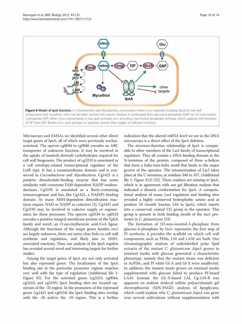
Figure 8 Model of IpsA function. In Corynebacteria and Mycobacteria, myo-inositol (mIno) is an important building block for cell wallcomponents and mycothiol. mIno can be taken up from the culture medium or synthesized from glucose-6-phosphate (G6P) via 1D-myo-inositol-3-phosphate (I3P). When mIno concentration is low, IpsA activates ino1, encoding myo-inositol phosphate synthase, which catalyzes the formationof I3P from G6P. Besides ino1, IpsA activates or represses several other targets of unknown function.
Baumgart et al. BMC Biology 2013, 11:122 Page 10 of 14http://www.biomedcentral.com/1741-7007/11/122
Microarrays and EMSAs we identified several other directtarget genes of IpsA, all of which were previously unchar-acterized. The operon cg0044 to cg0046 encodes an ABCtransporter of unknown function. It may be involved inthe uptake of inositol(-derived) carbohydrates required forcell wall biogenesis. The product of cg3210 is annotated asa cell envelope-related transcriptional regulator of theLytR type. It has a transmembrane domain and is con-served in Corynebacteria and Mycobacteria. Cg1421 is aputative dinucleotide-binding enzyme that has somesimilarity with coenzyme F420-dependent NADP oxidore-ductases. Cg3195 is annotated as a flavin-containingmonooxygenase and has, like Cg1421, a NAD(P) bindingdomain. As many MSH-dependent detoxification reac-tions require NAD or NADP as cofactors [3], Cg1421 andCg3195 may be involved in cofactor supply or regener-ation for these processes. The operon cg0534 to cg0533encodes a putative integral membrane protein of the YphAfamily and menE, an O-succinylbenzoic acid-CoA ligase.Although the functions of the target genes besides ino1are largely unknown, there are some clear links to cell wallsynthesis and regulation, and likely also to MSH-associated reactions. Thus, our analysis of the IpsA regulonhas revealed several novel and interesting targets for furtherstudies.Among the target genes of IpsA are not only activated
but also repressed genes. The localization of the IpsAbinding site in the particular promoter regions matchesvery well with the type of regulation (Additional file 1:Figure S5). For the activated genes (cg3323, cg0044,cg3210, and cg3195) IpsA binding sites are located up-stream of the -35 region. In the promoters of the repressedgenes (cg1421 and cg0534) the IpsA binding sites overlapwith the -35 and/or the -10 region. This is a further
indication that the altered mRNA level we see in the DNAmicroarrays is a direct effect of the IpsA deletion.The structure-function relationship of IpsA is compar-
able to other members of the LacI family of transcriptionalregulators. They all contain a DNA-binding domain at theN-terminus of the protein, composed of three α-helicesthat form a helix-turn-helix motif that binds to the majorgroove of the operator. The tetramerization of LacI takesplace at the C-terminus, at residues 340 to 357, (Additionalfile 1: Figure S12) [32]. These residues are missing in IpsA,which is in agreement with our gel filtration analysis thatindicated a dimeric conformation for IpsA. A computa-tional analysis of many LacI regulators and binding sitesrevealed a highly conserved hydrophobic amino acid atposition 54 (mostly leucine, L64 in IpsA), which insertsinto a conserved, central CG group in the operator. Thisgroup is present in both binding motifs of the ino1 pro-moter in C. glutamicum [33].The formation of 1D-myo-inositol-3-phosphate from
glucose-6-phosphate by Ino1 represents the first step ofPI synthesis; it provides the scaffold on which cell wallcomponents such as PIMs, LM and LAM are built. Ourchromatographic analysis of radiolabelled polar lipidextracts of the mutant C. glutamicum ΔipsA grown inminimal media with glucose generated a characteristicphenotype, namely that the mutant strain was deficientin AcPIM2 and PI while Gl-A and Gl-X were unaffected.In addition, the mutant strain grown on minimal mediasupplemented with glucose failed to produce PI-basedLAM. Instead, the GL-X-based LM, Cg-LM-B wasapparent on sodium dodecyl sulfate polyacrylamide gelelectrophoresis (SDS-PAGE) analysis of lipoglycans,which could explain why C. glutamicum ΔipsA can growover several cultivations without supplementation with

Baumgart et al. BMC Biology 2013, 11:122 Page 11 of 14http://www.biomedcentral.com/1741-7007/11/122
myo-inositol [1]. This LM-B was not found in Mycobac-teria. The polar lipid and lipoglycan extracts from thecomplemented strain C. glutamicum ΔipsA::pK18-ipsAgrown on minimal media supplemented with glucosedisplayed normal levels of AcPIM2, PI, LM and LAMbiosynthesis, with profiles identical to wild-type. Thesedata are consistent with the view that IpsA affects anearly stage of PI biosynthesis, namely the expression ofino1. In fact, the polar lipid and lipoglycan extracts ob-tained from ΔipsA complemented with plasmid-encodedino1 also displayed lipid profiles identical to that of thewild-type, indicating involvement of IpsA in transcrip-tional activation of ino1 expression. The results implythat in absence of IpsA, PI-based PIMs, LM and LAMare not synthesized as the mutant strain no longer pro-duces the inositol monophosphate synthase required forsynthesis of myo-inositol, a substrate for PI production.The polar lipid extracts of C. glutamicum ΔipsA comple-mented with the homolog from C. diphtheriae exhibiteda profile akin to wild-type. Thus DIP1969 is a functionalequivalent of IpsA and can be designated as transcrip-tional activator of ino1 (DIP0115) in this pathogenic spe-cies. For the homolog of Rv3575 the situation is not asclear as for DIP1969. Although we see a positive effecton growth and a partial complementation in terms ofmycothiol biosynthesis and morphological phenotype,no PI derived lipids could be identified on the TLCresults. One possible explanation for this is that most ofthe inositol formed is channeled towards mycothiol.Consequently, a low amount of PI derived lipids mightbe formed, sufficient for partial complementation of thephenotype but still too low for detection by TLC. Fur-thermore, the differences in M. tuberculosis IpsA struc-ture, binding affinity and specificity can also be a reasonfor the only partial complementation. The fact that wedid observe partial complementation with respect tomost phenotypes tested hints at least to a very similarfunction in M. tuberculosis.
ConclusionsIn this study, we have characterized the LacI regulatorIpsA as a first regulator of inositol anabolism in theCorynebacteriales. The findings provide insights into theregulatory mechanisms involved in processes that havebeen identified as promising targets for drug develop-ment. IpsA itself could be such a target, if a myo-inositolanalog could be developed that blocks activation of ino1;it is noteworthy that a trehalose analog with a bacterio-static effect on Mycobacterium aurum has been reported[34]. Finally, the IpsA regulon delivers a promising start-ing point for the analysis of further, so far unstudiedplayers in cell wall synthesis in this important class ofbacteria.
MethodsBacterial strains, plasmids and growth mediaThe bacterial strains and plasmids used in this study arelisted in Additional file 1: Table S1. The C. glutamicumtype strain ATCC 13032 was used as wild-type. Growthexperiments were performed at 30°C and 1,200 rpm in aBiolector system (m2p-labs, Baesweiler, Germany) in 48-well FlowerPlates containing 750 μL CGXII minimalmedium [35] supplemented with 3,4-dihydroxybenzoate(30 mg/L) and carbon source as indicated. If appropriate,25 μg/mL kanamycin was added. All cloning was per-formed in Escherichia coli DH5α cultivated at 37°C inlysogeny broth (LB, [36]) with 50 μg/L kanamycin.
Recombinant DNA workRoutine methods such as polymerase chain reaction(PCR), DNA restriction and ligation were performedusing standard protocols [36-38]. The oligonucleotidesused in this study were obtained from Eurofins MWGOperon (Ebersberg, Germany) and are listed in Add-itional file 1: Table S2. DNA sequencing was performedby Eurofins MWG Operon (Ebersberg, Germany). TheΔipsA mutant of C. glutamicum was constructed via atwo-step homologous recombination protocol as describedpreviously [39]. For further details regarding plasmid andmutant construction, see Additional file 1.
DNA microarraysFor transcriptome analysis, C. glutamicum wild-type andΔipsA cells were grown in 5 mL BHI (brain-heart infusion,Becton, Dickinson and Company, Le Pont de Claix,France) for about 6 h at 30°C. A second precultivation wasperformed in CGXII minimal medium containing 2% (w/v)glucose as carbon source. The main cultures were inocu-lated to an OD of 0.5 in CGXII minimal medium with 2%(w/v) glucose. At an OD of 5 the cells were harvested bycentrifugation (4,120 g, 10 minutes and 4°C). The cell pelletwas subsequently frozen in liquid nitrogen and stored at-70°C. The preparation of total RNA was performed asdescribed previously with the RNeasy Kit from Qiagen(Hilden, Germany) [40]. Synthesis of fluorescently-labeledcDNA was carried out as described in [40,41]. PurifiedcDNA samples to be compared were pooled and theprepared two-color samples were hybridized at 65°Cwhile rotating for 17 h using Agilent’s Gene ExpressionHybridization Kit (Agilent, Böbligen, Germany), hybridizationoven and hybridization chamber. After hybridization, thearrays were washed using Agilent’s Wash Buffer Kitaccording to the manufacturer’s instructions. Fluorescenceof hybridized DNAmicroarrays was determined at 532 nm(Cy3) and 635 nm (Cy5) at 5 μm resolution with a GenePix4000B laser scanner and GenePix Pro 6.0 software(Molecular Devices, Sunnyvale, CA, USA). Fluorescenceimages were saved to raw data files in TIFF format (Gene

Baumgart et al. BMC Biology 2013, 11:122 Page 12 of 14http://www.biomedcentral.com/1741-7007/11/122
Pix Pro 6.0). Quantitative TIFF image analysis was carriedout using GenePix image analysis software and results weresaved as GPR-file (GenePix Pro 6.0). For backgroundcorrection of spot intensities, ratio calculation and rationormalization, GPR-files were processed using the BioConductor R-packages limma and marray [42]. Array datawere deposited in the GEO database [43] under accessionnumber GSE50210.
Overproduction and purification of IpsAE. coli BL21(DE3) carrying the expression plasmidspET-TEV-ipsA or pAN6-ipsA-STREP were grown in LBmedium at 37°C and 120 rpm. IpsA overproduction was in-duced by addition of 50 μM isopropyl β-D-1-thiogalactopy-ranoside (IPTG) followed by cultivation at 16°C for 20 hbefore the cells were harvested by centrifugation. Nickelchelate and StrepTactin affinity chromatography were per-formed as described previously [44,45]. For further purifica-tion and determination of the molecular weight, gelfiltration was performed using a Superdex™ 200 10/300 GLcolumn (GE Healthcare, Munich, Germany) (buffer:50 mM Tris-HCl pH 8, 250 mM NaCl, 1 mM dithiothreitol(DTT)). Note that the IpsA-variant with the C-terminalSTREP-tag formed less aggregates than the variant with theHis10-tag and eluted at a volume corresponding to 79 kDa,suggesting dimer formation (Additional file 1: Figure S2).The protein was concentrated, flash frozen in liquidnitrogen and stored in the gel filtration buffer at -70°C.
Electrophoretic mobility shift assays (EMSAs)EMSAs were performed as described previously with thefollowing modifications [46]. Unless otherwise stated,the STREP-tag variant was used for the EMSAs. PurifiedIpsA was incubated with DNA fragments (30 to 500 bp,final concentration 0.028 to 1 μM) in binding buffer(50 mM Tris-HCl pH 7.5, 40 mM KCl, 5 mM MgCl2) ina total volume of 10 μL. Electrophoresis was performedwith 10% to 15% native polyacrylamide gels at roomtemperature and 150 or 180 V for 45 to 60 minutes(depending on the size of the DNA fragments). For thetesting of effector molecules, IpsA was first incubatedwith the putative effector for 20 minutes at roomtemperature, followed by addition of the DNA, further 20minutes incubation and electrophoresis as described above.As control AcnR protein and the acn promoter fragmentwas used as described in [47] (Additional file 1: Figure S7).
Fluorescence microscopyFor recording fluorescence microscopy images, cells werecentrifuged and resuspended in phosphate-buffered saline(PBS, 137 mM NaCl, 2.7 mM KCl, 4.3 mM Na2HPO4,1.4 mM KH2PO4, pH 7.3) containing 200 ng/mL DAPIand 300 ng/mL nile red. After 5 to 10 minutes incubationat room temperature, the cells were analyzed on agar pads
using a Zeiss Axioplan 2 imaging microscope that wasequipped with an AxioCam MRm camera and a Plan-Apochromat 100×, 1.40 Oil DIC oil-immersion objective.Digital images were acquired and analyzed with AxioVision4.6 software (Zeiss, Göttingen, Germany).
Promoter fusion studiesTo analyze the regulation of the ino1 promoter by IpsAin vivo, a DNA fragment covering the cg3323 promoterregion was fused to the eyfp-coding sequence (pJC1-Pcg3323-eYFP). Wild-type and ΔipsA cells were trans-formed with the resulting plasmid and the fluorescenceoutput was analyzed on minimal medium with 2% (w/v)glucose or myo-inositol in the Biolector system.
Extraction and determination of mycothiolThe determination of mycothiol was carried out as de-scribed [21]. Briefly, cells were first cultivated in BHImedium for 6 h and then twice in CGXII minimal mediumwith 4% (w/v) as carbon source at 30°C and 120 rpm for16 h and 24 h, respectively. 200 mg of wet cells were resus-pended in 1 mL of warm (60°C) acetonitrile containing20 mM Tris-HCl, pH 8.0 and 2 mM bromobimane(Sigma-Aldrich, Taufkirchen near Munich, Germany), soni-ficated for 20 s and further incubated in a 60°C water bathfor 15 minutes in the dark. The samples were acidified with5 μL of 5 M methanesulfonic acid and the cellular debriswas removed by centrifugation at 16,000 g for 10 minutes.The supernatants were filtered and diluted fivefold in10 mM methanesulfonic acid. A wild-type control samplewas incubated with 5 mM NMM to block the thiol groupsprior to derivatization with bromobimane. The samplewere analyzed using a C18 column (LiChrospher RP 18,125 × 4 mm) and the solvents A (100 mM sodium acetatebuffer pH 7.2) and B (100% methanol) a flow rate of0.7 mL/minute and the following gradient: 0 minutes, 15%B; 1 minute, 15% B; 1.5 minutes, 35% B; 11 minutes, 80%B; 12 minutes, 95% B; 13 minutes 100% B. The peakassumed to be mycothiol (5.61 minutes) was identified bycomparison of the wild-type chromatogram with the onefrom the ΔmshC strain and the NMM treated sample. Fur-thermore this peak appears shortly after glutathione (5.14)as published for a similar method [48].
Growth of bacteria for lipid and lipoglycan analysisOvernight cultures of C. glutamicum ATCC 13032, C.glutamicum ΔipsA, C. glutamicum ΔipsA::pK18int-ipsA,and C. glutamicum ΔipsA harboring ino1 (cg3323),Rv3575 and DIP1969 in plasmid pAN6 were pregrown in20 mL of 3.7% brain-heart-glucose bouillon (Sigma-Aldrich, Gillingham, UK) containing Kanamycin (25 μg/mL). The cells were washed once with PBS, used to inocu-late a second preculture to an OD600 of 1 in 50 mL CGXIIminimal medium (50 mL) supplemented with 2% glucose

Baumgart et al. BMC Biology 2013, 11:122 Page 13 of 14http://www.biomedcentral.com/1741-7007/11/122
and incubated with gentle shaking overnight at 30°C. Thecells of the second preculture were washed once with PBSand used to inoculate the main cultures to an OD600 of 0.1in 10 mL CGXII minimal medium (10 mL), supplementedwith either 2% glucose or 2% myo-inositol and grown untilearly log phase. At early log phase, the cells were labeledwith 1 μCi/mL [1,2-14C]-acetate (1.66 to 2.22GBq/mmol,PerkinElmer Inc., Waltham, Massachusetts, USA) andincubated overnight at 30°C with gentle shaking. The cellswere recovered following centrifugation at 3,500 g and dried.
Analysis of polar lipids and lipoglycansThe cell cultivation procedure for the following experi-ments is described in the Additional file 1. Polar and apolarlipid extracts were prepared from the dried cell pelletsusing established procedures [49]. The dried polar lipidextracts were resuspended in CHCl3:CH3OH (100 μL, 2: 1,v/v) and the incorporation of [1,2-14C] acetate in lipid ex-tracts determined by counting an aliquot (5%) using scintil-lation fluid (5 mL). To analyze the lipid profiles, equalcounts of polar lipid extracts (20,000 cpm) were applied tosilica thin layer chromatography (silica gel 60 F254 Merck5554, TLC plates) and developed using the solvent system:CHCl3:CH3OH:H2O (60:30:6, v/v/v) in the first dimensionand CHCl3:CH3CO2H:CH3OH:H2O (40:25:3:6, v/v/v/v) inthe second dimension. The autoradiograms were obtainedby exposing the TLC results to X-ray films (Kodak-BiomaxMR Kodak, Rochester, New York, USA) for 48 h.Lipoglycans were extracted using previously described
protocols [50]. Briefly, dried cell pellets from a 10 mLvolume culture were resuspended in water and refluxedfive times with equal volume of 50% aqueous C2H5OH at85°C, for 6 h intervals. The suspension was centrifugedand the supernatant recovered between each extractionfollowing centrifugation. The combined supernatant frac-tions were dried and subjected to hot phenol-H2O treatmentat 65°C. The aqueous phase containing the crude lipoglycanfraction was recovered and dialyzed (molecular weight cut-off 3.5 KDa) against water. The dialyzed fraction was driedand 20 μg of lipoglycans loaded on a 15% SDS-PAGE gel.The lipoglycans were visualized using Pro-Q emeraldglycoprotein stain (Life Technologies, Darmstadt, Germany).
Additional file
Additional file 1: Additional methods, figures and tables.
AbbreviationsBHI: brain-heart infusion; CDP-DAG: cytidine diphosphate-diacylglycerol;DAG: diacyl-glycerol; eyfp: enhanced yellow fluorescent protein;GL-A: GlcAGroAc2; 1,2-di-O-C16/C18:1-(α-D-glucopyranosyluronic acid)-(1→3)-glycerol; GL-X: ManGlcAGroAc2; 1,2-di-O-C16/C18:1-(α-D-mannopyranosyl)-(1→4)-(α-D-glucopyranosyluronic acid)-(1→3)-glycerol; IMP: inositolmonophosphatase; LAM: liporarabinomannan; LM: lipomannan;MSH: mycothiol; PBS: phosphate-buffered saline; PI: phosphatidylinositol;PIMs: PI mannosides.
Competing interestsThe authors declare that they have no competing interests.
Authors’ contributionsMB designed and coordinated the study, constructed the mutant, performedthe microarray experiments, the mycothiol measurements and fluorescencemicroscopy and prepared the figures. KL and CG carried out therecombinant DNA work, the electrophoretic mobility shift assays (EMSAs), theprotein purification, the promoter fusion studies and the growthexperiments. SG and GSB performed and analyzed the cell wall lipid studiesand prepared the respective parts of the manuscript. MB and JF conceivedof the study and wrote the manuscript. All authors read and approved thefinal manuscript.
AcknowledgementsThis work was supported by the German Ministry of Education and Research(BMBF, grant 0316017B) and by the Helmholtz Association (YoungInvestigator grant VH-NG-716). We thank Tino Polen for help with themicroarray analyses, Jan van Ooyen for help with high-performance liquidchromatography (HPLC) measurements, the students Jakob Schlegel andSebastian Schnorrenberg for their contribution to this project, Antonia Heyerfor providing plasmid pJC1-venus-term, Jörn Kalinowski for informationabout transcriptional start sites as well as Lothar Eggeling and Simon Klafflfor discussion.
Author details1Institut für Bio- und Geowissenschaften, IBG-1: Biotechnologie, Forschungs-zentrum Jülich, 52425 Jülich, Germany. 2School of Biosciences, University ofBirmingham, Edgbaston, Birmingham, B15 2TT, UK.
Received: 13 September 2013 Accepted: 17 December 2013Published: 30 December 2013
References1. Mishra AK, Driessen NN, Appelmelk BJ, Besra GS: Lipoarabinomannan and
related glycoconjugates: structure, biogenesis and role in Mycobacteriumtuberculosis physiology and host-pathogen interaction. FEMS MicrobiolRev 2011, 35:1126–1157.
2. Eggeling L, Besra GS, Alderwick L: Structure and synthesis of the cell wall.In Corynebacteria: genomics and molecular biology. Edited by Burkovski A.Wymondham, UK: Caister Academic Press; 2008:267–294.
3. Fahey RC: Glutathione analogs in prokaryotes. Biochim Biophys Acta 1830,2013:3182–3198.
4. Morita YS, Fukuda T, Sena CBC, Yamaryo-Botte Y, McConville MJ, Kinoshita T:Inositol lipid metabolism in mycobacteria: biosynthesis and regulatorymechanisms. Biochim Biophys Acta 1810, 2011:630–641.
5. http://www.microbesonline.org.6. Alm EJ, Huang KH, Price MN, Koche RP, Keller K, Dubchak IL, Arkin AP: The
MicrobesOnline web site for comparative genomics. Genome Res 2005,15:1015–1022.
7. Bachhawat N, Mande SC: Identification of the INO1 gene ofMycobacterium tuberculosis H37Rv reveals a novel class of inositol-1-phosphate synthase enzyme. J Mol Biol 1999, 291:531–536.
8. Nigou J, Besra GS: Characterization and regulation of inositolmonophosphatase activity in Mycobacterium smegmatis. Biochem J 2002,361:385–390.
9. Movahedzadeh F, Wheeler PR, Dinadayala P, Av-Gay Y, Parish T, Daffe M,Stoker NG: Inositol monophosphate phosphatase genes of Mycobacter-ium tuberculosis. BMC Microbiol 2010, 10:50.
10. Movahedzadeh F, Smith DA, Norman RA, Dinadayala P, Murray-Rust J,Russell DG, Kendall SL, Rison SC, McAlister MS, Bancroft GJ, McDonald NQ,Daffe M, Av-Gay Y, Stoker NG: The Mycobacterium tuberculosis ino1 gene isessential for growth and virulence. Mol Microbiol 2004, 51:1003–1014.
11. Jackson M, Crick DC, Brennan PJ: Phosphatidylinositol is an essentialphospholipid of mycobacteria. J Biol Chem 2000, 275:30092–30099.
12. Haites RE, Morita YS, McConville MJ, Jacobe HB: Function ofphosphatidylinositol in mycobacteria. J Biol Chem 2005, 280:10981–10987.
13. Hernick M: Mycothiol: a target for potentiation of rifampin and otherantibiotics against Mycobacterium tuberculosis. Expert Rev Anti Infect Ther2013, 11:49–67.

Baumgart et al. BMC Biology 2013, 11:122 Page 14 of 14http://www.biomedcentral.com/1741-7007/11/122
14. Krings E, Krumbach K, Bathe B, Kelle R, Wendisch VF, Sahm H, Eggeling L:Characterization of myo-inositol utilization by Corynebacteriumglutamicum: the stimulon, identification of transporters, and influenceon L-lysine formation. J Bacteriol 2006, 188:8054–8061.
15. Klaffl S, Brocker M, Kalinowski J, Eikmanns B, Bott M: Complex regulation ofthe PEP carboxykinase gene pck and characterization of its GntR-typeregulator IolR as a repressor of myo-inositol utilization genes inCorynebacterium glutamicum. J Bacteriol 2013, 195:4283–4296.
16. Swint-Kruse L, Matthews KS: Allostery in the Lacl/GaIR family: variationson a theme. Curr Opin Microbiol 2009, 12:129–137.
17. www.ncbi.nlm.nih.gov/geo18. Wennerhold J, Bott M: The DtxR regulon of Corynebacterium glutamicum.
J Bacteriol 2006, 188:2907–2918.19. Wennerhold J, Krug A, Bott M: The AraC-type regulator RipA represses
aconitase and other iron proteins from Corynebacterium under ironlimitation and is itself repressed by DtxR. J Biol Chem 2005,280:40500–40508.
20. Bailey TL, Elkan C: “Fitting a mixture model by expectation maximization todiscover motifs in biopolymers”, Proceedings of the Second InternationalConference on Intelligent Systems for Molecular Biology. Menlo Park, Ca:AAAI Press; 1994:28–36.
21. Feng J, Che YS, Milse J, Yin YJ, Liu L, Rückert C, Shen XH, Qi SW, Kalinowski J,Liu SJ: The gene ncgl2918 encodes a novel maleylpyruvate isomerase thatneeds mycothiol as cofactor and links mycothiol biosynthesis andgentisate assimilation in Corynebacterium glutamicum. J Biol Chem 2006,281:10778–10785.
22. Tatituri RV, Illarionov PA, Dover LG, Nigou J, Gilleron M, Hitchen P,Krumbach K, Morris HR, Spencer N, Dell A, Eggeling L, Besra GS: Inactivationof Corynebacterium glutamicum NCgl0452 and the role of MgtA in thebiosynthesis of a novel mannosylated glycolipid involved in lipomannanbiosynthesis. J Biol Chem 2007, 282:4561–4572.
23. Mishra AK, Klein C, Gurcha SS, Alderwick LJ, Babu P, Hitchen PG, Morris HR,Dell A, Besra GS, Eggeling L: Structural characterization and functionalproperties of a novel lipomannan variant isolated from aCorynebacterium glutamicum pimB′ mutant. Antonie Van Leeuwenhoek2008, 94:277–287.
24. Mishra AK, Batt S, Krumbach K, Eggeling L, Besra GS: Characterization ofthe Corynebacterium glutamicum ΔpimB′ ΔmgtA double deletion mutantand the role of Mycobacterium tuberculosis orthologues Rv2188c andRv0557 in glycolipid biosynthesis. J Bacteriol 2009, 191:4465–4472.
25. Vilchèze C, Av-Gay Y, Attarian R, Liu Z, Hazbón MH, Colangeli R, Chen B, Liu W,Alland D, Sacchettini JC, Jacobs WR Jr: Mycothiol biosynthesis is essential forethionamide susceptibility in Mycobacterium tuberculosis.Mol Microbiol 2008, 69:1316–1329.
26. Turner BL, Paphazy MJ, Haygarth PM, McKelvie ID: Inositol phosphates inthe environment. Philos T Roy Soc B 2002, 357:449–469.
27. Schröder J, Jochmann N, Rodionov DA, Tauch A: The Zur regulon ofCorynebacterium glutamicum ATCC 13032. BMC Genomics 2010, 11:12.
28. Tropis M, Meniche X, Wolf A, Gebhardt H, Strelkov S, Chami M, SchomburgD, Kramer R, Morbach S, Daffe M: The crucial role of trehalose andstructurally related oligosaccharides in the biosynthesis and transfer ofmycolic acids in corynebacterineae. J Biol Chem 2005, 280:26573–26585.
29. Murphy HN, Stewart GR, Mischenko VV, Apt AS, Harris R, McAlister MSB,Driscoll PC, Young DB, Robertson BD: The OtsAB pathway is essential fortrehalose biosynthesis in Mycobacterium tuberculosis. J Biol Chem 2005,280:14524–14529.
30. Norman RA, McAlister MSB, Murray-Rust J, Movahedzadeh F, Stoker NG,McDonald NQ: Crystal structure of inositol 1-phosphate synthase fromMycobacterium tuberculosis, a key enzyme in phosphatidylinositolsynthesis. Structure 2002, 10:393–402.
31. Roback P, Beard J, Baumann D, Gille C, Henry K, Krohn S, Wiste H, VoskuilMI, Rainville C, Rutherford R: A predicted operon map for Mycobacteriumtuberculosis. Nucleic Acids Res 2007, 35:5085–5095.
32. Bell CE, Lewis M: The Lac repressor: a second generation of structural andfunctional studies. Curr Opin Struct Biol 2001, 11:19–25.
33. Camas FM, Alm EJ, Poyatos JF: Local gene regulation details a recognitioncode within the LacI transcriptional factor family.PLoS Comput Biol 2010, 6:11.
34. Belisle JT, Vissa VD, Sievert T, Takayama K, Brennan PJ, Besra GS: Role of themajor antigen of Mycobacterium tuberculosis in cell wall biogenesis.Science 1997, 276:1420–1422.
35. Keilhauer C, Eggeling L, Sahm H: Isoleucine synthesis in Corynebacteriumglutamicum - Molecular analysis of the Ilvb-Ilvn-Ilvc Operon. J Bacteriol1993, 175:5595–5603.
36. Sambrook J, Russell DW: Molecular cloning: A Laboratory Manual. 3rd edition.Cold Spring Harbor, NY: Cold Spring Harbor Laboratory Press; 2001.
37. Hanahan D: Studies on transformation of Escherichia coli with plasmids.J Mol Biol 1983, 166:557–580.
38. van der Rest ME, Lange C, Molenaar D: A heat shock followingelectroporation induces highly efficient transformation ofCorynebacterium glutamicum with xenogeneic plasmid DNA.Appl Microbiol Biotechnol 1999, 52:541–545.
39. Niebisch A, Bott M: Molecular analysis of the cytochrome bc1-aa3 branchof the Corynebacterium glutamicum respiratory chain containing anunusual diheme cytochrome c1. Arch Microbiol 2001, 175:282–294.
40. Möker N, Brocker M, Schaffer S, Krämer R, Morbach S, Bott M: Deletion ofthe genes encoding the MtrA-MtrB two-component system ofCorynebacterium glutamicum has a strong influence on cell morphology,antibiotics susceptibility and expression of genes involved inosmoprotection. Mol Microbiol 2004, 54:420–438.
41. Lange C, Rittmann D, Wendisch VF, Bott M, Sahm H: Global expressionprofiling and physiological characterization of Corynebacteriumglutamicum grown in the presence of L-valine. Appl Environ Microbiol2003, 69:2521–2532.
42. Smyth GK: Limma: linear models for microarray data. In Bioinformatics andComputational Biology Solutions Using R and Bioconductor. Edited byGentleman R, Carey VJ, Huber W, Irizarry RA, Dudoit S. New York, NY:Springer; 2005:397–420.
43. Edgar R, Domrachev M, Lash AE: Gene Expression Omnibus: NCBI geneexpression and hybridization array data repository. Nucleic Acids Res 2002,30(1):207–210.
44. Garcia-Nafria J, Baumgart M, Bott M, Wilkinson AJ, Wilson KS: TheCorynebacterium glutamicum aconitase repressor: scratching around forcrystals. Acta Crystallogr Sect F Struct Biol Cryst Commun 2010,66:1074–1077.
45. Niebisch A, Kabus A, Schultz C, Weil B, Bott M: Corynebacterial proteinkinase G controls 2-oxoglutarate dehydrogenase activity via the phosphor-ylation status of the OdhI protein. J Biol Chem 2006, 281:12300–12307.
46. Heyer A, Gätgens C, Hentschel E, Kalinowski J, Bott M, Frunzke J: Thetwo-component system ChrSA is crucial for haem tolerance and interfereswith HrrSA in haem-dependent gene regulation in Corynebacteriumglutamicum. Microbiology 2012, 158:3020–3031.
47. Garcia-Nafria J, Baumgart M, Turkenburg JP, Wilkinson AJ, Bott M, Wilson KS:Crystal and solution studies reveal that the transcriptional regulatorAcnR of Corynebacterium glutamicum is regulated by Citrate-Mg2+
binding to a non-canonical pocket. J Biol Chem 2013, 288:15800–15812.48. Anderberg SJ, Newton GL, Fahey RC: Mycothiol biosynthesis and
metabolism - Cellular levels of potential intermediates in the biosynthesisand degradation of mycothiol in Mycobacterium smegmatis. J Biol Chem1998, 273:30391–30397.
49. Dobson G, Minnikin DE, Minnikin SM, Parlett JH, Goodfellow M, Ridell M,Magnusson M: Systematic analysis of complex mycobacterial lipids. InChemical Methods in Bacterial Systematics, Volume 1. Edited by M. G, E. MD.London, UK: Academic Press; 1985:237–265.
50. Ludwiczak P, Brando T, Monsarrat B, Puzo G: Structural characterization ofMycobacterium tuberculosis lipoarabinomannans by the combination ofcapillary electrophoresis and matrix-assisted laser desorption/ionizationtime of-flight mass spectrometry. Anal Chem 2001, 73:2323–2330.
doi:10.1186/1741-7007-11-122Cite this article as: Baumgart et al.: IpsA, a novel LacI-type regulator, isrequired for inositol-derived lipid formation in Corynebacteria and Myco-bacteria. BMC Biology 2013 11:122.