Embed Size (px)
Citation preview

RESEARCH ARTICLE Open Access
Design and development of a peptide-basedadiponectin receptor agonist for cancertreatmentLaszlo Otvos Jr1, Eva Haspinger2,3, Francesca La Russa2,3, Federica Maspero2,3, Patrizia Graziano2,3, Ilona Kovalszky4,Sandor Lovas5, Kaushik Nama1, Ralf Hoffmann6, Daniel Knappe6, Marco Cassone1, John Wade7 and Eva Surmacz2*
Abstract
Background: Adiponectin, a fat tissue-derived adipokine, exhibits beneficial effects against insulin resistance,cardiovascular disease, inflammatory conditions, and cancer. Circulating adiponectin levels are decreased in obeseindividuals, and this feature correlates with increased risk of developing several metabolic, immunological andneoplastic diseases. Thus, pharmacological replacement of adiponectin might prove clinically beneficial, especiallyfor the obese patient population. At present, adiponectin-based therapeutics are not available, partly due to yetunclear structure/function relationships of the cytokine and difficulties in converting the full size adiponectinprotein into a viable drug.
Results: We aimed to generate adiponectin-based short peptide that can mimic adiponectin action and besuitable for preclinical and clinical development as a cancer therapeutic. Using a panel of 66 overlapping 10 aminoacid-long peptides covering the entire adiponectin globular domain (residues 105-254), we identified the 149-166region as the adiponectin active site. Three-dimensional modeling of the active site and functional screening ofadditional 330 peptide analogs covering this region resulted in the development of a lead peptidomimetic, ADP355 (H-DAsn-Ile-Pro-Nva-Leu-Tyr-DSer-Phe-Ala-DSer-NH2). In several adiponectin receptor-positive cancer cell lines,ADP 355 restricted proliferation in a dose-dependent manner at 100 nM-10 μM concentrations (exceeding theeffects of 50 ng/mL globular adiponectin). Furthermore, ADP 355 modulated several key signaling pathways (AMPK,Akt, STAT3, ERK1/2) in an adiponectin-like manner. siRNA knockdown experiments suggested that ADP 355 effectscan be transmitted through both adiponectin receptors, with a greater contribution of AdipoR1. In vivo,intraperitoneal administration of 1 mg/kg/day ADP 355 for 28 days suppressed the growth of orthotopic humanbreast cancer xenografts by ~31%. The peptide displayed excellent stability (at least 30 min) in mouse blood orserum and did not induce gross toxic effects at 5-50 mg/kg bolus doses in normal CBA/J mice.
Conclusions: ADP 355 is a first-in-class adiponectin receptor agonist. Its biological activity, superior stability inbiological fluids as well as acceptable toxicity profile indicate that the peptidomimetic represents a true leadcompound for pharmaceutical development to replace low adiponectin levels in cancer and other malignancies.
BackgroundAdiponectin is a relatively large (244 amino acid) cyto-kine normally produced by the fat tissue and found inhuman serum at concentrations of 2-20 μg/mL [1-5].Circulating adiponectin levels are inversely correlatedwith body mass index (BMI) [6]. Adiponectin is
considered a protective hormone exhibiting beneficialeffects against insulin resistance, cardiovascular disease,inflammatory conditions, and cancer [5-11].Adiponectin circulates in trimeric, hexameric, and
higher order complexes [12]. The C-terminal half ofprotein representing the globular domain (gAd) exhibitspotent metabolic effects in various tissues [13-15]. Twoadiponectin receptors have been identified, AdipoR1 andAdipoR2. Both receptors are 7-channel integral mem-brane proteins containing an N-terminal intracellular
* Correspondence: [email protected] University, Sbarro Institute for Cancer Research and MolecularMedicine, Philadelphia, PA 19122, USAFull list of author information is available at the end of the article
Otvos et al. BMC Biotechnology 2011, 11:90http://www.biomedcentral.com/1472-6750/11/90
© 2011 Otvos et al; licensee BioMed Central Ltd. This is an Open Access article distributed under the terms of the Creative CommonsAttribution License (http://creativecommons.org/licenses/by/2.0), which permits unrestricted use, distribution, and reproduction inany medium, provided the original work is properly cited.

portion and a C-terminal extracellular-transmembranedomain [16,17]. AdipoR1 is a high-affinity receptor forgAd and a low affinity receptor for the full-size ligand[18]. AdipoR1 has 4 very short extracellular domainscomposed of 13, 6, 11 and 16 residues, respectively [17].The major intracellular signal induced by adiponectin
is the energy-sensing AMP-activated protein kinase(AMPK) pathway [13,19]. However, some adiponectin-dependent effects appear to be AMPK-independent [20].In addition, adiponectin can modulate in a tissue con-text-dependent manner several other signaling effectors,such as extracellular-signal-regulated kinases 1 and 2(ERK1/2), p38 kinase, peroxisome proliferator-activatedreceptor-a (PPARa), stress-responsive c-Jun N-terminalkinase (JNK), Wnt receptor, nitric oxide (NO), signaltransducer and activator of transcription 3 (STAT3) fac-tor, nuclear factor-�B (NF-�B), and ceramide [19-29].Targeted disruption experiments suggested that Adi-poR1 transmits signals mainly through AMPK, whileAdipoR2 acts through PPARa-related pathways [19].Recent evidence implicated adiponectin in the preven-
tion of cancer [4,30,31]. Epidemiological studies foundan inverse correlation between adiponectin and the riskof developing several obesity-related malignancies,including cancers of the breast, endometrium, colon,and prostate [7,32-34]. The best-documented associa-tions in breast cancer show that adiponectin levels arereduced in cancer patients vs. controls [34-36], and lowadiponectin levels correlate with more aggressive tumorsand higher frequency of lymph node metastasis [10,37].In agreement with this, in vitro studies demonstratedthat adiponectin or its globular form can inhibit theproliferation of breast, colorectal and prostate cancercells [1,26,38-44].Depending on the experimental model, cytostatic/
apoptotic effects of adiponectin can be associated withan increased activation of AMPK, reduced ERK1/2 sig-naling [40], inhibition of the Akt kinase and glycogensynthase kinase/b-catenin pathway [45], and/orenhanced expression of Bax and p53 pro-apoptoticgenes [44]. In addition, adiponectin can also reduce can-cer cell migration and invasion [46]. In animal models,adiponectin suppresses the growth of T47D and MDA-MB-231 breast cancer xenografts, and in some cases,inhibits tumor neoangiogenesis [45,47].The adiponectin receptors, AdipoR1 and AdipoR2,
have been detected in human breast cancer specimens,but not clearly associated with other biomarkers[26,48-50]. AdipoR1 appears to play a more definite rolein breast cancer, as adiponectin-dependent antiprolifera-tive effects are abolished by siRNA knockdown of thisreceptor [1,51]. However, in colon cancer cells, bothAdipoR1 and AdipoR2 can transmit cytostatic effects[52]. While data on AdipoR1/2 expression in other
malignancies are limited, the receptors have been foundin normal colon and colon cancer tissue [53] as well asin gastrointestinal stromal tumors [54].Although some anti-diabetic drugs (e.g., metformin, a
biguanide) [55-57] as well as caloric restriction [58,59]can partially mimic adiponectin action and induceAMPK signaling in cancer tissues, specific and selectivecompounds targeting AdipoR still await development.At present, adiponectin-based therapeutics are not avail-able, partly due to difficulties in converting the full sizeadiponectin protein into a viable drug. Here we reporton the design and initial preclinical development of adi-ponectin-based peptide compounds acting as AdipoRagonists in cancer cells.
MethodsInitial model buildingThe three-dimensional (3D) structure of the globulardomain (residues 105-254) of human adiponectin wasobtained with the YASARA molecular modeling package(Ver. 10.10.29) [60]. The hm_build.mcr macro of theYASARA package with default parameters, except themaximum oligomerization state set to one, was used tobuild the model. YASARA identified a protein with pro-tein databank (PDB) [61] i.d. 1C3H, which correspondsto a murine isoform of adiponectin, as a sole template.The model was subjected to further refinement usingthe md_refine.mcr macro of YASARA and to 1 ns con-stant temperature (300 K) and pressure (1 bar) molecu-lar dynamics (MD) simulations using the AMBER03force field. Simulation parameters were kept at thevalues defined by the macro. The structure of the pro-tein was simulated in an 8 × 8 × 8 nm rectangular boxwith periodic boundaries and endcapping with an N-acetyl protecting group to preserve the electronic struc-ture of the backbone. The box containing the proteinwas filled with 8390 water molecules, 48 Cl- and 53 Na+
ions. The final structure from the simulation was usedas a starting parameter to structure calculations of pep-tide 25 and its derivative, ADP 355.
Detailed molecular dynamics simulations of peptidesMD simulations were performed with the GROMACS4.0.7 software package [62] using the OPLS-AA/L forcefield [63]. Peptides were solvated with 2457 water mole-cules and one chloride ion to neutralize the charge ofthe system. The solvated structures were energy mini-mized by the steepest descent method. Simulations ranfor 500 ps at 300 K. At 500.5 ns the simulations contin-ued at 1 bar pressure by coupling the system to externalheat and pressure bath. Snapshots of the trajectorieswere saved at every 0.1 ns. The first 0.5 ns point wasconsidered as equilibration period and was not used forsubsequent analysis. A reaction-field correction was
Otvos et al. BMC Biotechnology 2011, 11:90http://www.biomedcentral.com/1472-6750/11/90
Page 2 of 14

used for long-range electrostatic interactions, and anenergy dispersion correction was implemented. Trajec-tories were submitted to cluster analysis using the GRO-MOS method [64]. Peptide conformations wereexamined using the middle structure of the largest clus-ter and using the DSSP program [65]. The root-mean-square deviation (RMSD) of the backbone of the peptidestructures was calculated using the g_rms utility ofGROMACS.
Peptide arrayA panel of 66 overlapping peptides, each 10 amino acid-long, covering the entire globular domain of the humanadiponectin protein was synthesized on a b-alanine deri-vatized cleavable cellulose membrane [66]. Each conse-cutive peptide was shifted by 2 residues along thesequence. Another panel of 330 peptides representinganalogs of the 66 original peptides was assembled onthe same membrane support. In these 10-residue-longoverlapping peptides, residues 1, 4, 7 and 10 of the firstarray were replaced with non-natural amino acids. Thesubstituting amino acids were norvaline for aliphaticresidues, D-asparagine for residues with amide-sidechain, D-serine for hydroxy-amino acids, D-lysine forcationic residues and 1-amino cyclopentane carboxylicacid for aromatic residues and proline. All peptidesequences are listed in Additional file 1.The peptides were assembled by Fmoc-synthesis tech-
niques [67], individually cut from the solid support andcleaved from the cellulose membrane by using 2% aqu-eous triethyl amine overnight [68]. Peptides 23-27 andtheir modified analogs were purified by reversed-phasehigh performance liquid chromatography (RP-HPLC)and characterized by matrix-assisted laser desorption/ionization mass spectroscopy (MALDI-MS).
Synthesis and purification of individual peptidesThe adiponectin-based peptide 25, a six-residue middlefragment of peptide 25, and peptidomimetic ADP 355 aswell as biotin-labeled analogs of the AdipoR1 extracellu-lar loops were synthesized on the solid-phase by using aCEM Liberty microwave-assisted peptide synthesizerand utilizing Fmoc-chemistry [67]. Biotin was coupledto the peptide while still attached the solid-phase car-rier. After cleavage with 95% aqueous trifluoroaceticacid (TFA) containing 2% thioanisole, the peptides werepurified by RP-HPLC. MALDI-MS verified the high pur-ity of the peptide preparations. After purification, ADP355 was lyophilized twice from 2% aqueous acetic acidsolution prior to cellular efficacy studies.
Screening of AdipoR1/peptide bindingThe 66 unmodified adiponectin array peptides wereindividually dried down to wells of an ELISA plate, and
tested for binding to biotin-labeled linear synthetic mod-els of the 4 extracellular loops of AdipoR1. The recep-tor/peptide interaction was detected by horseradish-peroxidase conjugated streptavidin.
In vitro screening of adiponectin-based peptidesBiological activity of the peptides was first assessedusing MCF-7 breast cancer cells that are known toexpress AdipoR1 [38]. MCF-7 cell line was obtainedfrom ATCC (Manassas, VA) and routinely grown inDMEM:F12 plus 5% calf serum (Cellgro Mediatech,Manassas, VA) at 37°C, 5% CO2. For screening experi-ments, MCF-7 cells were plated in 24-well plates at theconcentration of 30,000 cells/well. After 12 h of culturein the growth medium, the cells were synchronized inserum-free medium (SFM) (DMEM:F12 supplementedwith 0.42 g/mL bovine serum albumin, 1 mM FeSO4
and 2 mM L-glutamine) for 24 h, and then shifted backto the full growth medium containing either gAd (Phoe-nix Secretomics, Burlingame, CA) at 50 ng/mL, indivi-dual peptides, or no test compounds. After 24 h, thecells were counted under the microscope with trypan-blue exclusion. Each experiment was performed in tripli-cate and repeated at least three times.The array-extracted peptides (1, 2, 3, 19, 20, 21, 22,
23, 24, 26, 27, 28, 29, 30, 31, 37, 55, 56, 57, 58, 59, 60)solubilized at 65°C for 30 min were tested at an approxi-mate concentration of 8-50 ng/mL. In addition, the fol-lowing peptides: 22 (and its modifications 88, 154, 220,286, 352), 23 (and its modifications 89, 155, 221, 287,353), 24 (and its modifications 90, 156, 222, 288, 353),25 (and its modifications 91, 157, 223, 289 and 355), 26(and its modifications 92, 158, 224, 290, 356), 27 (andits modifications 93, 159, 225, 291, 357), 28, 29, 30 werefurther purified by RP-HPLC and screened togetherwith the individually synthesized adiponectin peptides atthe exact concentration of 50 ng/mL.
Cell growth experimentsThe effects of the lead active peptide 355 (ADP 335) oncell proliferation were tested in breast cancer cell lines(MCF-7, MDA-MB-231) and in glioblastoma cells(LN18), all obtained from ATCC and routinely culturedas described previously [69-71]. The peptide was testedat 10 pM-100 μM concentrations under conditionsdescribed above under peptide screening.
Detection of AdipoR1 and AdipoR2 and signaling analysisAdipoR1 and AdipoR2 were detected by Western immu-noblotting (WB) using 100 μg of total proteins isolatedfrom growing cell cultures, as described by us previously[72,73] using goat polyclonal AdipoR1 M18 Ab and goatpolyclonal AdipoR2 C12 Ab (Santa Cruz Biotechnology,Santa Cruz, CA).
Otvos et al. BMC Biotechnology 2011, 11:90http://www.biomedcentral.com/1472-6750/11/90
Page 3 of 14

Signaling analysis was performed using MCF-7 andMDA-MB-231 breast cancer cells and LN18 glioblas-toma cells. The cells at 70-80% confluence were shiftedto SFM for 24 h, then SFM was removed, the cultureswere washed 2 x with PBS, placed in normal growthmedium for 1 h, and then treated with ADP 355 at 100nM (MCF-7 cells) or 10 μM (MDA-MB-231 and LN 18cells) for 0-60 min, while gAd at 50 ng/mL was appliedfor 60 min only. Untreated cells were used as negativecontrol. After the treatment, the cells were lysed, as pre-viously described [72,73] and 100 μg of proteins wereanalyzed by WB for the expression of phosphorylated(p) and total forms of several signaling molecules. Thefollowing primary Abs from Cell Signaling (Danvers,MA) were used: 1) pAMPKa (T172) D79.5E rabbit mAb1: 750; 2) total AMPKa rabbit mAb 1:1000; 3) p44/42MAPK (T202/Y204) rabbit mAb 1:1000; 4) total 44/42MAP kinase rabbit mAb 1:1000; 5) pSTAT3 (Y705)D3A7 rabbit mAb 1: 500; 6) total STAT3 79D7 rabbitmAb 1:2000; 7) pAkt (Ser 473) rabbit mAb 1:1000; 8)total Akt rabbit mAb 1:1000. Protein loading was veri-fied by evaluating the expression of a constitutiveenzyme glyceraldehyde-3-phosphate dehydrogenase(GAPDH) using 6C51 mAbs 1:1000 (Santa Cruz). Thefollowing secondary Abs (Santa Cruz) were used whereappropriate: 1) donkey anti-goat IgG-HRP; 2) goat anti-mouse IgG-HRP; 3) goat anti-rabbit IgG-HRP, allapplied at 1:1000 dilution. The intensity of specific pro-tein bands was quantified by the ImageJ software (dis-tributed by the National Institutes of Health, Bethesda,USA). The ratio of phosphorylated to total levels wascalculated for each protein, and expressed as % changevs. untreated controls (taken as 100%).
siRNA experimentsMCF-7 cells expressing approximately equal levels ofAdipoR1 and AdipoR2 were used to assess the contribu-tion of each receptor in the response to ADP355. Adi-poR1 and AdipoR2 siRNA as well scrambled siRNAwere purchased from Santa Cruz Biotechnology. Dilu-tion of siRNA reagents and transfection of cells was per-formed following the manufacturer’s protocol. Forgrowth experiments, the cells were plated in 24-wellplates at 50 × 104 cells/well and transfected with 5 μl of10 μM stock siRNA. For WB, the cells were grown in60 mm plates at 5 × 10⁵ cells/plate and transfected with15 μl of 10 μM stock siRNA. The cells were processedfor WB or treated with ADP355 at 48 h followingtransfection.
Peptide stability in mouse blood and mouse serumSixty μg of ADP 355 were dissolved in 100 μL water,and 10 μL aliquots were mixed with 100 μL of freshlydrawn mouse blood. After 30 min of incubation at 37°C,
blood cells were centrifuged at 10,000 × g. Fifty μLserum was mixed with 50 μL phosphate buffered salinepH 6.8 (PBS), and serum proteins were precipitated byaddition of 45 μL aqueous 15% trichloroacetic acid(TCA) for 10 min at 4°C. After centrifugation at 12,000× g, the supernatant was neutralized with 0.1 M aqu-eous sodium hydroxide and 0.5 μL of this solution wasmixed with 0.5 μL a-cyano-4-hydroxycinnamic acid (4mg/mL in 60% aqueous acetonitrile containing 0.1%TFA) as matrix on a sample plate. Analysis was per-formed using a MALDI time-of-flight tandem massspectrometer (MALDI-TOF/TOF-MS, 4700 proteomicanalyzer, Applied Biosystems, Weiterstadt, Germany).Additionally, the neutralized supernatant was loaded ona Jupiter C18 RP-HPLC column (4.6 mm internal dia-meter, 150 mm length, 5 μm particle size, 30 nm poresize) previously calibrated with known amounts of ADP355 dissolved in PBS. Absorbance was measured at 214nm.ADP 355 was also incubated at 37°C with 25% aqu-
eous mouse serum at a final concentration of 150 μg/mL. After 0, 15, 30, 60, 120, and 240 min, 95 μL ali-quots were mixed with 25 μL 15% aqueous TCA andwere incubated for 10 min at 4°C. Sample analysis fol-lowed the protocols described above.
In vivo activity of ADP 355 in an orthotopic xenograftbreast cancer modelTen 8-week-old female immunocompromised (scid)mice (genetic background CB17/Icr) were anesthetizedto allow the implantation of 2.5 × 10⁶ MCF-7 cells intothe two inguinal mammary glands. When tumors werepalpable in all animals (34 days after cell transplanta-tion), the mice were divided into 2 groups containinganimals with comparable tumor sizes. One group of 5mice was treated daily with 1 mg/kg peptide ADP 355intraperitoneally (ip), while the other group of 5 micereminded untreated. After 28 days of treatment, the ani-mals were killed by CO2 inhalation, and tumors werecarefully removed, photographed and weighed. All verte-brate animals of this study were maintained and handledin accordance with the recommendations of the Guide-lines for the Care and Use of Laboratory Animals andwere approved by the Animal Care Committee of Sem-melweis University (permission No.:399/003/2005).
In vivo toxicityADP 355 was injected into 4 groups of three 10-12 weekold female CBA/J mice. Bolus ip doses were administeredat 5 mg/kg, 10 mg/kg, 25 mg/kg or 50 mg/kg in sterile sal-ine and the animals were observed for signs of systemictoxicity (tremor, head tilt, reduced activity and squinting)for 4 days. On day 5, the mice were sacrificed by CO2
inhalation. The potential peptide elimination organs: the
Otvos et al. BMC Biotechnology 2011, 11:90http://www.biomedcentral.com/1472-6750/11/90
Page 4 of 14

livers, spleens and kidneys were removed and weighed. Allprocedures for vertebrate animal experiments wereapproved by the Animal Health and Food Control Com-mittee of Budapest, protocol number 399/033/2005.
ResultsIdentification of the active site of adiponectin proteinThe peptides extracted from the array were tested forcytostatic activity in adiponectin sensitive MCF-7 cells[38]. In control experiments, we used gAd at 50 ng/mL,a concentration that induced maximal growth inhibitionin our dose response experiments in MCF-7 cells (datanot shown) and has previously been described as cyto-static in breast cancer cells [1]. Peptides 23-27 at 50 ng/mL inhibited MCF-7 cell proliferation by 19-26% rela-tive to untreated controls, while other peptides, eitherflanking this domain or distant, were ineffective or pro-duced only minimal (3% or less) cytostatic effects (Fig-ure 1A and Additional file 1). gAd restricted MCF-7 cellgrowth by ~18% (Figure 1A).The sequence covered by the active peptides 23-27 is:
H-Lys-Phe-His-Cys-Asn-Ile-Pro-Gly-Leu-Tyr-Tyr-Phe-Ala-Tyr-His-Ile-Thr-Val-NH2, and this fragment corre-sponds to amino acids 149-166 of human adiponectinprotein (Table 1).According to the currently most accepted model, the
globular domain of human adiponectin is a b-barrel-type structure where the b-sheets are connected with ω-loops. The identified active peptides are located on theloop-b-sheet region of the protein (Figure 1B). Whileapproximately half of the sequence, covering peptides 26and 27, is located inside the trimer bundle, the N-term-inal region falls slightly outside the trimer boundaries.The side-chains of the C-terminal 2/3 of the identifiedactive site are facing outside (Figure 1B). The center ofthe active peptides has homology only with spastin,immunoglobulin and complement proteins according toa BLAST homology search.
Identification of minimal adiponectin active site anddevelopment of its pharmacologically improved analogsNext, we generated multiple analogs of peptides 23-27in order to identify the minimal adiponectin active siteas well as introduce chemical modifications improvingpeptide activity and stability. The activities of all analogsat 50 ng/mL were determined relative to the effects of50 ng/mL gAd (Table 1).While peptide 25 was fully active in cell growth inhibi-
tion assays, its center 6 residue-long fragment 157-162 didnot exhibit any biological activity. Therefore, we generatedand tested several longer, 10-residue peptides encompass-ing the 149-166 adiponectin stretch. We attempted toidentify residues in this region that could be freelyreplaced with non-natural amino acid analogs in order to
improve pharmacological properties of the lead peptides.Biological assays identified a highly active short site: Ile-Pro-Gly-Leu-Tyr-Tyr-Phe-Ala, and further structure-func-tion analysis indicated that conservative substitutions inthe minimal active site can be introduced at Gly 155 andTyr 158 residues, without compromising biological activ-ity. Additions of non-natural amino acids at N- and C-ter-mini were envisioned to provide stability againstexopeptidase cleavage in vitro and in vivo (Table 1).
Identification of ADP 355 as an optimal adiponectinreceptor agonistBiological screening of the analogs of the minimal adi-ponectin active site resulted in the identification of apeptidomimetic ADP 355 (H-DAsn-Ile-Pro-Nva-Leu-Tyr-DSer-Phe-Ala-DSer-NH2) as the most promisingadiponectin receptor agonist. The compound is basedon the precursor peptide 25 and contains the minimalactive site with allowed modifications (Table 1).
Figure 1 Identification of the active site of adiponectin. A)Effects of adiponectin fragments encompassing the active site onthe growth of MCF7 cells. The activity of the entire globular domainof adiponectin (gAd) is included for comparison. The data areaverages from 3 different assays and represent average results +/-SE and were analyzed by Student t-test, p < 0.05. The sequences ofall tested peptides are listed in Additional file 1. B) High-resolutionstructure of the adiponectin monomer with the peptide 25 andactive site amino acid side-chains colored. Conservativesubstitutions of residues marked in green could be made withoutloss of biological activity; residues marked with red could not besubstituted.
Otvos et al. BMC Biotechnology 2011, 11:90http://www.biomedcentral.com/1472-6750/11/90
Page 5 of 14

ADP 355 inhibits the growth of AdipoR1/AdipoR2-positivecancer cell linesIn a preliminary, qualitative study, we examined theinteraction of peptide 25 to biotin-labeled fragments ofthe 4 AdipoR1 extracellular loops. The peptide boundthe first extracellular loop of AdipoR1, but not otherloops (data not shown). BLAST analysis identified an86% homology between the first loops of AdipoR1 andAdipoR2, suggesting that ADP 355 potentially can inter-act with both receptors.Dose-dependent effects of ADP 355 were tested in dif-
ferent cancer cells lines expressing AdipoR1 and Adi-poR2. The highest levels of AdipoR1 were found inMCF-7 cells, while the receptor was less abundant inMDA-MB-231 and LN18 cells (Figure 2A). On theother hand, AdipoR2 was undetectable in MDA-MB-231cells, and expressed at intermediate and high levels inLN18 and MCF-7 cells, respectively. Preliminary experi-ments suggested that all cell lines are sensitive to gAd,and the maximal growth inhibition can be achieved withgAd at 50-100 ng/mL (data not shown).In all cell lines, ADP 355 restricted normal cell growth in
a dose-dependent manner. In MCF-7 cells, the best growthinhibition was achieved with ADP 355 at 100 nM-10 μM,while 10 pM-10 nM concentrations were less effective (Fig-ure 2B). In MDA-MB-231 and LN 18 cells, the maximalgrowth inhibition was noted at 10 μM. In all cell lines,ADP 355 at maximal effective doses produced greater cyto-static effects then gAd at 50 ng/mL (Figure 2B).
Effects of AdipoR1 or AdipoR2 downregulation onADP355 activityTo assess the contribution of AdipoR1 and AdipoR2 inmediating ADP 355 effects, we selectively downregulatedthe expression of each receptor in MCF-7 cells usingsiRNA technology. The decrease of AdipoR1 by ~60%
reduced ADP 355 activity by 52%, while downregulationof AdipoR2 by 90% diminished ADP 355 effects by 20%(Figure 3 and Additional file 2). These results suggestedthat the peptide can transmit signals through bothreceptors, but the majority of activity was mediatedthrough AdipoR1. Of note, AdipoR2-negative MDA-MB-231 cells exhibited sensitivity to gAd and ADP355,suggesting that AdipoR1 was sufficient to activate theresponse.
ADP 355 differentially modulates AdipoR signalingpathwaysWe examined the effects of ADP 355 on different adipo-nectin signaling pathways in MCF-7, MDA-MB-231, andLN18 cells (Figure 4 and Additional file 3). The peptidewas used at concentrations that produced maximal cyto-static effects and the treatment was carried out for 0-60min.Remarkably, depending on cell line, ADP 355 exerted
differential signaling effects. In MCF-7 cells, the peptideincreased the phosphorylation of AMPK at 15 and 30min and decreased ERK1/2 phosphorylation at 30-60min. ADP 355 did not significantly affect the activationof Akt in these cells, but it increased the phosphoryla-tion of STAT3 at 15-60 min (Figure 4 and Additionalfile 3). In MDA-MB-231 cells, the major pathwayaffected by ADP 355 was ERK1/2, which was measur-ably inhibited at 15-60 min of treatment. The peptidetransiently increased STAT3 phosphorylation at 15 and30 min. In MDA-MB-231 cells, ADP 355 did not stimu-late AMPK activation, while Akt phosphorylation wasmoderately activated at 30-60 min. In LN18 cells, ADP355 decreased STAT3 phosphorylation at 15-60 minand dramatically downregulated total levels of Akt at15-60 min. However, the peptide did not significantlyaffect AMPK in these cells. In all cell lines, gAd
Table 1 Summary of structure-function analysis of adiponectin fragments
Original Peptide (aa numberin adiponectin)
Cytostatic Activity of Original Peptidevs. gAd (% Increase)
Sequence Modifications ofOriginal Peptide
Cytostatic Activity of ModifiedPeptides vs. gAd (% Increase)
ADP 23(149-158)
17 Lys-Phe-His-Cys-Asn-Ile-Pro-Gly-Leu-Tyr * * # *
0-61
ADP 24(151-160)
39 His-Cys-Asn-Ile-Pro-Gly-Leu-Tyr-Tyr-Phe # # # #
0-40
ADP 25(153-162)
11 Asn-Ile-Pro-Gly-Leu-Tyr-Tyr-Phe-Ala-Tyr * * * *
21-126
ADP 26(155-164)
33 Pro-Gly-Leu-Tyr-Tyr-Phe-Ala-Tyr-His-Ile # # # #
0-33
ADP 27(157-155)
33 Leu-Tyr-Tyr-Phe-Ala-Tyr-His-Ile-Thr-Val # # * #
0-66
Proposed minimal active site: Ile-Pro-Gly-Leu-Tyr-Tyr-Phe-Ala
Conservative substitutions allowed (bolded): X-Ile-Pro-Gly-Leu-Tyr-Tyr-Phe-Ala-X
The cytostatic activity of original and modified peptides was evaluated in MCF-7 cells, as described in Methods and was calculated relative to the activity of gAd(baseline). Underlined amino acids indicate the residues where conservative modifications were made. Replaceable residues are marked with * and non-replaceable residues are marked with #. X, non-natural amino acid.
Otvos et al. BMC Biotechnology 2011, 11:90http://www.biomedcentral.com/1472-6750/11/90
Page 6 of 14

Figure 2 Effects of ADP 355 on the growth of cancer cells in vitro. A) Expression of AdipoR1 (49 kDa) and AdipoR2 (44 kDa) in MCF-7, MDA-MB-231 and LN18 cells was examined by WB, as described in Methods. B) Cytostatic activity of ADP 355 at 10-100 μM was assessed in MCF-7,MDA-MB-231, and LN18 cancer cell lines, as described in Methods. Bars represent % growth inhibition relative to untreated cells +/- SE.
Otvos et al. BMC Biotechnology 2011, 11:90http://www.biomedcentral.com/1472-6750/11/90
Page 7 of 14

regulated signaling pathways similar to ADP 355, how-ever its effects were usually less pronounced (Figure 4and Additional file 3).
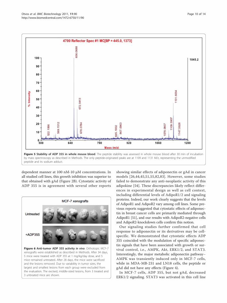
ADP 355 exhibits superior stability in mouse serum andbloodCalculating from the degradation rate measured in 25%aqueous serum [74], ADP 355 had a 75 min half-life inmouse serum (data not shown). In whole mouse blood,the peptide was present even after 30 min withoutnoticeable degradation (Figure 5).
ADP 355 inhibits the growth of MCF-7 xenografts inimmunocompromised miceThe efficacy of ADP355 was assessed in MCF-7 orthoto-pic xenograft model. The peptidomimetic was injectedip into scid mice carrying palpable MCF-7 xenografts ata 1 mg/kg/day dose. After 28 days of treatment, themice were sacrificed and tumors removed. Due to varia-bility in tumor sizes, the largest and smallest lesionsfrom each group were excluded from the evaluation.Figure 6 shows the remaining 6 tumor lesions (from 3animals in each group). The total tumor weight inuntreated animals was 10.27 g (average tumor weightsper mice: 2.56 g, 1,51 g and 1.07 g), and in ADP 355-treated animals 7.10 g (average tumor weights per mice:1.38 g, 1.36 g and 0.81 g). Thus, ADP 355 therapy
reduced established tumor growth by 31% (statisticallysignificant, p < 0.05), relative to untreated controls.
Preliminary assessment of ADP 355 toxicity in vivoHealthy mice receiving up to 50 mg/kg peptide ADP355 ip showed no signs of systemic toxicity. Four daysafter peptide administration the potential peptide elimi-nation organs were removed and weighed. While thespleens of treated and untreated animals were identicalin size, the kidney and liver weights were slightlyincreased in treated mice relative to total body weight atthe highest dose of 50 mg/kg (Table 2). Since peptidedrugs undergo renal and hepatic clearance [69], anincrease of the elimination organ size might indicate theactive metabolic processes. In addition, AdipoR1/2, asphysiological targets of adiponectin, are found in theliver, and might respond to agonist treatment [75].However, these limited toxic effects were not observedbelow the 10 mg/kg dose, a magnitude higher than thetherapy dose, identifying ADP 355 as a safe treatmentoption.
DiscussionNumerous epidemiological and experimental studiesprovided evidence linking obesity to an increased risk ofdeveloping different malignancies, including breast, col-orectal, prostate and endometrial cancers [76-79]. Inaddition, calorie-rich diet has been shown to induceinflammatory responses in microglia cells, which poten-tially can promote development of brain neoplasms[80,81].In obese individuals, especially in those with high visc-
eral fat content, adiponectin levels are low [11]. Accord-ing to epidemiological studies, low adiponectin levelsare associated with elevated cancer risk and develop-ment of more aggressive neoplasms [4,11,48]. Howexactly adiponectin might prevent or restrict cancer isyet not clear. The relevant mechanisms could involveactivation of intracellular metabolic changes similar tothose produced by calorie restriction, i.e., stimulation ofintracellular signals, such as AMPK, and inhibition ofabnormal growth and survival pathways [11]. Thus,pharmacological activation of adiponectin signaling inobese individuals that are refractory to lifestyle modifi-cations could help to restore beneficial pathways nor-mally controlled by this adipokine.However, development of the whole adiponectin pro-
tein as a drug is difficult because of the extreme insolu-bility of the C-terminal globular domain and its largerpeptide fragments. In addition, until now, the adiponec-tin active site has not been mapped. Consequently, weattempted to generate small peptides that would pro-duce biological effects similar or superior to that of
Figure 3 Effects of siRNA-mediated downregulation of AdipoR1or AdipoR2 on ADP 355 activity. The expression of AdipoR1,AdipoR2, and control protein GAPDH were assessed by WB incontrol (C) cells (transfection medium only), cells treated withscrambled siRNA (Sc siRNA), siRNA targeting AdipoR1, or siRNAtargeting AdipoR2, as described in Methods. The relative levels ofAdipoR1 and AdipoR2 proteins were calculated by densitometryscanning, as described in Methods, and are provided in Additionalfile 2. The relative % of growth inhibition upon ADP 355 treatmentin cells with different levels of AdipoR1 and AdipoR2 vs. untreatedcells is shown in the lower panel table.
Otvos et al. BMC Biotechnology 2011, 11:90http://www.biomedcentral.com/1472-6750/11/90
Page 8 of 14

gAd, but would be suitable for pharmaceuticalmodifications.First, using peptide arrays and biological screening
assays, we mapped the adiponectin active site to aminoacids 149-166 within the globular domain of the wholeadipokine (Figure 1A). In parallel experiments, we foundthat peptides covering the active site displayed high affi-nity to an extended version of the AdipoR1 loop 1(sequence: Arg-Pro-Asn-Met-Tyr-Fen-Met-Ale-Pro-Leu-Gln-Glu-Lys-Val-Val) that shares 86% homology withthe loop 1 in AdipoR2. Further modifications of theactive site, followed by structure-function screeningresulted in the development of the lead peptidomimetic,ADP 355, as optimal AdipoR agonist.The identified active site of adiponectin can be charac-
terized as a turn region followed by a b-pleated sheetfragment (Figure 1B). When removed from the proteinenvironment, MD studies indicated that the isolatednative peptide 25 loses the b-pleated sheet character
and forms a series of turns (Figure 7). During MD simu-lations, the initial turn- b-sheet structures of both pep-tide 25 and ADP 355 peptides were substantiallychanged and showed high flexibility. The backboneRMSD values fluctuated with high frequency between0.1 and 0.7 mm. However, in the case of ADP 355, from80 ns to 250 ns, the RMSD remained around 0.6 nm,indicting that the peptidomimetic folded into a morestable conformation characterized by a hairpin incorpor-ating almost the entire peptide. In the cluster analysis,the most populated cluster of the peptidomimetic con-tained more than twice as many structures as the nativefragment (31.6% vs 12.4%). If the dominant b-hairpinstructure is indeed the active conformation, the signifi-cantly increased population of this conformer canexplain the improved in vitro activity of ADP 355 rela-tive to that of its precursor peptide 25.Functional assays with ADP 355 demonstrated that the
peptide restricts cancer cell proliferation in a dose-
Figure 4 Effects of ADP 355 on intracellular cell signaling in cancer cells. The effects of ADP 355 on signaling pathways in MCF-7, MDA-MB-231, and LN18 cells at 0-60 min of treatment were studied by WB, as described in Methods. The expression of GAPDH was used asdetermination of protein loading. The relative levels of phosphorylated/total proteins were calculated by densitometry scanning, as described inMethods, and are provided in Additional file 3.
Otvos et al. BMC Biotechnology 2011, 11:90http://www.biomedcentral.com/1472-6750/11/90
Page 9 of 14
dependent manner at 100 nM-10 μM concentrations. Inall studied cell lines, this growth inhibition was superior tothat obtained with gAd (Figure 2B). Cytostatic activity ofADP 355 is in agreement with several other reports
showing similar effects of adiponectin or gAd in cancermodels [26,44,45,51,55,82,83]. However, some studiesfailed to demonstrate any anti-neoplastic activity of thisadipokine [54]. These discrepancies likely reflect differ-ences in experimental design as well as cell context,including differential levels of AdipoR1/2 and signalingproteins. Indeed, our work clearly suggests that the levelsof AdipoR1 and AdipoR2 vary among cell lines. Some pre-vious reports suggested that cytostatic effects of adiponec-tin in breast cancer cells are primarily mediated throughAdipoR1 [51], and our results with AdipoR2-negative cellsand AdipoR2-knockdown cells confirm this notion.Our signaling studies further confirmed that cell
response to adiponectin or its derivatives may be cell-specific. We demonstrated that cytostatic effects ADP355 coincided with the modulation of specific adiponec-tin signals that have been associated with growth or sur-vival control, i.e., AMPK, Akt, ERK1/2, and STAT3.Interestingly, the major metabolic adiponectin pathway–AMPK was transiently induced only in MCF-7 cells,while in MDA-MB-231 and LN18 cells, the peptide orgAd did not have any effects (Figure 4).In MCF-7 cells, ADP 355, but not gAd, decreased
ERK1/2 signaling. STAT3 was activated in this cell line
Figure 5 Stability of ADP 355 in whole mouse blood. The peptide stability was assessed in whole mouse blood after 30 min of incubationby mass spectroscopy as described in Methods. The only peptide-originated peaks are at 1109 and 1131 M/z, representing the unmodifiedpeptide and its sodium adduct.
Figure 6 Anti-tumor ADP 355 activity in vivo. Orthotopic MCF-7xenografts were established as described in Methods. After 34 days,5 mice were treated with ADP 355 at 1 mg/kg/day dose, and 5mice remained untreated. After 28 days, the mice were sacrificedand the lesions removed. Due to variability in tumor sizes, thelargest and smallest lesions from each group were excluded fromthe evaluation. The excised, middle-sized lesions, from 3 treated and3 untreated mice are shown.
Otvos et al. BMC Biotechnology 2011, 11:90http://www.biomedcentral.com/1472-6750/11/90
Page 10 of 14

by both ADP 355 and gAd. In MDA-MB-231 cells, likein MCF-7 cells, ADP 355 decreased ERK1/2 activationand transiently increased STAT3 signaling. In bothbreast cancer cell lines, ADP 355 did not affect themajor growth/survival Akt pathway. In contrast, ADP355 and gAd significantly inhibited Akt and STAT3 sig-nals in LN18 cells. Interestingly, the effects on Akt con-cerned total levels of the enzyme, suggesting that ADP355 might affect its turnover.Published data on adiponectin signaling in cancer cells
seem to support the notion that the cytokine mightinduce different signaling pathways in different celllines. For instance, in many cancer cell lines (breastMCF-7, MDA-MB-231, T47D; colorectal HT-29,CaCO2, SW480; prostate PC3) adiponectin activatedAMPK [26,40,54,55]. On the other hand, adiponectineither reduced or did not affect ERK1/2 in MCF-7 orMDA-MB-231 cells, but stimulated the pathway insome colorectal cancer cell lines [1,40,54]. Akt wasinhibited by adiponectin in MDA-MB-231 breast cancercells, but activated in prostate cancer cells LNCaP[82,84]. The upregulation of AMPK and reduction ofAkt in response to adiponectin in MDA-MB-231 cells
[82] is in contrast with our study and might be relatedto significantly lower gAd and ADP 355 concentrationsused in our experiments, while high doses used by Kimet al. were toxic in our system. Consistent with ourresults, moderate STAT3 stimulation by adiponectinwas noted in MDA-MB-231 cells, while the transcrip-tion factor was inhibited in DU145 prostate cancer cells[1,25]. These differences, in part, could reflect variableexperimental settings, such as baseline growth condi-tions, adiponectin reagents used as well as treatmenttiming and dosage.To further assess the efficacy of ADP 355, we carried
out a preliminary in vivo study. In scid mice carryingMCF-7 orthotopic xenografts, ADP 355 treatmentreduced the growth of established tumors by ~31%, vali-dating AdipoR as a target for breast cancer therapy.
ConclusionsHere we report on the design and development of afirst-in-class AdipoR agonist. AdipoR agonists areviewed as future drugs to treat multiple diseases relatedto obesity and insulin resistance. The biological activityof our novel ADP 355, including its in vivo efficacy, itssuperior stability in biological fluids, as well as accepta-ble toxicity profile and low production costs indicatethat the peptidomimetic represents a true lead com-pound for ensuing pharmaceutical development.
Additional material
Additional file 1: Sequences of tested adiponectin-derived peptides.Designation and amino acid sequences of tested adiponectin-derivedpeptides.
Additional file 2: Quantification of AdipoR1 and AdipoR2 amounts.Densitometry quantification of AdipoR1 and AdipoR2 levels followingtargeted siRNA knockdown experiments.
Additional file 3: Quantification of signaling pathways in ADP 355-treated cancer cells. Densitometry quantification of pAMPK/AMPK,pSTAT3/STAT3, pAkt/Akt, pERK 1/2/ERK1/2 levels in MCF-7, MDA-MB-231,and LN18 cells treated with gAd or ADP 355.
AcknowledgementsThis work was supported by grants Department of Defense W81XWH-09-1-0332 (LO, ES), NIH-INBRE 1 P20 RR16469 (SL), and the Sbarro Health ResearchOrganization (ES, EH, FLR, FM, PG).
Table 2 Toxicity analysis of ADP 355
Peptide dose (bolus ip) Liver weight (g); relative to total weight (%) Spleen weight (g) Kidney weight (g); relative to total weight (%)
Untreated 0.98; 0.054 0.07 0.29; 0.015
5 mg/kg 1.02; 0.054 0.07 0.30; 0.016
10 mg/kg 1.09; 0.059 0.06 0.29; 0.016
25 mg/kg 1.24; 0.059 0.07 0.35; 0.017
50 mg/kg 1.07; 0.062 0.07 0.31; 0.017
CBA/J mice were treated with ADP 355 and toxicity parameters were assessed as described in Methods.
Figure 7 ADP 355 energy analysis . Representative energyminimized structures of peptide 25 (red) and ADP 355 (purple)overlaid to the conformation of the 153-162 sequence found inadiponectin protein (grey).
Otvos et al. BMC Biotechnology 2011, 11:90http://www.biomedcentral.com/1472-6750/11/90
Page 11 of 14

Author details1Temple University, Department of Biology, Philadelphia, PA 19122, USA.2Temple University, Sbarro Institute for Cancer Research and MolecularMedicine, Philadelphia, PA 19122, USA. 3University of Verona, Department ofMedical Oncology, 37189 Verona, Italy. 4Semmelweis University MedicalSchool, 1st Department of Pathology and Experimental Cancer Research,1085 Budapest, Hungary. 5Creighton University, Department of BiomedicalSciences, Omaha, NE 68178, USA. 6Leipzig University, Institute of BioanalyticalChemistry, Leipzig 04103, Germany. 7Florey Neuroscience Institutes,Melbourne, 2010 Victoria, Australia.
Authors’ contributionsLO conceived the study, participated in its coordination and manuscriptwriting; EH, FLR, and FM analyzed biological activity and intracellularsignaling of peptides and peptidomimetics; PG performed siRNAexperiments; KN carried peptide cleavage, purification and measurements;DK and RH analyzed peptide stability in biological fluids; IK carried out andanalyzed all animal experiments; SL performed all energy calculations; JWsynthesized the peptides in solution; ES conceived the study, participated inits design and coordination, and drafted the manuscript. All authors readand approved the final manuscript.
Received: 23 August 2011 Accepted: 5 October 2011Published: 5 October 2011
References1. Grossmann ME, Nkhata KJ, Mizuno NK, Ray A, Cleary MP: Effects of
adiponectin on breast cancer cell growth and signaling. Br J Cancer 2008,98(2):370-379.
2. Ryan AS, Berman DM, Nicklas BJ, Sinha M, Gingerich RL, Meneilly GS,Egan JM, Elahi D: Plasma adiponectin and leptin levels, bodycomposition, and glucose utilization in adult women with wide rangesof age and obesity. Diabetes Care 2003, 26(8):2383-2388.
3. Chiarugi P, Fiaschi T: Adiponectin in health and diseases: from metabolicsyndrome to tissue regeneration. Expert Opin Ther Targets 2010,14(2):193-206.
4. Chen X, Wang Y: Adiponectin and breast cancer. Med Oncol 2010.5. Shibata R, Ouchi N, Murohara T: Adiponectin and cardiovascular disease.
Circ J 2009, 73(4):608-614.6. Galic S, Oakhill JS, Steinberg GR: Adipose tissue as an endocrine organ.
Mol Cell Endocrinol 2010, 316(2):129-139.7. Barb D, Williams CJ, Neuwirth AK, Mantzoros CS: Adiponectin in relation to
malignancies: a review of existing basic research and clinical evidence.Am J Clin Nutr 2007, 86(3):s858-866.
8. Hu PF, Bao JP, Wu LD: The emerging role of adipokines in osteoarthritis:a narrative review. Mol Biol Rep 2010, 38(2):873-878.
9. Ziemke F, Mantzoros CS: Adiponectin in insulin resistance: lessons fromtranslational research. Am J Clin Nutr 2010, 91(1):258S-261S.
10. Schaffler A, Scholmerich J, Buechler C: Mechanisms of disease: adipokinesand breast cancer - endocrine and paracrine mechanisms that connectadiposity and breast cancer. Nat Clin Pract Endocrinol Metab 2007,3(4):345-354.
11. Brochu-Gaudreau K, Rehfeldt C, Blouin R, Bordignon V, Murphy BD,Palin MF: Adiponectin action from head to toe. Endocrine 2010,37(1):11-32.
12. Fang X, Sweeney G: Mechanisms regulating energy metabolism byadiponectin in obesity and diabetes. Biochem Soc Trans 2006, 34(Pt5):798-801.
13. Wu X, Motoshima H, Mahadev K, Stalker TJ, Scalia R, Goldstein BJ:Involvement of AMP-activated protein kinase in glucose uptakestimulated by the globular domain of adiponectin in primary ratadipocytes. Diabetes 2003, 52(6):1355-1363.
14. Tsao TS, Tomas E, Murrey HE, Hug C, Lee DH, Ruderman NB, Heuser JE,Lodish HF: Role of disulfide bonds in Acrp30/adiponectin structure andsignaling specificity. Different oligomers activate different signaltransduction pathways. J Biol Chem 2003, 278(50):50810-50817.
15. Tomas E, Tsao TS, Saha AK, Murrey HE, Zhang Cc C, Itani SI, Lodish HF,Ruderman NB: Enhanced muscle fat oxidation and glucose transport byACRP30 globular domain: acetyl-CoA carboxylase inhibition and AMP-activated protein kinase activation. Proc Natl Acad Sci USA 2002,99(25):16309-16313.
16. Yamauchi T, Kamon J, Ito Y, Tsuchida A, Yokomizo T, Kita S, Sugiyama T,Miyagishi M, Hara K, Tsunoda M, et al: Cloning of adiponectin receptorsthat mediate antidiabetic metabolic effects. Nature 2003,423(6941):762-769.
17. Kadowaki T, Yamauchi T: Adiponectin and adiponectin receptors. EndocrRev 2005, 26(3):439-451.
18. Wang H, Zhang H, Jia Y, Zhang Z, Craig R, Wang X, Elbein SC: Adiponectinreceptor 1 gene (ADIPOR1) as a candidate for type 2 diabetes andinsulin resistance. Diabetes 2004, 53(8):2132-2136.
19. Yamauchi T, Nio Y, Maki T, Kobayashi M, Takazawa T, Iwabu M, Okada-Iwabu M, Kawamoto S, Kubota N, Kubota T, et al: Targeted disruption ofAdipoR1 and AdipoR2 causes abrogation of adiponectin binding andmetabolic actions. Nat Med 2007, 13(3):332-339.
20. Kadowaki T, Yamauchi T: Adiponectin receptor signaling: a new layer tothe current model. Cell Metab 2011, 13(2):123-124.
21. Wijesekara N, Krishnamurthy M, Bhattacharjee A, Suhail A, Sweeney G,Wheeler MB: Adiponectin-induced ERK and Akt phosphorylation protectsagainst pancreatic beta cell apoptosis and increases insulin geneexpression and secretion. J Biol Chem 2010, 285(44):33623-33631.
22. Handy JA, Saxena NK, Fu P, Lin S, Mells JE, Gupta NA, Anania FA:Adiponectin activation of AMPK disrupts leptin-mediated hepatic fibrosisvia suppressors of cytokine signaling (SOCS-3). J Cell Biochem 2010,110(5):1195-1207.
23. Akifusa S, Kamio N, Shimazaki Y, Yamaguchi N, Nonaka K, Yamashita Y:Involvement of the JAK-STAT pathway and SOCS3 in the regulation ofadiponectin-generated reactive oxygen species in murine macrophageRAW 264 cells. J Cell Biochem 2010, 111(3):597-606.
24. Liu J, Lam JB, Chow KH, Xu A, Lam KS, Moon RT, Wang Y: Adiponectinstimulates Wnt inhibitory factor-1 expression through epigeneticregulations involving the transcription factor specificity protein 1.Carcinogenesis 2008, 29(11):2195-2202.
25. Miyazaki T, Bub JD, Uzuki M, Iwamoto Y: Adiponectin activates c-Jun NH2-terminal kinase and inhibits signal transducer and activator oftranscription 3. Biochem Biophys Res Commun 2005, 333(1):79-87.
26. Korner A, Pazaitou-Panayiotou K, Kelesidis T, Kelesidis I, Williams CJ,Kaprara A, Bullen J, Neuwirth A, Tseleni S, Mitsiades N, et al: Total andhigh-molecular-weight adiponectin in breast cancer: in vitro and in vivostudies. J Clin Endocrinol Metab 2007, 92(3):1041-1048.
27. Hattori Y, Hattori S, Kasai K: Globular adiponectin activates nuclear factor-kappaB in vascular endothelial cells, which in turn induces expression ofproinflammatory and adhesion molecule genes. Diabetes Care 2006,29(1):139-141.
28. Haugen F, Drevon CA: Activation of nuclear factor-kappaB by high molecularweight and globular adiponectin. Endocrinology 2007, 148(11):5478-5486.
29. Plant S, Shand B, Elder P, Scott R: Adiponectin attenuates endothelialdysfunction induced by oxidised low-density lipoproteins. Diab Vasc DisRes 2008, 5(2):102-108.
30. Goktas S, Yilmaz MI, Caglar K, Sonmez A, Kilic S, Bedir S: Prostate cancerand adiponectin. Urology 2005, 65(6):1168-1172.
31. Housa D, Housova J, Vernerova Z, Haluzik M: Adipocytokines and cancer.Physiol Res 2006, 55(3):233-244.
32. Mantzoros C, Petridou E, Dessypris N, Chavelas C, Dalamaga M, Alexe DM,Papadiamantis Y, Markopoulos C, Spanos E, Chrousos G, et al: Adiponectinand breast cancer risk. J Clin Endocrinol Metab 2004, 89(3):1102-1107.
33. Miyoshi Y, Funahashi T, Kihara S, Taguchi T, Tamaki Y, Matsuzawa Y,Noguchi S: Association of serum adiponectin levels with breast cancerrisk. Clin Cancer Res 2003, 9(15):5699-5704.
34. Chen DC, Chung YF, Yeh YT, Chaung HC, Kuo FC, Fu OY, Chen HY, Hou MF,Yuan SS: Serum adiponectin and leptin levels in Taiwanese breast cancerpatients. Cancer Lett 2006, 237(1):109-114.
35. Cleary MP, Grossmann ME, Ray A: Effect of obesity on breast cancerdevelopment. Vet Pathol 2010, 47(2):202-213.
36. Cleary MP, Ray A, Rogozina OP, Dogan S, Grossmann ME: Targeting theadiponectin:leptin ratio for postmenopausal breast cancer prevention.Front Biosci (Schol Ed) 2009, 1:329-357.
37. Hou WK, Xu YX, Yu T, Zhang L, Zhang WW, Fu CL, Sun Y, Wu Q, Chen L:Adipocytokines and breast cancer risk. Chin Med J (Engl) 2007,120(18):1592-1596.
38. Arditi JD, Venihaki M, Karalis KP, Chrousos GP: Antiproliferative effect ofadiponectin on MCF7 breast cancer cells: a potential hormonal linkbetween obesity and cancer. Horm Metab Res 2007, 39(1):9-13.
Otvos et al. BMC Biotechnology 2011, 11:90http://www.biomedcentral.com/1472-6750/11/90
Page 12 of 14

39. Bub JD, Miyazaki T, Iwamoto Y: Adiponectin as a growth inhibitor inprostate cancer cells. Biochem Biophys Res Commun 2006,340(4):1158-1166.
40. Dieudonne MN, Bussiere M, Dos Santos E, Leneveu MC, Giudicelli Y,Pecquery R: Adiponectin mediates antiproliferative and apoptoticresponses in human MCF7 breast cancer cells. Biochem Biophys ResCommun 2006, 345(1):271-279.
41. Fenton JI, Birmingham JM: Adipokine regulation of colon cancer:adiponectin attenuates interleukin-6-induced colon carcinoma cellproliferation via STAT-3. Mol Carcinog 2010, 49(7):700-709.
42. Fujisawa T, Endo H, Tomimoto A, Sugiyama M, Takahashi H, Saito S,Inamori M, Nakajima N, Watanabe M, Kubota N, et al: Adiponectinsuppresses colorectal carcinogenesis under the high-fat diet condition.Gut 2008, 57(11):1531-1538.
43. Kang JH, Lee YY, Yu BY, Yang BS, Cho KH, Yoon DK, Roh YK: Adiponectininduces growth arrest and apoptosis of MDA-MB-231 breast cancer cell.Arch Pharm Res 2005, 28(11):1263-1269.
44. Dos Santos E, Benaitreau D, Dieudonne MN, Leneveu MC, Serazin V,Giudicelli Y, Pecquery R: Adiponectin mediates an antiproliferativeresponse in human MDA-MB 231 breast cancer cells. Oncol Rep 2008,20(4):971-977.
45. Wang Y, Lam JB, Lam KS, Liu J, Lam MC, Hoo RL, Wu D, Cooper GJ, Xu A:Adiponectin modulates the glycogen synthase kinase-3beta/beta-catenin signaling pathway and attenuates mammary tumorigenesis ofMDA-MB-231 cells in nude mice. Cancer Res 2006, 66(23):11462-11470.
46. Taliaferro-Smith L, Nagalingam A, Zhong D, Zhou W, Saxena NK, Sharma D:LKB1 is required for adiponectin-mediated modulation of AMPK-S6K axisand inhibition of migration and invasion of breast cancer cells. Oncogene2009, 28(29):2621-2633.
47. Saxena NK, Sharma D: Metastasis suppression by adiponectin: LKB1 risesup to the challenge. Cell Adh Migr 2010, 4(3).
48. Pfeiler G, Hudelist G, Wulfing P, Mattsson B, Konigsberg R, Kubista E,Singer CF: Impact of AdipoR1 expression on breast cancer development.Gynecol Oncol 2010, 117(1):134-138.
49. Pfeiler G, Treeck O, Wenzel G, Goerse R, Hartmann A, Schmitz G,Ortmann O: Influence of insulin resistance on adiponectin receptorexpression in breast cancer. Maturitas 2009, 63(3):253-256.
50. Takahata C, Miyoshi Y, Irahara N, Taguchi T, Tamaki Y, Noguchi S:Demonstration of adiponectin receptors 1 and 2 mRNA expression inhuman breast cancer cells. Cancer Lett 2007, 250(2):229-236.
51. Nakayama S, Miyoshi Y, Ishihara H, Noguchi S: Growth-inhibitory effect ofadiponectin via adiponectin receptor 1 on human breast cancer cellsthrough inhibition of S-phase entry without inducing apoptosis. BreastCancer Res Treat 2008, 112(3):405-410.
52. Kim AY, Lee YS, Kim KH, Lee JH, Lee HK, Jang SH, Kim SE, Lee GY, Lee JW,Jung SA, et al: Adiponectin represses colon cancer cell proliferation viaAdipoR1- and -R2-mediated AMPK activation. Mol Endocrinol 2010,24(7):1441-1452.
53. Yoneda K, Tomimoto A, Endo H, Iida H, Sugiyama M, Takahashi H,Mawatari H, Nozaki Y, Fujita K, Yoneda M, et al: Expression of adiponectinreceptors, AdipoR1 and AdipoR2, in normal colon epithelium and coloncancer tissue. Oncol Rep 2008, 20(3):479-483.
54. Williams CJ, Mitsiades N, Sozopoulos E, Hsi A, Wolk A, Nifli AP, Tseleni-Balafouta S, Mantzoros CS: Adiponectin receptor expression is elevated incolorectal carcinomas but not in gastrointestinal stromal tumors. EndocrRelat Cancer 2008, 15(1):289-299.
55. Zakikhani M, Dowling RJ, Sonenberg N, Pollak MN: The effects ofadiponectin and metformin on prostate and colon neoplasia involveactivation of AMP-activated protein kinase. Cancer Prev Res (Phila) 2008,1(5):369-375.
56. Gonzalez-Angulo AM, Meric-Bernstam F: Metformin: a therapeuticopportunity in breast cancer. Clin Cancer Res 2010, 16(6):1695-1700.
57. Hadad SM, Fleming S, Thompson AM: Targeting AMPK: a new therapeuticopportunity in breast cancer. Crit Rev Oncol Hematol 2008, 67(1):1-7.
58. Fay JR, Steele V, Crowell JA: Energy homeostasis and cancer prevention:the AMP-activated protein kinase. Cancer Prev Res (Phila) 2009,2(4):301-309.
59. Jiang W, Zhu Z, Thompson HJ: Dietary energy restriction modulates theactivity of AMP-activated protein kinase, Akt, and mammalian target ofrapamycin in mammary carcinomas, mammary gland, and liver. CancerRes 2008, 68(13):5492-5499.
60. Krieger E, Koraimann G, Vriend G: Increasing the precision of comparativemodels with YASARA NOVA–a self-parameterizing force field. Proteins2002, 47(3):393-402.
61. Berman HM, Westbrook J, Feng Z, Gilliland G, Bhat TN, Weissig H,Shindyalov IN, Bourne PE: The Protein Data Bank. Nucleic Acids Res 2000,28(1):235-242.
62. Van Der Spoel DLE, Hess B, Groenhof G, Mark AE, Berendsen HJC:GROMACS: fast, flexible, and free. J Comp Chem 2005, 26:1701-1718.
63. Kaminski GAFR, Tirado-Rives J, Jorgensen WL: Evaluation andreparametrization of the OPLS-AA force field for proteins via comparisonwith accurate quantum chamical calculations on peptides. J Phys Chem B2001, 105:6474-6487.
64. Daura XGK, Jaun B, Steebach D, van Gunsteren WF, Mark AE: Peptidefolding: When simulation meets experiment. Angew Chem Int Ed Engl1999, 38:236-240.
65. Kabsch W, Sander C: Dictionary of protein secondary structure: patternrecognition of hydrogen-bonded and geometrical features. Biopolymers1983, 22(12):2577-2637.
66. Frank R: The SPOT-synthesis technique. Synthetic peptide arrays onmembrane supports–principles and applications. J Immunol Methods2002, 267(1):13-26.
67. Fields GB, Noble RL: Solid phase peptide synthesis utilizing 9-fluorenylmethoxycarbonyl amino acids. Int J Pept Protein Res 1990,35(3):161-214.
68. Hilpert K, Winkler DF, Hancock RE: Peptide arrays on cellulose support:SPOT synthesis, a time and cost efficient method for synthesis of largenumbers of peptides in a parallel and addressable fashion. Nat Protoc2007, 2(6):1333-1349.
69. Otvos L Jr, Kovalszky I, Riolfi M, Ferla R, Olah J, Sztodola A, Namma K,Molino A, Piubello Q, Wade JD, Surmacz E: Efficacy of a Leptin ReceptorAntagonist Peptide in a Mouse Model of Triple-negative Breast Cancer.European J Cancer 2011.
70. Riolfi M, Ferla R, Del Valle L, Pina-Oviedo S, Scolaro L, Micciolo R, Guidi M,Terrasi M, Cetto GL, Surmacz E: Leptin and its receptor are overexpressedin brain tumors and correlate with the degree of malignancy. BrainPathol 2010, 20(2):481-489.
71. Bartucci M, Morelli C, Mauro L, Ando S, Surmacz E: Differential insulin-likegrowth factor I receptor signaling and function in estrogen receptor(ER)-positive MCF-7 and ER-negative MDA-MB-231 breast cancer cells.Cancer Res 2001, 61(18):6747-6754.
72. Bartella V, Cascio S, Fiorio E, Auriemma A, Russo A, Surmacz E: Insulin-dependent leptin expression in breast cancer cells. Cancer Res 2008,68(12):4919-4927.
73. Garofalo C, Sisci D, Surmacz E: Leptin interferes with the effects of theantiestrogen ICI 182,780 in MCF-7 breast cancer cells. Clin Cancer Res2004, 10(19):6466-6475.
74. Powell MF, Grey H, Gaeta F, Sette A, Colon S: Peptide stability in drugdevelopment: a comparison of peptide reactivity in different biologicalmedia. J Pharm Sci 1992, 81(8):731-735.
75. Kadowaki T, Yamauchi T, Kubota N: The physiological andpathophysiological role of adiponectin and adiponectin receptors in theperipheral tissues and CNS. FEBS Lett 2008, 582(1):74-80.
76. Maiti B, Kundranda MN, Spiro TP, Daw HA: The association of metabolicsyndrome with triple-negative breast cancer. Breast Cancer Res Treat 2009.
77. Vona-Davis L, Howard-McNatt M, Rose DP: Adiposity, type 2 diabetes andthe metabolic syndrome in breast cancer. Obes Rev 2007, 8(5):395-408.
78. Calle EE, Thun MJ: Obesity and cancer. Oncogene 2004, 23(38):6365-6378.79. Pischon T, Nothlings U, Boeing H: Obesity and cancer. Proc Nutr Soc 2008,
67(2):128-145.80. Velloso LA: The brain is the conductor: diet-induced inflammation
overlapping physiological control of body mass and metabolism. ArqBras Endocrinol Metabol 2009, 53(2):151-158.
81. Reynes G, Vila V, Martin M, Parada A, Fleitas T, Reganon E, Martinez-Sales V:Circulating markers of angiogenesis, inflammation, and coagulation inpatients with glioblastoma. J Neurooncol 2010, 102(1):35-41.
82. Kim KY, Baek A, Hwang JE, Choi YA, Jeong J, Lee MS, Cho DH, Lim JS,Kim KI, Yang Y: Adiponectin-activated AMPK stimulatesdephosphorylation of AKT through protein phosphatase 2A activation.Cancer Res 2009, 69(9):4018-4026.
83. Fenton JI, Birmingham JM, Hursting SD, Hord NG: Adiponectin blocksmultiple signaling cascades associated with leptin-induced cell
Otvos et al. BMC Biotechnology 2011, 11:90http://www.biomedcentral.com/1472-6750/11/90
Page 13 of 14

proliferation in Apc Min/+ colon epithelial cells. Int J Cancer 2008,122(11):2437-2445.
84. Barb D, Neuwirth A, Mantzoros CS, Balk SP: Adiponectin signals in prostatecancer cells through Akt to activate the mammalian target of rapamycinpathway. Endocr Relat Cancer 2007, 14(4):995-1005.
doi:10.1186/1472-6750-11-90Cite this article as: Otvos et al.: Design and development of a peptide-based adiponectin receptor agonist for cancer treatment. BMCBiotechnology 2011 11:90.
Submit your next manuscript to BioMed Centraland take full advantage of:
• Convenient online submission
• Thorough peer review
• No space constraints or color figure charges
• Immediate publication on acceptance
• Inclusion in PubMed, CAS, Scopus and Google Scholar
• Research which is freely available for redistribution
Submit your manuscript at www.biomedcentral.com/submit
Otvos et al. BMC Biotechnology 2011, 11:90http://www.biomedcentral.com/1472-6750/11/90
Page 14 of 14