Embed Size (px)
Citation preview

Kwok et al. BMC Microbiology 2014, 14:176http://www.biomedcentral.com/1471-2180/14/176
RESEARCH ARTICLE Open Access
Complete genome assembly and characterization ofan outbreak strain of the causative agent of swineerysipelas – Erysipelothrix rhusiopathiae SY1027Amy HY Kwok1†, Yufeng Li2†, Jingwei Jiang1, Ping Jiang2 and Frederick C Leung1,3*
Abstract
Background: Erysipelothrix rhusiopathiae is the causative agent of animal erysipelas and, to a fewer occurrences,human erysipeloid. It is ubiquitous in nature and commensal in diverse species of animals, wild or domestic, frommammals and birds to reptiles and fish. Mechanisms of its virulence and pathogenicity are poorly understood.
Results: Making use of the complete genome sequencing of E. rhusiopathiae strain SY1027 and comparative genomeanalysis between the three highly pathogenic strains (SY1027, Fujisawa and ATCC19414), the genomic structure andputative functional elements, such as pathogenicity island (PAI)-like regions, potential virulence factors and horizontaltransferring genes of the bacteria are identified. Strain SY1027 genome is 1,752,910 base pairs long, just 30 kilobasessmaller than strain Fujisawa, with the same GC level of 36.36%. It contains 1,845 open reading frames (ORF) predicted byGLIMMER 3.02, of which 1,775 were annotated by PGAAP, 1,757 (~95.23%) were annotated by NCBI nr blast, 1,209 byCOG database and 1,076 by KEGG database. 37 potential virulence factors were annotated in strain SY1027 by VFDB,while 19 (~51.35%) of them are common in the 2 strains, 7 of which are potentially related to antibiotic resistance andhighly conserved (~98-100% match identity (ID)) amongst the three strains of E. rhusiopathiae and modestly homologousto other gastrointestinal tract-inhabiting Firmicutes (~40% match ID), e.g. Clostridium spp., Enterococcus spp. Genomicisland- and pathogenicity island-like regions were also predicted, in which some showed association with tRNA andpotential virulence factors.
Conclusion: Complete genome sequencing of Erysipelothrix rhusiopathiae, the causative agent of animal erysipelas, wasperformed. Molecular identification of various genomic elements pave the way to the better understanding ofmechanisms underlying metabolic capabilities, pathogenicity of swine erysipelas and prospective vaccine targets besidesthe widely used SpaA antigens.
Keywords: Erysipelothrix rhusiopathiae, Complete genome assembly, Genome characterization, Erysipelas, Virulence factors
BackgroundSince its first isolation from mice in 1876, efforts havebeen devoted to the studies of Erysipelothrix rhusiopathiae(see review: [1]). E. rhusiopathiae is a Gram-positive bac-terium that has diverse cell morphology, remarkablechemical tolerance and poorly understood cellular struc-ture and molecular mechanisms, especially those related
* Correspondence: [email protected]†Equal contributors1Bioinformatics Center, Nanjing Agricultural University, F202, South Block,Faculty of Science Complex, 1 Weigang, Nanjing 210095, China3School of Biological Sciences, University of Hong Kong, Hong Kong, SAR,ChinaFull list of author information is available at the end of the article
© 2014 Kwok et al.; licensee BioMed Central LCommons Attribution License (http://creativecreproduction in any medium, provided the orDedication waiver (http://creativecommons.orunless otherwise stated.
to its virulence in a wide range of hosts, ranging frommammals and birds to reptiles and fish. Best known as theetiological agent of swine erysipelas– a disease that cancause acute symptoms such as septicaemia or lead tochronic syndromes like arthritis and endocarditis in pigs[1], E. rhusiopathiae is generally believed to be transmittedvia the gastrointestinal (GI) tract by intake of contami-nated water or food and causes great economical loss inhusbandry worldwide. Given its ubiquitous nature in theenvironment and reservoir of asymptomatic carriersamongst both domestic and wild animals, the preventionand control of swine erysipelas is often challenging. Morethan 23 serovars have so far been described [2]. However,due to the considerable variance in their morphology, host
td. This is an Open Access article distributed under the terms of the Creativeommons.org/licenses/by/4.0), which permits unrestricted use, distribution, andiginal work is properly credited. The Creative Commons Public Domaing/publicdomain/zero/1.0/) applies to the data made available in this article,

Kwok et al. BMC Microbiology 2014, 14:176 Page 2 of 12http://www.biomedcentral.com/1471-2180/14/176
specificity and/or pathogenicity, the practicality of sero-typing remains debatable. The heat labile capsule ofE. rhusiopathiae has been associated with its resistanceto phagocytosis and virulence by transposon mutagen-esis study [3]. Surface proteins such as neuraminidase,SpaA antigen, two adhesive surface proteins containingthe C-terminal anchoring LPXTG motif (RspA andRspB) have shown positive correlation to virulence ofthe bacteria [4–6], however, little is known about themechanisms of its pathogenicity.In China, outbreaks of swine erysipelas have surfaced in
recent years despite its relative tranquility in the past 3 de-cades, with an alarming trend of developing from scat-tered, domestic occurrences in a few farms to systemicoutbursts in provincial scale. Three representative local re-ports, which are only available in Chinese, are included inAdditional file 1. In present study, we isolated an outbreakfield strain SY1027 from SiYang, Jiangsu and completedits bacterial genome sequencing and assembly. Thecomplete genome of E. rhusiopathiae strain Fujisawa hasjust recently been published [7]. Together with the draftgenome of strain ATCC19414, data-mining of potentialvirulence factors, especially those related to capsular pro-tein biosynthesis, would offer us a more reliable compara-tive genomic analysis and a better understanding of thecommon genomic structure and a more precise predictionof potential virulence factors for improvement of vaccinetargets or strategy of disease control.
MethodsIsolation and total DNA extraction of E. rhusiopathiaestrain SY1027E. rhusiopathiae strain SY1027 was collected from a pigfarm in SiYang, Jiangsu. Anticoagulant-treated blood wascollected from diseased pigs by in-house veterinariansfrom pig farms following the Nanjing Agricultural Univer-sity Animal Ethics Committee guidelines (Approval No.IACECNAU20100902) and sent to our laboratory. Thesample was inoculated in Martin’s broth supplementedwith 400 μg/ml neomycin and 70 μg/ml vancomycin. Asingle colony was isolated by inoculation of the culture onstreaked agar plate supplemented with horse serum.The single colony of the bacterium was inoculated in
Modified Feist Broth (6 g glucose, 5 g proteose peptone no.2(BD BioSciences, San Jose, US), 5 g yeast extract, 0.5 g L-ar-ginine, 0.5 ml Tween 80 (oleic acid) in 1 L of 0.2 M sodiumphosphate buffer (pH8.0) [8] supplemented with 5% (v/v)fetal bovine serum (Invitrogen, Grand Island, US) and 50 μg/ml kanamycin and grown to its exponential growth phase.Bacteria were harvested by centrifuge and its genomicDNA was extracted according to the JGI bacterial DNAisolationCTABprotocol (http://1ofdmq2n8tc36m6i46scovo2e.wpengine.netdna-cdn.com/wp-content/uploads/2014/02/JGI-Bacterial-DNA-isolation-CTAB-Protocol-2012.pdf).
Pyrosequencing and complete genome assembly ofE. rhusiopathiae strain SY1027To confirm the purity of the genomic DNA of E. rhusio-pathiae strain SY1027, 16S rDNA-specific region was amp-lified and 20 individual positive clones were sequenced byGenetic Analyzer 3130 (Invitrogen, Grand Island, US).BLASTn analysis [9] revealed that E. rhusiopathiae strainSY1027 gDNA sequences have high similarity to those fromthe other 2 strains of E. rhusiopathiae publicly accessible.The quality and quantity of genomic DNA were evaluatedby 0.7% agarose gel electrophoresis and Nanodrop2000(Thermo Scientific, Waltham, US), and using the Quant-iTPicogreen dsDNA kit (Invitrogen), respectively.A whole genome shotgun library was generated with
500 ng of E. rhusiopathiae strain SY1027genomicDNA. The shotgun sequencing procedure was per-formed using 454 GS Junior General Library Prepar-ation Kit, following the manufacturer’s instruction(Roche, Basel, Switzerland). In addition, an 8 kb spanpaired end library was generated with 15 μg of E. rhu-siopathiae strain SY1027 genomic DNA. The pairedend sequencing procedure was performed using 454GS Junior Paired end Library Preparation Kit, follow-ing the manufacturer’s instruction (Roche). Paired endreads were used as orientation guide for assemblingthe contigs into scaffolds. The DNA libraries wereamplified by emPCR and sequenced by FLX Titaniumsequencing chemistry (Roche). One shotgun run andone paired end run were performed on individual li-braries prepared with same genomic DNA sample.After sequencing, the raw data were assembled byNewbler 2.7 (Roche) with default parameters. Primerpairs were designed along the sequences flanking thegap regions for PCR gap filling. The complete genomeof E. rhusiopathiae strain SY1027 was submitted toNCBI Genbank and is publicly accessible [GenBank:CP005079].
Genome annotation of E. rhusiopathiae strain SY1027Glimmer 3.02 [10] was used for gene prediction in E. rhu-siopathiae strain SY1027 complete genome. All predictedORF sequences were translated into amino acid sequencesby in-house Perl scripts. BLASTp [9] was applied to alignthe amino acid sequences against the NCBI non-redundant(nr) database (January, 2013). Amino acid sequences withalignment length over 90% of its own length and over 40%match identity were chosen and the description of the besthit (with highest alignment length percentage and matchidentity) was assigned as the annotation of predicted gene.Intergenic regions were annotated by RepeatMasker (http://www.repeatmasker.org) with default parameters. For faircomparison, genome annotation was simultaneously con-ducted using PGAAP (NCBI).

Kwok et al. BMC Microbiology 2014, 14:176 Page 3 of 12http://www.biomedcentral.com/1471-2180/14/176
Functional gene analysisBLASTp [9] was applied to align the amino acid sequencesagainst the COG database [11]. Amino acid sequenceswith alignment length over 90% of its own length and over20% match identity were chosen and the description ofthe best hit (with highest alignment length percentage andmatch identity) was assigned as the annotation of pre-dicted gene. All annotated genes were then classifiedbased on their COG classes and compared to that ofE. rhusiopathiae strain Fujisawa.
Pathway analysisGLIMMER-predicted ORF sequences of E. rhusiopathiaestrain SY1027were translated into amino acid sequences byin-house Perl scripts. All sequences were submitted toKEGG database [12] for automatic pathway annotation(http://www.genome.jp/kaas-bin/kaas_main) and thenmanually downloaded and curated by in-house Perl scripts.
Virulence gene analysisBLASTp [9] was applied to align the amino acid sequencesagainst the VFDB database [13]. Amino acid sequenceswith alignment length over 90% of its own length and over20% match identity were chosen and the description ofthe best hit (with the highest alignment length percentageand match identity) was assigned as the annotation of pre-dicted gene.
Genomic island (GI) and pathogenicity island (PAI)analysesGenomic islands and Pathogenicity islands were annotatedusing IslandViewer (http://www.pathogenomics.sfu.ca/islandviewer/about.php) [14] and PAI Finder (https://www.gem.re.kr/paidb/pai_finder.php?m=f) on PAIDB [15]respectively.
Drug resistant gene analysisBLASTp [9] was applied to align the amino acid sequencesagainst the ARDB database [16]. Amino acid sequenceswith alignment length over 90% of its own length and over40% match identity were chosen and the description ofthe best hit (with the highest alignment length percentageand match identity) was assigned as the annotation of pre-dicted gene. All annotated genes were designated by theantibiotics to which they render the bacteria resistant andcompared to that of E. rhusiopathiae strains SY1027.
Annotation of plasmid/phage/prophage-derived genesusing ACLAME databaseBLASTp [9] was applied to align the amino acid se-quences against the ACLAME database [17]. Amino acidsequences with alignment length over 90% of its ownlength and over 40% match identity were chosen and thedescription of the best hit (with highest alignment length
percentage and match identity) was assigned as the an-notation of predicted gene. All annotated genes wereclassified according to their corresponding potentialhorizontal transferring vectors (“virus” or phages in bac-teria, plasmid or prophage) and then compared to thatidentified in strain Fujisawa.
Comparative genomic analysis of E. rhusiopathiae strainSY1027 and strain FujisawaComplete genomes of E. rhusiopathiae strain Fujisawa[GenBank: NC_015601] was downloaded from NCBIGenbank. Orthologous genes were identified by reciprocalBLAT [18] using GLIMMER-predicted E. rhusiopathiaestrains SY1027 and Fujisawa genes. Predicted genes of E.rhusiopathiae strain SY1027 which are found as singlecopies and with 90% minimum alignment length in strainFujisawa were designated as the core genes.
Results and discussionSequencing and assembly of Erysipelothrix rhusiopathiaestrain SY1027 complete genomeErysipelothrix rhusiopathiae strain SY1027 genome wassequenced and its complete de novo assembly was achievedby one shotgun run and one 8 kb-span paired end runfollowed by PCR gap filling. A total of 65,982 raw shotgunreads (26,499,381 bases) and 65,032 raw paired end reads(19,179,868 bases) were generated, ~99.83% and ~85.72%of them, respectively, were aligned into 59 contigs and 4scaffolds, yielding an average sequencing depth of ~23-fold.Average read lengths for the shotgun run and the pairedend run are ~400 bp and ~300 bp, respectively. The largestscaffold is 1,733,324 base pairs (bp) in size and contains 43large contigs, while that of the N50 contig is 73,030 bp, in-dicating that the raw assembly is highly continuous. AfterPCR gap filling by Sanger sequencing, the complete circulargenome of E. rhusiopathiae strain SY1027 is 1.7 Mb (i.e.1,752,910 bp) with 36.36% GC content, which is just~30 kb smaller than and share the same GC content to thatin strain Fujisawa (Table 1).
Genome annotation of E. rhusiopathiae strain SY10271,845 open reading frames (ORF) were predicted byGLIMMER version 3.02, and 1,757 of them (i.e. 95.32%)were annotated by BLASTp search against the NCBI non-redundant (nr) database (Mar., 2013). The full annotationresult was attached as Additional file 2. Simultaneous an-notation using PGAAP yielded 1,775 CDS (NCBI).Bothnumbers of annotated CDS were similar to that in strainFujisawa (Table 1). 10 rRNA and 53 tRNA were identifiedvia PGAAP. Majority of them were arranged as large RNAislands– 3 ribosomal RNA (rrn) operons (loci located onnucleotide positions of 623,505 to 626,780 bp, 1,386,290to 1,391,831 bp, and 1,747,947 to 1,752,878 bp respect-ively) and 5 tRNA islands (located on 703,967 to
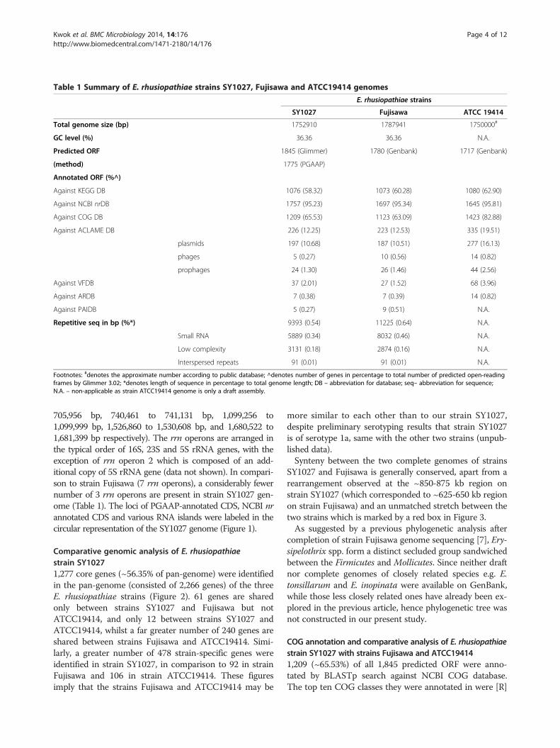
Table 1 Summary of E. rhusiopathiae strains SY1027, Fujisawa and ATCC19414 genomes
E. rhusiopathiae strains
SY1027 Fujisawa ATCC 19414
Total genome size (bp) 1752910 1787941 1750000#
GC level (%) 36.36 36.36 N.A.
Predicted ORF 1845 (Glimmer) 1780 (Genbank) 1717 (Genbank)
(method) 1775 (PGAAP)
Annotated ORF (%^)
Against KEGG DB 1076 (58.32) 1073 (60.28) 1080 (62.90)
Against NCBI nrDB 1757 (95.23) 1697 (95.34) 1645 (95.81)
Against COG DB 1209 (65.53) 1123 (63.09) 1423 (82.88)
Against ACLAME DB 226 (12.25) 223 (12.53) 335 (19.51)
plasmids 197 (10.68) 187 (10.51) 277 (16.13)
phages 5 (0.27) 10 (0.56) 14 (0.82)
prophages 24 (1.30) 26 (1.46) 44 (2.56)
Against VFDB 37 (2.01) 27 (1.52) 68 (3.96)
Against ARDB 7 (0.38) 7 (0.39) 14 (0.82)
Against PAIDB 5 (0.27) 9 (0.51) N.A.
Repetitive seq in bp (%*) 9393 (0.54) 11225 (0.64) N.A.
Small RNA 5889 (0.34) 8032 (0.46) N.A.
Low complexity 3131 (0.18) 2874 (0.16) N.A.
Interspersed repeats 91 (0.01) 91 (0.01) N.A.
Footnotes: #denotes the approximate number according to public database; ^denotes number of genes in percentage to total number of predicted open-readingframes by Glimmer 3.02; *denotes length of sequence in percentage to total genome length; DB – abbreviation for database; seq– abbreviation for sequence;N.A. – non-applicable as strain ATCC19414 genome is only a draft assembly.
Kwok et al. BMC Microbiology 2014, 14:176 Page 4 of 12http://www.biomedcentral.com/1471-2180/14/176
705,956 bp, 740,461 to 741,131 bp, 1,099,256 to1,099,999 bp, 1,526,860 to 1,530,608 bp, and 1,680,522 to1,681,399 bp respectively). The rrn operons are arranged inthe typical order of 16S, 23S and 5S rRNA genes, with theexception of rrn operon 2 which is composed of an add-itional copy of 5S rRNA gene (data not shown). In compari-son to strain Fujisawa (7 rrn operons), a considerably fewernumber of 3 rrn operons are present in strain SY1027 gen-ome (Table 1). The loci of PGAAP-annotated CDS, NCBI nrannotated CDS and various RNA islands were labeled in thecircular representation of the SY1027 genome (Figure 1).
Comparative genomic analysis of E. rhusiopathiaestrain SY10271,277 core genes (~56.35% of pan-genome) were identifiedin the pan-genome (consisted of 2,266 genes) of the threeE. rhusiopathiae strains (Figure 2). 61 genes are sharedonly between strains SY1027 and Fujisawa but notATCC19414, and only 12 between strains SY1027 andATCC19414, whilst a far greater number of 240 genes areshared between strains Fujisawa and ATCC19414. Simi-larly, a greater number of 478 strain-specific genes wereidentified in strain SY1027, in comparison to 92 in strainFujisawa and 106 in strain ATCC19414. These figuresimply that the strains Fujisawa and ATCC19414 may be
more similar to each other than to our strain SY1027,despite preliminary serotyping results that strain SY1027is of serotype 1a, same with the other two strains (unpub-lished data).Synteny between the two complete genomes of strains
SY1027 and Fujisawa is generally conserved, apart from arearrangement observed at the ~850-875 kb region onstrain SY1027 (which corresponded to ~625-650 kb regionon strain Fujisawa) and an unmatched stretch between thetwo strains which is marked by a red box in Figure 3.As suggested by a previous phylogenetic analysis after
completion of strain Fujisawa genome sequencing [7], Ery-sipelothrix spp. form a distinct secluded group sandwichedbetween the Firmicutes and Mollicutes. Since neither draftnor complete genomes of closely related species e.g. E.tonsillarum and E. inopinata were available on GenBank,while those less closely related ones have already been ex-plored in the previous article, hence phylogenetic tree wasnot constructed in our present study.
COG annotation and comparative analysis of E. rhusiopathiaestrain SY1027 with strains Fujisawa and ATCC194141,209 (~65.53%) of all 1,845 predicted ORF were anno-tated by BLASTp search against NCBI COG database.The top ten COG classes they were annotated in were [R]

100
kb
200 k
b
300 kb
400 kb
500 kb
600 kb
700 kb
800 kb
900
kb
1000
kb
1100
kb
1200 kb
1300 kb
1400 kb
1500 kb
1600 kb
1700 kb
rRNA
islan
d 1
tRNA
island 1
tRNA
island 2
tRNA
island 3
rRN
A is
land
2
tRNA
islan
d 4
tRNA
island 5
rRNA island 3
PAI-
L01PAI-L02
PAI-L03
PAI-L04
PAI-
L05
Figure 1 Circular representation of E. rhusiopathiae strain SY1027 genome. From the outer to inner layers, the circle shows (i) nucleotidepositions in kilobases (kb) (black); (ii) RNA region whereas rrn operons(light purple) and tRNA islands (dark purple) are labeled accordingly; (iii)PGAAP-annotated CDSs encoded by plus strand (red) and minus strand (dark yellow); (iv) NCBI non-redundant (nr) database-annotated CDSs(blue); (v) genes shared with strain Fujisawa (dark blue); (vi) ACLAME database-annotated potential horizontal transferring genes, classified by theirputative origins–plasmid (green), prophage (light red) and phage (orange); (vii) ARDB-annotated potential antibiotics resistance genes (dark red);(viii) percent G + C content (gray).
Kwok et al. BMC Microbiology 2014, 14:176 Page 5 of 12http://www.biomedcentral.com/1471-2180/14/176
General function prediction only (~12.08%), [J] Transla-tion, ribosomal structure and biogenesis (~11.41%), [G]Carbohydrate transport and metabolism (~9.02%), [L]Replication, recombination and repair (~8.52%), [S] Func-tion unknown (~7.28%), [K] Transcription (~6.20%), [P]Inorganic ion transport and metabolism (~5.46%), [E]
Amino acid transport and metabolism (~5.29%), [V]Defense mechanisms (~4.63%), and [M] Cell wall/mem-brane/envelope biogenesis (~4.30%). As expected, majorityof the genes were involved in basic cellular functions, suchas replication, transcription, translation and metabolism,however, up to ~19.36% of them only have predicted or

Figure 2 Pan-genome between E. rhusiopathiae strains. This Venn diagram is not drawn in proportion and aims only for the illustration ofpan-genome and distribution of common genes including core genes. Circles represent genomes, overlapping regions between circles indicategenes shared with respective genomes. Numeral figures within respective regions denote the number of genes found therein.
Kwok et al. BMC Microbiology 2014, 14:176 Page 6 of 12http://www.biomedcentral.com/1471-2180/14/176
unknown functions on COG database. COG class distri-bution of strain SY1027 was illustrated (Figure 4). Num-bers and percentage of each COG class were tabulated asAdditional file 3 and their detailed annotations attached asAdditional file 4.Similar numbers of COG-annotated genes were found
in strains Fujisawa (1,123 genes; 63.09%) and ATCC 19414(1,423 genes; 82.88%) (Table 1). Percentage of gene anno-tation in ATCC 19414 is possibly higher due to incom-plete sequencing coverage and assembly. COG classdistributions are highly similar between strains SY1027and Fujisawa (Additional files 5 and 6).
Figure 3 Mauve alignment between E. rhusiopathiae strains SY1027 adepicted as filled colored blocks in the order they are presented in the resto ~1140 kb in Fujisawa genome is marked by a red box.
Virulence gene/pathogenicity island-like gene annotationand comparative analysis of E. rhusiopathiae strainSY1027 and with strains Fujisawa and ATCC1941437 potential virulence genes (~2.01% of total predictedORF) were identified by BLASTp search against VFDB.Categorized by their respective COG classes, their acces-sion numbers and descriptions are listed in Additional file7. Many of these potential virulence genes are groupedunder or partially involved in cell wall/membrane/envelopebiogenesis (COG class [M]) or inorganic ion transport andmetabolism (COG class [P]). The former mainly includesenzymes or proteins involved in capsular polysaccharide
nd Fujisawa genomes. The complete genome assemblies arepective genomes. The short unmatched stretch found from ~1120

Figure 4 COG class annotation distribution of E. rhusiopathiae strain SY1027 genome. The COG-annotated genes are grouped under theirrespective COG classes. Only their class abbreviations are used in this graph, their corresponding class descriptions are listed as below: N, Cellmotility; Q, Secondary metabolite biosynthesis, transport and catabolism; U, Intracellular trafficking, secretion, and vesicular transport; D, Cell cyclecontrol, cell division, chromosome partitioning; T, Signal transduction mechanisms; I, Lipid transport and metabolism; H, Coenzyme transport andmetabolism; V, Defense mechanisms; O, Posttranslational modification, protein turnover, chaperones; C, energy production and conversion; F,Nucleotide transport and metabolism; P, Inorganic ion transport and metabolism; M, Cell wall/membrane/envelope biogenesis; K, Transcription; L,Replication, recombination and repair; E, Amino acid transport and metabolism; G, Carbohydrate transport and metabolism; S, Function unknown; R,General function prediction only; J, Translation, ribosomal structure and biogenesis. Percentages of the top ten classes are labeled for easy reference.
Kwok et al. BMC Microbiology 2014, 14:176 Page 7 of 12http://www.biomedcentral.com/1471-2180/14/176
and glycoprotein biosynthesis, and the latter include trans-porters for iron and magnesium uptake and manganese-dependent superoxide dismutase. Metal ions are scarceand limited in biological systems and their uptake by bac-teria upon invasion of various host cells have shown posi-tive correlation to bacterial virulence [19–21]. However,their functions and possible relevance to pathogenicity inE. rhusiopathiae remain unresolved. A modestly highernumber of virulence genes were identified in strainSY1027 than in strain Fujisawa in VFDB, while the num-ber in strain ATCC19414 might be overestimated due toits draft genome nature (Table 1).5 genomic island-like regions were annotated via Island
Viewer, while 5 pathogenicity island (PAI)-like islandswere identified in strain SY1027 via PAI finder in PAIDB(Figure 5, Tables 2 and 3). In comparison to the search re-sult in VFDB, only one PAI-virulence gene (groEL) wasannotated in PAI-L02, it may suggest strain SY1027 gen-ome has PAI-like islands atypical of that found in strainFujisawa and the present entries in the PAIDB. ThoughGroEL is a chaperonin widely conserved in bacteria, its as-sociation to cell adherence and virulence has been de-scribed in Clostridium spp. and other bacteria [22,23].Suggestion of a PAI-island with groEL in our strain may
warrant further studies in elucidating its contribution topathogenicity, as it may offer interesting insights into po-tential antigenic targets for vaccine development in E. rhu-siopathiae. In addition to the difference in GC content tothe rest of the genome, tRNA were found to flank GI-01,PAI-L03 and PAI-L04, providing further support to theseputative genomic islands (Tables 2 and 3). Nevertheless,the virulence of these putative regions in E. rhusiopathiaeneeds further elucidation, preferably by gene-knockout ex-periments. Since ATCC19414 genome was only completedto the level of scaffolds, search for GI/PAI-like islandswas unfeasible.Apart from the public databases, a list of potential viru-
lence factors previously suggested in E. rhusiopathiae wereidentified in the strain SY1027 genome, and they spanacross various categories, including surface proteins, anti-oxidant proteins, phospholipases, hemolysins, capsularpolysaccharide biosynthesis and extracellular proteins/en-zymes (Additional file 8).Surface proteins were annotated based on their LPXTG
motifs, which act as sortase recognition sequence for thecovalent binding of their carboxyl termini by sortases inGram-positive bacteria [4], or GW (glycine-tryptophan)repeats for non-covalent interactions as observed in E.

100
kb
200 k
b
300 kb
400 kb
500 kb
600 kb
700 kb
800 kb
900
kb
1000
kb
1100
kb
1200 kb
1300 kb
1400 kb
1500 kb
1600 kb
1700 kb
GI-01
GI-02
GI-03GI-04
GI-05
tRNA-01
tRNA-02
tRNA-03
PAI-L01
PAI-L02PAI-L03
PAI-L04
PAI-L05
groEL
virR
cap5E, cap5F,cap5G
hlyB
prrA
sodA
Erysipelothrix rhusiopathiae
SY1027GI-L, PAI-L
& potential VFs
Figure 5 Circular representation of GI-L, PAI-L and potential virulence factors in E. rhusiopathiae strain SY1027 genome. From the outerto inner layers, the circle shows (i) nucleotide positions in kilobases (kb) (black); (ii)Island Viewer-annotated potential genomic islands (GI) arelabeled accordingly (red); (iii) tRNAs flanking the potential GI or potential PAI-like regions are labeled accordingly, tRNAs found within 5000-bpflanking regions are marked in dark yellow while that found between5000 to 7000-bp flanking region is marked in grey; (iv) PAIDB-annotatedPAI-like regions are labeled accordingly (dark red); (v) VFDB-annotated potential virulence factors (blue); (vi) VFDB-annotated potential virulencefactors that might have originated from horizontal gene transfer are classified by their putative origins–plasmid (green) and prophage (orange);(vii) VFDB-annotated potential virulence factors that are previously mentioned in the literature are marked in dark green and their positions boxedin yellow and their gene names labeled accordingly. In cases they are predicted to be horizontally transferred from plasmids, they are boxed inpurple. (viii) Other potential virulence factors suggested by previous studies that may be horizontally transferred from plasmids (green); (ix) Other potentialvirulence factors found in the literature suggested by previous studies, divided into 8 categories – two component system with orientation of 5’histidinekinase-3’ response regulator (cyan), two component system with orientation of 3’histidine kinase-5’ response regulator (orange), surface proteins (brown),antioxidant proteins (vivid green), phospholipases (avocado green), hemolysins (lime), proteins involved in capsular polysaccharide synthesis (pink) andothers (pistachio green); (x) percent G + C content (gray). Horizontal transferring genes were annotated by ACLAME database. groEL, VFDB-annotatedpotential virulence factor that is found within putative PAI-L02, is boxed in red.
Kwok et al. BMC Microbiology 2014, 14:176 Page 8 of 12http://www.biomedcentral.com/1471-2180/14/176

Table 2 Genomic island-like regions in E. rhusiopathiaestrain SY1027 genome
GI # Start End Size %GC flanked by tRNA
GI-01 232197 263359 31162 30.5 tRNA-Leu (tRNA01)
GI-02 644770 648766 3996 34.6 N.F.
GI-03 831571 843384 11813 32 N.F.
GI-04 846580 880804 34224 31.3 N.F.
GI-05 1068490 1076408 7918 33.3 N.F.
Footnote: Genomic island-like regions were predicted by IslandPath-DIMOB onIslandViewer (http://www.pathogenomics.sfu.ca/islandviewer/about.php) [14].N.F.- Not found.
Kwok et al. BMC Microbiology 2014, 14:176 Page 9 of 12http://www.biomedcentral.com/1471-2180/14/176
rhusiopathiae [24]. These include SpaA antigen which iscommonly used as vaccine target [5], hyaluronidases andneuraminidases which may promote bacterial-host cellsurface association [25], RspA and RspB proteins whichbind fibronectin and collagen I and II and may participatein biofilm formation [6]. Interestingly, a premature stopcodon (TAA) was found near the middle of putative rspAgene (data not shown), leading to the annotation of 2RspA-like proteins, each representing the N-terminallyand C-terminally truncated proteins in strain SY1027 gen-ome (shaded in gray in Additional file 8), instead of 1 instrain Fujisawa. Though our preliminary results suggestthat these truncated proteins do not influence the serotyp-ing of strain SY1027 (unpublished data), their conform-ation, collagen binding capability and influence on host-bacterial interaction may need further elucidation.E. rhusiopathiae was hypothesized to have an atypical
capsule that renders it escape from phagocytosis andantioxidant proteins that facilitates its survival insidepolymorphonuclear leukocytes and macrophages [26].Transposon mutagenesis study showed that the loss of acluster of genes, putatively a polycistronic mRNA-codingoperon, in strain Fujisawa created a mutant deprived ofcapsule and pathogenicity [3]. The operon was identified,including all 7 proteins potentially involved in capsularpolysaccharide biosynthesis, in strain SY1027. Similarly,superoxide dismutase, thioredoxins, thiol peroxidase,alkyl-hydroperoxide reductases and other proteins whichconfer resistance to oxidative stress, were also found instrain SY1027.
Table 3 Pathogenicity island-like regions in E. rhusiopathiae s
PAI-L # Start End Size %GC flanked by tRNA
PAI-L01 806018 811037 5020 37.5 N.F.
PAI-L02 826289 835010 8721 34.2 N.F. PAI CFT07
PAI-L03 886571 889469 2898 35.8 tRNA-Lys (tRNA02) Not na
PAI-L04 973651 985197 11546 36.7 tRNA-Arg (tRNA03) Not na
PAI-L05 1619897 1622633 2736 36.4 N.F. Not na
Footnote: Pathogenicity island-like regions were predicted by PAI Finder (https://wwfrom PAIDB hits; Фas annotated by VFDB; *homo. PAI – abbreviation for homologou
Phospholipases are often considered as potential viru-lence factors for intracellular pathogens, hypothesizedvia acquisition of host membrane lipids and disruptionof phagosomal membrane. In strain SY1027 genome, 6phospholipases were identified, same as in strain Fuji-sawa (Additional file 8).As E. rhusiopathiae is highly tolerant and almost ubiqui-
tous in nature, two-component regulatory systems whichoften regulate responses to changes in the environmentwere also searched for in strain SY1027 genome. 9 pairs ofhistidine kinase-response regulator were identified– 8shown similarity to PhoR-PhoB two-component regula-tory system and the remaining one to AgrC-AgrA two-component regulatory system (Table 4).The highly potential virulence factors annotated both
via VFDB and literature search - hlyB, cap5E, cap5F,cap5G, prrA, sodA and virR (Figure 5) – may require fur-ther elucidation on their correlation to pathogenicity, es-pecially in intracellular survival of E. rhusiopathiae.
Potential drug resistance gene in E. rhusiopathiaestrain SY10277 potential drug resistance genes were annotated in E.rhusiopathiae strain SY1027, suggesting it may be re-sistant to macrolides, vancomycin and teicoplanin(Additional file 9). Their orthologs were also found instrain Fujisawa, sharing ~98-100% match ID (data notshown). Interestingly, a richer reservoir of potential drugresistance genes, both in number and variety, was anno-tated in strain ATCC19414 (Table 5). Our preliminary anti-microbial results suggested that despite the presence ofpotential macrolide-resistant genes (which share ~40-46%match IDs to ARDB entries), the antimicrobial susceptibil-ities to different macrolide-class drugs (e.g. erythromycin,roxithromycin and lincomycin) vary from moderate to min-imal levels (unpublished data), hinting that further func-tional elucidation, preferably gene-knockout experiments,of the potential drug resistance genes are required to sub-stantiate the in silico deduction. In addition, a previousinvestigation on antimicrobial susceptibilities of E. rhusio-pathiae suggested that it has partial or complete resistanceto other classes of antibiotics [27] which were not identified
train SY1027 genome
PAIs homologous to this region^ No. of ORF inhomo. PAI*
PotentialVFф
VPI-2 (Vibrio cholerae O1) 8 N.F.
3 (E. coli CFT073), SHI-2 (ShigellaflexneriM90T),HPI (Yersinia enterocoliticaWA 314)
11 groEL
med (Enterococcus faecalis MMH594, V583) 7 N.F.
med (Enterococcus faecalis MMH594, V583) 15 N.F.
med (Enterococcus faecalis MMH594, V583) 5 N.F.
w.gem.re.kr/paidb/pai_finder.php?m=f) on PAIDB [15]. ^representative PAIss PAI; N.F.- Not found.

Table 4 Two-component system genes in E. rhusiopathiae strain SY1027 genome
Contig_orf Gene Match ID (%) Order Phylum COG class and function annotation
Similar to AgrC-AgrA (exoprotein synthesis) two-component regulatory system
contig00001_orf00402 lin0044 39.42 Bacillales Firmicutes [KT]_COG3279 Response regulator of the LytR/AlgR family
contig00001_orf00401 lin0043 36.19 Bacillales Firmicutes [T]_COG2972 Predicted signal transduction proteinwith a C-terminal ATPase domain
Similar to PhoR-PhoB (phosphate starvation response) two-component regulatory system
contig00001_orf00481 SP2193 43.98 Lactobacillales Firmicutes [TK]_COG0745 Response regulators consisting of a CheY-likereceiver domain and a winged-helix DNA-binding domain
contig00001_orf00482 SP0604 27.08 Lactobacillales Firmicutes [T]_COG0642 Signal transduction histidine kinase
contig00001_orf00536 CAC2435 49.08 Clostridiales Firmicutes [TK]_COG0745 Response regulators consisting of a CheY-likereceiver domain and a winged-helix DNA-binding domain
contig00001_orf00535 CAC2434 25.87 Clostridiales Firmicutes [T]_COG0642 Signal transduction histidine kinase
contig00001_orf00866 SP1633 52.87 Lactobacillales Firmicutes [TK]_COG0745 Response regulators consisting of a CheY-likereceiver domain and a winged-helix DNA-binding domain
contig00001_orf00865 all3587 21.10 Nostocales Cyanobacteria [T]_COG0642 Signal transduction histidine kinase
contig00001_orf01180 BH0372 29.26 Bacillales Firmicutes [TK]_COG0745 Response regulators consisting of a CheY-likereceiver domain and a winged-helix DNA-binding domain
contig00001_orf01181 BH0754 20.07 Bacillales Firmicutes [T]_COG0642 Signal transduction histidine kinase
contig00001_orf01262 CAC0524 41.47 Clostridiales Firmicutes [TK]_COG0745 Response regulators consisting of a CheY-likereceiver domain and a winged-helix DNA-binding domain
contig00001_orf01261 Cgl2903 24.50 Actinomycetales Actinobacteria [T]_COG0642 Signal transduction histidine kinase
contig00001_orf01760 BH1153 47.39 Bacillales Firmicutes [TK]_COG0745 Response regulators consisting of a CheY-likereceiver domain and a winged-helix DNA-binding domain
contig00001_orf01761 BS_yrkQ 27.27 Bacillales Firmicutes [T]_COG0642 Signal transduction histidine kinase
contig00001_orf01866^ CAC0564 53.33 Clostridiales Firmicutes [TK]_COG0745 Response regulators consisting of a CheY-likereceiver domain and a winged-helix DNA-binding domain
contig00001_orf01868 BS_yvrG 24.84 Bacillales Firmicutes [T]_COG0642 Signal transduction histidine kinase
contig00001_orf01870^ CAC0830 45.02 Clostridiales Firmicutes [TK]_COG0745 Response regulators consisting of a CheY-likereceiver domain and a winged-helix DNA-binding domain
contig00001_orf01872^ BS_yrkQ 25.19 Bacillales Firmicutes [T]_COG0642 Signal transduction histidine kinase
Kwok et al. BMC Microbiology 2014, 14:176 Page 10 of 12http://www.biomedcentral.com/1471-2180/14/176
in our present search, hinting the presence of other poten-tial drug resistance genes, either novel or unlisted in theARDB, in the bacteria.
Potential horizontal transferring elements inE. rhusiopathiae strain SY1027Horizontal transferring elements are common in bacteriaand generally believed to be a significant driving force inprokaryotic evolution [28]. They are mainly divided intothree types based on their respective vectors – plasmids,phages (or viri) and prophages [28]. In strain SY1027 gen-ome, 226 potential horizontal transferring elements
Table 5 ARDB-annotated genes in E. rhusiopathiae strains SY1
Numb
Species Macrolide Vancomycin Tei
E. rhusiopathiaeSY1027 4 2
E. rhusiopathiae Fujisawa 4 2
E. rhusiopathiaeATCC19414 3 2
(~12.25% of all predicted ORFs) were annotated viaBLASTp against the ACLAME database (Additional file10). Amongst them, the majority (197 genes; ~87.18%)were putatively derived from plasmids, while the rest werefrom phages (5 genes; ~2.21%) and from prophages (24genes;~10.61%). Similar figures were observed in strainFujisawa genome, with a slightly fewer number of putativeplasmid-derived genes and doubled number of putativephage-derived genes. 355 potential horizontal transferringelements were found in the draft genome of strainATCC19414, yet it might be an over-estimated figure dueto incomplete or possible mis-assembly.
027, Fujisawa and ATCC19414 genomes
er of potential resistant genes to…
coplanin Lincosamide Tetracycline Streptogramin a
1 0 0 0
1 0 0 0
1 4 2 2
Kwok et al. BMC Microbiology 2014, 14:176 Page 11 of 12http://www.biomedcentral.com/1471-2180/14/176
As observed in Figure 5, some of the potential viru-lence factors identified might have originated fromhorizontal gene transfer. Two of them –prrA andsodA–have high virulence potential owing to theiridentification via both VFDB and literature search. Theformer is the transcriptional regulator in a PrrA-PrrBtwo-component regulatory system related to oxygencontrol and intracellular replication in Mycobacterium[29], while the latter encodes a superoxide dismutasewhich may offer a mean of antioxidant defense whichis critical for intracellular survival and growth in E.rhusiopathiae [26].
KEGG pathway analysis of E. rhusiopathiae strain SY1027KEGG pathway analysis was performed for E. rhusio-pathiae strain SY1027, and the full list of KEGG pathwayannotation was listed in Additional file 11. Strain SY1027genome contains glucose-specific IIA component in phos-photransferase system (PTS), endoglucanase and glucose-6-phosphate isomerase, which suggest that the bacteria iscapable in breaking down maltose and trehalose, celluloseand fructose, respectively. The potential of utilizing othersugars besides glucose may partly explain the ubiquitousnature of E. rhusiopathiae in the environment. Similar tostrain Fujisawa, many critical genes in the tricarboxylicacid (TCA) cycle were found missing in strain SY1027,with the exception of fumarate hydratase, citrate CoA-transferase, dihydrolipoamidesuccinyltransferase and dihy-drolipoamide dehydrogenase. Genome reduction was pre-viously hypothesized in E. rhusiopathiae strain Fujisawa,given the partial loss of fatty acid biosynthesis and DNArepair system similar in the Mollicutes [7]. Likewise, strainSY1027 seems to display these characteristics. Apart fromthe partial loss of TCA cycle genes, only 34 genes relatedto DNA repair were identified.
ConclusionThe complete genome of Erysipelothrix rhusiopathiaestrain SY1027 was sequenced and assembled. It is1,752,910 base pairs long, just 30 kilobases smallerthan strain Fujisawa, with the same GC level of 36.36%.It contains 1845 open reading frames (ORF) predictedby GLIMMER 3.02, of which 1757 (95.23%) are anno-tated by NCBI nr blast, 1209 by COG database and 1076by KEGG database. 37 potential virulence factors are an-notated in strain SY1027 by VFDB, while 19 (51.35%) ofthem are common in the 2 strains, 7 of which are poten-tially related to antibiotic resistance and highly conserved(~98-100% match ID) amongst the three strains ofE. rhusiopathiae and only modestly homologous toother gastrointestinal tract-inhabiting Firmicutes (~40%match ID), e.g. Clostridium spp., Enterococcus spp. Mo-lecular identification of these genomic elements and po-tential virulence factors offer insights into testing
prospective vaccine targets besides the widely used SpaAantigens in swine erysipelas and development of more ef-fective treatment or prevention in control of the disease.
Additional files
Additional file 1: Local Chinese swine erysipelas outbreak reports.Three articles (titles shaded in yellow) on local swine erysipelas outbreakspublished in Chinese scientific journals.
Additional file 2: NCBI nr database annotation for E. rhusiopathiaestrain. The full list of orfs in E. rhusiopathiae strain SY1027 annotatedagainst NCBI non-redundant database.
Additional file 3: COG-annotated genes in E. rhusiopathiae strainSY1027 genome. Tabulated summary of orfs in E. rhusiopathiae strainSY1027 annotated against COG database under various COG classes.
Additional file 4: COG functional annotation for E. rhusiopathiaestrain SY1027 genome. The full list of orfs in E. rhusiopathiae strainSY1027 annotated against COG database.
Additional file 5: COG class annotation distribution between E.rhusiopathiae strains SY1027 and Fujisawa genomes. A bar chartcomparing the percentage of each single-letter COG class found betweenthe 2 complete E. rhusiopathiae genomes.
Additional file 6: Comparison of COG-annotated genes betweenE. rhusiopathiae strains SY1027 and Fujisawa. Tabulated comparisonbetween COG-annotated genes under each single-letter COG class fromthe 2 complete E. rhusiopathiae genomes.
Additional file 7: VFDB-annotated genes between E. rhusiopathiaestrain SY1027 genome. The list of VFDB-annotated genes in strainSY1027 genome, classified by COG classes. Genes shared betweenstrains SY1027, Fujisawa and ATCC19414 were shaded in light graywhile those shared between strains SY1027 and Fujisawa werehighlighted in dark gray.
Additional file 8: Other potential virulence factors in E.rhusiopathiae strain SY1027 genome. A panel of virulence factorsmentioned in the literature was searched in strain SY1027 genome andlisted here. ^orf00231, orf00462 and orf00466 are pseudogenes withframe-shift or point mutation(s).
Additional file 9: ARDB-annotated genes in E. rhusiopathiae strainSY1027 genome. List of ARDB-annotated genes and their putativeantibiotic resistance.
Additional file 10: ACLAME database-annotated genes inE. rhusiopathiae strain SY1027 genome. Full list of orf annotated viaACLAME database in strain SY1027 genome.
Additional file 11: KEGG database-annotated genes in E. rhusiopathiaestrain SY1027 genome. Full list of KEGG database-annotated genes in strainSY1027 genome.
Competing interestsThe authors declare that they have no competing interests.
Authors’ contributionsAHYK prepared the genomic DNA for next-generation sequencing (NGS),post-NGS sequence alignment, genome assembly and annotation andcomparative genomic analyses, and the drafting of the manuscript. YL andPJ isolated the bacteria from field sample and participated in design of studyand coordination. JJ helped in writing in-house perl script and subsequentbioinformatics analyses. FCL participated in the design of study andcoordination. All authors read and approved the final manuscript.
AcknowledgementsThe authors would like to thank Raymond K. H. Hui for handling Roche GSJunior runs for this study.

Kwok et al. BMC Microbiology 2014, 14:176 Page 12 of 12http://www.biomedcentral.com/1471-2180/14/176
Author details1Bioinformatics Center, Nanjing Agricultural University, F202, South Block,Faculty of Science Complex, 1 Weigang, Nanjing 210095, China. 2College ofVeterinary Medicine, Nanjing Agricultural University, Room 4031, 4th floor,Shaw Building, 1 Weigang, Nanjing 210095, China. 3School of BiologicalSciences, University of Hong Kong, Hong Kong, SAR, China.
Received: 1 March 2014 Accepted: 26 June 2014Published: 2 July 2014
References1. Wang Q, Chang BJ, Riley TV: Erysipelothrix rhusiopathiae. Vet Microbiol
2010, 140:405–417.2. To H, Nagai S: Genetic and antigenic diversity of the surface protective
antigen proteins of Erysipelothrix rhusiopathiae. Clin Vaccine Immunol2007, 14:813–820.
3. Shimoji Y, Yokomizo Y, Sekizaki T, Mori Y, Kubo M: Presence of a capsule inErysipelothrix rhusiopathiae and its relationship to virulence for mice.Infect Immun 1994, 62:2806–2810.
4. Navarre WW, Schneewind O: Surface proteins of gram-positive bacteriaand mechanisms of their targeting to the cell wall envelope. MicrobiolMol Biol Rev 1999, 63:174–229.
5. Shimoji Y, Mori Y, Fischetti VA: Immunological characterization of a protectiveantigen of Erysipelothrix rhusiopathiae: identification of the regionresponsible for protective immunity. Infect Immun 1999, 67:1646–1651.
6. Shimoji Y, Ogawa Y, Osaki M, Kabeya H, Maruyama S, Mikami T, Sekizaki T:Adhesive surface proteins of Erysipelothrix rhusiopathiae bind topolystyrene, fibronectin, and type I and IV collagens. J Bacteriol 2003,185:2739–2748.
7. Ogawa Y, Ooka T, Shi F, Ogura Y, Nakayama K, Hayashi T, Shimoji Y: Thegenome of Erysipelothrix rhusiopathiae, the causative agent of swineerysipelas, reveals new insights into the evolution of firmicutes and theorganism’s intracellular adaptations. J Bacteriol 2011, 193:2959–2971.
8. Groschup MH, Timoney JF: Modified Feist broth as a serum-free alternativefor enhanced production of protective antigen of Erysipelothrixrhusiopathiae. J Clin Microbiol 1990, 28:2573–2575.
9. Altschul SF, Madden TL, Schaffer AA, Zhang J, Zhang Z, Miller W, Lipman DJ:Gapped BLAST and PSI-BLAST: a new generation of protein databasesearch programs. Nucleic Acids Res 1997, 25:3389–3402.
10. Delcher AL, Bratke KA, Powers EC, Salzberg SL: Identifying bacterial genesand endosymbiont DNA with Glimmer. Bioinformatics 2007, 23:673–679.
11. Tatusov RL, Fedorova ND, Jackson JD, Jacobs AR, Kiryutin B, Koonin EV,Krylov DM, Mazumder R, Mekhedov SL, Nikolskaya AN, et al: The COGdatabase: an updated version includes eukaryotes. BMC Bioinformatics2003, 4:41.
12. Kanehisa M, Goto S, Sato Y, Furumichi M, Tanabe M: KEGG for integrationand interpretation of large-scale molecular data sets. Nucleic Acids Res2012, 40:D109–D114.
13. Chen L, Yang J, Yu J, Yao Z, Sun L, Shen Y, Jin Q: VFDB: a reference databasefor bacterial virulence factors. Nucleic Acids Res 2005, 33:D325–D328.
14. Langille MG, Brinkman FS: IslandViewer: an integrated interface forcomputational identification and visualization of genomic islands.Bioinformatics 2009, 25:664–665.
15. Yoon SH, Park YK, Lee S, Choi D, Oh TK, Hur CG, Kim JF: Towardspathogenomics: a web-based resource for pathogenicity islands.Nucleic Acids Res 2007, 35:D395–D400.
16. Liu B, Pop M: ARDB–Antibiotic Resistance Genes Database. Nucleic AcidsRes 2009, 37:D443–D447.
17. Leplae R, Hebrant A, Wodak SJ, Toussaint A: ACLAME: a CLAssification ofMobile genetic Elements. Nucleic Acids Res 2004, 32:D45–D49.
18. Kent WJ: BLAT–the BLAST-like alignment tool. Genome Res 2002, 12:656–664.19. Poyart C, Pellegrini E, Gaillot O, Boumaila C, Baptista M, Trieu-Cuot P: Contribution
of Mn-Cofactored Superoxide Dismutase (SodA) to the Virulence ofStreptococcus agalactiae. Infect Immun 2001, 69:5098–5106.
20. Smith RL, Kaczmarek MT, Kucharski LM, Maguire ME: Magnesium transport inSalmonella typhimurium: regulation of mgtA and mgtCB during invasion ofepithelial and macrophage cells. Microbiology 1998, 144:1835–1843.
21. Wyckoff EE, Payne SM: The Vibrio cholerae VctPDGC system transportscatechol siderophores and a siderophore-free iron ligand. Mol Microbiol2011, 81:1446–1458.
22. Hennequin C, Porcheray F, Waligora-Dupriet A-J, Collignon A, Barc M-C,Bourlioux P, Karjalainen T: GroEL (Hsp60) of Clostridium difficile is involvedin cell adherence. Microbiology 2001, 147:87–96.
23. Zügel U, Kaufmann SHE: Role of heat shock proteins in protection fromand pathogenesis of infectious diseases. Clin Microbiol Rev 1999, 12:19–39.
24. Yother J, White JM: Novel surface attachment mechanism of theStreptococcus pneumoniae protein PspA. J Bacteriol 1994, 176:2976–2985.
25. Abrashev I, Orozova P: Erysipelothrix rhusiopathiae neuraminidase and itsrole in pathogenicity. Z Naturforsch C J Biosci 2006, 61:434–438.
26. Shimoji Y, Yokomizo Y, Mori Y: Intracellular survival and replication ofErysipelothrix rhusiopathiae within murine macrophages: failure of inductionof the oxidative burst of macrophages. Infect Immun 1996, 64:1789–1793.
27. Venditti M, Gelfusa V, Tarasi A, Brandimarte C, Serra P: Antimicrobialsusceptibilities of Erysipelothrix rhusiopathiae. Antimicrob AgentsChemother 1990, 34:2038–2040.
28. Gogarten JP, Townsend JP: Horizontal gene transfer, genome innovationand evolution. Nat Rev Microbiol 2005, 3:679–687.
29. Ewann F, Jackson M, Pethe K, Cooper A, Mielcarek N, Ensergueix D, Gicquel B,Locht C, Supply P: Transient Requirement of the PrrA-PrrB Two-ComponentSystem for Early Intracellular Multiplication of Mycobacterium tuberculosis.Infect Immun 2002, 70:2256–2263.
doi:10.1186/1471-2180-14-176Cite this article as: Kwok et al.: Complete genome assembly andcharacterization of an outbreak strain of the causative agent of swine erysipelas –Erysipelothrix rhusiopathiae SY1027. BMC Microbiology 2014 14:176.
Submit your next manuscript to BioMed Centraland take full advantage of:
• Convenient online submission
• Thorough peer review
• No space constraints or color figure charges
• Immediate publication on acceptance
• Inclusion in PubMed, CAS, Scopus and Google Scholar
• Research which is freely available for redistribution
Submit your manuscript at www.biomedcentral.com/submit