Embed Size (px)
Citation preview

Research ArticleEvaluating the Impact of Human Amnion EpithelialCells on Angiogenesis
Dandan Zhu,1,2 Ruth Muljadi,1,2 Siow Teng Chan,1,2 Patricia Vosdoganes,2 Camden Lo,3
Joanne C. Mockler,1,2 Euan M. Wallace,1,2 and Rebecca Lim1,2
1The Ritchie Centre, Hudson Institute of Medical Research, Monash University, Clayton, VIC, Australia2Department of Obstetrics and Gynaecology, Monash University, Clayton, VIC, Australia3Monash Microimaging, Monash University, Clayton, VIC, Australia
Correspondence should be addressed to Rebecca Lim; [email protected]
Received 14 July 2015; Accepted 29 September 2015
Academic Editor: Peter J. Quesenberry
Copyright © 2016 Dandan Zhu et al. This is an open access article distributed under the Creative Commons Attribution License,which permits unrestricted use, distribution, and reproduction in any medium, provided the original work is properly cited.
The effects of human amnion epithelial cells (hAECs) on angiogenesis remain controversial. It is yet unknown if the presence ofinflammation and/or gestational age of hAEC donors have an impact on angiogenesis. In this study, we examined the differencesbetween term and pretermhAECs on angiogenesis in vitro and in vivo. Conditionedmedia from termhAECs induced the formationof longer huVEC tubules on Matrigel. Both term and preterm hAECs expressed VEGFA, PDGFB, ANGPT1, and FOXC1, whichsignificantly increased after TNF𝛼 and IFN𝛾 stimulation. In the presence of TNF𝛼 and IFN𝛾, coculture with term hAECs reducedgene transcription of Tie-2 and Foxc1 in huVECs, while coculture with preterm hAECs increased gene transcription of PDGFR𝛼and PDGFR𝛽 and reduced gene transcription of FOXC1 in huVECs. In vivo assessment of angiogenesis using vWF immunostainingrevealed that hAEC treatment decreased angiogenesis in a bleomycinmodel of lung fibrosis but increased angiogenesis in a neonatalmodel of hyperoxia-induced lung injury. In summary, our findings suggested that the impact of hAECs on angiogenesis may beinfluenced by the presence of inflammation and underlying pathology.
1. Background
Human amnion epithelial cells (hAECs) isolated from theamniotic membrane are an attractive source of cell therapy.In addition to their anti-inflammatory and antifibrotic effects,they are nontumorigenic and they exhibit low antigenicityandmultipotent differentiation potential [1–3]. Furthermore,they can be isolated in large numbers adequate for clinicalapplications without requiring serial expansion [2]. We pre-viously applied hAECs isolated from term pregnancies to amousemodel of bleomycin-induced lung fibrosis.We showedthat hAEC administration prevented lung inflammation andfibrosis and prevented decline in lung function [4]. In studies,we have also shown that term hAECs can reduce mark-ers of lung inflammation and mitigate structural damagewhen administered following lung injury induced by eitherhyperoxia [5] or ventilation [6]. Furthermore, studies on thetherapeutic effects of hAECs showed that the engraftment
of amnion cells is very rare [5, 7], and hAEC-conditionedmedia have been shown to contain soluble factors that exertprofound biological effects [8]. These studies indicate thathAECs may exert their function in a paracrine fashion.
Given that angiogenesis plays a critical role in woundhealing and resolution of inflammation [9, 10], it is impor-tant to assess the contribution of angiogenesis to hAEC-augmented repair. There have been contradictory reports onthe angiogenic effects of hAECs to date. Specifically, hAECshave been reported to secrete several angiogenic factors invitro. These include tissue inhibitors of metalloproteinases-(TIMP-) 1 and 2, epidermal growth factor (EGF), angiogenin,vascular endothelial growth factor (VEGF), platelet derivedgrowth factor B (PDGFB), and angiogenin [11]. However,when functionally assessed in a rodent dorsal skinfold cham-ber model hAECs did not increase vessel lengths or vesselsprouts number [12]. It is possible that the effect of hAECson angiogenesis is altered in an inflammatory environment.
Hindawi Publishing CorporationStem Cells InternationalVolume 2016, Article ID 4565612, 13 pageshttp://dx.doi.org/10.1155/2016/4565612

2 Stem Cells International
Table 1: Gestational age of both term and preterm birth.
Gestational age (d) ComplicationsMinimum Maximum Mean ± SEM
Preterm birth (𝑛 = 9) 196 257 230.4 ± 7.37 IUGR (𝑛 = 5); PE (𝑛 = 4)Term birth (𝑛 = 16) 261 277 268.3 ± 1.03 Nil
Indeed, when administered to a bleomycin-induced lunginjury model a week following bleomycin administrationwhen lung inflammation was at its peak, hAECs were unableto mitigate injury [3].
We have also shown that preterm hAECs exert signifi-cantly less protective effects in vivo compared to term hAECs[13]. This suggests that there may be functional differencesbetween term and preterm hAECs. As such, we soughtto assess potential differences in the angiogenic effects ofhAECs isolated from different gestational ages. Furthermore,angiogenesis can be either beneficial or detrimental to woundhealing, depending on the disease context. Accordingly, wesought to assess the impact of hAECs on angiogenesis usingtwo models of lung injury. The first is a bleomycin-inducedlung fibrosis model where angiogenesis is detrimental to theoutcome, and the second is a hyperoxia-induced lung injurymodel where angiogenesis is beneficial.
2. Materials and Methods
2.1. huVEC and hAEC Isolation. Human umbilical veinendothelial cells (huVECs) were isolated from healthy termhuman umbilical cords and hAECs were isolated fromamnion collected from women undergoing a caesarean sec-tion preterm (<37 weeks’ gestation) or at term as previouslydescribed [2, 14]. Preterm donors included women deliveringprematurely due to preeclampsia, gestational hypertension,fetal growth restriction, placenta praevia, and discordant twingrowth. Donors with pregnancies complicated by chorioam-nionitis and preexisting maternal diseases were excluded.The term donors were women with a healthy pregnancyundergoing an elective repeat caesarean section. The meangestational age for preterm birth was 230 ± 7 days and forterm birth 268 ± 1 days (Table 1). All collection and isolationprocedures were undertaken with the approval fromMonashHealth Human Ethics Committee and with written informedconsent.
2.2. Collection of hAEC-Conditioned Media. The collectionof hAEC-conditioned media followed previous protocol [15].Briefly, hAECs were plated at a density of 10 million cells perT175 flask and conditionedmedia were collected following 96hours under standard tissue culture conditions.
2.3. In Vitro Angiogenesis Assay. On a Matrigel (50 𝜇L/well;Corning Life Sciences) coated 96-well plate, 1 × 10⁴ huVECsat passage 3 were seeded in each well. Cells were cultured ineither M199 media (Invitrogen) [16] or hAEC-conditionedmedia. Phase contrast images were taken every 30 min-utes for a total of 40 hours. The capillary-like structureswere detected and the average length of tubes between
branches was calculated using Image J (National Institutes ofHealth, Bethesda, MA) and Metamorph software (MolecularDevices, Sunnyvale, CA) programs.
2.4. Stimulation of hAECs. Preterm (𝑛 = 6) and term(𝑛 = 6) hAECs were cultured in the presence or absence oftumor necrosis factor 𝛼 (TNF𝛼) (20 ng/mL, PHC3015, LifeTechnologies) and interferon 𝛾 (IFN𝛾) (20 ng/mL, PHC4031,Life technologies) for 24 hrs and 48 hrs. Concentrations ofTNF𝛼 and IFN𝛾 were based on a previous study by Liu andcolleagues who reported an increase in angiogenic potentialof MSCs following exposure to inflammatory cytokines [17].
2.5. Coculture of huVECs and hAECs. hAECs were seeded ata density of 5 × 10⁵ cells in each well of a 6-well plate, while1 × 105 huVECs were seeded into each 0.4 𝜇m pore transwellinsert (BD Bioscience, San Jose, CA) in M199 media in thepresence or absence of TNF𝛼 and IFN𝛾.
2.6. Gene Expression Assays. RNA was isolated using theRNeasy Mini Kit (Qiagen, Limburg, Netherlands) and 1 𝜇gtotal RNA was converted to cDNA using the ThermoscriptReverse Transcription System (Invitrogen). qRT-PCR wasperformed using SensiMix SYBR and Rotor-Gene (Qiagen).Primers for hAECs were directed against VEGFA, PDGFB,angiogenin-1 (ANGPT1), and Forkhead box (FOX) transcrip-tion factor FOXC1. Primers for huVECs were directed againstvascular endothelial growth factor receptors 1 and 2 (VEGFR1and VEGFR2), platelet derived growth factor receptors alphaand beta (PDGFR𝛼 and PDGFR𝛽), Tie-2, and FOXC1. Geneexpression was normalised to 18S and expressed relative toeither unstimulated hAECs or huVECs. Detailed informationon primer sequences is listed in Table 2.
2.7. Animals and Experimental Groups. All animal experi-ments were approved by the Monash Medical Centre Ani-mal Ethics Committee and were conducted in accordancewith the Australia Code of Practice for Care and Use ofAnimals for Scientific Purpose (2006). In the bleomycin-induced mouse lung injury model, 6–8-week-old femaleC57/BL6 mice weighing 16–20 g were housed in a specificpathogen-free animal facility during this study. Experimentalgroups included saline + saline, saline + term hAECs,bleomycin + saline, bleomycin + term hAECs, and bleomycin+ preterm hAECs. The mice were given either saline or0.3 IU bleomycin (Blenoxane, Hospira, Lake Forest, IL, USA)intranasally, followed by intraperitoneal administration of 4million term hAECs or preterm hAECs, or 200𝜇L saline 24hours later, as previously described [13]. Mice were culled14 days following intranasal instillation of bleomycin by

Stem Cells International 3
Table 2: Primer sequences and annealing temperatures.
Gene Primer sequence Annealing temperature
VEGFA Fwd: CTACCTCCACCATGCCAAGTG 60∘CRev: TGATTCTGCCCTCCTCCTTCT
PDGFB Fwd: AATGGTCACCCGAGTTTGG 60∘CRev: CTGGCATGCAAGTGTGAGAC
ANGPT1 Fwd: CCTGATCTTACACGGTGCTGATT 60∘CRev: GTCCCGCAGTATAGAACATTCCA
VEGFR1 Fwd: CGGGGATTTCACTGTACATCT 60∘CRev: AAGCAAACCACACTGGCTTC
VEGFR2 Fwd: CCCTGCCGTGTTGAAGAGTT 60∘CRev: GGACAGGGGGAAGAACAAAA
PDGFR𝛼 Fwd: AGCTGGCAGAGGATTAGGCT 60∘CRev: CTCCATGTGTGGGACATTCA
PDGFR𝛽 Fwd: CAGGAGAGACAGCAACAGCA 60∘CRev: TGTCCAGAGCCTGGAACTGT
Tie-2 Fwd: AGTCTTATGTGTTCTGTCTCCCTGACC 60∘CRev: TCATCCTCGGTATGCCTTCTCTCTCAC
FOXC1 Fwd: ACCTTGACGAAGCACTCGTT 60∘CRev: CGGCATCTACCAGTTCATCA
carbon dioxide asphyxiation. The right lungs were inflatedand fixed with 4% (w/v) paraformaldehyde processed forimmunofluorescence.
In the neonatal mouse model of hyperoxia-induced lunginjury, newborn C57/BL6 mouse pups were randomisedto either normoxia (inspired O
2content (FiO
2) = 0.21)
or hyperoxia (FiO2= 0.85). Experimental groups included
normoxia + saline, normoxia + term hAECs, hyperoxia +saline, and hyperoxia + term hAECs. On postnatal days(PND) 4, 5, and 6, a total of 4.5 million term hAECs or 50 𝜇Lsterile saline (control) was administered intraperitoneally aspreviously described [5]. On postnatal day 14, mice wereculled and lungs were collected in 4%PFA prior to processingand embedding in paraffin.
2.8. Immunohistochemistry Staining for vWF. Pulmonaryvessels were assessed by von Willebrand factor (vWF) stain-ing of paraffin embedded lung tissue sections.The slides weresubjected to proteinase K retrieval, followed by incubationwith primary antibody polyclonal rabbit anti-human vWF(1 : 400, Dako, A0082, Germany) overnight at 4∘C. Afterwashingwith PBS, LSAB+/HRPkit (Dako, K0690,Germany)was applied to the sections for 1 hour at room temperature,followed by labelling with streptavidin-horseradish peroxi-dase and diaminobenzidine (DAB, Dako, K3408, Germany).Nuclei were counterstained with haematoxylin. The percent-age of vWF positive area was determined in bleomycin miceby Image J (NIH). In the hyperoxia study, the number of vWFpositive vessels with diameter less than 50 𝜇mwas counted in15 random images at 200xmagnification using Image J (NIH).
2.9. Data Analysis. Data were expressed as mean ± standarderror of mean (SEM). Statistical significance was determinedusing GraphPad Prism (GraphPad Software Inc., San Diego,
CA, USA) with one-way ANOVA accompanied by theBonferroni post hoc test for multiple groups or the Mann-Whitney test when comparing between two groups. Statisticalsignificance was accorded when 𝑝 < 0.05.
3. Results
3.1. Vascular Tube Formation in huVECs on Matrigel. Themaximum length of vascular tubes formed by huVECsappeared at 6 hours following culture in term hAEC-conditioned media and M199 media and appeared at 4 hoursfollowing culture in preterm hAEC-conditioned media.Changes in average vascular tube lengths over a 40-hourperiod are depicted in Figure 1(a). The average maximumlength of vascular tubes formed in term hAEC-conditionedmedia was significantly greater compared with M199 controlmedia (Figure 1(b), 88.03 ± 5.77 𝜇m versus 59.76 ± 2.19 𝜇m,𝑝 < 0.05) but not the preterm hAEC-conditioned media(73.52 ± 2.86 𝜇m), which was not significantly different toeither control conditions or term hAEC-conditioned media.On average, the tubules of huVECs cultured in term hAEC-conditionedmedia and controlmedia were stable for up to 40hours. In contrast, the average length of huVEC tubules cul-tured in preterm hAEC-conditionedmedia decreased after 16hours, such that by 20 hours they were significantly shorterthan tubules cultured in either term hAEC-conditionedmedia or control media (𝑝 < 0.05). This suggests thatpreterm hAEC-conditioned media contain soluble factorsthat reduce tubule stability. Representative images of huVECtubules formed following culture in term hAEC-conditionedmedia and control media are shown in Figure 1(c).
3.2. Gene Expression of Angiogenic Ligands by hAECs. Weassessed gene transcription ofVEGFA, PDGFB,ANGPT1, andFOXC1 in term and preterm hAECs under basal conditions

4 Stem Cells International
100
80
60
40
20
0
Tube
leng
th (𝜇
M)
2 4 6 8 10 12 16 20 30 40
(h)
∗
∗
M199 control mediaTerm hAEC-conditioned mediaPreterm hAEC-conditioned media
(a)
∗
Control
M199 control mediaTerm hAEC-conditioned mediaPreterm hAEC-conditioned media
Term CM Preterm CM
100
80
60
40
20
0
Max
imum
tube
leng
th (𝜇
M)
(b)
Preterm hAEC-CMTerm hAEC-CMM199
(c)
Figure 1:The vascular tube formation in huVECs onMatrigel in vitro. ((a) and (b)) huVECs formed longer vascular tubules following culturein term hAEC-conditioned media compared to control media (𝑝 < 0.05), and tubule lengths were stable for 40 hours. huVECs cultured inpreterm hAEC-conditionedmedia did not support tubule formation where tubule length was significantly reduced at 20 hours (𝑝 < 0.05). (c)Representative pictures of vascular tubules of huVECs following culture in term hAEC-conditioned media and control media (∗𝑝 < 0.05).
and following exposure to TNF𝛼 and IFN𝛾, which were usedto mimic an inflammatory environment. All of these geneswere transcribed by both term and preterm hAECs underbasal conditions. In term hAECs, stimulation with TNF𝛼and IFN𝛾 significantly increased transcription of VEGFA,PDGFB, and FOXC1 (Figure 2(a), 2.82 ± 0.66, 𝑝 = 0.0411;5.27 ± 2.68, 𝑝 = 0.0087, and 7.72 ± 3.69, 𝑝 = 0.0411, resp.).Gene transcription of ANGPT1 remained unchanged. Inpreterm hAECs, stimulation with TNF𝛼 and IFN𝛾 increasedgene transcription of PDGFB and ANGPT1 (Figure 2(b),6.14 ± 2.73, 𝑝 = 0.0462 and 90.92 ± 55.21, 𝑝 = 0.0079,resp.). Gene transcription of VEGFA and FOXC1 remainedunchanged.
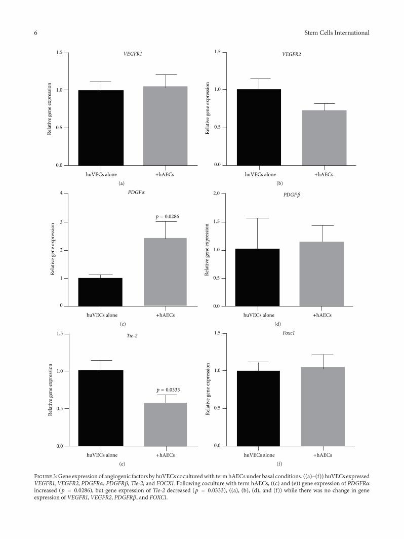
3.3. Gene Expression of Angiogenic Receptors by huVECs. Wenext assessed the impact of term hAECs on huVECs underbasal conditions and in the presence of TNF𝛼 and IFN𝛾.Under basal conditions, huVECs expressed receptors forpro- and antiangiogenic factors includingVEGFR1,VEGFR2,PDGFR𝛼, PDGFR𝛽, Tie-2, and FOXC1 (Figures 3(a)–3(f)).Following coculture with term hAECs, we noted a significantincrease in transcription of PDGFR𝛼 (Figure 3(c), 2.39 ±0.62 versus 1.0 ± 0.11, 𝑝 = 0.0286) and decrease in Tie-2(Figure 3(e), 0.56 ± 0.11 versus 1.0 ± 0.15, 𝑝 = 0.0333).
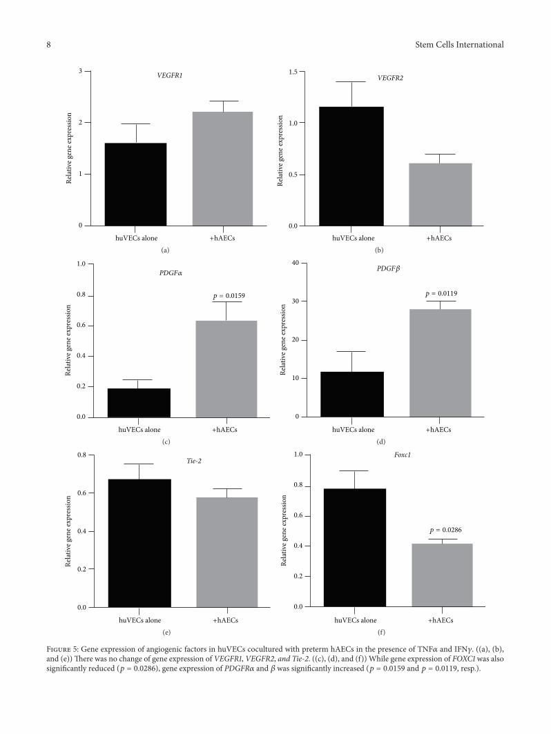
We next assessed the impact of term and preterm hAECson huVECs stimulated by TNF𝛼 and IFN𝛾. When coculturedwith stimulated term hAECs, huVECs significantly reducedgene transcription of Tie-2 (Figure 4(e), 0.40 ± 0.07 versus0.67 ± 0.08, 𝑝 = 0.0476) and Foxc1 (Figure 4(f), 0.59 ± 0.04versus 0.88 ± 0.09, 𝑝 = 0.0238). When cocultured withstimulated preterm hAECs, huVECs significantly increasedgene transcription of PDGFR𝛼 (Figure 5(c), 0.64±0.12 versus0.18 ± 0.06, 𝑝 = 0.0159) and 𝛽 (Figure 5(d), 27.92 ± 1.96versus 11.6 ± 5.33, 𝑝 = 0.0119). Gene expression of Foxc1was also significantly reduced (Figure 5(f), 0.42 ± 0.03 versus0.78 ± 0.12, 𝑝 = 0.0286).
3.4. Assessment of Angiogenesis In Vivo. Term but notpreterm hAEC administration reduced pulmonary fibrosisfollowing bleomycin challenge (Figure 6(a)). The percentageof vWF positive staining increased significantly in the lungsof bleomycin-alone animals (9.31± 0.69%versus 3.12± 0.47%,𝑝 < 0.0001). This was mitigated by the administration ofterm (5.34 ± 0.25%, 𝑝 < 0.001) but not preterm hAECs(Figure 6(b)). Representative images of immunohistochem-ical staining are shown in Figure 6(c).
Given that only term but not preterm hAECs result in aneffect in the adult mice, we proceeded to assess the impact

Stem Cells International 5
VEGFA PDGFB
Unstimulated term hAECsStimulated term hAECs
15
10
5
0
Relat
ive g
ene e
xpre
ssio
n
p = 0.0411
p = 0.0411
p = 0.0087
ANGPT1 FOXC1
(a)
Unstimulated preterm hAECsStimulated preterm hAECs
15
50
100
150
200
250
10
5
0
Relat
ive g
ene e
xpre
ssio
n
p = 0.0462
p = 0.0079
VEGFA PDGFB ANGPT1 FOXC1
(b)
Figure 2: Gene expression of angiogenic ligands by hAECs following stimulation with TNF𝛼 and IFN𝛾. (a) Gene expressions of VEGFA(𝑝 = 0.0411), PDGFB (𝑝 = 0.0087), and Foxc1 (𝑝 = 0.0411) increased in term hAECs. (b) Gene expression of PDGFB (𝑝 = 0.0462) andANGPT1 (𝑝 = 0.0079) increased in preterm hAECs.
of term hAECs on angiogenesis in a hyperoxia-induced lunginjurymodel in neonatalmice. Here we observed thatmice inhyperoxia group had simplified lung structure and enlargedalveoli, and term hAEC treatment improved lung structure(Figure 7(a)).The number of small pulmonary vessels (diam-eter < 50 𝜇m) decreased in hyperoxia-injured neonatal micecompared with normoxia mice (5.84 ± 0.58% versus 8.80 ±0.21%, 𝑝 < 0.001). Term hAEC administration restored thenumber of small pulmonary vessels in hyperoxia-inducedlung injury animals (7.88 ± 0.33%, 𝑝 < 0.05) (Figure 7(b)).Representative images of immunohistochemical staining areshown in Figure 7(c).
4. Discussion
Theability of hAECs to support angiogenesis is poorly under-stood. Additionally, little is known about the importanceof gestational age of hAEC donors to the contribution ofangiogenesis during repair. In this study, we showed that invitro tubule formation by huVECs was best supported byterm hAECs compared to preterm hAECs. Both term andpreterm hAECs transcribed genes of proangiogenic ligandsVEGFA, PDGFB, and ANGPT1 and transcription factor,FOXC1. These were upregulated by inflammatory cytokines,IFN𝛾 and TNF𝛼. However, coculture with both term andpreterm hAECs did not consistently increase gene transcrip-tion of proangiogenic receptor ligands in huVECs. Whenwe assess the effects of hAEC treatment on angiogenesis invivo using a bleomycin model of lung fibrosis, we foundthat while term hAECs reduced vWF staining in the lungs,
consistent with resolution of lung fibrosis, treatment withpreterm hAECs had no effect. This observation coincidedwith our previous study showing that preterm hAECs haddiminished reparative effects. When we assess the effectsof term hAEC treatment on angiogenesis in a model ofhyperoxia-induced neonatal lung injury, we found insteadthat hAEC treatment was associated with improvement inpathological lung remodelling.
The Matrigel tubule formation assay is an establishedmethod for evaluating the angiogenic effects of solublefactors in endothelial cells in vitro [18]. Using this assay, wedetermined that term hAECs release more proangiogenicfactors compared to their preterm counterparts, supportingendothelial cell tubule formation as previously reported withbone marrow-derived MSCs [19]. In order to elucidate thenature of these proangiogenic factors, we compared the geneexpressions of proangiogenic ligands. Angiogenic factors,such as ANGPT1, PDGFB, and VEGFA, have been previouslydetected in the secretome of human MSC from differenttissue sources [20]. Stimulation by TNF𝛼 and LPS increasedthe production of VEGFA by adipose-derived MSCs [21]while transforming growth factor 𝛼 (TGF-𝛼) induced thesecretion of VEGFA and PDGFB in bone marrow-derivedMSCs [22]. Similarly, amnion derived mesenchymal stromalcells (MSCs) secrete angiogenic factors including EGF,VEGF,TIMP-1, and TIMP-2 [11].
In our current study we found that both preterm andterm hAECs expressed VEGFA, PDGFB, ANGPT1, andFOXC1 under basal conditions. We then looked to see if thehAECs altered transcription of these genes in response to
6 Stem Cells International
1.5
1.0
0.5
0.0
Relat
ive g
ene e
xpre
ssio
n
huVECs alone +hAECs
VEGFR1
(a)
1.5
1.0
0.5
0.0
Relat
ive g
ene e
xpre
ssio
n
huVECs alone +hAECs
VEGFR2
(b)
huVECs alone +hAECs
4
3
2
1
0
Relat
ive g
ene e
xpre
ssio
n
p = 0.0286
PDGF𝛼
(c)huVECs alone +hAECs
2.0
1.5
1.0
0.5
0.0
Relat
ive g
ene e
xpre
ssio
n
PDGF𝛽
(d)
huVECs alone +hAECs
p = 0.0333
1.5
1.0
0.5
0.0
Relat
ive g
ene e
xpre
ssio
n
Tie-2
(e)huVECs alone +hAECs
1.5
1.0
0.5
0.0
Relat
ive g
ene e
xpre
ssio
n
Foxc1
(f)
Figure 3: Gene expression of angiogenic factors by huVECs coculturedwith term hAECs under basal conditions. ((a)–(f)) huVECs expressedVEGFR1, VEGFR2, PDGFR𝛼, PDGFR𝛽, Tie-2, and FOCX1. Following coculture with term hAECs, ((c) and (e)) gene expression of PDGFR𝛼increased (𝑝 = 0.0286), but gene expression of Tie-2 decreased (𝑝 = 0.0333), ((a), (b), (d), and (f)) while there was no change in geneexpression of VEGFR1, VEGFR2, PDGFR𝛽, and FOXC1.

Stem Cells International 7
2
3
1
0
Relat
ive g
ene e
xpre
ssio
n
huVECs alone +hAECs
VEGFR1
(a)huVECs alone +hAECs
1.5
1.0
0.5
0.0
Relat
ive g
ene e
xpre
ssio
n
VEGFR2
(b)
huVECs alone +hAECs
0.0
0.2
0.4
0.6
0.8
Relat
ive g
ene e
xpre
ssio
n
PDGF𝛼
(c)huVECs alone +hAECs
0
10
20
30
40
Relat
ive g
ene e
xpre
ssio
n
PDGF𝛽
(d)
huVECs alone +hAECs
0.0
0.2
0.4
0.6
0.8
Relat
ive g
ene e
xpre
ssio
n
p = 0.0476
Tie-2
(e)huVECs alone +hAECs
1.5
1.0
0.5
0.0
Relat
ive g
ene e
xpre
ssio
n
p = 0.0238
Foxc1
(f)
Figure 4: Gene expression of angiogenic factors by huVECs cocultured with term hAECs in the presence of TNF𝛼 and IFN𝛾. ((a)–(d))Therewas no change in gene expression of VEGFR1, VEGFR2, PDGFR𝛼, and PDGFR𝛽, ((e) and (f)) while gene expression of Tie-2 and FOXC1 wassignificantly reduced (𝑝 = 0.0476 and 𝑝 = 0.0238, resp.).
8 Stem Cells International
2
3
1
0
Relat
ive g
ene e
xpre
ssio
n
huVECs alone +hAECs
VEGFR1
(a)huVECs alone +hAECs
1.5
1.0
0.5
0.0
Relat
ive g
ene e
xpre
ssio
n
VEGFR2
(b)
huVECs alone +hAECs
0.0
0.2
0.4
0.6
0.8
1.0
Relat
ive g
ene e
xpre
ssio
n
PDGF𝛼
p = 0.0159
(c)huVECs alone +hAECs
p = 0.0119
0
10
20
30
40
Relat
ive g
ene e
xpre
ssio
n
PDGF𝛽
(d)
huVECs alone +hAECs
0.0
0.2
0.4
0.6
0.8
Relat
ive g
ene e
xpre
ssio
n
Tie-2
(e)huVECs alone +hAECs
0.0
0.2
0.4
0.6
0.8
1.0
Relat
ive g
ene e
xpre
ssio
n
p = 0.0286
Foxc1
(f)
Figure 5: Gene expression of angiogenic factors in huVECs cocultured with preterm hAECs in the presence of TNF𝛼 and IFN𝛾. ((a), (b),and (e))There was no change of gene expression of VEGFR1, VEGFR2, and Tie-2. ((c), (d), and (f)) While gene expression of FOXC1 was alsosignificantly reduced (𝑝 = 0.0286), gene expression of PDGFR𝛼 and 𝛽 was significantly increased (𝑝 = 0.0159 and 𝑝 = 0.0119, resp.).

Stem Cells International 9
Saline + saline Bleomycin + saline
Bleomycin + preterm hAECs Bleomycin + term hAECs
200𝜇m
200𝜇m 200𝜇m
200𝜇m
(a)
Saline + Saline + Bleomycin + Bleomycin + Bleomycin +saline salineterm
hAECsterm
hAECs hAECspreterm
∗
∗∗∗∗ ∗∗∗
15
10
5
0
vWF
posit
ive s
tain
ing
(%)
(b)
Saline + saline Bleomycin + saline
Bleomycin + preterm hAECs Bleomycin + term hAECs
200𝜇m200𝜇m
200𝜇m 200𝜇m
(c)
Figure 6: Collagen staining and vWF immunohistochemistry in bleomycin challenged mouse lung tissue. (a) Bleomycin challenged micehadmore fibrotic tissues in the lung compared to control group; term hAEC treatment, but not preterm hAEC treatment, reduced the fibrotictissues. (b) The percentage of vWF positive staining increased in bleomycin-injured animals compared to control group (𝑝 < 0.0001) anddecreased after term (𝑝 < 0.001) but not preterm hAEC administration. (c) The representative images for vWF immunohistochemistry inmouse lung tissues (∗𝑝 < 0.05, ∗∗∗𝑝 < 0.001, and ∗∗∗∗𝑝 < 0.0001).
proinflammatory stimuli by exposing them to a combinationof TNF𝛼 and IFN𝛾. VEGFA stimulates the generation ofnew, immature, and leaky blood vessels by disrupting thebasement membrane of the preexisting vessels, inducingendothelial cell migration and proliferation [23, 24], whilePDGF and ANGPT1 are essential for the stabilisation ofnew vessels. These angiogenic ligands promote angiogenesis,induce vascular maturation, and decrease vascular perme-ability by mediating migration, adhesion, and survival ofendothelial cells [25]. Interestingly, transcription of VEGFA,PDGFB, and FOXC1 was elevated in term hAECs, whilePDGFB andANGPT1were increased in preterm hAECs.Thissuggests that the gestational age of the hAEC donor can influ-ence differential response of hAECs towards an inflammatorystimulus. Given our current understanding of how stem cells
and stem-like cells can respond to environmental priming[26, 27], the findings from this study may have implicationson the application of hAECs collected from donors acrossdifferent gestational ages.
While hAECs have been reported to secrete angiogenicfactors in vitro [11], this is the first time that FOXC1 expressionby hAECs has been reported. FOXC1 is a transcriptionfactor involved in regulating vascular development. It iscritical for pericyte regulation of vascular development inthe mouse fetal brain [28] and is essential for maintainingthe integrity of basement membrane and decreasing vascularpermeability in zebrafish [29]. FOXC1 also reportedly regu-lates proangiogenic factors such asmatrixmetalloproteinases(MMPs) as well as VEGF receptor-ligand signalling [30,31]. Although VEGFR1 has higher affinity for VEGF, it has

10 Stem Cells International
Normoxia + saline
Hyperoxia + saline
Normoxia + hAECs
Hyperoxia + hAECs
200𝜇m200𝜇m
200𝜇m 200𝜇m
(a)
Normoxia + Normoxia + Hyperoxia + Hyperoxia +
∗∗∗∗
Vess
el n
umbe
rs p
er v
iew
15
10
5
0
saline salineterm hAECs term hAECs(b)
Normoxia + saline Hyperoxia + saline Hyperoxia + hAECs
200𝜇m200𝜇m200𝜇m
(c)
Figure 7: H&E staining and vWF immunohistochemistry in neonatal mouse lung tissue. (a) Hyperoxia-induced lung injury mice hadsimplified lung structure and enlarged alveoli, and term hAEC treatment improved lung structure. (b) The number of vessels (diameter< 50 𝜇m) decreased in hyperoxia-injured animals compared with normoxia animals (𝑝 < 0.001). Term hAEC administration restoredthe number of small pulmonary vessels in hyperoxia-induced lung injury animals (𝑝 < 0.05). (c) The representative images for vWFimmunohistochemistry in mouse lung tissues (∗𝑝 < 0.05, ∗∗∗𝑝 < 0.001).
amuchweaker kinase activity and is thus unable to generate aproangiogenic effect [32]. Indeed VEGFR1 can competitivelyinhibit the proangiogenic effects of VEGFR2 and thus canbe considered as being “antiangiogenic” [33]. In contrast,both PDGFR𝛼 and PDGFR𝛽, the two subunits of PDGFreceptors, can bind to PDGFB and contribute to angiogenesis.Although there is no clear separation between the operatingmechanisms of the two receptor subunits, PDGFR𝛽 plays amajor role in angiogenic processes in huVECs [34]. PDGFR𝛽is required for the stabilisation of newly formed blood vessels,while PDGFR𝛼 works in significant synergy [35]. PDGFR𝛽supports pericyte/endothelial cell interactions and pericyteformation by mediating VEGF expression [36]. IncreasedPDGFR-kinase activity is associated with elevated expressionof VEGFA and VEGFR2, acting directly on endothelialcells and resulting in increased vessel formation [37]. Tie-2
is also an endothelium-specific receptor. When ANGPT1binds to and activates Tie-2, it induces vascular stabilisationand triggers angiogenesis. The Ang-1/Tie-2 system stabilisespreexisting vessels and accelerates angiogenesis when cell-celladhesion is disrupted [38].
Given the transcriptional changes to proangiogenic lig-ands in hAECs, we next assessed changes to their receptorsin huVECs in a coculture system. When huVECs werecocultured with term hAECs under basal conditions, we sawan increase in PDGFR𝛼 transcription but a reduction in Tie-2. In the presence of IFN𝛾 and TNF𝛼, however, we observeda reduction in Tie-2 as well as FOXC1. When huVECs werecocultured with preterm hAECs in the presence of IFN𝛾 andTNF𝛼, we observed an increase in PDGFR𝛼 and PDGFR𝛽but a reduction in FOXC1 gene transcription. These findingsindicate that the relationship between hAECs and endothelial

Stem Cells International 11
cells is complex, and multiple receptor-ligand signallingpathways are likely to be activated during hAEC-mediatedangiogenesis. For example, MSCs exert proangiogenic effectsthrough VEGF during wound healing [26] and yet theysuppress neovascularisation in chemically injured rat corneas[27].
Next we employed two animal models of lung injury todetermine how hAECs affect angiogenesis in vivo and if theseeffects are dependent on the underlying pathology of theinjury. Bleomycin-induced pulmonary fibrosis is associatedwith neovascularisation, where the imbalance of pro- andantiangiogenic mediators is a perpetuator of lung fibrosis[39]. Indeed inhibitors of angiogenesis such as intedanib havebeen investigated as treatments for lung fibrosis [40]. In ourcurrent study we observed that excessive angiogenesis wasinhibited in bleomycin challenged mice after the adminis-tration of term but not preterm hAECs. This concurs withour previous findings where we showed that term but notpreterm hAECs mitigated bleomycin-induced lung injury[13]. In keeping with our in vitro findings where we showedthat the expression levels of Tie-2 and FoxC1 in huVECswere reduced following coculture with term hAECs in thepresence of IFN𝛾 and TNF𝛼, hAECs may reduce excessiveangiogenesis by downregulating angiogenic factor receptorson the endothelial cells.
Since there were only observable differences in thevWF staining following term and hAEC treatment in thebleomycin model, we next applied only term hAECs tothe hyperoxia neonatal lung injury model. Alveolarisationand angiogenesis are essential for normal lung developmentand the blood vessels in the lung promote normal alveolardevelopment and contribute to maintenance of alveolarstructure [41]. We observed that the number of smallerblood vessels was reduced following hyperoxia and this wasrestored by hAEC treatment, indicating that hAEC treatmentmay have affected angiogenesis in conjunction with thereversal of alveolar simplification as previously reported [5].Our findings from the two lung injury models suggest thatangiogenesis may be the driving force for hAEC-mediatedlung repair; however, it is equally important to appreciate thatangiogenic factors such as VEGF and PDGFB can also act asproinflammatory cytokines rather than solely as angiogenicfactors during lung injury [4, 6]. In particular, the processof angiogenesis can perpetuate inflammation depending onconcurrent events such as enhanced adhesion and increasedendothelial permeability [42].
In conclusion, we showed that angiogenesis may be oneof the mechanisms through which hAECs augment lungrepair. We also report on differential angiogenic potentialsbetween term and pretermhAECs, whichmay have profoundimplications on donor sourcing and clinical applications ofthese cells. Further, we show that inflammatory cytokinessuch as IFN𝛾 andTNF𝛼 can impact the angiogenic propertiesof hAECs and these findings may extend to other stem cellsand stem-like cells as well as other mechanisms of action.Given that stem cell priming has become a topical discussionpoint of late, the impact of microenvironmental cues onstem cell functionality should be consideredwhen identifyingoptimal times of cell administration.
Abbreviations
huVECs: Human umbilical vein endothelialcells
hAECs: Human amnion epithelial cellsTNF𝛼: Tumor necrosis factor 𝛼IFN𝛾: Interferon 𝛾VEGFA: Vascular endothelial growth factorPDGFB: Platelet derived growth factor BANGPT1: Angiogenin-1Foxc1: Forkhead box (Fox) transcription
factor c1VEGFR1andVEGFR2:
Vascular endothelial growth factorreceptors 1 and 2
PDGFR𝛼andPDGFR𝛽:
Platelet derived growth factorreceptors alpha and beta
vWF: von Willebrand factor.
Conflict of Interests
The authors declare that there is no conflict of interestsregarding the publication of this paper.
Acknowledgments
The authors would like to thank Dr. Nicole Alers for assistingwith consenting and collection of human placenta and umbil-ical cords used for the isolation of hAECs and huVECs usedin this study. This work was partly supported by VictorianGovernment’s Operational Infrastructure Support Programand the National Health and Medical Research CouncilProject Grant APP1083744.
References
[1] G. Bilic, S. M. Zeisberger, A. S. Mallik, R. Zimmermann, andA. H. Zisch, “Comparative characterization of cultured humanterm amnion epithelial and mesenchymal stromal cells forapplication in cell therapy,” Cell Transplantation, vol. 17, no. 8,pp. 955–968, 2008.
[2] S. Murphy, S. Rosli, R. Acharya et al., “UNIT 1E.6 Amnionepithelial cell isolation and characterization for clinical use,” inCurrent Protocols in Stem Cell Biology, chapter 1, 2010.
[3] P. Vosdoganes, E. M. Wallace, S. T. Chan, R. Acharya, T. J.M. Moss, and R. Lim, “Human amnion epithelial cells repairestablished lung injury,” Cell Transplantation, vol. 22, no. 8, pp.1337–1349, 2013.
[4] S. Murphy, R. Lim, H. Dickinson et al., “Human amnionepithelial cells prevent bleomycin-induced lung injury andpreserve lung function,” Cell Transplantation, vol. 20, no. 6, pp.909–923, 2011.
[5] P. Vosdoganes, R. Lim, E. Koulaeva et al., “Human amnionepithelial cells modulate hyperoxia-induced neonatal lunginjury in mice,” Cytotherapy, vol. 15, no. 8, pp. 1021–1029, 2013.
[6] R. J. Hodges, G. Jenkin, S. B. Hooper et al., “Human amnionepithelial cells reduce ventilation-induced preterm lung injuryin fetal sheep,”American Journal of Obstetrics &Gynecology, vol.206, no. 5, pp. 448.e8–448.e15, 2012.

12 Stem Cells International
[7] P. Vosdoganes, R. J. Hodges, R. Lim et al., “Human amnionepithelial cells as a treatment for inflammation-induced fetallung injury in sheep,” American Journal of Obstetrics & Gyne-cology, vol. 205, no. 2, pp. 156.e26–156.e33, 2011.
[8] A. Hodge, D. Lourensz, V. Vaghjiani et al., “Soluble factorsderived from human amniotic epithelial cells suppress collagenproduction in human hepatic stellate cells,” Cytotherapy, vol. 16,no. 8, pp. 1132–1144, 2014.
[9] U. Fiedler and H. G. Augustin, “Angiopoietins: a link betweenangiogenesis and inflammation,” Trends in Immunology, vol. 27,no. 12, pp. 552–558, 2006.
[10] J. M. Reinke and H. Sorg, “Wound repair and regeneration,”European Surgical Research, vol. 49, no. 1, pp. 35–43, 2012.
[11] S. Wolbank, F. Hildner, H. Redl, M. van Griensven, C. Gabriel,and S. Hennerbichler, “Impact of human amniotic membranepreparation on release of angiogenic factors,” Journal of TissueEngineering andRegenerativeMedicine, vol. 3, no. 8, pp. 651–654,2009.
[12] H. Niknejad, G. Paeini-Vayghan, F. A. Tehrani, M. Khayat-Khoei, and H. Peirovi, “Side dependent effects of the humanamnion on angiogenesis,” Placenta, vol. 34, no. 4, pp. 340–345,2013.
[13] R. Lim, S. T. Chan, J. L. Tan, J. C. Mockler, S. V. Murphy, andE. M. Wallace, “Preterm human amnion epithelial cells havelimited reparative potential,” Placenta, vol. 34, no. 6, pp. 486–492, 2013.
[14] R. Lim, R. Acharya, P. Delpachitra et al., “Activin and NADPH-oxidase in preeclampsia: insights from in vitro and murinestudies,”American Journal ofObstetrics andGynecology, vol. 212,no. 1, pp. 86.e1–86.e12, 2015.
[15] J. L. Tan, S. T. Chan, E.M.Wallace, andR. Lim, “Human amnionepithelial cells mediate lung repair by directly modulatingmacrophage recruitment and polarization,” Cell Transplanta-tion, vol. 23, no. 3, pp. 319–328, 2014.
[16] B. Umaru, A. Pyriochou, V. Kotsikoris, A. Papapetropoulos,and S. Topouzis, “ATP-sensitive potassium channel activationinduces angiogenesis in vitro and in vivo,” Journal of Pharma-cology and Experimental Therapeutics, vol. 354, no. 1, pp. 79–87,2015.
[17] Y. Liu, Z.-P. Han, S.-S. Zhang et al., “Effects of inflammatoryfactors on mesenchymal stem cells and their role in thepromotion of tumor angiogenesis in colon cancer,”The Journalof Biological Chemistry, vol. 286, no. 28, pp. 25007–25015, 2011.
[18] D. S. Grant, J. L. Kinsella, R. Fridman et al., “Interaction ofendothelial cells with a laminin A chain peptide (SIKVAV) invitro and induction of angiogenic behavior in vivo,” Journal ofCellular Physiology, vol. 153, no. 3, pp. 614–625, 1992.
[19] A. Aguirre, J. A. Planell, and E. Engel, “Dynamics of bonemarrow-derived endothelial progenitor cell/mesenchymal stemcell interaction in co-culture and its implications in angiogene-sis,” Biochemical and Biophysical Research Communications, vol.400, no. 2, pp. 284–291, 2010.
[20] R. A. Boomsma and D. L. Geenen, “Mesenchymal stem cellssecrete multiple cytokines that promote angiogenesis and havecontrasting effects on chemotaxis and apoptosis,” PLoS ONE,vol. 7, no. 4, Article ID e35685, 2012.
[21] P. R. Crisostomo, Y.Wang, T. A.Markel,M.Wang, T. Lahm, andD. R.Meldrum, “Humanmesenchymal stem cells stimulated byTNF-𝛼, LPS, or hypoxia produce growth factors by an NF𝜅B-but not JNK-dependent mechanism,” The American Journal ofPhysiology—Cell Physiology, vol. 294, no. 3, pp. C675–C682,2008.
[22] A. De Luca, M. Gallo, D. Aldinucci et al., “Role of the EGFRligand/receptor system in the secretion of angiogenic factorsin mesenchymal stem cells,” Journal of Cellular Physiology, vol.226, no. 8, pp. 2131–2138, 2011.
[23] N. Ferrara, H.-P. Gerber, and J. LeCouter, “The biology of VEGFand its receptors,” Nature Medicine, vol. 9, no. 6, pp. 669–676,2003.
[24] E. Fagiani and G. Christofori, “Angiopoietins in angiogenesis,”Cancer Letters, vol. 328, no. 1, pp. 18–26, 2013.
[25] J. Hori, M. Wang, K. Kamiya, H. Takahashi, and N. Sakura-gawa, “Immunological characteristics of amniotic epithelium,”Cornea, vol. 25, pp. S53–S58, 2006.
[26] S. Maxson, E. A. Lopez, D. Yoo, A. Danilkovitch-Miagkova, andM. A. LeRoux, “Concise review: role of mesenchymal stem cellsin wound repair,” Stem Cells Translational Medicine, vol. 1, no.2, pp. 142–149, 2012.
[27] J. Y. Oh, M. K. Kim, M. S. Shin et al., “The anti-inflammatoryand anti-angiogenic role of mesenchymal stem cells in cornealwound healing following chemical injury,” STEM CELLS, vol.26, no. 4, pp. 1047–1055, 2008.
[28] J. A. Siegenthaler, Y. Choe, K. P. Patterson et al., “Foxc1 isrequired by pericytes during fetal brain angiogenesis,” BiologyOpen, vol. 2, no. 7, pp. 647–659, 2013.
[29] J. M. Skarie and B. A. Link, “FoxC1 is essential for vascular base-ment membrane integrity and hyaloid vessel morphogenesis,”Investigative Ophthalmology and Visual Science, vol. 50, no. 11,pp. 5026–5034, 2009.
[30] S. De Val, N. C. Chi, S. M. Meadows et al., “Combinatorialregulation of endothelial gene expression by ets and forkheadtranscription factors,” Cell, vol. 135, no. 6, pp. 1053–1064, 2008.
[31] H.-Y. Koo and T. Kume, “FoxC1-dependent regulation of vascu-lar endothelial growth factor signaling in corneal avascularity,”Trends in Cardiovascular Medicine, vol. 23, no. 1, pp. 1–4, 2013.
[32] A. Hoeben, B. Landuyt, M. S. Highley, H. Wildiers, A. T. VanOosterom, and E. A. De Bruijn, “Vascular endothelial growthfactor and angiogenesis,” Pharmacological Reviews, vol. 56, no.4, pp. 549–580, 2004.
[33] H. F. Dvorak, “Vascular permeability factor/vascular endothe-lial growth factor: a critical cytokine in tumor angiogenesis anda potential target for diagnosis and therapy,” Journal of ClinicalOncology, vol. 20, no. 21, pp. 4368–4380, 2002.
[34] M. Wyler von Ballmoos, Z. Yang, J. Volzmann, I. Baumgartner,C. Kalka, and S. Di Santo, “Endothelial progenitor cells inducea phenotype shift in differentiated endothelial cells towardsPDGF/PDGFR𝛽 axis-mediated angiogenesis,” PLoS ONE, vol.5, no. 11, Article ID e14107, 2010.
[35] J. Zhang, R. Cao, Y. Zhang, T. Jia, Y. Cao, and E. Wahlberg,“Differential roles of PDGFR-𝛼 and PDGFR-𝛽 in angiogenesisand vessel stability,” The FASEB Journal, vol. 23, no. 1, pp. 153–163, 2009.
[36] N. Reinmuth, R. Liersch, M. Raedel et al., “Combined anti-PDGFR𝛼 and PDGFR𝛽 targeting in non-small cell lung cancer,”International Journal of Cancer, vol. 124, no. 7, pp. 1535–1544,2009.
[37] P. U. Magnusson, C. Looman, A. Ahgren, Y. Wu, L. Claesson-Welsh, and R. L. Heuchel, “Platelet-derived growth factorreceptor-𝛽 constitutive activity promotes angiogenesis in vivoand in vitro,” Arteriosclerosis, Thrombosis, and Vascular Biology,vol. 27, no. 10, pp. 2142–2149, 2007.

Stem Cells International 13
[38] S. Fukuhara, K. Sako, K. Noda, K. Nagao, K. Miura, and N.Mochizuki, “Tie2 is tied at the cell-cell contacts and to extra-cellular matrix by Angiopoietin-1,” Experimental and MolecularMedicine, vol. 41, no. 3, pp. 133–139, 2009.
[39] A. Johnson and L. A. di Pietro, “Apoptosis and angiogenesis: anevolvingmechanism for fibrosis,”TheFASEB Journal, vol. 27, no.10, pp. 3893–3901, 2013.
[40] S. A. Antoniu and M. R. Kolb, “Intedanib, a triple kinaseinhibitor of VEGFR, FGFR and PDGFR for the treatment ofcancer and idiopathic pulmonary fibrosis,” IDrugs, vol. 13, no.5, pp. 332–345, 2010.
[41] B. Thebaud, “Angiogenesis in lung development, injury andrepair: implications for chronic lung disease of prematurity,”Neonatology, vol. 91, no. 4, pp. 291–297, 2007.
[42] M. E. J. Reinders, T. J. Rabelink, and D. M. Briscoe, “Angio-genesis and endothelial cell repair in renal disease and allograftrejection,” Journal of the American Society of Nephrology, vol. 17,no. 4, pp. 932–942, 2006.

Submit your manuscripts athttp://www.hindawi.com
Hindawi Publishing Corporationhttp://www.hindawi.com Volume 2014
Anatomy Research International
PeptidesInternational Journal of
Hindawi Publishing Corporationhttp://www.hindawi.com Volume 2014
Hindawi Publishing Corporation http://www.hindawi.com
International Journal of
Volume 2014
Zoology
Hindawi Publishing Corporationhttp://www.hindawi.com Volume 2014
Molecular Biology International
GenomicsInternational Journal of
Hindawi Publishing Corporationhttp://www.hindawi.com Volume 2014
The Scientific World JournalHindawi Publishing Corporation http://www.hindawi.com Volume 2014
Hindawi Publishing Corporationhttp://www.hindawi.com Volume 2014
BioinformaticsAdvances in
Marine BiologyJournal of
Hindawi Publishing Corporationhttp://www.hindawi.com Volume 2014
Hindawi Publishing Corporationhttp://www.hindawi.com Volume 2014
Signal TransductionJournal of
Hindawi Publishing Corporationhttp://www.hindawi.com Volume 2014
BioMed Research International
Evolutionary BiologyInternational Journal of
Hindawi Publishing Corporationhttp://www.hindawi.com Volume 2014
Hindawi Publishing Corporationhttp://www.hindawi.com Volume 2014
Biochemistry Research International
ArchaeaHindawi Publishing Corporationhttp://www.hindawi.com Volume 2014
Hindawi Publishing Corporationhttp://www.hindawi.com Volume 2014
Genetics Research International
Hindawi Publishing Corporationhttp://www.hindawi.com Volume 2014
Advances in
Virolog y
Hindawi Publishing Corporationhttp://www.hindawi.com
Nucleic AcidsJournal of
Volume 2014
Stem CellsInternational
Hindawi Publishing Corporationhttp://www.hindawi.com Volume 2014
Hindawi Publishing Corporationhttp://www.hindawi.com Volume 2014
Enzyme Research
Hindawi Publishing Corporationhttp://www.hindawi.com Volume 2014
International Journal of
Microbiology