Embed Size (px)
Citation preview
Research ArticleComparative Performance Evaluation of Routine MalariaDiagnosis at Ho Municipal Hospital
James Osei-Yeboah,1 Gameli Kwame Norgbe,2 Sylvester Yao Lokpo,3
Mohammed Khadijah Kinansua,1 Loverage Nettey,3 and Emmanuel Alote Allotey1
1Department of Medical Laboratory Sciences, School of Allied Health Sciences, University of Health and Allied Sciences, Ho, Ghana2School of Allied Health Sciences, University of Health and Allied Sciences, Ho, Ghana3Laboratory Department, Ho Municipal Hospital, Ghana Health Service, Ho, Volta Region, Ghana
Correspondence should be addressed to James Osei-Yeboah; [email protected]
Received 18 May 2016; Revised 10 August 2016; Accepted 24 August 2016
Academic Editor: Jose F. Silveira
Copyright © 2016 James Osei-Yeboah et al. This is an open access article distributed under the Creative Commons AttributionLicense, which permits unrestricted use, distribution, and reproduction in any medium, provided the original work is properlycited.
Differences in quality performance score had been reported for the routinely used diagnostic methods for malaria at differentsettings. There is therefore a need to evaluate the test performance of the routine diagnostic methods for malaria detection inHo, a setting with no recorded quality evaluation on malaria diagnosis. The hospital-based cross-sectional study was conductedcomprising 299 outpatients. Patients were first seen and presumptively diagnosed with malaria by a clinician and were referred tothe laboratory for confirmation (microscopy and Rapid Diagnostic Test).The performance analysis included sensitivity, specificity,receiver operating characteristics (ROC), weighted kappa, Youden index, and 𝑝 value. Out of the 299 patients, 221 patients werepositive by presumptive diagnosis, 35 were positive by Rapid Diagnostic Test (RDT), and 25 were positive by microscopy. Usingmicroscopy as the gold standard, RDT had sensitivity of 62.5% and specificity of 92.73%, whilst presumptive diagnosis had asensitivity of 70.83% and specificity of 25.82%.The RDT recorded ROC of 0.697 with 𝑝 value of 0.0001. The presumptive diagnosisrecorded ROC of 0.506 with 𝑝 value of 0.7304. Though none of the test methods evaluated over the gold standard achieved theWHO recommended diagnostic sensitivity and specificity, the RDT achieved an acceptable agreement with the gold standard.
1. Introduction
Malaria continues to be a worldwide burden despite globalefforts to curb the disease [1]. In Ghana, malaria is one ofthe main causes of adult morbidity and the leading cause ofworkdays loss to illness [2]. Malaria also accounts for 44%of outpatient attendance, 13% of all hospital deaths, and 22%of mortality among children less than five years of age [2].The need for effective and practical diagnostic tests for globalmalaria control is increasing since effective diagnosis reducesboth complications and mortality from malaria [3]. Thelack of precise malaria diagnosis remains an obstacle to thetreatment adherence [1].Misdiagnosis ofmalariawill result inoverdiagnosis, overprescription of antimalaria drugs, underdiagnosis, and inappropriate treatment of nonmalaria febrilepatients [4]. The World Health Organization recommendsthat every suspected malaria case should undergo prompt
parasitological confirmation by microscopy or alternativelyby Rapid Diagnostic Test [5]. Thus treatment solely onthe basis of clinical suspicion should only be consideredwhen a parasitological diagnosis is not available [5]. Clinicaldiagnosis ofmalaria is traditional amongmedical doctors andthis method which is based on patients’ signs and symptomsor on physical findings during examination is least expensiveand most widely used [3]. Clinical diagnosis is widely usedin areas where laboratory facilities are not available; however,it is unreliable due to the signs and symptoms of malariabeing similar to other diseases [3]. Microscopy remains thegold standard for routine laboratory diagnosis of malaria,although it is not accessible and affordable in most periph-eral health facilities [6]. Rapid Diagnostic Test (RDT), animmunochromatographic capture procedure, was developedto improve the timeless sensitivity and objectivity of malariadiagnosis through less reliance on expert microscopy [7].
Hindawi Publishing CorporationJournal of Parasitology ResearchVolume 2016, Article ID 5837890, 7 pageshttp://dx.doi.org/10.1155/2016/5837890
2 Journal of Parasitology Research
Although RDTs clearly show promise as new diagnostictool for Africa, it is not clear whether RDTs should replacepresumptive therapy or light microscope nor is it clearwhich RDT is more appropriate for different epidemiologicalsettings [8].
Despite an obvious need for improvement, malaria diag-nosis is the most neglected area of malaria research [9].Prompt and accurate diagnosis is critical to the effectivemanagement of malaria [3]. For an effective and timelytreatment of malaria, the diagnostic method used should beaccurate [9].This will preventmisdiagnosis which can lead todrug misuse, increase in cost of antimalaria drugs, and alsodeath of the patient [4]. It is estimated that a diagnostic testwith 95% sensitivity and 95% specificity requiring minimalinfrastructure would avert more than 100,000 deaths andabout 400 million unnecessary treatments [10]. There is noknown study carried out at the Ho Municipal Hospital thatsought to assess the diagnostic efficiency of malaria in thefacility. This study therefore seeks to comparatively evaluatethe diagnosis efficiency of the various malaria diagnosticmethods in the facility.
2. Materials and Methods
2.1. Study Design and Study Population. A purposive conve-nient cross-sectional study was carried out between January2016 and April 2016 at the Ho Municipal Hospital in theVolta Region of Ghana. The study population is comprisedof all outpatients who presented with signs and symptomscommon tomalaria infection (fever, bodily pains, headaches,chills, general weakness and loss of appetite, etc.). Thesepatients, aged between five (5) months and eighty-five (85)years, were first seen, presumptively diagnosed with malariaby clinicians, and referred to the laboratory for confirmation(microscopy and Rapid Diagnostic Test). Participation wasvoluntary and patients who were excluded were those whowere unwilling to participate, inpatients, patients reportingfor review, and those with unrelated cause of ailment tomalaria.
2.2. Sample Size Determination. Using the average monthlytotal malaria test requested (528) for two previous months(November 2015 and December 2015), a total study pop-ulation of 2112 was generated for the four months studyduration, using the Raosoft online sample size calculator(Raosoft. Inc, 2004). The recommended minimum sample of289 participants was calculated at 95% confidence level, 5%margin of error, and a response distribution of 68% based onthe average routine laboratorymalaria positive test in the twoprevious months irrespective of the method used.
2.3. Blood Sample Collection. Using standard phlebotomyprocedure, about 2mL of venous blood was drawn anddispensed into ethylenediaminetetraacetic acid anticoagulant(EDTA) tubes by qualified technicians working in the hos-pital facility. The sample was then taken to the laboratoryand used for the Rapid Diagnostic Test and field microscopytesting.
2.4. Rapid Diagnostic Test. Rapid Diagnostic Test was carriedout as routinely done without any special attention given tothe samples by biomedical scientist working in the munic-ipal hospital laboratory. All Rapid Diagnostic Tests weredone using Bioline SD malaria antigen Pf. manufactured byStandard Diagnostic, Inc, Korea. Assays were carried out asdescribed by the manufacturer. In brief, all kit componentsand specimen were brought to room temperature priorto testing. Using a 5 𝜇l disposable capillary pipette, wholeblood was drawn and transferred into the round samplewell. Four (4) drops of assay diluent was added into thesquare assay diluent well holding the diluent bottle vertically.Reading of test was done in 15 minutes and for samples thattested negative repeated reading was done in 30 minutes.The presence of one colour band (“C” Control line) withinthe result window was interpreted as negative results. Thepresence of two colour bands (“T” Test line and “C” Controlline) within the result window was interpreted as positiveresults. In the event where the control line fails to appearwithin the results window, the test was invalidated.
2.5. Microscopy. An amount of 6 𝜇L and 2 𝜇L of the bloodsample was pipetted for the preparation of thick and thinblood films, respectively. Thin film was fixed with methanolfor 5 minutes and both thin and thick film were stained with10% Giemsa for 10 minutes. Stained slides were left to air-dry before examination using a 100x objective oil immersionlightmicroscope.The smears were independently read by twomicroscopists who were blinded to the results of the RDTas well as the diagnosis made by the clinicians and betweeneach other. Parasites were counted against 200 white bloodcells (WBCs) from the thick film. The parasite density wasobtained by assuming a total WBC count of 8000/mL andat least 200 fields were examined before being taken as anegative result.
2.6. Data Analysis. The sensitivity and specificity of each ofthe three test methods were calculated by comparing to acomposite reference gold standard generated from the threemethods. The composite reference method was defined as amethod that was positive for malaria parasites by all of thethreemethods (Presumptive, RDT, andMicroscopy) and alsonegative formalaria parasites by all of the threemethods.Thisgives the method 100% hypothetical sensitivity, specificity,and positive and negative predictive values [7].
Taking blood slide microscopy as the gold standard, theperformance of the presumptive diagnosis method and theRapid Diagnostic Test was evaluated to generate diagnosticaccuracy summary statistics including receiver operativecharacteristics test, weighted kappa, 𝑝 values, and YoudenJ. statistics using MedCalc Version 14.2.0.0 for Windows(Vienna, Austria, https://www.medcalc.be/).
3. Results
Using a composite reference as gold standard which wasgenerated from the three diagnostic methods, only 10 peoplewere found to be truly positive for malaria and 65 people
Journal of Parasitology Research 3
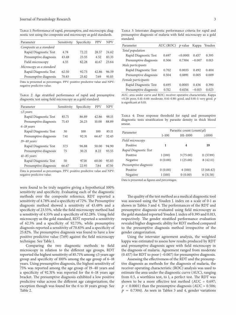
Table 1: Performance of rapid, presumptive, and microscopic diag-nostic test using the composite and microscopy as gold standards.
Parameter Sensitivity Specificity PPV NPVComposite as a standard
Rapid Diagnostic Test 4.78 72.22 28.57 24.62Presumptive diagnosis 43.48 23.55 4.52 83.33Field microscopy 4.55 82.28 41.67 23.64
Microscopy as a standardRapid Diagnostic Test 62.50 92.73 42.86 96.59Presumptive diagnosis 70.83 25.82 7.69 91.03
Data is presented as percentages. PPV: positive predictive value and NPV:negative predictive value.
Table 2: Age stratified performance of rapid and presumptivediagnostic test using field microscopy as a gold standard.
Parameter Sensitivity Specificity PPV NPV≤5 years
Rapid Diagnostic Test 85.71 86.89 42.86 98.15Presumptive diagnosis 71.43 26.23 10.00 88.89
6–18 yearsRapid Diagnostic Test 30 100 100 85.11Presumptive diagnosis 7.41 92.31 66.67 32.43
19–40 yearsRapid Diagnostic Test 37.5 96.88 50.00 94.90Presumptive diagnosis 75 30.21 8.22 93.55
41–85 yearsRapid Diagnostic Test 50 97.18 60.00 95.83Presumptive diagnosis 66.67 22.95 7.84 87.50
Data is presented as percentages. PPV: positive predictive value and NPV:negative predictive value.
were found to be truly negative giving a hypothetical 100%sensitivity and specificity. Evaluating each of the diagnosticmethods over the composite reference, RDT reported asensitivity of 4.78% and a specificity of 72%.The Presumptivediagnosis method showed a sensitivity of 43.48% and aspecificity of 23.55%, while the field microscopy method hada sensitivity of 4.55% and a specificity of 82.28%. Using fieldmicroscopy as the gold standard, RDT reported a sensitivityof 62.5% and a specificity of 92.73%, while presumptivediagnosis reported a sensitivity of 70.83% and a specificity of25.82%. The presumptive diagnosis was found to have a lowpositive predictive value (7.69) against the field microscopytechnique. See Table 1.
Comparing the two diagnostic methods to fieldmicroscopy in relation to the different age groups, RDTreported the highest sensitivity of 85.71% among ≤5 years agegroup and specificity of 100% among the age group of 6–18years. Using presumptive diagnosis, the highest sensitivity of75% was reported among the age group of 19–40 years anda specificity of 92.31% was reported for the 6–18 years agebracket. The presumptive diagnosis exhibited a low positivepredictive value across the different age categorization; theexception though was found for the 6 to 18 years group. SeeTable 2.
Table 3: Interrater diagnostic performance criteria for rapid andpresumptive diagnosis of malaria with field microscopy as a goldstandard.
Parameter AUC (ROC) 𝑝 value Kappa YoudenTotal populationRapid Diagnostic Test 0.697 <0.0001 0.457 0.395Presumptive diagnosis 0.506 0.7304 −0.007 0.013
Male participantsRapid Diagnostic Test 0.702 0.0035 0.492 0.404Presumptive diagnosis 0.504 0.8891 0.005 0.009
Female participantsRapid Diagnostic Test 0.695 0.0005 0.436 0.390Presumptive diagnosis 0.512 0.6156 −0.013 0.023
AUC: area under curve and ROC: receiver operative characteristic. Kappa<0.20: poor, 0.41–0.60: moderate, 0.61–0.80: good, and 0.81–1: very good. 𝑝is significant at 0.05.
Table 4: Dose response threshold for rapid and presumptivediagnostic tests stratification by parasite density in thick bloodsmear.
Parameter Parasitic count (count/𝜇l)1–100 101–1000 ≥1000
Field microscopyPositive 1 4 19
Rapid Diagnostic TestPositive 1 (100) 3 (75.00) 11 (57.89)Negative 0 (0.00) 1 (25.00) 8 (42.11)
Presumptive diagnosisPositive 0 (0.00) 4 (100) 13 (68.42)Negative 1 (100) 0 (0.00) 6 (31.58)
Data is presented as figures and percentages.
The quality of the testmethod as amedical diagnostic toolwas assessed using the Youden J. index on a scale of 0-1 asshown in Tables 3 and 4. The performances of the RDT andpresumptive diagnosis evaluated using field microscopy asthe gold standard reported Youden J. index of 0.395 and 0.013,respectively. The gender stratified performance evaluationrevealed higher diagnostic ability for RDTmethod comparedto the presumptive diagnosis method irrespective of thegender categorization.
Using the interrater agreement analysis, the weightedkappa was estimated to assess how results produced by RDTand presumptive diagnosis agree with field microscopy inthe diagnosis of malaria. Agreement ranged from moderate(0.457) for RDT to poor (−0.007) for presumptive diagnosis.
Assessing the effectiveness of the RDT and the presump-tive diagnosis as methods for the diagnosis of malaria, thereceiver operating characteristic (ROC) analysis was used toestimate the area under the diagnostic curve (AUC), rangingfrom 0.5, a worthless test, to 1, a perfect test. The RDT wasshown to be a more effective test method (AUC = 0.697;𝑝 < 0.0001) than the presumptive diagnosis (AUC = 0.506;𝑝 = 0.7304). As seen in Tables 3 and 4, gender variations

4 Journal of Parasitology Research
in performance were not prominent for both diagnosticmethods (RDT and presumptive diagnosis). See Table 3.
Out of the 24 patients that were reported as havingpositive malaria parasite by the field microscopy method, 15and 17 were positive for RDT and presumptive diagnosis,respectively (true positive). Among the 264 patients whotested negative for RDT, 9 were positive for field microscopy(false negatives). Among the 78 patients reported to benegative by presumptive diagnosis, 7 tested positive usingthe field microscopy technique (false negative). Among thefalse negative recording using the RDT 11.11% was within aparasitic count of 101–1000/𝜇l and the rest (88.89%) had aparasitic count of 1000/𝜇l or greater. Among the false negativerecording using presumptive diagnosis 14.29%was within theparasitic density of 1–100/𝜇l and the rest (85.71%) presentedwith parasitic density of 1000/𝜇l and above. See Table 4.
4. Discussion
Malaria is a deadly disease especially in children and, forthat reason, it is important to have a prompt and accuratediagnosis of the disease [11]. In this study, the test per-formance of the three routine malaria diagnostic methods,namely, Rapid Diagnostic Test (RDT), presumptive diagnosismethod, and field microscopy technique, was evaluated. Thediagnostic accuracy of these methods was measured againsta composite and field microscopy as gold standards. Ingeneral, the study established that the sensitivity of the threediagnostic methods was very low when evaluated over thecomposite reference.This is an indication of wider differencesor substantial nonoverlap between the test methods in thepositive detection ofmalaria among the study population [6].In contrast, the sensitivities recorded by the individual testmethods were lower than that reported in a similar study inNigeria by Ojurongbe et al. [7] which had 62.3% and 77.2%for RDT and field microscopy, respectively.This difference inrecorded sensitivity could be accounted for by the makeupof the composite reference where polymerase chain reactionwas inclusive in the previous study but this is not included inthis study. Again, whereas this studymade use of presumptivediagnosis, the study by Ojurongbe et al. [7] did not. Thus,problems arise in comparison when established methodsare used as a composite gold standard without allowing forinherent accuracies for the individual test methods [12].
Diagnosis of malaria has traditionally been based onpresumptive diagnosis [4] and its principles are based onnonspecific signs and symptoms like headaches, fever, weak-ness, dizziness, vomiting, abdominal pains, myalgia, chills,and pruritus [7]. In the current study presumptive diagnosisdemonstrated a generally low specificity when comparedwith the two gold standards: composite (23.55%) and fieldmicroscopy (25.82%) as shown in Tables 1 and 2, respectively.These findings were consistent with a previous work whichrecorded a specificity ranging from 0 to 9% [13]. Thus,presumptive diagnosis is unable to identify patients withoutmalaria as truly uninfected (false positives). This leads totreating all fevers presumptively as malaria, thereby maskingunderlying potentially fatal conditions [14]. From the presentstudy, out of 221 people who were clinically diagnosed with
malaria, 17 and 24 were positive by field microscopy andRDT, respectively.This shows that about 70% of patients whowere diagnosed as having malaria presumptively turned outto be parasite negative. This massive malaria overdiagnosisaccording to Reyburn et al. [15] threatens the sustainabilityof deployment of artemisinin combination treatment, andtreatable bacterial diseases are likely to be missed.
After age stratification, the highest sensitivity for the twotest methods against the field microscopy technique wasrecorded among ≤5 years group (Table 2). This agrees withthe findings of Nkrumah et al. [16] where an increase inthe sensitivity of a histidine-rich protein 2 (Pfhrp2) assay wasfound in children compared to adults. Nkrumah et al. [16]posited that lower immunity and possibly less interferenceby antibodies among children could be attributable to suchoutcome. Parasitaemia in older aged groups often remainsvery low and frequently undetectable by conventionalmalariadiagnostics (microscopy) and Rapid Diagnostic Test [17].
Rapid Diagnostic Tests (RDTs) based on histidine-richprotein 2 (Pfhrp2) have shown a varied accuracy for malariainfection in field studies, with field microscopy taken asthe gold standard [1, 18–20]. In the present study, RDTreported a sensitivity of 62.5% and specificity of 92.93%whencompared with field microscopy. This is within the samerange as previous studies [21, 22]. However, the sensitivitywas lower than the World Health Organization (WHO)recommended minimal standard of 95% sensitivity for Plas-modium falciparum densities of 100/𝜇L and a specificity of95% for an acceptable Rapid Diagnostic Test for malaria [23].The diagnostic accuracy of RDTs can be affected by severalfactors such as quality of the products, storage temperature,humidity, and end users’ performance [6].
Persistence of Pfhrp2 protein in circulation after parasiteclearance contributes to lower specificity level of RDT [9, 19,21]. This may explain the observation in the current studywhere 9 participants on malaria treatment tested positive forRDT with no parasitic detection by field microscopy (falsepositives). In the case where the parasite density is belowthe threshold for detection by microscopy (submicroscopicparasitic density), it is still possible for RDT to report positivemalaria test [19] or low density parasitaemia might be missedand wrongly classified as negative with microscopy; howeverthe double blind reading of blood films by two experiencedmicroscopists was aimed at reducing the latter scenario [24].
In the current study, out of the 264 patients that RDTconfirmed as negative, field microscopy detected 9 of them aspositive with 8 patients recording a parasitic density of ≥1000parasites/𝜇L (Table 4). Studies have reported patients withhigh levels of parasitaemia that give false negativeRDT resultsdue to the deletion of Pfhrp2 antigens or genetic variability inthe Pfhrp2 gene in certain Plasmodium falciparum parasites[25, 26].
In a study by Abeku et al. [24], false positive errorrates declined with increasing age of patients and this wasprobably a result of acquired immunity in clearing parasiteantigen. Contrary to this, the present study noted falsepositive results by RDT increased with increasing age ofpatients. According to the manufacturer’s manual, internalevaluation was performed on the SD Bioline malaria Ag Pf.
Journal of Parasitology Research 5
test which reported a 100% sensitivity for parasitic countbetween 101 and 500 parasites/𝜇L but the present studyreported a sensitivity of 75% for a similar parasitic countof 101–1000 parasites/𝜇L. Manufacturer’s specified sensitivitywas 100% for a parasitic count greater than 1000 parasites/𝜇Lwhilst this study had a sensitivity of 57.89% for that sameparasitic count. The manufacturer’s overall sensitivity whenmicroscopy was used as a reference was greater or equal to99% whilst this study reported 62.5% overall sensitivity.
A patient who reported Plasmodium malariae infectionwas also positive with the RDT which has been designed todetect only Plasmodium falciparum. This revelation may bedue to cross-reactivity [9]. RDT also was positive to a parasitecount that was as low as 40 parasites/𝜇L and this is in contrastto other studies [6, 10, 27].
The weighted kappa statistic, widely used as a chance-corrected measure for nominal agreement [28], was usedto test interrater agreement between microscopy and theother two diagnostic methods (RDT and presumptive diag-nosis). There was a moderate level of agreement betweenRDT (kappa: 0.457) and microscopy. This observation is inaccordance with the findings of Ali et al. [29] who reporteda moderate agreement between RDT and field microscopy inCameroon. But the agreement observed was slightly highercompared to that reported by Kilonzo et al. [30] (kappa:0.354) in Tanzania. A recent study in Yemen by Alareqi et al.[31] also reported a kappa of 0.379 for both febrile and afebrileparticipants and a higher weighted kappa of 0.638 amongonly febrile patients. Thus RDT could be used for malariadiagnosis in settings where microscopy is not available [32].Presumptive diagnosis (kappa: 0.007) however had a pooror no agreement with microscopy and thus it is likely tomisdiagnose malaria if it is not used alongside a laboratorybased diagnostic tool.
The discriminating power of RDT and presumptive diag-nosis for the detection of malaria in patients was furtherinvestigated by the area under the receiver operative char-acteristics (ROC) as seen in Table 3. It was found that RDT(area under the diagnostic curve (AUC: 0.697)) was moreeffective in predictingmalaria infection; however in the workof Djimde et al. [33] in Mali, RDT recorded a higher areaunder the ROC curve (0.97). The presumptive diagnosismethod had an area under the ROC curve of −0.506, whichimplies that it is a worthless test for malaria diagnosis.
Youden index was carried out to measure the medicalusefulness of the diagnostic methods for malaria detection.RDT proved to be a useful diagnostic test (Youden: 0.395)as compared to presumptive diagnosis (Youden: 0.013).The performance of the RDT agrees with the findings ofSamadoulougou et al. [34] in Burkina Faso who reportedan overall Youden of 0.40. This shows that presumptivediagnosis is less effective and an unreliable diagnostic tool formalaria diagnosis as a standalone test.
5. Conclusion
Based on the results of this study, none of the test meth-ods evaluated over the gold standard (field microscopytechnique) achieved the WHO recommended diagnostic
sensitivity and specificity. However, the RDT achieved anacceptable agreement with the gold standard. Factors such asmedication and age also played a role in influencing the testperformance of the various diagnostic methods. Diagnosis ofmalaria by field microscopy should still be the gold standardalthough it requires a level of expertise. In settings wheremicroscopy is not available, however, RDTsmust be preferredas a confirmation of presumptive diagnosis.
Ethical Approval
Approval for this study was granted by the authoritiesof the facility. The study was anonymous and nonlinked;participation was voluntary and all patients or their parentsconsented to participation in this study. All patients in thisstudy had been referred to the laboratory as part of the clinicalcare they were receiving from the hospital. No patient wasdenied the appropriate care for declining to participate in thisstudy.
Competing Interests
Authors have declared that no competing interests exist.
Authors’ Contributions
This work was carried out in collaboration between allauthors. J. Osei-Yeboah, G. K. Norgbe, S. Y. Lokpo, M.K. Kinansua, L. Nettey, and E. A. Allotey conceptualizedand designed the study. J. Osei-Yeboah, S. Y. Lokpo, M. K.Kinansua, and L. Nettey recruited participants and generatedthe data. J. Osei-Yeboah, S. Y. Lokpo, M. K. Kinansua, and E.A. Allotey analyzed the data. J. Osei-Yeboah, G. K. Norgbe,S. Y. Lokpo, M. K. Kinansua, and E. A. Allotey draftedthe manuscript. J. Osei-Yeboah, G. K. Norgbe, S. Y. Lokpo,M. K. Kinansua, L. Nettey, and E. A. Allotey reviewed themanuscript for intellectual content and each author approvedthe final manuscript.
Acknowledgments
The authors want to thank the management of the HoMunicipal Hospital for giving them the permission to carryout this study in their facility. Their sincere gratitude goesto all biomedical scientists at the Ho Municipal Hospital, forwithout their participation this project would not have comeinto fruition.
References
[1] B. B. Andrade, A. Reis-Filho, A. M. Barros et al., “ResearchTowards a precise test for malaria diagnosis in the BrazilianAmazon: comparison among field microscopy, a Rapid Diag-nostic Test, nested PCR, and a computational expert systembased on artificial neural networks,” 2010.
[2] A. Abdul-Aziz, E. Harris, and L. Munyakazi, “Risk factors inmalaria mortality among children in Northern Ghana: a casestudy at the tamale teaching hospital,” International Journal ofBusiness and Social Research, vol. 2, no. 5, pp. 35–45, 2012.
6 Journal of Parasitology Research
[3] N. Tangpukdee, C. Duangdee, P. Wilairatana, and S. Krudsood,“Malaria diagnosis: a brief review,” The Korean Journal ofParasitology, vol. 47, no. 2, pp. 93–102, 2009.
[4] B. S. C. Uzochukwu, E. N. Obikeze, O. E. Onwujekwe, C. A.Onoka, and U. K. Griffiths, “Cost-effectiveness analysis of rapiddiagnostic test, microscopy and syndromic approach in thediagnosis of malaria in Nigeria: implications for scaling-updeployment of ACT,” Malaria Journal, vol. 8, no. 1, article 265,2009.
[5] A. Bjorkman andA.Martensson, “Risks and benefits of targetedmalaria treatment based on rapid diagnostic test results,”Clinical Infectious Diseases, vol. 51, no. 5, pp. 512–514, 2010.
[6] T. Endeshaw, T. Gebre, J. Ngondi et al., “Evaluation of lightmicroscopy and rapid diagnostic test for the detection ofmalaria under operational field conditions: A household surveyin Ethiopia,”Malaria Journal, vol. 7, no. 1, article 118, 2008.
[7] O. Ojurongbe, O. O. Adegbosin, S. S. Taiwo et al., “Assessmentof clinical diagnosis, microscopy, rapid diagnostic tests, andpolymerase chain reaction in the diagnosis of Plasmodiumfalciparum in Nigeria,” Malaria Research and Treatment, vol.2013, Article ID 308069, 5 pages, 2013.
[8] H. Hopkins, L. Bebell, W. Kambale, C. Dokomajilar, P. J.Rosenthal, and G. Dorsey, “Rapid diagnostic tests for malaria atsites of varying transmission intensity in Uganda,” The Journalof Infectious Diseases, vol. 197, no. 4, pp. 510–518, 2008.
[9] C. Wongsrichanalai, M. J. Barcus, S. Muth, A. Sutamihardja,and W. H. Wernsdorfer, “A review of malaria diagnostic tools:microscopy and Rapid Diagnostic Test (RDT),” The AmericanJournal of Tropical Medicine and Hygiene, vol. 77, no. 6, supple-ment, pp. 119–127, 2007.
[10] E. G. Long, “Requirements for diagnosis of malaria at differentlevels of the laboratory network in Africa,” American Journal ofClinical Pathology, vol. 131, no. 6, pp. 858–860, 2009.
[11] P. Mens, N. Spieker, S. Omar, M. Heijnen, H. Schallig, and P. A.Kager, “Is molecular biology the best alternative for diagnosisof malaria to microscopy? A comparison between microscopy,antigen detection and molecular tests in rural Kenya and urbanTanzania,” Tropical Medicine and International Health, vol. 12,no. 2, pp. 238–244, 2007.
[12] D. R. Bell, D. W. Wilson, and L. B. Martin, “False-positiveresults of a Plasmodium falciparum histidine-rich protein 2-detecting malaria rapid diagnostic test due to high sensitivityin a community with fluctuating low parasite density,” TheAmerican Journal of Tropical Medicine and Hygiene, vol. 73, no.1, pp. 199–203, 2005.
[13] J. Chipeta, S. Mharakurwa, O. Thuma, and N. Kumar, “Asynopsis of current malaria diagnosis trends,” Medical Journalof Zambia, vol. 36, no. 2, 2009.
[14] M. Amexo, R. Tolhurst, G. Barnish, and I. Bates, “Malariamisdiagnosis: effects on the poor and vulnerable,”Rapid Review,vol. 364, 2004.
[15] H. Reyburn, H. Mbakilwa, R. Mwangi et al., “Rapid diagnostictests compared with malaria microscopy for guiding outpatienttreatment of febrile illness in Tanzania: randomised trial,”British Medical Journal, vol. 334, no. 7590, pp. 403–406, 2007.
[16] B. Nkrumah, S. E. K. Acquah, L. Ibrahim et al., “Comparativeevaluation of two rapid field tests for malaria diagnosis: PartecRapidMalaria Test� and Binax Now�Malaria Rapid DiagnosticTest,” BMC Infectious Diseases, vol. 11, no. 1, article 143, pp. 1–8,2011.
[17] L. Golassa, F. N. Baliraine, N. Enweji, B. Erko, G. Swedberg,andA.Aseffa, “Microscopic andmolecular evidence of the pres-ence of asymptomatic Plasmodium falciparum and Plasmodiumvivax infections in an area with low, seasonal and unstablemalaria transmission in Ethiopia,” BMC Infectious Diseases, vol.15, article 310, 2015.
[18] Z. Bisoffi, S. B. Sirima, J. Menten et al., “Accuracy of a rapiddiagnostic test on the diagnosis of malaria infection and ofmalaria—attributable fever during low and high transmissionseason in Burkina Faso,” Malaria Journal, vol. 9, no. 1, article192, 2010.
[19] D. R. Bell, D. W. Wilson, and L. B. Martin, “False-positiveresults of a Plasmodium falciparum histidine-rich protein 2–detecting malaria rapid diagnostic test due to high sensitivityin a community with fluctuating low parasite density,” TheAmerican Journal of Tropical Medicine and Hygiene, vol. 73, no.1, pp. 199–203, 2005.
[20] V. Batwala, P. Magnussen, and F. Nuwaha, “Are rapid diagnostictests more accurate in diagnosis of Plasmodium falciparummalaria compared to microscopy at rural health centres?”Malaria Journal, vol. 9, no. 1, article 349, 2010.
[21] M. L. Mcmorrow, M. I. Masanja, S. M. K. Abdulla, E. Kahigwa,and S. P. Kachur, “challenges in routine implementation andquality control of Rapid Diagnostic Tests for malaria—Rufijidistrict,”TheAmerican Society of TropicalMedicine andHygiene,vol. 79, no. 3, pp. 385–390, 2008.
[22] D. O. Acheampong, M. G. Appiah, L. K. Boamponsem, J. N.Boampong, and R. Afoakwa, “the efficacy of Rapid Diagnos-tic Test(rdt) in diagnosing plasmodium falciparum in someselected health facilities in the capecoast metropolis of Ghana,”Advances in Applied Science Research, vol. 2, no. 4, pp. 348–356,2011.
[23] C. K. Murray, R. A. Gasser Jr., A. J. Magill, and R. S. Miller,“Update on rapid diagnostic testing formalaria,”ClinicalMicro-biology Reviews, vol. 21, no. 1, pp. 97–110, 2008.
[24] T. A. Abeku, M. Kristan, C. Jones et al., “Determinants of theaccuracy of rapid diagnostic tests in malaria case management:evidence from low and moderate transmission settings in theEast African highlands,”Malaria Journal, vol. 7, no. 1, article 202,2008.
[25] J. C. Mouatcho and J. P. D. Goldring, “Malaria rapid diagnostictests: challenges and prospects,” Journal of Medical Microbiol-ogy, vol. 62, no. 10, pp. 1491–1505, 2013.
[26] M. L. Wilson, laboratory Diagnosis of Malaria, Archives ofPathology & Laboratory Medicine, 2013.
[27] L. M. Singer, R. D. Newman, A. Diarra et al., “Evaluation of amalaria rapid diagnostic test for assessing the burden ofmalariaduring pregnancy,” The American Journal of Tropical Medicineand Hygiene, vol. 70, no. 5, pp. 481–485, 2004.
[28] M. A. Billo, M. Diakite, A. Dolo et al., “Inter-observer agree-ment according to malaria parasite density,” Malaria Journal,vol. 12, no. 1, article 335, pp. 1–6, 2013.
[29] I.M. Ali, J. D. Bigoga, D. A. Forsah et al., “Field evaluation of the22 rapid diagnostic tests for communitymanagement ofmalariawith artemisinin combination therapy in Cameroon,” MalariaJournal, vol. 15, no. 1, article 31, pp. 1–7, 2016.
[30] S. B. Kilonzo, E. Kamugisha, J. A. Downs et al., “Malariaamong adult inpatients in two Tanzanian referral hospitals: aprospective study,”Acta Tropica, vol. 134, no. 1, pp. 95–100, 2014.
[31] L. M. Q. Alareqi, M. A. K. Mahdy, Y.-L. Lau et al., “Fieldevaluation of a PfHRP-2/pLDH rapid diagnostic test and light

Journal of Parasitology Research 7
microscopy for diagnosis and screening of falciparum malariaduring the peak seasonal transmission in an endemic area inYemen,”Malaria Journal, vol. 15, no. 1, article 49, 9 pages, 2016.
[32] E. K. Ansah, S. Narh-Bana, M. Epokor et al., “Rapid testing formalaria in settings wheremicroscopy is available and peripheralclinics where only presumptive treatment is available: a ran-domised controlled trial in Ghana,” British Medical Journal, vol.340, article c930, 2010.
[33] A. A. Djimde, H. Maiga, I. Sagara et al., “Field assessment ofSD biolinemalaria species antigen detection by rapid diagnostictests in Mali,” Journal of Parasitology and Vector Biology, vol. 8,no. 1, pp. 1–9, 2016.
[34] S. Samadoulougou, F. Kirakoya-Samadoulougou, S. Sarrassatet al., “Paracheck� rapid diagnostic test for detecting malariainfection in under five children: a population-based survey inBurkina Faso,”Malaria Journal, vol. 13, no. 1, article 101, 2014.

Submit your manuscripts athttp://www.hindawi.com
Hindawi Publishing Corporationhttp://www.hindawi.com Volume 2014
Anatomy Research International
PeptidesInternational Journal of
Hindawi Publishing Corporationhttp://www.hindawi.com Volume 2014
Hindawi Publishing Corporation http://www.hindawi.com
International Journal of
Volume 2014
Zoology
Hindawi Publishing Corporationhttp://www.hindawi.com Volume 2014
Molecular Biology International
GenomicsInternational Journal of
Hindawi Publishing Corporationhttp://www.hindawi.com Volume 2014
The Scientific World JournalHindawi Publishing Corporation http://www.hindawi.com Volume 2014
Hindawi Publishing Corporationhttp://www.hindawi.com Volume 2014
BioinformaticsAdvances in
Marine BiologyJournal of
Hindawi Publishing Corporationhttp://www.hindawi.com Volume 2014
Hindawi Publishing Corporationhttp://www.hindawi.com Volume 2014
Signal TransductionJournal of
Hindawi Publishing Corporationhttp://www.hindawi.com Volume 2014
BioMed Research International
Evolutionary BiologyInternational Journal of
Hindawi Publishing Corporationhttp://www.hindawi.com Volume 2014
Hindawi Publishing Corporationhttp://www.hindawi.com Volume 2014
Biochemistry Research International
ArchaeaHindawi Publishing Corporationhttp://www.hindawi.com Volume 2014
Hindawi Publishing Corporationhttp://www.hindawi.com Volume 2014
Genetics Research International
Hindawi Publishing Corporationhttp://www.hindawi.com Volume 2014
Advances in
Virolog y
Hindawi Publishing Corporationhttp://www.hindawi.com
Nucleic AcidsJournal of
Volume 2014
Stem CellsInternational
Hindawi Publishing Corporationhttp://www.hindawi.com Volume 2014
Hindawi Publishing Corporationhttp://www.hindawi.com Volume 2014
Enzyme Research
Hindawi Publishing Corporationhttp://www.hindawi.com Volume 2014
International Journal of
Microbiology