Embed Size (px)
Citation preview

Open Journal of Radiology, 2016, 6, 39-48 Published Online March 2016 in SciRes. http://www.scirp.org/journal/ojrad http://dx.doi.org/10.4236/ojrad.2016.61006
How to cite this paper: Karjalainen, M., et al. (2016) Reproducibility of 3 mm-Slice-Thick Reconstruction of Paranasal Sinus Computed Tomography Scans. Open Journal of Radiology, 6, 39-48. http://dx.doi.org/10.4236/ojrad.2016.61006
Reproducibility of 3 mm-Slice-Thick Reconstruction of Paranasal Sinus Computed Tomography Scans Matti Karjalainen1*, Anna Julkunen2,3*, Antti Markkola4, Prasun Dastidar5, Heini Huhtala6, Mikko Suvinen1,7, Anna-Maija Kuukka1, Markus Rautiainen1,8, Jura Numminen1#, Sanna Toppila-Salmi2,9# 1Department of Otorhinolaryngology, University of Tampere, Tampere, Finland 2Transplantation Laboratory, Haartman Institute, University of Helsinki, Helsinki, Finland 3Department of Oral and Maxillofacial Diseases, University of Helsinki and Helsinki University Hospital, Helsinki, Finland 4University of Helsinki and HUS Imaging, Helsinki, Finland 5Medical Imaging Centre, Department of Radiology, Tampere University Hospital, Finland 6School of Public Health, University of Tampere, Tampere, Finland 7Department of Otorhinolaryngology, Tampere City Hospital, Tampere, Finland 8Department of Ear and Oral diseases, Tampere University Hospital, Tampere, Finland 9Department of Allergy, University of Helsinki and Helsinki University Hospital, Helsinki, Finland
Received 19 January 2016; accepted 23 March 2016; published 28 March 2016
Copyright © 2016 by authors and Scientific Research Publishing Inc. This work is licensed under the Creative Commons Attribution International License (CC BY). http://creativecommons.org/licenses/by/4.0/
Abstract Background: After the failure of medical treatment, the surgery of chronic rhinosinusitis (CRS) is planned according to endoscopic and paranasal sinus computed tomography (CT) findings. Objec-tive: The aim of this prospective study was to evaluate whether this study method might be eligi-ble in studies aiming at radiation dose reduction. Sinus CT scans were chosen as a model because of the high variation of the radiological anatomy of surgically important sinonasal structures. We hypothesized that 3 mm-slice-thick reconstruction CT had poor reproducibility. Methods: 59 CRS patients underwent routine multi-detector sinus CT (CTMD). CT3mm was reconstructed from CTMD data-sets. Lund-Mackay (LM) scores and 43 other structural parameters were analyzed blinded. Agreement was studied between CTMD and CT3mm (intra-observer reproducibility), and between three observers (inter-observer reproducibility) by using Cohen’s kappa. Results: The inter-ob- server agreement was moderate (kappa 0.4 - 0.6, p < 0.01) in the majority of structures of CT3mm scans. The intra-observer reproducibility of CT3mm scans was very good in most structures, how-ever, it was poor in important structures such as frontal and spheno-ethmoid recess, lamina pa-
*Matti Karjalainen and Anna Julkunen have equally contributed to this work. #Jura Numminen and Sanna Toppila-Salmi are shared last authors.
M. Karjalainen et al.
40
pyracae, and location of optic nerve or anterior ethmoidal artery. The grade of surgeon’s confi-dence of CT3mm in comparison to CTMD was lower (kappa 0.2 - 0.4, P < 0.05). Conclusion: This me-thodology might have some use in studies aiming at radiation dose reduction. As was expected, 3 mm-slice-thick reconstruction CT had poor reproducibility and surgeon’s confidence. More recent methods such as cone beam computed tomography scans have nowadays more relevant dose re-duction potential.
Keywords Chronic Rhinosinusitis, Computed Tomography, Image Reconstruction, Inter-Observer Agreement, Intra-Observer Agreement, Kappa, Radiation-Dose Reduction, Reproducibility, Paranasal Sinus, Sinus Imaging
1. Introduction Chronic rhinosinusitis (CRS) is a common, multifactorial and variable disease with a prevalence of around 10% - 16% [1]. The diagnostics of CRS is based on typical symptoms and clinical findings [1]. Computed tomogra-phy (CT) scans are the imaging modality of choice confirming the extent of pathology and the need for surgery. The main findings in CRS are mucosal changes within the osteomeatal complex and/or sinuses. Other characte-ristic findings are air-fluid levels, mucosal thickening and opacification of the normally aerated sinus lumen. The only CRS indicating change can be sclerotic, thickened bone of the sinus wall [1]. The number of anatomi-cal variants is very high in paranasal sinuses [2] [3]. Several of them are located close to sinonasal surgical area and thus the intraoperative lesions of them may lead to severe illness or be life threatening. The operatively im-portant structures are insertion of middle turbinate and the uncinated process, the location of anterior ethmoidal artery, and Keros class, Infraorbital cells, position of the Agger nasi cell, and the anatomical variants located in the operative area [4]-[6]. Studies demonstrate that there is dose-reduction potential of CT scans [7]-[10].
The aim of this prospective study was to evaluate whether our method might be eligible in studies aiming at radiation dose reduction. Sinus CT scans were chosen as a model because of the high variation of the radiologi-cal anatomy of surgically important sinonasal structures. We hypothesized that 3 mm-slice-thick reconstruction of sinus CT scans had poor dose-reduction potential and inter-observer agreement.
2. Materials and Methods 2.1. Patients This prospective cohort study was carried out in the Department of Otorhinolaryngology at Tampere University Hospital, Finland from 2006 to 2015. The study was approved by the ethical committee of the Pirkanmaa Hos-pital District (no 96032). Written informed consent was obtained of each participant. A random sample of 59CRS patients, who were evaluated to benefit from sinus CT scans during 2006-2007, was enrolled to this study. Patient data was collected from medical records and by a questionnaire at the time the sinus CT was per-formed. The follow-up data of sinonasal operations and time of surgery was collected form patient records of Tampere University Hospital or Tampere City Hospital in 2015 in average 9 years after the time of performing sinus CT scans. None of the subjects had used Aspirin desensitization, allergen immunotherapy, or anti IgE therapy prior to or during the sinus CT scans or during the follow-up.
2.2. CT Scans The patients underwent routine sinus multiple detector computed tomography (CTMD) examinations for clinical purposes. Two different CTMD machines were used: GE Light Speed 16 (GE Healthcare, Milwaukee, Wisconsin) and Philips Brilliance 64 (Philips, Best, the Netherlands). The patients were imaged in supine position with a kilovoltage of 120 kV and a milliampere second of 100 mAs. In the GE machine, the slice thickness was 0.625 mm with coronal reconstructions at 1.5 mm. In the Philips machine, the slice thickness was 0.9 mm with coronal reconstructions at 0.9 mm. Both were three dimensional (3D) in nature without any gap. In all cases, the imag-

M. Karjalainen et al.
41
ing was performed using a bone filter technique. The imaging covered the entire sinonasal area both in the axial and coronal directions. The coronal reformations spanned through the entire area, starting from the anterior wall of the frontal sinuses and ending to the level of the posterior wall of the sphenoid sinuses. We performed 3 mm-slice-thick reconstructions (CT3mm) from coronal, sagittal and axial data sets of CTMD. The coronal reforma-tions in the CTMD and CT3mm spanned through the entire area, starting from the anterior wall of the frontal si-nuses and ending at the level of the posterior wall of the sphenoid sinuses.
2.3. Evaluation of CT Scans CTMD and CT3mm scans were observed by three independent observers blinded to each other and to the patient history data. The observation of the same patient’s scans occurred at least one week apart. The focus was to compare agreement between CTMD and CT3mm scans and between three observers in evaluating CRS-related changes and radiological anatomy of the structures. The observers were an experienced head and neck radiolo-gist (AM), an experienced ENT- and rhinosurgeon (JN), and a fifth year ENT resident (ST-S). They filled in a 49 -item form of sinonasal structures from both scans and from both sides of each patient (Table 3). Similar forms were filled from CTMD and CT3mm scans. Evaluation of the same patient’s scans occurred at least 7 days apart. All asked structures had 2 - 5 different choices. Before starting the evaluation of the CT scans, all choices were carefully discussed by the observers. Observers also made a pilot of 15 CT scans in order to make sure that all observers understood how to fill in the forms. The Radiologist did not respond to the questions: “Need for septoplasty” and “Grade of surgeon’s confidence based on images”.
2.4. Data Analysis Statistical analysis was carried out by the SPSS Base 15.0 Statistical Software Package (SPSS Inc., Chicago, IL, USA). Cohen’s kappa was used to compare the degree of agreement of CTMD and CT3mm scans (e.g. intraob-server agreement); and the inter-observer agreement of CT3mm scans. The calculation is based on the difference between how much agreement is actually present compared to how much agreement would be expected to be present by chance alone. The established interpretation of Kappa-value is classified into 6 subgroups: Poor ≤ 0.2, Fair 0.21 - 0.4, Moderate 0.41 - 0.6, Good 0.61 - 0.8, and Very Good 0.81 - 1.0. A value under zero means that the agreement is worse than by chance, and the value range is from −1 to +1. Two-tailed P-values of <0.05 were considered statistically significant.
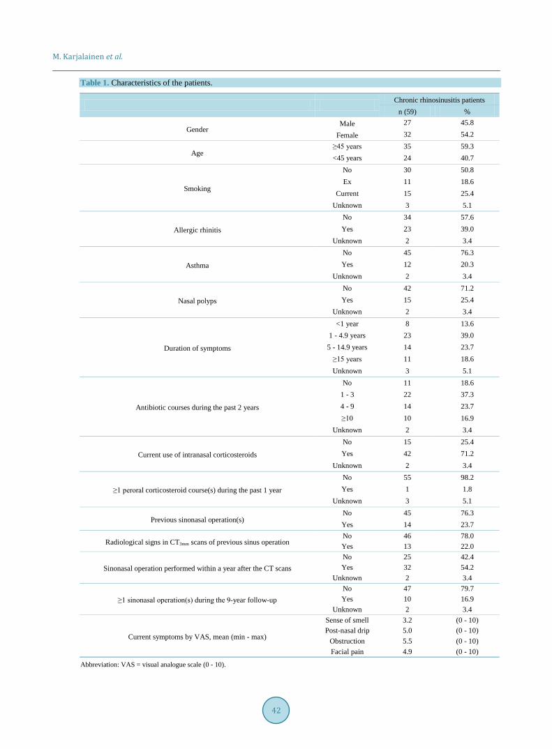
3. Results 3.1. Patient Characteristics The characteristics of patients are shown in Table 1. The mean (min-max) age was 43 (13 - 77) years by the time of taking the CT scans. 54.2% of patients underwent sinonasal operation within a year after the CT scans were performed (Table 1). Of these patients that were operated at the time of CT scans, 7 (21.9%) underwent revision sinonasal surgery in average 3 years later. 47.5% of patients reported suffering from diseases (other than CRS) with regular need of medications. The most frequent diseases were (number of parients): heart and vascular diseases [11], hypothyreosis [8], migraine [3] and arthrosis/arthritis [4].
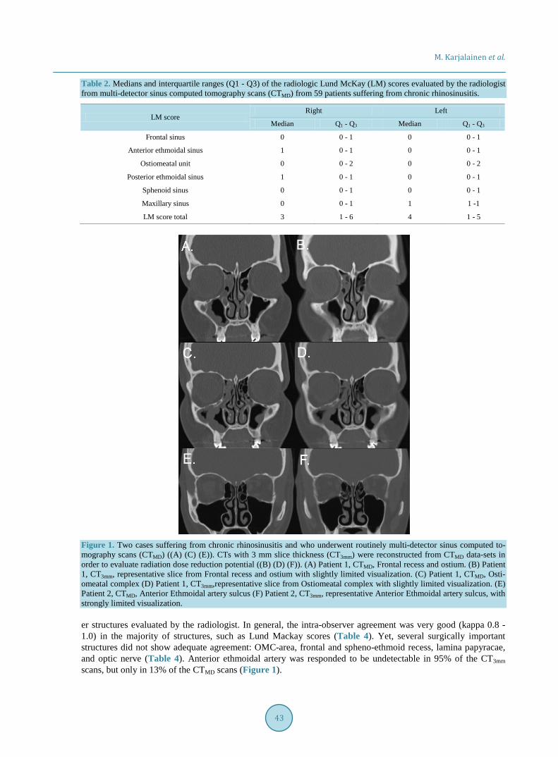
3.2. Inter-Observer Agreement of CT3mm Scans Lund-MacKay scores (Table 2) and 43 other structures were evaluated by a radiologist, an ENT surgeon and an ENT resident from both CTMD and reconstructed CT3mm scans (Table 2, Table 3). The inter-observer agreement for CT3mm scans was moderate (kappa 0.4 - 0.6) in the majority of structures (Table 3). In CT3mm scans, the sur-gically important structures with greatest disagreement were found in Keros classification (radiologist vs. ENT resident), the location of anterior ethmoidal artery and optic nerve (all observers), prominent ethmoid bulla (ENT resident vs. ENT surgeon), and thickness and contact to middle turbinate of orbital lamina of ethmoidal bone (radiologist vs. ENT surgeon) (Figure 1, Table 3).
3.3. Intra-Observer Agreement We compared the degree of agreement between CTMD and CT3mm scans of the Lund-MacKay scores and 43 oth-
M. Karjalainen et al.
42
Table 1. Characteristics of the patients.
Chronic rhinosinusitis patients n (59) %
Gender Male 27 45.8
Female 32 54.2
Age ≥45 years 35 59.3 <45 years 24 40.7
Smoking
No 30 50.8 Ex 11 18.6
Current 15 25.4 Unknown 3 5.1
Allergic rhinitis No 34 57.6 Yes 23 39.0
Unknown 2 3.4
Asthma No 45 76.3 Yes 12 20.3
Unknown 2 3.4
Nasal polyps No 42 71.2 Yes 15 25.4
Unknown 2 3.4
Duration of symptoms
<1 year 8 13.6 1 - 4.9 years 23 39.0
5 - 14.9 years 14 23.7 ≥15 years 11 18.6 Unknown 3 5.1
Antibiotic courses during the past 2 years
No 11 18.6 1 - 3 22 37.3 4 - 9 14 23.7 ≥10 10 16.9
Unknown 2 3.4
Current use of intranasal corticosteroids No 15 25.4 Yes 42 71.2
Unknown 2 3.4
≥1 peroral corticosteroid course(s) during the past 1 year No 55 98.2 Yes 1 1.8
Unknown 3 5.1
Previous sinonasal operation(s) No 45 76.3 Yes 14 23.7
Radiological signs in CT3mm scans of previous sinus operation No 46 78.0 Yes 13 22.0
Sinonasal operation performed within a year after the CT scans No 25 42.4 Yes 32 54.2
Unknown 2 3.4
≥1 sinonasal operation(s) during the 9-year follow-up No 47 79.7 Yes 10 16.9
Unknown 2 3.4
Current symptoms by VAS, mean (min - max)
Sense of smell 3.2 (0 - 10) Post-nasal drip 5.0 (0 - 10)
Obstruction 5.5 (0 - 10) Facial pain 4.9 (0 - 10)
Abbreviation: VAS = visual analogue scale (0 - 10).
M. Karjalainen et al.
43
Table 2. Medians and interquartile ranges (Q1 - Q3) of the radiologic Lund McKay (LM) scores evaluated by the radiologist from multi-detector sinus computed tomography scans (CTMD) from 59 patients suffering from chronic rhinosinusitis.
LM score Right Left
Median Q1 - Q3 Median Q1 - Q3
Frontal sinus 0 0 - 1 0 0 - 1
Anterior ethmoidal sinus 1 0 - 1 0 0 - 1
Ostiomeatal unit 0 0 - 2 0 0 - 2
Posterior ethmoidal sinus 1 0 - 1 0 0 - 1
Sphenoid sinus 0 0 - 1 0 0 - 1
Maxillary sinus 0 0 - 1 1 1 -1
LM score total 3 1 - 6 4 1 - 5
Figure 1. Two cases suffering from chronic rhinosinusitis and who underwent routinely multi-detector sinus computed to-mography scans (CTMD) ((A) (C) (E)). CTs with 3 mm slice thickness (CT3mm) were reconstructed from CTMD data-sets in order to evaluate radiation dose reduction potential ((B) (D) (F)). (A) Patient 1, CTMD, Frontal recess and ostium. (B) Patient 1, CT3mm, representative slice from Frontal recess and ostium with slightly limited visualization. (C) Patient 1, CTMD, Osti-omeatal complex (D) Patient 1, CT3mm,representative slice from Ostiomeatal complex with slightly limited visualization. (E) Patient 2, CTMD, Anterior Ethmoidal artery sulcus (F) Patient 2, CT3mm, representative Anterior Ethmoidal artery sulcus, with strongly limited visualization.
er structures evaluated by the radiologist. In general, the intra-observer agreement was very good (kappa 0.8 - 1.0) in the majority of structures, such as Lund Mackay scores (Table 4). Yet, several surgically important structures did not show adequate agreement: OMC-area, frontal and spheno-ethmoid recess, lamina papyracae, and optic nerve (Table 4). Anterior ethmoidal artery was responded to be undetectable in 95% of the CT3mm scans, but only in 13% of the CTMD scans (Figure 1).

M. Karjalainen et al.
44

M. Karjalainen et al.
45

M. Karjalainen et al.
46

M. Karjalainen et al.
47
3.4. Operative Confidence of CT3mm Scans ENT surgeon and ENT resident estimated operative certainty of CT3mm in comparison to CTMD. ENT surgeon responded that the operative confidence is good in 69.6% of CTMD scans, and 64.3% of CT3mm scans. The intra- observer agreement in the grade of ENT surgeon’s confidence of CT3mm in comparison to CTMD was moderate (kappa 0.4, P = 0.005). ENT resident responded that the operative confidence is good in 73.2% of CTMD scans, but only in 37.5% of CT3mm scans. The intra-observer agreement in the grade of ENT resident’s confidence of CT3mm in comparison to CTMD was thus poor (kappa 0.2, P = 0.035).
4. Discussion This study was carried out to evaluate whether the used method would be eligible when studying radiation dose reduction potential. As an example, we compared two techniques of paranasal sinus CT scans. From the CT scans we evaluated important structures of CRS diagnostics and operation. By reformatting the CT3mm images, we were able to evaluate the same patient exactly without additional imagining. Patient selection was performed randomly from a doctor’s reception. A heterogeneous patient group made the extrapolation of the results possi-ble for clinical practice
We found that inter-observer agreement was only moderate in CT3mm scans. This finding is in line with our unpublished data that inter-observer agreement is at similar level also in the conventional CTMD scans. This might be due to the fact that sinonasal anatomy is highly variable. Intra-observer agreement between CT3mm and CTMD scans was very good in most structures. However, it was fair to poor in several surgically important structures. The grade of young surgeons’ confidence was relatively good with CTMD scans whereas the grade of confidence was poor with CT3mm scans. Taking together, CT3mm scans seemed not to be clinically relevant im-aging method.
Previously, it has been shown that CRS diagnostics is possible with other reduced radiation CT-techniques. However, the surgical aspects have not been dealt with in these studies [7]-[9]. Our current study showed that it was possible to reduce the radiation dose and increase slice thickness without compromising the excellent bone and soft tissue contrast. However, successful endoscopic sinus surgery (ESS) requires detailed knowledge of the highly variable anatomy of the nasal cavities, the ostiomeatal unit, and the skull base. In addition, the vital structures especially the optic nerve and the anterior ethmoid artery and their relationship to operational areas need to be clear.
Multi-detector CT technology comprises multiple detectors that are equivalent to slices. Conventional CTMD comprises consecutive slices with a thickness of 0.6 - 1.5 mm and causes a radiation dose of 0.5 mSv on average. Technological improvement has been rapid resulting in 4- to 8-, 16-, 32-, 40- and 64-detector machines. It is known that there is no safe radiation dose. In all exposure quantities, there is a corresponding mutational risk [11]. The head and neck region, the eyes and thyroid gland are the most radiosensitive organs. In addition to the cancer risk, radiation-induced cataracts are possible after multiple exposures [3]. Image quality is affected by image noise or quantum mottle and is almost always directly related to the radiation dose. Keeping this in mind, the cone beam computed tomography seems to be relevant recent implementation in CT imaging. Essentially, it could be the best compromise so far between low radiation and excellent image quality [12]-[15].
As a conclusion, our study demonstrated that this methodology was easy to use and might have some use in studies aiming at radiation dose reduction. As was expected, 3 mm-slice-thick reconstruction CT had poor re-producibility and surgeon’s confidence. More recent methods such as cone beam computed tomography scans have nowadays more relevant dose reduction potential.
Acknowledgements The authors thank research nurse Marja-Leena Oksanen for her excellent assistance. The study was supported in part by research grants from the Ahokas Foundation, Competitive Research Funding of the Tampere University Hospital (Grants 9H067, 9J108, 9L087), the Finnish Medical Society Duodecim, the Finnish Society of Aller-gology and Immunology, Finnish Society of Otorhinolaryngology-Head and Neck Surgery, Helsinki University Central Hospital Research Funds, the Ida Monti Foundation, the Jane and Aatos Erkko Foundation, the Väinö and Laina Kivi Foundation, and the Yrjö Jahnsson Foundation.
M. Karjalainen et al.
48
Author Contributions STS and JN provided the study plan (with MR, PD and MS), made the applications and recruited the subjects. AM, STS and JN evaluated the CT scans. AMK, MS and STS collected the clinical data. AJ, MK, AMK, HH, and STS performed the data management. MK, AJ, JN and STS wrote the manuscript. All authors reviewed critically the manuscript.
References [1] Fokkens, W.J., Lund, V.J., Mullol, J., Bachert, C., Alobid, I., Baroody, F., et al. (2012) European Position Paper on
Rhinosinusitis and Nasal Polyps 2012. Rhinology Supplement, 23, 3 p Preceding Table of Contents, 1-298. [2] Food and Drug Administration (2002) FDA Public Health Notification: Reducing Radiation Risk from Computed To-
mography for Pediatric and Small Adult Patients. Pediatric Radiology, 32, 314-316. http://dx.doi.org/10.1007/s00247-002-0687-6
[3] Zammit-Maempel, I., Chadwick, C.L. and Willis, S.P. (2003) Radiation Dose to the Lens of Eye and Thyroid Gland in Paranasal Sinus Multislice CT. The British Journal of Radiology, 76, 418-420. http://dx.doi.org/10.1259/bjr/82798696
[4] Lund, V.J. and Kennedy, D.W. (1997) Staging for Rhinosinusitis. Otolaryngology—Head and Neck Surgery, 117, S35-S40. http://dx.doi.org/10.1016/s0194-5998(97)70005-6
[5] Holbrook, E.H., Brown, C.L., Lyden, E.R. and Leopold, D.A. (2005) Lack of Significant Correlation between Rhino-sinusitis Symptoms and Specific Regions of Sinus Computer Tomography Scans. American Journal of Rhinology, 19, 382-387.
[6] Hopkins, C., Browne, J.P., Slack, R., Lund, V. and Brown, P. (2007) The Lund-Mackay Staging System for Chronic Rhinosinusitis: How Is It Used and What Does It Predict? Otolaryngology—Head and Neck Surgery, 137, 555-561. http://dx.doi.org/10.1016/j.otohns.2007.02.004
[7] Brem, M.H., Zamani, A.A., Riva, R., Zou, K.H., Rumboldt, Z., Hennig, F.F., et al. (2007) Multidetector CT of the Pa-ranasal Sinus: Potential for Radiation Dose Reduction. Radiology, 243, 847-852. http://dx.doi.org/10.1148/radiol.2433050207
[8] Mulkens, T.H., Broers, C., Fieuws, S., Termote, J.L. and Bellnick, P. (2005) Comparison of Effective Doses for Low-Dose MDCT and Radiographic Examination of Sinuses in Children. AJR American Journal of Roentgenology, 184, 1611-1618. http://dx.doi.org/10.2214/ajr.184.5.01841611
[9] Abul-Kasim, K., Strombeck, A. and Sahlstrand-Johnson, P. (2011) Low-Dose Computed Tomography of the Paranasal Sinuses: Radiation Doses and Reliability Analysis. American Journal of Otolaryngology, 32, 47-51. http://dx.doi.org/10.1016/j.amjoto.2009.08.004
[10] Demeslay, J., Vergez, S., Serrano, E., Chaynes, P., Cantet, P., Chaput, B., et al. (2015) Morphological Concordance between CBCT and MDCT: A Paranasal Sinus-Imaging Anatomical Study. Surgical and Radiologic Anatomy, 38, 71-78.
[11] Frush, D.P., Slack, C.C., Hollingsworth, C.L., Bisset, G.S., Donnelly, L.F., Hsieh, J., et al. (2002) Computer-Simulated Radiation Dose Reduction for Abdominal Multidetector CT of Pediatric Patients. AJR American Journal of Roentge-nology, 179, 1107-1113. http://dx.doi.org/10.2214/ajr.179.5.1791107
[12] Miracle, A.C. and Mukherji, S.K. (2009) Conebeam CT of the Head and Neck, Part 1: Physical Principles. AJNR American Journal of Neuroradiology, 30, 1088-1095. http://dx.doi.org/10.3174/ajnr.A1653
[13] Miracle, A.C. and Mukherji, S.K. (2009) Conebeam CT of the Head and Neck, Part 2: Clinical Applications. AJNR American Journal of Neuroradiology, 30, 1285-1292. http://dx.doi.org/10.3174/ajnr.A1654
[14] Campbell Jr., P.D., Zinreich, S.J. and Aygun, N. (2009) Imaging of the Paranasal Sinuses and In-Office CT. Otolaryn-gologic Clinics of North America, 42, 753-764. http://dx.doi.org/10.1016/j.otc.2009.08.015
[15] Viera, A.J. and Garrett, J.M. (2005) Understanding Interobserver Agreement: The Kappa Statistic. Family Medicine, 37, 360-363.
Abbreviations CT = Computed tomography; CRS = Chronic rhinosinusitis; ENT = Ear nose throat.