Embed Size (px)
Citation preview

1
Report on the Sixth Blind Test of OrganicCrystal-Structure Prediction Methods
Anthony M. Reilly,a* Richard I. Cooper,b Claire S. Adjiman,c
Saswata Bhattacharya,d A. Daniel Boese,e Jan Gerit Brandenburg,f1
Peter J. Bygrave,g Rita Bylsma,h Josh E. Campbell,g Roberto Car,i
David H. Case,g Renu Chadha,j Jason C. Cole,a Katherine Cosburn,k,l
Herma M. Cuppen,h Farren Curtis,k,m Graeme M. Day,g Robert
A. DiStasio Jr,i,n Alexander Dzyabchenko,o Bouke P. van Eijck,p
Dennis M. Elking,q Joost A. van den Ende,h Julio C. Facelli,r,s Marta
B. Ferraro,t Laszlo Fusti-Molnar,q Christina-Anna Gatsiou,c Thomas
S. Gee,g Rene de Gelder,h Luca M. Ghiringhelli,d Hitoshi Goto,u,v
Stefan Grimme,f Rui Guo,w Detlef W. M. Hofmann,x,y Johannes Hoja,d
Rebecca K. Hylton,w Luca Iuzzolino,w Wojciech Jankiewicz,z Daniel
T. de Jong,h John Kendrick,aa Niek J. J. de Klerk,h Hsin-Yu Ko,i
Liudmila N. Kuleshova,y Xiayue Li,k,bb Sanjaya Lohani,k Frank J.
J. Leusen,aa Albert M. Lund,q,cc Jian Lv,dd Yanming Ma,dd Noa Marom,k,ee
Artem E. Masunov,ff,gg,hh,ii Patrick McCabe,a David P. McMahon,g
Hugo Meekes,h Michael P. Metz,jj Alston J. Misquitta,kk
Sharmarke Mohamed,ll Bartomeu Monserrat,mm,nn Richard J. Needs,mm
Marcus A. Neumann,oo Jonas Nyman,g Shigeaki Obata,u
Harald Oberhofer,pp Artem R. Oganov,qq,rr,ss,tt Anita M. Orendt,r
Gabriel I. Pagola,t Constantinos C. Pantelides,c Chris J. Pickard,uu,vv
Rafa l Podeszwa,z Louise S. Price,w Sarah L. Price,w Angeles Pulido,g
1Present Address: Department of Chemistry, London Centre for Nanotechnology, University CollegeLondon, 20 Gordon Street, London WC1H 0AJ, England.
PREPRINT: Acta Crystallographica Section B A Journal of the International Union of Crystallography

2
Murray G. Read,a Karsten Reuter,pp Elia Schneider,ww
Christoph Schober,pp Gregory P. Shields,a Pawanpreet Singh,j Isaac
J. Sugden,c Krzysztof Szalewicz,jj Christopher R. Taylor,g
Alexandre Tkatchenko,d,xx Mark E. Tuckerman,ww,yy,zz
Francesca Vacarro,k,aaa Manolis Vasileiadis,c
Alvaro Vazquez-Mayagoitia,bb Leslie Vogt,ww Yanchao Wang,dd Rona
E. Watson,w Gilles A. de Wijs,h Jack Yang,g Qiang Zhuqq and Colin
R. Grooma
aThe Cambridge Crystallographic Data Centre, 12 Union Road, Cambridge CB2
1EZ, England, bChemical Crystallography, Chemistry Research Laboratory,
Mansfield Road, Oxford OX1 3TA, England, cDepartment of Chemical Engineering,
Centre for Process Systems Engineering, Imperial College London, London SW7
2AZ, England, dFritz-Haber-Institut der Max-Planck-Gesellschaft, Faradayweg 4-6,
14195, Berlin, Germany, eDepartment of Chemistry, Institute of Physical and
Theoretical Chemistry, University of Graz, Heinrichstraße 28/IV, 8010 Graz,
Austria, fMulliken Center for Theoretical Chemistry, Institut fur Physikalische und
Theoretische Chemie, Rheinische Friedrich-Wilhelms Universitat Bonn, Beringstraße
4, 53115 Bonn, Germany, gSchool of Chemistry, University of Southampton,
Southampton, SO17 1BJ, England, hRadboud University, Institute for Molecules and
Materials, Heyendaalseweg 135, 6525 AJ Nijmegen, The Netherlands, iDepartment
of Chemistry, Princeton University, Princeton, NJ 08544, USA, jUniversity Institute
of Pharmaceutical Sciences, Panjab University, Chandigarh, India, kDepartment of
Physics and Engineering Physics, Tulane University, New Orleans, Louisiana 70118,
USA, lDepartment of Physics, University of Toronto, Toronto, M5S 1A7, Canada,
mDepartment of Physics, Carnegie Mellon University, Pittsburgh, PA 15213, USA,
nDepartment of Chemistry and Chemical Biology, Cornell University, Ithaca, NY
IUCr macros version 2.1.6: 2014/10/01

3
14853, USA, oKarpov Institute of Physical Chemistry, Moscow, Russia, pUtrecht
University (retired), The Netherlands, qOpenEye Scientific Software, 9 Bisbee Court,
Suite D, Santa Fe, NM 87508, USA, rCenter for High Performance Computing,
University of Utah, 155 South 1452 East Room 405, Salt Lake City, UT 84112-0190,
USA, sDepartment of Biomedical Informatics, University of Utah, 155 South 1452
East Room 405, Salt Lake City, UT 84112-0190, USA, tDepartamento de Fısica and
Ifiba (CONICET) Facultad de Ciencias Exactas y Naturales, Universidad de Buenos
Aires, Ciudad Universitaria, Pab. I (1428), Buenos Aires, Argentina, uEducational
Programs on Advanced Simulation Engineering, Toyohashi University of Technology,
1-1 Hibarigaoka, Tempaku-cho, Toyohashi, Aichi 441-8580, Japan, vDepartment of
Computer Science and Engineering, Graduate School of Engineering, Toyohashi
University of Technology, 1-1 Hibarigaoka, Tempaku-cho, Toyohashi, Aichi
441-8580, Japan, wDepartment of Chemistry, University College London, 20 Gordon
St., London WC1H 0AJ, England, xCRS4, Parco Scientifico e Tecnologico,
POLARIS, Edificio 1, 09010 PULA, Italy, yFlexCryst, Schleifweg 23, 91080
Uttenreuth, Germany, zInstitute of Chemistry, University of Silesia, Szkolna 9,
40-006 Katowice, Poland, aaFaculty of Life Sciences, University of Bradford,
Richmond Road, Bradford, BD7 1DP, England, bbArgonne Leadership Computing
Facility, Argonne National Laboratory, Lemont, Illinois, 60439,USA, ccDepartment
of Chemistry, University of Utah, 155 South 1452 East Room 405, Salt Lake City,
UT 84112-0190, USA, ddState Key Laboratory of Superhard Materials, Jilin
University, Changchun 130012, China, eeDepartment of Materials Science and
Engineering and Department of Physics, Carnegie Mellon University, Pittsburgh, PA
15213, USA, ffNanoScience Technology Center, University of Central Florida,
12424 Research Parkway, PAV400, Orlando, FL 32826, USA, ggDepartment of
Chemistry, University of Central Florida, 4111 Libra Drive PSB225, Orlando, FL
IUCr macros version 2.1.6: 2014/10/01
4
32816, USA, hhDepartment of Physics, University of Central Florida, 4111 Libra
Drive PSB430, Orlando, FL 32816, USA, iiDepartment of Condensed Matter
Physics, National Research Nuclear University MEPhI, Kashirskoye shosse 31,
Moscow, 115409, Russia, jjDepartment of Physics and Astronomy, University of
Delaware, Newark, Delaware 19716, USA, kkSchool of Physics and Astronomy,
Queen Mary University of London, London E1 4NS, England, llKhalifa University,
Abu Dhabi, P.O. Box 127788, United Arab Emirates, mmCavendish Laboratory, 19,
J. J. Thomson Avenue, Cambridge CB3 0HE, England, nnDepartment of Physics
and Astronomy, Rutgers University, Piscataway, New Jersey 08854-8019, USA,
ooAvant-garde Materials Simulation, Germany, ppChair for Theoretical Chemistry
and Catalysis Research Center, Technische Universitat Munchen, Lichtenbergstr. 4,
D-85747 Garching, Germany, qqDepartment of Geosciences, Center for Materials by
Design, and Institute for Advanced Computational Science, SUNY Stony Brook, New
York 11794-2100, USA, rrSkolkovo Institute of Science and Technology, Skolkovo
Innovation Centers, Bldg. 3, Moscow Region, 143026, Russia, ssMoscow Institute of
Physics and Technology, 9 Institutskiy Lane, Dolgoprudny City, Moscow Region,
141700, Russia, ttInternational Center for Materials Discovery, School of Materials
Science and Engineering, Northwestern Polytechnical University, Xi’an, 710072,
China, uuDepartment of Materials Science & Metallurgy, University of Cambridge,
27 Charles Babbage Road, Cambridge CB3 0FS, England, vvDepartment of Physics
and Astronomy, University College London, Gower St., London WC1E 6BT,
England, wwDepartment of Chemistry, New York University, New York, New York
10003, USA, xxPhysics and Materials Science Research Unit, University of
Luxembourg, L-1511 Luxembourg, yyCourant Institute of Mathematical Sciences,
New York University, New York, New York 10012,USA, zzNYU-ECNU Center for
Computational Chemistry at NYU Shanghai, 3663 Zhongshan Road North, Shanghai
IUCr macros version 2.1.6: 2014/10/01
5
200062, China, and aaaDepartment of Chemistry, Loyola University, New Orleans,
Louisiana 70118, USA. E-mail: [email protected]
Abstract
The sixth blind test of organic crystal-structure prediction (CSP) methods has been
held, with five target systems: a small nearly rigid molecule, a polymorphic former
drug candidate, a chloride salt hydrate, a co-crystal, and a bulky flexible molecule.
This blind test has seen substantial growth in the number of submissions, with the
broad range of prediction methods giving a unique insight into the state of the art
in the field. Significant progress has been seen in treating flexible molecules, usage
of hierarchical approaches to ranking structures, the application of density-functional
approximations, and the establishment of new workflows and “best practices” for
performing CSP calculations. All of the targets, apart from a single potentially
disordered Z ′ = 2 polymorph of the drug candidate, were predicted by at least
one submission. Despite many remaining challenges, it is clear that CSP methods
are becoming more applicable to a wider range of real systems, including salts,
hydrates and larger flexible molecules. The results also highlight the potential for
CSP calculations to complement and augment experimental studies of organic solid
forms.
IUCr macros version 2.1.6: 2014/10/01
6
1. Introduction
The ability to predict or explore the solid-state properties of molecules has long been
a central aim of computational chemistry and materials science. The ultimate goal
of crystal-structure prediction (CSP) methods is to be able to explore the possible
polymorphs, co-crystals, salts, hydrates etc. of a molecule based solely on minimal
information such as its two-dimensional (2-D) chemical diagram. This information
could be used to predict or design novel solid forms, or determine the chance of
undesirable polymorphs or solid forms occurring. The latter application of CSP
methods is of particular importance for active pharmaceutical ingredients, due to
the time and material cost of experimental solid-form screening and the serious
consequences of unforeseen polymorphism or alternative solid forms.
Progress in the development of organic CSP methods over the past 15 years has
been charted in a series of blind tests, hosted by the Cambridge Crystallographic
Data Centre (CCDC). Five blind tests have been held to date, in 1999 (Lommerse
et al., 2000), 2001 (Motherwell et al., 2002), 2004 (Day et al., 2005), 2007 (Day
et al., 2009) and 2010 (Bardwell et al., 2011). Participants were provided with the
2-D chemical diagram and crystallisation conditions of a set of target systems where
the experimental structure had been determined but not yet reported.
These tests have shown many advances, with the range and size of the target
systems expanding from three relatively “simple” molecules (Lommerse et al., 2000),
to tackling “drug like” molecules, co-crystals and polymorphic systems in the most-
recent, fifth blind test (Bardwell et al., 2011). In the fourth and fifth blind tests,
all systems were predicted by at least one method (Neumann et al., 2008; Day
et al., 2009; Bardwell et al., 2011). However, the tests have highlighted many
challenges, including accuracy of ranking methods, their computational cost and the
applicability of methods for the full range of solid-form types, with salts, hydrates and
IUCr macros version 2.1.6: 2014/10/01
7
larger molecules proving challenging in previous blind tests.
For many years, the focus of CSP research and the blind tests was often on
predicting “the” crystal structure of a molecule, with participants in previous blind
tests submitting only three official predictions for each target. Recently, CSP methods
have moved towards understanding the solid-form landscape of putative structures
they generate, with various factors influencing which structures are likely to be
found experimentally (Price, 2013). At the same time, there has been considerable
interest in using CSP methods to augment and understand experimental solid-form
screening of pharmaceuticals [see, for example: Bhardwaj et al. (2013); Ismail et al.
(2013); Kuleshova et al. (2013); Neumann et al. (2015)], organic semiconductors (Valle
et al., 2008) and microporous materials (Pyzer-Knapp et al., 2014). Density-functional
approximations (DFAs), which have been some of the most promising tools for ranking
the stability of possible crystal structures have also developed considerably, with
many new vdW-inclusive methods (Klimes & Michaelides, 2012) particularly suited
to modelling molecular materials (Reilly & Tkatchenko, 2015; Kronik & Tkatchenko,
2014; Brandenburg & Grimme, 2014). New developments in CSP codes and algorithms
have also been reported (Habgood et al., 2015; Wang et al., 2012; Lund et al., 2015; Zhu
et al., 2012; Obata & Goto, 2015), while there have been a number of new insights
into conformational polymorphism (Cruz-Cabeza & Bernstein, 2014; Thompson &
Day, 2014).
On the basis of this shift in the focus of CSP and new methodological developments
and insights, a sixth blind test of organic CSP methods was launched in 2014. The
aims of this test were to provide a fair benchmark of the state of the art in CSP
methodology, to spur on the continued development of CSP methods, and to provide
a platform to communicate progress and challenges for CSP research with the wider
scientific community (Groom & Reilly, 2014). To this end, this blind test has seen more
IUCr macros version 2.1.6: 2014/10/01

8
challenging and “realistic” target systems and changes in the nature of submissions
to ensure as much information and as many insights as possible can be gained from
the blind test.
This paper reports the overall results of the blind test, and its structure is as
follows: the blind-test procedure and selection of targets is outlined in Section 2,
a brief report of the methods and approaches employed is given in Section 3 and a
summary and discussion of the results is presented in Section 4, including a discussion
of current challenges in Section 4.8. With 25 submissions, the volume of data and
information precludes a detailed discussion of every result. However, the supporting-
information documents of each submission (part of the supporting information of the
paper) provide important context for the trends and general results presented in the
main paper, and the interested reader is encouraged to consult these.
2. Organisation and Approach
Previous blind tests largely followed the same format with the number and complexity
of the target systems increasing over the years. Following dialogue with the CSP
community in early 2014, a number of changes were made to the organisation of the
sixth blind test, which are outlined in the following subsections.
2.1. Target Categories and Selection
In the previous blind test (Bardwell et al., 2011), six target categories were
employed, covering simple and more complex rigid molecules, partially flexible
molecules, salts and co-crystals, flexible molecules and polymorphic systems. Finding
unpublished crystal structures of small rigid molecules containing only CHNO atoms
proved very difficult in the fifth blind test, as did finding a polymorphic system
(Bardwell et al., 2011). Therefore, the target categories for the sixth blind test
IUCr macros version 2.1.6: 2014/10/01
9
were adjusted to remove the small rigid CHNO-molecule target and the separate
polymorphic system. In addition, co-crystals and salts, which had been a single
category previously, were split into two separate categories, resulting in five target
categories:
(1) Rigid molecules, with functional groups restricted to CHNO, halogens, S, P, B;
one molecule in the asymmetric unit; up to about 30 atoms.
(2) Partially flexible molecule with two to four internal degrees of freedom; one
molecule in the asymmetric unit; up to about 40 atoms.
(3) Partially flexible molecule with one or two internal degrees of freedom as a salt;
two charged components in the asymmetric unit, in any space group; up to about
40 atoms.
(4) Multiple partially flexible (one or two degrees of freedom) independent molecules
as a co-crystal or solvate in any space group; up to about 40 atoms.
(5) Molecules with four to eight internal degrees of freedom; no more than two
molecules in the asymmetric unit, in any space group; 50–60 atoms.
One of the most challenging aspects of organising the blind tests has been finding
suitable unpublished crystal structures that fit these categories. In addition to being
unpublished, the structures must be of high quality and have all atoms located.
As in previous blind tests, the structures were also required to be free of disorder.
The collection of potential experimental structures for these categories took place
in summer 2014. A number of crystallographers were contacted and asked to send
information on any suitable targets directly to an external referee, Prof. Richard
Cooper (University of Oxford). A general request for structures was also included
in the announcement of the blind test (Groom & Reilly, 2014). The full experimental
IUCr macros version 2.1.6: 2014/10/01

10
structures were known only to the external referee, who also made the final selection
of candidates, enabling the CCDC itself to participate in the blind test.
2.1.1. Selection of Suitable Targets Following the initial requests, 20 unpublished
structures were submitted for consideration. Of these, ten were considered candidates
for category 2, four were considered for category 4, and two fell into each of the
remaining categories. A further request yielded some additional possible category 1
and 2 structures. The final targets are given in Table 1 and are numbered XXII to
XXVI, following on from the 21 molecules and systems studied in previous blind tests.
All three potential category 1 molecules contained one or more ring systems with
more than one possible conformation. Molecule XXII contains no rotatable bonds but
the molecule is “hinged” about the six-membered ring, introducing some flexibility,
with the flat molecule representing a saddle point in vacuo. However, the hinged
conformation and flexibility was deemed to be predictable, although participants were
not provided with the conformation.
Molecule XXIII was disclosed along with five known crystal structures (A–E) and
experimental determination of the most-stable polymorphs at 257 K and 293 K
through slurrying experiments. The molecule formally has five rotatable bonds but an
intramolecular hydrogen bond between the amine and carboxylic acid group constrains
two of these to be almost planar in the observed crystal structures, although a complete
CSP calculation would need to explore the possibility of the molecule not forming such
a hydrogen bond. The presence of two Z ′ = 2 polymorphs (C and E) also stretches the
requirements of category 2, but given there were three other Z ′ = 1 crystal structures
as potential structure-prediction targets, it was decided that this would not make the
target too difficult. One of the two molecules in the asymmetric unit of form E has
significantly larger anisotropic displacement parameters than the other, particularly
IUCr macros version 2.1.6: 2014/10/01

11
for the ethyl linker between the two phenyl rings (see Figure S1 of the supporting
information). While this suggests that there is potentially disorder in the structure, it
was still deemed a valid target.
Structure XXIV was chosen from two candidates and satisfied the criteria of
category 3. Although containing only 11 non-hydrogen atoms, it did contain an
additional solvent of crystallisation, which increases the difficulty of the structure-
prediction problem.
Structure XXV was chosen from four candidates as the best example of a co-crystal
that satisfied the category 4 criteria. Both molecules in the structure appeared to be
quite rigid, but the two possible hydrogen-bonding interactions between the molecules
retained some of the complexity. The original experimental data for molecule XXV
were collected at room temperature. They were remeasured after the blind test at
100 K, which revealed that there is a significant amount of proton transfer from
the carboxylic acid group to the amine. A competitive refinement determined proton
occupancies of 0.58 on the carboxylic acid oxygen and 0.42 on the nitrogen.
Molecule XXVI was one of two possibilities for category 5 and contains five
rotatable bonds, with each half of the topologically symmetric molecule adopting
different conformations in the solid state. Molecule XXVI was screened for additional
polymorphs by Johnson Matthey (Pharmorphix). The study found one high-
temperature polymorph and several solvates.
2.2. Structure of the Blind Test
The primary aims of the sixth blind test were to enable the CSP community to
perform a fair benchmark of their methodologies, provide a platform to communicate
progress and state of the art in the field and to spur new development in the
methodologies. To further these aims, the format and structure of this latest blind
IUCr macros version 2.1.6: 2014/10/01

12
test differs from the previous one in a number of areas.
In previous blind tests, participants were allowed to submit three predicted crystal
structures for each target as their principal predictions, although they were encouraged
to submit extended lists of structures resulting from their predictions for further
analysis. This is not in keeping with the more recent focus of CSP methods on solid-
form landscapes and the insight they can provide on the multiple likely solid forms of
a molecule. The restriction of submitting only three structures as principal predictions
also created an arbitrary cut-off point for what was considered a successful prediction.
In choosing their three structures, some participants combined different analysis or
ranking approaches, highlighting that various information and calculations can be
complementary.
Reflecting all these points, each submission in the sixth blind test could contain up to
100 predicted structures ranked in order of their likelihood using some form of fitness
function. Participants were also allowed to submit a second list of 100 structures,
which could be generated or re-ranked using alternative methods. The purpose of
these changes was to maximise the information and insight gained from the blind
test. For this reason, re-ranking submissions, where a submission solely re-ranked
structures provided by other participants, were also permitted for this blind test.
This allowed a number of research groups developing ranking approaches [e.g. bespoke
potentials, density-functional theory (DFT) and quantum-chemical methods] to apply
their methods under blind-test conditions.
Participants were required to submit a supporting-information document that would
provide a clear summary of their methodology at the time of submission, as opposed to
optionally providing one afterwards. These changes in procedure were agreed through
dialogue with potential participants in spring and summer 2014. Previous participants
in blind tests and anyone who had expressed interest in any new blind tests were
IUCr macros version 2.1.6: 2014/10/01
13
invited via e-mail to take part in the sixth blind test, while an open invitation was
published on the CCDC and IUCr websites and in Acta Cryst. B (Groom & Reilly,
2014).
The 2-D chemical diagrams and crystallisation conditions (Table 1) were sent to
researchers interested in participating on September 12th 2014 by the referee, with a
deadline for submissions of August 31st 2015. As in previous blind tests, participants
were not required to attempt all five target systems. A number of researchers expressed
interest after the start date and were also allowed to participate. In the week following
the submission deadline the predicted structures were compared to the experimentally
known ones by the CCDC and the referee. Participants were then sent the experimental
structures on September 7th 2015, and the results confirmed by mid-September 2015.
A workshop was held to discuss the results in October 2015 in Cambridge, UK.
2.3. Assessment of Predictions
The predicted crystal structures submitted by participants were compared to the
experimentally known crystal structures using the Crystal Packing Similarity Tool
(Chisholm & Motherwell, 2005), as available through the CSD Python API (Groom
& Allen, 2014) and Mercury 3.6 (Macrae et al., 2008). The tool represents a crystal
structure using a cluster of N molecules comprised of a central reference molecule and
(N − 1) nearest-neighbour molecules. The distances and a subset of the triangles that
define the reference cluster are then used as a 3-D substructure-search query within
the comparison structure. For this search, two molecules within the packing shells
are considered to match if these distances agree within 25% and the angles of the
triangles agree within 25◦. Those molecules that match are then overlaid and a root
mean-squared deviation (RMSD) is calculated.
The result of the comparison is a number of molecules that match, n, between
IUCr macros version 2.1.6: 2014/10/01

14
the two packing shells and a corresponding RMSDn for those matching molecules.
Where multiple clusters can be defined for an input crystal (i.e. Z ′ > 1 or structures
submitted in P1 symmetry) the best result is retained. The Crystal Packing Similarity
Tool normally considers only heavy atoms when calculating distances and angles
within clusters and for the final RMSD analysis, ignoring H-atom positions due to their
limited accuracy in standard X-ray diffraction crystal structures. However, matching
and overlay heavy atoms does require the number of H atoms bonded to them to
be the same. Predicted structures were deemed to match an experimental structure
when 20 out of 20 molecules matched. The largest RMSD20 value was approximately
0.8 A. A single predicted structure of XXV approximately matched the experimental
structure but with an RMSD of more than 1.2 A, which was deemed too far from the
experimental geometry.
For XXIII, some of the predicted crystal structures have the same heavy-atom
positions as the experimental structure but place the carboxylic acid H atom on
the oxygen closest to the NH group. The analysis for these systems was therefore
performed twice, once requiring the H atom to be located as in the experimental
structure and a second time where the H-atom location and connectivity was not
considered.
In the case of XXIV, each of the three components in the asymmetric unit counts
towards N , therefore a cluster of 20 components does not amount to the same
physical extent as for the other systems. In addition, H-atom positions are particularly
important for this system. Therefore, initial analysis was performed ignoring H-atom
positions and with N = 20. If a match was found, the analysis for that structure was
re-run considering H-atom positions and with N = 60 to confirm the match.
Finally, after the blind test had concluded it was discovered that the hydrogen-
bonding proton in XXV is disordered, making the structure a mixture of a molecular
IUCr macros version 2.1.6: 2014/10/01
15
salt and a co-crystal. Therefore, the analysis of XXV was performed twice to find both
co-crystal and salt matches to the experimental heavy-atom coordinates.
3. Methodologies
There are a wide variety of approaches to predicting organic crystal structures. The
larger number of submissions in this blind test has seen a number of new approaches
being applied in a blind test for the first time. Broadly speaking, the CSP process can
be broken down into a series of steps:
(i) Exploration of the conformational preferences of the target molecules
(ii) Generating plausible crystal-packing arrangements of the target molecules
(iii) Ranking the likelihood of resulting crystal structures forming using some form
of scoring or fitness function
There are however, many variations on these steps. In this section we summarise
some of the approaches used in the current blind test. Brief details of the approach
used in each submission are given in Table 2, while full details are provided in the
supporting-information document that accompanied each submission.
3.1. Molecular-Structure Generation and Conformational Analysis
For many approaches to predicting crystal structures, the first stage is to explore
the conformational flexibility of the target molecules. This can help to define a set of
rigid conformations that some methods use for structure generation, while in other
methods this information is used to define and limit the flexible degrees of freedom
explored in tandem with the unit-cell degrees of freedom. Not all approaches require
this information though, with some exploring molecular degrees of freedom in the
search stage in an unbiased way or with implicit limits imposed by the search strategy.
IUCr macros version 2.1.6: 2014/10/01
16
In several approaches, the initial starting conformations for molecules were
determined using ab initio calculations of isolated molecules in the gas phase,
including “scans” of specific degrees of freedom (such as torsions), which have been
used to understand the extent of flexibility of a molecule and define conformations.
Information on conformational preferences from the Cambridge Structural Database
(Bruno et al., 2004) has been combined with ab initio data in some methods, and also
used to directly generate conformations in one approach.
In some cases, force fields have been used for the initial stages of exploring flexibility,
which allows one to apply more exhaustive methods for exploring conformational
flexibility, such as low-mode conformational searches (Kolossvary et al., 1996),
systematic grid searches and perturbations of initial conformations, including
CONFLEX conformational searches (Goto & Osawa, 1989; Goto & Osawa, 1993). In
many cases, the resulting conformations were then optimised using ab initio methods.
3.2. Crystal-Structure Generation
There are a plethora of methods for generating possible organic crystal structures,
which requires exploring the degrees of freedom of the unit cell (up to six lattice
parameters), the position and orientation of molecules in the unit cell and, in some
cases, internal molecular degrees of freedom. As in the previous blind test, the majority
of methods employ some variation on random or quasi-random searches to generate
trial crystal structures (Submissions 3, 5–7, 10, 11, 15, 16 and 18–20), with four
submissions (3, 15, 18, 19) using low-discrepancy Sobol’ sequences (Sobol’, 1967).
Monte Carlo simulated annealing (Submissions 1 and 13) and parallel tempering
(Submission 14) have also be used, as have systematic grid searches (Submissions
4, 9, 17) and evolutionary and genetic algorithms (Submissions 8, 12 and 21). Shape
matching of the target systems to known experimental structures in the CSD has been
IUCr macros version 2.1.6: 2014/10/01
17
employed in one submission to generate analogue crystal structures (Submission 2).
An important choice in the structure-generation process is the consideration of
the set of space groups or Z values to consider in the search. The majority of
submissions imposed crystallographic symmetry, explicitly exploring a set of space
groups, typically chosen on the basis of frequencies of occurrence in the CSD. For some
submissions, parts of the ranking or generation process, including some DFT codes and
MD simulations, do not fully conserve the crystallographic symmetry. Software and
utilities including PLATON (Spek, 2009), PyMatGen (Ong et al., 2013), FINDSYM
(Stokes & Hatch, 2005) and Spglib (Spglib, 2015) have been used to detect and enforce
such symmetry in the final submitted structures.
As noted above, some methods explore the molecular degrees of freedom as part
of the search for putative crystal structures. This can be important, as conformers
that appear unstable for the molecule in vacuo can be found in the stable crystal
structure of the molecule (Thompson & Day, 2014), while in some cases the solid-
state conformation may not even correspond to a conformer on the isolated molecule’s
potential-energy surface. More than half of the search methods in the present blind
test allowed for some molecular flexibility while exploring the search space and many of
those that performed only a rigid-conformation search used a set of likely or low-energy
conformations or were attempting only molecule XXII, which contains no rotatable
bonds.
3.3. Optimisation and Ranking
The final stage of predicting crystal structures is to optimise or minimise the energy
the raw crystal structures generated and then rank them in order of stability or
likelihood of occurrence. All of the submissions in this blind test used some form
of energy-based metric to rank structures.
IUCr macros version 2.1.6: 2014/10/01

18
In a number of methods, a hierarchical approach has been adopted, in which
a less intensive computational method or algorithm is used initially, for example,
generic or tailor-made empirical potentials (Neumann, 2008) or “coarse” evaluation
of DFT energies, including use of a modified Harris approximation to calculate
solid-state charge densities from molecular charge densities (Submission 12). More
computationally demanding methods and algorithms were then employed for the
final set of structures closest to the global minimum. In a number of submissions
the final ranking was performed using potentials based on distributed multipole
electrostatics (Stone, 2005; Price et al., 2010), ab initio intramolecular energies
(Kazantsev et al., 2011; Habgood et al., 2015) and various dispersion–repulsion
potentials. Other methods employed generic force fields, sometimes fitted to ab initio
or experimental data or augmented with ab initio conformational energies (van Eijck
et al., 2001a), while three submissions shared potentials derived from symmetry-
adapted perturbation theory based on DFT [SAPT(DFT)] calculations (Misquitta
et al., 2005) of XXII (Submissions 17, 19 and 20).
DFT has seen extensive use with a range of vdW-inclusive density-functional
approximations (DFAs) (Klimes & Michaelides, 2012) being applied. These include
the Neumann-Perrin (Neumann & Perrin, 2005), D2 (Grimme, 2006), TS (Tkatchenko
& Scheffler, 2009), XDM (Becke & Johnson, 2007), D3 (Grimme et al., 2010) and
MBD (Tkatchenko et al., 2012; Ambrosetti et al., 2014) methods, as well as two
vdW density functionals, vdW-DF (Dion et al., 2004) and optB86b-vdW (Klimes
et al., 2011). These treatments differ in the way the dispersion interaction is modelled.
Many of the methods are based on C6/R6 terms, and differ in the origin of the
C6 coefficients and whether higher-order terms (i.e. C8 and/or C10 term, as in D3
and XDM) are included. Many-body vdW effects, which have been shown to be
increasingly important for molecular materials (Reilly & Tkatchenko, 2015) including
IUCr macros version 2.1.6: 2014/10/01
19
for polymorphism (Marom et al., 2013), are also modelled by some methods, either
using three-body Axilrod-Teller-Muto (Axilrod & Teller, 1943) contributions (D3), or
a full many-body treatment using coupled atomic response functions (MBD). Most of
these have been combined with the Perdew, Burke and Ernzerhof (PBE) semi-local
density functional (Perdew et al., 1996), with the TPSS (Tao et al., 2003) and BLYP
(Lee et al., 1988; Becke, 1988) functionals also used. The two vdW density functionals
feature an additional density-dependent term in the functional to approximate long-
range or non-local correlation. See Table 2 and SI documents for details of the methods
used by each submission.
The ranking methods mentioned above are normally used to estimate a lattice-
energy difference between polymorphs. In reality, the relative thermodynamic stability
of polymorphs is governed by free-energy differences, which include the contributions
of zero-point and thermal motion to the enthalpy and entropy of the lattice, with
configurational entropy also important in cases of disorder. Such contributions can
affect the rank ordering of polymorphs (van Eijck et al., 2001b; Reilly & Tkatchenko,
2014; Nyman & Day, 2015). A number of methods have involved the use of lattice
dynamics (Born & Huang, 1954; Dove, 1993) to estimate harmonic Helmholtz free
energies. The effects of anharmonicity of the free energy have been captured using an
extension of lattice dynamics [vibrational self-consistent field theory; Monserrat et al.
(2013)], while molecular-dynamics (MD) simulations have been used to generate time-
and ensemble-averaged structures and lattice energies at experimental temperatures
and pressures. Finally, one submission considered kinetic aspects by ranking the
structures generated based on the smallest critical-nucleus size determined from kinetic
Monte Carlo simulations (Boerrigter et al., 2004; Deij et al., 2007). However, although
crystallisation conditions (e.g. solvent of crystallisation) were provided as part of the
blind test, none of the methods used this information as part of the CSP process.
IUCr macros version 2.1.6: 2014/10/01
20
3.4. Analysis and Post-Processing
Many CSP methods involve analysis and post-processing of the structures generated.
The nature of search algorithms frequently leads to the same structure being generated
multiple times. In some approaches this is used as a measure or indication of the search
completeness (Case et al., 2016), but in all cases further calculations on duplicate
structures waste computational resources. Many different approaches are used to
detect and remove duplicates, ranging from packing-similarity analysis (discussed
in Section 2.3), powder-pattern similarity (de Gelder et al., 2001; Hofmann &
Kuleshova, 2005), fingerprint functions (Oganov & Valle, 2009), and radial distribution
functions (Verwer & Leusen, 1998). In some cases, structures that were very similar
(e.g. structures with closely related hydrogen-bonding patterns or similar gross
packings) were also removed, on the basis that such structures are unlikely to exist as
distinct points or minima on the free-energy solid-form landscape. Filtering of results
based on CSD informatics has also been used.
Post-processing of structures has been used to investigate the sensitivity of the
results to the method used to rank them, e.g. to different repulsion–dispersion
parameters, different quality wavefunctions or a polarisable continuum model for
distributed multipoles and intramolecular energy contributions. As noted above,
MD simulations and lattice-dynamics calculations can be used to provide finite-
temperature estimates of relative stability of different structures. Such methods also
provide an indication of the inherent finite-temperature and mechanical stability of
the crystal structures generated. The crystal-adiabatic free-energy dynamics method
(Yu & Tuckerman, 2011) was used to explore the stability and relations of structures
in one submission.
IUCr macros version 2.1.6: 2014/10/01
21
3.5. Changes in the Methodologies
Comparing the present blind test with previous ones, we can see a number of changes
in the approaches and methods employed. Firstly, there has been a change in the aims
of some methods, which are not targeting an accurate prediction of the experimental
crystal structure, but rather explicitly aiming to generate the experimental lattice
somewhere within their low-energy structures. These results might then feed into
other re-ranking approaches or analysis.
The protocols and workflows used by the different methods have also been
developed and refined. Many approaches are now employing more exhaustive searches,
considering more space groups, as well as larger regions of conformational space or a
greater number of rigid conformations. In many instances, these expanded searches are
guided by analysis of the results to inform on their completeness or sensitivity to levels
of theory. This already feeds directly into the search process for some methods, while
in others it is used to refine future searches (see individual supporting-information
documents for more details).
One of the most significant changes is in the ranking methods employed. Solid-state
DFT calculations have been used by 12 submissions, a significant increase compared
to the fifth blind test, where only two submissions employed DFT. Many other
submissions used more computationally demanding or bespoke potentials than in the
past, with the use of generic empirical potentials and simple point-charge electrostatics
as a final ranking method further declining to only a few submissions. In addition to
focussing on better lattice energies, more methods are calculating free energies to rank
the experimental structures at finite temperatures.
IUCr macros version 2.1.6: 2014/10/01
22
4. Results and Discussion
The sixth blind test has been the biggest to date: 25 distinct submissions were received,
of which seven were full submissions, 14 attempted some of the targets, and four
involved re-ranking structures generated using another method (by another team).
This compares to 15 submissions in total in the previous blind test. Table 2 lists those
who contributed to each submission along with a very brief summary of the methods
employed, while Tables S10 and S11 in the supporting information provide a more
detailed summary of the methods employed.
The overall results of the blind test are presented in Table 1, which lists for each
system the number of attempts at prediction, the number of times the experimental
structure was generated and the best ranking of that structure within the submitted
lists. Table 3 provides the full results of each submission, broken down by target and
the two lists. Tables showing the relative deviation between the lattice parameters
of the predicted and experimental structures, as well as crystal and conformational
RMSD values, are provided in the supporting information.
Given the number of submissions and large volume of data produced, an exhaustive
account of the results is beyond the scope of this publication. Instead, we now focus
on describing the experimental structures of the target systems and the trends and
challenges in predicting and modelling them. A broad discussion of the results is then
presented in Section 4.7.
4.1. Target XXII
Tricyano-1,4-dithiino[c]-isothiazole (C8N4S3) was crystallised from an acetone:water
mixture with X-ray diffraction data collected at 150 K (Horton & Gossel, 2016). The
molecule crystallises in the monoclinic P21/n space group. In the experimental crystal
structure the molecules form rows of molecules clasped together but offset from one
IUCr macros version 2.1.6: 2014/10/01

23
another.
As Figure 1 shows, the six-membered ring containing two S atoms is hinged,
with an angle between the two C=C–S planes of 44.4◦. This makes the molecule
chiral, although calculations suggest the barrier to interconversion may be small. As
communicated to participants, no chiral precursors were used during synthesis and
therefore crystallisation in a centrosymmetric space group is not unexpected. A search
of the CSD (version 5.37; R-factor <7.5%; no errors, disorder or polymeric systems;
organics only) for the six-membered dithiino ring, finds 77 structures that contain it,
the majority of which feature the molecule in the hinged conformation with an angle
between the two C=C–S planes of >40◦. Around 15 molecules have angles close to
or at 0◦, but many sit on a symmetry element such as an inversion centre, which can
result in conformational bias (Cruz-Cabeza et al., 2012).
Some force fields fail to adequately represent the hinge of this molecule, instead
predicting that the molecule should be completely flat. Such a flat molecule is, as
noted by a number of groups, a saddle point between the S atoms being above or
below the mean plane of the molecule. Even some DFT methods have difficulty with
the conformation of the molecule, which can be traced back to issues treating the S
atoms with some vdW approaches. As a result, a number of submissions, even fully
ab initio ones, featured crystal structures with flat or nearly flat molecules, although
intermolecular interactions will also stabilise the planar conformation in some crystal
structures.
Overall though, the experimental crystal structure was successfully generated and
ranked by 12 out of 21 submissions, with all but one of those ranking the known
experimental structure within the top eight most-likely or stable structures and
four ranking it as number one. A comparison of the predicted structures with the
experimental one is given in Table S1. There is no definite trend in performance, with
IUCr macros version 2.1.6: 2014/10/01

24
a range of treatments from generic potentials, point and multipole electrostatics, and
DFAs ranking the experimental structure as being one of the most stable. Some of the
other predicted structures are similar to the experimental one (for example, featuring a
shift of the inversion centre), while others have more layered structures. Interestingly,
many low-energy putative structures were found by multiple submissions. Solid-form
screening of XXII may shed light on whether these predicted crystal structures could
be isolated experimentally.
A number of second lists of predicted structures were submitted for XXII and three
submissions re-ranked other structures, which gives an insight into the sensitivity of
the ranking to the method employed. Three submissions (Podeszwa et al., Szalewicz et
al., and Tuckerman, Szalewicz et al.) shared a set of potentials fitted to SAPT(DFT)
calculations. Different functional forms for the potential, necessitated by the different
software employed by the different methods, led to significantly different rankings
for the experimental structure, while the ranking was sensitive to errors in the
fitting procedure. Tkatchenko et al. re-ranked structures provided by Price et al.
using the PBE+MBD functional, which improved the ranking compared to that
with the FIT potential and multipole electrostatics. The second lists of Day et
al., Price et al. and Tkatchenko et al. all employed Helmholtz free energies, which
changed the rank order of the putative structures and, in all three cases, improved
the ranking of the experimentally known structure. In addition to free energies, two
methods (Tuckerman, Szalewicz et al. and Podeszwa et al.) used MD simulations to
obtain thermally averaged structures and potential energies at 300 K. The actual
temperature of the diffraction experiment (150 K) was not disclosed to participants.
These simulations confirm the stability of the experimental form on the potential-
energy surface of the SAPT(DFT)-fitted potential. In post-test analysis, Marom et
al. have also explored the rank ordering of low-energy structures of XXII using
IUCr macros version 2.1.6: 2014/10/01
25
the PBE0 hybrid functional (Adamo & Barone, 1999) alongside different dispersion
contributions.
4.2. Target XXIII
2-((4-(3,4-dichlorophenethyl)phenyl)amino)benzoic acid (C21H17Cl2N1O2) is a for-
mer drug candidate. XXIII targeted β-amyloid aggregation (Simons et al., 2009;
Augelli-Szafran et al., 2002), which is believed to play an important role in Alzheimer’s
disease. Five polymorphs of XXIII are known, three Z ′ = 1 structures [forms A
(Samas, 2016a), B (Samas, 2016b) and D (Samas, 2016d)] and two Z ′ = 2 structures
[forms C (Samas, 2016c) and E (Samas, 2016e)]. Forms A and D crystallise in
the monoclinic P21/c space group, while forms B, C and E crystallise as triclinic
P 1 structures. Slurrying experiments have identified form A as being the most
stable polymorph at 257 K, while at 293 K form D is the most stable polymorph
(Samas, 2015).
All five polymorphs feature R22(8) carboxylic acid hydrogen-bond dimers and
intramolecular hydrogen bonds between the NH group and the carbonyl oxygen of the
carboxylic acid, which is common in many fenamate structures. Figure 2 shows the
overlay of the conformations of XXIII in forms A–D. Forms B and D have a similar
conformation, while form A has the chloro-phenyl ring flipped approximately 180◦
compared to B and D. The two molecules in the asymmetric unit of form C are similar,
adopting the same torsions about the ethyl but differing in the twist of the phenyl
group. The two molecules in form E (see Figure S1) have distinct conformations from
those found in forms A–D, with one molecule having the central phenyl ring rotated by
approximately 120◦ compared to all of the other experimental conformations. Forms B
and C have a similar gross packing, but deviate due to the two different conformations
of the molecules in the asymmetric unit of form C. Forms A and D are also related
IUCr macros version 2.1.6: 2014/10/01

26
in terms of their packing, featuring similar layers or sheets of molecules as seen in
Figure 3, again, differing only due to the different conformations of the end phenyl
group. Given their close resemblance, interconversion of forms A and D, and forms B
and C, respectively, might be expected to be facile but conversion of A or D to B or
C might be much slower. Disorder might also be expected, with small energy barriers
between some of the conformations.
The three Z ′ = 1 forms of XXIII were the main targets for this molecule, with
14 attempted predictions and three submissions re-ranking structures. Form A was
generated four times in the top 100 structures, form B ten times and form D three
times, with two methods (Day et al.; Neumann, Leusen, Kendrick) generating all
three structures. In some cases, the heavy-atom positions of the polymorphs were
predicted but not the correct ordering of the protons of the carboxylic acid dimer.
These predictions are not counted in the totals above, as the proton environments
are likely to be very different and distinguishable, but are denoted in parenthesis in
Table 3.
The ranking of the experimental structures is more varied than for XXII, with
only a few of the predictions ranking the experimental structures as being one of the
ten most-stable structures, with form A having a best rank of 23 (Day et al.). A
number of submissions predicted form B to be the most stable of the three Z ′ = 1
polymorphs, with a highest rank of 1 (Price et al.). In all of the experimentally
observed conformations the molecule is extended. However, some of the low-energy
predicted crystal structures have more compact conformations, with the terminal
phenyl ring bending back towards the other end of the molecule. Such conformations
could be favoured in vacuo but not necessarily in solution or the solid state (Thompson
& Day, 2014). Conformation and packing are the main differences between many of
the predicted structures of XXIII, as the CO2H dimer motif is found in the majority
IUCr macros version 2.1.6: 2014/10/01
27
of low-energy structures.
As for XXII, second lists and re-ranking submissions shed some light on the
sensitivity of the results and methods. Price et al. predicted form D to be ranked
85th based on lattice energies from distributed multipoles and the FIT intermolecular
potential. Re-ranking by Tkatchenko et al. placed the experimental structure as 14th in
terms of lattice energy. Both submissions employed Helmholtz free energies (calculated
at 300 K) in their second lists, which also significantly changed the polymorph
rankings, and in the case of Tkatchenko et al., changed the relative ordering of the
B and D polymorphs, improving the rank of D to second. Shifting through different
levels of theory, from minimal basis set Hartree-Fock theory to DFT (Brandenburg
& Grimme, 2014), also altered Brandenburg & Grimme’s ranking of Form B from
number eleven to number one.
Four attempts were made at predicting the Z ′ = 2 polymorphs. Form C was
predicted by one method (Neumann, Kendrick and Leusen), ranking at number six in
a list of both Z ′ = 1 and 2 structures. The second Z ′ = 2 polymorph, form E, was
not predicted by any submission. The potential disorder in the experimental structure
might point to this being difficult to predict, but post-test analysis results suggest that
most ranking methods have a valid local minimum corresponding to the experimental
structure of form E, which means the structure should have been predictable with
these methods.
Following the disclosure of the structures after the submission deadline, the
experimental structures have been optimised and ranked using a number of different
methods. The resulting calculated relative stabilities of the five polymorphs are
presented in Table S12. Of the experimental structures, forms B and C are most often
found to be the lowest-energy polymorph, although they are not generally found as the
global minimum. This contrasts with the experimental stabilities from the slurrying
IUCr macros version 2.1.6: 2014/10/01
28
data, where form A is most stable at 257 K and form D at 293 K. Directly comparing
their rank or position on the energy landscape of each submission is difficult, as some
methods may generate more or fewer local minima than others. This is demonstrated
by the combined Z ′ = 1 and 2 list of Neumann, Kendrick and Leusen, where some of
the additional Z ′ = 2 structures are lower in energy than some of the Z ′ = 1 structures,
making the ranks of the latter worse. However, post-test analysis does suggest that
some of the more recent vdW-inclusive DFT methods (e.g. TPSS-D3 and PBE+MBD)
would have ranked the experimental structures better, perhaps within the top 10–15
putative structures, if applied to a larger set of initial crystal structures or combined
with different search methods.
4.3. Target XXIV
Target XXIV is a chloride salt hydrate of (Z)-3-((diaminomethyl)thio)acrylic acid
[(C4H8N2O2)+Cl−·H2O], which was crystallised in the monoclinic P21/c space group
from a 1M HCl solution, with the structure determined at 240 K (Foxman, 2016). The
experimental crystal structure is shown in Figure 4. Graph-set analysis (Etter et al.,
1990) yields over 25 distinct hydrogen-bond types. The Cl− ions are six coordinate,
with four short contacts and two longer ones, forming separate C12 (4) hydrogen-bond
chains with thiouronium groups of the acid and water molecules. An R22(16) ring
motif is also formed between carbonyl oxygens and the thiouronium groups of the
acid molecules. As the molecule has a relatively flat conformation, the combination of
the two motif types is to form interlocking layers or strands of acid molecules.
Of the eight full submissions for this target system, only the method of Neumann,
Kendrick and Leusen generated the known experimental structure, ranking it as
the second most stable structure with the PBE functional plus the Neumann-Perrin
dispersion correction. Other structures in this and other submissions contain a large
IUCr macros version 2.1.6: 2014/10/01

29
variety of different hydrogen-bonding patterns. The experimental hydrogen-bonding
set is found in a few predictions, while some of the individual motifs (in particular,
the C12 (4) Cl− · · ·water· · ·Cl− chains) are found in a number of structures generated
by other methods.
As there are three components in the asymmetric unit, this is one of the most
challenging target systems in the series of blind tests to date. This is both in terms
of generating the complex hydrogen-bond patterns of the crystal structure and the
demands of correctly ranking the strength of such interactions. Dealing with charged
species, modelling charge penetration (Stone, 2013), capturing the coordination
preferences of the Cl− ion, and modelling polarisation within the crystal are all serious
challenges for empirical potentials. A number of submissions reported significant re-
ordering of their predicted structures based on the type of Cl potential employed, and
the dielectric constant used to model the effect of polarisation on the electrostatic
interactions in the crystal structures. Post-test analysis has borne this out, with some
methods ranking the experimental structure more than 20 kJ/mol above the global
minimum. Standard density-functional approximations can also struggle to deal with
charged systems and charge transfer adequately due to self-interaction errors (Cohen
et al., 2008; Cohen et al., 2012), but in the case of XXIV, DFT provides a good basis
for fitting a bespoke potential and ranking the predicted structures.
4.4. Target XXV
XXV is a multi-component system consisting of 3,5-dinitrobenzoic acid (C7H4N2O6)
and 2,8-dimethyl-6H,12H-5,11-methanodibenzo[b, f ][1,5]diazocine (C17H18N2), also
known as Troger’s base. The nitrogen atoms of Troger’s base are unable to invert
and therefore the molecule is chiral, but the structure was crystallised from a
methanol solution that contained both enantiomers. X-ray diffraction data were
IUCr macros version 2.1.6: 2014/10/01

30
initially collected at 300 K (Wheeler & Breen, 2016a). The two components crystallise
in the monoclinic P21/c space group, with the asymmetric unit and unit cell shown
in Figure 5. Both molecules in the structure adopt their expected conformation, with
only a slight tilting of the NO2 groups of the acid. The position of the H atom between
the two co-formers was determined from a Fourier difference map, which shows that
the proton is mostly located on the O atom, forming a co-crystal. Experimental data
collected at 100 K after the blind test had concluded, show more clearly that the
system is disordered with a two-site refinement suggesting the proton occupancy on
the O atom is 0.58 and that on the N atom is 0.42 (Wheeler & Breen, 2016b). More
variable-temperature studies and neutron diffraction may resolve whether the proton
disorder is a dynamic, temperature-related effect. In a few experimental structures
of Troger’s base derivatives, the N atoms appear to be clearly protonated, forming
salts rather than co-crystals (see, for example, CSD refcodes: LEMBEL, CUNQAE),
while neutral hydrogen bonds are observed in other structures such as PECDIM and
PIPXAP.
In total, 14 attempted predictions were made for XXV, with five groups
generating the experimental structure and two re-ranking submissions also ranking
the experimental structure within their list of 100 structures. All of these predicted a
co-crystal, with no iso-structural salt being found in any submissions. Once generated,
XXV has generally been ranked as one of the most stable structures in the predicted
landscape, with three predictions (van Eijck; Pantelides, Adjiman et al.; Price et al.)
ranking it as being the most stable structure, and the worst rank being sixth. The
re-ranking submissions of Brandenburg & Grimme, and Tkatchenko et al. ranked it
as being the second-most or most stable structure, respectively.
The proton position in XXV is a significant challenge both for theory and
experiment. As XXV was stated to be a co-crystal in the blind-test announcement, it is
IUCr macros version 2.1.6: 2014/10/01
31
expected and understandable that no method explored the proton position explicitly,
and for a number of methods the protonation state is fixed on the basis of the
information given and cannot vary during the CSP calculation. Had the disorder
been known in advance, it is likely that many methods would have been adapted,
perhaps employing multiple searches with both neutral and charged co-formers and the
potential parameters or “typing” used for the N and O atoms would have been varied
or explored, as well, all of which could affect the results of the prediction (Mohamed
et al., 2011). Three methods (Facelli et al.; Neumann, Kendrick and Leusen; Zhu,
Oganov, Masunov) did predict a non-isostructural salt form as being the most stable
form for XXV, although the latter two submissions do rank the experimental form
as being one of the most stable structures. The prediction of a salt form for XXV is
possible due to their use of DFT in the final ranking stage, which allows for proton
migration and transfer to occur, although only if there is no barrier for this with the
DFA used. Many of the other methods that use DFAs also predicted salt structures
somewhere in their submitted lists.
While the disorder in XXV was an unexpected complication, it highlights the on-
going challenges of modelling proton positions and disorder. Salts and co-crystals are
often considered distinct types of solid forms but XXV also demonstrates the fine line
between the two and the challenges of predicting or even characterising them.
4.5. Target XXVI
N ,N ′-([1,1′-binaphthalene]-2,2′-diyl)bis(2-chlorobenzamide) (C34H22C12N2O2), was
crystallised from a 1:1 mixture of hexane and dichloromethane in the triclinic P 1
space group, with data collected at room temperature (Wheeler & Hopkins, 2016).
This crystal structure was the original target for this molecule and is referred to
as form 1. Polymorph screening (Sharp, 2016) found that form 1 undergoes a phase
IUCr macros version 2.1.6: 2014/10/01

32
transition to another polymorph at around 428 K. This high-temperature polymorph is
known as form 11 and has been characterised using high-resolution powder diffraction,
with structure solution on-going (Sharp, 2016). The polymorph screen also found nine
solvates of XXVI (known as forms 2–10).
Compounds containing the 1,1′-binaphthalene fragment can feature axial chirality,
however no chiral precursors were used in the synthesis of XXVI. While the category
for this target stated that the experimental crystal structure was Z ′ ≤ 2, the
experimental structure for form 1 is Z ′ = 1, with one molecule in the asymmetric
unit. In the crystal structure, shown in Figure 6, the two molecules in the unit cell
form an R22(18) dimer. There is also a close contact within the molecule between
the Cl and an amide hydrogen on one of the two amide groups in the molecule.
One of the two amide oxygens in the molecule is unsatisfied in terms of hydrogen
bonds. As noted by a number of groups, the bulky binaphthalene and phenyl groups
may well cause frustration in the molecular conformation, leading to difficulty in
forming a more extensive intermolecular hydrogen-bond network, although intra-
molecular NH· · ·O hydrogen bonds might be observed. Comparing the experimental
intramolecular geometry to CSD-derived angle and torsion distributions [using Mogul ;
Bruno et al. (2004)], suggests that the angle and torsions between the amide group
and phenyl ring that are involved in both hydrogen bonds are unusual compared to
expected CSD values.
There were 12 attempted predictions for molecule XXVI, five of which explicitly
considered the possibility of the experimental structure being Z ′ = 2. Three methods
(Elking & Fusti-Molnar, Neumann, Kendrick and Leusen, and Price et al.) generated
the experimental structure of form 1. All three submissions ranked form 1 as being the
most stable polymorph in at least one of their two lists. For one submission (Elking
& Fusti-Molnar), form 1 was ranked as number eight by an empirical potential, with
IUCr macros version 2.1.6: 2014/10/01

33
DFT (PBE+XDM) improving the ranking to be number one in the second list. A
comparison of the experimental structure of form 1 with the correction predictions is
given in Table S8.
In many of the submissions, high-ranking structures (e.g. within the ten highest
ranked predictions) do not feature intermolecular hydrogen bonds and conversely
in some cases low packing coefficients are reported. This reflects the difficulty the
molecule has in forming stable close-packed structures and intermolecular hydrogen
bonds simultaneously and perhaps tallies with the preponderance of solvates in the
experimental solid-form screen. For a number of methods, the failure to generate
the form 1 structure can be attributed to difficulties in generating the experimental
conformation due to its distorted nature. This posed a significant difficulty for searches
employing rigid conformations, but even with flexibility permitted some methods
would have needed more exhaustive searches to generate the correct conformation.
4.6. Computational Resources
As in previous blind tests, participants were asked to include a brief summary of the
computational resources and hardware used to carry out their predictions. Directly
comparing these data is difficult not only due to the different CPUs used but also the
wide range of architectures employed, ranging from standard desktop PCs to massively
parallel machines at national supercomputing facilities. As a result the data have not
been normalised. A summary of each submission’s usage is provided in Table S9 of the
supplementary material, with more details available in each submission’s SI document.
In general, the resources employed for predictions have increased significantly since
the last blind test, with 13 submissions employing more than 100,000 CPU hours,
compared to four in the fifth blind test. This is partly due to the increased use
of more sophisticated ranking and refinement methods (such as DFT, tailor-made
IUCr macros version 2.1.6: 2014/10/01

34
force fields and flexible multipoles) and partly due to more detailed and demanding
searches of the conformational and structural landscapes of the targets, increasing
the number of putative structures. A number of the full submissions that targeted
all five systems employed over 500,000 CPU hours. For a single target, 100,000
CPU hours would amount to approximately 16 days elapsed time on a 256-core
machine, representing a substantial investment of computational resources and time.
Nevertheless, the increased importance and potential of computational modelling in
general means that such computational resources are more widely available in both
academia and industry, and further advancements and optimisation in algorithms and
software might well yield significant reductions in computational costs.
However, as in previous blind tests, there is a significant disparity in the amount of
computational resources employed in obtaining a successful prediction. For XXII, a
number of successful predictions employed in the range of 10,000–30,000 CPU hours,
while a few submissions predicted the known experimental structure with less than
200 CPU hours, using comparatively simple empirical potentials and, at most, rigid
multipole electrostatics. Conversely, a number of full DFT/ab initio submissions for
XXII failed to predict the experimental structure, despite using orders of magnitude
more computational resources. A few methods generated some of the experimental
structures of XXIII and XXV with a fraction of the CPU resources of other approaches
and in some cases comparable ranking. This disparity suggests that there remains
considerable scope to improve our understanding of where simple potentials are
sufficient for some or all of the CSP calculation, where instead bespoke potentials
and ab initio information and calculations must be used, and where optimisations
and improvements in algorithms are possible.
As a final point, it is worth noting that as computational resources become more
widely available and cheaper, the personnel cost of the methods becomes more
IUCr macros version 2.1.6: 2014/10/01

35
important. This too likely varies significantly between the different methods and
approaches to the problem. Whereas ranking is the most time consuming process
from a computational perspective, conformational analysis and interpretation of the
CSP results are likely the most demanding parts of the calculation in terms of human
resources.
4.7. Performance and Progress of Crystal-Structure Prediction Methods
The performance and “success” of a CSP calculation is naturally first assessed in
terms of whether experimental structures are generated by the calculation and where
they are placed on the putative crystal-structure landscape. Generation relies on the
experimental structure corresponding to a local minimum of the fitness function (or
potential-energy surface) used. All the experimental structures in the sixth blind test,
apart from the potentially disordered form E of XXIII, were generated by one or
more methods and submissions, with one method (Neumann, Kendrick and Leusen)
generating all of them (apart from XXIII E).
While all of the structures have been generated, their ranking and placement on
the predicted landscapes is more variable. XXII, form B of XXIII, XXV and XXVI
were ranked as the lowest-energy, most-stable putative structure by a few methods
but not consistently by a single method. This inconsistency may be explained, in part,
by the possibility that some higher-ranked predicted structures might correspond to
undiscovered experimental forms of XXII, XXIV and XXV, which have not been
subject to extensive solid-form screening.
The extent to which experimental structures have been reproduced in terms of the
crystal structure is also variable. One measure of this is the RMSD between clusters
from the experimental and predicted crystal structures, with example structure
overlays for XXII shown in Figure 7 (see Tables S1–S8 and Section 2.3 for more values
IUCr macros version 2.1.6: 2014/10/01

36
and details, respectively). The values for this blind test are comparable to those in
the previous one, although some are relatively large at ≈0.8 A. The RMSD value is
often a combination of deviations in the gross packing and conformation, and therefore
expected values may vary depending on the conformational flexibility of a molecule and
the degree to which flexibility was permitted in the CSP calculation. In general, the
smallest RMSD values are found for methods using DFAs for the final optimisation and
ranking step. However, it is worth remembering that experimental structures feature
thermal-expansion effects, whereas the majority of the CSP methods are predicting 0 K
“equilibrium” geometries. MD simulations, which have been used by two submissions
(Podeszwa et al. and Tuckerman, Szalewicz et al.), should capture these effects and
provide better comparison with experiment. Such simulations require the temperature
of the diffraction experiment as input though, which was not disclosed to participants.
For XXII, MD simulations at 300 K gave an RMSD20 of 0.187 A (Tuckerman, Szalewicz
et al.), but a post-test MD simulation at the experimental temperature of 150 K, gives
a value of 0.140 A, which is smaller than the RMSD values for the submitted structures.
This demonstrates the significant contribution of thermal and zero-point motion to
RMSDs.
To understand how the field has progressed and developed we can compare the
sixth blind test with the previous fifth one. In that test the targets were generated
and ranked within the top 100 structures between three and five times with typically
10–15 submissions (Bardwell et al., 2011), leading to around 24 out of 68 predictions
generating the experimental structure, although it should be noted that the criteria
in the fifth blind test considered only the top-three predicted structures as a success
and not all submissions provided extended lists of structures. In the present blind
test, 36 predictions out of 70 (for Z ′ = 1 structures) generated the experimental
structure. Some systems have been generated by a number of methods, e.g. 10 of 14
IUCr macros version 2.1.6: 2014/10/01

37
submissions generating or ranking XXIII form B, while only one method predicted
the experimental structure of XXIV and none predicted XXIII E.
However, a key difference and development is the nature of the target molecules,
which represent a significantly increased challenge. XXIV is the first three-component
and salt–hydrate system, with both salts and hydrates having proven difficult
individually in the previous blind test (Bardwell et al., 2011). XXVI is the largest
molecule attempted in a blind test to date, while the polymorphic nature of XXIII,
its intramolecular flexibility and two Z ′ = 2 forms makes it a serious challenge and
test for methods as well, and XXII cannot be considered a strictly rigid molecule.
In this sense, the current blind test shows the advancement in the capabilities of CSP
methods in the five years since the last test, and the broadening of their applicability to
new types of solid forms and more complex molecules. While many challenges remain,
as will be discussed below, the wide range of methods, many of them applied for the
first time in this blind test, does bode well for the CSP in the future. There is a wealth
of information in the submissions that points to new and continuing developments,
as post-test analysis has already begun to show. Another important aspect of the
development of CSP methods, is the establishment of more well-defined protocols
and “best practice” guidelines for performing the calculations, which will be further
developed in light of the results of this blind test.
4.8. Challenges in CSP Methods
The sixth blind test highlights the continuing development of CSP methods but
also the challenges they face. The first of these is in the initial generation of the
experimental crystal structure. In many cases where methods failed this can be traced
back to issues in generating the experimental conformation, either due to the search
using rigid conformations significantly different from those in the experimentally
IUCr macros version 2.1.6: 2014/10/01
38
observed forms or not considering a wide enough search space in flexible CSP
calculations, which was seen in particular for XXVI. In other cases, assumptions or
limits placed on the search space or possible intermolecular interactions prevented
the search from finding the observed crystal structure, or the search was simply
not exhaustive enough. Experimental structures were initially generated by some
search algorithms but not ranked highly by the intermediate optimisation and ranking
methods, and therefore not brought forward to the final stages where these missing
structures could have ranked highly. Encouragingly, post-test analysis has suggested
a number of adjustments and refinements to different methods that should limit or
prevent these issues in future.
The final, definitive ranking of the predicted structures remains a long-standing issue
for CSP methods. The majority of methods based their final rankings on differences
in static, 0 K lattice energies. DFT is emerging as a leading method for calculating
these differences, being used by 12 CSP methods in this blind test. However, a
number of benchmark studies of density-functional approximations and models of
vdW interactions (Otero-de-la-Roza & Johnson, 2012; Reilly & Tkatchenko, 2013;
Moellmann & Grimme, 2014) suggest accuracies of 3–4 kJ/mol for absolute lattice
energies, while one of the most sophisticated quantum-chemical calculations of the
lattice energy of benzene is accurate to only 1 kJ/mol (Yang et al., 2014). Given the
small energy differences needed to resolve some polymorphs (Price, 2009), accuracies
of lattice-energy differences therefore may still involve fortuitous cancellation of errors,
which is not assured with so many different types of interactions, conformations and
packing arrangements possible. Post-test analysis of the XXIII polymorphs (Table S12)
highlights the differences between ranking methods with a range of different relative
orderings and absolute differences.
Many of the benchmark DFT studies have pointed towards ways of improving
IUCr macros version 2.1.6: 2014/10/01

39
accuracy and transferability, including many-body vdW interactions (Risthaus &
Grimme, 2013; Tkatchenko et al., 2012) and the use of hybrid and meta-GGA density
functionals (Reilly & Tkatchenko, 2013; Moellmann & Grimme, 2014). Affordable
periodic quantum-chemical calculations are also emerging (Wen & Beran, 2011;
Bygrave et al., 2012; Del Ben et al., 2012), and are already providing insights into
polymorphism (Wen & Beran, 2012b; Wen & Beran, 2012a; Bygrave et al., 2012).
The cost of ab initio calculations is a related issue for ranking, with less-intensive
intermediate ranking methods still important for making CSP calculations tractable.
The decline in the use of generic empirical potentials points to the need for better
potentials to be developed or wider use of bespoke potentials based on first-principles
methods, such as DFT [e.g. Neumann et al. (2008); Grimme (2014)] or SAPT(DFT)
(Misquitta et al., 2005). Such intermediate methods may lead to more confidence in
selecting the final set of structures for optimisation and ranking with more expensive
methods.
After considering static lattice energies, it is important to remember the
contributions of vibrations, disorder and, if it is an experimental variable, pressure
to the free-energy differences of crystal structures. Vibrational contributions can be
readily estimated in the harmonic limit using lattice dynamics (Born & Huang, 1954;
Dove, 1993), and have been used as part of a number of methods in this blind test and
shown to affect rankings of a number of systems and the ordering of the polymorphs of
XXIII. However, such calculations neglect the contributions of anharmonic vibrations
and thermal expansion, the role of which in polymorph free-energy differences is not
well understood. Wider use of anharmonic lattice dynamics (Monserrat et al., 2013)
and MD simulations may shed more light on this. Configurational disorder can
also be modelled, for example using ensemble approaches (Habgood et al., 2011) or
approaches based on Monte Carlo and substitution methods (Neumann et al., 2015).
IUCr macros version 2.1.6: 2014/10/01
40
However, the cost of all of these calculations is substantial, often more than an
order of magnitude more than the initial geometry optimisation (see the supporting-
information documents of a number of submissions), making a fully consistent estimate
of thermodynamic ordering very computationally demanding and challenging. Given
the small energy differences observed between some low-energy structures in this blind
test, it may become more important to include these contributions in future.
Beyond thermodynamics, there remains the fundamental role of kinetics in
determining the experimentally observed or accessible solid forms (Threlfall, 2003;
Blagden & Davey, 2003; Price, 2013). Some thermodynamically stable solid forms
may be slow to nucleate, for example, due to the required molecular conformation
being unstable in the crystallisation solution, while metastable solid forms favoured
by the fastest pathway to crystallisation may be slow to revert to other forms. The
similarities between some of the forms of XXIII and significant differences between
others, suggests that the balance between kinetics and thermodynamics might well be
important for XXIII. Only one method in the present blind test explicitly considered
kinetics (using kinetic Monte Carlo simulations to determine critical-nucleus sizes),
and no submission took account of the crystallisation conditions supplied. There have
been many advances in the modelling of nucleation (Anwar & Zahn, 2011) and crystal
growth (Piana et al., 2005; Salvalaglio et al., 2012), but again these are involved and
computationally demanding simulations, mostly limited, to date, to considering model
systems, with relatively generic empirical potentials.
While direct modelling of kinetics is not routine, some CSP methods do involve
considering differences between predicted structures, with the aim of rationalising
whether they would amount to distinct solid forms that would be expected to
crystallise separately (Price, 2013; Price, 2014). Structural informatics based on
experimental crystal structures, such as hydrogen-bond propensities (Galek et al.,
IUCr macros version 2.1.6: 2014/10/01
41
2007; Galek et al., 2009), could also be used to assess the experimental likelihood
of features in predicted structures. Approaches such as these may provide a bridge
between the thermodynamic ranking produced by CSP calculations and the more
demanding investigations of how kinetics affects the final solid form(s) of a molecule.
4.9. Beyond Predicting “The” Crystal Structure
While significant challenges remain for routine and definitive prediction of the
stable solid forms of organic molecules, this is not always the true aim of performing
CSP calculations, which are emerging as a general tool to complement experimental
studies of organic solid forms. On a fundamental level, CSP calculations represent
one of the most demanding challenges of the reliability of empirical potentials and
first-principles methods. Their role in providing information for solving or confirming
crystal structures from powder X-ray diffraction data is now well established, and they
can also aid alternative structure-characterisation methods, such as NMR or electron
diffraction (Baias et al., 2013; Eddleston et al., 2013).
CSP calculations also have a significant role in understanding the potential solid
forms of a molecule. This has been demonstrated by a number of studies combining
CSP calculations with experimental solid-form screening, as has already been noted in
the introduction, and the sixth blind test further illustrates this. For XXV, some of the
submitted lists show large gaps in terms of energy between the lowest-energy structure
and other putative structures. For other systems, the results show a range of structures
close to the global minimum, which is more indicative of potential polymorphism.
The experimental form of XXII was predicted by 12 out of 21 submissions, but a
number of the other structures predicted as being low in energy were found in multiple
submissions. While the exact predictions of the experimental structures are not always
correct, these observations might help guide where more experiments, e.g. solid-form
IUCr macros version 2.1.6: 2014/10/01
42
screening, are more likely to be needed. Indeed, it is worth remembering that the
practical use of CSP calculations is unlikely to be “blind”, with either structures
known experimentally or the difficulty of crystallisation having been established. A
CSP calculation that predicts many possible putative structures competitive with an
experimentally observed form, as seen for all of the submissions for XXIII, would
suggest more experimental studies as being advisable.
Beyond guiding experiment directly, the landscapes or sets of putative crystal
structures can inform on the general behaviour of a target molecule. For a number of
submissions, low-energy predictions that do not match the experimental structures are
nevertheless closely related to them, with a number of the unsuccessful submissions
for XXII predicting structures that matched the experimental form with 14 out of
20 molecules. Such structures might well have similar properties to the observed solid
form. In other cases, the submissions show how CSP enables one to explore the general
ability of a molecule to pack with itself. A number of submissions for XXVI show the
distorted nature of the molecular conformation and the difficulty the molecule has
in forming extended hydrogen-bonding networks. Low packing coefficients are also
reported, correlating well with the experimental observation of nine solvates in solid-
form screening.
In the context of these wider applications of CSP methods, the “success” of a CSP
calculation can only be measured in terms of its specific goals and aims, which will
rarely mean a completely blind prediction. These types of applications of CSP methods
will require not only developments in the methods themselves but clear protocols for
analysing the putative structures generated, as well as a greater understanding of
how to turn information on possible or putative structures into new experiments and
ultimately new solid forms. This will no doubt be one focus of on-going research in
CSP methods and future blind tests might well reflect this in the choice of target
IUCr macros version 2.1.6: 2014/10/01

43
systems and goals.
5. Conclusion
The sixth blind test of organic CSP methods has been the biggest to date, with 21
submissions attempting to predict one or more of the five target systems, and four
submissions re-ranking other predictions with different methods. The range of methods
and approaches show the development of the field, with progress in the treatment
of conformational flexibility in molecules, wider use of ab initio or ab initio-based
methods for optimising and ranking the final structures, as well as more well-defined
and systematic protocols for performing CSP calculations.
Apart from the potentially disordered form E of XXIII, all of the experimental
crystal structures of the five targets were predicted by one or more submissions, with
one method based on Monte Carlo parallel tempering for structure generation and final
ranking with DFT (Neumann, Kendrick and Leusen) generating all of them. While
the rate of success is comparable to the previous blind test, the target systems are
significantly more challenging, and include a polymorphic former drug candidate, a
three-component chloride salt hydrate and a bulky flexible molecule that is the largest
attempted in a blind test to date. In this context, we conclude that state-of-the-art
CSP calculations are now applicable to a wider range of solid forms, such as salts and
hydrates, as well as larger more flexible molecules.
However, significant challenges remain for routine and reliable CSP calculations.
One source of difficulties in generating structures was the conformational flexibility
and preferences of the targets. For XXII, force fields and even some density-functional
approximations had difficulty with the hinged nature of the molecule, while searches
with rigid conformations had difficulties for XXIII and XXVI. Encouragingly, post-
test analysis of the results has already suggested a number of refinements to the CSP
IUCr macros version 2.1.6: 2014/10/01

44
workflows used in the submissions.
The definitive ranking of the predicted crystal structures remains difficult and
computationally expensive. While the experimental structures of many of the targets
were ranked as being the most-stable or one of the most stable predicted crystal
structures, no method consistently ranked all of the experimental structures, as XXIII
highlights. Post-test analysis again suggests that state-of-the-art density-functional
approximations could improve upon the submitted results and ongoing developments
in ab initio and DFT methods, algorithms and the use of bespoke force fields bode well.
As ranking based on lattice energies improves, considering additional contributions
such as entropy will be more important, with this blind test also seeing an increase in
the number of submissions ranking structures based on Helmholtz free energies.
Overall, the results of this blind test have demonstrated the increased maturity of
CSP methods. They have also illustrated the role for CSP calculations to guide and
complement our understanding and experimental studies of organic solid forms. This
is likely to be an important focus for the application and development of CSP methods
moving forward.
The organisers and participants are very grateful to the crystallographers who
supplied the candidate structures: Dr. Peter Horton (XXII), Dr. Brian Samas (XXIII),
Prof. Bruce Foxman (XXIV), and Prof. Kraig Wheeler (XXV and XXVI). We are also
grateful to Dr. Emma Sharp and colleagues at Johnson Matthey (Pharmorphix) for
the polymorph screening of XXVI, as well as numerous colleagues at the CCDC for
assistance in organising the blind test. Submission 2: We acknowledge Dr. Oliver
Korb for numerous useful discussions. Submission 3: The Day group acknowledge
the use of the IRIDIS High Performance Computing Facility, and associated support
services at the University of Southampton, in the completion of this work. We
acknowledge funding from the EPSRC (grants EP/J01110X/1 and EP/K018132/1)
IUCr macros version 2.1.6: 2014/10/01

45
and the European Research Council under the European Union’s Seventh Framework
Programme (FP/2007-2013)/ERC through grant agreements n. 307358 (ERC-stG-
2012-ANGLE) and n. 321156 (ERC-AG-PE5-ROBOT). Submission 4: I am grateful
to Mikhail Kuzminskii for calculations of molecular structures on Gaussian 98 program
in the Institute of Organic Chemistry RAS. The Russian Foundation for Basic
Research is acknowledged for financial support (14-03-01091). Submission 5: Toine
Schreurs provided computer facilities and assistance. I am grateful to Matthew
Habgood at AWE company for providing a travel grant. Submission 6: We would
like to acknowledge support of this work by GlaxoSmithKline, Merck, and Vertex.
Submission 7: The research was financially supported by the VIDI Research Program
700.10.427, which is financed by The Netherlands Organisation for Scientific Research
(NWO), and the European Research Council (ERC-2010-StG, grant agreement n.
259510-KISMOL). We acknowledge the support of the Foundation for Fundamental
Research on Matter (FOM). Supercomputer facilities were provided by the National
Computing Facilities Foundation (NCF). Submission 8: Computer resources were
provided by the Center for High Performance Computing at the University of Utah and
the Extreme Science and Engineering Discovery Environment (XSEDE), supported by
NSF grant number ACI-1053575. MBF and GIP acknowledge the support from the
University of Buenos Aires and the Argentinian Research Council. Submission 9:
We thank Dr. Bouke van Eijck for his valuable advice on our predicted structure
of XXV. We thank the promotion office for TUT programs on advanced simulation
engineering (ADSIM), the leading program for training brain information architects
(BRAIN), and the information and media center (IMC) at Toyohashi University of
Technology for the use of the TUT supercomputer systems and application software.
We also thank the ACCMS at Kyoto University for the use of their supercomputer.
In addition, we wish to thank financial supports from Conflex Corp. and Ministry
IUCr macros version 2.1.6: 2014/10/01
46
of Education, Culture, Sports, Science and Technology. Submission 12: We thank
Leslie Leiserowitz from the Weizmann Institute of Science and Geoffrey Hutchinson
from the University of Pittsburgh for helpful discussions. We thank Adam Scovel
at the Argonne Leadership Computing Facility (ALCF) for technical support. Work
at Tulane University was funded by the Louisiana Board of Regents Award #
LEQSF(2014-17)-RD-A-10 “Toward Crystal Engineering from First Principles”, by
the NSF award # EPS-1003897 “The Louisiana Alliance for Simulation-Guided
Materials Applications (LA-SiGMA)”, and by the Tulane Committee on Research
Summer Fellowship. Work at the Technical University of Munich was supported
by the Solar Technologies Go Hybrid initiative of the State of Bavaria, Germany.
Computer time was provided by the Argonne Leadership Computing Facility (ALCF),
which is supported by the Office of Science of the U.S. Department of Energy under
contract DE-AC02-06CH11357. Submission 13: This work would not have been
possible without funding from Khalifa University’s College of Engineering. I would
like to acknowledge Prof. Robert Bennell and Prof. Bayan Sharif for supporting me in
acquiring the resources needed to carry out this research. Dr. Louise Price is thanked
for her guidance on the use of DMACRYS and NEIGHCRYS during the course of this
research. She is also thanked for useful discussions and numerous e-mail exchanges
concerning the blind test. Prof. Sarah Price is acknowledged for her support and
guidance over many years and for providing access to DMACRYS and NEIGHCRYS.
Submission 15: The work was supported by the United Kingdom’s Engineering
and Physical Sciences Research Council (EPSRC) (EP/J003840/1, EP/J014958/1)
and was made possible through access to computational resources and support
from the High Performance Computing Cluster at Imperial College London. We are
grateful to Professor Sarah L. Price for supplying the DMACRYS code for use within
CrystalOptimizer, and to her and her research group for support with DMACRYS
IUCr macros version 2.1.6: 2014/10/01

47
and feedback on CrystalPredictor and CrystalOptimizer. Submission 16: R. J. N.
acknowledges financial support from the Engineering and Physical Sciences Research
Council (EPSRC) of the U.K. [EP/J017639/1]. R. J. N. and C. J. P. acknowledge use
of the Archer facilities of the U.K.’s national high-performance computing service
(for which access was obtained via the UKCP consortium [EP/K014560/1]). C.
J. P. also acknowledges a Leadership Fellowship Grant [EP/K013688/1]. B. M.
acknowledges Robinson College, Cambridge, and the Cambridge Philosophical Society
for a Henslow Research Fellowship. Submission 17: The work at the University of
Delaware was supported by the Army Research Office under Grant W911NF-13-1-
0387 and by the National Science Foundation Grant CHE-1152899. The work at the
University of Silesia was supported by the Polish National Science Centre Grant No.
DEC-2012/05/B/ST4/00086. Submission 18: We would like to thank Constantinos
Pantelides, Claire Adjiman and Isaac Sugden of Imperial College for their support
of our use of CrystalPredictor and CrystalOptimizer in this and Submission 19. The
CSP work of the group is supported by EPSRC, though grant ESPRC EP/K039229/1,
and Eli Lilly. The PhD students support: RKH by a joint UCL Max-Planck Society
Magdeburg Impact studentship, REW by a UCL Impact studentship; LI by the
Cambridge Crystallographic Data Centre and the M3S Centre for Doctoral Training
(EPSRC EP/G036675/1). Submission 19: The potential generation work at the
University of Delaware was supported by the Army Research Office under Grant
W911NF-13-1-0387 and by the National Science Foundation Grant CHE-1152899.
Submission 20: The work at New York University was supported, in part, by
the U.S. Army Research Laboratory and the U.S. Army Research Office under
contract/grant number W911NF-13-1-0387 (MET and LV) and, in part, by the
Materials Research Science and Engineering Center (MRSEC) program of the National
Science Foundation under Award Number DMR-1420073 (MET and ES). The work
IUCr macros version 2.1.6: 2014/10/01

48
at the University of Delaware was supported by the U.S. Army Research Laboratory
and the U.S. Army Research Office under contract/grant number W911NF-13-1-
0387 and by the National Science Foundation Grant CHE-1152899. Submission
21: We thank the National Science Foundation (DMR-1231586), the Government
of Russian Federation (Grant No. 14.A12.31.0003), the Foreign Talents Introduction
and Academic Exchange Program (No. B08040) and the Russian Science Foundation,
project no. 14-43-00052, base organization Photochemistry Center of the Russian
Academy of Sciences. Calculations were performed on the Rurik supercomputer at
Moscow Institute of Physics and Technology. Submission 22: The computational
results presented have been achieved in part using the Vienna Scientific Cluster
(VSC). Submission 24: The potential generation work at the University of Delaware
was supported by the Army Research Office under Grant W911NF-13-1-0387 and
by the National Science Foundation Grant CHE-1152899. Submission 25: J.H.
and A.T. acknowledge the support from the Deutsche Forschungsgemeinschaft under
the program DFG-SPP 1807. H-Y.K., R.A.D., and R.C. acknowledge support from
the Department of Energy (DOE) under Grant Nos. DE-SC0008626. This research
used resources of the Argonne Leadership Computing Facility at Argonne National
Laboratory, which is supported by the Office of Science of the U.S. Department of
Energy under Contract No. DE-AC02-06CH11357. This research used resources of
the National Energy Research Scientific Computing Center, which is supported by
the Office of Science of the U.S. Department of Energy under Contract No. DE-
AC02-05CH11231. Additional computational resources were provided by the Terascale
Infrastructure for Groundbreaking Research in Science and Engineering (TIGRESS)
High Performance Computing Center and Visualization Laboratory at Princeton
University.
IUCr macros version 2.1.6: 2014/10/01
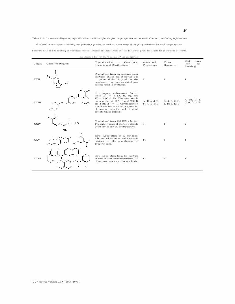
49
Table 1. 2-D chemical diagrams, crystallisation conditions for the five target systems in the sixth blind test, including information
disclosed to participants initially and following queries, as well as a summary of the full predictions for each target system.
Separate lists and re-ranking submissions are not counted in these totals but the best rank given does includes re-ranking attempts.
See Section 2.1 for more details of the categories.
Target Chemical DiagramCrystallisation Conditions,Remarks and Clarifications
AttemptedPredictions
TimesGenerated
Best Rank(Incl. Re-Ranking)
XXII
Crystallised from an acetone/watermixture; chiral-like character dueto potential flexibility of the six-membered ring, but no chiral pre-cursors used in synthesis.
21 12 1
XXIII
Five known polymorphs (A–E);three Z′ = 1 (A, B, D), twoZ′ = 2 (C & E). The most stablepolymorphs at 257 K and 293 Kare both Z′ = 1. Crystallisationconditions include slow evaporationof acetone solution and of ethylacetate:water mixture.
A, B and D:14; C & E: 3
A: 4, B: 8, C:1, D: 3, E: 0
A: 23, B: 1,C: 6, D: 2, E:–
XXIVCrystallised from 1M HCl solution.The substituents of the C=C doublebond are in the cis configuration.
8 1 2
XXV
Slow evaporation of a methanolsolution, which contained a racemicmixture of the enantiomers ofTroger’s base.
14 5 1
XXVISlow evaporation from 1:1 mixtureof hexane and dichloromethane. Nochiral precursors used in synthesis.
12 3 1
IUCr macros version 2.1.6: 2014/10/01

50
Table 2. List of members of each team/submission (* denotes corresponding author), as well as a brief summary of the generation
and ranking methods used. Please refer to Section 3 for an overview of the methods, Tables S10 and S11 of the supporting
information, and each submission’s supporting-information document for more details. Helmholtz free-energy contributions are
denoted by Fvib, polarisable continuum model is abbreviated PCM, while Monte Carlo is abbreviated MC.
Team Members Generation method Final ranking method(s)
List One (L1) List Two (L2)
1 Chadha,* Singh MC simulated anneal-ing
COMPASS (2.8) force field –
2 Cole,* McCabe, Read, Reilly,Shields
CSD analogues Fitted exp-6 potential –
3 Day*, Bygrave, Campbell,Case, Gee, McMahon, Nyman,Pulido, Taylor, Yang
Quasi-random search(Sobol’)
Atomic multipoles andexp-6
Fvib contributions (XXII& XXV), PCM ε = 3(XXIV & XXVI)
4 Dzyabchenko Grid search Empirical potential –5 van Eijck Random search Atomic charges, intra-
molecular 6-31G**energies and exp-6
–
6 Elking, Fusti-Molnar Random generation Empirical potential PBE-XDM7 de Jong, van den Ende,* de
Gelder, de Klerk, Bylsma, deWijs, Meekes, Cuppen
Random search q-GRID method Smallest critical nucleussize from kinetic MC sim-ulations
8 Lund, Pagola, Orendt, Ferraro,Facelli*
Genetic algorithm PBE-D2 PBE-D2 for all stages ofGA search
9 Obata, Goto* Grid search PBE+TS –10 Hofmann,* Kuleshova Random search Fitted potential –11 Lv, Wang, Ma* Random search optB86b-vdW –12 Curtis, Li, Schober, Cosburn,
Lohani, Vacarro, Oberhofer,Reuter, Bhattacharya,Vazquez-Mayagoitia,Ghiringhelli, Marom*
Genetic algorithm PBE+TS PBE+MBD
13 Mohamed MC simulated anneal-ing
Atomic multipoles andexp-6
–
14 Neumann, Kendrick, Leusen MC parallel temper-ing
PBE+Neumann-Perrin Includes Z′ = 2 structuresfor XXIII & XXVI
15 Sugden, Gatsiou, Vasileiadis,Adjiman,* Pantelides*
Quasi-random search(Sobol’)
Atomic multipoles andexp-6
–
16 Pickard,* Monserrat,Misquitta, Needs
Random search PBE+MBD –
17 Jankiewicz, Metz, Podeszwa,*Szalewicz
Grid search SAPT(DFT) fitted poten-tial
Alternative SAPT(DFT)fitted potential
18 S.L. Price,* Hylton, L.S. Price,Guo, Watson, Iuzzolino
Quasi-random search(Sobol’)
Atomic multipoles andexp-6
Different PCM treatments(all); Fvib for all butXXIV
19 Metz, Hylton, S.L. Price, Sza-lewicz*
Quasi-random search(Sobol’)
SAPT(DFT) fitted poten-tial
–
20 Vogt, Schneider, Metz, Tucker-man,* Szalewicz*
Random search SAPT(DFT) fitted poten-tial
–
21 Zhu,* Oganov, Masunov Evolutionaryalgorithm
vdW-DF –
22 Boese Re-ranking 10 PBE+TS & BLYP-D3 –
23 Brandenburg, Grimme Re-ranking 18 HF-3catm TPSS-D3atm
24 Metz, Guo, Szalewicz Re-ranking 18 SAPT(DFT) fitted poten-tial
–
25 Hoja, Ko, Car, DiStasio Jr.,Tkatchenko*
Re-ranking 18 PBE+MBD Fvib contributions
IUCr macros version 2.1.6: 2014/10/01

51
Table 3. Results of each submission in the sixth blind test, broken down by target system and the two lists (L1 and L2; cf. Table 2)
that could be submitted. Numbers indicate the position in the submitted list at which an an experimental structure was found, a
dash (–) indicates that the experimental structure was not found in the submitted predicted structures, and a blank entry indicates
no prediction was attempted. For re-ranking submissions, an asterisk (*) indicates that the experimental structure was not present
in the set of re-ranked structures. For XXIII C and E, only submissions that explicitly considered Z′ = 2 searches are noted in the
table. Numbers in parentheses for XXIII indicate that the heavy-atom positions were predicted but not the correct position of the H
atom of the carboxylic acid.
Team Members XXII XXIII XXIV XXV XXVI
A B C D E
L1 L2 L1 L2 L1 L2 L1 L2 L1 L2 L1 L2 L1 L2 L1 L2 L1 L2
1 Chadha & Singh – – – – –2 Cole et al. – – – – – – – –3 Day et al. 3 1 23 – – 75 75 – – – – – – –4 Dzyabchenko 1 – –5 van Eijck 4 83 20 – – 1 –6 Elking & Fusti-Molnar – – – – 78 – (73) – – – – – 8 17 van den Ende, Cuppen et al. 9 90 – – – – – – – –8 Facelli et al. – – – – – – –9 Obata & Goto 2 – 13 (66) –10 Hofmann & Kuleshova – – – – – – – – –11 Lv, Wang, Ma – –12 Marom et al. – –13 Mohamed 1 – 88 – – –14 Neumann, Kendrick, Leusen 2 26 85 2 4 – 6 11 39 – – 2 6 1 115 Pantelides, Adjiman et al. 6 70 13 – 1 –16 Pickard et al. –17 Podeszwa et al. 8 318 Price et al. 6 2 – – 1 2 85 44 – – 1 1 2 119 Szalewicz et al. –20 Tuckerman, Szalewicz et al. 421 Zhu, Oganov, Masunov 3 – – – – – 2 –
22 Boese * * * * * * * * *23 Brandenburg & Grimme – – – – 11 1 – – * * 2 – –24 Szalewicz et al. *25 Tkatchenko et al. 3 1 – – 2 5 14 2 * 1
Fig. 1. Experimental crystal structure of XXII; carbon atoms are in grey, nitrogen inblue and sulfur in yellow.
IUCr macros version 2.1.6: 2014/10/01

52
Fig. 2. Molecular conformations found in forms A–D of XXIII, overlaid onto thefenemate group of the molecule; form A is in blue, form B in grey, form C moleculeone is in red, form C molecule two in purple and form D in orange. Hydrogens areomitted for clarity.
Fig. 3. Crystal structures of form A (left) and form D (right) of XXIII, showing thesimilar layers found in the two structures. Hydrogens are omitted for clarity.
IUCr macros version 2.1.6: 2014/10/01
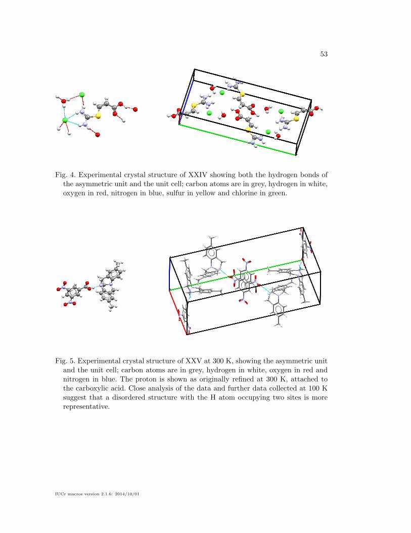
53
Fig. 4. Experimental crystal structure of XXIV showing both the hydrogen bonds ofthe asymmetric unit and the unit cell; carbon atoms are in grey, hydrogen in white,oxygen in red, nitrogen in blue, sulfur in yellow and chlorine in green.
Fig. 5. Experimental crystal structure of XXV at 300 K, showing the asymmetric unitand the unit cell; carbon atoms are in grey, hydrogen in white, oxygen in red andnitrogen in blue. The proton is shown as originally refined at 300 K, attached tothe carboxylic acid. Close analysis of the data and further data collected at 100 Ksuggest that a disordered structure with the H atom occupying two sites is morerepresentative.
IUCr macros version 2.1.6: 2014/10/01
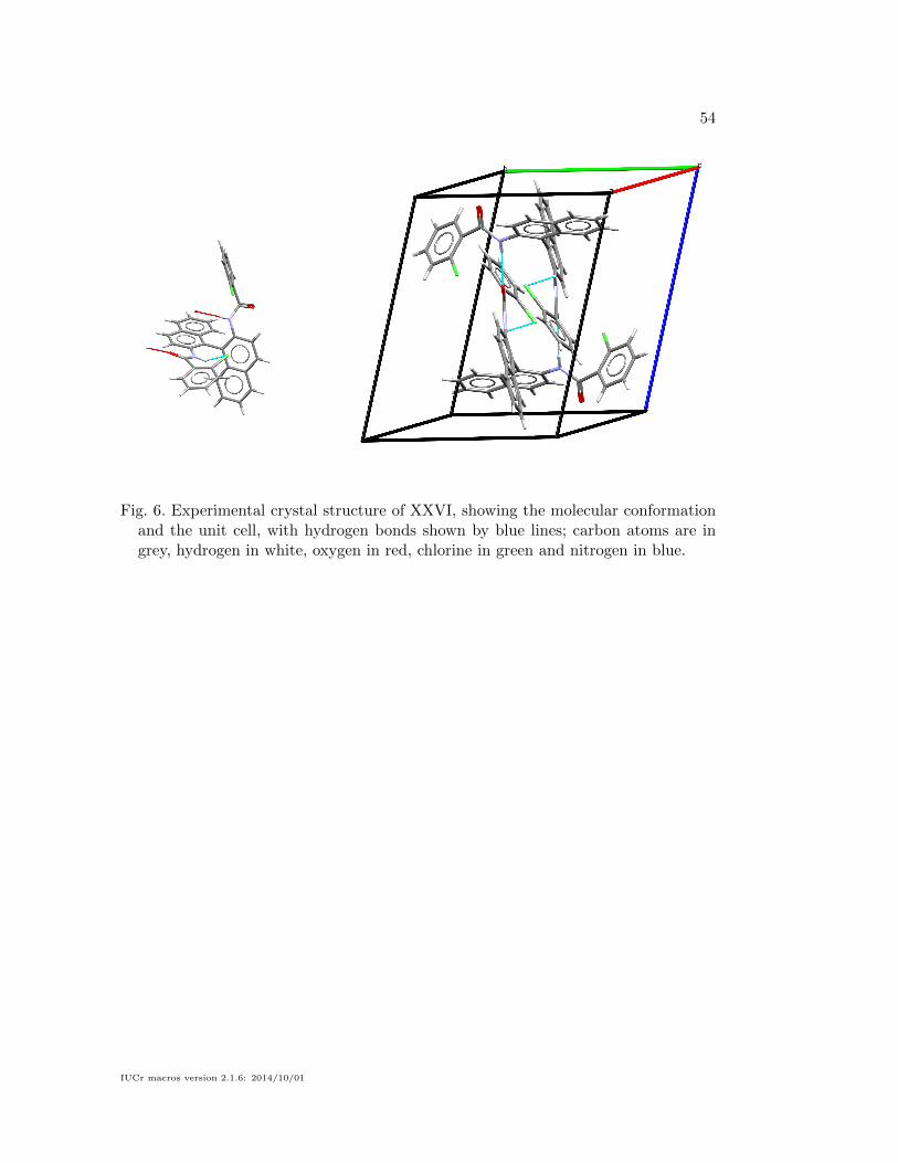
54
Fig. 6. Experimental crystal structure of XXVI, showing the molecular conformationand the unit cell, with hydrogen bonds shown by blue lines; carbon atoms are ingrey, hydrogen in white, oxygen in red, chlorine in green and nitrogen in blue.
IUCr macros version 2.1.6: 2014/10/01
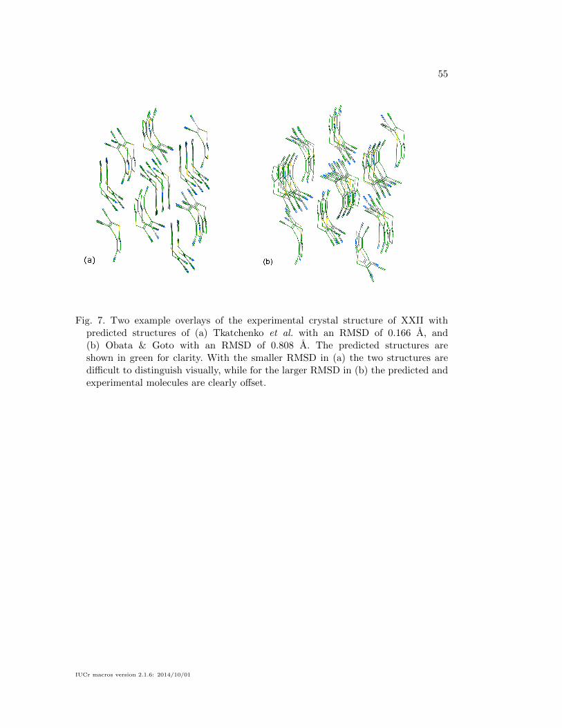
55
Fig. 7. Two example overlays of the experimental crystal structure of XXII withpredicted structures of (a) Tkatchenko et al. with an RMSD of 0.166 A, and(b) Obata & Goto with an RMSD of 0.808 A. The predicted structures areshown in green for clarity. With the smaller RMSD in (a) the two structures aredifficult to distinguish visually, while for the larger RMSD in (b) the predicted andexperimental molecules are clearly offset.
IUCr macros version 2.1.6: 2014/10/01
56
References
Adamo, C. & Barone, V. (1999). J. Chem. Phys. 110(13), 6158–6170.
Ambrosetti, A., Reilly, A. M., DiStasio Jr., R. A. & Tkatchenko, A. (2014). J. Chem. Phys.140(18), 18A508.
Anwar, J. & Zahn, D. (2011). Angew. Chem., Int. Ed. 50(9), 1996–2013.
Augelli-Szafran, C., Barvian, M., Bigge, C., Glase, S., Hachiya, S., Keily, J., Kimura, T., Lai,Y., Sakkab, A., Suto, M. et al., (2002). Method of inhibiting amyloid protein aggregationand imaging amyloid deposits. WO Patent App. PCT/US2000/015,071.
Axilrod, B. M. & Teller, E. (1943). J. Chem. Phys. 11(6), 299–300.
Baias, M., Dumez, J.-N., Svensson, P. H., Schantz, S., Day, G. M. & Emsley, L. (2013). J.Am. Chem. Soc. 135(46), 17501–17507.
Bardwell, D. A., Adjiman, C. S., Arnautova, Y. A., Bartashevich, E., Boerrigter, S. X. M.,Braun, D. E., Cruz-Cabeza, A. J., Day, G. M., Della Valle, R. G., Desiraju, G. R.,van Eijck, B. P., Facelli, J. C., Ferraro, M. B., Grillo, D., Habgood, M., Hofmann, D.W. M., Hofmann, F., Jose, K. V. J., Karamertzanis, P. G., Kazantsev, A. V., Kendrick,J., Kuleshova, L. N., Leusen, F. J. J., Maleev, A. V., Misquitta, A. J., Mohamed, S.,Needs, R. J., Neumann, M. A., Nikylov, D., Orendt, A. M., Pal, R., Pantelides, C. C.,Pickard, C. J., Price, L. S., Price, S. L., Scheraga, H. A., van de Streek, J., Thakur,T. S., Tiwari, S., Venuti, E. & Zhitkov, I. K. (2011). Acta Cryst. B, 67(6), 535–551.
Becke, A. D. (1988). Phys. Rev. A, 38, 3098–3100.
Becke, A. D. & Johnson, E. R. (2007). J. Chem. Phys. 127(15), 154108.
Bhardwaj, R. M., Price, L. S., Price, S. L., Reutzel-Edens, S. M., Miller, G. J., Oswald, I.D. H., Johnston, B. F. & Florence, A. J. (2013). Cryst. Growth Des. 13(4), 1602–1617.
Blagden, N. & Davey, R. J. (2003). Cryst. Growth Des. 3(6), 873–885.
Boerrigter, S. X. M., Josten, G. P. H., van de Streek, J., Hollander, F. F. A., Los, J., Cuppen,H. M., Bennema, P. & Meekes, H. (2004). J. Phys. Chem. A, 108(27), 5894–5902.
Born, M. & Huang, K. (1954). Dynamical theory of crystal lattices. International series ofmonographs on physics. Clarendon Press.
Brandenburg, J. G. & Grimme, S. (2014). Topics in Current Chemistry, vol. 345, chap.Dispersion Corrected Hartree-Fock and Density Functional Theory for Organic CrystalStructure Prediction, pp. 1–23. Springer Berlin Heidelberg.
Bruno, I. J., Cole, J. C., Kessler, M., Luo, J., Motherwell, W. D. S., Purkis, L. H., Smith,B. R., Taylor, R., Cooper, R. I., Harris, S. E. & Orpen, A. G. (2004). J. Chem. Inf.Comput. Sci. 44(6), 2133–2144.
Bygrave, P. J., Allan, N. L. & Manby, F. R. (2012). J. Chem. Phys. 137(16), 164102.
Case, D. H., Campbell, J. E., Bygrave, P. J. & Day, G. M. (2016). J. Chem. Theory Comput.12(2), 910–924.
Chisholm, J. A. & Motherwell, S. (2005). J. Appl. Cryst. 38(1), 228–231.
Cohen, A. J., Mori-Sanchez, P. & Yang, W. (2008). Science, 321(5890), 792–794.
Cohen, A. J., Mori-Sanchez, P. & Yang, W. (2012). Chem. Rev. 112(1), 289–320.
Cruz-Cabeza, A. J. & Bernstein, J. (2014). Chem. Rev. 114(4), 2170–2191.
Cruz-Cabeza, A. J., Liebeschuetz, J. W. & Allen, F. H. (2012). CrystEngComm, 14, 6797–6811.
Day, G. M., Cooper, T. G., Cruz-Cabeza, A. J., Hejczyk, K. E., Ammon, H. L., Boerrigter, S.X. M., Tan, J. S., Della Valle, R. G., Venuti, E., Jose, J., Gadre, S. R., Desiraju, G. R.,Thakur, T. S., van Eijck, B. P., Facelli, J. C., Bazterra, V. E., Ferraro, M. B., Hofmann,D. W. M., Neumann, M. A., Leusen, F. J. J., Kendrick, J., Price, S. L., Misquitta, A. J.,Karamertzanis, P. G., Welch, G. W. A., Scheraga, H. A., Arnautova, Y. A., Schmidt,M. U., van de Streek, J., Wolf, A. K. & Schweizer, B. (2009). Acta. Cryst. B, 65(2),107–125.
IUCr macros version 2.1.6: 2014/10/01
57
Day, G. M., Motherwell, W. D. S., Ammon, H. L., Boerrigter, S. X. M., Della Valle, R. G.,Venuti, E., Dzyabchenko, A., Dunitz, J. D., Schweizer, B., van Eijck, B. P., Erk, P.,Facelli, J. C., Bazterra, V. E., Ferraro, M. B., Hofmann, D. W. M., Leusen, F. J. J.,Liang, C., Pantelides, C. C., Karamertzanis, P. G., Price, S. L., Lewis, T. C., Nowell,H., Torrisi, A., Scheraga, H. A., Arnautova, Y. A., Schmidt, M. U. & Verwer, P. (2005).Acta Cryst. B, 61(5), 511–527.
Deij, M. A., ter Horst, J. H., Meekes, H., Jansens, P. & Vlieg, E. (2007). J. Phys. Chem. B,111(7), 1523–1530.
Del Ben, M., Hutter, J. & VandeVondele, J. (2012). J. Chem. Theory Comput. 8(11), 4177–4188.
Dion, M., Rydberg, H., Schroder, E., Langreth, D. C. & Lundqvist, B. I. (2004). Phys. Rev.Lett. 92, 246401.
Dove, M. (1993). Introduction to Lattice Dynamics. Cambridge Topics in Mineral Physics andChemistry. Cambridge University Press.
Eddleston, M. D., Hejczyk, K. E., Bithell, E. G., Day, G. M. & Jones, W. (2013). Chem. Eur.J. 19(24), 7874–7882.
van Eijck, B. P., Mooij, W. T. M. & Kroon, J. (2001a). J. Comput. Chem. 22(8), 805–815.
van Eijck, B. P., Mooij, W. T. M. & Kroon, J. (2001b). J. Phys. Chem. B, 105(43), 10573–10578.
Etter, M. C., MacDonald, J. C. & Bernstein, J. (1990). Acta. Cryst. B, 46(2), 256–262.
Foxman, B. M. (2016). CSD Communication: CCDC 1447530. DOI: 10.5517/cc1kl8jy.
Galek, P. T. A., Allen, F. H., Fabian, L. & Feeder, N. (2009). CrystEngComm, 11, 2634–2639.
Galek, P. T. A., Fabian, L., Motherwell, W. D. S., Allen, F. H. & Feeder, N. (2007). ActaCryst. B, 63(5), 768–782.
de Gelder, R., Wehrens, R. & Hageman, J. A. (2001). J. Comput. Chem. 22(3), 273–289.
Goto, H. & Osawa, E. (1989). J. Am. Chem. Soc. 111(24), 8950–8951.
Goto, H. & Osawa, E. (1993). J. Chem. Soc., Perkin Trans. 2, pp. 187–198.
Grimme, S. (2006). J. Comput. Chem. 27(15), 1787–1799.
Grimme, S. (2014). J. Chem. Theory Comput. 10(ja), 4497–4514.
Grimme, S., Antony, J., Ehrlich, S. & Krieg, H. (2010). J. Chem. Phys. 132(15), 154104.
Groom, C. R. & Allen, F. H. (2014). Angew. Chem., Int. Ed. 53(3), 662–671.
Groom, C. R. & Reilly, A. M. (2014). Acta Cryst. B, 70(4), 776–777.
Habgood, M., Grau-Crespo, R. & Price, S. L. (2011). Phys. Chem. Chem. Phys. 13, 9590–9600.
Habgood, M., Sugden, I., Kazantsev, A. V., Adjiman, C. S. & Pantelides, C. C. (2015). J.Chem. Theory Comput. 11(4), 1957–1969.
Hofmann, D. W. M. & Kuleshova, L. (2005). J. Appl. Cryst. 38(6), 861–866.
Horton, P. N. & Gossel, M. C. (2016). CSD Communication: CCDC 1451239. DOI:10.5517/cc1kq45l.
Ismail, S. Z., Anderton, C. L., Copley, R. C. B., Price, L. S. & Price, S. L. (2013). Cryst.Growth Des. 13(6), 2396–2406.
Kazantsev, A. V., Karamertzanis, P. G., Adjiman, C. S. & Pantelides, C. C. (2011). J. Chem.Theory Comput. 7(6), 1998–2016.
Klimes, J., Bowler, D. R. & Michaelides, A. (2011). Phys. Rev. B, 83, 195131.
Klimes, J. & Michaelides, A. (2012). J. Chem. Phys. 137(12), 120901.
Kolossvary, I., & Guida, W. C. (1996). J. Am. Chem. Soc. 118(21), 5011–5019.
Kronik, L. & Tkatchenko, A. (2014). Acc. Chem. Res. 47, 3208–3216.
Kuleshova, L., Hofmann, D. & Boese, R. (2013). Chem. Phys. Lett. 564, 26–32.
Lee, C., Yang, W. & Parr, R. G. (1988). Phys. Rev. B, 37, 785–789.
IUCr macros version 2.1.6: 2014/10/01
58
Lommerse, J. P. M., Motherwell, W. D. S., Ammon, H. L., Dunitz, J. D., Gavezzotti, A.,Hofmann, D. W. M., Leusen, F. J. J., Mooij, W. T. M., Price, S. L., Schweizer, B.,Schmidt, M. U., van Eijck, B. P., Verwer, P. & Williams, D. E. (2000). Acta Cryst. B,56(4), 697–714.
Lund, A. M., Pagola, G. I., Orendt, A. M., Ferraro, M. B. & Facelli, J. C. (2015). Chem. Phys.Lett. 626(0), 20–24.
Macrae, C. F., Bruno, I. J., Chisholm, J. A., Edgington, P. R., McCabe, P., Pidcock, E.,Rodriguez-Monge, L., Taylor, R., van de Streek, J. & Wood, P. A. (2008). J. Appl.Cryst. 41(2), 466–470.
Marom, N., DiStasio Jr., R. A., Atalla, V., Levchenko, S., Reilly, A. M., Chelikowsky, J. R.,Leiserowitz, L. & Tkatchenko, A. (2013). Angew. Chem., Int. Ed. 52, 6629–6632.
Misquitta, A. J., Podeszwa, R., Jeziorski, B. & Szalewicz, K. (2005). J. Chem. Phys. 123(21),214103.
Moellmann, J. & Grimme, S. (2014). J. Phys. Chem. C, 118(14), 7615–7621.
Mohamed, S., Tocher, D. A. & Price, S. L. (2011). Int. J. Pharm. 418(2), 187 – 198.
Monserrat, B., Drummond, N. D. & Needs, R. J. (2013). Phys. Rev. B, 87, 144302.
Motherwell, W. D. S., Ammon, H. L., Dunitz, J. D., Dzyabchenko, A., Erk, P., Gavezzotti, A.,Hofmann, D. W. M., Leusen, F. J. J., Lommerse, J. P. M., Mooij, W. T. M., Price, S. L.,Scheraga, H., Schweizer, B., Schmidt, M. U., van Eijck, B. P., Verwer, P. & Williams,D. E. (2002). Acta Cryst. B, 58(4), 647–661.
Neumann, M. A. (2008). J. Phys. Chem. B, 112(32), 9810–9829.
Neumann, M. A., Leusen, F. J. J. & Kendrick, J. (2008). Angew. Chem., Int. Ed. 47(13),2427–2430.
Neumann, M. A. & Perrin, M.-A. (2005). J. Phys. Chem. B, 109(32), 15531–15541.
Neumann, M. A., van de Streek, J., Fabbiani, F. P. A., Hidber, P. & Grassmann, O. (2015).Nat. Commun. 6, 7793.
Nyman, J. & Day, G. M. (2015). CrystEngComm, 17, 5154–5165.
Obata, S. & Goto, H. (2015). AIP Conf. Proc. 1649(1), 130–134.
Oganov, A. R. & Valle, M. (2009). J. Chem. Phys. 130(10), 104504.
Ong, S. P., Richards, W. D., Jain, A., Hautier, G., Kocher, M., Cholia, S., Gunter, D., Chevrier,V. L., Persson, K. A. & Ceder, G. (2013). Comp. Mat. Sci. 68, 314–319.
Otero-de-la-Roza, A. & Johnson, E. R. (2012). J. Chem. Phys. 136(17), 174109.
Perdew, J. P., Burke, K. & Ernzerhof, M. (1996). Phys. Rev. Lett. 77, 3865–3868.
Piana, S., Reyhani, M. & Gale, J. D. (2005). Nature, 438(7064), 70–73.
Price, S. L. (2009). Acc. Chem. Res. 42(1), 117–126.
Price, S. L. (2013). Acta Cryst. B, 69(4), 313–328.
Price, S. L. (2014). Chem. Soc. Rev. 43, 2098–2111.
Price, S. L., Leslie, M., Welch, G. W. A., Habgood, M., Price, L. S., Karamertzanis, P. G. &Day, G. M. (2010). Phys. Chem. Chem. Phys. 12(30), 8478–8490.
Pyzer-Knapp, E. O., Thompson, H. P. G., Schiffmann, F., Jelfs, K. E., Chong, S. Y., Little,M. A., Cooper, A. I. & Day, G. M. (2014). Chem. Sci. 5, 2235–2245.
Reilly, A. M. & Tkatchenko, A. (2013). J. Chem. Phys. 139(2), 024705.
Reilly, A. M. & Tkatchenko, A. (2014). Phys. Rev. Lett. 113, 055701.
Reilly, A. M. & Tkatchenko, A. (2015). Chem. Sci. 6, 3289–3301.
Risthaus, T. & Grimme, S. (2013). J. Chem. Theory Comput. 9(0), 1580–1591.
Salvalaglio, M., Vetter, T., Giberti, F., Mazzotti, M. & Parrinello, M. (2012). J. Am. Chem.Soc. 134(41), 17221–17233.
Samas, B. (2015). Private Communication.
Samas, B. (2016a). CSD Communication: CCDC 1447522. DOI: 10.5517/cc1kl88p.
Samas, B. (2016b). CSD Communication: CCDC 1447523. DOI: 10.5517/cc1kl89q.
IUCr macros version 2.1.6: 2014/10/01
59
Samas, B. (2016c). CSD Communication: CCDC 1447524. DOI: 10.5517/cc1kl8br.
Samas, B. (2016d). CSD Communication: CCDC 1447525. DOI: 10.5517/cc1kl8cs.
Samas, B. (2016e). CSD Communication: CCDC 1447526. DOI: 10.5517/cc1kl8dt.
Sharp, E. et al. (2016). Manuscript in preparation.
Simons, L. J., Caprathe, B. W., Callahan, M., Graham, J. M., Kimura, T., Lai, Y., LeVineIII, H., Lipinski, W., Sakkab, A. T., Tasaki, Y., Walker, L. C., Yasunaga, T., Ye, Y.,Zhuang, N. & Augelli-Szafran, C. E. (2009). Bioorg. Med. Chem. Lett. 19(3), 654–657.
Sobol’, I. (1967). Comp. Math. Math. Phys. 7(4), 86 – 112.
Spek, A. L. (2009). Acta Cryst. D, 65(2), 148–155.
Spglib, (2015). http://spglib.sourceforge.net/.
Stokes, H. T. & Hatch, D. M. (2005). J. Appl. Cryst. 38(1), 237–238.
Stone, A. J. (2005). J. Chem. Theory Comput. 1(6), 1128–1132.
Stone, A. J. (2013). The Theory of Intermolecular Forces. Oxford University Press, UK, 2nded.
Tao, J., Perdew, J. P., Staroverov, V. N. & Scuseria, G. E. (2003). Phys. Rev. Lett. 91, 146401.
Thompson, H. P. G. & Day, G. M. (2014). Chem. Sci. 5, 3173–3182.
Threlfall, T. (2003). Org. Process Res. Dev. 7(6), 1017–1027.
Tkatchenko, A., DiStasio Jr., R. A., Car, R. & Scheffler, M. (2012). Phys. Rev. Lett. 108,236402.
Tkatchenko, A. & Scheffler, M. (2009). Phys. Rev. Lett. 102, 073005.
Valle, R. G. D., Venuti, E., Brillante, A. & Girlando, A. (2008). J. Phys. Chem. A, 112(29),6715–6722.
Verwer, P. & Leusen, F. J. J. (1998). Computer Simulation to Predict Possible CrystalPolymorphs, vol. 12 of Reviews in Computational Chemistry, pp. 327–365. John Wiley& Sons, Inc.
Wang, Y., Lv, J., Zhu, L. & Ma, Y. (2012). Comput. Phys. Commun. 183(10), 2063–2070.
Wen, S. & Beran, G. J. O. (2011). J. Chem. Theory Comput. 7(11), 3733–3742.
Wen, S. & Beran, G. J. O. (2012a). Cryst. Growth Des. 12(5), 2169–2172.
Wen, S. & Beran, G. J. O. (2012b). J. Chem. Theory Comput. 8(8), 2698–2705.
Wheeler, K. A. & Breen, M. E. (2016a). CSD Communication: CCDC 1447527.DOI:10.5517/cc1kl8fv.
Wheeler, K. A. & Breen, M. E. (2016b). CSD Communication: CCDC 1447528. DOI:10.5517/cc1kl8gw.
Wheeler, K. A. & Hopkins, G. W. (2016). CSD Communication: CCDC 1447529. DOI:10.5517/cc1kl8hx.
Yang, J., Hu, W., Usvyat, D., Matthews, D., Schutz, M. & Chan, G. K.-L. (2014). Science,345, 640–643.
Yu, T.-Q. & Tuckerman, M. E. (2011). Phys. Rev. Lett. 107, 015701.
Zhu, Q., Oganov, A. R., Glass, C. W. & Stokes, H. T. (2012). Acta Cryst. B, 68(3), 215–226.
Synopsis
The results of the sixth blind test of organic crystal-structure prediction methods are presentedand discussed, highlighting progress for salts, hydrates and bulky flexible molecules, as wellas on-going challenges.
IUCr macros version 2.1.6: 2014/10/01


![Blackness Primary School184140]Newslette… · Scottish Saying “A nod’s as guid as a wink tae a blind horse”, meaning, explain yourself properly, and make your meaning crystal](https://img.dokumen.tips/doc/110x75/5f17a5bb00db2a1eff6419e7/blackness-primary-school-184140newslette-scottish-saying-aoea-nodas-as-guid.jpg)