Embed Size (px)
Citation preview

The Journal of Neuroscience, August 1995, 15(8): 5478-5485
Reorganization of Ascending 5HT Axon Projections in Animals Previously Exposed to the Recreational Drug (+)3,4- Methylenedioxymethamphetamine (MDMA, “Ecstasy”)
C. Fischer,’ G. Hatzidimitriou,’ J. WIOS,~ J. Katz,’ and G. Ricaurtel
‘Department of Neurology, Johns Hopkins Medical Institutions, and 2NIDA Addiction Research Center, Baltimore, Maryland 21224
The recreational drug (rt)3,4-methylenedioxymethamphe- tamine (MDMA, “ecstasy”) is a methamphetamine deriva- tive that selectively destroys central 5-HT axons and axon terminals in animals and, possibly, humans. The fate of 5-HT neurons following MDMA injury is uncertain. In par- ticular, while it is known that central 5-HT axons can un- dergo regenerative sprouting after MDMA injury, it has not been determined whether they reestablish the original in- nervation pattern. To address this question, the present studies examined 5-HT innervation patterns in animals le- sioned with MDMA 12-18 months previously. Both rodents (rats) and nonhuman primates (squirrel monkeys) were ex- amined, since there is indication that serotonergic recov- ery after MDMA injury may be species dependent. 5-HT axon projections were studied neurochemically, autoradi- ographically and immunocytochemically. In both rodents and nonhuman primates previously lesioned with MDMA, substantial serotonergic axonal sprouting was observed. However, in a few rats and in most squirrel monkeys, the reinnervation pattern was highly abnormal: distant targets (e.g., dorsal neocortex) remained denervated, while some proximal targets (e.g., amygdala, hypothalamus) were rein- nervated or hyperinnervated. Although the specific deter- minants of axonal recovery after MDMA injury remain to be identified, it appears that axons which initially sustain more severe damage, are longer, or are more highly arborized have low probability of recovering. The observation that some brain regions remain denervated, while others are reinnervated or hyperinnervated suggests that, under some circumstances, MDMA injury can lead to a lasting reorganization of ascending 5-HT axon projections. Such lasting changes in brain innervation, documented here in MDMA-treated animals, may have implications for humans using MDMA recreationally.
[Key words: 5-HT, neurotoxicity, amphetamines, sprout- ing, regeneration, reinnervation]
Despite multiple regulatory efforts (Anonymous, 1985; Lawn, 1986, 1988), the synthetic amphetamine derivative (k-)3,4-meth- ylenedioxymethamphetamine (MDMA, “ecstasy”) has gained
Received Dec. 28, 1994; revised Mar. 22, 1995; accepted Mar. 28, 1995. We thank Lynda Roggio for technical assistance. This work was supported
by PHS Grants DA05707 and DA06275. Correspondence should be addressed to Dr. George A. Ricaurte, Department
of Neurology, Johns Hopkins University School of Medicine, 5501 Bayview Drive, Baltimore, MD 21224. Copyright 0 1995 Society for Neuroscience 0270-6474/95/155476-10$05.00/O
status as a popular recreational “designer” drug in various parts of the world (Haislip, 1989; Abbot and Concar, 1992; Solowij and Hall, 1992; Cregg and Tracey, 1993). In the United States, where recreational use of MDMA first became apparent in 1985 (Baum, 1985; Siegel, 1986), recent reports indicate that MDMA use is increasing (Cuomo and Dyment, 1994; McDowell and Kleber, 1994; Newmeyer, 1994). In Great Britain, increasing use of MDMA has also been noted (Henry, 1992; Randall, 1992), and one survey indicates that illicit use of MDMA and related drugs in the United Kingdom ranks second only to the use of marijuana (Anonymous, 1992).
This apparent widespread increase in recreational MDMA use is of concern because there is extensive preclinical evidence that MDMA is a potent 5-HT neurotoxin (for reviews, see McKenna and Peroutka, 1990; Steele et al. 1994). Animals given MDMA show large depletions of brain 5-HT, as well as marked reduc- tions in the activity of tryptophan hydroxylase, the concentration of 5hydroxyindoleacetic acid (5-HIAA), and the number of 5-HT transporter sites (Stone et al., 1986; Battaglia et al., 1987; Commins et al., 1987; Schmidt, 1987; Ricaurte et al., 1988b; Slikker et al., 1988; Insel et al., 1989; Kleven et al., 1989; De Souza et al., 1990). Correlative anatomic studies indicate that loss of presynaptic 5-HT axonal markers in MDMA-treated an- imals is related to damage of 5-HT axons and axon terminals (Battaglia et al., 1987; Commins et al., 1987; O’Hearn et al., 1988; Wilson et al., 1989; Molliver et al., 1990; Mamounas and Molliver, 1991). Since doses of MDMA that produce neurotoxic effects in nonhuman primates (Ricaurte et al., 1988) closely ap- proach those typically used by humans (Seymour, 1986; Doblin, 1989), increasing recreational use of MDMA may pose a sig- nificant public health problem.
Although the neurotoxic potential of MDMA in animals is now well established, the fate of 5-HT neurons after MDMA injury remains uncertain. In particular, while it is known that central 5-HT neurons can undergo regenerative sprouting after MDMA injury (Battaglia et al., 1988; Molliver et al., 1990; Scanzello et al., 1993), it has not been determined whether they reestablish a normal innervation pattern. Further, there is indi- cation that axonal sprouting after MDMA injury may be species dependent, occurring in rodents (Battaglia et al., 1988; Molliver et al., 1990; Scanzello et al., 1993) but not nonhuman primates (Insel et al., 1989; Ricaurte et al., 1992). In light of these un- certainties, the present studies were undertaken to characterize the organization of ascending 5-HT axon projections in rodents and nonhuman primates lesioned with MDMA 12-18 months previously. The purpose of these studies was to determine if
The Journal of Neuroscience, August 1995, 75(8) 5477
80
g 80
5 0
s 40
20
0
5-HT s-HIAA [3~Citalopram binding
250
200
5 150 b 5 0
s 100
50
0
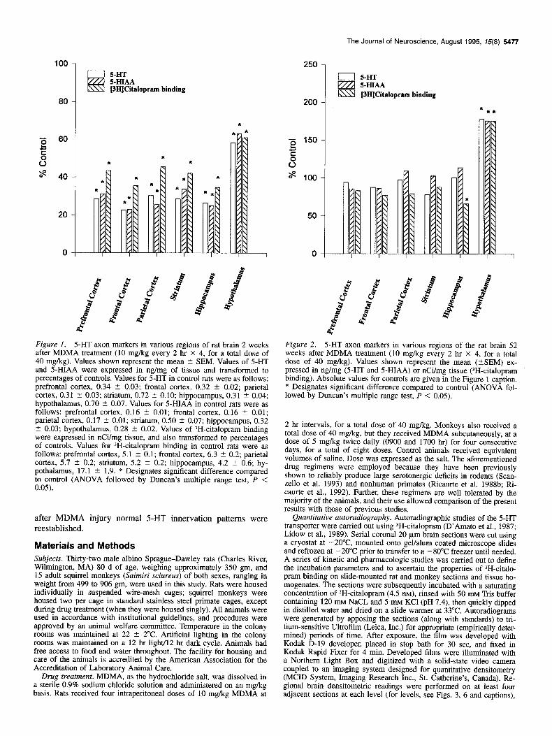
Figure I. 5-HT axon markers in various regions of rat brain 2 weeks after MDMA treatment (10 mg/kg every 2 hr X 4, for a total dose of 40 mg/kg). Values shown represent the mean ? SEM. Values of 5-HT and 5-HIAA were expressed in ng/mg of tissue and transformed to percentages of controls. Values for 5-HT in control rats were as follows: prefrontal cortex, 0.34 t 0.03; frontal cortex, 0.32 + 0.02; parietal cortex, 0.31 ? 0.03; striatum, 0.72 2 0.10; hippocampus, 0.31 t 0.04; hypothalamus, 0.70 t 0.07. Values for 5-HIAA in control rats were as follows: prefrontal cortex, 0.16 t 0.01; frontal cortex, 0.16 2 0.01; parietal cortex, 0.17 IT 0.01; striatum, 0.50 t 0.07; hippocampus, 0.32 2 0.03; hypothalamus, 0.28 ? 0.02. Values of 3H-cita10pram binding were expressed in nCi/mg tissue, and also transformed to percentages of controls. Values for 3H-citalopram binding in control rats were as follows: prefrontal cortex, 5.1 2 0.1; frontal cortex, 6.3 ? 0.2; parietal cortex, 5.7 ? 0.2; striatum, 5.2 + 0.2; hippocampus, 4.2 2 0.6; hy- pothalamus, 17.1 + 1.9. * Designates significant difference compared to control (ANOVA followed by Duncan’s multiple range test, P < 0.05).
after MDMA injury normal .5-HT innervation patterns were reestablished.
Materials and Methods
Subjects. Thirty-two male albino Sprague-Dawley rats (Charles River, Wilmington, MA) 80 d of age, weighing approximately 350 gm, and 15 adult squirrel monkeys (Saimiri sciureus) of both sexes, ranging in weight from 499 to 906 gm, were used in this study. Rats were housed individually in suspended wire-mesh cages; squirrel monkeys were housed two per cage in standard stainless steel primate cages, except during drug treatment (when they were housed singly). All animals were used in accordance with institutional guidelines, and procedures were approved by an animal welfare committee. Temperature in the colony rooms was maintained at 22 + 2°C. Artificial lighting in the colony rooms was maintained on a 12 hr light/l2 hr dark cycle. Animals had free access to food and water throughout. The facility for housing and care of the animals is accredited by the American Association for the Accreditation of Laboratory Animal Care.
Drug treatment. MDMA, as the hydrochloride salt, was dissolved in a sterile 0.9% sodium chloride solution and administered on an mg/lcg basis. Rats received four intraperitoneal doses of 10 mg/kg MDMA at
0 5-HT pm 5-HIAA m [3H]Citalopram binding
Figure 2. 5-HT axon markers in various regions of the rat brain 52 weeks after MDMA treatment (10 mg/kg every 2 hr X 4, for a total dose of 40 mg/kg). Values shown represent the mean (+SEM) ex- pressed in ng/mg (5-HT and 5-HIAA) or nCi/mg tissue (3H-citalopram binding). Absolute values for controls are given in the Figure 1 caption. * Designates significant difference compared to control (ANOVA fol- lowed by Duncan’s multiple range test, P < 0.05).
2 hr intervals, for a total dose of 40 mg/kg. Monkeys also received a total dose of 40 mg/kg, but they received MDMA subcutaneously, at a dose of 5 mg/kg twice daily (0900 and 1700 hr) for four consecutive days, for a total of eight doses. Control animals received equivalent volumes of saline. Dose was expressed as the salt. The aforementioned drug regimens were employed because they have been previously shown to reliably produce large serotonergic deficits in rodents (Scan- zello et al. 1993) and nonhuman primates (Ricaurte et al. 1988b; Ri- caurte et al., 1992). Further, these regimens are well tolerated by the majority of the animals, and their use allowed comparison of the present results with those of previous studies.
Quantitative autoradiography. Autoradiographic studies of the 5-HT transporter were carried out using 3H-citalopram (D’Amato et al., 1987; Lidow et al., 1989). Serial coronal 20 urn brain sections were cut using a cryostat at -2O”C, mounted onto gel/alum coated microscope slides and refrozen at - 20°C prior to transfer to a - 80°C freezer until needed. A series of kinetic and pharmacologic studies was carried out to define the incubation parameters and to ascertain the properties of 3H-citalo- pram binding on slide-mounted rat and monkey sections and tissue ho- mogenates. The sections were subsequently incubated with a saturating concentration of 3H-citalopram (4.5 nM), rinsed with 50 mM Tris buffer containing 120 mu NaCL and 5 mu KC1 (pH 7.4), then quickly dipped in distilled water and dried on a slide warmer at 33°C. Autoradiograms were generated by apposing the sections (along with standards) to tri- tium-sensitive Ultrofilm (Leica, Inc.) for appropriate (empirically deter- mined) periods of time. After exposure, the film was developed with Kodak D-19 developer, placed in stop bath for 30 set, and fixed in Kodak Rapid Fixer for 4 min. Developed films were illuminated with a Northern Light Box and digitized with a solid-state video camera coupled to an imaging system designed for quantitative densitometry (MCID System, Imaging Research Inc., St. Catherine’s, Canada). Re- gional brain densitometric readings were performed on at least four adjacent sections at each level (for levels, see Figs. 3, 6 and captions),
5478 Fischer et al. * Reorganization of 5-HT Axons after MDMA Injury

The Journal of Neuroscience, August 1995, 75(8) 5479
and a calibration curve was obtained from standards processed in par- allel. Standards containing the same isotope as that used to label the ligand were used, and optical density values outside the linear range were discarded. Specific binding was defined as the difference between total binding and nonspecific binding, with nonspecific binding being defined as the amount of binding taking place in the presence of 1uM paroxetine hydrochloride. Data were expressed in nCi/mg tissue. Scat- chard transformation and calculation of KD and B,,, values were per- formed using EBDA and LIGAND software (Biosoft).
Indole determinations. Regional brain levels of 5-HT and 5-HIAA were measured by means of high performance liquid chromatography coupled with electrochemical detection (HPLC-EC), as described pre- viously (Ricaurte et al., 1992).
Immunocytochemical studies. Anatomic studies of ascending 5-HT- containing axon projections were performed using a rabbit antiserum directed at 5-HT, employing the method of Wilson et al. (1989), with minor modification (Scanzello et al., 1993). Briefly, l-2 hr prior to sacrifice, animals received 10 mg/kg of the monoamine oxidase inhib- itor trans-2-phenylcyclopropylamine (Lp.). Under deep chloral hydrate anesthesia (400 mg/kg, i.p.j,-the animals were perfused by an intracar- disc route. After an initial rinse with ice-cold PBS (850 ml uerfused at a rate of approximately 150 ml/min), perfusion was continuei using 4% paraformaldehyde and 0.1% glutaraldehyde (pH 7.4) (3600 ml perfused at a rate of approximately 150 ml/min). Tissue blocks were placed in buffered 4% paraformaldehyde for 4-6 hr and then in 10% dimethyl- sulfoxide in PBS overnight. Frozen sections (30 km) were incubated in an anti-5-HT antisera diluted 1:14,000 in PBS with 0.2% Triton X-100 and 1% normal serum at 4°C for 3 d. The antibody was visualized with the Vectastain ABC-peroxidase method (Vector Laboratories, Inc., Bur- lingame, CA), and staining was enhanced with the osmiophilic reaction sequence of Gerfen (1985).
Statistical analysis. One-way analysis of variance (ANOVA) was used to detect significant overall effects. Post hoc Duncan’s multiple range tests were performed to assess the significance of differences between group means. Correlations were assessed by Pearson’s product moment. Differences were considered significant when a P value less than 0.05 was obtained. All tests were two-tailed.
Drugs and chemicals. 5-Hydroxytryptamine (5-HT, 5-HT) creatinine sulfate complex and 5-hydroxyindole-3-acetic acid (dicyclohexylam- monium salt) were purchased from Sigma Chemical Co. (St. Louis, MO). Perchloric acid was purchased from J. T Baker Inc. (Phillipsburg, NJ). 3H-Citalopram (specific activity, 38.1 Ci/mmol) was purchased from New England Nuclear (Boston, MA). (?)3,4-Methylenedioxy- methamphetamine hydrochloride was obtained from the National Insti- tute on Drug Abuse. The 5-HT antibody was purchased from the Incstar Corp., Stillwater, MN.
Results
Rodents. Two weeks after MDMA treatment, serotonergic ax- onal markers were markedly reduced in multiple regions of the rat brain. Reductions were most severe in the cerebral cortex, striatum, and hippocampus; serotonergic deficits were least pro- nounced in the hypothalamus (Fig. 1). In keeping with previous results (Battaglia et al. 1988; Molliver et al., 1990; Scanzello et al., 1993), rats examined 52 weeks after MDMA treatment, as a group, showed evidence of serotonergic recovery (Fig. 2). Re- covery was complete or nearly complete by 52 weeks for all markers in all brain regions examined (no intermediate time points were examined). The only exception was ‘H-citalopram binding in the hippocampus, which remained partially reduced. In the hypothalamus, all markers were above control levels 52 weeks after MDMA treatment (Fig. 2).
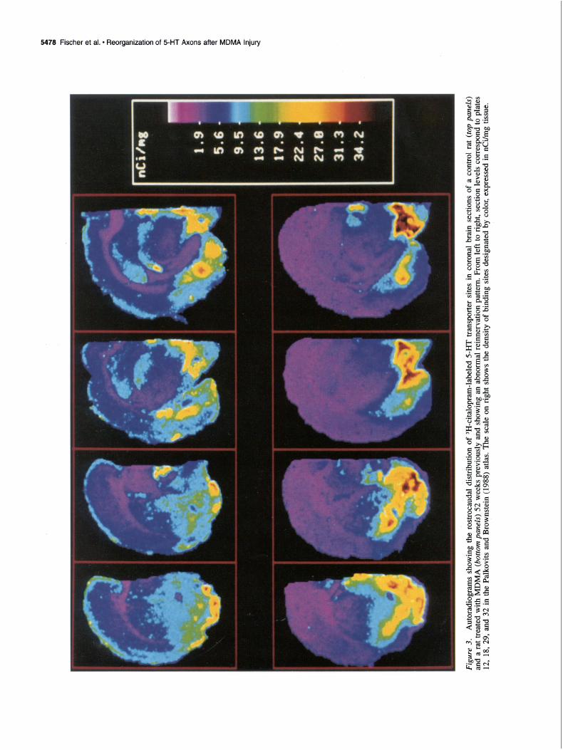
Within the group of rats examined 52 weeks after MDMA treatment, a few (3 of 16) rats were found that had large, per- sistent reductions in serotonergic axonal markers in multiple regions of the forebrain (e.g., dorsal neocortex, striatum, hip- pocampus) but an increase in serotonergic markers in the basal forebrain (Fig. 3). For example, as can be seen in the figure, these animals had marked decreases in 3H-citalopram binding in
Figure 4. Density of 3H-citalopram-labeled 5-HT uptake sites in the dorsal neocortex and lateral hypothalamus of five rats treated with MDMA 52 weeks previously. Note that the total number of sites is relatively constant across animals. Also note the inverse relation be- tween the number of sites in the dorsal neocortex and hypothalamus.
the dorsal neocortex, but increases in the hypothalamus and oth- er regions of the basal forebrain.
Further analysis of the autoradiographic data revealed a high correlation between the magnitude of decreases in binding sites in the dorsal neocortex and the magnitude of increases in bind- ing sites in the hypothalamus (r = 0.94) (Fig. 4). Moreover, while there was a redistribution of 3H-citalopram binding sites in these animals, the density of transporter sites at three different coronal brain levels (plates 15, 25, and 31 of the rat brain atlas by Palkovits and Brownstein, 1988) was the same as in controls (control, 9.25 5 1.46 fmol/mg; MDMA, 9.41 + 1.85 fmol/mg).
Changes in 5-HT, 5-HIAA, and lH-citalopram binding were associated with changes in the density of 5-HT-immunoreactive axons (Fig. 5). Of 16 rats examined 52 weeks after MDMA exposure, 3 showed an altered pattern of distribution of 5-HT axon markers such as that depicted in Figures 3 and 5; the others had patterns that approached, to varying degrees, those of con- trol animals.
Nonhuman primates. To explore the species generality of these findings, similar studies were carried out in squirrel mon- keys treated with MDMA (5 mg/kg, s.c., twice daily for 4 d) 2 and 72 weeks previously. Two weeks after MDMA treatment, profound decrements in 5-HT axonal markers were evident in all regions of the monkey brain examined (see below). Seventy- two weeks after MDMA treatment, autoradiographic studies re- vealed persistent and equally profound reductions in 3H-citalo- pram binding in many regions of the monkey forebrain, includ- ing the neocortex, hippocampus, caudate nucleus, putamen, and many nuclei of the thalamus (Fig. 6). However, as is evident in the figure, there were marked increases in 3H-citalopram binding in the hypothalamus and other basal forebrain regions.
Immunocytochemical studies revealed a close association be- tween changes in the density of 5-HT-immunoreactive axons and changes in the density of 3H-citalopram binding sites. For ex- ample, the marked decrease in 3H-citalopram binding in the dor- sal neocortex (Fig. 6) was associated with a marked decrease in the density of 5-HT-immunoreactive axons (Fig. 7); similarly, the increase in 3H-citalopram binding in the hypothalamus (Fig. 6) was associated with an increase in the density of 5-HT-im- munoreactive axons (Fig. 7). Of 17 MDMA-treated monkeys

5480 Fischer et al. l Reorganization of 5-HT Axons after MDMA Injury
Figure 5. SHT-immunoreactive axons in the dorsal neocortex and hypothalamus of a control rat (A, C) and a rat treated with MDMA 52 weeks previously (C, D). Note the reduction in axon density in the cerebral cortex (B), and the apparent increase in axon density in the hypothalamus (D). Dark-field photomicrographs; scale bar, 100 pm.

The Journal of Neuroscience, August 1995, 15(8) 5481

Control MDMA-2wks MDMA - 18 Mos
Cerebral Cortex
Hypothalamus
Globus Pallidus

thus far evaluated, 14 have shown changes consistent with ab- normal reinnervation, although the degree of abnormality has varied among animals.
Discussion
The major finding of the present study is ‘that following MDMA injury there is a lasting reorganization of ascending 5-HT axon projections, with projections to distant forebrain targets (e.g., dorsal neocortex) showing little or no evidence of recovery, while projections to some more proximal targets (e.g., hypo- thalamus) recover fully or in excess. The present results also indicate that altered reinnervation patterns develop much more frequently in MDMA-treated primates than in MDMA-treated rodents. While the factors responsible for this striking species difference are poorly understood, it may be related to the size or severity of the initial lesion, since 5-HT axon markers are typically more severely reduced by MDMA than those in rodents (Insel et al. 1989; Ricaurte et al., 1992). Other factors that could contribute to the observed species difference include species dif- ferences in (1) the length of 5-HT axon projections; (2) the de- gree of axon myelination (5HT axons in the monkey are thinly myelinated, whereas those in the rat are unmyelinated) (Azmitia and Ganong, 1983); (3) possible involvement of the nerve cell body in monkeys (Ricaurte et al., 1991a); (4) the composition of the extracellular matrix; (5) the ability to generate growth factors; or (6) the nature and extent of the glial response to MDMA injury (Wilson and Molliver, 1994). Which, if any, of these factors plays a role in the species differences in seroto- nergic recovery observed after MDMA injury remains to be established.
The redistribution of 3H-citalopram-labeled 5-HT transporter sites observed in MDMA-treated animals is in keeping with pre- vious results indicating that neurons may have an inherent ten- dency to preserve the quantity of their terminal field. Based on studies involving lesions of retinotectal projections in newborn hamsters, Schneider (1973) was the first to note that axons had a tendency to conserve the quantity of their terminal arboriza- tions, such that loss of synaptic contacts in one brain region was compensated by increased synaptic contacts in another. This “pruning” effect has been observed in numerous neuron sys- tems, including monoamine neuron systems (for review, see Bjorklund et al., 1979). Although most often observed after le- sions inflicted during the neonatal period (Jonsson et al., 1974; Sachs and Jonsson, 1975; Jonsson et al., 1978; Levitt and Moore, 1980; Jonsson et al., 1982; Gustafson and Moore, 1987), synaptic rearrangements consistent with a pruning effect have also been documented in animals lesioned as adults (Wiklund et al., 1978; Bjorklund and Wiklund, 1980; Frankfurt and Azmitia, 1984; Frankfurt and Beaudet, 1987; Pare et al., 1987, 1992; Frit- schy and Grzanna, 1992). The fact that altered reinnervation patterns develop after MDMA injury is consistent with these reports, and noteworthy in two respects: (1) it mitigates against the possibility that the changes in organization of axonal pro- jections previously observed after intraventricular administration of neurotoxins are related to the mechanical insult associated
t
The Journal of Neuroscience, August 1995, 75(8) 5483
with intracerebroventricular injection, and (2) it establishes that such neuronal changes can occur after exposure to a drug abused by humans. The possibility that similar long-term changes in neuronal organization may occur after exposure to other toxic amphetamine derivatives such as methamphetamine (for review on neurotoxicity, see Seiden and Ricaurte, 1986) or dexfenflur- amine (Ricaurte et al., 1991b; McCann et al., 1994a) used by humans for recreational (Cho, 1990) or medical (Guy-Grand et al. 1989) purposes, respectively, needs to be considered.
The present results add to growing evidence that, in addition to their acute neurotoxic effects, amphetamine derivatives such as MDMA can produce tardive effects on brain 5-HT neurons. For example, Molliver and colleagues have reported develop- ment of premature age-related morphologic changes in 5-HT fibers, as well as delayed 5-HT axon loss, in the neocortex of rats previously treated with p-chloroamphetamine (p-CA) (Wil- son et al., 1993; Axt et al., 1994). Using biochemical markers, Zaczek et al. (I 990) have made similar observations after fen- fluramine. The extent to which such delayed neuronal changes contribute to development of aberrant reinnervation patterns such as those characterized in this study is unclear. However, it is conceivable that axonal reorganization observed after MDMA injury represents the combined effect of regionally specific re- generative processes and subsequent degenerative changes.
The factors which influence the extent and pattern of seroto- nergic recovery after MDMA injury have yet to be identified. Initial lesion severity would appear to be important, since axons in brain regions that are initially least severely injured (e.g., hypothalamus) recover to the greatest degree, while axons in more severely damaged brain regions (e.g., dorsal neocortex) show little or no evidence of recovery (Figs. 3, 5). Lesion se- verity alone, however, is not the sole determining factor, since there are some regions of the hypothalamus that are more se- verely injured than others (e.g., dorsomedial hypothalamus is more severely injured than the lateral hypothalamus), yet the entire hypothalamus shows evidence of reinnervation. This ob- servation suggests that the regional pattern of reinnervation is influenced not only by initial lesion severity but also perhaps by specific anatomic features of the lesioned neuron system. Spe- cifically, brain regions such as the occipital cortex that are dis- tant from 5-HT nerve cell bodies in the raphe, and/or far re- moved from a major 5-HT axon bundle (e.g., MFB) may be less likely to be reinnervated than regions such as the lateral hypo- thalamus, which are close to the raphe as well as the MFB (and therefore contain 5-HT axons that are less arborized). Further studies are needed to better define the factors that govern the extent and pattern of serotonergic axonal regeneration after MDMA injury.
The clinical implications of the present findings are uncertain, although the more frequent occurrence of altered reinnervation patterns in MDMA-treated monkeys than in MDMA-treated ro- dents raises the question of whether similar neuronal alterations might develop in MDMA-exposed humans. As noted previously, the toxic dose of MDMA in the monkey closely approaches that typically used by humans (5 mg/kg vs 1.7 mg/kg) (Seymour,
Figure 7. 5-HT-immunoreactive axons in several brain regions of a control monkey (A, D, G), a monkey treated with MDMA 2 weeks previously (B, E, H), and a monkey treated with MDMA 18 months previously (C, F, r). Note the reduction in axon density in the cerebral cortex (B), hypothalamus (E), and globus pallidus (H) 2 weeks after MDMA exposure. Also note that by 18 months, there is little or no evidence of reinnervation in the cerebral cortex (C), whereas in the hypothalamus (F) and globus pallidus (I) of the same animal, there is indication of reinnervation and/or hyperinnervation. Dark-field photomicrographs; scale bar, 100 pm.

5484 Fischer et al. l Reorganization of 5HT Axons after MDMA lnjuty
1986; Ricaurte et al., 1988; Doblin, 1989), and many humans use MDMA repeatedly and at high dosage (for reviews, see McKenna and Perot&a, 1990; Steele et al., 1994). Further, re- sults from a recent controlled study indicate that MDMA-ex- posed humans have lasting reductions in cerebrospinal fluid (CSF) 5-HIAA (McCann et al., 1994b), a finding that in MDMA-treated monkeys is known to reflect 5-HT neurotoxicity (Ricaurte et al., 1988a).
The functional consequences of aberrant serotonergic brain reinnervation are unknown. Hyperinnervation of the hypothal- amus may lead to neuroendocrine abnormalities, but this has yet to be documented. Hyperinnervation of other limbic structures (e.g., amygdala) might be anticipated to produce changes in emotion, motivation, learning or memory (Aggleton, 1992) but, again, few such changes have been documented (for review, see Steele et al., 1994). Along with the possible functional conse- quences of hyperinnervation, behavioral changes related to per- sistent denervation of certain brain regions (e.g., dorsal neocor- tex) should also be considered. For instance, as discussed by Hunter (1989), serotonergic denervation of the neocortex could result in cognitive impairments. Under usual circumstances, such impairments might not develop in later life because of sufficient neural reserve; however, if 5-HT function declines with age, MDMA-exposed individuals could be at increased risk for de- veloping age-related cognitive impairment.
In summary, the results of the present study indicate that in some rats and in most monkeys there is a lasting reorganization of ascending 5-HT axon projections following severe MDMA injury. In particular, while some projections (e.g., those to the neocortex) fail to recover for up to 18 months after drug ad- ministration, others (e.g., projections to the basal forebrain) re- cover fully, sometimes in excess. The factors governing 5-HT axon regeneration after MDMA injury await elucidation, as do the functional consequences of aberrant reinnervation. The pos- sibility that aberrant reinnervation such as that seen after MDMA injury may also occur during the course of neurode- generative diseases (e.g., Parkinson’s disease, Alzheimer’s dis- ease) should be considered, since aberrant reinnervation could influence not only the manifestations of the disease process but also its treatment.
References Abbot A, Concar D (1992) A trip into the unknown. New Scientist 29:
3&34. Aggleton J (1992) The amygdala. New York: Wiley-Liss. Anonymous (1985) 22nd Report of the Expert Committee on Drug
Dependence. 729:34. Anonymous (1992) Drug culture. Lancet 339:117. Axt KJ, Mamounas LA, Molliver ME (1994) Structural features of
amphetamine neurotoxicity in the brain. In: Amphetamine and its analogs: psychopharmacology, toxicology and abuse (Cho A, Segal D, eds), pp 315-358. San Diego: Academic.
Azmitia EC, Ganong PJ (1983) The ultrastructural localization of se- rotonin immunoreactivity in myelinated and unmyelinated axons within the medial forebrain bundle of the rat and monkey. J Neurosci 3:2083-2090.
Battaglia G, Yeh S, O’Hearn E, Molliver M, Kuhar M, DeSouza E (1987) 3,4-Methylenedioxymethamphetamine and 3,4-methylene- dioxyamphetamine destroy serotonin terminals in rat brain: qualifi- cation of neurodegeneration by measurement of [3H] paroxetine-la- beled serotonin uptake sites. J Pharmacol Exp Ther 242:911-916.
Battaglia G, Yeh SY, DeSouza EB (1988) MDMA-induced neurotox- icity: parameters of degeneration and recovery. Pharmacol Biochem Behav 29~269-274.
Baum R (1985) New variety of street drugs poses growing problem. Chem Eng News 63:7-16.
Bjorklund A, Stenevi U (1979) Regeneration of monoaminergic and cholinergic neurons in the mammalian central nervous system. Phys- iol Rev 59:62-100.
Bjorklund A, Wiklund L (1980) Mechanisms of regrowth of the bul- bospinal serotonin system following 5,6-dihydroxytryptamine-in- duced axotomy: biochemical correlates. Brain Res 191:109-127.
Bjorklund A, Wiklund L, Descarries L (1979) Regeneration and plas- ticity of central serotonergic neurons: a review. J Physiol (Paris) 77: 247-255.
Cho AK (1990) Ice: a new dosage form of an old drug. Science 249: 631-634.
Commins DL, Virus RM, Woolverton WL, Schuster CR, Seiden LS (1987) Biochemical and histological evidence that methylenedioxy- methylamphetamine (MDMA) is toxic to neurons in the rat brain. J Pharmacol Exp Ther 241:338-245.
Cregg MT, Tracey JA (1993) Ecstasy abuse in Ireland. Ir Med J 86: 118-120.
Cuomo M, Dyment P Gammino V (1994) Increasing use of ecstasy (MDMA) and other hallucinogens on a college campus. J Am Co11 Health 42:271-274.
D’Amato R, Largent, Snyder S (1987) Selective labeling of serotonin uptake sites in rat brain by [3H]citalopram contrasted to labeling of multiple sites by r3H]imipramine. J Pharmacol Exp Ther 242:364 371.
De Souza EB, Battaglia G, Insel TR (1990) Neurotoxic effects of MDMA on brain serotonin neurons: evidence from neurochemical and radioligand binding studies. Ann NY Acad Sci 600:682-698.
Doblin R (1989) Risk Assessment, the FDA and MDMA research. In: Ecstasy: the MDMA story (Eisner B, ed), pp 163-170. Berkeley, CA: Ronin.
Frankfurt M, Azmitia E (1984) Regeneration of serotonergic fibers in the rat hypothalamus following unilateral 5,7-dihydroxytyptamine in- jection. Brain Res 298:273-282.
Frankfurt M, Beaudet A (1987) Ultrastructural organization of regen- erated serotonin axons in the dorsomedial hypothalamus of the adult rat. J Neurocytol 16:799-809.
Fritschy J, Grzanna R (1992) Restoration of ascending noradrenergic projections by residual locu coeruleus neurons: compensatory re- sponse to neurotoxin-induced cell death in the adult rat brain. J Comp Neurol 321:421-441.
Gerfen C (1985) The neostriatal mosaic. I. Compartmental organization of projections from the striatum to the substantia nigra in the rat. J Comp Neurol 236:454-463.
Gustafson E, Moore R (1987) Noradrenaline neuron plasticity in de- veloping rat brain: effects of neonatal 6-hydroxydopamine demon- strated by dopamine+-hydroxylase immunocytochemistry. Dev Brain Res 37:143-155.
Guy-Grand B, Crepaldi G, Lefebvre P Apfelbaum M, Gries A, Turner P (1989) International trial of long-term dexfenfluramine in obesity. Lancet 11:1142-1145.
Haislip G (1989) The evolution of designer drugs. In: Clandestinely produced drugs, analogues, and precursors (Klein M, Sapienza E McClain H, eds), pp 3-6. Washington, DC: United States Department of Justice, Drug Enforcement Administration Monograph.
Henry JA (1992) Ecstasy and the dance of death. Br Med J 305:5-6. Hunter A (1988) Serotonergic involvement in learning and memory.
Biochem Sot Trans 17:79-81. Insel TR, Battaglia G, Johannessen JN, Marra S, DeSouza EB (1989)
(+)3,4-Methylenedioxymethamphetamine (MDMA; “ecstasy”) se- lectively destroys brain serotonin terminals in rhesus monkey. J Phar- macol Exp Ther 249:713-720.
Jonsson G, Sachs C (1976) Regional changes in [3H]noradrenaline up- take, catecholamines, and catecholamine synthetic and catabolic en- zymes in rat brain following neonatal 6-hydroxydopamine treatment. Med Biol 54:28&297.
Jonsson G, Sachs C (1982) Changes in the development of central noradrenergic neurons after neonatal axon lesions. Brain Res Bull 9:641-650.
Jonsson G, Pycock C, Fuxe K, Sachs C (1974) Changes in the devel- opment of central noradrenaline neurons after neonatal administration of 6-hydroxydopamine. J Neurochem 22:419-426.
Jonsson G, Pollare T, Hallman H, Sachs C (1978) Developmental plas- ticity of central serotonin neurons after 5,7-dihydroxytryptamine treatment. Ann NY Acad Sci 305:328-345.
Kleven MS, Woolverton WL, Seiden LS (1989) Evidence that both

The Journal of Neuroscience, August 1995, 75(8) 5465
intragastric and subcutaneous administration of methylenedioxyme- thylamphetamine (MDMA) produce serotonin neurotoxicity in rhesus mbnkeis. Brain Res 4883121-125.
Lawn JC (1986) Schedules of controlled substances: extension of tem- porary placement of 3,4-methylenedioxymethamphetamine (MDMA) in Schedule 1. Fed Reg 51:211911-211912.
Lawn JC (1988) Schedules of controlled substances: scheduling of 3,4- methvlenedioxymethamphetamine (MDMA) into schedule I of the controlled substances; remand. Fed Reg 53:5156.
Levitt P, Moore R (1980) Organization of brainstem noradrenaline hy- perinnervation following neonatal 6-hydroxydopamine treatment in the rat. Anat Embryo1 158:133-150.
Lidow M. Goldman-Rakic P(1989) Ouantitative autoradiograohic mau- ping of serotonin 5-HTl and 5-HT2 receptors and uptakve s&es in tie neocortex of the rhesus monkey. J Comp Neurol 280:27-42.
Mamounas LA, Molliver ME (1991) Dual serotonergic projections to forebrain in the rat: morphologically distinct 5-HT axon terminals exhibit differential vulnerability to neurotoxic amphetamine deriva- tives. J Comp Neurol 314:558-586.
McCann UD, Hatzidimitriou G, Ridenour A, Fischer C, Yuan J, Katz J, Ricaurte G (1994a) Dexfenfluramine and serotonin neurotoxicity: further preclinical evidence that clinical caution is indicated. J Phar- macol Exp Ther 269:792-798.
McCann UD, Ridenour A, Shaham Y, Ricaurte GA (1994b) Serotonin neurotoxicity after (+)3,4-methylenedioxymethamphetamine (MDMA; “ecstasy”): a controlled study in humans. Neuropsychopharmacology 10:129-138.
McDowell DM, Kleber HD (1994) MDMA: its history and pharma- cology. Psychiatr Ann 24:127-130.
McKenna DJ, Peroutka SJ (1990) Neurochemistry and neurotoxicity of 3,4-methylenedioxymethamphetamine (MDMA, “ecstasy”). J Neurochem 54: 14-22.
Molliver ME, Berger UV, Mamounas LA, Molliver DC, Wilson MA (1990) Neurotoxicity of MDMA and related compounds: anatomic studies. Ann NY Acad Sci 600:682-698.
Newmeyer JA (1994) Drug use in the San Francisco Bay area. NIH Publication No. 93-3560.
O’Hearn L, Battaglia G, Desouza E, Kuhar M, Molliver M (1988) Methylenedioxyamphetamine (MDA) and methylenedioxymetham- phetamine (MDMA) cause ablation of serotonergic axon terminals in forebrain: immunocytochemical evidence. J Neuroscience 8:2788- 2803.
Palkovits M, Brownstein J (1988) Maps and guide to microdissection of the rat brain. New York: Elsevier.
Pare M, Descarries L, Wiklund L (1987) Innervation and reinnervation of rat inferior olive by neurons containing serotonin and substance P: an immunocytochemical study after 5&dihydroxytryptamine. J Neurocytol 16:155-167. -
_ _._
Pare M. Descarries L. Ouirion R (1992) Uo-reculation of 5-HT2 and NK-i receptors assbciated with serot&inisubGance P hyperinnerva- tion in the rat inferior olive. Neuroscience 51:97-106.
Peroutka SJ (1990) Ecstasy: the clinical, pharmacological and neuro- toxicological of the drug MDMA. Norwell, MA: Kluwer.
Randall T (1992) Ecstasy-fueled “rave” parties become dances of death for English youths. J Am Med Assoc 268:1505-1506.
Ricaurte GA, McCann UD (1992) Neurotoxic amphetamine analogues: effects in monkeys and implications for humans. Ann NY Acad Sci 6481371-382.
Ricaurte GA, Delanney LE, Irwin I, Langston JW (1988) Toxic effects of MDMA on central serotonergic neurons in the primate: importance of route and frequency of drug administration. Brain Res 446:165- 168.
Ricaurte GA, Delanney LE, Wiener SG, Irwin I, Langston JW (1988a) 5-Hydroxyindoleacetic acid in cerebrospinal fluid reflects serotoner-
gic damage induced by 3,4-methylenedioxymethamphetamine in CNS of non-human primates. Brain Res 474:359-363.
Ricaurte GA, Forno LS, Wilson MA, DeLanney LE, Irwin I, Molliver ME, Langston JW (198813) (+)3,4-Methylenedioxymethamphetam- ine selectively damages central serotonergic neurons in nonhuman primates. J Am MedAssoc 260:51-55. -
Ricaurte GA. Katz JL. Hatzidimitriou G (1991a) Cell bodv loss un- derlies persistent seiotonergic deficits iiduced’by (+)3,4lmethlene- dioxymethamphetamine (MDMA) in primates. Sot Neurosci Abstr 17:1182.
Ricaurte GA, Molliver ME, Martello MB, Katz JL, Wilson MA, Mar- tello AL (1991b) Dexfenfluramine neurotoxicity in brains of non- human primates. Lancet 338:1487-1488.
Ricaurte G, Katz J, Martello M (1992) Lasting effects of 3,4-methy- lenedioxymethamphetamine on central serotonergic neurons in non- human primates. J Pharmacol Exp Ther 261:61&622.
Sachs C, Jonsson G (1975) Effects of 6-hydroxydopamine on central noradrenaline neurons during ontogeny. Brain Res 99:277-291.
Scanzello CR, Hatzidimitriou G, Martello A, Katz J, Ricaurte GA (1993) Serotonergic recovery after (?)3,4-methylenedioxymetham- phetamine injury: observations in rats. J Pharmacol Exp Ther 264: 1484-1491.
Schmidt CJ (1987) Neurotoxicity of the psychedelic amphetamine, methylenedioxymethamphetamine. J Pharmacol Exp Ther 240:1-7.
Schneider GE (1973) Early lesions of the superior colliculus: factors affecting the formation of abnormal retinal projections. Brain Behav Evol 8:73-109.
Seiden LS, Ricaurte GA (1987) Neurotoxicity of methamphetamine and related drugs. In: Psychopharmacology-the third generation of progress (Meltzer HY ed), pp 359-366. Raven: New York.
Seymour RB (1986) MDMA. San Francisco, CA: Haight-Ashbury Publications.
Siegel RK (1986) MDMA nonmedical use and intoxication. J Psycho- active Drugs 18:349-354.
Slikker W, Ali SE Scallet C, Frith CH, Newport GD, Bailey JR (1988) Neurochemical and neurohistological alterations in the rat and mon- key produced by orally administered methylenedioxymethamphetam- ine (MDMA). Toxic01 Appl Pharmacol 94:448457.
Solowij N, Hall (1992) Recreational MDMA use in Sydney: a profile of “ecstasy” users and their experiences with the drug. Br J Addict 87:1161-1172.
Steele TD, McCann UD, Ricaurte GA (1994) 3,4-Methvlenedioxv- methamphetamine (MDMA, ecstasy): pharm&ology and toxicology in animals and humans. Addiction 89:539-551.
Stone DM, Stahl DC, Hanson GR, Gibb JW (1986) The effects of 3,4- methylenedioxymethamphetamine (MDMA) and 3,4-methylenediox- vamphetamine (MDA) on monoaminergic systems in the rat brain. &r i Pharmacoi 128:41-48.
I _
Wiklund L, Bjorklund A, Nobin A (1978) Regeneration of serotonin neurons in the rat brain after 5,6-dihydroxytryptamine-induced axo- tomy. Ann NY Acad Sci 305:370-384.
Wilson MA. Molliver ME (1994) Microglial resoonse to degeneration of serotonergic axon terminals: Glia 1 < 18-34.’
Wilson MA, Ricaurte GA, Molliver ME (1989) Distinct morphologic classes of serotonergic axons in primates exhibit differential vulner- ability to the psychotropic drug (+)3,4-methylenedioxymethamphe- tamine. Neuroscience 28:121-137.
Wilson MA, Mamounas LA, Fasman KA, Axt KJ, Molliver ME (1993) Reactions of 5-HT neurons to drugs of abuse: neurotoxicity and plas- ticitv. NIDA Res Monogr 136:155-187.
Zaczei R, Battaglia G, Cub S, Appel N, Con&era J, De Souza E (1990) Effects of repeated fenfluramine administration on indices of mono- amine function in rat brain: pharmacokinetic, dose-response, regional specificity, and time-course data. J Pharmacol Exp Ther 253:104- 112.