Embed Size (px)
Citation preview

Perinephric Compartment
Gerota’s space
A cone with its apex in the iliac
fossa
Communicates across the
midline plane
Contents: Adrenal gland, kidney,
renal vessels,
fat, collecting system

Normal renal CT

Renal enhancement
patterns

Renal CT [ reconstruction ,
angiography ]
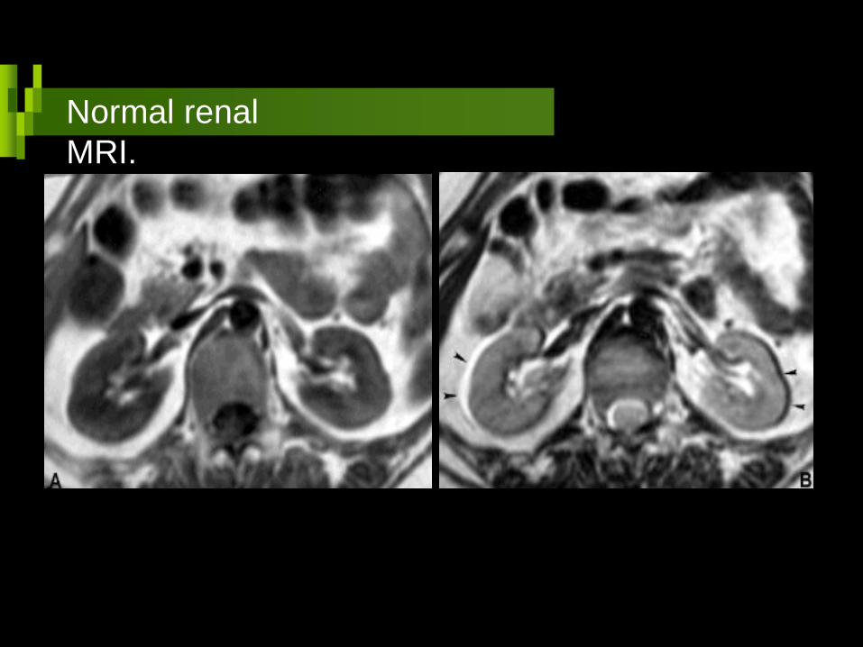
Normal renal
MRI.

T
1
T1+
C T
2
Normal renal
MRI.

Kidneys
CT density 30-50HU
Cortex and medulla are not
differentiated without contrast
Anatomic location from xiphoid
to umbilicus

Kidneys CT Techniques
Pre contrast scans 1cm intervals
Post contrast scans conventional, dynamic 1cm intervals
Reconstructed images (sagittal, coronal)

Renal
pathology
Masses
Trauma
Obstruction
Congenital
lesions

Renal
masses
Cystic lesions
Solid masses
Mixed lesions

Solid renal masses
The most common lesion
1-3% of all visceral
neoplasms
About 90% of all renal
tumors in adults
Male: female 3:1
Renal cell carcinoma

Renal cell carcinoma
The classical clinical triad of hematuria,
abdominal mass, and pain is an uncommon
presentation of RCC and in fact a late one.
Micro-hematuria may exist but hematuria
can be absent in up to 40% of cases.
A much more common clinical scenario
includes, fever, malaise, anemia, weight
loss, or a paraneoplastic syndrome.
Clinical aspects

Initial imaging of renal cell carcinoma is often
by intravenous urography with tomography or
ultrasonography.
The urographic appearance of a renal mass
includes:
Renal axis rotation by a large lesion
Focal contour bulge
Displacement or amputation of a part
of the collecting system
Focal or global hydronephrosis
Urography and
US

Indications for imaging of suspected RCC include:
Incidental detection of renal mass, presumed to be solid.
Suspicious or positive excretory urogram for tumor.
Persistent hematuria after normal excretory urography
Possible para-neoplastic syndrome, e.g.,hypercalcemia, erythrocytosis.
Evaluation for unknown primary neoplasm.
Patients with conditions known to be associated with renal
neoplasia, as Von Hippel -Lindau disease,….
Previous known renal neoplasm.
CT and MRI

Diagnosis of renal cell carcinoma
by CT or MR depends on the
distortion of the collecting system
and change in the attenuation or
signal intensity and contour of
renal parenchyma.
CT and
MRI
58Y M

The features of renal cell carcinoma on CT are:
Mass with attenuation similar to or less than renal parenchyma
Irregular parenchymal interface or margins (pseudo capsule)
Contour deformity (mass effect)
Enhancement with contrast media
Calcification either central, peripheral, or both
Secondary features such as:
Renal vein invasion
IVC invasion
Perinephric invasion
Lymph node enlargement
Adrenal metastasis
Computed
Tomography
A patient with pulmonary metastases.

64Y
M
Renal cell carcinoma with venous invasion

Is no longer part of the initial imaging
and staging evaluation of patients with
renal cell carcinoma being totally
replaced by CT or MR angiograms.
These angiograms are specially
needed when nephron sparing
surgery is contemplated especially in
the solitary kidneys, or patients with
polycystic kidney disease, or Von-
Hippel Lindau disease.
Venograms are needed to define the
cephalic extent of an inferior vena cava
thrombus. The need for conventional
angiography is only for embolization of
large hyper vascular tumors.
Conventional angiography
?!

Evaluation of tumor size and shape
Perinephric extension
Tumor vascularity [ CT angiography]
Lymph node enlargement
Major vascular (venous) involvement
Contiguous organ spread [ adrenal ]
Local or distant Metastatic spread.
Interpretation of CT & MR imaging should include

41Y M
RCC with local invasion of the adrenal gland and IVC as well
as metastatic extension to liver, other kidney and adrenal gland
Renal cell carcinoma

Solitary kidney
Synchronously in both kidneys
Poor renal function
Rates of local tumor recurrence
ranged from 4% to 10%,
The overall patient survival has not
significantly differed from that of
patients with similar stage disease who
have undergone radical nephrectomy.
Partial nephrectomy

Peripheral location of the tumor
Lack of invasion of
The renal sinus or perinephric fat
The renal collecting system
Renal vein
Absence of lymphadenopathy or distant metastases.
Imaging parameters for partial nephrectomy
Small tumor
less than 3 cm

Renal cell carcinoma Staging
I Tumor within the renal capsule
II Tumor invaded the capsule but not the Gerota’s fascia
III Renal vein or lymph node invasion
IV Beyond the Gerota’s fascia or metastases

Stage I RCC [ Tumor within the renal capsule ]

Progression of RCC over 2
years

Precontrast Post contrast
Stage I RCC [ Tumor within the renal capsule ]

A 7.2-cm heterogeneously
enhancing mass with invasion
of the perinephric fat
Stage II RCC [ Tumor invading perinephric fat ]

Stage III RCC [ Tumor with lymphadenopathy ]

Stage III RCC [ Tumor invading the liver ]

Renal cell carcinoma Stage ?! ONE

Renal cell carcinoma Stage ?! ONE

Pelvic filling defect was seen on a urogram in this
patient with hematuria; post contrast CT shows a
mass protruding into the pelvis and involves the
lower pole parenchyma
Renal cell carcinoma Stage ?! ONE

F 59Y

Renal cell carcinoma Stage ?! II by pathology

T2
Stage I RCC [ Tumor within the renal capsule ]

Stage I RCC [ Tumor within the renal capsule ][MRI]

Renal cell carcinoma Stage ?! Four

Patient presented with pulmonary metastases, a 3.5 tumor is shown on post contrast CT .
Renal cell carcinoma Stage ?! Four

Renal cell carcinoma Stage ?! Three
Invasion of the renal vein and IVC

Renal cell carcinoma with renal vein and IVC invasion

Cystic renal cell
carcinoma

76Y M Cystic renal cell
carcinoma

Cystic renal cell
carcinoma
Two different patients

Two different patients
Cystic renal cell
carcinoma

Contrast enhanced CT abdominal scan showing a large well marginated soft tissue mass lesion of heterogeneous texture and dense matrix calcifications seen arising from the ventral aspect of the left kidney.
Renal cell carcinoma with matrix calcification

Wilms’ tumor is the most common
primary malignant renal tumor of
childhood, accounting for about 7% of
all childhood cancers.
The mean age at diagnosis is 3.5Y
Clinical presentation : abdominal
mass with abdominal pain, fever, and
hematuria.
About10% of children have
metastases, at presentation.
Bilateral tumors occur in 5% to 10%
Nephroblastoma Wilm’s tumor
F 3Y

Wilm’s
Tumor

Wilm’s
Tumor
Male 6Y

Renal lymphoma
Multiple parenchymal renal masses 60% Helpful diagnostic criteria Known patient with lymphoma Immune compromised patient Other lesions specially in the spleen Retroperitoneal bulky nodes

Lymphoma
liver, spleen, kidneys, nodes, stomach

Post contrast CT Chest and abdominal scans showing a large infiltrative heterogenously enhancing mass lesion involving the wall of the left ventricle. Other lesions are seen in the spleen as well as both kidneys appearing as hypodense focal lesions
Cardiac and abdominal lymphoma

Renal
lymphoma

Renal lymphoma in an AIDS
patient

Renal
lymphoma

Fat containing renal masses
Angiomyolipoma
Simple lipoma
Teratoma
Liposarcoma
Xanthogranulomatous pyelonephritis

Renal Angiomyolipoma
Common benign renal mass
Formed of blood vessels, smooth
muscles, fat
40- 80% of patients with tuberous
sclerosis have AML
Usually small, bilateral, multiple,
asymptomatic, M=F

Tuberous
sclerosis:
Contrast-
enhanced
CTshows
numerous
small, fat-
density
lesions
scattered
throughout
both kidneys,
all are
angiomyolipo
Renal Angiomyolipoma

Tuberous sclerosis
Autosomal dominated disorder male= female
Pulmonary changes are seen almost only in females in 3rd -4th decades
Changes are similar to lymphangioleiomyomatosis
Except chylous effusion (rare in T. sclerosis)
Angiomyolipomas of the kidney and liver help in diagnosis

Renal Angiomyolipoma
Large, single, unilateral, symptomatic Middle age M:F = 1:4 Heterogeneously enhancing, fat containing renal mass Do not obstruct or destruct the calyces US, MRI, Angiography

Renal Angiomyolipoma

F35Y Renal Angiomyolipoma

Angiomyolipoma, growth on follow up

Transitional cell carcinoma
The most common tumor of the renal
pelvis
Multiple lesions in about 30% of cases
M:F = 4:1 above 60Years
Diagnosed by IVP, CTU , MRU
Filling defect in the pelvis
CT Urogram

Transitional Cell
Carcinoma

Transitional Cell
Carcinoma

Transitional Cell
Carcinoma

Transitional Cell
Carcinoma

Transitional Cell
Carcinoma

Transitional Cell
Carcinoma

Transitional cell carcinoma extending along the upper ureter
Transitional Cell
Carcinoma

56Y M
Transitional cell carcinoma filling the renal pelvis
Transitional Cell
Carcinoma

Transitional Cell
Carcinoma

Renal pelvic filling defect
Non opaque stones Blood clots Polyps Hypertrophied renal papilla Vascular impression Inflammatory conditions
Transitional cell carcinoma.

Oncocytoma
Rare, benign, solid renal tumor
Usually asymptomatic
Single, multiple
Usually larger than 2cm
3-5% of renal parenchymal tumors

Oncocytoma
Differentiation from RCC is difficult Helpful diagnostic criteria
• Homogenous solid lesion • Sharply hypodense star- shaped scar 33% • Large size may reach 4kg

Oncocytoma.

Oncocytoma.

Oncocytoma.

Multilocular cystic neophroma
Multilocular cystic lesion Occurs in children (<5years) and adults (40-70Y Well defined lesion with thick internal septations Calcium 10-50%
Unknown origin

Multilocular cystic neophroma

Multilocular cystic neophroma

سبحانك الهم و بحمدك نشهد ان ال اله اال انت نستغفرك و نتوب اليك
Thank you

TCC ,2CASES
hemangioma

RCC
Angiomyolipoma 2 cases


LYMPHOMA

CYSTIC RCC


F 3Y

TNM Stage
TNM Description Robson Stage
Tumor confined to renal capsule I
I T1 Small tumor (< 2.5 cm)
II T2 Large tumor (> 2.5 cm)
III Tumor spread to perinephric fat or adrenal gland II
III III
T3b T3c
Venous tumor thrombus Renal vein thrombus only IVC thrombus
III A
III/IV N1-N3 Regional lymph node metastases III B
III/IV T3b/c, N1-N3 Venous tumor thrombus and regional nodes III C
IV T4 Direct invasion of adjacent organs outside Gerota’s fascia IV A
IV M1 Distant metastases IV B

Tuberous sclerosis. Both kidneys contain many cysts . More inferiorly, an
angiomyolipoma is present (arrow (

Wilm’s
tumor

Angiomyolipoma.

Angiomyolipoma with hemorrhage.