Embed Size (px)
Citation preview
RELIABLE OPTION FOR RECONSTRUCTION OF AMPUTATIONSTUMPS: THE FREE ANTEROLATERAL THIGH FLAP
SERKAN YILDIRIM, M.D.,* GAYE TAYLAN CALIKAPAN, M.D., and TAYFUN AKOZ, M.D.
The increased use of microsurgery has enabled reconstructive surgeons to deal with tissue defects of various sizes and compositions.The limited amount of qualified tissue for covering is the primary problem in stump reconstruction. Free flaps offer the ideal solution by pro-viding the optimal cover, and by preserving the length of the amputation site. Anterolateral thigh flaps were preferred for reconstruction oflower extremity amputation sites of nine patients admitted both in the subacute and chronic periods. All underwent previous stump recon-struction with local flaps in other clinics. Anterolateral thigh flaps avoided further shortening of the extremities, and provided stable tissuefor prosthesis use. The flap offers reliable soft-tissue reconstruction of amputation stumps. VVC 2006 Wiley-Liss, Inc. Microsurgery 26:386–390, 2006.
Though replantation of the proximal amputate is the first
plan in an emergency setting, preserving an adequate
length of the stump and a good functional recovery
should also be kept in mind when there is no chance for
extremity salvage.1 Durable and reliable amputation
stump coverage is required in order to increase the qual-
ity of life of a patient already debilitated due to a major
trauma. The options for soft-tissue reconstruction of an
amputation stump include using a flap from the ampu-
tated distal part, a local flap, or a free flap.1 Free flaps
are usually the primary choice in secondary procedures
when local tissues fail to provide adequate coverage. The
aims of amputation are preserving maximal length, and
provision of a sensate, durable, and cylindrical stump
which is pain-free.2 The anterolateral thigh flap was the
choice of flap in our limited series of stump reconstruc-
tions. This flap can be customized to individual needs in
terms of sensation, composition, and function.3 Both ten-
sor fascia lata and sensory nerves can be harvested at the
same donor site.4 We did not observe any complications
regarding flap viability or wound healing. All patients are
comforted with a reliable stump, which provided them a
qualified daily living with their prosthesis.
PATIENTS AND METHODS
Between 2002–2005, nine patients with lower extremity
amputations were operated upon for local infection and
unstable wound coverage. The mean age of patients was
33.3 (range, 17–48) years, and the female/male ratio was
2/7. The levels of amputations were the forefoot (2), below
the knee (4), and the distal tibia (3). All patients had
undergone surgery in other clinics on an emergency basis,
and local flaps were the choice for reconstruction. Two
patients with foot-level amputation had additional osteomy-
elitis, confirmed both radiologically and clinically. None of
the patients were comfortable with their prostheses, and
they complained about frequent wound occurrence requir-
ing dressing changes, which negatively affected their social
life. All patients insisted on length preservation, and free
flap transfers were planned. Anterolateral thigh flaps from
the same or the contralateral leg (6/3) were harvested. Flap
dimensions ranged between 7 3 10 and 11 3 15 cm. The
lateral femoral cutaneous nerves were included in the flaps.
Donor sites were primarily closed in three patients, and
skin-grafted in the rest. The mean hospitalization time was
15.6 (range, 11–17) days.
CASE REPORTS
Case 1
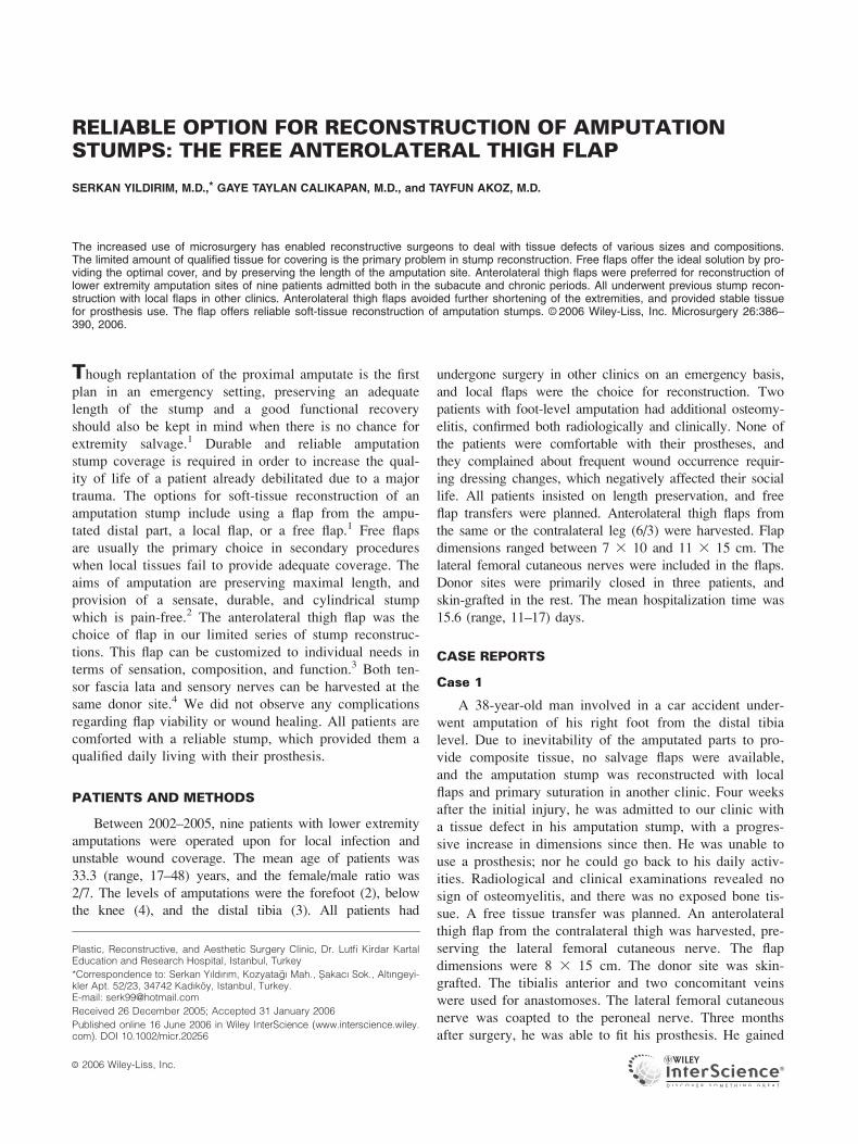
A 38-year-old man involved in a car accident under-
went amputation of his right foot from the distal tibia
level. Due to inevitability of the amputated parts to pro-
vide composite tissue, no salvage flaps were available,
and the amputation stump was reconstructed with local
flaps and primary suturation in another clinic. Four weeks
after the initial injury, he was admitted to our clinic with
a tissue defect in his amputation stump, with a progres-
sive increase in dimensions since then. He was unable to
use a prosthesis; nor he could go back to his daily activ-
ities. Radiological and clinical examinations revealed no
sign of osteomyelitis, and there was no exposed bone tis-
sue. A free tissue transfer was planned. An anterolateral
thigh flap from the contralateral thigh was harvested, pre-
serving the lateral femoral cutaneous nerve. The flap
dimensions were 8 3 15 cm. The donor site was skin-
grafted. The tibialis anterior and two concomitant veins
were used for anastomoses. The lateral femoral cutaneous
nerve was coapted to the peroneal nerve. Three months
after surgery, he was able to fit his prosthesis. He gained
Plastic, Reconstructive, and Aesthetic Surgery Clinic, Dr. Lutfi Kirdar KartalEducation and Research Hospital, Istanbul, Turkey
*Correspondence to: Serkan Yıldırım, Kozyatagı Mah., Sakacı Sok., Altıngeyi-kler Apt. 52/23, 34742 Kadıkoy, Istanbul, Turkey.E-mail: [email protected]
Received 26 December 2005; Accepted 31 January 2006
Published online 16 June 2006 in Wiley InterScience (www.interscience.wiley.com). DOI 10.1002/micr.20256
VVC 2006 Wiley-Liss, Inc.
deep sensibility of the flap. A follow-up period of 3 years
called for no revisional surgery of the flap (Figs. 1, 2).
Case 2
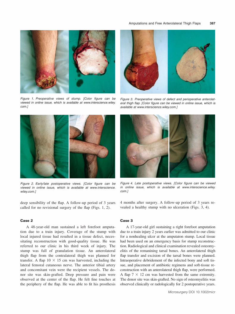
A 48-year-old man sustained a left forefoot amputa-
tion due to a train injury. Coverage of the stump with
local injured tissue had resulted in a tissue defect, neces-
sitating reconstruction with good-quality tissue. He was
referred to our clinic in his third week of injury. The
stump was full of granulation tissue. An anterolateral
thigh flap from the contralateral thigh was planned for
transfer. A flap 10 3 15 cm was harvested, including the
lateral femoral cutaneous nerve. The anterior tibial artery
and concomitant vein were the recipient vessels. The do-
nor site was skin-grafted. Deep pressure and pain were
observed at the center of the flap. He felt fine touches at
the periphery of the flap. He was able to fit his prosthesis
4 months after surgery. A follow-up period of 3 years re-
vealed a healthy stump with no ulceration (Figs. 3, 4).
Case 3
A 17-year-old girl sustaining a right forefoot amputation
due to a train injury 2 years earlier was admitted to our clinic
for a nonhealing ulcer at the amputaton stump. Local tissue
had been used on an emergency basis for stump reconstruc-
tion. Radiological and clinical examination revealed osteomy-
elitis of the remanining tarsal bones. An anterolateral thigh
flap transfer and excision of the tarsal bones were planned.
Intraoperative debridement of the infected bony and soft tis-
sue, and placement of antibiotic regimens and soft-tissue re-
construction with an anterolateral thigh flap, were performed.
A flap 7 3 12 cm was harvested from the same extremity.
The donor site was skin-grafted. No sign of osteomyelitis was
observed clinically or radologically for 2 postoperative years.
Figure 1. Preoperative views of stump. [Color figure can be
viewed in online issue, which is available at www.interscience.wiley.
com.]
Figure 2. Early/late postoperative views. [Color figure can be
viewed in online issue, which is available at www.interscience.
wiley.com.]
Figure 3. Preoperative views of defect and perioperative anterolat-
eral thigh flap. [Color figure can be viewed in online issue, which is
available at www.interscience.wiley.com.]
Figure 4. Late postoperative views. [Color figure can be viewed
in online issue, which is available at www.interscience.wiley.
com.]
Amputations and Free Anterolateral Thigh Flaps 387
Microsurgery DOI 10.1002/micr

She feels light touches and pain in the flap-normal skin neigh-
borhood (Figs. 5, 6).
RESULTS
Local infection resolved in the two patients involved,
and no wound dehiscence occurred. All flaps survived.
The mean follow-up period was 2.4 years (range, 1–3 years).
Flap sensibility was assessed with two-point discrimination
and the Semmes-Weinstein monofilament system. All patients
recovered deep pressure, and only two felt light touches
and pain at the periphery of the flap. The patients are
comfortable with their prostheses, and none have required
reoperation so far (Table 1).
DISCUSSION
Amputation of a major limb is one of the most debili-
tating traumas. It is usually not very easy for patients to
deal with problems in their new life with a prosthesis. A
nonhealing wound creates further discomfort in the social
life to which they are adapting. The surgeon must offer
the best solution, with minimal morbidity.
Although secondary revisions of amputation stumps by
bone-shortening can be performed with unstable stump-heal-
ing, it should be kept in mind that the length of the extremity
matters in terms of prosthesis fitting and the psychological
health of the patient.5 Isik et al. reconstructed troublesome
amputation stumps in 4 patients with neurosensorial free
medial plantar flaps from unaffected feet without shortening.5
Gallico et al.6 used a variety of flaps for reconstruction
of below-knee amputation stumps to preserve a functional
prosthetic level, including latissimus dorsi myocutaneous
flaps, groin flaps, foot-fillet flaps, and latissimus dorsi mus-
cle flaps with skin grafts. They mentioned the importance
of a stable and functional knee joint after lower leg trauma
for the rehabilitation of the patient.6 The major problem
their patients developed in long-term follow-up was ulcera-
tion on or adjacent to flaps. We did not observe any ulcers,
though continuing follow-up may show these as well.
The ‘‘free fillet flap’’ is an established strategy that
allows the creation of flaps without additional donor-site
morbidity.7–10 However, the patient must be admitted in
the acute period, both for revascularization-replantation and
for spare-part transfer choices. All our patients were already
operated upon primarily by other clinics, and were in the sub-
acute or chronic period.
Erdmann et al. mentioned their preference for free flaps in
elective reconstructions of amputation stumps.7 Among the
flaps that have been transferred are scapular flaps, fillet flaps
from the amputated extremity, anterolateral thigh flaps, and
lateral arm flaps.7 They summarized the indications for micro-
surgical free flap transfers to amputation sites as follows:
� Microsurgical free flap transfer in emergency settings;
� Unstable tissue and/or localized soft-tissue infection; and
� Reconstrction after extensive tumor resection.7
Figure 5. Preoperative view of defect and elevated flap. [Color figure
can be viewed in online issue, which is available at www.interscience.
wiley.com.]
Figure 6. Early/late postoperative views. [Color figure can be viewed
in online issue, which is available at www.interscience.wiley.com.]
388 Yıldırım et al.
Microsurgery DOI 10.1002/micr

Kasabian et al. preferred the foot as first choice if
available. Otherwise, the parascapular free flap was their
donor site of choice.10
Tukiainen et al. used latissimus dorsi free flaps for
the reconstruction of amputation stumps to avoid further
amputation. The reconstructions were done in the acute
period, posttraumatic phase, and chronic period.1
Both latissimus dorsi and parascapular flaps necessi-
tate positional changes during flap harvest, which in turn
avoids simultaneous two-team work. Furthermore, flap thin-
ning is not an option for these flaps. While sensory branches
are divided during flap elevation, a sensory flap is not feasi-
ble for both flaps.
Ghali et al. preserved leg length with pedicled fillets
of foot flaps after traumatic amputations. When planned well,
the procedure avoids donor-site morbidity and microsurgery
as well.2
The anterolateral thigh flap can be used for reconstruction
of various defects. Among its advantages are; long and large-
caliber vascular pedicle, wide, reliable skin pedicle, elevation
as a sensory flap, different types of tissue composition (mus-
culocutaneous or fasciacutaneous), available for thinning
procedure, and permission for a two team work.11 Yıldırım
et al. demonstrated excellent functional and cosmetic results
with anterolateral thigh flaps used for soft-tissue reconstruc-
tion of various regions of the body.12 The anterolateral thigh
flap was postulated by Ozkan et al. as an ideal and versatile
flap for lower extremity reconstruction.13 Yıldırım et al.
mentioned the various advantages of the anterolateral thigh
flap for lower extremity reconstruction in its maximal recon-
structive capacity.14
The superior results of neural continuity as mentioned by
Arnez et al. encourage the use of sensorial flap transfers.15
The lateral femoral cutaneous nerves were included in the
flaps. All patients had deep-pressure sensibility, and two had
a moderate amount of protective sensation. No ulcers oc-
curred in a maximal follow-up period of 5 years.
The fascia component of the flap serves different re-
constructive purposes, as studied by Yıldırım et al.16 Sekido
et al. studied anterolateral thigh flaps for reconstruction
of large-sized defects at the weight-bearing plantar re-
gion. They demonstrated that the anterolateral thigh flap,
while providing adequate bulk and contour of the foot,
also withstands weight pressure and shearing force. It has
the ability to provide recovery of sensation.17 Though both
thighs were used as donor sites, our primary choice was the
same thigh, in order to protect the unviolated healthy thigh.
However, when this was not possible, we used the contralat-
eral thigh as the donor site.
The variations in the vascular anatomy of the flap is
compensated with the surgeon’s experience as this flap is
routinely used by our clinic.
Reconstructive requirements for amputation stumps are
variable, for that reason every stump reconstruction must be
handled individually by means of flap characteristics. While
forefoot amputations necessitate very thin flaps, below-knee
sites, dead spaces, or the existence of an infection may re-
quire bulky flaps with a muscle component. Anterolateral
thigh flaps enable both thin and thick flaps, with fascia and
muscle components added or excluded as required.
As a promising multipurpose flap, the anterolateral
thigh flap provides a durable and reliable cover for ampu-
tation stumps. It offers both soft tisssue and some amount
of sensation required for stump reconstruction. The ease
of dissection when it is frequently performed diminishes
the operative time and failure rate. Among various alter-
natives, we prefer the anterolateral thigh flap for elective
reconstruction of amputation stumps.
REFERENCES
1. Tukiainen EJ, Saray A, Kuokkanen HO, Asko-Seljavaara SL. Sal-vage of major amputation stumps of the lower extremity with latissi-mus dorsi free flaps. Scand J Plast Reconstr Surg Hand Surg 2002;36:85–90.
2. Ghali S, Haris PA, Khan U, Pearse M, Nanchahal J. Leg length pres-ervation with pedicled fillet of foot flaps after traumatic amputations.Plast Reconstr Surg 2005;115:498–505.
3. Lutz BS. Aesthetic and functional advantages of the anterolateralthigh flap in reconstruction of tumor-related scalp defects. Microsur-gery 2002;22:258–264.
4. Pribaz JJ, Orgill DP, Epstein MD, Sampson CE, Hergrueter CA.Anterolateral thigh flap. Ann Plast Surg 1995;34:585–592.
Table 1. Patients’ Characteristics
Patient
(years)
age/sex
Amputation
level
Follow-up
(years)
Hospitalzation
time (days)
Presence of
osteomyelitis Sensibility
Flap
dimensions
(cm)
38/M Distal tibia 3 12 No Deep pressure 8 3 15
48/M Forefoot 3 15 No Deep pressure 10 3 15
17/F Forefoot 2 21 Yes Light touch, pain 7 3 12
26/F Below knee 2 11 No Deep pressure 9 3 12
39/M Below knee 3 13 No Deep pressure 9 3 15
47/M Distal tibia 1 17 Yes Light touch, pain 7 3 11
31/M Below knee 3 15 No Deep pressure 8 3 13
33/M Below knee 3 14 No Deep pressure 7 3 13
21/M Distal tibia 2 13 No Deep pressure 10 3 14
Amputations and Free Anterolateral Thigh Flaps 389
Microsurgery DOI 10.1002/micr

5. Isik S, Guler MM, Selmanpakoglu N. Salvage of foot amputationstumps of chopart level by free medial plantar flap. Plast ReconstrSurg 1998;101:745–750.
6. Gallico GG III, Ehrlichman RJ, Jupiter J, May JW Jr. Free flaps topreserve below-knee amputation stumps: long-term evaluation. PlastReconstr Surg 1987;79:871–877.
7. Erdmann D, Sundin BM, Yasui K, Wong MS, Levin LS. Microsurgi-cal free flap transfer to amputation sites: indications and results. AnnPlast Surg 2002;48:167–172.
8. Chiang YC, Wei FC, Wang JW, Chen WS. Reconstruction ofbelow-knee stump using the salvaged foot fillet flap. Plast ReconstrSurg 1995;96:731–738.
9. Hammond DC, Matloub HS, Kadaz BB, Yousif NJ, Sanger JR, Lar-son DL. The free fillet flap for reconstruction of the upper extremity.Plast Reconstr Surg 1994;94:507–512.
10. Kasabian AK, Glat PM, Eidelman Y, Colen S, Longaker MT,Attinger C, Shaw W. Salvage of traumatic below-knee amputationstumps utilizing the fillet of foot free flap: critical evaluation of sixcases. Plast Reconstr Surg 1995;96:1145–1153.
11. Ozkan O, Coskunfirat OK, Ozgentas HE. An ideal and versatile ma-terial for spft-tissue coverage: experiences with most modifications
of the anterolateral thigh flap. J Reconstr Microsurg 2004;20:377–383.
12. Yıldırım S, Avci G, Akoz T. Soft-tissue reconstruction using a freeanterolateral thigh flap: experience with 28 patients. Ann Plast Surg2003;51:37–44.
13. Ozkan O, Coskunfirat OK, Ozgentas HE. The use of free anterolat-eral thigh flap for reconstructing soft tissue defects of the lowerextremities. Ann Plast Surg 2004;53:455–461.
14. Yıldırım S, Gideroglu K, Akoz T. Anterolateral thigh flap: ideal freeflap choice for lower extremity soft-tissue reconstruction. J ReconstrMicrosurg 2003;19:225–233.
15. Arnez ZM, Valdatta L, Sassoon E, Planinsek F, Ahcan U. Salvageof a below knee amputation stump with a free sensate total sole flappreserving continuity of the posterior tibial nerve. Br J Plast Surg1998;51:470–472.
16. Yıldırım S, Taylan G, Akoz T. Use of fascia component of the an-terolateral thigh flap for different reconstructive purposes. Ann PlastSurg 2005;55:479–484.
17. Sekido M, Yamamoto Y, Furukawa H, Sugihara T. Change ofweight-bearing pattern before and after plantar reconstruction withfree anterolateral thigh flap. Microsurgery 2004;24:289–292.
390 Yıldırım et al.
Microsurgery DOI 10.1002/micr