Embed Size (px)
Citation preview
RELATIONSHIP BETWEEN
NUTRITIONAL STATUS AND THE
ABILITY OF SIX TO TWELVE- WEEKS
INFANTS TO SERO-CONVERT TO
ROTAVIRUS VACCINATION
By
Chishimba Katema
A Dissertation submitted to the University of Zambia in partial
fulfillment of the requirements of the degree of Masters of Medical
Microbiology
UNIVERSITY OF ZAMBIA
Lusaka
2020

ii
DECLARATION
This Dissertation represents Chishimba Katema’s own work and has not been previously submitted
for a degree, diploma or other qualification at this or any other University.
Candidate Name: Chishimba Katema
Signature: ............................
Date: ............................

iii
CERTIFICATE OF APPROVAL
This dissertation of Chishimba Katema has been approved in partial fulfilment of the requirements
for the degree of Master of Medical Microbiology by the University of Zambia.
……………… ………………… …….………………
Supervisor Signature Month/Date/Year
……………… ………………… …….………………
Examiner 1 Signature Month/Date/Year
……………… ……………… …..…………………..
Examiner 2 Signature Month/Date/Year
……………… ……… ..…… …..………………..
Examiner 3 Signature Month/Date/Year

iv
DEDICATION
This dissertation is dedicated to my wonderful Family. I also dedicate this work to the Love of
My life, to all my children and my sisters.

v
ACKNOWLEDGEMENTS
Firstly, thanks and praises go to the Lord Almighty, Jesus Christ for He has been and always will
be faithful in all ups and downs. In no particular order, I would like to thank the following for
helping in my successful completion of this work. I would like to acknowledge the efforts and
contributions of my supervisors Dr Sody Munsaka, Dr Roma Chilengi and co-supervisor Dr.
Michelo Simuyandi who gave me valuable support regardless of their limited and precious time.
Other professional contributors to this work, Dr Samuel Bosomprah, Ms. Katayi Mwila
Kazimbaya, who offered their expert opinion in the design and implementation of this work. I am
grateful to the parents of all the infants who participated in this study.
vi
ABSTRACT
Live attenuated oral vaccines against rotavirus have been shown to be less efficacious in children
from developing countries compared to developed countries. Reasons for this disparity are not
fully understood.
This study aimed to investigate the potential association between indicators of nutritional status
(vitamin A status, weight for age, height for age and mid-upper arm circumference) of the infants
and sero-conversion among Zambian infants routinely immunized with rotavirus vaccine,
Rotarix™.
A total of 1320 infants were assessed for enrolment and420 infants were recruited at infant age
6-12 weeks in Lusaka, Zambia. Clinical information and samples were collected at baseline and
at one month following the second dose of rotavirus vaccine. Only 208 infant samples were
analyzed at baseline and one month after vaccination to determine infant nutritional status
(vitamin A status, weight for age, height for age status and mid-upper arm circumference.
Vitamin A status and serum rotavirus-specific IgA were determined using standardized Enzyme
Linked Immuno-sorbent Assay methods. The anthropometric indices were interpreted using
WHO growth standards. Sero-conversion was defined as a ≥ 4 fold rise in serum IgA titre from
baseline to one-month post Rotarix™ dose 2, while sero-positivity of IgA was defined as serum
titre ≥ 40. Pearson Chi-squared test was used to investigate the association of sero-conversion
and categorical factors (i.e. sex, vitamin A status, weight for age, height for age, mid-upper arm
circumference at baseline and serum IgA sero-positivity).
The sero-conversion frequency to rotavirus was 57.2% (119/208) and baseline infant sero-
positivity to rotavirus was 23.1% (48/208). Baseline vitamin A deficiency, underweight and
stunting were 78.9% (164/208), 6.7% (14/208) and 52.4% (109/208), respectively. Base-line
Infant moderate acute malnutrition was 38.0% (79/208208) and severe acute malnutrition was
12.50% (26/208) as determined by the infant mid-upper arm circumference. There was no
evidence of association between infant serum IgA sero-conversion and nutritional variables;
serum vitamin A deficiency (p=0.882), stunting (p=0.905), underweight (p=0.243), Mid-Uper
arm circumference (p=0.565) and infant baseline serum IgA sero-positivity (p=0.832).
Poor IgA sero-conversion frequency observed in this cohort was not influenced by nutritional
factors indicated by infant serum vitamin A status, weight for age, height for age and mid-upper
arm circumference. Early infant exposure to rotavirus infection, determined by infant serum IgA
sero-positivity levels did not influence sero-conversions frequencies in this cohort. These factors
may have other effects later in childhood, as no difference was observed among seroconvertors
and non-seroconvertors to rotavirus vaccination in the 6-12 weeks age group. Further research is
needed to better understand vaccine sero-conversion.
Key word: Rotavirus vaccine, rotavirus, seroconvert, Zambia, Infant
vii
TABLE OF CONTENTS
DECLARATION ................................................................................................................................... ii
CERTIFICATE OF APPROVAL ........................................................................................................ iii
DEDICATION ...................................................................................................................................... iv
ACKNOWLEDGEMENTS................................................................................................................... v
ABSTRACT .......................................................................................................................................... vi
TABLE OF CONTENTS .................................................................................................................... vii
LIST OF TABLES................................................................................................................................ ix
LIST OF FIGURES ............................................................................................................................... x
LIST OF ABBREVIATIONS............................................................................................................... xi
CHAPTER 1: INTRODUCTION ......................................................................................................... 1
1.1 Background ................................................................................................................................... 1
1.2 Statement of the Problem ............................................................................................................... 4
1.3 Justification of the Study ................................................................................................................ 5
1.4 Research Questions ........................................................................................................................ 5
1.5 Objectives ...................................................................................................................................... 6
1.5.1 General Objective ................................................................................................................... 6
1.5.2 Specific Objectives ................................................................................................................. 6
CHAPTER 2: LITERATURE REVIEW .............................................................................................. 7
2.1 Virology of Rotavirus .................................................................................................................... 7
2.1.1 Structure and Taxonomy ......................................................................................................... 7
2.1.2 Classification .......................................................................................................................... 8
2.1.3 Epidemiology ........................................................................................................................ 10
2.1.4 Replication Cycle .................................................................................................................. 11
2.1.5 Immunity to Rotavirus Infection ............................................................................................ 12
2.2 Rotavirus Acute Gastroenteritis (RV-AGE) .................................................................................. 14
2.3 Clinical Picture of Rotavirus Gastroenteritis ................................................................................. 15
2.4 Transmission and Shedding.......................................................................................................... 16
2.6 Rotavirus Vaccines ...................................................................................................................... 17
2.6.1 Rotashield™: Rhesus Rotavirus Vaccine ............................................................................... 18
2.6.2 Rotateq™: Bovine-Human Reassorted Rotavirus Vaccine ..................................................... 18
2.6.3 Rotarix™: Human Attenuated Rotavirus Vaccine .................................................................. 19
2.6.4 Rotavirus Vaccine Effectiveness ........................................................................................... 21
2.7 Nutrition Status ............................................................................................................................ 23
2.8 Weight for Age Status .................................................................................................................. 24
2.9 Height for Age Status................................................................................................................... 24
2.10 Mid-upper Arm Circumference (MUAC) ................................................................................... 25
viii
2.11 Serum Vitamin A Status of Children .......................................................................................... 26
2.12 Consequences of Under-nutrition on Mortality/Morbidity .......................................................... 26
2.13 Malnutrition and the Immune System ......................................................................................... 28
2.14 Malnutrition and Vaccination ..................................................................................................... 29
CHAPTER 3: METHODOLOGY ...................................................................................................... 31
3.1 Study Design ............................................................................................................................... 31
3.2 Study site ..................................................................................................................................... 31
3.3 Study Population.......................................................................................................................... 31
3.3.1 Inclusion Criteria .................................................................................................................. 32
3.3.2 Exclusion Criteria ................................................................................................................. 33
3.3.3 Sample Size Determination ................................................................................................... 33
3.3.2 Clinic Procedures .................................................................................................................. 34
3.4 Laboratory Procedures. ................................................................................................................ 35
3.4.1 Measurement of Serum IgA for the Determination of Sero-positivity and Sero-conversion .... 35
3.4.2 Measurement of Serum Vitamin A to Determine the Prevalence of Vitamin A Deficiency ..... 36
3.5 Data Analysis .............................................................................................................................. 36
3.6 Ethical Considerations ................................................................................................................. 36
3.7 Limitations .................................................................................................................................. 37
CHAPTER 4: RESULTS .................................................................................................................... 38
4.1 Patient Recruitment and Descriptives ........................................................................................... 38
4.2 Prevalence of Poor Nutritional Status in Infants and Infant Early Exposure to Rotavirus Infection 38
4.3 Effects of Nutritional Status on Sero-conversion Among Children 3 months in Lusaka Zambia .... 39
CHAPTER 5: DISCUSSION, CONCLUSION AND RECOMENDATIONS ................................... 42
5.1 Discussion ................................................................................................................................... 42
5.2 Conclusion................................................................................................................................... 45
5.3 Recommendations........................................................................................................................ 46
REFERENCES .................................................................................................................................... 47
APPENDICES ..................................................................................................................................... 61
Appendix A: Letters of support .......................................................................................................... 61
Appendix B: Study questionnaire and clinical examination form. ....................................................... 64
ix
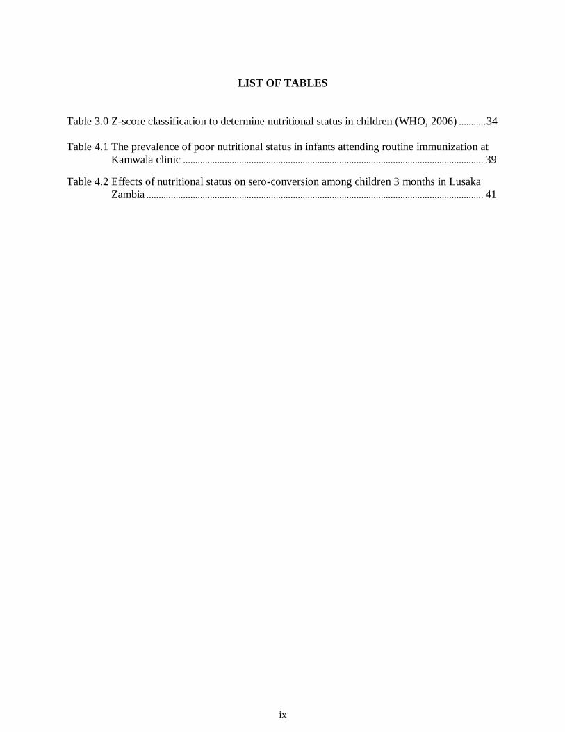
LIST OF TABLES
Table 3.0 Z-score classification to determine nutritional status in children (WHO, 2006) ........... 34
Table 4.1 The prevalence of poor nutritional status in infants attending routine immunization at
Kamwala clinic ........................................................................................................................... 39
Table 4.2 Effects of nutritional status on sero-conversion among children 3 months in Lusaka Zambia .......................................................................................................................................... 41

x
LIST OF FIGURES
Figure 3: Recruitment and sample processing flow diagram .........................................................32
xi
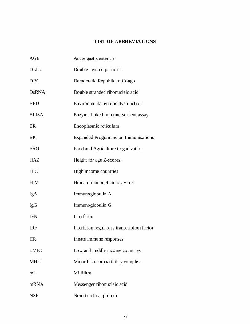
LIST OF ABBREVIATIONS
AGE Acute gastroenteritis
DLPs Double layered particles
DRC Democratic Republic of Congo
DsRNA Double stranded ribonucleic acid
EED Environmental enteric dysfunction
ELISA Enzyme linked immune-sorbent assay
ER Endoplasmic reticulum
EPI Expanded Programme on Immunisations
FAO Food and Agriculture Organization
HAZ Height for age Z-scores,
HIC High income countries
HIV Human Imunodeficiency virus
IgA Immunoglobulin A
IgG Immunoglobulin G
IFN Interferon
IRF Interferon regulatory transcription factor
IIR Innate immune responses
LMIC Low and middle income countries
MHC Major histocompatibility complex
mL Millilitre
mRNA Messenger ribonucleic acid
NSP Non structural protein
xii
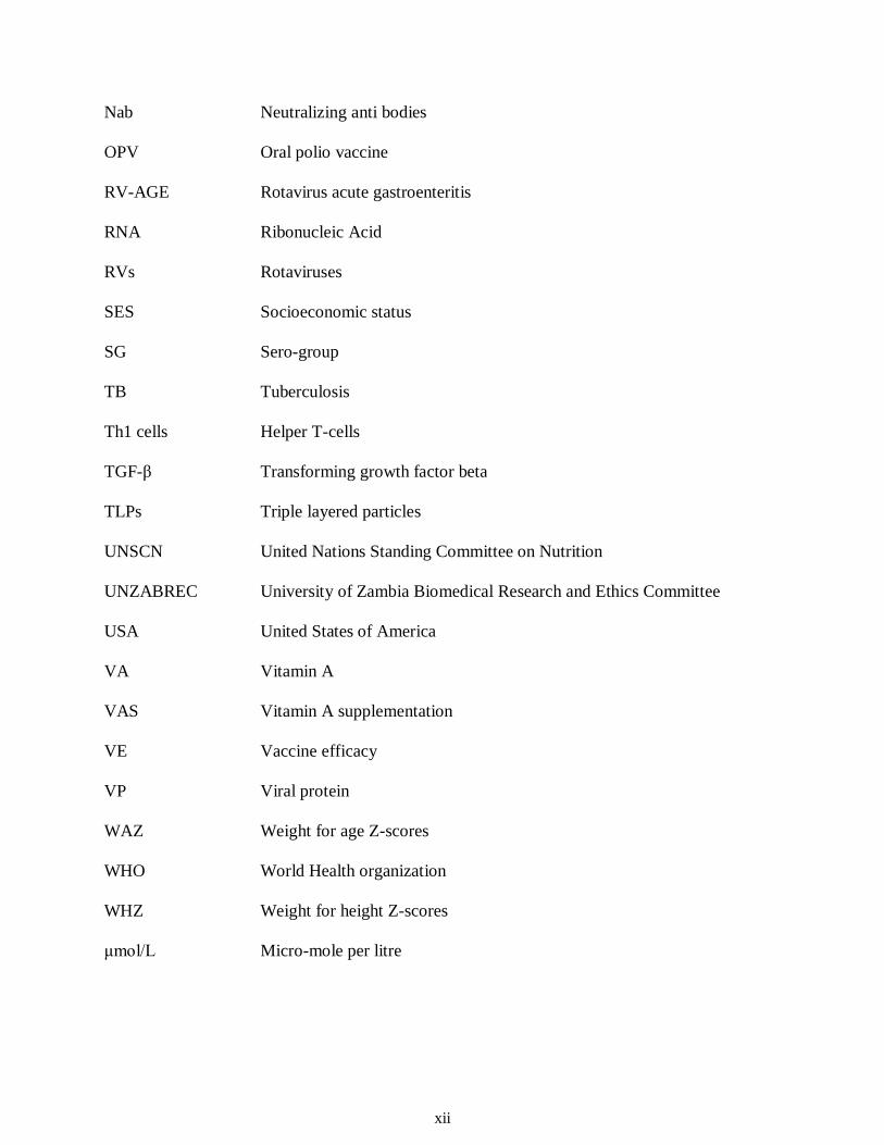
Nab Neutralizing anti bodies
OPV Oral polio vaccine
RV-AGE Rotavirus acute gastroenteritis
RNA Ribonucleic Acid
RVs Rotaviruses
SES Socioeconomic status
SG Sero-group
TB Tuberculosis
Th1 cells Helper T-cells
TGF-β Transforming growth factor beta
TLPs Triple layered particles
UNSCN United Nations Standing Committee on Nutrition
UNZABREC University of Zambia Biomedical Research and Ethics Committee
USA United States of America
VA Vitamin A
VAS Vitamin A supplementation
VE Vaccine efficacy
VP Viral protein
WAZ Weight for age Z-scores
WHO World Health organization
WHZ Weight for height Z-scores
μmol/L Micro-mole per litre
1
CHAPTER 1: INTRODUCTION
1.1 Background
Diarrhoeal diseases are one of the world’s leading killers of children and rotavirus is the most
common cause of severe diarrhoea among children under the age of five globally, and almost every
child is infected during the first five years of life (Tate et al., 2016). Rotavirus disease is typically
associated with vomiting and fever, followed by profuse watery diarrhoea, and the main cause of
death is dehydration. The most important therapy is oral or intravenous rehydration, as there is no
specific antimicrobial treatment for rotavirus disease. Although oral rehydration therapy had
reduced mortality by the time the first rotavirus vaccine was licensed in 2005, there still were about
450 000 deaths due to rotavirus gastroenteritis (RVGE) around the world (Tate et al., 2012). With
over 80% of those deaths occurring in low-income countries, rotavirus infection was so prevalent
that nearly every child in the world was estimated to have had at least one episode of rotavirus
disease by age two (Walker et al., 2013).
Globally, diarrhoeal diseases of any kind account for approximately 578,000 deaths among
children under the age of five. In this age group, rotavirus infection is still the leading cause of
severe acute gastroenteritis, accounting for an estimated 215,000 (37%) deaths annually and is
responsible for millions of hospitalizations and clinic visits (Tate et al., 2016, Walker et al., 2013).
Nearly 150,000 African children die from the dehydrating diarrhoea caused by rotavirus infection
every year, accounting for more than 56% of the global total of rotavirus deaths (WHO, 2013;
(Tate et al., 2016).
2
In Zambia, children under five years of age experience 10 million annual episodes of
diarrhoea, which result in an estimated 63,000 hospitalizations and 15,000 deaths (Liu et al.,
2015b, Mpabalwani et al., 1995). This makes diarrhoea Zambia’s third leading cause of under-5
mortality (after pneumonia and malaria) causing approximately 9% of deaths in children under
five years of age (Walker et al., 2013). One-quarter to one-third of the severe diarrhoea resulting
in hospitalization and death are attributable to vaccine-preventable rotavirus (Walker et al., 2013).
Thus, while recent years have seen significant reductions in morbidity and mortality due to
rotavirus infection, there is still much work to be done. Improvements in hygiene and sanitation
have been helpful in decreasing the incidence of many causes of diarrhoea; these measures alone
do not prevent rotavirus transmission (Armah and Binka, 2014). Rather, based on findings that
early rotavirus infections provide infants increased protection against subsequent bouts of disease
(Dennehy, 2008).The most important public health approach to fight the disease has been the
widespread use of immunizations. Researchers have tried various strategies since the 1980s to
develop effective vaccines (Cortese et al., 2013), and after two vaccines, Rotarix™
(GlaxoSmithKline, Biologicals) and RotaTeq™ (Merck & co, inc), were found to demonstrate
safety and efficacy in Europe and North America in the mid-2000s, efforts in the past decade have
focused on rolling them out them globally (Armah et al., 2016, Jiang et al., 2010). A third vaccine
called ROTAVAC™ was recently introduced. ROTAVAC™ is based on the116E rotavirus strain
and is manufactured by Bharat Biotech International Limited of India (Bhandari et al., 2014). In
efforts to battle the diarrhoeal scourge, in 2013, Zambia rolled out the Rotarix™
(GlaxoSmithKline, Biologicals) rotavirus vaccine in its Expanded Programme on Immunization
(EPI). Rotarix™ is an attenuated human oral vaccine based on the G1P[8] strain.
3
Its safety and efficacy have been established in healthy infants across multiple countries, including
Tanzania, Kenya and Guinea (Linhares et al., 2008, Ruiz-Palacios et al., 2006, Vesikari et al.,
2007). However, the vaccine has been found less efficacious among children in poor, developing
countries as compared with middle income and industrialized countries for reasons that are not yet
completely understood (Armah et al., 2010, Glass et al., 2006, Richardson et al., 2010). There is
substantial variation in vaccine effectiveness within developing world settings. A trial of Rotarix™
conducted in South Africa and Malawi demonstrated substantially better efficacy in South Africa
(76.9% efficacy) than in Malawi (49.4% efficacy) (Madhi et al., 2010). It has been proposed that
high background rates of severe rotavirus disease, other enteric co-infections, chronic diseases
such as Human Immunodeficiency Virus (HIV), Tuberculosis (TB) and recurrent malaria
prevalent in these populations, co-administration of rotavirus vaccine with Oral Polio Vaccine
(OPV), breastfeeding at the time of vaccination, and interference from passively acquired maternal
antibody, may all play a role in this reduced vaccine responsiveness (Jiang et al., 2010). Other
factors include; suspected high levels of environmental enteric dysfunction (EED), individual
genetic factors and malnutrition. We Hypothesized that poor infant nutritional status may affect
effectiveness of the rotavirus vaccine.
A child’s nutritional status is most commonly assessed through measurement of a child’s
weight and height, as well as through biochemical, clinical assessment. Underweight, stunting and
wasting are the most common clinical manifestations of malnutrition for which children are
screened in developing countries by anthropometry. Malnutrition is the most common form of
immunodeficiency and although the immunology of malnutrition remains poorly characterized
(Heilskov et al., 2014). The effects of malnutrition on vaccine efficacy, sero-conversion rates and
immunoglobulin titres have been the subject of extensive research for several decades and studies
show that the role of malnutrition on vaccine responsiveness still needs to be characterized (Savy
et al., 2009). Malnutrition also affects physical growth, morbidity, mortality, cognitive
4
development, reproduction, and physical work capacity (Prendergast, 2015). Malnutrition is
associated with impairments in mucosal barrier integrity, and innate and adaptive immune
dysfunction (Myatt et al., 2006). Malnutrition remains one of Zambia’s biggest public health
problems with about 40% of under five children being malnourished (Demographic, 2015). The
immunity that vaccines elicit is a selective pressure to which rotaviruses may adapt. Therefore the
different strains of the rotaviruses must be clearly identified to understand the different levels of
immunogenicity each or a group of strains have on the vaccines. Vaccine responsiveness is
measured through serum immunoglobulin titres, IgA and IgG titres are both used use to measure
the protectiveness of the vaccine. Different end points are chosen to depict sero-conversion rates;
usually the base-line and post vaccination time points are taken to measure the vaccine
immunogenicity (Jiang et al., 2010, Franco et al., 2006a, Clarke and Desselberger, 2014)
1.2 Statement of the Problem
Despite good progress with global introduction of the rotavirus vaccines (RV), diarrhoea is still a
leading cause of death in children under the age of 5 years, and as earlier mentioned, diarrhoea is
Zambia’s third leading cause of under-5 mortality (after pneumonia and malaria) causing
approximately nine percent of deaths in children under the age of 5 years (Walker et al., 2013).
Rotavirus takes the lives of over 3,600 Zambian children under the age of five each year and
accounts for approximately 40% of all under five diarrhoeal deaths and hospitalizations in Zambia
(Demographic, 2015). Following RV introduction, there has been a notable reduction in the
number of deaths due to diarrhoea (Tate et al., 2016). However, there is consistent evidence from
clinical trials that RV have lower efficacy in low and middle income countries (LMIC): efficacy
of vaccine is between 80-90% in high income countries (HICs) as compared to 40-60% in LMIC
(Cortese et al., 2013, Ruiz-Palacios et al., 2006, Vesikari et al., 2013). Many Hypotheses exist as
5
to why RV perform desperately poorer in LMIC as compared to HIC but none seem to evaluate
the role of poor infant nutritional status on RV1 effectiveness in LMIC like Zambia.
1.3 Justification of the Study
With the introduction of Rotarix™ (RV1) in its EPI, Zambian children under five years of age
have experienced a reduction in RV-AGE of 51% (Mpabalwani et al., 2016). Although the vaccine
has reduced under five RV-AGE, its effectiveness still remains low at only 57% (Beres et al.,
2016). Many factors have been proposed as to why Rotarix™ is not as effective as in other
countries but the potential role that indicators of nutritional status [vitamin A status, weight for
age (WAZ), height for age (HAZ) and mid-upper arm circumference of the infants (MUAC)] and
sero-conversion among Zambian infants routinely immunized with rotavirus vaccine, (RV1) have
not been explored. Knowing the exact causes of poor vaccine effectiveness would help further
reduce RV-AGE and help change policy in relation to food supplementation regimes before
vaccination.
1.4 Research Questions
1.4.1 What is the nutritional status (height for age, weight for age, mid-upper arm circumference
and serum vitamin A status) of infants receiving RV vaccines at the Kamwala Clinic in Lusaka,
Zambia?
1.4.2 Could the poor nutritional status of these infants be associated with low sero- conversion
rates after rotavirus vaccination?
6
1.5 Objectives
1.5.1 General Objective
To determine the relationship between poor nutritional status of infants and rotavirus vaccine
sero-conversion rates.
1.5.2 Specific Objectives
i. To determine the prevalence of early stunting, underweight, acute malnutrition determined
by MUAC and vitamin A deficiency in Zambian infants attending routine immunization at
Kamwala clinic by age three months.
ii. To determine any relationship between poor nutritional status and infant sero-conversion
following routine rotavirus immunization.
7
CHAPTER 2: LITERATURE REVIEW
2.1 Virology of Rotavirus
2.1.1 Structure and Taxonomy
RVs are non-enveloped double-stranded RNA (dsRNA) viruses that belong to the Reoviridae
family, with a characteristic morphology of wheel-like particles (as seen by electron microscopy)
from which their name is derived. The mature and infectious rotavirus particle is approximately
100 nm in diameter including the spikes, and composed of a three-layered icosahedral protein
capsid surrounding the genome (Greenberg and Estes, 2009). The viral genome is constituted by
11 segments of dsRNA encoding six structural proteins (VPs), which provide structural support
and mediate cell entry, and six non-structural proteins (NSPs), which are only produced by RVs
in infected cells and are implicated in viral replication, morphogenesis, and evasion of the host
immune response. Each segment encodes a single protein, except segment 11 that encodes two
proteins (NSP5 and NSP6, in some viral strains) (Greenberg and Estes, 2009). The infective virion
consists of three concentric protein layers that surround and cover its genome. The inner layer or
core is constituted by 120 copies of VP2 (scaffolding protein), and anchored to each segment of
dsRNA are VP1 (RNA-dependent RNA-polymerase) and VP3 (guanylyltransferase and
methylase), both proteins implicated in genome transcription. The intermediate layer surrounds
the core and it is composed of VP6 organized as pentamers and hexamers, giving rise to 132
channels of three classes that play an important role in the entrance of compounds to the capsid
and the export of newly formed mRNAs. VP6 is the most abundant structural protein and it is
assembled to form double layered particles (DLPs), which are non-infectious but transcriptionally
active (Angel et al., 2012). The outer capsid, part of the entire infectious triple layered particle
(TLP) is made up of two proteins, the calcium-binding glycoprotein VP7 and VP4 spikes, involved
8
in cellular attachment during infection, both of which induce neutralizing antibodies (Hu et al.,
2012).
The non-structural proteins NSP1-NSP6 are essential for rotavirus replication because they
modify the cell functions to enable the release of new virions from the infected cells. The NSP1
protein is engaged in inhibition of IFN-α responses, NSP2 is required for dsRNA synthesis, and
NSP3 is essential for translational regulation and inhibition of host protein synthesis. The viral
enterotoxin NSP4 increases the concentration of Ca2+, which disrupts the cytoskeleton of
microvilli and the cellular homeostasis of the host. NSP4 is the major contributing factor to
electrolyte and fluid malabsorption causing diarrhea. In addition, NSP5 interacts with NSP2 to
form cytoplasmic structures known as viroplasms, inside of which RNA replication and
morphogenesis of new viral particles take place. NSP6 interacts with NSP5 in the viroplasms, but
its function is unknown, being not coded by all rotavirus strains (Hu et al., 2012).
2.1.2 Classification
RV strains have a high genomic and antigenic diversity that can be classified into four
different specificities: group, subgroup, serotype and genotypes (Matthijnssens et al., 2012). The
VP6 protein, the major capsid viral protein, confers the group specificity; groups (A-H) have been
established according to antigenic properties and sero-epidemiological studies (Greenberg and
Estes, 2009, Matthijnssens et al., 2012). However, only groups A-C and H can infect humans,
group A viruses being responsible for over 90% of all infections and this group is considered a
relevant target for vaccination (Greenberg and Estes, 2009), based on the amino acid sequences of
their VP6 protein, RVs can be grouped into subgroups (SG) and can be referred to as SG-I, SG-II,
SG-I/II, and non SGI/ II, depending on the presence or absence of subgroup-specific epitopes.
(Matthijnssens et al., 2012).
9
Within each group, rotaviruses can be further classified into serotypes defined by reactivity
of viruses in plaque reduction neutralization assays using polyclonal or monoclonal antisera raised
against the viral capsid proteins VP7 and VP4 (Marcelin et al., 2011). Given that the genes
encoding these proteins can segregate independently, a dual nomenclature system was established
in which the serotypes determined by the VP7 protein (termed G serotypes because VP7 is a
glycoprotein) and the VP4 serotypes (designated P serotypes since VP4 is protease-sensitive) are
considered (Matthijnssens et al., 2008). The G- and P-genotyping system is based on reverse
transcription polymerase chain reaction (RT-PCR), where different genotypes may be recognized
by their length and further sequenced (Matthijnssens et al., 2011b). VP7 types are classified as
serotypes by neutralization assays or as genotypes by sequencing; these 2 assays yield concordant
results, so viruses are referred to by their G serotype alone (e.g., G1, G2, G3, and so forth). VP4
serotypes are also classified by neutralization and sequencing assays, but the results do not always
agree, so there is a dual system for P typing. P serotypes are referred to by their serotype numbers
(e.g., P1, P2, and so forth) and P genotypes are denoted in brackets (e.g., P[8], P[4], and so forth).
G genotyping is the most widely used method for classification because of difficulties in
standardizing VP4 serotype assays. Currently, 27 G types (according to the nucleotide sequence
of VP7) and 37 P types (according to the nucleotide sequence of VP4) are known (Matthijnssens
et al., 2011a)
Rotaviruses may also be classified by their whole genome, where genome segments forVP7-
VP4-VP6-VP1-VP2-VP3-NSP1-NSP2-NSP3-NSP4-NSP5/6 are represented by the acronym Gx-
P[x]-Ix-Rx-Cx-Mx-Ax-Nx-Tx-Ex-Hx (where x= an Arabic numeral≤1) (Matthijnssens et al.,
2014). Each of the nine other internal gene segments (other than G- and P-typing gene segments)
have more than 8 genotype alternatives. Sequencing of the full genome has revealed that the
internal gene segments of the most common genotypes with P[8] P-type (G1P[8], G3P[8], G4P[8],
10
and G9P[8]) usually belong to genogroup 1, whereas the internal gene segments from G2P[4]
strains belong to genogroup 2 (Maes et al., 2009, McDonald and Patton, 2008). In addition,
phylogenetic analyses have revealed that the human genogroup 1 rotaviruses have developed from
the same origin as porcine rotaviruses, whereas the genogroup 2 viruses have a link to rotavirus
strains of bovine origin (Matthijnssens et al., 2014).The most common G and P type associations
worldwide related with RV infection in humans are G1P1A[8], G2P1B[4], G3P1A[8], G4P1A[8]
and G9P1A[8] (Matthijnssens et al., 2008, Matthijnssens et al., 2011a).
Sequencing of the full genome has revealed that the internal gene segments of the most
common genotypes with P[8] P-type (G1P[8], G3P[8], G4P[8], and G9P[8]) usually belong to
genogroup 1, whereas the internal gene segments from G2P[4] strains belong to genogroup 2
(Matthijnssens et al., 2014). In addition, phylogenetic analyses have revealed that the human
genogroup 1 rotaviruses have developed from the same origin as porcine rotaviruses, whereas the
genogroup 2 viruses have a link to rotavirus strains of bovine origin (Matthijnssens et al., 2014).
2.1.3 Epidemiology
Before rotavirus vaccinations in 2000-2004, rotavirus infections were estimated to cause over
500,000 deaths annually worldwide (Parashar et al., 2006). The majority of rotavirus related deaths
occur in developing countries (especially in India and Africa), where access to health care is
limited (Desselberger, 2014). Each child is normally infected at least once before the age of five,
the majority of them before two-years of age. Neonatal and adult RV infections are more
uncommon and often asymptomatic. In regions with a temperate climate, such as Europe, RV has
a clear seasonal distribution, the most active months being in the winter and/or early spring. In
subtropical and tropical climates the distribution of RV disease is not as clear as in Europe; the
most active months are during the cool and dry season, but sporadic infections may be detected
11
during the whole year (Chilengi et al., 2016, Hashizume et al., 2008, Luchs et al., 2014, Parashar
et al., 2006)
2.1.4 Replication Cycle
After ingestion, rotaviruses infect the mature enterocytes at the tip of the villi of the small
intestine and replicate in the cell cytoplasm. Upon contact with the cellular receptor, the VP4
spikes of RV three layered particles (TLPs) undergo conformational changes in such a way that
the lipophilic domains of VP5* which are normally hidden below VP8* are exposed on the surface
in form of a ‘post-penetration umbrella’ conformation (Kim et al., 2010, Settembre et al., 2011,
Trask et al., 2010). Following binding, the mechanism of cell penetration of RV particles remains
unclear; it may occur by receptor-mediated endocytosis or direct membrane penetration, with
solubilization of the outer capsid proteins VP7 due to low Ca2+
concentrations in endosomes to
yield double layered particles (DLPs) in the cytosol (Wolf et al., 2011). In the transcriptionally
active DLPs, the dsRNA is used as template and the VP1 protein produces positive sense, capped,
non-polyadenylated transcripts (McDonald and Patton, 2011). These transcripts are used as
templates for translation and for strand synthesis in the replication process to generate a complete
set of eleven dsRNA genome segments. RNA molecules are ejected from DLPs into the cytoplasm
through a series of channels (Desselberger, 2014, Trask et al., 2010). After translation, the
structural proteins VP1, VP2, VP3, and VP6; the non-structural proteins NSP2 and NSP5, and
RNA molecules aggregate in viral inclusion bodies (viroplasms) located in the cytoplasm. In these
viroplasms take place replication, RNA packaging, and DLPs assembly. Viral proteins VP4, VP7,
NSP1, NSP3, and NSP4 are not present in the viroplasm but are necessary for other processes.
NSP4 is attached to the endoplasmic reticulum (ER) membrane and binds VP6 in DLPs to facilitate
its translocation to the intra luminal side of the ER. There, DLPs are covered with VP7 and VP4
12
(by an incompletely understood mechanism) to give rise to TLPs, which will be expulsed from the
ER and subsequently from the cell before or during cell lysis (Criglar et al., 2014).
2.1.5 Immunity to Rotavirus Infection
Upon RV infection, acquired immune responses are elicited, both from B cells producing
antibodies directed against virus-specific proteins, and from T cells recognizing T cell-specific RV
epitopes on the surface of infected cells in complexes with major histocompatibility complex
(MHC) classes I and II antigens. Many antibodies directed against VP7 andVP4 on the surface of
RV particles are neutralizing antibodies (NAb) in vitro and protective in vivo, as demonstrated by
passive transfer in mice and gnotobiotic piglets as animal models (Yuan et al., 2008).
Passive transfer of RV-specific cluster of differentiation T cells (CD8+
) has also shown to be
protective. Transplacentally acquired RV-specific antibodies likely protect new-borns from
infection (Ray et al., 2007) and interfere with immune responses to RV vaccination (Appaiahgari
et al., 2014, Chilengi et al., 2016, Johansson et al., 2008). The availability of various knockout
mutant mice has permitted researchers to dissect the relative contributions of humoral and cellular
immune responses to protection while RV-specific T cells help eliminate RV after primary
infection, it is the RV-primed memory B cells which provide more long-term protection (Franco
et al., 2006b). Humoral antibodies boosted after repeated infection are directed against both
serotype-specific and cross-reactive epitopes on VP4 and VP7 molecules, thus also providing
heterotypic protection (Franco et al., 2006b). Plasma-cytoid dendritic cells were found to be
necessary and sufficient to induce B cell activation after RV infection in mice (in vivo) and
inhuman cells (in vitro) (Deal et al., 2013). Human RV-specific CD4+
T-cells circulating in the
blood express the intestinal homing receptor α4β7 (Parra et al., 2014). However, protection from
RV infection is not entirely correlated with the concentration of VP4- and VP7-specific NAbs.
Upon natural infection by or vaccination with RV, infants and young children develop antibodies
13
against other structural (VP6, VP2) and non-structural (NSP4) proteins. VP6-specific antibodies
do not neutralize in vitro, but were shown to be protective in vivo (Weitkamp et al., 2005), when
VP6-specific antibodies of the IgA class were applied. It was suggested and has recently been
proven that VP6-specific IgA antibodies are taken up (‘transcytosed’) by epithelial gut cells
through J protein receptors at the basolateral membrane and form complexes with new DLPs
released from viroplasm, thus preventing their maturation to TLPs (‘intracellular neutralization’)
(Aiyegbo et al., 2013, Sapparapu et al., 2014). However, it has also been shown that mice and
patients who are IgA-deficient eliminate RV after infection, probably due to a compensatory IgG
protective immunity (Corthesy et al., 2006). Passive transfer of NSP4 antibody has produced some
protection in mice (Hou et al., 2008), but actively acquired NSP4 antibody does not protect
gnotobiotic piglets from a challenge with RV (Aiyegbo et al., 2013). Prospective studies have
shown that, after 1 or 2 natural RV infections, children appear to be highly protected against severe
disease following infection by various, also heterotypic, RVs (Verkerke et al., 2016)
RV infection immediately triggers various mechanisms of innate immune responses (IIR),
which occur earlier than acquired RV-specific immune responses, at least after primary infection
(Angel et al., 2012). Many immune-inflammatory responses (IIRs) appear to be RV strain-specific
and also cell type-specific (Feng et al., 2009). At present it is not fully clear to what extent the IIRs
after RV infection modify disease outcome. It has been shown that the RV NSP1 is able to interact
with the following cellular proteins: interferon (IFN) interferon regulatory transcription factor
(IRF) 3 (Sen et al., 2011). NSP1 targets the pro-apoptotic cellular protein p53, leading to its
proteasomal degradation and thus delaying cell death during the early stages of RV replication
(Bhowmick et al., 2013).
14
In studies using animal models, rotavirus infection has been shown to induce type I (IFN-γ)
and type III (IFN- λ) interferon (IFN) messenger ribonucleic acid (mRNA) expression, which
reduces viral replication (Pott et al., 2011). In addition, the capacity of RV strains to inhibit the
IFN system has been shown to affect the degree of extra-intestinal spread of the virus in mouse
models. Although rotaviruses may potentially inhibit all types of IFN response by inhibiting their
transcription factors (IRF3, IRF5, IRF7, and NF-κВ) by NSP1 or inhibiting their signal complexes
(STAT1 and STAT2) (Sen et al., 2011), the human RVs have been shown to inhibit the IFN
response less efficiently than the animal RV strains (Rodriguez-Limas et al., 2014). In addition,
rotaviruses potentially inhibit the capacity of dendritic cells to activate type1 helper T-cells (Th1
cells) by stimulating secretion of a regulatory cytokine, transforming growth factor beta (TGF-β),
in Caco-2 cells in vitro (Feng et al., 2009).
2.2 Rotavirus Acute Gastroenteritis (RV-AGE)
Acute gastroenteritis (AGE) is one of the infectious disease entities that cause major morbidity
and mortality in the world. After pneumonia and malaria, diarrheal diseases are the third most
important infectious cause of death in children under the age of 5 (Liu et al., 2015a, Walker et al.,
2013). It is estimated that AGE causes 580 000 – 750 000 deaths in children of this age in the
world every year (Liu et al., 2015a). Most of these fatal cases occur in countries with poor health
care systems and a lack of safe water, sanitation and poor hygiene e.g. in Sub-Saharan Africa and
Southeast Asia. In developed countries, deaths due to AGE are very rare. However, AGE viruses
circulate in both developed and poor countries, causing an estimated 1.5 billion cases every year
in children and adults (Mandeville et al., 2009). As a consequence, the disease burden, morbidity,
heath care utilization and expenses caused by AGE is enormous.
15
Rotavirus acute gastroenteritis (RV-AGE) is a serious global issue that is associated with
substantial morbidity and mortality among infants and young children. In 2013, an estimated
47,100 rotavirus deaths occurred in India, which represented 22% of all deaths due to rotavirus
that occurred globally that year. Four countries (India, Nigeria, Pakistan, and Democratic Republic
of Congo) accounted for approximately half (49%) of all rotavirus deaths in 2013, and 10 countries
(India, Nigeria, Pakistan, Democratic Republic of Congo, Angola, Ethiopia, Afghanistan, Chad,
Niger, and Kenya) accounted for almost two-thirds of all deaths (65%) in 2013 (Tate et al., 2016).
Tate and co-workers estimated that 37% of the 578 000 diarrheal deaths in children <5 years of
age in 2013 were due to rotavirus, resulting in 215 000 rotavirus deaths in this age group (Tate et
al., 2016).
2.3 Clinical Picture of Rotavirus Gastroenteritis
The symptoms of RV-AGE consist of watery diarrhoea, vomiting and fever. Because of the
wide spectrum of symptom severity, the causative agent of an individual AGE episode cannot be
reliably differentiated by its symptoms (Gray et al., 2008). In the bigger picture, RV-AGE is often
more severe compared to AGE caused by other viruses: the risk of dehydration is higher, fever is
seen more often, and RV-AGE more often leads to hospitalization (Aupiais et al., 2009, Giaquinto
et al., 2007). Clinically, RV-AGE caused by different genotypes cannot be distinguished, although
it has been proposed that G1P[8] and G9P[8] cause more severe symptoms on average than other
RV genotypes. Diarrhoea with or without vomiting is the predominant feature in RV-AGE. In a
Finnish community-based prospective study of AGE in children less than two years of age, it was
showed that there was a difference between AGEs caused by different viruses: diarrhoea was more
pronounced in RV-AGE, whereas vomiting was seen more often in norovirus AGE (Greenberg
and Estes, 2009). The incubation period of RV-AGE is typically one to three days, with an average
of two days (Aupiais et al., 2009, Desselberger, 2014, Lee et al., 2013). The symptoms begin
16
acutely, most often with vomiting followed by watery diarrhoea. Sometimes stomach ache
precedes the major symptoms. Vomiting lasts on average for two to three days, and diarrhoea for
four to five days (Desselberger, 2014), but there is a great deal of variation. Fever is often seen,
and febrile seizures are seen in children with a predisposition for them. Bloody diarrhoea is rare.
Antigenaemia and viraemia can be seen but systemic complications are rare (Blutt et al., 2007).
Many children, however, are infected by RV without marked clinical symptoms. Especially infants
less than six months of age seem to have protection by maternal antibodies, and if infected during
these first months, the infection often remains silent (Desselberger, 2014). Also in older children
RV infection can sometimes be passed with nonspecific symptoms or no clinical symptoms at all
(Desselberger, 2014), usually if the child has already had a primary RV infection (Franco et al.,
2006b).
2.4 Transmission and Shedding
The main route of transmission of RV-AGE is faecal-oral route (Dormitzer et al., 2004), but
it can also occur via respiratory droplets containing mucus and viruses (Aupiais et al., 2009). The
spreading of the infection is very difficult to control. The virus is shed in large numbers, up to 1011
infectious viral particles/mL in stools of an infected person and, on the other hand, the contagious
dose of RV is very small: about 10 viral particles (Aupiais et al., 2009). Besides these, knowing
that RVs are highly resistant to environmental factors such as temperature and pH, it is easy to
understand why RV-AGE is very easily transmitted, especially at day-care centres, where young
children prone to infection are in close contact with each other, or in a hospital ward treating
children with and without AGE. Shedding of viruses in stools has been demonstrated for about
two or three weeks by reverse transcriptase polymerase chain reaction (RT-PCR), but in immune-
compromised persons the shedding can last as long as three months (Corcoran et al., 2014,
Mukhopadhya et al., 2013).
17
2.5 Diagnosis of Rotavirus Gastroenteritis
The presence of RV is commonly detected in stools by the Enzyme Linked Immuno-sorbent
Assay (ELISA) antigen test or RT-PCR. ELISA tests are commercially available and fast to
perform. The sensitivity of ELISA tests is good (Greenberg, 2011), but false positives can also
occur. Compared to the ELISA test, RT-PCR detects more RV-positive in stool specimens, the
sensitivity of the two methods is about the same in detecting severe cases. This suggests that the
sensitivity of ELISA is good enough to be used at the hospital level and in vaccine efficacy studies,
and the studies conducted using ELISA are comparable to the ones using RT-PCR (Desselberger,
2014). However, the ELISA cannot be used if the size of the stool sample is very small or if the
stool is soaked into a diaper. Furthermore, ELISA cannot determine RV genotype. Compared to
ELISA, RT-PCR is better in detecting RVs of genotype G2 and G4, and perhaps of more
uncommon strains as well. The RT-PCR method is highly sensitive, and the test can be done from
a minimal amount of stool, such as from diapers or a rectal swab. The genotype of RV can be
determined by sequencing the VP7 and VP4 coding regions. Until lately, RT-PCR testing has
mainly been used in research, but along with rapid-PCR techniques, the test is becoming more
commonly available, also in clinical practice (Desselberger, 2014).
2.6 Rotavirus Vaccines
The RV vaccines in clinical use are orally administered live vaccines with re-assorted human-
animal RVs or attenuated human RVs. The efficacy of these vaccines relies upon heterotypic cross-
immunity: after a natural or introduced infection caused by one RV type, the induced immunity
protects against consecutive RV infections, and after two infections the protective immunity
against moderate to severe RV AGE – also against heterotypic RVs – is excellent and long-lasting
(Fischer et al., 2002). Of the five most common RV genotypes in Western countries (G1P[8],
G9P[8], G2P[4], G3P[8] and G4P[8]), the G2P[4] genotype has been suspected to have the ability
18
to escape vaccine-induced cross-immunity better than the other common RV genotypes (Franco
et al., 2006b, Ruiz-Palacios et al., 2006). However, RV vaccines still show efficacy against severe
AGE associated with G2P[4] (Vesikari et al., 2007).
2.6.1 Rotashield™: Rhesus Rotavirus Vaccine
The first vaccine against RV that became commercially available was derived from rhesus
monkey RV G3P[3] re-assorted with human VP7 antigens G1, G2 and G3. The full vaccine series
consisted of three doses of oral live vaccine scheduled at two, four and six months of age. The
vaccine was highly reactogenic and induced febrile reactions in many vaccines. This vaccine,
tetravalent rhesus RV (RRV-TV) or Rotashield™, was launched in the USA in 1998. It was
recommended in the routine immunization of children in the above-mentioned schedule, and a
catch-up programme of children up to nine months of age in the first year. About 600 000 children
did enter the immunization program until the vaccine was withdrawn in 1999 due to association
with vaccination and intestinal intussusception. After withdrawal, in further studies, the relative
risk of intussusception following vaccination was assessed to be 58.9% (Rosillon et al., 2015) or
between 1/10 000 and 1/32 000 vaccinated children. The risk was associated with the age of the
vaccinated child: if the first dose of vaccine was given at between three and nine months of age,
as in the catch-up schedule, the risk of intussusception was elevated (Lynch et al., 2006, Simonsen
et al., 2005). The observed efficacy of Rotashield™ against severe rotavirus AGE was 82- 91%
against severe AGE, and efficacy against any RV AGE was 57-66% (Vesikari et al., 2006).
2.6.2 Rotateq™: Bovine-Human Reassorted Rotavirus Vaccine
Shadowed by Rotashield™, the second generation of RV vaccines were exposed to extensive
preclinical trials before entering clinical practice. Rotateq™ is a five-component human-bovine
re-assortant rotavirus vaccine in which human RV VP7 capsid proteins G1, G2, G3 and G4 are re-
assorted with bovine virus expressing P7[5], and one human VP4 P-type P[8] re-assorted with
bovine VP7 G6 (Hemming and Vesikari, 2014, Vesikari et al., 2006). The result is “pentavalent”
19
(RV5) vaccine. The full vaccination series consists of three doses. The first dose is given between
six and twelve weeks of age, and the subsequent doses at 4-10-week intervals until the third dose
is administered before 32 weeks of age. Diarrhoea, vomiting and low-grade fever, associated with
vaccination with RV5 have been reported at a similar or slightly higher rate in vaccinated and in
placebo recipients (Block et al., 2007). More severe diarrhoea associated with the emergence of a
human-bovine double re-assortant RV in vaccinated has been seen in case reports (Hemming and
Vesikari, 2014, Markkula et al., 2014). The major adverse effect of interest, intussusception, has
been continuously studied. A small risk has been found of intussusception associated with 3-7 days
after the first dose of RV vaccine. The relative risk of intussusception associated with RV5 has
been between 2.9 and 9.9 after the first dose, accounting for 0.79-7.0 excess cases of
intussusception per 100 000 children (Haber et al., 2008).
2.6.3 Rotarix™: Human Attenuated Rotavirus Vaccine
The other currently available vaccine against RV is also an orally administered live vaccine.
Rotarix™ has been attenuated from human RV-type G1P[8], and it is a “monovalent” vaccine
(RV1). The vaccination consists of two doses given between 6 and 24 weeks of age with at least
four-week intervals between doses. Adverse effects associated with RV1 have been sparse.
Irritability and vomiting have been seen, but no significant association with fever reactions
(Vesikari et al., 2013). Recently, a similar slightly elevated risk of intussusception following the
first dose of RV1 vaccine than with RV5 has been noted, the relative risk being between 1.1 and
6.8 (Rha et al., 2014), accounting for 1.2 to 2.8 per 100 000 children after the first dose of RV1
(Haber et al., 2015). The efficacy of RV1 against severe rotavirus AGE has been about 90% and
against all AGE between 58% and 90%, with the lowest efficacy against G2P[4]. (Parashar et al.,
2006, Ruiz-Palacios et al., 2006).
20
In India, candidate rotavirus vaccines are being developed using two strains (116E strain and
I321 strain) isolated from new-borns. The 116E strain is a P8[121] G9 natural re-assortant between
a human parent strain and a VP4 gene of bovine origin. This strain was isolated in 1985 from an
outbreak of asymptomatic rotavirus infections in New Delhi (Bhandari et al., 2014). The sequence
of the VP4 gene is homologous to that of P[11], a genotype commonly found in cattle. In addition,
the I321 strain was identified from an outbreak of a nosocomial infection at a maternity centre in
Bangalore and was identified to be a bovine-human re-assortant strain (Bhandari et al., 2014). The
genome of the I321 strain is different from the genome of the 116E strain. The I321 strain includes
nine bovine gene segments. Among them, only gene segments five and seven, which encoded non-
structural proteins 1 and 3, are of human origin. A new strain with the identical G and P segments
as the I321 strain has emerged in Vellore, India as a cause of gastroenteritis in children (Bhandari
et al., 2014).
Published results from a phase 3 clinical trial of an oral, attenuated rotavirus vaccine
(ROTAVAC™, Bharat Biotech International, Limited, of India), manufactured in India and based
on the natural human-bovine reassortant strain G10P (Kawamura et al., 2011), which causes
asymptomatic infection in neonates, demonstrated that the vaccine effectiveness for protection
from severe rotavirus gastroenteritis in the first year of children’s lives was 56% (Bhandari et al.,
2014). The results from the Indian multicentre trial showed that this vaccine efficacy was
comparable to that of the 2 internationally licensed rotavirus vaccines in low-income settings, and
alternate dosing schedules, including neonatal dosing schedules, are being evaluated as well
(Armah and Binka, 2014, Leshem et al., 2014). According to this observation, both commercially
available vaccines have been shown to be highly effective against severe rotavirus disease, despite
one being monovalent and the other pentavalent (Payne et al., 2013). This is important because
data from countries in Asia and Africa show greater strain diversity with several rotavirus types
circulating simultaneously (Todd et al., 2010).
21
2.6.4 Rotavirus Vaccine Effectiveness
The World Health Organization (WHO) in 2009 issued a global recommendation for the
inclusion of rotavirus vaccines into routine immunization programs, based on results from
successful clinical trials from Asia and Africa. (Agócs et al., 2014, Danchin and Bines, 2009).
These vaccines have great potential to prevent the severe morbidity and mortality from rotavirus,
but studies consistently demonstrate a gradient of reduced efficacy in low socio-economic settings
(SES) where the burden of severe rotavirus disease, particularly mortality, is greatest (Nelson and
Glass, 2010). Clinical trials and observational studies of oral rotavirus vaccines performed in
infants in high income settings demonstrated vaccine efficacy (VE) exceeding 90% (Boom et al.,
2010, Buttery et al., 2011). In middle income settings of Latin America, South Africa and Vietnam,
VE ranged from 72 to 83%, (Madhi et al., 2010) while in low income settings in Asia and Africa,
VE ranged from 39 to 49% and 57% in Zambia (Beres et al., 2016). A research after the
introduction of RV1 in Zambia’s EPI recorded declines in RV-AGE in infants (Mpabalwani et al.,
2016). In the same study it was observed that a marked reduction in RV-AGE occurred in the
rotavirus positivity rate from 44.6% to 26.2% in the pre– and post– rotavirus vaccine eras,
respectively, representing a reduction of 51%. These same patterns are apparent for both currently
licensed vaccines (single-strain Rotarix™ (GlaxoSmithKline Biologicals) and pentavalent
Rotateq™ (Merck & Co) referred to as RV1 and RV5, respectively). Understanding the biological
basis for this poorer performance may be crucial for maximizing the impact of current vaccines
and for guiding the development of new ones.
With all existing live oral vaccines against enteric infections (including typhoid, cholera and
oral polio), the immune response and efficacy are diminished amongst certain populations living
in developing countries (Pasetti et al., 2011). While the exact reasons for this phenomenon are
unclear, a range of hypotheses has been proposed. These can be broadly categorized as (1) factors
22
leading to a poor immune response to natural infection, (2) reduced immunogenicity of the
vaccines, and (3) very high incidence rate of infection that overwhelms immunity from vaccination
(Moon et al., 2010). In very young infants in low Social and Economic Status (SES), greater levels
of maternal antibody acquired trans-placentally or from breast milk may serve to neutralize vaccine
virus so as to reduce replication and antigen load and thereby the decrease the Immunogenicity
(Chilengi et al., 2016). The greater diversity of rotavirus strains circulating in many developing
countries may also lead to weaker natural and vaccine-derived immunity (Santos and Hoshino,
2005). Furthermore, micronutrient deficiency and nutritional status are specific factors that have
all been associated with diminished immune response to live oral vaccination, and would also
affect immunity to natural infection (Ahmed et al., 2009). The extent to which nutritional factors
affects vaccine responsiveness still remains poorly characterized and identifying whether
nutritional factors can explain the reduced vaccine efficacy observed in low socioeconomic status
(SES), and which factors are most important, can help to prioritize future research aimed at
improving vaccine performance.
2.6.4.1 Rotavirus Vaccines in Zambia
The Zambian government, in 2012 initiated a 2-year pilot introduction of the RV1, oral
rotavirus vaccine in all public health facilities in Lusaka Province and in 2013 introduced it in the
routine immunization programme (Chilengi et al., 2016). The RV1 is given at 6 and 10 weeks of
age (without a catch-up dose). The first dose of monovalent rotavirus vaccine is administered early
in life (6 weeks) due to early exposure of rotavirus infection and allowing the first dose to be
administered between 6 and 20 weeks of age. (Chilengi et al., 2016). Following the introduction
of RV1 in the national EPI, a research was conducted to estimate the vaccine effectiveness and the
overall trend of VE, ranged from 17% to 60% for at least 1 dose, this was consistent with other
studies from other developing countries (Mpabalwani et al., 2016). These findings support prior
23
reports of the relatively lower VE of live oral vaccines in low- to middle-income countries such as
Zambia, compared with more developed countries (Madhi et al., 2010).
Another study conducted the University Teaching Hospital (UTH) after the introduction of
RV1 in Zambia’s EPI recorded declines in, all-cause diarrhoea and rotavirus AGE hospitalizations
as well as in-hospital diarrhoea deaths as alluded to earlier (Mpabalwani et al., 2016).
2.7 Nutrition Status
Nutritional status of a population is a prominent reflection of a nation’s economic
development and public welfare policies. Adequate growth and nutritional status of children are
monitored by the use of anthropometric measurements, specifically height and weight, which in
combination with the age of the child forms the anthropometric indices (Janevic et al., 2010).
Nutritional status is defined as “a person’s physiological level of nourishment in terms of energy
and protein stores, micronutrient status and metabolic functioning” (Kennedy, 2005). The
assessment of nutritional status is defined as “the science of determining nutrition status by
analyzing an individual’s medical, dietary, and social history; anthropometric data, biochemical
data, clinical data and drug-nutrient interactions” (Hammond et al., 2014, Janevic et al., 2010).
Thus, nutritional status is often used as a measure of social development. Furthermore, nutritional
status is strongly connected to health outcomes such as malnutrition (Masibo and Makoka, 2012).
Malnutrition is the condition that results from taking an unbalanced diet in which certain
nutrients are lacking, in excess (too high an intake), or in the wrong proportions (Dorland`s
Medical Dictionary). The term malnutrition therefore, refers to both under nutrition and over
nutrition. Under nutrition is the outcome of inadequate intake of nutrients. Under nutrition includes
being underweight for one’s age, too short for one’s age (stunted), dangerously thin (wasted), and
deficient in vitamins and minerals (Meshram et al., 2011). Over nutrition (obesity) is the outcome
24
of excess intake of nutrients. “For the purposes of this study, Malnutrition refers to under
nutrition”.
2.8 Weight for Age Status
Weight for age is used to measure a child’s weight in relation to his age (Furlong et al., 2016,
WHO, 2006). Underweight is defined as a weight for age below -2SD of the reference population,
while a weight for age of below -3SD of the reference population is classified as severe
underweight (Furlong et al., 2016, Bloem, 2007). Furthermore, classifications for assessing the
public health significance of malnutrition indicated that a prevalence of underweight that is less
than 10% indicates a low prevalence of malnutrition, whereas 10 to 19% indicates a medium
prevalence (Levin et al., 2016, Organization, 2016). In addition, 20 to 29% indicates a high
prevalence, while > 30% indicates a very high prevalence of underweight.
Nationally, 15% of children under age 5 are underweight (low weight-for-age), and 3% are
severely underweight. The proportion of underweight children is highest among those age 18-23
months (18%) (Jones et al., 2014). Regionally the national underweight prevalence is modest,
being lower than that observed in other countries (Botswana 11.2%, Egypt 7%, Ghana 11%,Kenya
11%, South Africa 8.7% and Zimbabwe 11.2%) and lower than some (Ethiopia 25.2%, Malawi
16.5%, Nigeria 19.8% and Niger 37.8%) (Jones et al., 2014, Organization, 2016, Demographic,
2015).
2.9 Height for Age Status
Height for age is a measure of how tall or short the child is relative to his age (Bloem, 2007,
WHO, 2006). Height does not increase rapidly in children and a low height for age reflects chronic
malnutrition, which is due to long-term starvation or shortage of food or repeated illness. Height
for age helps to identify children who are stunted or those who are very tall or above normal height.
25
Stunting is defined as a height for age of below -2SD of the reference population. In addition, a
height for age of below -3SD of the reference population is classified as severe stunting (Bloem,
2007, WHO, 2006).
Nationally, 40% of children under age 5 are stunted, and 17% are severely stunted. Analysis
by age groups shows that stunting is highest (54%) in children age 18-23 months and lowest (14%)
in children less than 6 months of age. Severe stunting shows a similar pattern, with the highest
proportion among children age 18-23 months (25%) (Jones et al., 2014). The stunting prevalence
of under-five children has been shown to be higher than many other African countries such as
Egypt having the stunting prevalence of 22.3%, Ghana 18.8%, Kenya 26.0%, Zimbabwe 27.6%
South Africa 22.0% and Nigeria 32.9%. Although other African countries have reported slightly
higher stunting prevalence compared to the national one of 40%, (Ethiopia 40.4%, DRC 42.6%,
Malawi 42.1%, Niger 43%), Zambia is ranked 116th
in the World and 10th
in Africa due to high
under five stunting prevalence levels (Organization, 2016, Demographic, 2015).
2.10 Mid-upper Arm Circumference (MUAC)
Mid-Upper Arm Circumference (MUAC) is the circumference of the left upper arm, measured
at the mid-point between the tip of the shoulder and the tip of the elbow. MUAC is used for the
assessment of nutritional status. The cut-offs commonly used are <11.5cm for severe acute
malnutrition, 11.5 to 12.4cm for moderate acute malnutrition, 12.5 to 13.4cm for risk of
malnutrition and > 13.5 as being well nourished. Using MUAC is simple and cheap, it is more
sensitive and is a better indicator of mortality risk associated with malnutrition than Weight-for-
Height. It is therefore used as surrogate measure of SAM than WAZ (Fiorentino et al., 2016, WHO,
2006).
26
2.11 Serum Vitamin A Status of Children
Micronutrient malnutrition, such as vitamin A and iron deficiency, is still a major public
health problem in developing countries. It was estimated that 163 million children in developing
countries were vitamin A deficient (Organization, 2009), Sanders et al., (2014) earlier estimated
that 140 million children younger than five years had vitamin A deficiency globally. It has also
been estimated that, globally, 127 million pre-school children were sub-clinically vitamin A
deficient (Sanders et al., 2014). Furthermore, nearly 100 million of those children with vitamin A
deficiency live in South Asia and sub-Saharan Africa (Sanders et al., 2014). In Sub- Saharan
Africa, 36 million pre-school children were affected by vitamin A deficiency (Sanders et al., 2014).
Although the extent of clinical vitamin A deficiency in South Africa is not as severe as it is in
some of the other sub-Saharan countries, one out of three children were identified as marginally
vitamin A deficient in the South Africa Vitamin A Consultative Group study (Labadarios et al.,
2008). Recent data indicate that two out of three children in South Africa have poor vitamin A
status (Labadarios et al., 2008). Nationally, not much has been documented concerning infant
serum vitamin A levels, but according to the estimates of Hotz and others, 56% of preschool aged
children are deficient in vitamin A (Hotz et al., 2012). In effort to reduce under 5 vitamin A
deficiency, Zambia has achieved high rates of vitamin A supplementation:96% of children 6–59
months of age receive the recommended two doses of vitamin approximately six month apart
(Organization, 2009).
2.12 Consequences of Under-nutrition on Mortality/Morbidity
Malnutrition in children is a global public health problem with wide implications.
Malnourished children have increased risk of dying from infectious diseases, and it is estimated
that malnutrition is the underlying cause of 45% of global deaths in children below 5 years of age
(Black et al., 2013). Although it has been debated whether malnutrition increases incidence of
27
infections, or whether it only increases severity of disease, solid data indicates that malnourished
children are at higher risk of dying once infected (Page et al., 2013). In a large analysis of data
derived from 10 prospective studies in Asia, Africa and South America, stunting, wasting and
underweight were each associated with excess mortality; even at Z-scores between -1 and -2
(Olofin et al., 2013). Other studies also reported malnutrition to be the reason for hospital
admissions in under five children for gastroenteritis (Mondal et al., 2009, Petri et al., 2008) and
Pneumonia (Olofin et al., 2013). Mondal also reported an increased risk to cryptosporidiosis,
amoebiasis and E. histolytica infections in Indian children (Mondal et al., 2009). Other studies
have also shown that poor nutritional status in children have a negative effect on brain and nervous
system development (Cusick and Georgieff, 2012, Martorell et al., 2009, Uauy et al., 2011). There
was a clear dose–response relationship between the degree of malnutrition and mortality, with the
risk among those with wasting greater than among those with stunting. Children with the most
profound anthropometric defects had an elevated risk of death from a variety of infections,
including sepsis, meningitis, measles and tuberculosis. Although some datasets in this analysis
were not adjusted for all potential confounders, and under nutrition may to some degree reflect
general socioeconomic disadvantage, nevertheless the body of evidence indicates that
malnourished children have a clear excess risk of infectious morbidity and mortality (Black et al.,
2013). Other specific nutritional deficiencies such as vitamin A deficiency (VAD) also increase
risk of death. In children, VAD results in increased risks of mortality and morbidity from measles
and diarrhoeal infections blindness, and anaemia (Black et al., 2013).
28
2.13 Malnutrition and the Immune System
Malnutrition has been described has the most common form of immunodeficiency and that
the increased susceptibility to infections may in part be caused by impairment of immune function
by malnutrition (Prendergast, 2015). The first-line defence of the immune system has been
considered to be the barrier function of the skin and mucosal surfaces, upheld by the physical
integrity of the epithelia, anti-microbial factors in secretions (e.g. lysozyme, secretory IgA and
gastric acidity) and the commensal bacterial flora. Studies have been conducted to describe the
barrier function in malnourished children, skin structure and function, structure and permeability
of intestinal mucosa, the protective factors in secretions and the microbial flora colonizing mucosal
surfaces. The skin barrier has mostly been studied in children with oedematous malnutrition, who
may develop a characteristic dermatosis, characterized by hyper-pigmentation, cracking and
scaling of the epidermis, resembling ‘‘peeling paint’’, providing a potential entry port for
pathogens (Heilskov et al., 2014). The intestinal mucosa of malnourished children has been studied
from as early as 1965 and investigators have described a thin-walled intestine in malnourished
children. Small-intestinal biopsies showed thinning of the mucosa (Korpe and Petri, 2012),
decrease in villous height (Ahmed et al., 2009, Fagbemi et al., 2008) altered villous morphology;
and infiltration of lymphocytes (Korpe and Petri, 2012). Electron-microscopy studies have shown
sparse brush border with shortened microvilli and sparse endoplasmic reticulum (Garrett et al.,
2010, Korpe and Petri, 2012). Such an intestine may predispose to bacterial and viral translocation,
making the malnourished infants more susceptible to infectious diseases. Two studies carried in
Gambia and Zambia, described immune cells in small intestinal biopsies from malnourished
children compared to well-nourished children and both reported increased lymphocyte infiltration,
more T-cells, and cells expressing HLA-DR in malnourished children compared to malnourished
children (Fagbemi et al., 2008). In a study to describe the colony in malnourished children,
29
investigators reported an increased vascularity, atrophy of the mucosa and a tendency to rectal
prolapse in children with oedematous malnutrition (Heilskov et al., 2014).
Vitamin A and its active metabolites have been shown to be involved in many steps of
protective immunity from the antigen uptake to Secretory IgA (SIgA) secretion in the lumen and
imprinting T and B cells with gut-homing receptors (Beijer et al., 2014, Mora et al., 2008, Pino-
Lagos et al., 2008). SIgA is the most abundant class of antibodies found in the human intestine
and it is considered as a first line of defence in protecting the intestinal epithelium from enteric
pathogens and toxins. Studies show that vitamin A-deficient persons may experience increased
susceptibility to a variety of pathogens including rotavirus due to decreased amounts of SIgA
(Zeng et al., 2013).
2.14 Malnutrition and Vaccination
The effects of malnutrition on vaccine performance, sero-conversion rates and serum
immunoglobulin titres have been the subject of extensive research for several decades (Gaayeb et
al., 2014) Studies show that malnourished children generally appear to respond adequately to
vaccines and other studies have conflicting results. However, the evidence base remains relatively
weak because most are small, old studies, using laboratory techniques and definitions of
malnutrition that are now outdated. The anthropometric indices of many of these studies were not
clearly defined neither was the time point of infant enrolment clearly indicated. There is, therefore,
an urgent need to better understand the fundamental immunology of malnutrition. Only a few of
such studies were carried out in Africa and the vaccines studied were not rotavirus vaccines.
Despite these limitations, it appears that malnourished children can generally mount protective
responses to protein (Deloria-Knoll et al., 2006), live (Savy et al., 2009), killed (Savy et al., 2009)
and polysaccharide vaccines (Savy et al., 2009, von Gottberg et al., 2014). To evaluate the effect
of nutritional status on the effectiveness of RV1 vaccine, a Meta–analysis was performed on the
efficacy studies conducted in Brazil, Mexico, and Venezuela. The results of this analysis showed
30
that malnutrition did not seem to influence efficacy of the RV1 vaccine. In this study, VE against
RVGE was similar among well-nourished and malnourished infants at 74% and 73% respectively
against severe RVGE and 61% for both against RVGE of any severity (Ruiz-Palacios et al., 2006).
In another study carried out in Brazil to evaluate the effectiveness of a different type of the
rotavirus vaccine RV5 in relation to infant nutritional status, it suggested that rotavirus vaccine
may achieve higher levels of protection among well-nourished than in malnourished infants (Savy
et al., 2009).Other studies did not directly look at association between nutritional status and
rotavirus vaccine effectiveness but rather looked at the possible association between the nutritional
status and rotavirus disease outcomes. A study carried out in Bangladesh to evaluate the impact of
nutritional status and Rotavirus diarrhoea found that better nutritional status among Bangladeshi
infants was strongly associated with higher risk of rotavirus diarrhea in the first three years of life.
This study had a limitation in that it failed to explain why well-nourished children were a more
risk of having RV-age than vice versa and secondly all diarrhea cases within the study cohort could
not be collected and analyzed suggesting that the results obtained were a not true indication of the
association in the population (Verkerke et al., 2016). Data regarding the potential impact of
malnutrition on rotavirus vaccines and other oral vaccines other than OPV are limited (Verkerke
et al., 2016). One of such studies conducted to determine sero-conversion rates after OPV
vaccination recorded significantly lower poliovirus antibody titers in conjunction with
underweight (weight-for-age Z score ≤2) after at least three doses of OPV (Haque et al., 2014).
However, other studies in India, Nigeria and Pakistan have not observed an association between
malnutrition and OPV immunogenicity (Haque et al., 2014).
Little work has been done in the context of malnutrition and rotavirus vaccines, in Zambia this
was the first study undertaken to study the relationship between nutritional status on rotavirus
vaccine sero-conversion in infants.
31
CHAPTER 3: METHODOLOGY
3.1 Study Design
This was a prospective cross-sectional study whose purpose was to evaluate if poor nutritional
status (vitamin A, weight for age, height for age and mid-upper arm circumference) affect the
ability of 6-12 weeks infants to sero-convert to rotavirus vaccination?
3.2 Study site
The study was conducted at Kamwala clinic, a peri-urban primary care health facility in Lusaka,
which provides basic outpatient and maternal child health services. Kamwala has a catchment
population of over 10,000 with approximately 120 new infants presenting each month. The
majority of the attendees at this public health facility are mostly low-income residents of two
nearby high density residential areas.
3.3 Study Population
Study recruitment was conducted between April 2013 and March 2014. 1320 Participants were
recruited as they presented to the facility for routine immunization services described in detail
previously and as described in figure 3 (Chilengi et al., 2016).
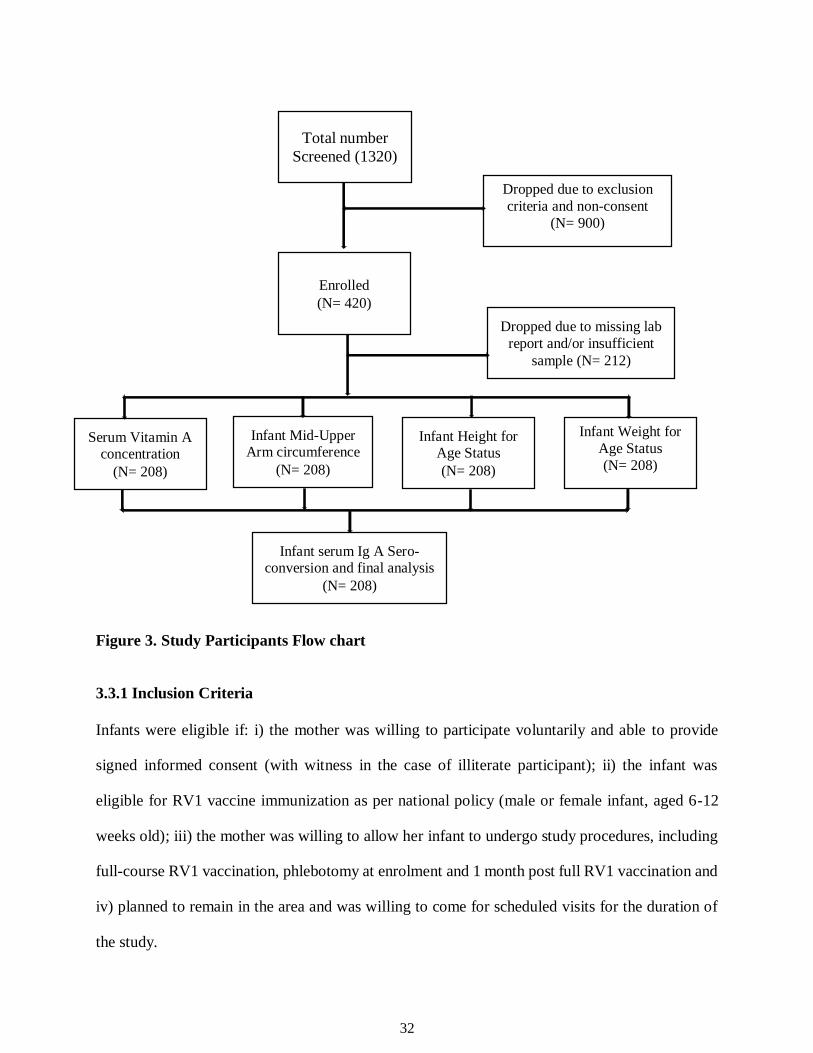
32
Figure 3. Study Participants Flow chart
3.3.1 Inclusion Criteria
Infants were eligible if: i) the mother was willing to participate voluntarily and able to provide
signed informed consent (with witness in the case of illiterate participant); ii) the infant was
eligible for RV1 vaccine immunization as per national policy (male or female infant, aged 6-12
weeks old); iii) the mother was willing to allow her infant to undergo study procedures, including
full-course RV1 vaccination, phlebotomy at enrolment and 1 month post full RV1 vaccination and
iv) planned to remain in the area and was willing to come for scheduled visits for the duration of
the study.
Total number
Screened (1320)
Infant Height for Age Status
(N= 208)
Infant Mid-Upper Arm circumference
(N= 208)
Serum Vitamin A concentration
(N= 208)
Enrolled
(N= 420)
Infant Weight for
Age Status
(N= 208)
Dropped due to exclusion
criteria and non-consent
(N= 900)
Dropped due to missing lab
report and/or insufficiTotal number Screened (1320)
Total number Screened
(1320)
ent sample (N= 212)
(N= 900)
Dropped due to missing lab
report and/or insufficient
sample (N= 212)
Infant serum Ig A Sero-conversion and final analysis
(N= 208)
33
3.3.2 Exclusion Criteria
Infants were not eligible for the study if they had received any rotavirus vaccine prior to the study,
suffered from any congenital or chronic gastrointestinal diseases including any uncorrected
congenital malformation of the gastrointestinal tract, received any immunosuppressant or
immunoglobulin, or had a history of gastroenteritis seven days prior to their visit to the clinic.
3.3.3 Sample Size Determination
Sample size calculation was achieved by using the population data which suggests a sero-
conversion rate of 60% (Chilengi et al., 2016) and a total of 232 infant paired samples were
required to detect 50% reduction in sero-conversion at 80% power with a 2 sided Chi-squared test
with a 5% level of significance.
3.3.3.1 Sampling Method
Simple random sampling was used to select participants/samples for analysis.
Sample size was established by the following formula;
Sample Size= 2SD2−(𝑍∝ + 𝑍1−𝛽)2
𝑑2
Where; SD is the Standard Deviation of the population =1.15,
Zα = 1.96 (From the Z table) at type 1 error of 5%, Z = 0.842 (from the Z table) at 80% power,
d = 0.30 expected effect size.
This gives us;
Sample Size =2*(1.15)2(1.96 + 0.842)
2/ (0.30)
2
Sample Size = 232 were required to detect the difference. Convenient sampling was used for there
was not enough samples to be included in the final analysis.
34
3.3.2 Clinic Procedures
Enrolled infants received a physical examination and a baseline serum sample was taken for
rotavirus-specific IgA antibodies to determine natural infection (serum anti RV-IgA sero-
positivity) and serum vitamin A concentration. Infants were then administered the full expanded
programme on immunization (EPI) vaccine regimen, which included the oral RV1 and
poliovaccines, and parenteral pentavalent vaccines (containing diphtheria, tetanus, pertussis,
haemophilus influenza, and hepatitis B). They were given an appointment card highlighting the
next scheduled visit (one month post the second RV dose) and were escorted home by a research
assistant in order to record the physical location of their residence, as there are no street addresses
in the clinic catchment area. The nutritional status of children was also assessed by anthropometric
measurements at base-line just before rotavirus vaccination. Child nutritional indicators of height
for age Z-scores (HAZ), weight for age Z scores (WAZ) and mid upper arm circumference
(MUAC) were calculated according to the WHO 2009 growth standard (WHO, 2006). We used
the most common thresholds: HAZ < −2 Z scores to define stunting or chronic malnutrition, WAZ
< −2 Z scores to define underweight or global malnutrition and MUAC of <11.5cm for severe
acute malnutrition, 11.5 to 12.4cm for moderate acute malnutrition, 12.5 to 13.4cm as risk of
malnutrition and > 13.5 as being well nourished. One month after the second dose of RV1 serum
sample was taken for rotavirus-specific IgA antibodies to determine infant sero-conversion
following vaccination.
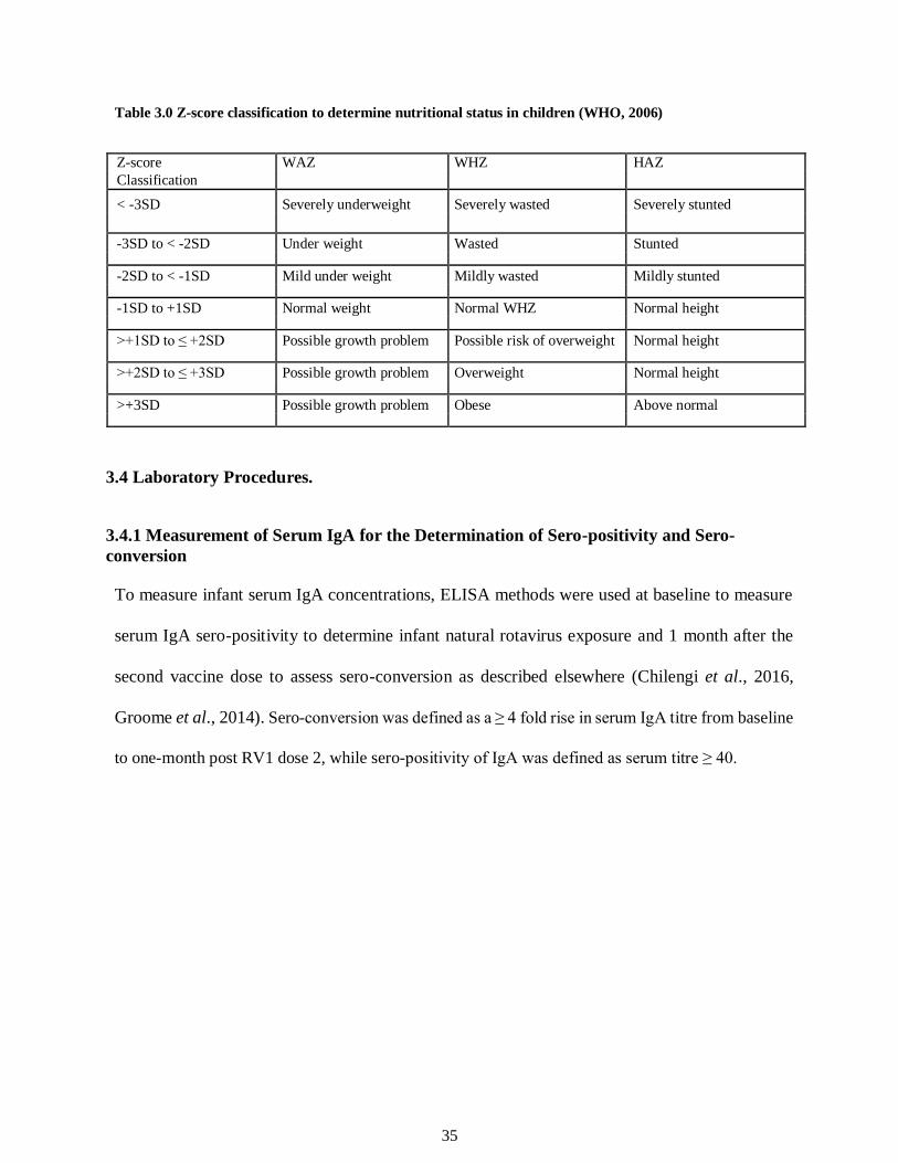
35
Table 3.0 Z-score classification to determine nutritional status in children (WHO, 2006)
Z-score WAZ WHZ HAZ
Classification
< -3SD Severely underweight Severely wasted Severely stunted
-3SD to < -2SD Under weight Wasted Stunted
-2SD to < -1SD Mild under weight Mildly wasted Mildly stunted
-1SD to +1SD Normal weight Normal WHZ Normal height
>+1SD to ≤ +2SD Possible growth problem Possible risk of overweight Normal height
>+2SD to ≤ +3SD Possible growth problem Overweight Normal height
>+3SD Possible growth problem Obese Above normal
3.4 Laboratory Procedures.
3.4.1 Measurement of Serum IgA for the Determination of Sero-positivity and Sero-
conversion To measure infant serum IgA concentrations, ELISA methods were used at baseline to measure
serum IgA sero-positivity to determine infant natural rotavirus exposure and 1 month after the
second vaccine dose to assess sero-conversion as described elsewhere (Chilengi et al., 2016,
Groome et al., 2014). Sero-conversion was defined as a ≥ 4 fold rise in serum IgA titre from baseline
to one-month post RV1 dose 2, while sero-positivity of IgA was defined as serum titre ≥ 40.
36
3.4.2 Measurement of Serum Vitamin A to Determine the Prevalence of Vitamin A
Deficiency Serum samples were evaluated for Vitamin A level using Quantikine® ELISA human retinol
binding protein4 (RBP4) Immunoassay which is used as a surrogate measure for serum VA (RnS
systems, MN, USA) according to the manufacturer’s instructions. Vitamin A status was defined
as normal serum vitamin A concentration when serum RBP4 was > 0.70μmol/L, moderate deficiency
was defined as < 0.70μmol/L and severely deficient as ≤ 0.35 μmol/L of serum RBP4 concentration
respectively.
3.5 Data Analysis
Children were defined as having sero-converted when at 1 month post immunization, the rotavirus
specific-IgA titres increased by four fold or greater compared with the titre recorded before the
first dose of RV. Pearson Chi-squared test was used to investigate the association of sero-
conversion and categorical factors (i.e. sex, vitamin A status, underweight, stunting and infant
sero-positivity at baseline) at 95% confidence level and p value less than 0.05 was considered
statistically significant.
3.6 Ethical Considerations
Ethical Clearance was sought from the University of Zambia Biomedical Research Ethic
committee (UNZABREC) of the School of Medicine (REF. No. 013-02-16). The study posed no
additional risks to the participants as no extra samples were collected from them apart from those
indicated by the attending physician as part of routine standard of care. Patient names were not
used; instead, unique identification codes were used in order to ensure confidentiality. Written
consent was sought from parents/guardians of the participants prior to sample collection.
Information obtained from the patients was strictly confined to academic use only, unless
37
otherwise there was clinical indication necessary to allow a shared confidentiality in good faith of
the patient concerned.
3.7 Limitations
This study had several limitations, including the small sample size, and the lack of collection of
serum samples between doses. Only serum vitamin A was measured and other micronutrients like
Iron and Zinc whose deficiency have been implicated in diarrhoeal diseases and poor vaccine
response were not included in the study.
38
CHAPTER 4: RESULTS
4.1 Patient Recruitment and Descriptives
Of a total of 1320 infants that were screened, 420 were successfully enrolled. The most common
reason for declining to participate was unwillingness for the infant’s mother to have the infant’s
blood drawn (3.5ml of blood was drawn from each infant), methods of enrolment have been
published elsewhere (Chilengi et al., 2016). From the 420 infants who were successfully enrolled,
208 serum samples were analyzed to determine infant serum vitamin A concentration and serum
anti RV IgA at base-line and one month post vaccination.
4.2 Prevalence of Poor Nutritional Status in Infants and Infant Early Exposure to
Rotavirus Infection The nutritional status of the infants was determined by infant serum vitamin A status and weight
for age status, height for age status. Overall baseline serum vitamin A deficiency was 78.8%
(164/208). Baseline infant vitamin A moderate deficiency was 63.9%(133/208) and vitamin A
severe deficiency was14.9%(31/208). Infants who were categorized as being underweight or
stunted had Z-scores of ≤-2 and as having normal weight and height Z-scores of >-2 respectively.
Infant base-line underweight was 6.3%(13/208) and stunting was at 52.4%(109/208). MUAC
status was also used to determine infant nutritional status and those whose were well nourished
were 10.1%(21/208), 39.4%(82/208) of the infants were at risk of malnutrition, 37.95%(79/208)
of the infants had moderate acute malnutrition and 12.5% (26/208) infants had severe acute
malnutrition. Infant early exposure to rotavirus infection was determined by the presence of anti-
rotavirus specific IgA in infant serum at base-line and infant sero-positivity was 48(23.2%) as
shown in Table 4.1.
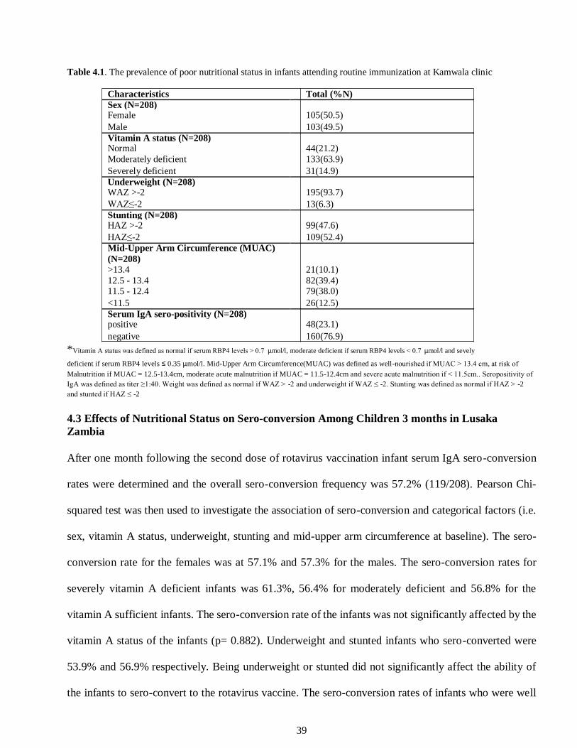
39
Table 4.1. The prevalence of poor nutritional status in infants attending routine immunization at Kamwala clinic
Characteristics Total (%N)
Sex (N=208)
Female 105(50.5)
Male 103(49.5)
Vitamin A status (N=208)
Normal 44(21.2)
Moderately deficient 133(63.9)
Severely deficient 31(14.9)
Underweight (N=208)
WAZ >-2 195(93.7)
WAZ≤-2 13(6.3)
Stunting (N=208)
HAZ >-2 99(47.6)
HAZ≤-2 109(52.4)
Mid-Upper Arm Circumference (MUAC)
(N=208)
>13.4 21(10.1)
12.5 - 13.4 82(39.4) 11.5 - 12.4 79(38.0)
<11.5 26(12.5)
Serum IgA sero-positivity (N=208)
positive 48(23.1)
negative 160(76.9) *Vitamin A status was defined as normal if serum RBP4 levels ˃ 0.7 μmol/l, moderate deficient if serum RBP4 levels ˂ 0.7 μmol/l and sevely deficient if serum RBP4 levels ≤ 0.35 μmol/l. Mid-Upper Arm Circumference(MUAC) was defined as well-nourished if MUAC ˃ 13.4 cm, at risk of
Malnutrition if MUAC = 12.5-13.4cm, moderate acute malnutrition if MUAC = 11.5-12.4cm and severe acute malnutrition if ˂ 11.5cm.. Seropositivity of
IgA was defined as titer ≥1:40. Weight was defined as normal if WAZ ˃ -2 and underweight if WAZ ≤ -2. Stunting was defined as normal if HAZ ˃ -2
and stunted if HAZ ≤ -2
4.3 Effects of Nutritional Status on Sero-conversion Among Children 3 months in Lusaka
Zambia After one month following the second dose of rotavirus vaccination infant serum IgA sero-conversion
rates were determined and the overall sero-conversion frequency was 57.2% (119/208). Pearson Chi-
squared test was then used to investigate the association of sero-conversion and categorical factors (i.e.
sex, vitamin A status, underweight, stunting and mid-upper arm circumference at baseline). The sero-
conversion rate for the females was at 57.1% and 57.3% for the males. The sero-conversion rates for
severely vitamin A deficient infants was 61.3%, 56.4% for moderately deficient and 56.8% for the
vitamin A sufficient infants. The sero-conversion rate of the infants was not significantly affected by the
vitamin A status of the infants (p= 0.882). Underweight and stunted infants who sero-converted were
53.9% and 56.9% respectively. Being underweight or stunted did not significantly affect the ability of
the infants to sero-convert to the rotavirus vaccine. The sero-conversion rates of infants who were well
40
nourished as determined by their MUAC was 13(61.9%), 50(61.0%) for those who at were risk of
malnutrition, 44(55.70%) for those who were moderately malnourished and for the infants who were
severely malnourished was 12(46.2). Moderate or severe acute malnutrition did not significantly affect
the ability of these infants to sero-convert to rotavirus vaccine (p=0.565). Infant who were exposed to
rotavirus infection before RV1 vaccine administration had a sero-conversion rate of 28(48.3%) and early
exposure to rotavirus infection did not significantly affect the ability of these infants to sero-convert to
rotavirus vaccine (p=0.832) as shown in Table 4.2.
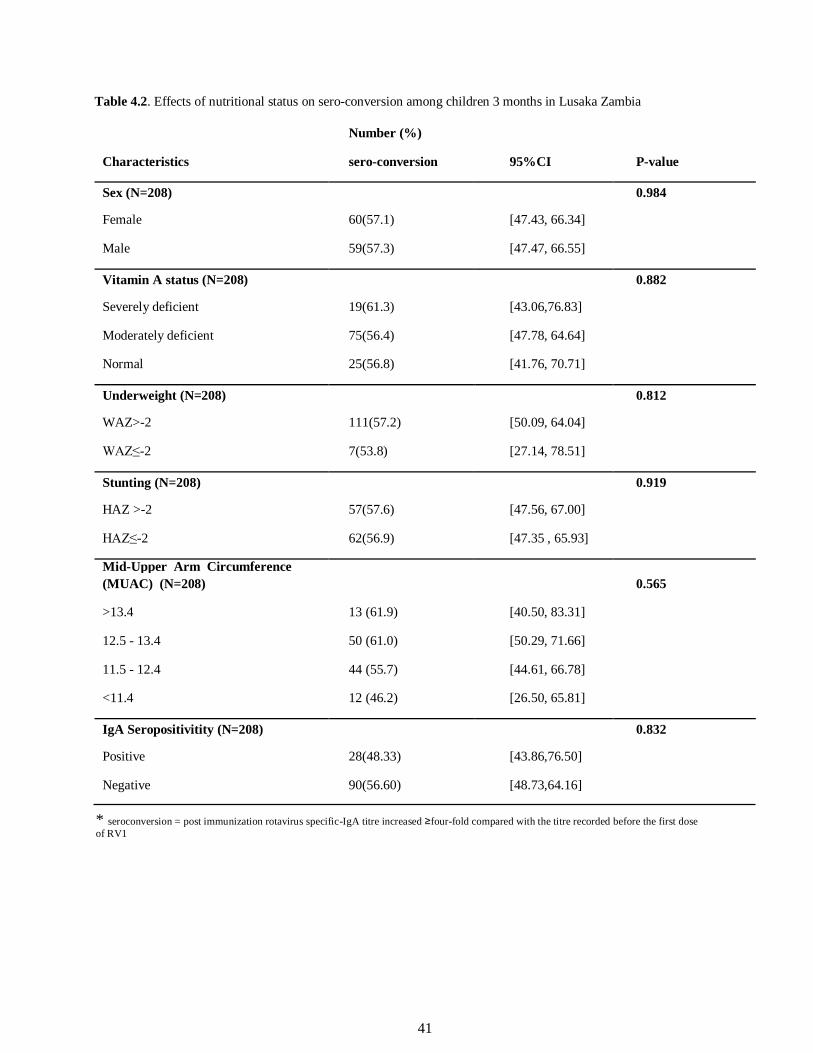
41
Table 4.2. Effects of nutritional status on sero-conversion among children 3 months in Lusaka Zambia
Number (%)
Characteristics sero-conversion 95%CI P-value
Sex (N=208) 0.984
Female 60(57.1) [47.43, 66.34]
Male 59(57.3) [47.47, 66.55]
Vitamin A status (N=208) 0.882
Severely deficient 19(61.3) [43.06,76.83]
Moderately deficient 75(56.4) [47.78, 64.64]
Normal 25(56.8) [41.76, 70.71]
Underweight (N=208) 0.812
WAZ>-2 111(57.2) [50.09, 64.04]
WAZ≤-2 7(53.8) [27.14, 78.51]
Stunting (N=208) 0.919
HAZ >-2 57(57.6) [47.56, 67.00]
HAZ≤-2 62(56.9) [47.35 , 65.93]
Mid-Upper Arm Circumference
(MUAC) (N=208) 0.565
>13.4 13 (61.9) [40.50, 83.31]
12.5 - 13.4 50 (61.0) [50.29, 71.66]
11.5 - 12.4 44 (55.7) [44.61, 66.78]
<11.4 12 (46.2) [26.50, 65.81]
IgA Seropositivitity (N=208) 0.832
Positive 28(48.33) [43.86,76.50]
Negative 90(56.60) [48.73,64.16]
* seroconversion = post immunization rotavirus specific-IgA titre increased ≥four-fold compared with the titre recorded before the first dose
of RV1
42
CHAPTER 5: DISCUSSION, CONCLUSION AND RECOMENDATIONS
5.1 Discussion
The main focus of this study was to investigate if the nutritional status (Vitamin A, WAZ, HAZ
and MUAC) have an effect on the sero-conversion following rotavirus vaccination. There were
four key findings from this study: a) Infant serum specific RV-IgA sero-conversion after RV
vaccination was not influenced by the base-line VA status of the infants although VAD was
generally high in this cohort. b) base-line infant underweight was low and that underweight did
not influence the level of Infant serum specific anti RV-IgA sero-conversion rate following RV
vaccination; c) base-line infant stunting was very high and that stunting did not influence the level
of Infant serum specific RV-IgA sero-conversion rate following RV vaccination; d) base-line
severe acute malnutrition determined by the infant mid-upper arm circumference (MUAC) did not
significantly affect infant sero-conversion rates following RV vaccination.
The effect of VA status and malnutrition on vaccine responses in children in recent times has
received particular attention, although their effect on rotavirus vaccines has not been extensively
studied (Heilskov et al., 2014, Savy et al., 2009). Our findings of no association between VA status
and rotavirus IgA sero-conversion rates have been consistent with the results of randomised
controlled trials, observational and cross-sectional studies on the effects of vitamin A
supplementation (VAS) and VAD in children on sero-conversion rates of vaccines. VAS was
shown to be associated with a small decrease in response to the tuberculin skin test after BCG
vaccination in children from Malawi and Guinea-Bissau respectively (Diness et al., 2007). It has
also being shown that although mean antibody titters tended to be higher in participants who
received VA, sero-conversion rates were universally high and did not significantly differ between
supplemented and un-supplemented participants in measles vaccine (MV) in studies carried out in
43
India, Jamaica and Guinea-Bissau respectively (Benn et al., 2002, Rytter et al., 2014). VA has
been shown to have no effect on sero-conversion rates to any of the 3 serotypes of poliomyelitis
vaccine, (Bahl et al., 2002) and has no discernable effect on other vaccines, including,
cholera,(Qadri et al., 2016) influenza (Gardner et al., 2000), Hib (Zandoh et al., 2007), and
pneumococcal vaccines (Deloria-Knoll et al., 2006).
Our observed low prevalence of under-weight and high prevalence of stunting was in-line with the
report of WHO on global nutritional trends 2016 (Organization, 2016), this report suggested that
some countries have low underweight prevalence but unacceptably high stunting rates, for
example, in Guatemala, Liberia, Malawi, Mozambique, Rwanda, and the United Republic of
Tanzania, child underweight prevalence is lower than 20 per cent, while stunting prevalence
remains above 40%. Our infant underweight prevalence was also in line with the findings of the
Zambia Demographic of Health Survey of 2013- 2014 (Demographic, 2015) but stunting rates are
higher in our observation ((51.2% (103/201)) than the predicted rates of the ZDHS for Lusaka
Province stunting rates (35.7%) (Demographic, 2015). Malnutrition defined as low HAZ and WAZ
was shown to have no detectable effect on antibody responses to other vaccines, like the measles
vaccine (Bhaskaram, 1995). A study carried out in Nigeria to assess the effect of malnutrition on
responses to OPV in young children, found that the mean increase in antibody titres after 3 doses
of OPV did not significantly differ between infants severely or moderately malnourished
(according to their weight-for-age) and well-nourished infants (Greenwood et al., 1986). In
addition, no correlation was found between the antibody response to OPV and serum albumin
levels, used as an indicator of nutritional status. Similarly, a study conducted in 62 Indian children
aged 1–12 mo showed no difference in sero-conversion rates after 5 doses of the trivalent OPV
between under-weight and non-underweight children (L Heymann et al., 1987). However, the
analyses were not adjusted for any variables, not even the pre-vaccination antibody levels, which
44
were found to be sero-positive for some of the children. These findings were not in agreement with
an Egyptian study that was conducted in 11- to 23-mo-old children (Hafez et al., 1977). Children
suffering from kwashiorkor and marasmus were matched with healthy children of the same age
and sex and they all received a single dose of the OPV. The authors reported that antibody titers
to the vaccine were significantly lower in children with kwashiorkor or marasmus than normal
controls, suggesting a depression in antibody production against poliovirus vaccine in children
with malnutrition. A study carried out in Brazil to evaluate the effects of malnutrition of the sero-
conversion rates to RRV-TV infants, suggested that RRV-TV may achieve higher levels of ser-
conversion among well-nourished than in malnourished infants (Linhares et al., 2002). On the
MUAC basis, a study carried out in the Gambia by Moore (Moore et al., 2003) found no significant
association between MUAC indices and sero-conversion rates following the double doses of rabies
vaccines. The findings of these studies are not all in agreement with the findings of our study,
although similarities where seen in the findings of the Nigerian and Indian studies (Greenwood et
al., 1986, Hafez et al., 1977), the study populations for these studies were quiet small, the effects
of natural infections were over-looked and the studies were done over 30 ago using rather old
methods. The age of the study participants were usually older than the cohort in our study, 6-12
weeks old infants. This age group is important for such studies because it is at this age that most
vaccines are recommended for administration including OPV, rotavirus vaccines, and parenteral
pentavalent vaccines (containing diphtheria, tetanus, pertussis, haemophilus influenza, and
hepatitis B) therefore, understanding the nutritional status of the infants just before vaccination
would better the understanding of the performance of vaccines in relation to their nutritional status.
45
Another important finding our study was the relatively high rate of pre-vaccination IgA sero-
positivity. This finding suggests that almost quarter of the infants were already exposed to wild
rotavirus infection very early in life before rotavirus vaccination. There was no evidence of
association between infant serum IgA sero-conversion and serum IgA sero-positivity, it remains
to be concluded whether early natural exposure could also negatively influence vaccine sero-
conversion as previously observed in South Africa and in Nicaragua (Becker-Dreps et al., 2014,
Groome et al., 2014).
Our observation of no evidence of association between infant nutritional status (vitamin A status,
HAZ, WAZ and MUAC) was not always consistent with the findings of most of the studies
conducted on malnutrition and vaccination and we therefore, suggest more research in this light to
investigate the particular arm of the immune response poor infant nutritional status may have on
the rotavirus vaccine (Prendergast, 2015, Savy et al., 2009). As many LMICs seek to attain
millennium development goals, reduction of child mortality through interventions that include
“effective child vaccines” is an important strategy. Research such as ours, seeking to understand
and explain challenges of achieving rotavirus vaccine impact is of great importance.
5.2 Conclusion
The complexity and lack of understanding of sero-conversion to vaccines and its meaning in terms
of clinical protection or vaccine efficacy still prevails at this point. Based on our findings, we
conclude that poor IgA sero-conversion frequency observed in this cohort was not influenced by
nutritional factors indicated by infant serum Vitamin A, HAZ, WAZ and MUAC. These factors
may have other effects later in childhood, as no difference was observed among sero-convertors
and non-sero-convertors to rotavirus vaccination in the 6-12 weeks age group.

46
5.3 Recommendations
Further research is needed to better understand vaccine sero-conversion. The relationship between
EED and RV vaccine efficacy remains a subject of interest for our on-going research.
47
REFERENCES
Agócs, M. M., Serhan, F., Yen, C., Mwenda, J. M., De Oliveira, L. H., Teleb, N., Wasley, A.,
Wijesinghe, P. R., Fox, K. & Tate, J. E. 2014. WHO global rotavirus surveillance network:
a strategic review of the first 5 years, 2008–2012. MMWR. Morbidity and mortality weekly
report, 63, 634.
Ahmed, T., Arifuzzaman, M., Lebens, M., Qadri, F. & Lundgren, A. 2009. CD4+ T-cell responses
to an oral inactivated cholera vaccine in young children in a cholera endemic country and
the enhancing effect of zinc supplementation. Vaccine, 28, 422-9.
Aiyegbo, M. S., Sapparapu, G., Spiller, B. W., Eli, I. M., Williams, D. R., Kim, R., Lee, D. E.,
Liu, T., Li, S., Woods, V. L., Jr., Nannemann, D. P., Meiler, J., Stewart, P. L. & Crowe, J.
E., Jr. 2013. Human rotavirus VP6-specific antibodies mediate intracellular neutralization
by binding to a quaternary structure in the transcriptional pore. PLoS One, 8, e61101.
Angel, J., Franco, M. A. & Greenberg, H. B. 2012. Rotavirus immune responses and correlates of
protection. Current Opinion in Virology, 2, 419-25.
Appaiahgari, M. B., Glass, R., Singh, S., Taneja, S., Rongsen-Chandola, T., Bhandari, N., Mishra,
S. & Vrati, S. 2014. Transplacental rotavirus IgG interferes with immune response to live
oral rotavirus vaccine ORV-116E in Indian infants. Vaccine, 32, 651-6.
Armah, G., Pringle, K., Enweronu- Laryea, C. C., Ansong, D., Mwenda, J. M., Diamenu, S. K.,
Narh, C., Lartey, B., Binka, F., Grytdal, S., Patel, M., Parashar, U. & Lopman, B. 2016.
Impact and Effectiveness of Monovalent Rotavirus Vaccine Against Severe Rotavirus
Diarrhea in Ghana. Clinical Infectious Diseases, 62 Suppl 2, S200-7.
Armah, G. E. & Binka, F. N. 2014. Sustaining rotavirus vaccination in Africa: measuring vaccine
effectiveness. Lancet Infectious Diseases, 14, 1031-1032.
Armah, G. E., Sow, S. O., Breiman, R. F., Dallas, M. J., Tapia, M. D., Feikin, D. R., Binka, F. N.,
Steele, A. D., Laserson, K. F., Ansah, N. A., Levine, M. M., Lewis, K., Coia, M. L., Attah-
Poku, M., Ojwando, J., Rivers, S. B., Victor, J. C., Nyambane, G., Hodgson, A., Schodel,
F., Ciarlet, M. & Neuzil, K. M. 2010. Efficacy of pentavalent rotavirus vaccine against
severe rotavirus gastroenteritis in infants in developing countries in sub- Saharan Africa: a
randomised, double-blind, placebo-controlled trial. Lancet, 376, 606-14.
Aupiais, C., De Rougemont, A., Menager, C., Vallet, C., Brasme, J. F., Kaplon, J., Pothier, P. &
Gendrel, D. 2009. Severity of acute gastroenteritis in infants infected by G1 or G9
rotaviruses. Journal of Clinical Virology, 46, 282-5.
Bahl, R., Bhandari, N., Kant, S., Molbak, K., Ostergaard, E. & Bhan, M. K. 2002. Effect of vitamin
A administered at Expanded Program on Immunization contacts on antibody response to
oral polio vaccine. European Journal of Clinical Nutrition, 56, 321-5.
48
Becker-Dreps, S., Bucardo, F., Vilchez, S., Zambrana, L. E., Liu, L., Weber, D. J., Peña, R.,
Barclay, L., Vinjé, J., Hudgens, M. G., Nordgren, J., Svensson, L., Morgan, D. R.,
Espinoza, F. & Paniagua, M. 2014. Etiology of childhood diarrhea after rotavirus vaccine
introduction: a prospective, population-based study in Nicaragua. The Pediatric infectious
disease journal, 33, 1156-1163.
Beijer, M. R., Kraal, G. & Den Haan, J. M. 2014. Vitamin A and dendritic cell differentiation.
Immunology, 142, 39-45.
Benn, C. S., Balde, A., George, E., Kidd, M., Whittle, H., Lisse, I. M. & Aaby, P. 2002. Effect of
vitamin A supplementation on measles-specific antibody levels in Guinea-Bissau. Lancet,
359, 1313-4.
Beres, L. K., Tate, J. E., Njobvu, L., Chibwe, B., Rudd, C., Guffey, M. B., Stringer, J. S., Parashar,
U. D. & Chilengi, R. 2016. A Preliminary Assessment of Rotavirus Vaccine Effectiveness
in Zambia. Clinical Infectious Diseases, 62 Suppl 2, S175-82.
Bhandari, N., Rongsen-Chandola, T., Bavdekar, A., John, J., Antony, K., Taneja, S., Goyal, N.,
Kawade, A., Kang, G., Rathore, S. S., Juvekar, S., Muliyil, J., Arya, A., Shaikh, H.,
Abraham, V., Vrati, S., Proschan, M., Kohberger, R., Thiry, G., Glass, R., Greenberg, H.
B., Curlin, G., Mohan, K., Harshavardhan, G. V., Prasad, S., Rao, T. S., Boslego, J. &
Bhan, M. K. 2014. Efficacy of a monovalent human-bovine (116E) rotavirus vaccine in
Indian children in the second year of life. Vaccine, 32 Suppl 1, A110-6.
Bhaskaram, P. 1995. Measles & malnutrition.
Bhowmick, R., Halder, U. C., Chattopadhyay, S., Nayak, M. K. & Chawla-Sarkar, M. 2013.
Rotavirus-encoded nonstructural protein 1 modulates cellular apoptotic machinery by
targeting tumor suppressor protein p53. Journal of virology, 87, 6840-6850.
Black, R. E., Victora, C. G., Walker, S. P., Bhutta, Z. A., Christian, P., De Onis, M., Ezzati, M.,
Grantham-Mcgregor, S., Katz, J., Martorell, R. & Uauy, R. 2013. Maternal and child
undernutrition and overweight in low-income and middle-income countries. Lancet, 382,
427-451.
Block, S. L., Vesikari, T., Goveia, M. G., Rivers, S. B., Adeyi, B. A., Dallas, M. J., Bauder, J.,
Boslego, J. W. & Heaton, P. M. 2007. Efficacy, immunogenicity, and safety of a
pentavalent human-bovine (WC3) reassortant rotavirus vaccine at the end of shelf life.
Pediatrics, 119, 11-8.
Bloem, M. 2007. The 2006 WHO child growth standards. BMJ (Clinical research ed.), 334, 705-
706.
Blutt, S. E., Matson, D. O., Crawford, S. E., Staat, M. A., Azimi, P., Bennett, B. L., Piedra, P. A.
& Conner, M. E. 2007. Rotavirus antigenemia in children is associated with viremia.
PLoS Med, 4, e121.
49
Boom, J. A., Tate, J. E., Sahni, L. C., Rench, M. A., Hull, J. J., Gentsch, J. R., Patel, M. M., Baker,
C. J. & Parashar, U. D. 2010. Effectiveness of pentavalent rotavirus vaccine in a large
urban population in the United States. Pediatrics, 125, e199-207.
Buttery, J. P., Lambert, S. B., Grimwood, K., Nissen, M. D., Field, E. J., Macartney, K. K.,
Akikusa, J. D., Kelly, J. J. & Kirkwood, C. D. 2011. Reduction in rotavirus-associated
acute gastroenteritis following introduction of rotavirus vaccine into Australia's National
Childhood vaccine schedule. The Pediatric infectious disease journal, 30, S25-9.
Chilengi, R., Simuyandi, M., Beach, L., K. Becker-Dreps S, M., Emperador, D., Velasquez, D.,
Bosomprah, S. & Jiang, B. 2016. Association of Maternal Immunity with Rotavirus Vaccine
Immunogenicity in Zambian Infants.
Clarke, E. & Desselberger, U. 2014. Correlates of protection against human rotavirus disease and
the factors influencing protection in low-income settings. Mucosal Immunology, 8, 1.
Corcoran, M., Well, G. & Loo, I. 2014. Diagnosis of viral gastroenteritis in children:
interpretation of real-time PCR results and relation to clinical symptoms.
Cortese, M. M., Immergluck, L. C., Held, M., Jain, S., Chan, T., Grizas, A. P., Khizer, S., Barrett,
C., Quaye, O., Mijatovic-Rustempasic, S., Gautam, R., Bowen, M. D., Moore, J., Tate, J.
E., Parashar, U. D. & Vazquez, M. 2013. Effectiveness of monovalent and pentavalent
rotavirus vaccine. Pediatrics, 132, e25-33.
Corthesy, B., Benureau, Y., Perrier, C., Fourgeux, C., Parez, N., Greenberg, H. & Schwartz-Cornil,
I. 2006. Rotavirus anti- VP6 secretory immunoglobulin A contributes to protection via
intracellular neutralization but not via immune exclusion. Journal of Virology, 80, 10692-
9.
Criglar, J. M., Hu, L., Crawford, S. E., Hyser, J. M., Broughman, J. R., Prasad, B. V. & Estes, M.
K. 2014. A novel form of rotavirus NSP2 and phosphorylation-dependent NSP2-NSP5
interactions are associated with viroplasm assembly. Journal of Virology, 88, 786-98.
Cusick, S. E. & Georgieff, M. K. 2012. Nutrient supplementation and neurodevelopment: timing
is the key. Archives of Pediatrics and Adolescent Medicine, 166, 481-2.
Danchin, M. H. & Bines, J. E. 2009. Defeating rotavirus? The global recommendation for rotavirus
vaccination. New England Journal of Medicine, 361, 1919-1921.
Deal, E. M., Lahl, K., Narvaez, C. F., Butcher, E. C. & Greenberg, H. B. 2013. Plasmacytoid
dendritic cells promote rotavirus-induced human and murine B cell responses. Journal of
Clinical Investigations, 123, 2464-74.
Deloria-Knoll, M., Steinhoff, M., Semba, R. D., Nelson, K., Vlahov, D. & Meinert, C. L. 2006.
Effect of zinc and vitamin A supplementation on antibody responses to a pneumococcal
conjugate vaccine in HIV-positive injection drug users: a randomized trial. Vaccine, 24,
1670-9.
Demographic, Z. 2015. Health survey (ZDHS) 2013-2014. House hold population by Age and sex.
Lusaka: Zambia.
50
Dennehy, P. H. 2008. Rotavirus Vaccines: an Overview. Clinical Microbiology Reviews, 21, 198-
208.
Desselberger, U. 2014. Rotaviruses. Virus Research, 190, 75-96.
Diness, B. R., Fisker, A. B., Roth, A., Yazdanbakhsh, M., Sartono, E., Whittle, H., Nante, J. E.,
Lisse, I. M., Ravn, H., Rodrigues, A., Aaby, P. & Benn, C. S. 2007. Effect of high-dose
vitamin A supplementation on the immune response to Bacille Calmette-Guerin vaccine.
American Journal of Clinical Nutrition, 86, 1152-9.
Dormitzer, P. R., Nason, E. B., Prasad, B. V. & Harrison, S. C. 2004. Structural rearrangements
in the membrane penetration protein of a non-enveloped virus. Nature, 430, 1053-8.
Fagbemi, A. O., Amadi, B., Salvestrini, C., Eklund, E. A., Torrente, F., Freeze, H. H., Golden, M.
H., Mwiya, M., Kelly, P., Day, R. & Murch, S. H. 2008. Reduced production of sulfated
glycosaminoglycans occurs in Zambian children with kwashiorkor but not marasmus. The
American Journal of Clinical Nutrition, 89, 592-600.
Feng, N., Sen, A., Nguyen, H., Vo, P., Hoshino, Y., Deal, E. M. & Greenberg, H. B. 2009.
Variation in Antagonism of the Interferon Response to Rotavirus NSP1 Results in
Differential Infectivity in Mouse Embryonic Fibroblasts. Journal of Virology, 83, 6987-
6994.
Fiorentino, M., Sophonneary, P., Laillou, A., Whitney, S., De Groot, R., Perignon, M., Kuong, K.,
Berger, J. & Wieringa, F. T. 2016. Current MUAC Cut-Offs to Screen for Acute
Malnutrition Need to Be Adapted to Gender and Age: The Example of Cambodia. PloS
one, 11, e0146442-e0146442.
Fischer, T. K., Valentiner-Branth, P., Steinsland, H., Perch, M., Santos, G., Aaby, P., Mølbak, K.
& Sommerfelt, H. 2002. Protective Immunity after Natural Rotavirus Infection: A
Community Cohort Study of Newborn Children in Guinea-Bissau, West Africa. The
Journal of Infectious Diseases, 186, 593-597.
Franco, M. A., Angel, J. & Greenberg, H. B. 2006a. Immunity and correlates of protection for
rotavirus vaccines. Vaccine, 24, 2718-2731.
Franco, M. A., Angel, J. & Greenberg, H. B. 2006b. Immunity and correlates of protection for
rotavirus vaccines. Vaccine, 24, 2718-31.
Furlong, K. R., Anderson, L. N., Kang, H., Lebovic, G., Parkin, P. C., Maguire, J. L., O'connor, D. L. & Birken, C. S. 2016. BMI-for-Age and Weight-for-Length in Children 0 to 2 Years. Pediatrics, 138.
Gaayeb, L., Pincon, C., Cames, C., Sarr, J. B., Seck, M., Schacht, A. M., Remoue, F., Hermann,
E. & Riveau, G. 2014. Immune response to Bordetella pertussis is associated with season
and undernutrition in Senegalese children. Vaccine, 32, 3431-7.
51
Gardner, E. M., Bernstein, E. D., Popoff, K. A., Abrutyn, E., Gross, P. & Murasko, D. M. 2000.
Immune response to influenza vaccine in healthy elderly: lack of association with plasma
beta-carotene, retinol, alpha-tocopherol, or zinc. Mechanism of Ageing Development
Journal, 117, 29-45.
Garrett, W. S., Gordon, J. I. & Glimcher, L. H. 2010. Homeostasis and inflammation in the
intestine. Cell, 140, 859-70.
Giaquinto, C., Van Damme, P., Huet, F., Gothefors, L., Maxwell, M., Todd, P. & Da Dalt, L. 2007.
Clinical consequences of rotavirus acute gastroenteritis in Europe, 2004-2005: the
REVEAL study. Journal of Infectious Diseases, 195 Suppl 1, S26-35.
Glass, R. I., Parashar, U. D., Bresee, J. S., Turcios, R., Fischer, T. K., Widdowson, M. A., Jiang,
B. & Gentsch, J. R. 2006. Rotavirus vaccines: current prospects and future challenges.
Lancet, 368, 323-32.
Gray, J., Vesikari, T., Van Damme, P., Giaquinto, C., Mrukowicz, J., Guarino, A., Dagan, R.,
Szajewska, H. & Usonis, V. 2008. Rotavirus. Journal of Pediatric Gastroenterology and
Nutrition, 46, S24-S31.
Greenberg, H. B. 2011. Rotavirus vaccination and intussusception--act two. New England Journal
of Medicine, 364, 2354-5.
Greenberg, H. B. & Estes, M. K. 2009. Rotaviruses: from pathogenesis to vaccination.
Gastroenterology, 136, 1939-51.
Greenwood, B. M., Bradley-Moore, A. M., Bradley, A. K., Kirkwood, B. R. & Gilles, H. M. 1986.
The immune response to vaccination in undernourished and well-nourished Nigerian
children. Annals of Tropical Medicine & Parasitology, 80, 537-544.
Groome, M. J., Page, N., Cortese, M. M., Moyes, J., Zar, H. J., Kapongo, C. N., Mulligan, C.,
Diedericks, R., Cohen, C., Fleming, J. A., Seheri, M., Mphahlele, J., Walaza, S., Kahn, K.,
Chhagan, M., Steele, A. D., Parashar, U. D., Zell, E. R. & Madhi, S. A. 2014. Effectiveness
of monovalent human rotavirus vaccine against admission to hospital for acute rotavirus
diarrhoea in South African children: a case-control study. The Lancet Infectious Diseases,
14, 1096-1104.
Haber, P., Parashar, U. D., Haber, M. & Destefano, F. 2015. Intussusception after monovalent
rotavirus vaccine-United States, Vaccine Adverse Event Reporting System (VAERS),
2008-2014. Vaccine, 33, 4873-7.
Haber, P., Patel, M., Izurieta, H. S., Baggs, J., Gargiullo, P., Weintraub, E., Cortese, M., Braun,
M. M., Belongia, E. A., Miller, E., Ball, R., Iskander, J. & Parashar, U. D. 2008.
Postlicensure monitoring of intussusception after RotaTeq vaccination in the United
States, February 1, 2006, to September 25, 2007. Pediatrics, 121, 1206-12.
Hafez, M., Aref, G. H., Mehareb, S. W., Kassem, A. S., El-Tahhan, H., Rizk, Z., Mahfouz, R. &
Saad, K. 1977. Antibody production and complement system in protein energy
malnutrition. The Journal of tropical medicine and hygiene, 80, 36-39.
52
Hammond, M. I., Myers, E. F. & Trostler, N. 2014. Nutrition Care Process and Model: An
Academic and Practice Odyssey. Journal of the Academy of Nutrition and Dietetics, 114,
1879-1891.
Haque, R., Snider, C., Liu, Y., Ma, J. Z., Liu, L., Nayak, U., Mychaleckyj, J. C., Korpe, P., Mondal,
D., Kabir, M., Alam, M., Pallansch, M., Oberste, M. S., Weldon, W., Kirkpatrick, B. D. &
Petri, W. A., Jr. 2014. Oral polio vaccine response in breast fed infants with malnutrition
and diarrhea. Vaccine, 32, 478-482.
Hashizume, M., Armstrong, B., Wagatsuma, Y., Faruque, A. S., Hayashi, T. & Sack, D. A. 2008. Rotavirus infections and climate variability in Dhaka, Bangladesh: a time-series analysis. Epidemiology and Infections, 136, 1281-9.
Heilskov, S., Rytter, M. J. H., Vestergaard, C., Briend, A., Babirekere, E. & Deleuran, M. S. 2014.
Dermatosis in children with oedematous malnutrition (Kwashiorkor): a review of the
literature. Journal of the European Academy of Dermatology and Venereology, 28, 995-
1001.
Hemming, M. & Vesikari, T. 2014. Detection of rotateq vaccine-derived, double-reassortant
rotavirus in a 7-year- old child with acute gastroenteritis. The Pediatric infectious disease
journal, 33, 655-656.
Hotz, C., Chileshe, J., Siamusantu, W., Palaniappan, U. & Kafwembe, E. 2012. Vitamin A intake
and infection are associated with plasma retinol among pre-school children in rural Zambia.
Public Health and Nutrition, 15, 1688-96.
Hou, Z., Huang, Y., Huan, Y., Pang, W., Meng, M., Wang, P., Yang, M., Jiang, L., Cao, X. & Wu,
K. K. 2008. Anti-NSP4 antibody can block rotavirus-induced diarrhea in mice. Journal of
pediatric gastroenterology and nutrition, 46, 376-385.
Hu, L., Crawford, S. E., Hyser, J. M., Estes, M. K. & Prasad, B. V. 2012. Rotavirus non-
structural proteins: structure and function. Current Opinions in Virology, 2, 380-8.
Janevic, T., Borrell, L. N., Savitz, D. A., Herring, A. H. & Rundle, A. 2010. Neighbourhood food
environment and gestational diabetes in New York City. Paediatric and Perinatal
Epidemiology, 24, 249-254.
Jiang, V., Jiang, B., Tate, J., Parashar, U. D. & Patel, M. M. 2010. Performance of rotavirus
vaccines in developed and developing countries. Human Vaccines, 6, 532-42.
Johansson, E., Istrate, C., Charpilienne, A., Cohen, J., Hinkula, J., Poncet, D., Svensson, L. &
Johansen, K. 2008. Amount of maternal rotavirus-specific antibodies influence the
outcome of rotavirus vaccination of newborn mice with virus-like particles. Vaccine, 26,
778-85.
Jones, A. D., Ickes, S. B., Smith, L. E., Mbuya, M. N., Chasekwa, B., Heidkamp, R. A., Menon,
P., Zongrone, A. A. & Stoltzfus, R. J. 2014. World Health Organization infant and young
child feeding indicators and their associations with child anthropometry: a synthesis of
recent findings. Maternal Child Nutrition, 10, 1-17.
53
Kawamura, N., Tokoeda, Y., Oshima, M., Okahata, H., Tsutsumi, H., Van Doorn, L. J., Muto, H.,
Smolenov, I., Suryakiran, P. V. & Han, H. H. 2011. Efficacy, safety and immunogenicity
of RIX4414 in Japanese infants during the first two years of life. Vaccine, 29, 6335-41.
Kennedy, E. T. 2005. The global face of nutrition: what can governments and industry do? The
Journal of nutrition, 135, 913-915.
Kim, I. S., Trask, S. D., Babyonyshev, M., Dormitzer, P. R. & Harrison, S. C. 2010. Effect of
mutations in VP5 hydrophobic loops on rotavirus cell entry. Journal of Virology, 84, 6200-
7.
Korpe, P. S. & Petri, W. A., Jr. 2012. Environmental enteropathy: critical implications of a poorly
understood condition. Trends in Molecular Medicine, 18, 328-36.
L Heymann, D., Murphy, K., Brigaud, M., Aymard, M., Tembon, A. & K Maben, G. 1987. Oral
poliovirus vaccine in tropical Africa: Greater impact on incidence of paralytic disease than
expected from coverage surveys and seroconversion rates.
Labadarios, D., Swart, E. & Maunder, E. 2008. Executive summary of the National Food
Consumption Survey Fortification Baseline (NFCS-FB-I) South Africa, 2005.
Lee, R. M., Lessler, J., Lee, R. A., Rudolph, K. E., Reich, N. G., Perl, T. M. & Cummings, D. a.
T. 2013. Incubation periods of viral gastroenteritis: a systematic review. BMC Infectious
Diseases, 13, 446.
Leshem, E., Lopman, B., Glass, R., Gentsch, J., Bányai, K., Parashar, U. & Patel, M. 2014.
Distribution of rotavirus strains and strain-specific effectiveness of the rotavirus vaccine
after its introduction: a systematic review and meta-analysis. The Lancet Infectious
Diseases, 14, 847-856.
Levin, D., Marryat, L., Cole, T. J., Mccoll, J., Harjunmaa, U., Ashorn, P. & Wright, C. 2016. Fit
to WHO weight standard of European infants over time. Archives of Disease in Childhood,
101, 455-60.
Linhares, A. C., Carmo, K. B. D., Oliveira, K. K., Oliveira, C. S., Freitas, R. B. D., Bellesi, N.,
Monteiro, T. a. F., Gabbay, Y. B. & Mascarenhas, J. D. a. P. 2002. Nutritional status in
relation to the efficacy of the rhesus-human reassortant, tetravalent rotavirus vaccine
(RRV-TV) in infants from Belém, pará state, Brazil. Revista do Instituto de Medicina
Tropical de Sao Paulo, 44, 13-16.
Linhares, A. C., Velazquez, F. R., Perez-Schael, I., Saez-Llorens, X., Abate, H., Espinoza, F.,
Lopez, P., Macias-Parra, M., Ortega-Barria, E., Rivera-Medina, D. M., Rivera, L., Pavia-
Ruz, N., Nunez, E., Damaso, S., Ruiz-Palacios, G. M., De Vos, B., O'ryan, M., Gillard, P.
& Bouckenooghe, A. 2008. Efficacy and safety of an oral live attenuated human rotavirus
vaccine against rotavirus gastroenteritis during the first 2 years of life in Latin American
infants: a randomised, double-blind, placebo-controlled phase III study. Lancet, 371, 1181-
9.
54
Liu, L., Oza, S., Hogan, D., Perin, J., Rudan, I., Lawn, J. E., Cousens, S., Mathers, C. & Black, R.
E. 2015a. Global, regional, and national causes of child mortality in 2000-13, with
projections to inform post-2015 priorities: an updated systematic analysis. Lancet, 385,
430-40.
Liu, L., Oza, S., Hogan, D., Perin, J., Rudan, I., Lawn, J. E., Cousens, S., Mathers, C. & Black, R.
E. 2015b. Global, regional, and national causes of child mortality in 2000–13, with
projections to inform post-2015 priorities: an updated systematic analysis. The Lancet, 385,
430-440.
Luchs, A., Cilli, A., Morillo, S. G., De Cassia Compagnoli Carmona, R. & Do Carmo Sampaio
Tavares Timenetsky, M. 2014. Rotavirus in adults, Brazil, 2004-2011: G2P[4] dominance
and potential impact on vaccination. Brazilian Journal of Infectious Diseases, 18, 53-9.
Lynch, M., Shieh, W. J., Bresee, J. S., Tatti, K. M., Gentsch, J. R., Jones, T., Jiang, B.,
Hummelman, E., Zimmerman, C. M., Zaki, S. R. & Glass, R. I. 2006. Intussusception after
administration of the rhesus tetravalent rotavirus vaccine (Rotashield): the search for a
pathogenic mechanism. Pediatrics, 117, e827-32.
Madhi, S. A., Cunliffe, N. A., Steele, D., Witte, D., Kirsten, M., Louw, C., Ngwira, B., Victor, J.
C., Gillard, P. H., Cheuvart, B. B., Han, H. H. & Neuzil, K. M. 2010. Effect of Human
Rotavirus Vaccine on Severe Diarrhea in African Infants. New England Journal of
Medicine, 362, 289-298.
Maes, P., Matthijnssens, J., Rahman, M. & Van Ranst, M. 2009. RotaC: a web-based tool for the
complete genome classification of group A rotaviruses. BMC Microbiology, 9, 238.
Mandeville, K. L., Krabshuis, J., Ladep, N. G., Mulder, C. J. J., Quigley, E. M. M. & Khan, S. A.
2009. Gastroenterology in developing countries: issues and advances. World journal of
gastroenterology, 15, 2839-2854.
Marcelin, G., Miller, A. D., Blutt, S. E. & Conner, M. E. 2011. Immune mediators of rotavirus
antigenemia clearance in mice. Journasl of Virology, 85, 7937-41.
Markkula, J., Hemming-Harlo, M. & Vesikari, T. 2014. Detection of Vaccine-Derived Rotavirus
Strains in Non-Immunocompromised Children up to 3-6 Months after RotaTeq®
Vaccination.
Martorell, R., Horta, B., Adair, L., Stein, A., Richter, L., H.D. Fall, C., Bhargava, S., Dey Biswas,
S. K., Perez, L., C Barros, F. & G Victora, C. 2009. Weight Gain in the First Two Years of
Life Is an Important Predictor of Schooling Outcomes in Pooled Analyses from Five Birth
Cohorts from Low- and Middle-Income Countries.
Masibo, P. K. & Makoka, D. 2012. Trends and determinants of undernutrition among young
Kenyan children: Kenya Demographic and Health Survey; 1993, 1998, 2003 and 2008-
2009. Public Health and Nutrition, 15, 1715-27.
55
Matthijnssens, J., Ciarlet, M., Heiman, E., Arijs, I., Delbeke, T., Mcdonald, S. M., Palombo, E. A.,
Iturriza-Gomara, M., Maes, P., Patton, J. T., Rahman, M. & Van Ranst, M. 2008. Full
genome-based classification of rotaviruses reveals a common origin between human Wa-
Like and porcine rotavirus strains and human DS-1-like and bovine rotavirus strains.
Journal of Virology, 82, 3204-19.
Matthijnssens, J., Ciarlet, M., Mcdonald, S. M., Attoui, H., Banyai, K., Brister, J. R., Buesa, J.,
Esona, M. D., Estes, M. K., Gentsch, J. R., Iturriza-Gomara, M., Johne, R., Kirkwood, C.
D., Martella, V., Mertens, P. P., Nakagomi, O., Parreno, V., Rahman, M., Ruggeri, F. M.,
Saif, L. J., Santos, N., Steyer, A., Taniguchi, K., Patton, J. T., Desselberger, U. & Van
Ranst, M. 2011a. Uniformity of rotavirus strain nomenclature proposed by the Rotavirus
Classification Working Group (RCWG). Archives of Virology, 156, 1397-413.
Matthijnssens, J., De Grazia, S., Piessens, J., Heylen, E., Zeller, M., Giammanco, G. M., Banyai,
K., Buonavoglia, C., Ciarlet, M., Martella, V. & Van Ranst, M. 2011b. Multiple
reassortment and interspecies transmission events contribute to the diversity of feline,
canine and feline/canine- like human group A rotavirus strains. Infections and Genetic
Evolutions, 11, 1396-406.
Matthijnssens, J., Otto, P. H., Ciarlet, M., Desselberger, U., Van Ranst, M. & Johne, R. 2012. VP6-sequence-based cutoff values as a criterion for rotavirus species demarcation. Archives of Virology, 157, 1177-82.
Matthijnssens, J., Zeller, M., Heylen, E., De Coster, S., Vercauteren, J., Braeckman, T., Van Herck,
K., Meyer, N., Pircon, J. Y., Soriano-Gabarro, M., Azou, M., Capiau, H., De Koster, J.,
Maernoudt, A. S., Raes, M., Verdonck, L., Verghote, M., Vergison, A., Van Damme, P. &
Van Ranst, M. 2014. Higher proportion of G2P[4] rotaviruses in vaccinated hospitalized
cases compared with unvaccinated hospitalized cases, despite high vaccine effectiveness
against heterotypic G2P[4] rotaviruses. Clinical Microbiology and Infections, 20, O702-
10.
Mcdonald, S. M. & Patton, J. T. 2008. Molecular characterization of a subgroup specificity
associated with the rotavirus inner capsid protein VP2. Journal of virology, 82, 2752-2764.
Mcdonald, S. M. & Patton, J. T. 2011. Rotavirus VP2 Core Shell Regions Critical for Viral
Polymerase Activation. Journal of Virology, 85, 3095-3105.
Meshram, I., Arlappa, N., Balakrishna, N., Laxmaiah, A., Kodavanti, M., Chitty, G. R.,
Ravindranath, M., Sharad Kumar, S. & Brahmam, G. 2011. Prevalence and Determinants
of Undernutrition and its Trends among Pre-School Tribal Children of Maharashtra State,
India.
Mondal, D., Haque, R., Sack, R. B., Kirkpatrick, B. D. & Petri, W. A., Jr. 2009. Attribution of
malnutrition to cause-specific diarrheal illness: evidence from a prospective study of
preschool children in Mirpur, Dhaka, Bangladesh. American Journal of Tropical Medicine
and Hygiene, 80, 824-6.
Moon, S. S., Wang, Y., Shane, A. L., Nguyen, T., Ray, P., Dennehy, P., Baek, L. J., Parashar, U.,
Glass, R. I. & Jiang, B. 2010. Inhibitory effect of breast milk on infectivity of live oral
rotavirus vaccines. Pediatric Infectious Diseases Journal, 29, 919-23.
56
Moore, S. E., Goldblatt, D., Bates, C. J. & Prentice, A. M. 2003. Impact of nutritional status on
antibody responses to different vaccines in undernourished Gambian children. Acta
Paediatrica, 92, 170-6.
Mora, J. R., Iwata, M. & Von Andrian, U. H. 2008. Vitamin effects on the immune system:
vitamins A and D take centre stage. Nature Reviews Immunology, 8, 685-98.
Mpabalwani, E. M., Simwaka, C. J., Mwenda, J. M., Mubanga, C. P., Monze, M., Matapo, B.,
Parashar, U. D. & Tate, J. E. 2016. Impact of Rotavirus Vaccination on Diarrheal
Hospitalizations in Children Aged <5 Years in Lusaka, Zambia. Clinical Infectious
Diseases, 62 Suppl 2, S183-7.
Mpabalwani, M., Oshitani, H., Kasolo, F., Mizuta, K., Luo, N., Matsubayashi, N., Bhat, G.,
Suzuki, H. & Numazaki, Y. 1995. Rotavirus gastro-enteritis in hospitalized children with
acute diarrhoea in Zambia. Annals of tropical paediatrics, 15, 39-43.
Mukhopadhya, I., Sarkar, R., Menon, V., Babji, S., Paul, A., Rajendran, P., Thuppal, S., Moses,
P., Iturriza-Gòmara, M., Gray, J. & Kang, G. 2013. Rotavirus Shedding in Symptomatic
and Asymptomatic Children using Reverse Transcription-Quantitative PCR.
Myatt, M., Khara, T. & Collins, S. 2006. A review of methods to detect cases of severely
malnourished children in the community for their admission into community-based
therapeutic care programs. Food and Nutrition Bulletin, 27, S7-23.
Nelson, E. A. & Glass, R. I. 2010. Rotavirus: realising the potential of a promising vaccine.
Lancet, 376, 568-70.
Olofin, I., Mcdonald, C. M., Ezzati, M., Flaxman, S., Black, R. E., Fawzi, W. W., Caulfield, L. E.
& Danaei, G. 2013. Associations of suboptimal growth with all-cause and cause-specific
mortality in children under five years: a pooled analysis of ten prospective studies. PLoS
One, 8, e64636.
Organization, W. H. 2009. Global prevalence of vitamin A deficiency in populations at risk 1995-
2005: WHO global database on vitamin A deficiency.
Organization, W. H. 2016. World health statistics 2016: monitoring health for the SDGs
sustainable development goals, World Health Organization.
Page, A.-L., De Rekeneire, N., Sayadi, S., Aberrane, S., Janssens, A.-C., Rieux, C., Djibo, A.,
Manuguerra, J.-C., Ducou-Le -Pointe, H., Grais, R. F., Schaefer, M., Guerin, P. J. & Baron,
E. 2013. Infections in children admitted with complicated severe acute malnutrition in
Niger. PloS one, 8, e68699-e68699.
Parashar, U. D., Gibson, C. J., Bresee, J. S. & Glass, R. I. 2006. Rotavirus and severe childhood
diarrhea. Emerging infectious diseases, 12, 304-306.
Parra, M., Herrera, D., Calvo-Calle, J. M., Stern, L. J., Parra-López, C. A., Butcher, E., Franco,
M. & Angel, J. 2014. Circulating human rotavirus specific CD4 T cells identified with a
class II tetramer express the intestinal homing receptors α4β7 and CCR9. Virology, 452-
453, 191-201.
57
Pasetti, M. F., Simon, J. K., Sztein, M. B. & Levine, M. M. 2011. Immunology of gut mucosal
vaccines. Immunology Review, 239, 125-48.
Payne, D. C., Boom, J. A., Staat, M. A., Edwards, K. M., Szilagyi, P. G., Klein, E. J., Selvarangan,
R., Azimi, P. H., Harrison, C., Moffatt, M., Johnston, S. H., Sahni, L. C., Baker, C. J.,
Rench, M. A., Donauer, S., Mcneal, M., Chappell, J., Weinberg, G. A., Tasslimi, A., Tate,
J. E., Wikswo, M., Curns, A. T., Sulemana, I., Mijatovic-Rustempasic, S., Esona, M. D.,
Bowen, M. D., Gentsch, J. R. & Parashar, U. D. 2013. Effectiveness of pentavalent and
monovalent rotavirus vaccines in concurrent use among US children <5 years of age, 2009-
2011. Clinical Infectious Diseases, 57, 13-20.
Petri, W. A., Jr., Miller, M., Binder, H. J., Levine, M. M., Dillingham, R. & Guerrant, R. L. 2008.
Enteric infections, diarrhea, and their impact on function and development. Journal of
Clinical Investigations, 118, 1277-90.
Pino-Lagos, K., Benson, M. J. & Noelle, R. J. 2008. Retinoic acid in the immune system. Annals
of the New York Academy of Sciences, 1143, 170-87.
Pott, J., Mahlakoiv, T., Mordstein, M., Duerr, C. U., Michiels, T., Stockinger, S., Staeheli, P. &
Hornef, M. W. 2011. IFN-lambda determines the intestinal epithelial antiviral host defense.
Proceedings of the National Academy of Sciences of the United States of America, 108,
7944-9.
Prendergast, A. J. 2015. Malnutrition and vaccination in developing countries. Philosophical
transactions of the Royal Society of London. Series B, Biological sciences, 370, 20140141.
Qadri, F., Wierzba, T. F., Ali, M., Chowdhury, F., Khan, A. I., Saha, A., Khan, I. A., Asaduzzaman,
M., Akter, A., Khan, A., Begum, Y. A., Bhuiyan, T. R., Khanam, F., Chowdhury, M. I.,
Islam, T., Chowdhury, A. I., Rahman, A., Siddique, S. A., You, Y. A., Kim, D. R., Siddik,
A. U., Saha, N. C., Kabir, A., Cravioto, A., Desai, S. N., Singh, A. P.
& Clemens, J. D. 2016. Efficacy of a Single-Dose, Inactivated Oral Cholera Vaccine in
Bangladesh. New England Journal of Medicine, 374, 1723-32.
Ray, P. G., Kelkar, S. D., Walimbe, A. M., Biniwale, V. & Mehendale, S. 2007. Rotavirus
immunoglobulin levels among Indian mothers of two socio-economic groups and
occurrence of rotavirus infections among their infants up to six months. Journal of Medical
Virology, 79, 341-349.
Rha, B., Tate, J. E., Weintraub, E., Haber, P., Yen, C., Patel, M., Cortese, M. M., Destefano, F.
& Parashar, U. D. 2014. Intussusception following rotavirus vaccination: an updated
review of the available evidence. Expert Review on Vaccines, 13, 1339-48.
Richardson, V., Hernandez-Pichardo, J., Quintanar-Solares, M., Esparza-Aguilar, M., Johnson, B.,
Gomez-Altamirano, C. M., Parashar, U. & Patel, M. 2010. Effect of rotavirus vaccination
on death from childhood diarrhea in Mexico. The New England journal of medicine, 362,
299-305.
58
Rodriguez -Limas, W. A., Pastor, A. R., Esquivel-Soto, E., Esquivel-Guadarrama, F., Ramirez, O.
T. & Palomares, L. A. 2014. Immunogenicity and protective efficacy of yeast extracts
containing rotavirus-like particles: a potential veterinary vaccine. Vaccine, 32, 2794-8.
Rosillon, D., Buyse, H., Friedland, L. R., Ng, S. P., Velazquez, F. R. & Breuer, T. 2015. Risk of
Intussusception After Rotavirus Vaccination: Meta-analysis of Postlicensure Studies.
Pediatic Infectious Diseases Journal, 34, 763-8.
Ruiz-Palacios, G. M., Perez-Schael, I., Velazquez, F. R., Abate, H., Breuer, T., Clemens, S. C.,
Cheuvart, B., Espinoza, F., Gillard, P., Innis, B. L., Cervantes, Y., Linhares, A. C., Lopez,
P., Macias-Parra, M., Ortega-Barria, E., Richardson, V., Rivera-Medina, D. M., Rivera, L.,
Salinas, B., Pavia- Ruz, N., Salmeron, J., Ruttimann, R., Tinoco, J. C., Rubio, P., Nunez,
E., Guerrero, M. L., Yarzabal, J. P., Damaso, S., Tornieporth, N., Saez-Llorens, X.,
Vergara, R. F., Vesikari, T., Bouckenooghe, A., Clemens, R., De Vos, B. & O'ryan, M.
2006. Safety and efficacy of an attenuated vaccine against severe rotavirus gastroenteritis.
New England Journal of Medicine, 354, 11-22.
Rytter, M., Kolte, L., Briend, A., Friis, H. & Brix Christensen, V. 2014. The Immune System in
Children with Malnutrition-A Systematic Review.
Sanders, D., Yukich, J., Shrimpton, R., Greiner, T. & Mason, J. 2014. Vitamin A policies need
rethinking. International Journal of Epidemiology, 44, 283-292.
Santos, N. & Hoshino, Y. 2005. Global distribution of rotavirus serotypes/genotypes and its
implication for the development and implementation of an effective rotavirus vaccine.
Review of Medical Virology, 15, 29-56.
Sapparapu, G., Sims, A. L., Aiyegbo, M. S., Shaikh, F. Y., Harth, E. M. & Crowe, J. E., Jr. 2014. Intracellular neutralization of a virus using a cell-penetrating molecular transporter. Nanomedicine (London), 9, 1613-24.
Savy, M., Edmond, K., Fine, P. E., Hall, A., Hennig, B. J., Moore, S. E., Mulholland, K., Schaible,
U. & Prentice, A. M. 2009. Landscape analysis of interactions between nutrition and
vaccine responses in children. Journal of Nutrition, 139, 2154s-218s.
Sen, A., Pruijssers, A. J., Dermody, T. S., Garcia-Sastre, A. & Greenberg, H. B. 2011. The early
interferon response to rotavirus is regulated by PKR and depends on MAVS/IPS-1, RIG-I,
MDA-5, and IRF3. Journal of Virology, 85, 3717-32.
Settembre, E. C., Chen, J. Z., Dormitzer, P. R., Grigorieff, N. & Harrison, S. C. 2011. Atomic
model of an infectious rotavirus particle. Emboryology Journal, 30, 408-16.
Simonsen, L., Viboud, C., Elixhauser, A., Taylor, R. J. & Kapikian, A. Z. 2005. More on
RotaShield and intussusception: the role of age at the time of vaccination. Journal of
Infectious Diseases, 192 Suppl 1, S36-43.
59
Tate, J. E., Burton, A. H., Boschi-Pinto, C., Steele, A. D., Duque, J. & Parashar, U. D. 2012. 2008
estimate of worldwide rotavirus-associated mortality in children younger than 5 years
before the introduction of universal rotavirus vaccination programmes: a systematic review
and meta-analysis. The Lancet Infectious Diseases, 12, 136-141.
Tate, J. E., Ngabo, F., Donnen, P., Gatera, M., Uwimana, J., Rugambwa, C., Mwenda, J. M. &
Parashar, U. D. 2016. Effectiveness of Pentavalent Rotavirus Vaccine Under Conditions
of Routine Use in Rwanda. Clinical Infections and Diseases, 62 Suppl 2, S208-12.
Todd, S., Page, N. A., Duncan Steele, A., Peenze, I. & Cunliffe, N. A. 2010. Rotavirus strain types
circulating in Africa: Review of studies published during 1997-2006. Journal of Infectious
Diseases, 202 Suppl, S34-42.
Trask, S. D., Kim, I. S., Harrison, S. C. & Dormitzer, P. R. 2010. A rotavirus spike protein
conformational intermediate binds lipid bilayers. Journal of Virology, 84, 1764-70.
Uauy, R., Kain, J. & Corvalan, C. 2011. How can the Developmental Origins of Health and Disease
(DOHaD) hypothesis contribute to improving health in developing countries? American
Journal of Clinical Nutrition, 94, 1759s-1764s.
Verkerke, H., Sobuz, M. S., Z. Ma, J., E. Petri, S., Reichman, D., Qadri, F., Rahman, M., Haque,
R. & A. Petri, W. 2016. Malnutrition Is Associated with Protection from Rotavirus
Diarrhea: Evidence from a Longitudinal Birth Cohort Study in Bangladesh.
Vesikari, T., Karvonen, A., Prymula, R., Schuster, V., Tejedor, J. C., Cohen, R., Meurice, F., Han,
H. H., Damaso, S. & Bouckenooghe, A. 2007. Efficacy of human rotavirus vaccine against
rotavirus gastroenteritis during the first 2 years of life in European infants: randomised,
double-blind controlled study. Lancet, 370, 1757-63.
Vesikari, T., Matson, D. O., Dennehy, P., Van Damme, P., Santosham, M., Rodriguez, Z., Dallas,
M. J., Heyse, J. F., Goveia, M. G., Black, S. B., Shinefield, H. R., Christie, C. D., Ylitalo,
S., Itzler, R. F., Coia, M. L., Onorato, M. T., Adeyi, B. A., Marshall, G. S., Gothefors, L.,
Campens, D., Karvonen, A., Watt, J. P., O'brien, K. L., Dinubile, M. J., Clark, H. F.,
Boslego, J. W., Offit, P. A. & Heaton, P. M. 2006. Safety and efficacy of a pentavalent
human-bovine (WC3) reassortant rotavirus vaccine. New England Journal of Medicine,
354, 23-33.
Vesikari, T., Uhari, M., Renko, M., Hemming, M., Salminen, M., Torcel-Pagnon, L., Bricout, H.
& Simondon, F. 2013. Impact and effectiveness of RotaTeq(R) vaccine based on 3 years
of surveillance following introduction of a rotavirus immunization program in Finland.
Pediatric Infectious Diseases Journal, 32, 1365-73.
Von Gottberg, A., De Gouveia, L., Tempia, S., Quan, V., Meiring, S., Von Mollendorf, C., Madhi,
S. A., Zell, E. R., Verani, J. R., O’brien, K. L., Whitney, C. G., Klugman, K. P. & Cohen,
C. 2014. Effects of Vaccination on Invasive Pneumococcal Disease in South Africa. New
England Journal of Medicine, 371, 1889-1899.
60
Walker, C. L. F., Rudan, I., Liu, L., Nair, H., Theodoratou, E., Bhutta, Z. A., O'brien, K. L.,
Campbell, H. & Black, R. E. 2013. Global burden of childhood pneumonia and diarrhoea.
Lancet, 381, 1405-1416.
Weitkamp, J. H., Kallewaard, N. L., Bowen, A. L., Lafleur, B. J., Greenberg, H. B. & Crowe, J.
E., Jr. 2005. VH1-46 is the dominant immunoglobulin heavy chain gene segment in
rotavirus-specific memory B cells expressing the intestinal homing receptor alpha4beta7.
ournal of Immunology, 174, 3454-60.
Who, M. 2006. Growth. Reference, Study, Group: WHO Child Growth Standards: Length/height-
for-age, weight-for-age, weight -for-length, weight-for-height and body mass index-for-
age: Methods and development.
Wolf, M., Vo, P. T. & Greenberg, H. B. 2011. Rhesus Rotavirus Entry into a Polarized Epithelium
Is Endocytosis Dependent and Involves Sequential VP4 Conformational Changes. Journal
of Virology, 85, 2492-2503.
Yuan, L., Wen, K., Azevedo, M. S. P., Gonzalez, A. M., Zhang, W. & Saif, L. J. 2008. Virus-
specific intestinal IFN-gamma producing T cell responses induced by human rotavirus
infection and vaccines are correlated with protection against rotavirus diarrhea in
gnotobiotic pigs. Vaccine, 26, 3322-3331.
Zandoh, C., Adjei, G., Tchum, S., Newton, S., Owusu-Agyei, S., Kirkwood, B. R., Filteau, S.,
Ampofo, W. & Adjuik, M. 2007. Vitamin A Supplementation Enhances Infants' Immune Responses to Hepatitis B Vaccine but Does Not Affect Responses to Haemophilus
influenzae Type b Vaccine. The Journal of Nutrition, 137, 1272-1277.
Zeng, R., Oderup, C., Yuan, R., Lee, M., Habtezion, A., Hadeiba, H. & Butcher, E. C. 2013.
Retinoic acid regulates the development of a gut-homing precursor for intestinal dendritic
cells. Mucosal Immunology, 6, 847-56.
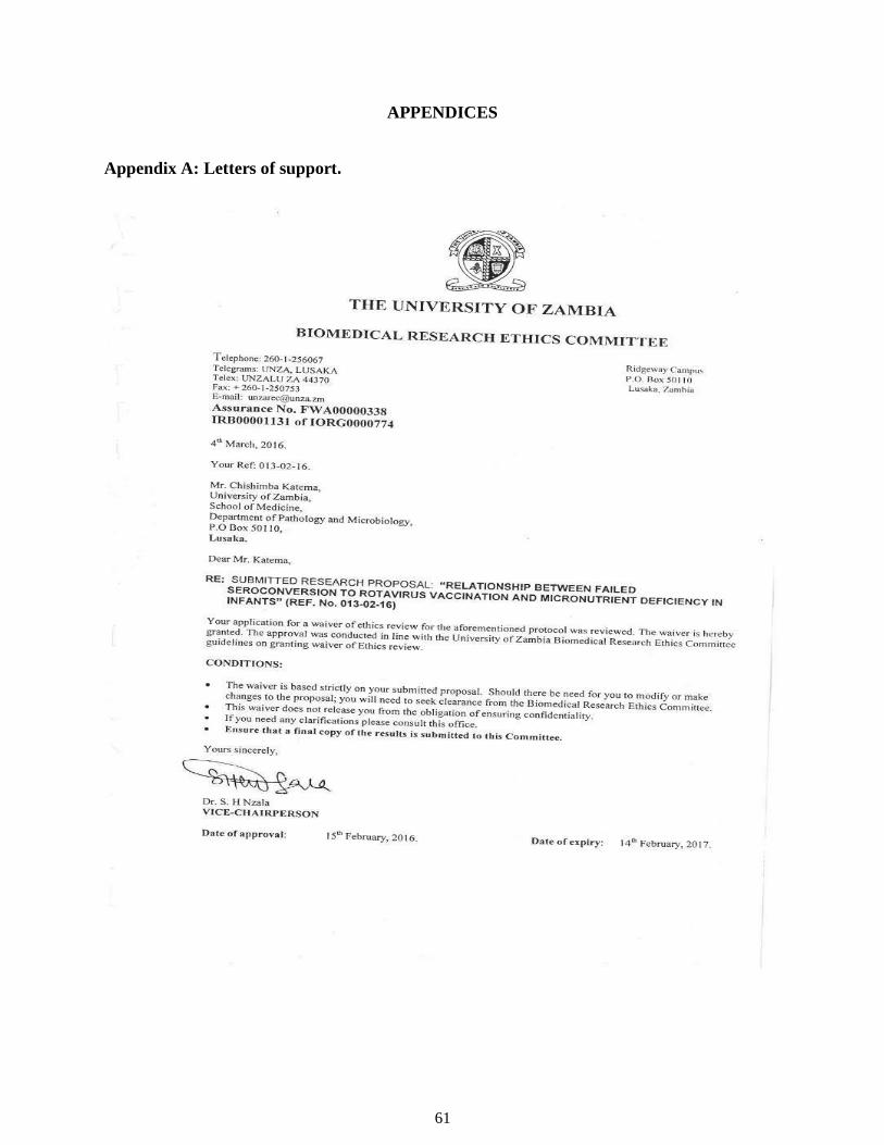
61
APPENDICES
Appendix A: Letters of support.
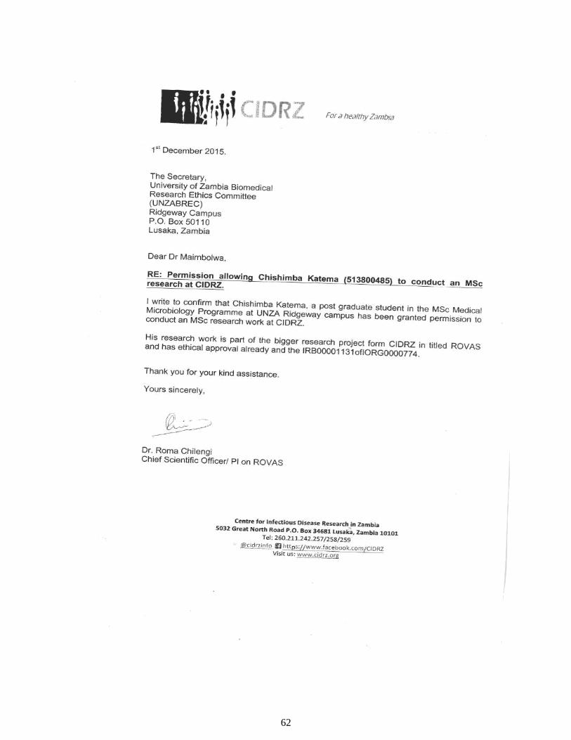
62
63
64
Appendix B: Study questionnaire and clinical examination form.
CENTRE FOR INFECTIOUS DISEASE RESEARCH IN ZAMBIA
CASE RECORD FORM
ID NUMBER:
CAUSES OF ROTAVIRUS VACCINE FAILURE IN ZAMBIA ROVAS STUDY
PART A –VISIT 1.0
ELIGIBILITY CHECK
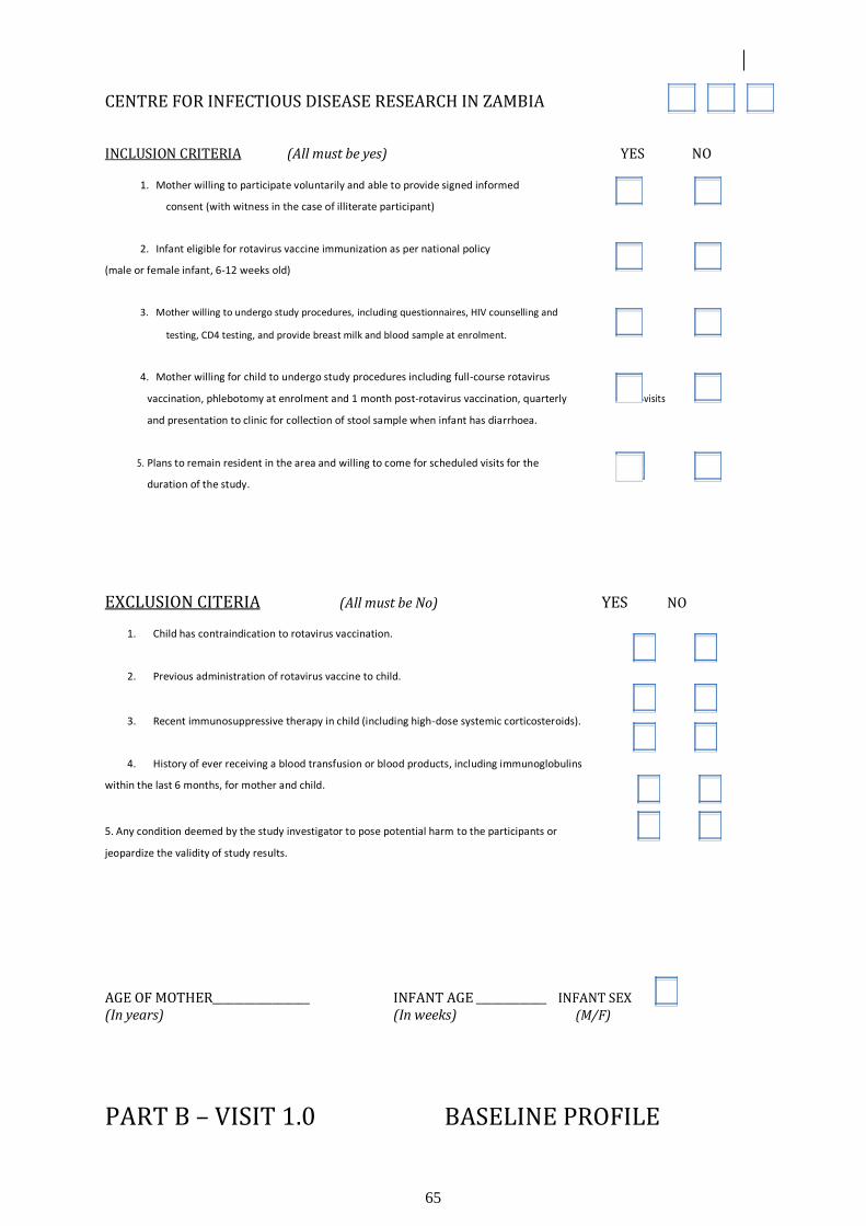
65
CENTRE FOR INFECTIOUS DISEASE RESEARCH IN ZAMBIA
INCLUSION CRITERIA (All must be yes) YES NO
1. Mother willing to participate voluntarily and able to provide signed informed
consent (with witness in the case of illiterate participant)
2. Infant eligible for rotavirus vaccine immunization as per national policy (male or female infant, 6-12 weeks old)
3. Mother willing to undergo study procedures, including questionnaires, HIV counselling and
testing, CD4 testing, and provide breast milk and blood sample at enrolment.
4. Mother willing for child to undergo study procedures including full-course rotavirus vaccination, phlebotomy at enrolment and 1 month post-rotavirus vaccination, quarterly visits
and presentation to clinic for collection of stool sample when infant has diarrhoea.
5. Plans to remain resident in the area and willing to come for scheduled visits for the
duration of the study.
EXCLUSION CITERIA (All must be No) YES NO
1. Child has contraindication to rotavirus vaccination.
2. Previous administration of rotavirus vaccine to child.
3. Recent immunosuppressive therapy in child (including high-dose systemic corticosteroids).
4. History of ever receiving a blood transfusion or blood products, including immunoglobulins within the last 6 months, for mother and child. 5. Any condition deemed by the study investigator to pose potential harm to the participants or
jeopardize the validity of study results.
AGE OF MOTHER__________________ (In years)
INFANT AGE _____________ INFANT SEX (In weeks) (M/F)
PART B – VISIT 1.0 BASELINE PROFILE
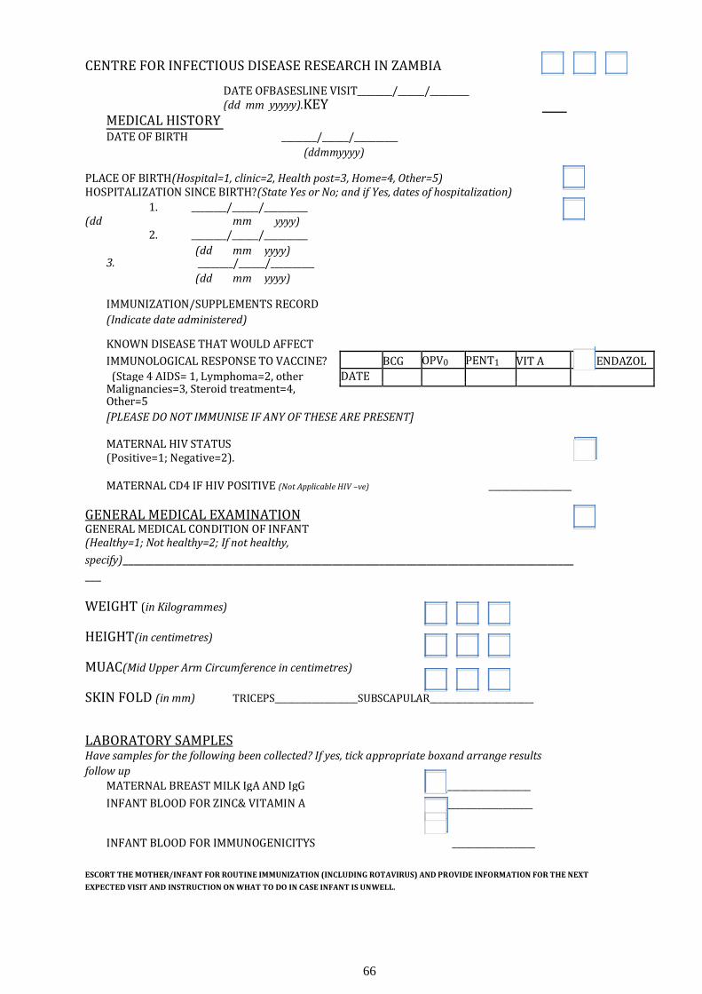
66
CENTRE FOR INFECTIOUS DISEASE RESEARCH IN ZAMBIA
DATE OFBASESLINE VISIT________/______/_________ (dd mm yyyyy).KEY MEDICAL HISTORY
DATE OF BIRTH ________/______/__________
(ddmmyyyy) PLACE OF BIRTH(Hospital=1, clinic=2, Health post=3, Home=4, Other=5) HOSPITALIZATION SINCE BIRTH?(State Yes or No; and if Yes, dates of hospitalization)
1. ________/______/__________ (dd mm yyyy)
2. ________/______/__________
3. (dd mm yyyy) ________/______/__________
(dd mm yyyy)
IMMUNIZATION/SUPPLEMENTS RECORD
(Indicate date administered)
KNOWN DISEASE THAT WOULD AFFECT
IMMUNOLOGICAL RESPONSE TO VACCINE? BCG OPV0 PENT1 VIT A ALBENDAZOL
(Stage 4 AIDS= 1, Lymphoma=2, other DATE Malignancies=3, Steroid treatment=4,
Other=5
[PLEASE DO NOT IMMUNISE IF ANY OF THESE ARE PRESENT]
MATERNAL HIV STATUS
(Positive=1; Negative=2).
MATERNAL CD4 IF HIV POSITIVE (Not Applicable HIV –ve) ___________________
GENERAL MEDICAL EXAMINATION GENERAL MEDICAL CONDITION OF INFANT (Healthy=1; Not healthy=2; If not healthy,
specify)______________________________________________________________________________________ ___
WEIGHT (in Kilogrammes)
HEIGHT(in centimetres)
MUAC(Mid Upper Arm Circumference in centimetres)
SKIN FOLD (in mm) TRICEPS____________________SUBSCAPULAR_________________________
LABORATORY SAMPLES Have samples for the following been collected? If yes, tick appropriate boxand arrange results
follow up MATERNAL BREAST MILK IgA AND IgG ___________________
INFANT BLOOD FOR ZINC& VITAMIN A
____________________
INFANT BLOOD FOR IMMUNOGENICITYS ___________________
ESCORT THE MOTHER/INFANT FOR ROUTINE IMMUNIZATION (INCLUDING ROTAVIRUS) AND PROVIDE INFORMATION FOR THE NEXT
EXPECTED VISIT AND INSTRUCTION ON WHAT TO DO IN CASE INFANT IS UNWELL.
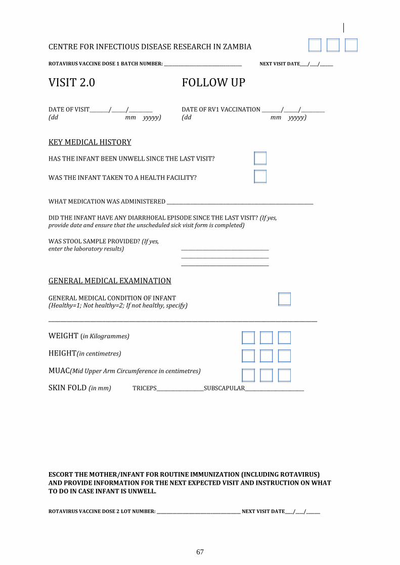
67
CENTRE FOR INFECTIOUS DISEASE RESEARCH IN ZAMBIA ROTAVIRUS VACCINE DOSE 1 BATCH NUMBER: _______________________________________ NEXT VISIT DATE____/____/_______
VISIT 2.0
FOLLOW UP
DATE OF VISIT________/______/__________ (dd mm yyyyy)
DATE OF RV1 VACCINATION ________/______/__________ (dd mm yyyyy)
KEY MEDICAL HISTORY
HAS THE INFANT BEEN UNWELL SINCE THE LAST VISIT?
WAS THE INFANT TAKEN TO A HEALTH FACILITY?
WHAT MEDICATION WAS ADMINISTERED ______________________________________________________________
DID THE INFANT HAVE ANY DIARRHOEAL EPISODE SINCE THE LAST VISIT? (If yes, provide date and ensure that the unscheduled sick visit form is completed)
WAS STOOL SAMPLE PROVIDED? (If yes, enter the laboratory results)
_____________________________________ _____________________________________ _____________________________________
GENERAL MEDICAL EXAMINATION GENERAL MEDICAL CONDITION OF INFANT (Healthy=1; Not healthy=2; If not healthy, specify)
__________________________________________________________________________________________
WEIGHT (in Kilogrammes)
HEIGHT(in centimetres)
MUAC(Mid Upper Arm Circumference in centimetres)
SKIN FOLD (in mm) TRICEPS____________________SUBSCAPULAR_________________________
ESCORT THE MOTHER/INFANT FOR ROUTINE IMMUNIZATION (INCLUDING ROTAVIRUS) AND PROVIDE INFORMATION FOR THE NEXT EXPECTED VISIT AND INSTRUCTION ON WHAT TO DO IN CASE INFANT IS UNWELL.
ROTAVIRUS VACCINE DOSE 2 LOT NUMBER: __________________________________________ NEXT VISIT DATE____/____/_______
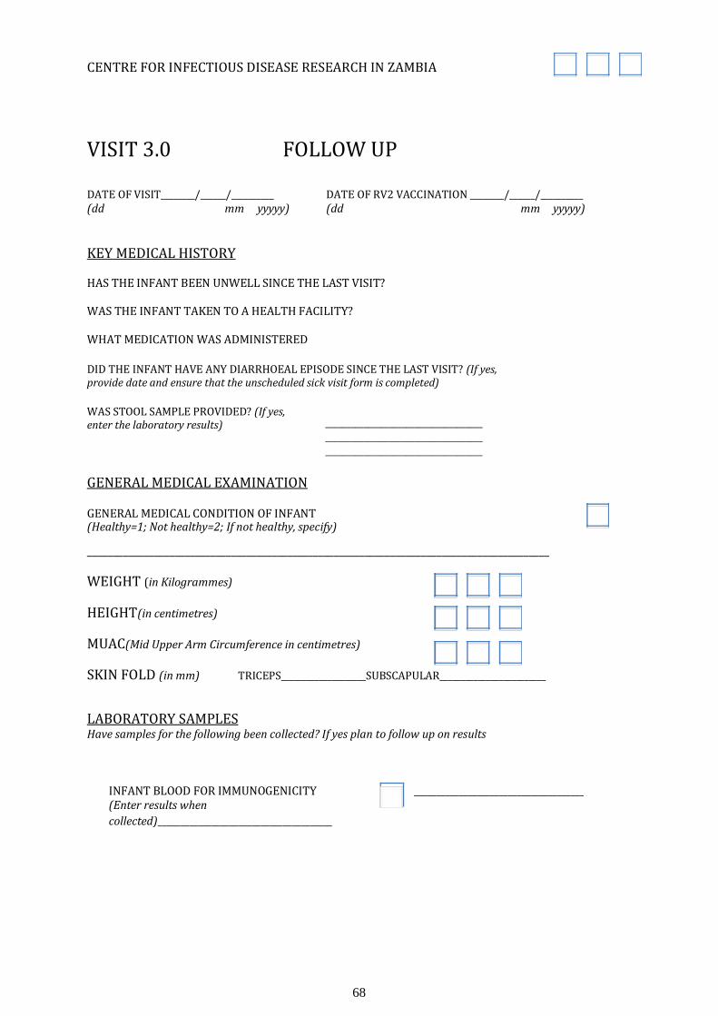
68
CENTRE FOR INFECTIOUS DISEASE RESEARCH IN ZAMBIA
VISIT 3.0
FOLLOW UP
DATE OF VISIT________/______/__________ (dd mm yyyyy)
DATE OF RV2 VACCINATION ________/______/__________ (dd mm yyyyy)
KEY MEDICAL HISTORY
HAS THE INFANT BEEN UNWELL SINCE THE LAST VISIT?
WAS THE INFANT TAKEN TO A HEALTH FACILITY?
WHAT MEDICATION WAS ADMINISTERED
DID THE INFANT HAVE ANY DIARRHOEAL EPISODE SINCE THE LAST VISIT? (If yes, provide date and ensure that the unscheduled sick visit form is completed)
WAS STOOL SAMPLE PROVIDED? (If yes, enter the laboratory results)
_____________________________________ _____________________________________ _____________________________________
GENERAL MEDICAL EXAMINATION GENERAL MEDICAL CONDITION OF INFANT (Healthy=1; Not healthy=2; If not healthy, specify)
__________________________________________________________________________________________
WEIGHT (in Kilogrammes)
HEIGHT(in centimetres)
MUAC(Mid Upper Arm Circumference in centimetres)
SKIN FOLD (in mm) TRICEPS____________________SUBSCAPULAR_________________________
LABORATORY SAMPLES Have samples for the following been collected? If yes plan to follow up on results
INFANT BLOOD FOR IMMUNOGENICITY ______________________________________ (Enter results when
collected)_______________________________________
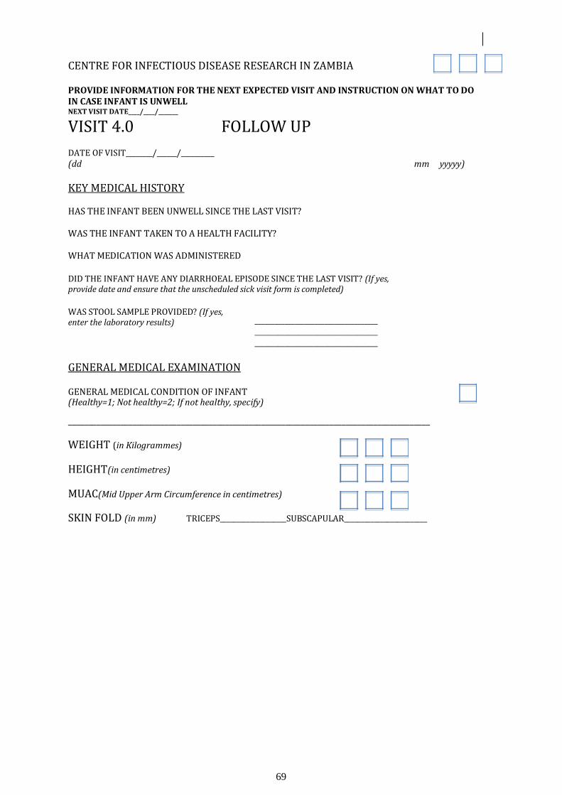
69
CENTRE FOR INFECTIOUS DISEASE RESEARCH IN ZAMBIA
PROVIDE INFORMATION FOR THE NEXT EXPECTED VISIT AND INSTRUCTION ON WHAT TO DO IN CASE INFANT IS UNWELL NEXT VISIT DATE____/____/_______
VISIT 4.0
FOLLOW UP DATE OF VISIT________/______/__________ (dd mm yyyyy)
KEY MEDICAL HISTORY
HAS THE INFANT BEEN UNWELL SINCE THE LAST VISIT?
WAS THE INFANT TAKEN TO A HEALTH FACILITY?
WHAT MEDICATION WAS ADMINISTERED
DID THE INFANT HAVE ANY DIARRHOEAL EPISODE SINCE THE LAST VISIT? (If yes, provide date and ensure that the unscheduled sick visit form is completed)
WAS STOOL SAMPLE PROVIDED? (If yes, enter the laboratory results)
_____________________________________ _____________________________________ _____________________________________
GENERAL MEDICAL EXAMINATION GENERAL MEDICAL CONDITION OF INFANT (Healthy=1; Not healthy=2; If not healthy, specify)
__________________________________________________________________________________________
WEIGHT (in Kilogrammes)
HEIGHT(in centimetres)
MUAC(Mid Upper Arm Circumference in centimetres)
SKIN FOLD (in mm) TRICEPS____________________SUBSCAPULAR_________________________
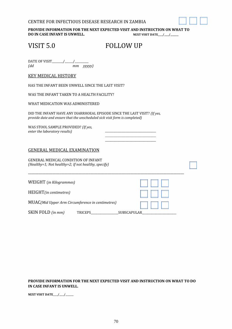
70
CENTRE FOR INFECTIOUS DISEASE RESEARCH IN ZAMBIA PROVIDE INFORMATION FOR THE NEXT EXPECTED VISIT AND INSTRUCTION ON WHAT TO DO IN CASE INFANT IS UNWELL. NEXT VISIT DATE____/____/_______
VISIT 5.0
FOLLOW UP
DATE OF VISIT________/______/__________ (dd mm yyyyy)
KEY MEDICAL HISTORY
HAS THE INFANT BEEN UNWELL SINCE THE LAST VISIT?
WAS THE INFANT TAKEN TO A HEALTH FACILITY?
WHAT MEDICATION WAS ADMINISTERED
DID THE INFANT HAVE ANY DIARRHOEAL EPISODE SINCE THE LAST VISIT? (If yes, provide date and ensure that the unscheduled sick visit form is completed)
WAS STOOL SAMPLE PROVIDED? (If yes, enter the laboratory results)
_____________________________________ _____________________________________ _____________________________________
GENERAL MEDICAL EXAMINATION
GENERAL MEDICAL CONDITION OF INFANT (Healthy=1; Not healthy=2; If not healthy, specify)
__________________________________________________________________________________________
WEIGHT (in Kilogrammes)
HEIGHT(in centimetres)
MUAC(Mid Upper Arm Circumference in centimetres)
SKIN FOLD (in mm) TRICEPS____________________SUBSCAPULAR_________________________
PROVIDE INFORMATION FOR THE NEXT EXPECTED VISIT AND INSTRUCTION ON WHAT TO DO
IN CASE INFANT IS UNWELL. NEXT VISIT DATE____/____/_______
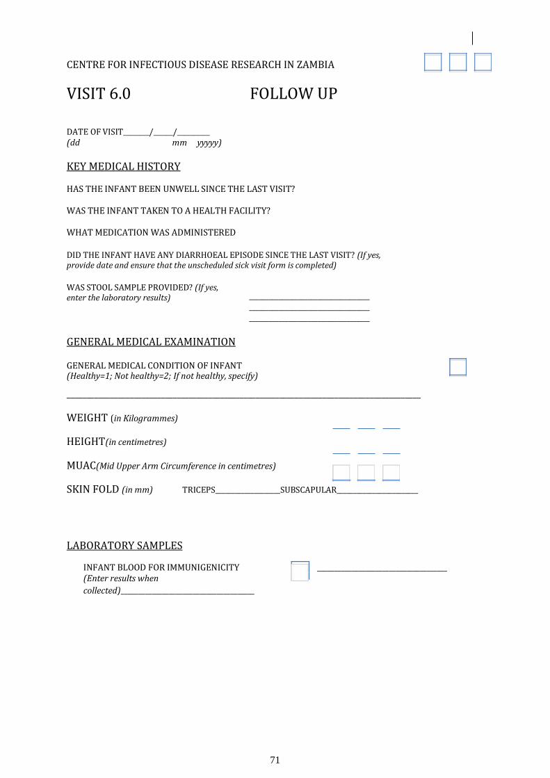
71
CENTRE FOR INFECTIOUS DISEASE RESEARCH IN ZAMBIA
VISIT 6.0
FOLLOW UP
DATE OF VISIT________/______/__________ (dd mm yyyyy)
KEY MEDICAL HISTORY
HAS THE INFANT BEEN UNWELL SINCE THE LAST VISIT?
WAS THE INFANT TAKEN TO A HEALTH FACILITY?
WHAT MEDICATION WAS ADMINISTERED
DID THE INFANT HAVE ANY DIARRHOEAL EPISODE SINCE THE LAST VISIT? (If yes, provide date and ensure that the unscheduled sick visit form is completed)
WAS STOOL SAMPLE PROVIDED? (If yes, enter the laboratory results)
_____________________________________ _____________________________________ _____________________________________
GENERAL MEDICAL EXAMINATION
GENERAL MEDICAL CONDITION OF INFANT (Healthy=1; Not healthy=2; If not healthy, specify)
__________________________________________________________________________________________
WEIGHT (in Kilogrammes)
HEIGHT(in centimetres)
MUAC(Mid Upper Arm Circumference in centimetres)
SKIN FOLD (in mm) TRICEPS____________________SUBSCAPULAR_________________________
LABORATORY SAMPLES
INFANT BLOOD FOR IMMUNIGENICITY ______________________________________ (Enter results when
collected)_______________________________________
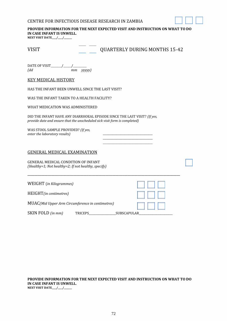
72
CENTRE FOR INFECTIOUS DISEASE RESEARCH IN ZAMBIA PROVIDE INFORMATION FOR THE NEXT EXPECTED VISIT AND INSTRUCTION ON WHAT TO DO IN CASE INFANT IS UNWELL. NEXT VISIT DATE____/____/_______
VISIT
QUARTERLY DURING MONTHS 15-42
DATE OF VISIT________/______/__________ (dd mm yyyyy)
KEY MEDICAL HISTORY
HAS THE INFANT BEEN UNWELL SINCE THE LAST VISIT?
WAS THE INFANT TAKEN TO A HEALTH FACILITY?
WHAT MEDICATION WAS ADMINISTERED
DID THE INFANT HAVE ANY DIARRHOEAL EPISODE SINCE THE LAST VISIT? (If yes, provide date and ensure that the unscheduled sick visit form is completed)
WAS STOOL SAMPLE PROVIDED? (If yes, enter the laboratory results)
_____________________________________ _____________________________________ _____________________________________
GENERAL MEDICAL EXAMINATION GENERAL MEDICAL CONDITION OF INFANT (Healthy=1; Not healthy=2; If not healthy, specify)
__________________________________________________________________________________________
WEIGHT (in Kilogrammes)
HEIGHT(in centimetres)
MUAC(Mid Upper Arm Circumference in centimetres)
SKIN FOLD (in mm) TRICEPS____________________SUBSCAPULAR_________________________
PROVIDE INFORMATION FOR THE NEXT EXPECTED VISIT AND INSTRUCTION ON WHAT TO DO IN CASE INFANT IS UNWELL. NEXT VISIT DATE____/____/_______
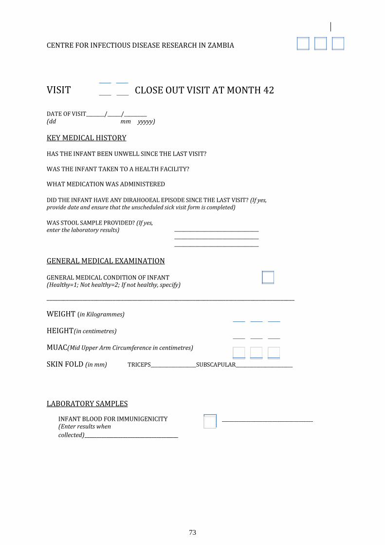
73
CENTRE FOR INFECTIOUS DISEASE RESEARCH IN ZAMBIA
VISIT
CLOSE OUT VISIT AT MONTH 42
DATE OF VISIT________/______/__________ (dd mm yyyyy)
KEY MEDICAL HISTORY
HAS THE INFANT BEEN UNWELL SINCE THE LAST VISIT?
WAS THE INFANT TAKEN TO A HEALTH FACILITY?
WHAT MEDICATION WAS ADMINISTERED
DID THE INFANT HAVE ANY DIRAHOOEAL EPISODE SINCE THE LAST VISIT? (If yes, provide date and ensure that the unscheduled sick visit form is completed)
WAS STOOL SAMPLE PROVIDED? (If yes, enter the laboratory results)
_____________________________________ _____________________________________ _____________________________________
GENERAL MEDICAL EXAMINATION GENERAL MEDICAL CONDITION OF INFANT (Healthy=1; Not healthy=2; If not healthy, specify)
__________________________________________________________________________________________
WEIGHT (in Kilogrammes)
HEIGHT(in centimetres)
MUAC(Mid Upper Arm Circumference in centimetres)
SKIN FOLD (in mm) TRICEPS____________________SUBSCAPULAR_________________________
LABORATORY SAMPLES
INFANT BLOOD FOR IMMUNIGENICITY ______________________________________ (Enter results when
collected)_______________________________________
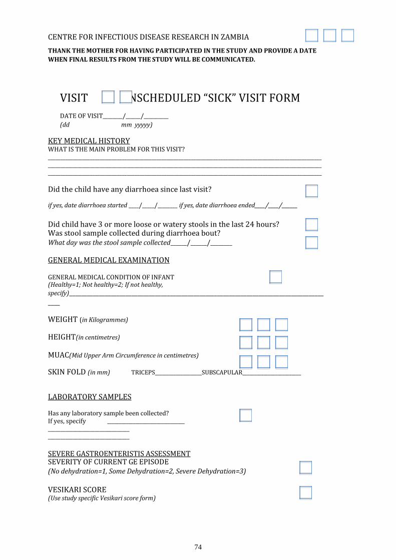
74
CENTRE FOR INFECTIOUS DISEASE RESEARCH IN ZAMBIA THANK THE MOTHER FOR HAVING PARTICIPATED IN THE STUDY AND PROVIDE A DATE
WHEN FINAL RESULTS FROM THE STUDY WILL BE COMMUNICATED.
VISIT UNSCHEDULED “SICK” VISIT FORM
DATE OF VISIT________/______/__________
(dd mm yyyyy)
KEY MEDICAL HISTORY WHAT IS THE MAIN PROBLEM FOR THIS VISIT? _______________________________________________________________________________________________________________ _______________________________________________________________________________________________________________ _______________________________________________________________________________________________________________
Did the child have any diarrhoea since last visit?
if yes, date diarrhoea started ____/_____/________ if yes, date diarrhoea ended____/____/______
Did child have 3 or more loose or watery stools in the last 24 hours? Was stool sample collected during diarrhoea bout? What day was the stool sample collected______/______/________
GENERAL MEDICAL EXAMINATION GENERAL MEDICAL CONDITION OF INFANT (Healthy=1; Not healthy=2; If not healthy,
specify)______________________________________________________________________________________ ____
WEIGHT (in Kilogrammes)
HEIGHT(in centimetres)
MUAC(Mid Upper Arm Circumference in centimetres)
SKIN FOLD (in mm) TRICEPS____________________SUBSCAPULAR_________________________
LABORATORY SAMPLES
Has any laboratory sample been collected? If yes, specify _________________________________ _________________________________ _________________________________
SEVERE GASTROENTERISTIS ASSESSMENT SEVERITY OF CURRENT GE EPISODE (No dehydration=1, Some Dehydration=2, Severe Dehydration=3)
VESIKARI SCORE (Use study specific Vesikari score form)
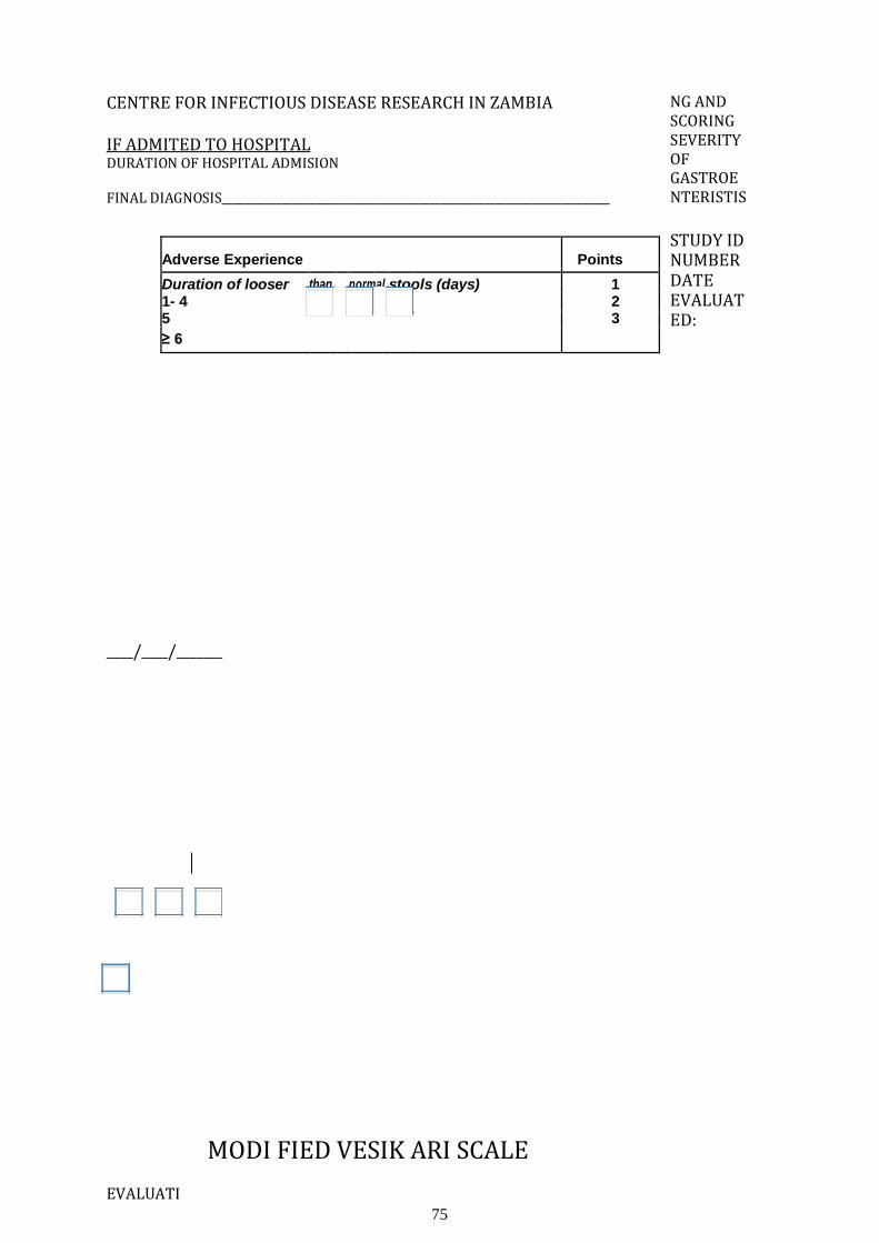
75
CENTRE FOR INFECTIOUS DISEASE RESEARCH IN ZAMBIA
IF ADMITED TO HOSPITAL DURATION OF HOSPITAL ADMISION
FINAL DIAGNOSIS_______________________________________________________________________
Adverse Experience Points
Duration of looser than normal stools (days) 1
1- 4 2 5
3
≥ 6
____/____/_______
MODI FIED VESIK ARI SCALE
EVALUATI
NG AND SCORING SEVERITY OF GASTROE NTERISTIS
STUDY ID NUMBER DATE EVALUAT ED:
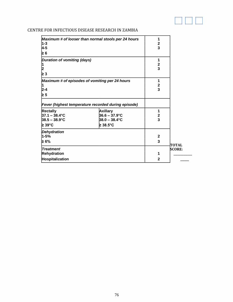
76
CENTRE FOR INFECTIOUS DISEASE RESEARCH IN ZAMBIA
Maximum # of looser than normal stools per 24 hours 1 1-3 2
4-5 3
≥ 6
Duration of vomiting (days) 1 1 2 2 3
≥ 3
Maximum # of episodes of vomiting per 24 hours 1 1 2
2-4 3
≥ 5
Fever (highest temperature recorded during episode)
Rectally Axillary 1 37.1 – 38.4°C 36.6 – 37.9°C 2
38.5 – 38.9°C 38.0 – 38.4°C 3
≥ 39°C ≥ 38.5°C
Dehydration 1-5% 2
≥ 6% 3 TOTAL
Treatment
SCORE: Rehydration 1 _____________
Hospitalization 2 ______