1
824 MANAGEMENT AND OUTCOME OF SEVERE COLITIS IN PREGNANCY Asher Kornbluth, M.D.* and Deepa Reddy. Medicine, Mt Sinai, New York, NY and Medicine, Elmhurst Hospital of Mt Sinai Health System, New York, NY. Purpose: The objective of this study is to analyze the course and man- nagement of maternal and fetal outcomes of women hospitalized for a course of iv steroids for management of severe colitis during pregnancy. Methods: We conducted a retrospective review of pregnant patients with severe colitis hospitalized for a course of iv steroids for the years 1989 – 2001. We compared the maternal and fetal outcomes to a control group of age matched patients with IBD who did not develop a severe flare during pregnancy and who had no other medical or gynecological diagnosis. Results: We identified a total of 12 patients; 7 with UC, 4 with Crohns colitis and 1 patient with indeterminate colitis. The mean age of patients was 30 years.The patients were admitted at a mean of 15.3 weeks of gestation and had a mean length of stay of 10.3 days. Prior to admission, 6 patients were on PO steroids as outpatients treated with a mean dose of 173mg HC equivalent and 2 patients were on IV steroids as inpatients at other hospitals with a mean dose of 220 mg HC equivalent. On admission, all patients received IV HCwith a mean dose of 202mg. Of the 12 patients, 4 were treated with immunomodulators: 2 with cyclo- sporine, 1 with azathioprine, and 1 with both cyclosporine and 6 –MP. Severe colitis patients delivered at a mean of 34 weeks, compared to 39 weeks for healthy IBD controls, with mean birth weights of 2121 gm , compared to 3105 gm for healthy IBD controls. Apgar scores at 1 and 5 minutes in both groups were equivalent (8.7 and 8.8). All 12 patients responded successfully to these courses of therapy for severe colitis. Of the 8 patients that had deliveries in Mt. Sinai, 6 patients had complications during delivery including preterm labor, postpartum hemmorrage, HELLP, and preeclampsia. Two of the patients had C–sec- tion, one due to preecclampsia and the other due to thrombocytopenia. In the control group, one patient had preeclampsia and two had elective C–sections. Conclusions: Severe colitis in IBD during pregnancy was successfully managed in all cases with agressive medical therapy using combinations of IV steroids, cylclosporine, 6 –MP and azathioprine. Although deliveries occured earlier and with lower birth weights, APGAR scores and fetal outcomes were comparable to a healthy IBD control group. 825 RELATIONSHIP BETWEEN HISTOLOGIC GRADE OF INFLAMMATION AND EXPRESSION OF FAS–FAS–LIGAND IN PATIENTS WITH ILEAL POUCH–ANAL ANASTOMOSIS B. Shen*, T. Gramlich, B. Lashner, P. Achkar, A. Brzezinski, J. Connor, M. Bambrick, F. Remzi, C. Bevins and V. Fazio. Digestive Disease Center, Cleveland Clinic, Cleveland, OH and Anatomic Pathology, Cleveland Clinic, Cleveland, OH. Purpose: The pathogenesis of pouchitis and ulcerative colitis (UC) may overlap. In UC, increased epithelial apoptosis as evidenced by the upregu- lation of Fas–Fas–L, leading to an accelerated epithelial cell turnover is attributed to the reduction in the crypt size and epithelial cell depletion. However, the role of Fas–Fas–L mediated apoptosis in the development of adaptive changes of pouch or pouchitis is not clear. It is possible that expression of Fas–Fas–L in the small intestinal epithelium of the pouch is altered. Aims: To analyze Fas–Fas–L expression in pts with pouchitis & controls, and to assess its association with histologic inflammation/adaptive changes. Methods: 14 consecutive pts with pouchitis (Pouchitis Disease Activity Index 7) and 17 pts with normal pouch were studied for histology and Fas–Fas–L immunohistochemistry. A GI pathologist blindly scored the pouch biopsy for acute (ulcer and polymorphic neutrophil infiltration) & chronic inflammation/adaptive changes (mononuclear cell infiltration and villous atrophy), and immunostaining, on a scale of 0 to 3 for each component. Results: Fas was readily detected in the pouch epithelial & mononuclear cells in both pouchitis and non–pouchitis pts (P 0.05). Only low level of Fas–L expression was detected in in 6 (43%) in the pouchitis group and 6 pts (35%)in the non–pouchitis group. Histology of inflammation/adaptive changes did not significantly correlate with expression of Fas or Fas–L, with r ranging from – 0.04 to 0.31 (P 0.05). Conclusions: Fas expression in the normal or inflammmed pouches was readily detectable, while Fas–L expression was uncommon in either group. Expression of Fas or Fas–L did not correlate with histologic grade of acute or chronic inflammation/adaptive changes. Fas–Fas–L pathway may not be a predominant mechanism for inflammation/adaptive changes of pouch mucosa, reflecting differences in pathophysiology of inflammation in the small intestine (pouch) and colon (UC). Non–Pouchitis N 17 Pouchitis N 14 P–value Acute histologic inflammation 0.8 0.8 2.6 1.2 0.0001 Chronic histologic inflammation 2.8 1.4 4.8 1.4 0.0003 Fas epithelia/mononuclear cells 1.9 0.9/2.2 0.8 2.1 0.8/2.4 0.8 0.38 Fas–L epithelia/mononuclear cells 0.4 0.5/0.1 0.2 0.4 0.6/0.4 0.9 0.097 826 MESALAMINE PREVENTS TNF–a INDUCED INCREASE IN INTESTINAL EPITHELIAL TIGHT JUNCTION PERMEABILITY Thomas Y. Ma, M.D.*, Neil Hoa, Vimesh Akotia, M.D. and Joseph Chen. Internal Medicine, University of New Mexico–Health Sciences Center, Albuquerque, NM; Internal Medicine, Albuquerque VA Medical Center, Albuquerque, NM and Internal Medicine, University of California, Irine, Orange, CA. Purpose: An abnormal increase in intestinal epithelial tight junction (TJ) permeability appears to be an important pathogenic factor of Crohn’s disease (CD). TNF–a plays a central role in intestinal inflammation of CD. Previous studies have shown that TNF–a induces an increase in intestinal TJ permeability. TNF–a induced increase in intestinal permeability has been proposed as an important pro–inflammatory mechanism. Mesalamine is an effective therapeutic agent in IBD. However, the therapeutic mech- anism of mesalamine remains unknown. We hypothesize that anti–inflam- matory mechanism of mesalamine involves inhibition of TNF–a induced increase in intestinal TJ permeability. The purpose of this study was to determine whether mesalamine protects against TNF–a induced increase in intestinal TJ permeability, using Caco–2 intestinal epithelial monolayers. Methods: Caco–2 epithelial TJ permeability was assessed by measuring trans– epithelial resistance or permeability to paracellular markers. Cellular localization of occludin and NF– kB activation were assessed by immuno– staining. Results: 1) TNF–a (0 –100 ng/ml) produced a dose– and time– dependent increase in Caco–2 TJ permeability. 2) TNF–a affect was associated with activation and nuclear translocation of NF– kB. 3) TNF–a also produced a decrease in expression of trans–membrane TJ protein occludin and disrup- tion of junctional localization of occludin. 4) Mesalamine (10 uM) pre- vented both TNF–a (10 ng/ml) induced increase in TJ permeability and TNF–a activation of the NF– kB pathways. 5) Mesalamine also prevented the TNF–a down–regulation of occludin expression and disruption of junctional localization. 6) Additionally, a known NF– kB inhibitor (triptol- ide) also prevented the TNF–a induced activation of the NF– kB and increase in Caco–2 TJ permeability. Conclusions: Our results demonstrate for the first time that mesalamine inhibits TNF–a induced increase in intestinal epithelial TJ permeability. Moreover, our findings indicate that the mechanism of mesalamine inhi- S271 AJG – September, Suppl., 2002 Abstracts

Relationship between histologic grade of inflammation and expression of Fas-Fas-ligand in patients with ileal pouch-anal anastomosis

  • Upload
    b-shen

  • View
    212

  • Download
    0

Embed Size (px)

Citation preview

824

MANAGEMENT AND OUTCOME OF SEVERE COLITIS INPREGNANCYAsher Kornbluth, M.D.* and Deepa Reddy. Medicine, Mt Sinai, NewYork, NY and Medicine, Elmhurst Hospital of Mt Sinai Health System,New York, NY.

Purpose: The objective of this study is to analyze the course and man-nagement of maternal and fetal outcomes of women hospitalized for acourse of iv steroids for management of severe colitis during pregnancy.Methods: We conducted a retrospective review of pregnant patients withsevere colitis hospitalized for a course of iv steroids for the years 1989–2001. We compared the maternal and fetal outcomes to a control group ofage matched patients with IBD who did not develop a severe flare duringpregnancy and who had no other medical or gynecological diagnosis.Results: We identified a total of 12 patients; 7 with UC, 4 with Crohnscolitis and 1 patient with indeterminate colitis. The mean age of patientswas 30 years.The patients were admitted at a mean of 15.3 weeks ofgestation and had a mean length of stay of 10.3 days.

Prior to admission, 6 patients were on PO steroids as outpatients treatedwith a mean dose of 173mg HC equivalent and 2 patients were on IVsteroids as inpatients at other hospitals with a mean dose of 220 mg HCequivalent.

On admission, all patients received IV HCwith a mean dose of 202mg.Of the 12 patients, 4 were treated with immunomodulators: 2 with cyclo-sporine, 1 with azathioprine, and 1 with both cyclosporine and 6–MP.Severe colitis patients delivered at a mean of 34 weeks, compared to 39weeks for healthy IBD controls, with mean birth weights of 2121 gm ,compared to 3105 gm for healthy IBD controls. Apgar scores at 1 and 5minutes in both groups were equivalent (8.7 and 8.8).

All 12 patients responded successfully to these courses of therapy forsevere colitis. Of the 8 patients that had deliveries in Mt. Sinai, 6 patientshad complications during delivery including preterm labor, postpartumhemmorrage, HELLP, and preeclampsia. Two of the patients had C–sec-tion, one due to preecclampsia and the other due to thrombocytopenia. Inthe control group, one patient had preeclampsia and two had electiveC–sections.Conclusions: Severe colitis in IBD during pregnancy was successfullymanaged in all cases with agressive medical therapy using combinations ofIV steroids, cylclosporine, 6–MP and azathioprine. Although deliveriesoccured earlier and with lower birth weights, APGAR scores and fetaloutcomes were comparable to a healthy IBD control group.

825

RELATIONSHIP BETWEEN HISTOLOGIC GRADE OFINFLAMMATION AND EXPRESSION OF FAS–FAS–LIGAND INPATIENTS WITH ILEAL POUCH–ANAL ANASTOMOSISB. Shen*, T. Gramlich, B. Lashner, P. Achkar, A. Brzezinski, J. Connor,M. Bambrick, F. Remzi, C. Bevins and V. Fazio. Digestive DiseaseCenter, Cleveland Clinic, Cleveland, OH and Anatomic Pathology,Cleveland Clinic, Cleveland, OH.

Purpose: The pathogenesis of pouchitis and ulcerative colitis (UC) mayoverlap. In UC, increased epithelial apoptosis as evidenced by the upregu-lation of Fas–Fas–L, leading to an accelerated epithelial cell turnover isattributed to the reduction in the crypt size and epithelial cell depletion.However, the role of Fas–Fas–L mediated apoptosis in the development ofadaptive changes of pouch or pouchitis is not clear. It is possible thatexpression of Fas–Fas–L in the small intestinal epithelium of the pouch isaltered. Aims: To analyze Fas–Fas–L expression in pts with pouchitis &controls, and to assess its association with histologic inflammation/adaptivechanges.Methods: 14 consecutive pts with pouchitis (Pouchitis Disease ActivityIndex � 7) and 17 pts with normal pouch were studied for histology andFas–Fas–L immunohistochemistry. A GI pathologist blindly scored thepouch biopsy for acute (ulcer and polymorphic neutrophil infiltration) &

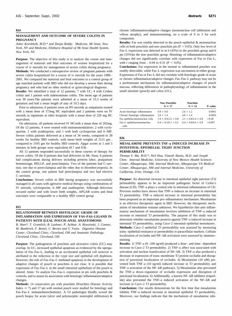
chronic inflammation/adaptive changes (mononuclear cell infiltration andvillous atrophy), and immunostaining, on a scale of 0 to 3 for eachcomponent.Results: Fas was readily detected in the pouch epithelial & mononuclearcells in both pouchitis and non–pouchitis pts (P � 0.05). Only low level ofFas–L expression was detected in in 6 (43%) in the pouchitis group and 6pts (35%)in the non–pouchitis group. Histology of inflammation/adaptivechanges did not significantly correlate with expression of Fas or Fas–L,with r ranging from – 0.04 to 0.31 (P � 0.05).Conclusions: Fas expression in the normal or inflammmed pouches wasreadily detectable, while Fas–L expression was uncommon in either group.Expression of Fas or Fas–L did not correlate with histologic grade of acuteor chronic inflammation/adaptive changes. Fas–Fas–L pathway may not bea predominant mechanism for inflammation/adaptive changes of pouchmucosa, reflecting differences in pathophysiology of inflammation in thesmall intestine (pouch) and colon (UC).

Non–PouchitisN � 17

PouchitisN � 14 P–value

Acute histologic inflammation 0.8 � 0.8 2.6 � 1.2 0.0001Chronic histologic inflammation 2.8 � 1.4 4.8 � 1.4 0.0003Fas epithelia/mononuclear cells 1.9 � 0.9/2.2 � 0.8 2.1 � 0.8/2.4 � 0.8 �0.38Fas–L epithelia/mononuclear

cells0.4 � 0.5/0.1 � 0.2 0.4 � 0.6/0.4 � 0.9 �0.097

826

MESALAMINE PREVENTS TNF–a INDUCED INCREASE ININTESTINAL EPITHELIAL TIGHT JUNCTIONPERMEABILITYThomas Y. Ma, M.D.*, Neil Hoa, Vimesh Akotia, M.D. and JosephChen. Internal Medicine, University of New Mexico–Health SciencesCenter, Albuquerque, NM; Internal Medicine, Albuquerque VA MedicalCenter, Albuquerque, NM and Internal Medicine, University ofCalifornia, Irine, Orange, CA.

Purpose: An abnormal increase in intestinal epithelial tight junction (TJ)permeability appears to be an important pathogenic factor of Crohn’sdisease (CD). TNF–a plays a central role in intestinal inflammation of CD.Previous studies have shown that TNF–a induces an increase in intestinalTJ permeability. TNF–a induced increase in intestinal permeability hasbeen proposed as an important pro–inflammatory mechanism. Mesalamineis an effective therapeutic agent in IBD. However, the therapeutic mech-anism of mesalamine remains unknown. We hypothesize that anti–inflam-matory mechanism of mesalamine involves inhibition of TNF–a inducedincrease in intestinal TJ permeability. The purpose of this study was todetermine whether mesalamine protects against TNF–a induced increase inintestinal TJ permeability, using Caco–2 intestinal epithelial monolayers.Methods: Caco–2 epithelial TJ permeability was assessed by measuringtrans–epithelial resistance or permeability to paracellular markers. Cellularlocalization of occludin and NF–kB activation were assessed by immuno–staining.Results: 1) TNF–a (0–100 ng/ml) produced a dose– and time– dependentincrease in Caco–2 TJ permeability. 2) TNF–a affect was associated withactivation and nuclear translocation of NF–kB. 3) TNF–a also produced adecrease in expression of trans–membrane TJ protein occludin and disrup-tion of junctional localization of occludin. 4) Mesalamine (10 uM) pre-vented both TNF–a (10 ng/ml) induced increase in TJ permeability andTNF–a activation of the NF–kB pathways. 5) Mesalamine also preventedthe TNF–a down–regulation of occludin expression and disruption ofjunctional localization. 6) Additionally, a known NF–kB inhibitor (triptol-ide) also prevented the TNF–a induced activation of the NF–kB andincrease in Caco–2 TJ permeability.Conclusions: Our results demonstrate for the first time that mesalamineinhibits TNF–a induced increase in intestinal epithelial TJ permeability.Moreover, our findings indicate that the mechanism of mesalamine inhi-

S271AJG – September, Suppl., 2002 Abstracts