Embed Size (px)
Citation preview

REGULATION AND FUNCTION OF
P2Y2 AND P2X 7 NUCLEOTIDE RECEPTORS
IN THE CENTRAL NERVOUS SYSTEM
A Dissertation
Presented to
the Faculty of the Graduate School
at the University of Missouri-Columbia
In Partial Fulfillment
of the Requirements for the Degree
Doctor of Philosophy
by
QIONGMAN KONG,
Dr. Gary A. Weisman, Dissertation Supervisor
AUGUST 2007

The undersigned, appointed by the dean of the Graduate School, have examined the dissertation entitled
REGULATION AND FUNCTION OF P2Y2 AND P2X 7 NUCLEOTIDE
RECEPTORS IN THE CENTRAL NERVOUS SYSTEM
presented by Qiongman Kong,
a candidate for the degree of doctor of philosophy,
and hereby certify that, in their opinion, it is worthy of acceptance.
Professor Gary A. Weisman
Professor Laurie Erb
Professor Mark Kirk
Professor Dennis Lubahn
Professor Grace Y. Sun

ACKNOWLEDGEMENTS
These projects would not have been possible without the support of many people.
I would like to express my deepest gratitude to my advisor, Dr. Gary A. Weisman for
his excellent guidance in both research and life, for providing me with an active
laboratory atmosphere and encouraging me to work independently on the research
projects with great patience, and for the great care during the long recovery term after
an auto accident in 2002. I also want to thank Dr. Cheikh I. Seye, Dr. Laurie Erb, and
Jean M. Camden in Weisman’s lab, and Drs. Grace Y. Sun and Agnes Simonyi in
Sun’s lab for their tremendous support on my research projects.
I extend my gratitude to members of my doctoral committee: Drs. Laurie Erb,
Grace Y. Sun, Mark Kirk, Dennis Lubahn and Cathy Krull, for their insightful
comments and continuous guidance on my work.
I want to thank Jun Liu and Zhongji Liao, my best friends at MU, who have
shared happiness with me and supported me to overcome my difficulties. I would also
like to express my appreciation to many other friends at MU: Yu Ningpu, Wang Min,
Qun Wang, Jennifer Lynn Hamilton, Sue Yu, Xiaolin Zhang, Brent Krugh, Olga Baker,
Antje Heese, Troy Scott Peterson, Ann Schrader, Nishant Jain, Shivaji Rikka,
Tixieanna Dissmore, Fernand Gendron, Sriparna Bagchi, Julie Hayes, Jessica Newton,
Korey Griffin, and Richard Garrad.
Finally, I would like to thank my parents, my sister, and my boyfriend, Xin He,
for their continuous love and support.
ii

TABLE OF CONTENTS
ACKNOWLEDGEMENTS ii
LIST OF TABLES vi
LIST OF FIGURES vii
LIST OF ABBREVIATIONS ix
ABSTRACT xiii
CHAPTER 1. INTRODUCTION 1
Adenosine 5’–triphosphate release ……………………………………………… 1
P2 nucleotide receptor classification …..…..…….…..….………………..…. 1
Expression and regulation of P2 nucleotide receptors in the brain …...….…….. 2
P2X7 nucleotide receptors and apoptosis ….……………….….…………..…... 3
P2 nucleotide receptors in Alzheimer’s disease ……….…….….……………... 5
P2 nucleotide receptors in brain ischemia ……….…..…….……………...…… 7
Outline of the Dissertation …………………………..…….…..………………. 9
CHAPTER 2. P2X7 NUCLEOTIDE RECEPTORS MEDIATE CASPASE-8/9/3- DEPENDENT APOPTOSIS IN RAT PRIMARY CORTICAL NEURONS 18
Abstract …………………...…………………………….……………………. 19
Introduction ……………...…………………………...………..…………...… 20
Materials and Methods ……………………..……………………….…………. 22
Results ………………..…………………………….………………………… 30
Discussion …………………..……………………….……....................... 33
CHAPTER 3. IL-1β ENHANCES NUCLEOTIDE-INDUCED α-SECRETASE- DEPENDENT APP PROCESSING IN RAT PRIMARY CORTICAL NEURONS VIA UPREGULATION OF THE P2Y2 RECEPTOR 59
Abstract …………………..…………………………………….………….. 60
Introduction …………………..……………………………..……………. 60
iii

Materials and Methods …………………….……….…………..…….... 63
Results …………………..………………………………………………... 67
Discussion ………………..………………………………………..……….… 70
CHAPTER 4. EXPRESSION AND REGULATION OF P2Y2 NUCLEOTIDE RECEPTORS IN RODENT BRAINS AND THE EFFECT OF CEREBRAL ISCHEMIA 93
Abstract ………………..………………………………………….………….. 94
Introduction ………………..…………………………………………………. 95
Materials and Methods ………………….……….………………………….... 96
Results .………………..…………………………………………………….... 100
Discussion ………………..……………………………………………….…103
CHAPTER 5. GENERAL DISCUSSION 116
The major contribution and significance ……………………………………… 118
APPENDIX. BRIEF REVIEW OF OTHER RESEARCH PROJECTS 121
A-1. FUNCTIONAL P2Y2 NUCLEOTIDE RECEPTORS MEDIATE URIDINE 5’-TRIPHOSPHATE-INDUCED INTIMAL HYPERPLASIA IN COLLARED RABBIT CAROTID ARTERIES 122
Abstract ………………..…….………………….……..…........……… 122
Introduction ………………...……………………………………….… 123
Materials and Methods …………………..………………………..…...... 123
Results ………………..……………………………………………….. 125
Discussion ……………..…………………………................................ 126
A-2. P2Y 2 RECEPTOR ACTIVITY IS MODULATED BY THE ECTO- NUCLEOTIDASE NTPASE (CD39) 132
Abstract ………………..….………………………………..........……. 132
Introduction ………………...………………………….………...….… 133
Materials and Methods ……………..……………….….………….….. 134
iv
Results ………………..…………………………………….................. 136
A-3. AN RGD SEQUENCE IN THE P2Y2 RECEPTOR INTERACTS WITH Vß3 INTEGRINS AND IS REQUIRED FOR Go-MEDIATED SIGNAL TRANSDUCTION 145
Abstract ………………..……………………………….…..……......... 145
Introduction …….……………..………………..……………..……... 146
Materials and Methods …………..……………………..……………... 147
Results ………………..…………………………….…………...…….. 148
REFERENCES 152
VITA 184
v

LIST OF TABLES
Table Page
1-1 Regulation of P2 nucleotide receptor expression after ischemia and in Alzheimer’s disease (AD) .………………….…………..…………………………...... 11
2-1 Sequence-specific oligonucleotide primers used for RT-PCR studies ……….. 39
vi
LIST OF FIGURES
Figure Page
1-1 Expression of P2 receptors in rat cortical astrocytes and neurons .….................. 12
1-2 Nerve cells, synapses and brain tissue in AD ………………....................…….. 14
1-3 Neuronal cell death in rodent ischemia models .……………..………….... 16
2-1 Purity of rPCNs and P2X7R Expression in rPCNs …………..………………. 40
2-2 ATP- and BzATP-induced increases in [Ca2+]i in rat PCNs ..…….………...… 42
2-3 ATP/BzATP-induced nuclear condensation mediated by the P2X7R is dependent on caspase-3 activation …………………………………….….……..….... 44
2-4 BzATP stimulates DNA fragmentation in rat PCNs ………..……………..…. 46
2-5 BzATP or ATP induces DNA strand breakage in rat PCNs ..…..…………..…. 48
2-6 P2X7Rs mediate caspase-3 activation in rat PCNs …………...….…………... 50
2-7 Roles of caspase-8 and caspase-9 in P2X 7 R-mediated apoptosis in rPCNs ………………………………………………………..…..…....... 53
2-8 BzATP or ATP induces ERK1/2 and JNK1 phosphoryla t ion in rPCNs ………………………………...…………………………….......… 55
2-9 Inhibitors of MEK and JNK decrease BzATP-induced caspase-3 cleavage in rat PCNs …………………………………………………………………...…. 57
3-1 IL-1β upregulates functional expression of P2Y2 receptors in rPCNs ………. 76
3-2 UTP stimulates phosphorylation of ERK1/2 in IL-1β-treated rPCNs ..… 78
3-3 Release of sAPPα is stimulated by UTP in IL-1β-treated rPCNs .............. 81
3-4 P2Y2 receptor expression in rPCNs enhances α-secretase-mediated APP processing induced by UTP …………………………………………………..… 83
3-5 A metalloprotease inhibitor prevents UTP-induced sAPPα release from rPCNs…………………………………………………………………..... 85
vii

3-6 UTP-induced sAPPα release occurs independently of PKC activation in IL-1β-treated rPCNs …………………………………………………….….. 87
3-7 Inhibition of a PI3K prevents UTP-induced sAPPα release in IL-1β-treated rPCNs. …………………………………………………………………....…. 89
3-8 UTP-stimulated sAPPα release in IL-1β-treated rPCNs is partially dependent on MAPK/ERK1/2 activation.. .……….……….……….……….……….…….….. 91
4-1 P2Y2 receptor mRNA expression in adult rodent brains …….................…. 105
4-2 Expression of mRNA for P2Y2, P2X7, P2Y1, P2Y4 and P2Y6 nucleotide receptors in adult rat brain …………..…………….………………………………….... 107
4-3 P2Y2R mRNA levels in the gerbil hippocampal CA1 region after ischemic injury …………………………………………….….................................. 109
4-4 Expression of P2Y2R mRNA in hippocampal subregions after global cerebral ischemia …...……..……..……..……..……..……..……..………...…... 112
4-5 Effect of resveratrol on P2Y2R mRNA expression in the CA1 region of gerbil brain after transient global cerebral ischemia …………………………….….... 114
A-1-1 Collared rabbit carotid artery model ..…………...………...……………….. 128
A-1-2 Nucleotide-induced OPN expression in collared carotid arteries and SMC cultures …………………………..………………………………………… 130
A-2-1 Effect of BG0136 on UTP-stimulated inositol phosphate (IP) release and ERK1/2 activation in COS-7 cells with or without CD39 transfection ………. 139
A-2-2 Effect of BG0136 on UTP-stimulated ERK1/2 activation in HEK cells with or without CD39 transfection ……………………………................................ 141
A-2-3 Effect of BG0136 on nucleotide-stimulated ERK activation in human 1321N1 astrocytoma cells expressing the human P2Y2 nucleotide receptor ………… 143
A-3-1 Effect of anti-integrin antibodies on P2Y2R-activated IP formation in 1321N1 cells ………………………..……………………………..…………………..... 150
viii

LIST OF ABBREVIATIONS
Aβ amyloid-β peptide
AD Alzheimer’s disease
ADAM a disintegrin and metalloprotease
AMP adenosine 5’-monophosphate
ADP adenosine 5’-diphosphate
APP amyloid-β precursor protein
ATP adenosine 5’-triphosphate
BACE β-site APP cleaving enzyme
BCAO bilateral carotid arteries occlusion
BG0136 1-naphthol-3, 6-disulfonic acid, disodium salt
[Ca2+]i concentration of cytoplasmic free calcium
CCAs common carotid arteries
CD39 ecto-nucleoside triphosphate diphosphohydrolase, NTPDase
C83 α-secretase generated membrane bound APP fragment
CE cerebellum
CNS central nervous system
CO cerebral cortex
COS7 African Green Monkey kidney fibroblast cell line
DAG diacylglycerol
ix

DIV days after in vitro culture
DND delayed neuronal death
EC50 half maximal effective concentration
ERK1/2 extracellular signal-related kinase 1/2
FAK focal adhesion kinase
GFR growth factor receptor
GPCR G protein-coupled receptor
HEK human embryonic kidney cells
HI hippocampus
IL-1 interleukin-1
IP inositol phosphate
IP3 Inositol 1, 4, 5-trisphosphate
JNK Jun N-terminal kinase
KPI Kunitz Protease Inhibitor
MAPK mitogen-activated protein kinase
MCAO middle cerebral artery occlusion
MDC9 meltrin-gamma/ADAM9, a disintegrin and metalloprotease-9
MEK MAPK kinase
2-MeSATP 2-methylthioadenosine 5’-triphosphate
mRNA messenger ribonucleic acid
NF-κB nuclear factor κB
NGF nerve growth factor
x

OPN osteopontin
PCR polymerase chain reaction
PI3K phosphatidylinositol 3-kinase
PKC protein kinase C
PLA2 phospholipases A2
PLC phospholipases C
P3 extracellular APP fragment released from C83 following γ
cleavage
PMA phorbol 12-myristate 13-acetate
rCBF regional cerebral blood flow
RGD arginine-glycine-aspartic acid
RGE arginine-glycine-glutamic acid
rPCN rat primary cortical neuron
RT reverse transcription
sAPPα secreted amyloid precursor protein α
sAPPβ secreted amyloid precursor protein β
SH3 Src homology domain 3
SMC smooth muscle cell
TACE tumor necrosis factor alpha converting enzyme
TNF-α tumor necrosis factor α
TUNEL terminal deoxynucleotidyl transferase biotin-dUTP nick end
labeling
xi

UDP uridine 5’-diphosphate
UTP uridine 5’-triphosphate
1321N1 cells human 1321N1 astrocytoma cells
xii

REGULATION AND FUNCTION OF P2Y2 AND P2X 7 NUCLEOTIDE
RECEPTORS IN THE CENTRAL NERVOUS SYSTEM
Qiongman Kong
Dissertation Supervisor: Dr. Gary A. Weisman
ABSTRACT
It is well known that ATP acts as a classic neurotransmitter in the central nervous
system. It is stored in molar concentrations in synaptic vesicles, released upon
neuronal excitation, and activates both ion channel receptors (P2X) and metabotropic
receptors (P2Y) on post-synaptic cells. Activation of these nucleotide receptors then
alters a variety of brain activities, such as the formation of long term memory and the
perception of pain. In addition to its role as a neurotransmitter, ATP and other
nucleotides are known to be released from injured or ischemic tissue and have been
reported to affect inflammatory events in the brain and many other tissues, however,
these inflammatory events as well as the subtype(s) of nucleotide receptor(s)
influencing these events have not been well defined. This dissertation examines the
expression patterns of P2X and P2Y nucleotide receptors in rat primary cortical
neurons and adult rodent brain tissue. Based on these studies, particular emphasis was
placed on delineating the role of the P2Y2 receptor subtype in neuronal inflammation
and the P2X7 receptor subtype in neuronal apoptosis.
The P2Y2 receptor is distinguished from other nucleotide receptor subtypes by an
equal-potency for activation by ATP and UTP and its ability to be up-regulated in
response to tissue injury or stress. Studies mentored by Dr. Cheikh Seye used a
xiii

collared rabbit carotid artery model to indicate for the first time a role for the P2Y2R
in development of intimal hyperplasia associated with atherosclerosis and restenosis
(Appendix-1). In vitro studies with various cell lines (1321N1, HEK, COS7, and
DITNC) in collaboration with others investigated signaling events regulated by the
P2Y2R, including activation of PI3K, Src, calcium mobilization, Akt, and ERK1/2 and
the importance of αVβ3/β5 integrin interaction with the P2Y2 receptor in selective (Go
but not Gq) signaling and the antagonist role of BG0136 on P2Y2R activity
(Appendix-2, 3).
In a gerbil transient global ischemia model, we analyzed the effect of ischemia on
the in vivo expression level of P2Y2R mRNA in the rodent brain by in situ
hybridization and RT-PCR (Chapter 4). P2Y2R mRNA was expressed at low levels in
normal rodent (rat, mouse and gerbil) brain. Transient ischemia (5 min occlusion) did
not affect P2Y2R mRNA levels in any region of gerbil brain after various reperfusion
times (4 h, 16 h and 48 h), indicating that the P2Y2R was not regulated at the
transcriptional level in the gerbil transient ischemia model.
The functional expression of the P2Y2R was also tested in rat primary cortical
neurons (rPCNs). In rPCNs after in vitro culture for 7-10 days, P2Y2R mRNA and
receptor activity were virtually absent in unstimulated cells, whereas overnight
treatment with IL-1β caused up-regulation of both P2Y2R mRNA and receptor
activity. Furthermore, activation of P2Y2R in rPCNs treated with IL-1β or transfected
with P2Y2R cDNA enhanced the release of sAPPα, a secrectory amyloid protein that
protects neurons as an injury-induced neurotrophic factor. P2Y2R-mediated sAPPα
xiv

release from rPCNs was dependent on ADAM10/17 and PI3K activity and was
partially dependent on ERK1/2 activity, but did not require activation of PKC. These
results suggest that under inflammatory conditions, IL-1β evokes functional
up-regulation of P2Y2R in neurons and subsequent activation of the P2Y2R signaling
cascade results in an increase of sAPPα release through activation of the α,γ-secretase
pathway (Chapter 3).
In contrast to the neuroprotective role of extracellular nucleotides just described,
ATP has also been reported to cause apoptosis in a neuronal cell line by activating the
P2X7 subtype of nucleotide receptors (P2X7R). Here, we extended the examination of
P2X7R-mediated apoptosis in rPCNs. Unlike the P2Y2R, P2X7R was functionally
expressed in unstimulated rPCNs (indicated by RT-PCR and ATP-induced
intracellular calcium mobilization). Activation of the P2X7R via extracellular
nucleotides in rPCNs caused biochemical (i.e., caspase activation) and morphological
(i.e., nuclear condensation and DNA fragmentation) changes characteristic of
apoptotic cell death through an intrinsic JNK1- and ERK-dependent caspase-8/9/3
activation pathway (Chapter 2).
Together, these data describe the expression patterns of P2 nucleotide receptors in
rat primary cultures of cortical neurons, in adult rodent brains, in cerebral ischemia
and in collared rabbit carotid arteries and demonstrate that the P2Y2R is up-regulated
in response to cytokine stimulation in rPCNs and after collar transplantation around
rabbit carotid arteries. Moreover, activation of the P2Y2R and P2X7R in rPCNs
caused α-secretase-dependent APP processing and caspase8/9/3-dependent apoptosis,
xv

respectively, whereas in vivo studies in the collared carotid artery model indicate that
P2Y2R also plays an important role in the development of intimal hyperplasia.
xvi
CHAPTER 1
INTRODUCTION
Adenosine 5’ –triphosphate release
Nucleotides can be released from various sources in the nervous system include
excitatory neurons, injured cells, cells undergoing mechanical or oxidative stress,
aggregating platelets, degranulating macrophages, and astrocytes (Bergfeld et al.,
1992; Ciccarelli et al., 1999; Ahmed et al., 2000; Ostrom et al., 2001). In addition to
acting as energy sources and neurotransmitters, purines such as adenosine 5’
–triphosphate (ATP) are trophic factors in both development/growth and
regeneration/proliferation and neurotoxic factors in neuronal cell death of different
cell types under normal physiological and developmental conditions (Burnstock and
Wood, 1996; Neary et al., 1996; Vitolo et al., 1998).
P2 nucleotide receptor classification
Extracellular nucleotides bind to and activate cell surface P2 receptors. To date,
these receptors have been classified into two main families: the G protein-coupled
receptors (GPCRs; P2YR) and the ligand-gated ion channels (P2XR). Nine P2Y
receptors have been cloned and identified as GPCRs with seven transmembrane
domains coupled via G proteins (Gq/11 or Gi/o) to phospholipase C (PLC) and/or
adenylate cyclase, including P2Y1, P2Y2, P2Y4, P2Y6, P2Y11, P2Y12, P2Y13, P2Y14,
and P2Y15 (Sak and Webb, 2002; Inbe et al., 2004), although the P2Y15 receptor has
not been conclusively established to be a P2 nucleotide receptor (Abbracchio et al.,
1

2004). Seven P2X receptors have been cloned and identified as ligand-gated ion
channels comprised of homo- or hetero-oligomers, including P2X1, P2X2, P2X3, P2X4,
P2X5, P2X6, and P2X7 (Burstock 2000). Activation of these P2 receptor subtypes in
neurons and glial cells under normal and pathological conditions can mediate cell
apoptosis, proliferation, migration, inflammation, ion transport, and
neurotransmission (Burstock 2000; Ciccarrelli et al., 2001). Therefore, P2 receptors in
the CNS might be potential targets for pharmaceutical therapies in neurological
disorders. Among these P2 receptors, our research has focused on the expression and
regulation of the P2Y2R and its signaling pathways associated with
α-secretase-dependent amyloid precursor protein (APP) processing, and on
P2X7R-mediated apoptosis in neurons.
Expression and regulation of P2 nucleotide receptors in the brain
In the rat brain, P2Y1 receptor mRNA expression was detected and regulated at
different embryonic and postnatal stages; P2Y2 receptor expression was detected at
P8-12 (postnatal 8-12 days; Zhu and Kimelberg, 2004) and in adult rat brain (Loesch
and Glass, 2006). P2Y4 receptor expression was detected at E14 (embryonic 14 days)
and P8 (postnatal 8 days) while P2Y6 receptor expression was detected at E14 (Webb
et al., 1998; Cheung et al., 2003; Zhu and Kimelberg, 2001 and 2004; Bennett et al.,
2003). P2Y12 receptor expression was low on E13 and E17, but it gradually increased
during development from postnatal day 0 to 6 weeks (Sasaki et al., 2003). P2X3
receptor mRNA was detected at E11, then declined in P7 rats, and was hardly
detectable in P14 animals (Kidd et al., 1998; Cheung & Burnstock, 2002). Other P2X
2

receptor sybtypes were also detected at certain developmental stages. P2X1R
expression was detected at P7, P2X2R expression was detected at E11 and P2X7
receptor expression was detected at E19 (Kidd et al., 1995; Collo et al., 1997).
In primary cultures of rat cortical astrocytes obtained at different postnatal
periods, P2Y1, P2Y2, P2Y4 and P2X1-P2X4, P2X6, and P2X7 receptor subtypes were
expressed in P1 pups (Lenz et al., 2000; Jacques-Silva et al., 2004); P2X3, P2X5-P2X7
and P2Y1, P2Y2, P2Y4, and P2Y6 receptor expression was detectable in P2-P3 pups
(postnatal 2-3 days; Figure 1A; Weisman et al., 2005); and all P2 receptor subtypes
except P2X6 were detected in P7 pups (Fumagalli et al., 2003). In rat primary cortical
neurons obtained at E17-19 days, mRNA for P2Y1, P2Y4, P2Y6, P2X3, P2X5, and
P2X7 receptor subtypes were present after 7-10 days in culture, whereas very low
levels of P2Y2, P2X2 and P2X6 receptor mRNA expression could be detected after 35
cycles of polymerase chain reaction (Figure 1B-C; Weisman et al., 2005; Kong et al.,
2005). ATP and NGF have been shown to promote sustained up-regulation of various
P2 nucleotide receptor subtypes including P2X2-P2X4 and P2Y2 in PC12 cells under
neurite regenerating (serum-containing) conditions (Rathbone et al., 1999;
D’Ambrosi et al., 2001).
P2X7 nucleotide receptors and apoptosis
As a mediator of cell-to-cell communication, ATP can trigger a variety of
biological responses after being rapidly released in large amounts from various
sources, including nerve terminals, activated glial cells, platelets, endothelial cells,
antigen-stimulated T cells, and other cell types following hypoxia, stress, and tissue
3

damage. (Bergfeld et al., 1992; Ciccarelli et al., 1999; Ahmed et al., 2000; Ostrom et
al., 2001) For example, in human umbilical cord vein endothelial cells (HUVECs),
substantial release of ATP (and UTP) is induced by shear stress (Burnstock 1999),
which may lead to alterations in the balance between proliferation and apoptosis
regulated by P1 adenosine and P2 (particularly P2X7) nucleotide receptors (Kaiser et
al., 1997). P2X7 and P1 receptors have been previously linked to apoptosis in other
cell types, including immune cells, astrocytes, and thymocytes (Zheng et al., 1991; Di
Virgilio et al., 1995; Jacobson et al., 1999). The P2X7R also has been shown to
mediate ATP/UTP-induced cell death in human embryonic kidney cells (Wen et al.,
2003), human cervical epithelial cells (Wang et al., 2004), and primary rat cortical
neurons (Chapter 2; Kong et al., 2005).
In human fibroblasts, P2X7 receptors were identified as the main nucleotide
receptor involved in high glucose concentration-dependent responses modulated by
ATP, including morphological changes, enhanced apoptosis, caspase-3 activation, and
IL-6 release (Solini et al., 2000). In the immune system, ATP also plays important
roles through nucleotide receptors in leukocyte functions. P2X7 receptor-mediated cell
death has been demonstrated in various cell types, including a lymphocytic cell line,
murine thymocytes, murine peritoneal macrophages, human macrophages, mesangial
cells, dendritic cells, and microglial cells (Zheng et al., 1991; Zanovello et al., 1990;
Lammas et al., 1997; Schulze-Lohoff et al., 1998; Coutinho-Silva et al., 1999; Ferrari
et al., 1999). It also has been reported that the P2X7 receptor modulates macrophage
production of TNF-α, IL-1β and NO following LPS exposure (Hu et al., 1998),
4
consistent with a role for the P2X7 receptor in inflammation. These studies have
provided compelling evidence suggesting a role for P2 receptors (especially P2X7R)
in cell apoptosis in the central nervous system; however, further studies are needed to
determine the precise pathways involved and to accumulate direct evidence that these
pathways contribute significantly to neurodegenerative diseases and brain injury.
Studies in Chapter 2 have investigated P2X7 receptor-mediated apoptosis in rat
primary cortical neurons and the signaling pathways involved (Kong et al., 2005).
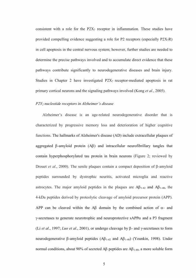
P2Y2 nucleotide receptors in Alzheimer’s disease
Alzheimer’s disease is an age-related neurodegenerative disorder that is
characterized by progressive memory loss and deterioration of higher cognitive
functions. The hallmarks of Alzheimer's disease (AD) include extracellular plaques of
aggregated β-amyloid protein (Aβ) and intracellular neurofibrillary tangles that
contain hyperphosphorylated tau protein in brain neurons (Figure 2; reviewed by
Drouet et al., 2000). The senile plaques contain a compact deposition of β-amyloid
peptides surrounded by dystrophic neuritis, activated microglia and reactive
astrocytes. The major amyloid peptides in the plaques are Aβ1-42 and Aβ1-40, the
4-kDa peptides derived by proteolytic cleavage of amyloid precursor protein (APP).
APP can be cleaved within the Aβ domain by the combined action of α- and
γ-secretases to generate neurotrophic and neuroprotective sAPPα and a P3 fragment
(Li et al., 1997; Luo et al., 2001), or undergo cleavage by β- and γ-secretases to form
neurodegenerative β-amyloid peptides (Aβ1-42 and Aβ1-40) (Younkin, 1998). Under
normal conditions, about 90% of secreted Aβ peptides are Aβ1-40, a more soluble form
5

than Aβ1-42 that can be eliminated from brain and can only slowly be converted to the
plaque-forming β-sheet configuration. In the AD brain, Aβ1-42 is first deposited in
plaques, whereas Aβ1-40 is deposited later as the disease progresses (Iwatsubo et al.,
1994). A major goal to date in the development of treatments for AD include
approaches that decrease Aβ production by blocking γ-secretase activity. However,
this may result in defective haematopoiesis or other side effects (e.g., inhibition of the
notch signaling pathway) due to loss of γ-secretase function (Rochette and Murphy,
2002). Clearly, further studies are needed to better understand the regulation of the
different APP processing enzymes (i.e., α-, β- and γ-secretases) to design effective
treatments for AD.
To date, the research on the functional involvement of P2 nucleotide receptors in
AD is very limited. P2X7 receptors mediate superoxide production in primary
microglia and are up-regulated in a transgenic mouse model of Alzheimer's disease
(Parvathenani et al., 2003). Chronic neuroinflammation is a contributor to the
pathophysiology of Alzheimer’s disease (AD; Eikelenboom et al., 2000;
Hauss-Wegrzyniak et al., 1998). Oxidative stress and oligomeric β-amyloid (Aβ)
peptide production in AD brain can increase extracellular levels of cytokines and
nucleotides that activate receptors in glial cells (i.e., astrocytes and microglia) to
stimulate intracellular signaling pathways and promote reactive gliosis, a universal
response of the brain to injury (Mehlhorn et al., 2000). In the CNS, we reported that
P2Y2 receptors mediate astrocyte migration through interactions with αV integrins in
rat primary cortical astrocytes, which suggests a role for P2Y2R-mediated signaling in
6

regulating astrogliosis in brain disorders (Wang et al., 2005). Moreover, previous
studies by our group showed that UTP enhances α- and γ-secretase-mediated APP
processing in P2Y2R-transfected 1321N1 astrocytoma cells (Camden et al., 2005).
However, relatively little is known about the contribution of P2Y2Rs to neuronal
functions or their co-regulation with cytokine receptors of activities related to AD.
Therefore, we have investigated the effect of cytokines on functional P2Y2R
expression in primary cortical neurons from E18 rat embryos, as described in Chapter
3. The role of P2Y2 receptors in APP processing and the signaling molecules involved
was also investigated in rat primary cortical neurons (see Chapter 3).
P2 nucleotide receptors in brain ischemia
Acute ischemic stroke is the leading cause of adult disability and also the most
frequent pathogenic mechanism of brain lesion in infancy (Guzzetta et al., 2000).
Interestingly, basic and clinical evidence shows that impaired cerebral perfusion
originating in the microvasculature triggered most of the key pathologic events
associated with Alzheimer’s disease (AD); thus the presence of a condition that
lowers cerebral perfusion together with advanced aging are proposed as two factors
necessary to induce a neurodegenerative process in the AD brain (de la Torre, 2000).
The cerebral circulation becomes inadequate under the following conditions that often
lead to global ischemia, rather than focal ischemia: 1) the perfusion pressure is
reduced; 2) the vascular resistance becomes too high; or 3) the oxygen or glucose
supply is reduced (Garcia, 1975).Transient global ischemia caused by a 5-minute
occlusion of gerbil bilateral carotid arteries (BCAOs) leads to selective and delayed
7

neuronal death (DND) of CA1 pyramidal neurons (Figure 3; Kirino, 1982), associated
with activation of surrounding astrocytes in the hippocampus (DeLeo et al., 1987).
Transient focal ischemia for 60 min consistently resulted in a large infarct confined to
the right MCA cortex in rats (Figure 3C). However, ischemic stroke cannot be
mimicked satisfactorily in vitro due to its complex pathophysiology involving the
interplay of many different cells and tissues including neurons, glia, endothelium, and
the immune system. Thus, several animal models of global and focal brain ischemia
have been developed for stroke research including the gerbil/mouse model of transient
global iscemia by occlusion of both common carotid arteries (CCAs) and the
rat/mouse model of focal ischemia model by middle cerebral artery occlusion (MCAO)
(Traystman, 2003).
After transient global ischemia, up-regulation of P2X2 and P2X4 receptors was
observed in neurons and microglia of gerbil hippocampus, respectively (Table 1;
Cavaliere et al., 2003). P2X7 mRNA was also up-regulated in cultured cerebellar
granule neurons of organotypic hippocampal cultures in vitro (Cavaliere et al., 2002,
2004) and in activated microglia surrounding necrotic regions in vivo (Table 1; Collo
et al., 1997), suggesting a role for P2X7Rs in the mechanisms of cellular damage
induced by hypoxia/ischemia. Up-regulation of the P2Y2R subtype also has been
found with stress and injury (Seye et al., 2002; Koshiba et al., 1997 and Turner et al.,
1997). For example, P2Y2R up-regulation is induced by a collar placed around a
rabbit carotid artery (Seye et al., 2002) and arterial P2Y2R up-regulation is associated
with enhanced neointimal hyperplasia, macrophage infiltration and osteopontin
8

expression in response to UTP (Seye et al., 1997, 2003, 2004 & 2006). These findings
indicate for the first time a role for the P2Y2R, in development of intimal hyperplasia
associated with atherosclerosis and restenosis (Seye et al., 2002; Appendix-1). In
Chapter 4, the expression of P2Y2 receptors was identified in normal adult rodent
brains and the relationship of P2Y2 receptor expression to ischemia-induced injury
was determined in a gerbil transient ischemia model (Weisman et al., 2005; Chapter
4).
Outline of the dissertation
This dissertation contains five parts concerning the Regulation and Function of
P2Y2 and P2X7 Nucleotide Receptors in the Central Nervous System from (i.e.,
Chapters 2-4 and the Appendix).
In Chapter 2, we examined P2X7R-mediated apoptosis in rat primary cortical
neurons (rPCNs). P2X7Rs were functionally expressed in the rPCNs and their
activation by extracellular nucleotides was shown to cause apoptotic cell death
through an intrinsic JNK1- and ERK-dependent caspase-8/9/3 activation pathway.
Chapter 3 extends the examination of nucleotide receptors in rPCNs to the P2Y2
receptor subtype. P2Y2R mRNA and receptor activity were virtually absent in
unstimulated rPCNs but was dramatically up-regulated upon overnight treatment with
IL-1β. Furthermore, activation of P2Y2R in rPCNs enhanced the release of sAPPα, a
putative neuroprotective secrectory amyloid protein, by an ADAM10/17- and
PI3K/ERK-dependent mechanism.
In Chapter 4, the expression pattern of P2Y2 receptor was studied in adult rodent
9

brain slices. Moreover, the effect of ischemia on the expression level of P2Y2Rs in the
rodent brain was analyzed by in situ hybridization and RT-PCR in a gerbil transient
global ischemia model.
This dissertation concludes with Chapter 5, which addresses future directions and
the scientific significance of the completed research followed by an Appendix that
presents data from other projects undertaken.
10

Table 1. Regulation of P2 nucleotide receptor expression after ischemia and in
Alzheimer’s Disease (AD)
P2R Injury Cell type Change
P2X2 Ischemia Neurons (hippocampus, organotypic culture, gerbil) ↑
P2X4 Ischemia Microglia (hippocampus, organotypic culture, gerbil) ↑
Ischemia Microglia (cortex, rat) ↑
Ischemia Neurons (hippocampus, organotypic culture) ↑
Ischemia Astrocytes, neurons, microglia (cortex, rat) ↑
P2X7
AD Around plaques (hippocampus, Tg2576 mice) ↑
P2Y1
AD
Neurofibrillary tangles, neuritic plaques, neuropil threads (human hippocampus) Cellular distribution pattern was altered in AD brains.
↓
(Collo et al., 1997; Moore et al., 2000; Cavaliere et al., 2003 &2004; Parvathenani et
al., 2003 ; Franke et al., 2004 ; modified from review in Franke and Illes, 2006b)
11

Figure 1. Expression of P2 receptors in rat cortical astrocytes and neurons. Total
RNA was isolated from primary rat cortical (A) astrocytes or (B, C) neurons and
RT-PCR was used to amplify mRNAs to P2Y and P2X receptors, as previously
described (Weisman et al., 2005; Kong et al., 2005). The amplified PCR products
were resolved by gel electrophoresis, and data shown are representative of results
from three independent experiments performed in the presence (+) or absence (-) of
reverse transcriptase (only the +RT samples are shown in (A) and ++ indicates
amplification of a G3PDH-positive control sample (B).
12

A
B
C
13

Figure 2. Nerve cells, synapses and brain tissue in Alzheimer’s disease. A.
Alzheimer’s disease tissue has fewer nerve cells and synapses than healthy brain
tissue and there are abnormal clusters of phosphorylated tau protein in dead and dying
neurons (white arrow) surrounded with Aβ plaques (white circles) in the extracellular
space. B. In advanced Alzheimer’s disease, most of the cortex is seriously damaged.
The brain shrinks dramatically due to widespread cell death. Pictures are cited from:
www.alz.org.
14

Figure 3. Neuronal cell death in rodent ischemia models. A, B: transient gerbil
global cerebral ischemia, Toshiki et al., 2005; and C: rat focal cerebral ischemia,
web.psych. ualberta.ca/~fcolbour/research.html. Neuronal densities in the CA1 region
of the gerbil hippocampus (B) dramatically decreased 5 days after transient cerebral
ischemia compared with the sham-operated gerbil brain (A). Arrows show location of
changes. Focal cerebral ischemia–reperfusion injury induced by temporary middle
cerebral artery occlusion (MCAO) for 60 min followed by reperfusion causes a large
infarct (C; top of right cortex) compared with the sham-operated right MCA cortex.
16

A B C
(A-B: Toshiki et al., 2005; C: ) web.psych.ualberta.ca/~fcolbour/research.html
17

CHAPTER 2
P2X7 NUCLEOTIDE RECEPTORS MEDIATE CASPASE-8/9/3-DEPENDENT
APOPTOSIS IN RAT PRIMARY CORTICAL NEURONS
Kong, Q. et al., (2005) Purinergic Signalling 1:337-347
18

ABSTRACT
Apoptosis is a major cause of cell death in the nervous system. It plays a role in
embryonic and early postnatal brain development and contributes to the pathology of
neurodegenerative diseases. Here, we report that activation of the P2X7 nucleotide
receptor (P2X7R) in rat primary cortical neurons (rPCNs) causes biochemical (i.e.,
caspase activation) and morphological (i.e., nuclear condensation and DNA
fragmentation) changes characteristic of apoptotic cell death. Caspase-3 activation
and DNA fragmentation in rPCNs induced by the P2X7R agonist BzATP were
inhibited by the P2X7R antagonist oxidized ATP (oATP) or by pretreatment of cells
with P2X7R antisense oligonucleotide indicating a direct involvement of the P2X7R in
nucleotide-induced neuronal cell death. Moreover, Z-DEVD-FMK, a specific and
irreversible cell permeable inhibitor of caspase-3, prevented BzATP-induced
apoptosis in rPCNs. In addition, a specific caspase-8 inhibitor, Ac-IETD-CHO,
significantly attenuated BzATP-induced caspase-9 and caspase-3 activation,
suggesting that P2X7R-mediated apoptosis in rPCNs occurs primarily through an
intrinsic caspase-8/9/3 activation pathway. BzATP also induced the activation of
JNK1 and ERK1/2 in rPCNs, and pharmacological inhibition of either JNK1 or
ERK1/2 significantly reduced caspase activation by BzATP. Taken together, these
data indicate that extracellular nucleotides mediate neuronal apoptosis through
activation of P2X7Rs and their downstream signaling pathways involving JNK1, ERK
and caspases 8/9/3.
19

INTRODUCTION
ATP is an important extracellular messenger generated from various sources
including release from presynaptic vesicles upon nerve stimulation in the peripheral
and central nervous systems (Burnstock et al., 1978). Extracellular ATP functions as a
neuromodulator by activation of cell surface P2 nucleotide receptors that are widely
expressed in the nervous system (Burnstock 1980; Inoue 2002). Based on their
distinct structure, P2 nucleotide receptors are classified into two families: G
protein-coupled P2Y receptors (P2YR1, 2, 4, 6, 11-14; Abbracchio et al., 2003) and
ligand-gated ion channels (P2XR1-7; Burnstock 2000). Functional responses to
activation of these P2 receptor subtypes in cells of the nervous system under normal
and pathological conditions include cell apoptosis, proliferation and migration,
inflammation, ion transport, and neurotransmission (Burnstock 2000; Norenberg &
Illes 2000; von Kugelgen & Wetter 2000; Khakh 2001 and Illes et al., 2004).
Therefore, P2 receptors in the nervous system may prove to be useful pharmaceutical
targets in the treatment of neurological disorders.
The seven subtypes of the ionotropic ATP-gated P2XR family, ranging from 379
to 595 amino acids in length, regulate the intracellular calcium concentration through
ligand-stimulated increases in the cell membrane permeability to extracellular Ca2+
ions (Benham 1990; Di Virgilio et al., 2001a). In contrast to other P2XRs, the P2X7R
subtype functions exclusively as a homomeric receptor (Chessell et al., 1998; North &
Surprenant 2000). The P2X7R is a 595-amino acid polypeptide containing two
membrane-spanning domains, a large extracellular loop, and intracellular N- and
20

C-terminal domains including a C-terminal tail that is the longest among the cloned
P2XRs (Surprenant et al., 1996; Rassendren et al., 1997). P2X7Rs have been reported
in microglial cells (Collo et al., 1997) where they play a role in cytokine release
(Ferrari et al., 1997). These receptors are also found in cultured astrocytes and
Schwann cells (Duan et al., 2003; Grafe et al., 1999), spinal cord neurons and in other
regions of the central nervous system (Deuchars et al., 2001; Atkinson et al., 2004).
Brief stimulation of the P2X7R with ATP results in formation of a non-selective
cationic channel that promotes the influx of Ca2+ and the equilibration of the
transmembrane sodium and potassium gradients leading to membrane depolarization
(Surprenant et al., 1996; Rassendren et al., 1997; Weisman et al., 1984; Virginio et al.,
1999). Subsequent responses to P2X7R activation include the formation of a
nonselective pore for molecules up to 900 Da (Weisman et al., 1984; Virginio et al.,
1999), extensive membrane blebbing, and eventual cell death (Virginio et al., 1999).
Activation of the P2X7R was reported to elicit Ca2+ influx in cerebrocortical nerve
terminals (Lundy et al., 2002) and hippocampal glutamate release (Sperlagh et al.,
2002), which has been confirmed by studies with P2X7R knockout mice (Papp et al.,
2004). In an ex vivo model of organotypic hippocampal cultures, P2X7R activation
was shown to directly participate in cell damage induced by oxygen/glucose
deprivation (Cavaliere et al., 2004). It has been shown that a Pro-451 to Leu
polymorphism within the C-terminal tail of the P2X7R interferes with the ability of
the receptor to induce cell death in murine thymocytes (Le Stunff et al., 2004). The
P2X7R also has been shown to mediate ATP-induced cell death in human embryonic
21

kidney cells (Wen et al., 2003) and human cervical epithelial cells (Wang et al., 2004).
A recent study demonstrated that spinal cord injury was associated with prolonged
P2X7R activation of spinal cord neurons by extracellular ATP, which resulted in
high-frequency spiking, increases in cytosolic calcium levels and neuronal
degeneration, responses that were reversed by P2X7R inhibition (Wang et al., 2004).
In the present study, we demonstrate that the P2X7R agonists BzATP or ATP
induce nuclear condensation and DNA fragmentation in rat primary cortical neurons
(rPCNs). These apoptotic responses in rPCNs were dependent on the functional
expression of P2X7Rs that mediate the ERK1/2- and JNK1-dependent activation of
caspases-8/9/3.
MATERIALS AND METHODS
Materials
Fetal bovine serum (FBS) was obtained from Hyclone (Logan, UT, USA).
Dulbecco’s Modified Eagle’s Medium (DMEM), Minimum Essential Medium
(MEM), Neurobasal Medium, penicillin (100 units/ml), streptomycin (100 units/ml)
and B27-AO (B27 without cortex antioxidants) were obtained from Gibco-BRL
(Carlsbad, CA, USA). Rabbit anti-rat ERK1/2, horseradish peroxidase
(HRP)-conjugated goat anti-rabbit IgG, and HRP-conjugated goat anti-mouse IgG
antibodies were obtained from Santa Cruz Biotechnology (Santa Cruz, CA, USA).
Goat anti-rat P2X7R antibody was obtained from Alomone (Jerusalem, Israel). Rabbit
anti-rat caspase-8 antibody was obtained from Biovision Inc. (Mountain View, CA,
22

USA). Mouse anti-NeuN antibody was obtained from Chemicon International Inc.
(Temecula, CA, USA). Alexa Fluor TM 488 goat anti-mouse IgG antibody, Alexa
Fluor TM 594 goat anti-rabbit IgG antibody, YO-PRO-1, TO-PRO-3 and the Prolong
anti-fade kit were obtained from Molecular Probes Inc. (Eugene, OR, USA). All other
antibodies were obtained from Cell Signaling Technology (Beverly, MA, USA).
Caspase-3 and caspase-9 inhibitors were obtained from R&D (Minneapolis, MN,
USA). Caspase-8 inhibitor was obtained from BioSource International Inc. (Camarillo,
CA, USA). The Precision Plus Protein Standards and nitrocellulose membranes (0.45
mm) were obtained from Bio-Rad (Hercules, CA, USA). LumiGLO
chemiluminescent substrates were obtained from New England Biolabs (Beverly, MA,
USA). The RNeasy Mini Kit was obtained from Qiagen (Chatsworth, CA, USA). The
Apoptotic DNA-ladder kit, the First Strand cDNA Synthesis kit and the TUNEL In
Situ Cell Death Detection kit were obtained from Roche (Indianapolis, IN, USA).
Nucleotides and all other biochemicals were obtained from Sigma Chemical Co. (St.
Louis, MO, USA).
Primary cell culture of cortical neurons
Experimental procedures for cell culture of rat primary cortical neurons (rPCNs)
were carried out essentially as described by Suen et al. (Suen et al., 2003). Briefly,
cerebral cortices from18-day-old embryos of Sprague-Dawley rats were removed and
the meninges discarded. The brain tissue was mechanically dissociated in HGGMEM
comprised of 1.78 g glucose, 100 IU/ml penicillin, 100 μg/ml streptomycin, 7.5 μg/ml
fungizone, 2 mM glutamine in 88 ml of MEM and 10% (v/v) horse serum. The tissue
23

clumps were dispersed with a 10 ml pipette and suspended in 6 ml of 0.25% (w/v)
trypsin at 37 °C for 10 min. After incubation, 2 ml of heat-inactivated horse serum
were added to block trypsin activity. Cells were centrifuged at 4000 g for 5 min and
the pellet was suspended in HGGMEM with 10% horse serum. The cell suspension
was filtered through a sterilized 75 mm cell strainer (nylon membrane; Becton
Dickinson, Franklin Lakes, NJ) and cells were counted and seeded into plastic culture
plates that were precoated with poly-D-lysine (0.1 μg/ml). After 16 h and every 3
days thereafter, the medium was replaced with B27-AO Neurobasal Medium (2 mM
glutamine, 8.9 g glucose, 100 IU/ml penicillin, 100 μg/ml streptomycin, 7.5 μg/ml
fungizone, 10 ml of B27 minus AO, and Neurobasal Medium (Gibco-BRL) to 500 ml)
and the neurons were ready for use after 7 days in culture.
RT-PCR analysis of P2XR mRNA expression
Total RNA was isolated from rPCNs using the RNeasy Mini Kit (Qiagen). cDNA
was synthesized from the purified RNA using the First Strand cDNA Synthesis Kit
for RT-PCR (AMV) (Roche). Five percent of the synthesized cDNA was used as a
template in PCR reactions with the Expand High Fidelity PCR System (Roche).
Specific oligonucleotide primers were designed to selectively amplify cDNA for
individual P2XR subtypes, as shown in Table 2. The amplification was performed
using 35 cycles of denaturation at 95 °C for 30 s, with annealing at 60 °C for 30 s and
extension at 72 °C for 1 min. The resulting PCR products were resolved on a 1% (w/v)
agarose gel containing 10 mg/ml ethidium bromide and photographed under UV
illumination.
24

Single cell calcium assay
The [Ca2+]i was quantified in single cells with the Ca2+-sensitive fluorescent dye
fura-2, using an InCyt Dual-Wavelength Fluorescence Imaging System (Intracellular
Imaging, Cincinnati, OH). Rat PCNs cultured on a poly-D-lysine-coated coverslip
were incubated with 2.5 μM fura-2-acetoxymethylester at 37 °C for 30 min in
physiological salt solution (PSS) containing (mM) NaCl 138, KCl 5, CaCl2 2, MgCl2
1, HEPES 10, glucose 10, pH 7.4, and washed with PSS. The coverslip with
fura-2-loaded cells was positioned on the stage of an inverted epifluorescence
microscope (Nikon; model TMD) and stimulated with agonists at 37 °C, as described
in the figure legends. Cells were exposed to 340/380 nm light and fluorescence
emission at 505 nm was converted to [Ca2+]i using a standard curve created with
solutions containing known concentrations of Ca2+. Increases in [Ca2+]i were
measured by subtracting basal [Ca2+]i from the maximum agonist-induced increase in
[Ca2+]i. The percentage of cells that responded to ATP/BzATP was also determined.
Viable cells were identified by responsiveness to carbachol and non-responding cells
were eliminated.
Detection of nuclear condensation
Rat PCNs were cultured in B27-AO Neurobasal medium on poly-D-lysine-coated
coverslips in 12-well plates until 50-70% confluence. After nucleotide treatments,
medium was removed and cells were washed once and incubated with
DAPI-methanol staining solution (1 mg/ml) for 15 min at 37 °C. The staining solution
was removed and the cells were washed with methanol. The inverted coverslip was
25

placed onto a microscope slide using glycerol as the mounting agent. Nuclear
morphology was observed under a fluorescence microscope (Nikon, Eclipse TE300)
with a 340/380 nm excitation filter and a LP 430 nm barrier filter. DAPI has an
absorbance maximum at 340 nm and an emission maximum at 488 nm in aqueous
solution (Kapus’cin’ski et al., 1978).
DNA laddering assay
Rat PCNs were cultured in B27-AO Neurobasal medium in 100 mm dishes to
70% confluence, and incubated in serum-free medium overnight with or without
BzATP, or treated for 2 h with 500 μM H2O2 and DNA was isolated using the
Apoptotic DNA-ladder kit (Roche, Indianapolis, IN) according to the manufacturer’s
instructions. DNA also was isolated from lyophilized apoptotic U937 cell lysate
(provided with the DNA-ladder assay kit) for use as a positive control. Briefly, the
medium was replaced with 200 μl of PBS and 200 μl of binding buffer (6 M
guanidine-HCl, 10 mM urea, 10 mM Tris-HCl, pH 4.4, 20% (v/v) Triton X-100).
After 10 min at 15-25 °C, 100 ml of isopropanol were added and the samples were
transferred to a filter tube and centrifuged for 1 min at 10,000 g (Spectrafuge Labnet,
Edison, NJ). The fluid in the collection tube was discarded and the upper filter column
was washed with 500 μl of washing buffer (20 mM NaCl and 2 mM Tris-HCl, pH 7.5,
40% (v/v) ethanol) followed by centrifugation twice for 1 min at 13,000 g. After the
residual washing buffer was removed, the DNA was eluted with 200 μl of
pre-warmed (70 °C) elution buffer (10 mM Tris, pH 8.5). The concentration and
purity of the eluted DNA was determined by measuring the optical density at 260 and
26

280 nm. DNA was electrophoresed in a 2% (w/v) agarose gel at 80 V for 1 h in TBE
buffer (89 mM Tris base, 89 mM boric acid, 2 mM EDTANa2-salt, pH 8.3) and
visualized under UV light.
TUNEL assay
After treatment with nucleotides or H2O2, rPCNs were fixed with fresh 4% (v/v)
paraformaldehyde in PBS and treated with 3% (v/v) H2O2 in methanol to inhibit
endogenous peroxidase activity. Then, DNA strand breaks in fixed cells were detected
with a TUNEL In Situ Cell Death Detection kit (Roche) according to the
manufacturer’s instructions. The number of TUNEL-positive cells and the total cell
number were determined by fluorescence (Nikon; Eclipse TE300) using an excitation
wavelength in the range of 450-500 nm and a detection wavelength in the range of
515-565 nm (green) and light microscopy with cell counting, respectively, at 200x
magnification.
Application of P2X7 antisense oligonucleotide
Antisense oligonucleotide treatment was performed as previously described (Seye
et al., 2002). Phosphorothioate-modified oligonucleotides to rat P2X7 were
synthesized and purified by Integrated DNA Technologies (ADT, Coralville, IA,
USA). Sequences were designed as follows: sense 5’-AGAGCGTGAATTACGGCA
CATCAA-3’, antisense 5’-TTGATGGTGCCGTAATTCACGCTCT-3’. Sequences
were checked for uniqueness with the National Center for Biotechnology
Information’s Basic Local Alignment Search Tool (http://www.ncbi.nlm.nih.
gov/BLAST). The rPCNs were incubated with 0.1 or 1 μmol/L P2X7 sense or
27

antisense S-oligonucleotide for 6 h in serum-free medium containing 1.4% (v/v)
DOTAP liposomal reagent (Roche Diagnostics). Fresh serum-free medium was added
with or without BzATP, and cells were cultured for an additional 18 h.
Western blot analysis
Rat PCNs cultured in 6-well plates were treated with 300 μM BzATP in the
presence or absence of inhibitors as indicated. The cells were washed twice with
ice-cold PBS, and lysed for 5 min in 200 ml of ice-cold lysis buffer (20 mM Tris-HCl,
pH 7.5, 2% (w/v) sodium dodecyl sulfate (SDS), and 1 mM sodium orthovanadate).
Lysate (30 mg of protein) was added to Laemmli sample buffer (187.5 mM Tris-HCl,
pH 6.8, 6% (w/v) SDS, 1.8% (v/v) β-mercaptoethanol and 0.003% (w/v)
bromophenol blue). Samples were heated for 5 min at 96-100 °C, and subjected to
7.5% (w/v) SDS-polyacrylamide gel electrophoresis (SDS-PAGE) and transferred to
nitrocellulose membranes for protein immunoblotting. After overnight blocking at 4
°C with 5% (w/v) fat-free milk in TBS-T (10 mM Tris-HCl, pH 7.4, 120 mM NaCl,
and 0.1% (v/v) Tween-20), membranes were incubated with either 1:1000 dilution of
anti-ERK1/2, anti-phospho-ERK1/2, anti-phospho-JNK, or anti-P2X7R antibodies for
2 h at room temperature, or 1:1000 dilution of anti-cleaved caspase-3, anti-caspase-3,
anti-cleaved caspase-9, anti-caspase-9, or anti-caspase-8 antibodies overnight at 4 °C
followed by incubation with HRP-conjugated anti-rabbit or anti-mouse IgG antibodies
(1:1000 dilution in TBS-T containing 5% (w/v) fat-free milk) for 1 h at room
temperature. For normalization of protein loading, the membranes were stripped of
antibodies by incubation for 30 min at 60 °C in stripping buffer (62.5 mM Tris-HCl,
28

pH 6.8, 100 mM 2-mercaptoethanol, and 2% (w/v) SDS), and re-probed with 1:2000
dilution of rabbit polyclonal anti-actin antibody (2 h at room temperature;
Cytoskeleton, Denver, CO) followed by 1:2000 dilution of HRP-conjugated
anti-rabbit IgG antibody (1 h at room temperature). Protein immunoreactivity was
visualized on autoradiographic film using the LumiGlo Chemiluminescence System
(New England BioLabs), according to the manufacturer’s instructions. The protein
bands detected on X-ray film were quantified using a computer-driven scanner and
Quantity One software (Bio-Rad, Hercules, CA). The activation levels of kinases or
caspases were calculated as a percentage of control (i.e., total kinase, caspase, or
actin).
Immunofluorescence
After fixation and permeabilization, rPCNs cultured on coverslips in 6-well plates
were incubated in 2.5% (w/v) BSA and 2.5% (v/v) goat serum in PBS for 1 h at room
temperature followed by incubation overnight with mouse anti-NeuN antibody (1:100
dilution; Chemicon) and rabbit anti-cleaved caspase-3 antibody (1:100 dilution; Cell
Signaling) in 0.25% BSA and 0.25% goat serum in PBS. Cells were washed three
times with PBS and then incubated with 1:200 dilution of goat anti-mouse Alexa
Fluor 488- or goat anti-rabbit Alexa Fluor 594-conjugated IgG antibody (Molecular
Probes, Eugene, OR) in 0.25% BSA and 0.25% goat serum in PBS for 1 h at 37 °C.
Nuclei were stained with TO-PRO-3 (Molecular Probes), cells were rinsed with PBS
and mounted on glass slides in ProLong antifade reagent (Molecular Probes). Images
were obtained on a Bio-Rad MRC 1024 laser scanning confocal imaging system
29

coupled to an Olympus IX70 inverted microscope. The imaging system was
controlled by Laser Sharp software (version 3.2; Bio-Rad, Hercules, CA). Alexa
488-labeled slices were excited by 488 nm laser light, and images were acquired with
a 522 nm emission filter with a bandwidth of 35 nm, whereas Alexa 594-labeled
slices were excited by 568 nm light and images acquired with a 585 nm longpass
emission filter. Settings of the confocal microscope were optimized for imaging in an
initial experiment and then equivalently applied to all sections. Laser power and
photomultiplier tube (PMT) gain were adjusted to ensure that even the brightest
fluorescence was below saturation levels. Neurons were randomly chosen and
scanned at 512 x 512 pixel resolution.
Statistical analysis
Results are expressed as the means ± S.E.M. Statistical analysis of data was
performed using Graph Pad Prism version 4.0. Statistical significance was determined
by student t-test. Differences were considered statistically significant when p < 0.05.
RESULTS
Purity of rat cortical neurons in primary culture
The purity of rPCNs was determined by monitoring morphology with phase
contrast microscopy and by immunofluorescence assay with the neuron-specific
marker NeuN and the nuclear stain, TO-PRO-3 (Figure 1A). NeuN staining was found
primarily in the nucleus of the neurons with lighter staining in the cytoplasm. About
95% of the cells were determined to be cortical neurons based on the ratio of NeuN
30

positive cells to total cells counted by TO-PRO nuclear staining (data not shown).
Functional P2X7Rs are expressed in rat PCNs
Rat PCNs express P2X7R mRNA, as well as mRNAs for P2X2, P2X3, P2X5, and
P2X6 receptors (Figure 1B). Furthermore, immunoblot analysis indicated that rPCNs
express P2X7R protein (Figure 1C). The P2X7R agonists ATP and BzATP (Erb et al.,
1990) caused a dose-dependent increase in [Ca2+]i in rPCNs (Figure 2), suggesting
that calcium influx is stimulated by activation of the endogenously expressed P2X7R
protein. Calcium responses were also observed by stimulation of rPCNs with
2-methylthio-ADP (data not shown), an agonist of the P2Y1R, indicating that other
P2R subtypes besides the P2X7R can regulate increases in [Ca2+]i.
ATP and BzATP induce nuclear condensation and DNA fragmentation in rPCNs
Treatment of rPCNs with either BzATP (300 μM) or ATP (100 μM) overnight
caused nuclear condensation as indicated by increased DAPI staining of nuclei
(Figure 3). BzATP-induced nuclear condensation was inhibited by the P2X7R
antagonist oATP, consistent with the involvement of P2X7Rs in this process.
Moreover, BzATP treatment of rPCNs caused DNA fragmentation, suggestive of
apoptotic cell death (Figure 4). BzATP or ATP also caused DNA strand breakage in
rPCNs, another indicator of apoptosis, detected as an increase in the number of
TUNEL-positive cells (Figure 5). These data suggest that BzATP or ATP can cause
apoptosis in rPCNs through a pathway involving P2X7R activation.
P2X7R-mediated caspase-3 and caspase-9 activation
31

Caspase-3 is an essential component of apoptotic pathways in many cell types,
including neurons (Salvesen and Dixit, 1997), and is activated by the sequential
release of cytochrome c from mitochondria and the activation of caspase-9 (Li et al.,
1997). Consistent with a role for caspase-3 in nucleotide-induced neuronal apoptosis,
nuclear condensation caused by BzATP was diminished by treatment of rPCNs with
the caspase-3 inhibitor Z-Asp(OMe)-Glu(OMe)-Val-Asp(OMe)-FMK (Z-DEVDFMK;
Figure 3C). Results also indicated that treatment of rPCNs with either ATP or BzATP
significantly increased the cleavage of caspase-3 in a time- and dose-dependent
manner (Figure 6A). BzATP-induced caspase-3 cleavage was inhibited by oATP
(Figure 6B), the caspase-3 inhibitor Z-DEVD-FMK (Figure 6B), or P2X7 antisense
oligonucleotide (Figure 6C). Although recent studies indicate that oATP might act
independently from P2 receptor inhibition (Beigi et al., 2003), inhibition of
BzATP-induced caspase-3 cleavage by P2X7R antisense oligonucleotide confirms the
involvement of the P2X7R. Since no preparation of primary neurons is devoid of glial
cells, dual immunofluorescence analysis for neuron-specific NeuN and cleaved
caspase-3 was performed to evaluate whether the apoptotic cells were neurons. The
majority of cells treated with BzATP were positive for cleaved caspase-3 and NeuN
(e.g., pink and white arrows in Figure 6D), indicating that a large percentage of cells
undergoing apoptosis were neurons. NeuN immunoreactivity has been reported to
decrease under several pathological conditions (Unal-Cevik et al., 2004), and
therefore NeuN detection may decrease with caspase-3 activation in apoptotic neurons,
suggesting that some apoptotic neurons may go undetected. BzATP also caused the
32

cleavage of caspase-9 (Figure 7A), whereas activation of caspase-9 and caspase-3
were significantly inhibited by the caspase-8 inhibitor Ac-IETD-CHO (Figure 7B) or
the caspase-9 inhibitor Z-LEHD-FMK (data not shown), indicating that caspase-8
also plays a role in activating downstream caspase family members (e.g., caspase-9/3)
of the P2X7R-mediated apoptosis pathway. We also detected an inhibitory effect of
the caspase-9 inhibitor, Z-LEHD-FMK, on caspase-8 cleavage (Figure 7C) suggesting
that caspase-8 and caspase-9 may regulate caspase-3 activity in a non-linear fashion.
Caspase-3 activation by BzATP in rPCNs is dependent upon ERK1/2 and JNK1
phosphorylation
P2X7R-mediated activation of extracellular signal-regulated kinases (ERK1/2) in
rat primary astrocytes (Panenka et al., 2001) and SAPK/JNK in human and rodent
macrophages occur independently of the activation of caspase-1- or caspase-3-like
proteases (Humphreys et al., 2000). Here, we tested whether ERK1/2 or JNK1
activities were involved in BzATP-induced apoptosis in rPCNs. As indicated in
Figure 8, both ERK1/2 and JNK1 were rapidly phosphorylated in response to BzATP
or ATP in a dose- and time-dependent manner. Inhibition of ERK1/2 or JNK1
phosphorylation in rPCNs with either the MEK inhibitor UO126 or the JNK inhibitor
SP600125, respectively, attenuated caspase-3 activation, indicating the involvement
of ERK1/2 and JNK1 in BzATP-induced neuronal cell death (Figure 9).
DISCUSSION
The results obtained with rat PCNs indicate that activation of the P2X7R by its
33

agonists BzATP or ATP promotes the cleavage of caspases-8/9/3 and the appearance
of cellular markers indicative of apoptosis including DNA degradation and nuclear
condensation and fragmentation. Since BzATP can activate other P2X receptor
subtypes, albeit at higher concentrations than the P2X7R (Bianchi et al., 1999), we
explored the role of the P2X7R in the apoptotic effects of BzATP in rPCNs using
P2X7 antisense oligonucleotide to downregulate P2X7R expression and the P2X7R
antagonist oATP. Results indicate that inhibition of P2X7R activity significantly
inhibited BzATP-induced nuclear condensation (by P2X7R antagonist oATP; Figure 3)
and caspase-3 cleavage (by both oATP and P2X7R antisense oligonucleotide; Figure
6), apoptotic responses that were diminished by incubation of rPCNs with the
caspase-3 inhibitor, Z-DEVD-FMK (Figures 3C and 6B). These data strongly suggest
that P2X7Rs mediate BzATP-induced apoptosis in rPCNs, although this does not rule
out a potential role for other P2 nucleotide receptors.
P2X7R-mediated neuronal apoptosis in vivo requires that ATP be released from
neurons or non-neuronal cells to activate cell surface receptors on neighboring
neurons. Neuronal P2X7 receptors are targeted to presynaptic terminals in the central
and peripheral nervous systems and activation of the P2X7R has been indicated to
promote release of vesicular contents from presynaptic terminals (Deuchars et al.,
2001). ATP is present at high concentrations in synaptic vesicles and is released as a
co-transmitter with noradrenaline, acetylcholine and other neurotransmitters
(Burnstock et al., 1978). ATP release also has been demonstrated in a model of spinal
cord injury (Sim et al., 2004). Thus, ATP release from excited neurons or damaged
34
cells is an assumed consequence of any brain injury or disease (e.g., ischemia, spinal
cord injury, or neurodegenerative diseases), suggesting that pro-apoptotic P2X7Rs are
likely activated under pathological conditions in vivo. Recent research indicates a lack
of P2X7R expression in adult rat hippocampal neurons (Khera et al., 2004), whereas
our studies demonstrate functional P2X7R expression in rPCNs from 18-day old
embryos. We have detected P2X7R mRNA in freshly isolated rPCNs (data not shown)
and 7-10 day-old cultures (Figure 1), suggesting that the in vitro cell culture
procedure alone was not required for P2X7R expression.
Caspase activation is known to play a central role in the execution of apoptosis by
causing nuclear condensation, DNA fragmentation and other apoptotic changes. In the
mitochondrial-initiated pathway of apoptosis, caspase activation is triggered by the
formation of a multimeric Apaf-1/cytochrome c complex that is fully functional in
recruiting and activating procaspase-9 (Li et al., 1997). Activated caspase-9 will then
cleave and activate downstream caspases such as caspase-3, -6, and -7. In rPCNs, it
was not surprising that activation of caspase-3 was significantly inhibited by the
caspase-9 inhibitor Z-LEHD-FMK (data not shown), since caspase-9 is known to
activate caspase-3. We also found that in rat PCNs, the caspase-8 inhibitor
Ac-IETD-CHO partially inhibited caspase-9 and caspase-3 activation (Figure 7B)
indicating that caspase-8 cleavage may be an early event in this neuronal apoptosis
pathway. Caspase-8 has been reported to play a role in cytochrome c-dependent
apoptosis (Viswanath et al., 2001) and caspase-8 activation is associated with Fas
receptor-induced apoptosis (Zhuang et al., 1999) and can result in the cleavage and
35

activation of Bid, a proapoptotic member of the Bcl-2 family (Li et al., 1998).
Truncated Bid translocates from the cytoplasm to the mitochondria, where it appears
to antagonize the actions of anti-apoptotic Bcl-2, thereby causing an efflux of
cytochrome c from the mitochondria and downstream caspase-9/3 activation (Li et al.,
1998; Kuwana et al., 1998; Luo et al., 1998; Schendel et al., 1999; Wei et al., 2001).
In human embryonic kidney HEK-293 cells expressing P2X7Rs, extracellular ATP
induced apoptosis associated with activation of poly (ADP-ribose) polymerase (PARP)
and a decrease in Bax protein expression (Wen et al., 2003). We examined the effect
of ATP/BzATP on the expression of Bcl-2 family members in rPCNs but failed to
detect a significant change in Bax and Bcl-2 protein expression (data not shown).
Evidence from cell-free and in vitro expression systems has indicated that caspase-3
can induce cleavage and activation of the upstream initiator caspase-8 (Slee et al.,
1999; Wolf and Green, 1999; Tang et al., 2000). Our data indicated that inhibition of
caspase-9 cleavage decreased caspase-8 cleavage (Figure 7C), consistent with the
ability of downstream caspases to regulate the cleavage of initiator caspases.
We have demonstrated that activation of P2X7Rs in rPCNs increases calcium
influx (Figure 2) and stimulates phosphorylation of the intracellular kinases ERK1/2
and JNK1 (Figure 8). Furthermore, inhibition of ERK1/2 or JNK1 phosphorylation
decreased BzATP-induced caspase-3 cleavage (Figure 9). Our previous studies
demonstrating that P2X7R-mediated ERK1/2 activation was dependent on
extracellular Ca2+ (Gendron et al., 2003) suggest a similar role for BzATP-stimulated
calcium influx in rPCNs (Figure 2). Recent studies indicate that activation of ERK
36

and phosphatidylinositol 3-kinase/Akt signaling pathways caused inhibition of
caspase-3 and cell apoptosis (Khreiss et al., 2002). It also has been reported that
inhibition of MEK/ERK signaling is required for the induction of apoptosis by
Sulindac metabolites (Rice et al., 2004). Nonetheless, our data support the hypothesis
that ERK1/2 and JNK1 activation can promote P2X7R-mediated apoptosis in rPCNs,
although the mechanism involved requires further investigation. P2X7Rs can promote
neuronal cell death by mediating the release of glutamate and inflammatory factors
from cortical neurons, RAW 264.7 macrophages, and astrocytes (Duan et al, 2003;
Hu et al., 1998; Guerra et al., 2003). Moreover, glutamate can induce ATP release
from astrocytes (Queiroz et al., 1999), suggesting that ATP and glutamate have
reciprocal roles that contribute to the propagation of an apoptotic signal. It also has
been reported that the P2X7R modulates macrophage production of TNF-α, IL-1β and
nitric oxide (NO) following LPS exposure (Hu et al., 1998), consistent with a role for
the P2X7R in inflammation (Di Virgilio et al., 2001b). In addition to these factors,
structural features within the P2X7R indicate that the mechanism whereby it regulates
apoptosis is likely to be complex. There are several functional domains located in the
P2X7R C-terminal tail including an LPS-binding domain, a SH3-binding domain, and
an overlapping Fcelldeath domain, which appear to be important for receptor
trafficking and various physiological functions (Denlinger et al., 2001), and the role
of these domains in P2X7R-mediated apoptosis of neurons warrants further
investigation. Our studies with the membrane impermeant fluorescent dye YO-PRO-1
(molecular weight: 375 Da) indicate that BzATP causes pore formation (YO-PRO-1
37

uptake) in rPCNs (data not shown), consistent with the report that apoptosis in the
retinal microvasculature is associated with P2X7 receptor-mediated pore formation
(Sugiyama et al., 2004).
A recent study indicated that spinal cord neurons expressed an abundance of
P2X7Rs, and exposure of freshly prepared spinal cord slices to ATP or BzATP evoked
high frequency firing and irreversible increases in the cytosolic Ca2+ concentration
(Wang et al., 2004). Significant cell death occurred 24 h after spinal cord injury in
areas of high ATP release surrounding the epicenter of the injury, while local
injection of oATP in the peritraumatic zone strongly reduced both apoptotic cell
number and the severity of histological injury, suggesting the involvement of a P2X7R.
Other studies indicate that the P2X7R is up-regulated in a mouse model of
Alzheimer’s disease (Parvathenani et al., 2003). Thus, the P2X7R may represent a
promising therapeutic target to inhibit neurodegeneration due to injury or disease.
In summary, the data presented indicate that BzATP and ATP can stimulate
P2X7Rs in embryonic rat primary cortical neurons leading to the activation of the
caspases-8/9/3 and the induction of DNA degradation and nuclear condensation and
fragmentation, apoptotic responses that appear to be regulated by ERK1/2 and JNK1.
Since neuronal apoptosis occurs in Alzheimer’s disease, Parkinson’s disease and other
neurodegenerative disorders, a better understanding of the mechanisms of
P2X7R-mediated apoptosis may lead to novel therapies to prevent apoptosis-related
brain injuries.
38

Table 2. Sequence-specific oligonucleotide primers used for RT-PCR studies.
Receptor Subtypes Oligonucleotide Sequences
Forward 5'-AAC AGC ATC AGC TTT CCA CG-3'P2X1
Reverse 5'-TGT AGT AGT GCC TCT TAG GC-3'
Forward 5'-ACC TGC CTC TCC GAC GCC GA-3'P2X2
Reverse 5'-GAA GTC AGA GCT GTG GCC AG-3'
Forward 5'-CAA CTT CAG GTT TGC CAA A-3' P2X3
Reverse 5'-TGA ACA GTG AGG GCC TAG AT-3'
Forward 5'-TAC GAC ACG CCG CGC ATC-3'P2X4
Reverse 5'-TGC ACG ATT TGA GGT AGG ACG-3'
Forward 5'-CAA AGT CCA TGC CAA CGG AT-3'P2X5
Reverse 5'-ACG GAA CTC TAC CCC ATT AG-3'
Forward 5'-GTA GTG CTG TGC CCA GGA AA-3'P2X6
Reverse 5'-GGA CTC CAC GCC TGA GGC TG-3'
Forward 5'-ACA ATG TTG AGA AAC GGA CTC TGA-3' P2X7
Reverse 5'-CCG GCT GTT GGT GGA ATC CAC ATC-3'
Forward 5’-TGA AGG TCG GTG TCA ACG GAT TTG GC-3’G3PDH
Reverse 5’-CAT GTA GGC CAT GAG GTC CAC CAC-3’
39

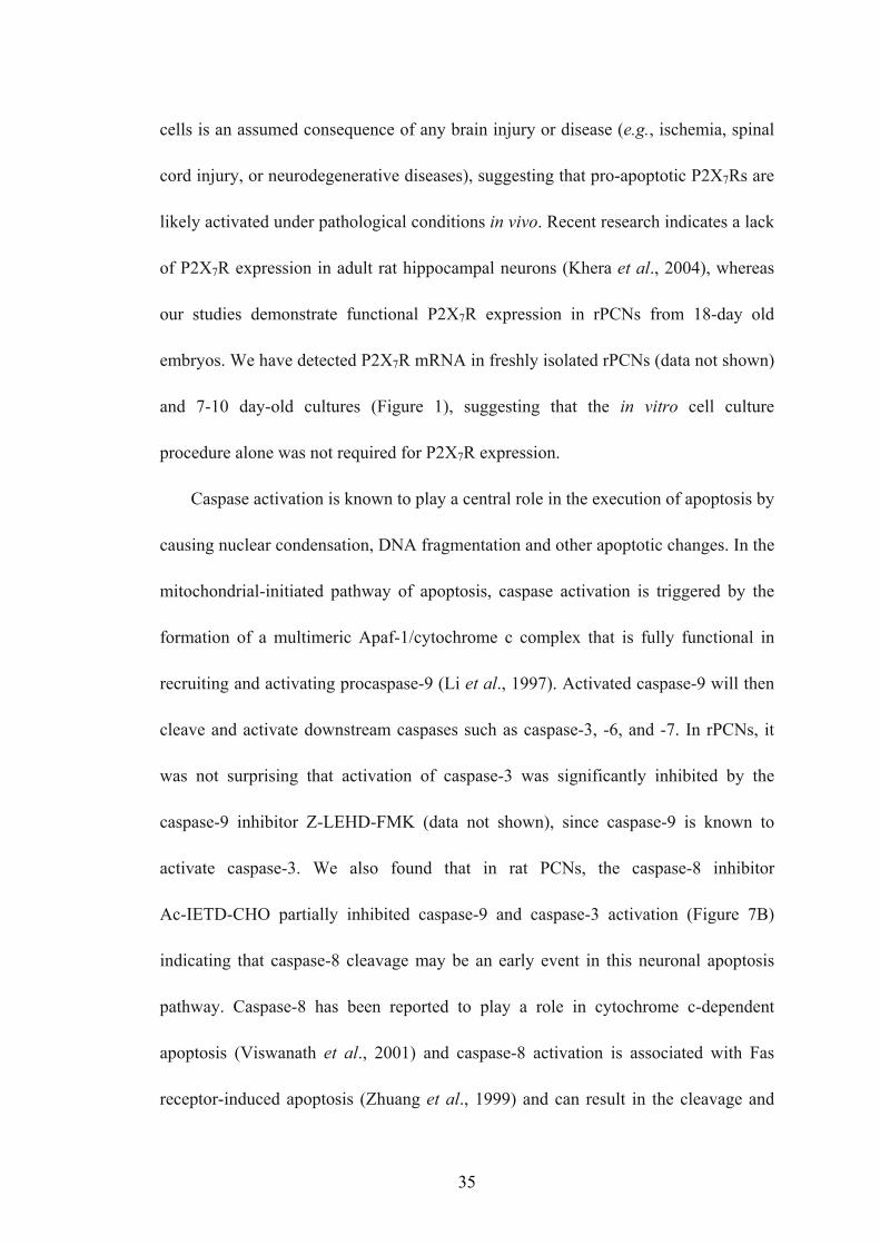
Figure 1. Purity of rat primary cortical neurons (rPCNs) and P2X7R expression.
(A) Rat PCNs were cultured on coverslips in 6-well plates and neurons were labeled
with mouse NeuN monoclonal antibody (Panel a) and cell nuclei with TO-PRO-3
(Panel b); merged picture shown in Panel c. (B) RNA was isolated from rPCNs
maintained in culture for 7-10 days and RT-PCR was used to amplify mRNAs to
P2X1-7Rs, as described in the “Materials and Methods”. The amplified PCR products
were resolved by gel electrophoresis, and data shown are representative of results
from three independent experiments. Results are shown of PCRs performed in the
presence (+) or absence (-) of reverse transcriptase (RT). G3PDH primers were used
to amplify G3PDH mRNA as a positive control. (C) P2X7R protein expression was
detected by Western blot analysis, as described in the “Materials and Methods”.
Human 1321N1 cells expressing the recombinant human P2X7R or the empty
expression vector pLXSN were used as positive (+) or negative (-) controls,
respectively. Precision Plus Protein Standards are indicated as ‘75KD’ and ‘100KD’.
40

A
B
C
41

Figure 2. ATP- and BzATP-induced increases in [Ca2+]i in rat PCNs. (A) Single
cell calcium assays were performed on rPCNs that were cultured for 7-10 days on
poly-D-lysine-coated coverslips. Cells were incubated in PSS with the indicated
concentration of BzATP or ATP and the maximum increase in [Ca2+]i was determined,
as described in the “Materials and Methods”. (B) The percentage of cells responding
to BzATP or ATP is shown as the mean +/- S.E.M. of results from three experiments.
42

A
B
43

Figure 3. ATP/BzATP-induced nuclear condensation mediated by the P2X7R is
dependent on caspase-3 activation. (A) Rat PCNs were cultured for 7-10 days,
incubated in serum-free HGGMEM for 6 h and stimulated with BzATP (300 μM) or
ATP (100 μM) for 16 h or with H2O2 (1 mM) for 2 h. When indicated, 500 μM oATP
was added 2 h prior to addition of BzATP. Cells cultured in B27-AO Neurobasal
medium (NB) or in serum-free HGGMEM (-) overnight were used as controls. Then,
nuclear condensation was determined by DAPI staining and detected by fluorescence
microscopy, as described in the “Materials and Methods”. (B) Cells were treated as in
(A) except that the data were expressed as a percentage of cells that exhibited DAPI
stained nuclei. Data are the means +/- S.E.M. of results from at least four experiments,
where *p < 0.05, and ***p < 0.001 indicate significant differences from the
serum-starved control (-), and where ###p < 0.001 indicates a significant difference
from BzATP treatment. (C) rPCNs were treated as in (A) except that 10 μM
Z-DEVD-FMK, a caspase-3 inhibitor, was added for 1 h prior to BzATP, when
indicated.
44

A
B C
45

Figure 4. BzATP stimulates DNA fragmentation in rat PCNs. Rat PCNs were
incubated in serum-free HGGMEM for 6 h and then stimulated with BzATP (300 μM)
for 16 h or H2O2 (1 mM) for 2 h. Cells cultured in serum-free HGGMEM (-)
overnight were used as the control. Then, cells were lysed, total DNA was purified,
the DNA concentration was determined, and 3 μg of purified DNA was loaded onto
each lane of a 2% (w/v) agarose gel and electrophoresed, as described in the
“Materials and Methods”. DNA fragmentation was visualized after electrophoresis of
DNA. Apoptotic U937 cells (+++; provided in the Apoptotic DNA-ladder kit) were
used as a positive control and “M” indicates the 100 bp DNA ladder marker.
46

47

Figure 5. BzATP or ATP induces DNA strand breakage in rat PCNs. Rat PCNs
were cultured for 7-10 days, incubated in serum-free HGGMEM for 6 h, and
stimulated with BzATP (300 μM) or ATP (100 μM) for 16 h or with H2O2 for 2 h.
Cells cultured in serum-free HGGMEM (-) overnight were used as the control. Then,
cells were fixed and DNA strand breakage was determined by TUNEL assay, as
described in the “Materials and Methods”. DNA strand breakage was observed as
green fluorescence using fluorescence microscopy.
48

49

Figure 6. P2X7Rs mediate caspase-3 activation in rat PCNs. (A) Rat PCNs were
incubated in serum-free medium for 6 h and then stimulated with the indicated
concentration of ATP or BzATP for 16 h or the time indicated. Cells cultured in
B27-AO Neurobasal medium (NB) or in serum-free HGGMEM (-) overnight were
used as controls. (B) Cells were treated as in (A) except that 500 μM oATP was added
for 2 h or the indicated concentration (1, 10 or 100 μM) of Z-DEVD-FMK was added
for 1 h prior to addition of 300 μM BzATP for 16 h. (C) Cells were treated as in (A)
except that P2X7 antisense or sense oligonucleotide was added at the indicated
concentration in μg/ml for 8 h prior to addition of 300 μM BzATP for 16 h or 1 mM
H2O2 for 2 h. Western analysis was performed to determine the relative amounts of
caspase-3, cleaved caspase-3 or P2X7R protein. Data are the means +/- S.E.M. of
results from at least three experiments, where *p < 0.05, **p < 0.01, and ***p < 0.001
indicate significant differences from either serum-starved (A) or BzATP-treated (B, C)
cells. (D) Rat PCNs were serum-starved overnight in the presence (Panels B, D and
F) or absence (Panels A, C, and E) of 300 μM BzATP and then immunofluorescence
of the neuron-specific marker NeuN (green; Panels A, B), cleaved caspase-3 (red;
Panels C and D), and the nuclear stain TO-PRO (blue; Panels E and F) were detected
as described in the “Materials and Methods”. Arrows (pink or white) indicate
apoptotic neurons that are both capase-3 and NeuN positive, whereas yellow arrows
indicate apoptosis in a NeuN-negative cell. Similar results were obtained in three
individually isolated groups of primary cultures.
50

A
B
C
51
D
52

Figure 7. Role of caspase-8 and caspase-9 in P2X7R-mediated apoptosis in rPCNs.
(A) Rat PCNs were incubated in serum-free HGGMEM and BzATP (300 μM) was
added for 16 h. Cells cultured in B27-AO Neurobasal medium (NB) or in serum-free
HGGMEM (-) overnight were used as controls. (B) Cells were treated as in (A)
except that the indicated concentration (μM) of a specific caspase-8 inhibitor,
Ac-IETD-CHO was added for 1 h prior to addition of 300 μM BzATP. (C) Cells were
treated as in (B) except that the indicated concentration of a specific caspase-9
inhibitor, Z-LEHD-FMK, was added. “Pan” (full-length) and cleaved caspase-8 were
detected by Western analysis, as described in the “Materials and Methods”. Data are
the means +/- S.E.M. of results from at least three experiments, where *p < 0.05, **p
< 0.01, and ***p < 0.001 indicate significant differences from either serum-starved (A)
or BzATP-treated (B, C) cells.
53

A
B
C
54

Figure 8. BzATP or ATP induces ERK1/2 and JNK1 phosphorylation in rPCNs.
Rat PCNs were incubated in serum-free HGGMEM for 6 h and treated with the
indicated concentration (μM) of BzATP or ATP for 5 min or with 100 μM ATP or 300
μM BzATP for the indicated time period. Cells cultured in serum-free HGGMEM (-)
overnight were used as the control. Then, cell lysates were prepared and (A) ERK1/2
or (B) JNK1 phosphorylation was detected by Western analysis, as described in the
“Materials and Methods”. Total ERK1/2 and actin were used as protein loading
controls for (A) and (B), respectively.
55

A
B
56

Figure 9. Inhibitors of MEK and JNK decrease BzATP-induced caspase-3
cleavage in rat PCNs. Rat PCNs were incubated in serum-free HGGMEM for 6 h
and treated for 30 min with the indicated concentration (μM) of (A) the MEK
inhibitor U0126, (B) the JNK inhibitor SP600125 or (A, B) 0.1% (v/v) DMSO as a
vehicle control. Then, the cells were incubated with 300 μM BzATP for 16 h, and
Western analysis was used to detect expression of cleaved or pan caspase-3, as
described in the “Materials and Methods”. Cells cultured in serum-free HGGMEM (-)
overnight were used as the control. Data are the means +/- S.E.M. of results from at
least three experiments, where *p < 0.05, **p < 0.01, and ***p < 0.001 indicate
significant differences from BzATP-treated cells.
57

A
B
58

CHAPTER 3
IL-1β ENHANCES NUCLEOTIDE-INDUCED α-SECRETASE-DEPENDENT
APP PROCESSING IN RAT PRIMARY CORTICAL NEURONS VIA
UPREGULATION OF THE P2Y2 RECEPTOR
Kong, Q. et al., to be submitted
59

ABSTRACT
The P2Y2 nucleotide receptor (P2Y2R) stimulates α-secretase-dependent
cleavage of the amyloid precursor protein (APP) in human 1321N1 astrocytoma cells,
causing extracellular release of the neuroprotective protein sAPPα. In this study, we
analyzed functional upregulation of the P2Y2R by cytokines and the P2Y2
receptor-mediated production of sAPPα in rat primary cortical neurons (rPCNs). In
rPCNs, P2Y2R mRNA and receptor activity were virtually absent in unstimulated
cells, whereas overnight treatment with the inflammatory cytokine IL-1β upregulated
both P2Y2R mRNA expression and receptor activity. Furthermore, the P2Y2R agonist
UTP enhanced the release of sAPPα in rPCNs treated with IL-1β or transfected with
P2Y2R mRNA. UTP-induced release of sAPPα from rPCNs was completely inhibited
by pretreatment of the cells with the metalloproteinase inhibitor TAPI-2 or the PI3K
inhibitor LY2904, partially inhibited by the MEK inhibitor U0126, and unaffected by
the PKC inhibitor GF109203, suggesting that P2Y2R-mediated release of sAPPα from
neurons is dependent on ADAM10/17 and PI3K activity, partially dependent on
ERK1/2 activity, and does not require activation of PKC. These results suggest that
under inflammatory conditions, IL-1β evokes the functional upregulation of the
P2Y2R in neurons and implicates the activation of the P2Y2R in neuroprotection.
60
INTRODUCTION
Inflammation is established as a central component of several chronic diseases.
Various inflammatory mediators, such as cytokines, chemokines, proteases and
protease inhibitors, have been detected in the AD brain (Parihar and Hemnani, 2004).
Interleukin-1 (IL-1) and tumor necrosis factor α (TNF-α) are rapidly upregulated in a
variety of tissues in response to pro-inflammatory stimuli to amplify downstream
signaling cascades that mediate inflammation (Cacquevel et al. 2004). In Alzheimer’s
disease, IL-1 directly regulates the expression and processing of β-amyloid precursor
protein (β-APP), the phosphorylation of tau and the increased production and activity
of acetylcholinesterase (AChE) in neurons. These processes result in neuronal stress
and injury, which in turn further enhance microglial activation and IL-1
overexpression in a self propagating cycle. Recent studies, however, indicate that
certain aspects of the inflammatory response may have therapeutic potential
(Wyss-Coray et al., 2001). Nucleotides co-released with neurotransmitters can
activate a widely distributed family of nucleotide receptors in the CNS. P2 nucleotide
receptors (i.e., P2X ion channels and G protein-coupled P2Y receptors) have been
implicated in the regulation of numerous functions in the mammalian nervous system,
including neurotransmission, glial cell migration, cell proliferation, ion transport,
neuronal cell apoptosis and survival (Burnstock, 2000; Ciccarelli et al., 2001;
Weisman et al., 2005). The P2Y2 nucleotide receptor is distinguished among other P2
receptors by its ability to be activated equipotently by ATP and UTP and has been
shown to be upregulated in response to stress or injury in various cell types, including
61
activated thymocytes, salivary gland epithelial cells, and models of vascular tissue
injury (Koshiba et al., 1997; Turner et al., 1997; Seye et al., 2002). In rat primary
cortical astrocytes, the P2Y2R is functionally expressed and promotes astrocyte
migration through receptor interaction with αV integrins, which suggests an important
role for P2Y2R-mediated signaling pathways in regulating astrogliosis in brain
disorders (Wang et al., 2005). However, very little is known about the functional role
of the P2Y2 nucleotide receptor subtype in neurons.
Previously, we demonstrated that the G protein-coupled P2Y2R mediates the
α-secretase-dependent release of sAPPα from human 1321N1 astrocytoma cells
(Camden et al., 2005). Cleavage of APP by α-secretase results in the shedding of
nearly the entire ectodomain of APP to yield a large soluble derivative called sAPPα,
and essentially precludes the generation of neurotoxic Aβ peptide. Several zinc
metalloproteinases, including TNF-α converting enzyme (TACE/ADAM17),
ADAM9, ADAM10 and MDC-9, and the aspartyl protease BACE2 can cleave APP
at the α-secretase site (Allison et al., 2003). While the proteolytic cleavage of APP is
constitutive, it can also be increased by substances such as GPCR agonists that
activate protein kinase C (PKC) or other signaling pathways (Nitsch et al., 1997;
Kirazov et al., 1997) and may function in the modulation of neuronal excitability,
synaptic plasticity, neurite outgrowth, synaptogenesis, and cell survival (Mattson,
1997).
62

The aim of this study was to determine whether cytokines such as IL-1β regulate
functional expression of the P2Y2R in rat primary cortical neurons and how P2Y2R
signaling pathways affect APP processing in these cells.
MATERIALS AND METHODS
Materials
Fetal bovine serum (FBS) was obtained from Hyclone (Logan, UT). Dulbecco’s
Modified Eagle’s Medium (DMEM), Minimum Essential Medium (MEM),
Neurobasal Medium, penicillin (100 units/ml), streptomycin (100 units/ml) and
B27-AO (B27 without cortex antioxidants) were obtained from Gibco-BRL (Carlsbad,
CA). Rabbit anti-rat ERK1/2, horseradish peroxidase (HRP)-conjugated goat
anti-rabbit IgG, and HRP-conjugated goat anti-mouse IgG antibodies were obtained
from Santa Cruz Biotechnology (Santa Cruz, CA). Rabbit anti-rat beta amyloid
antibody was obtained from Signet Laboratories (Dedham, MA). All other antibodies
were obtained from Cell Signaling Technology (Beverly, MA). The Dual Color
Protein Standards and nitrocellulose membranes (0.45 mm) were obtained from
Bio-Rad (Hercules, CA). LumiGLO chemiluminescent substrates were obtained from
New England Biolabs (Beverly, MA). The RNeasy Mini Kit was obtained from
Qiagen (Chatsworth, CA). The First Strand cDNA Synthesis Kit and the PCR Kit
were obtained from Roche (Indianapolis, IN). The GenCarrierTM2 Transfection
Reagent Kit was obtained from Epoch Biolabs (Sugar Land, TX). Nucleotides and all
63

other biochemicals were obtained from Sigma Chemical Co. (St. Louis, MO).
Primary cell culture of cortical neurons
Experimental procedures for cell culture of rat primary cortical neurons (rPCNs)
were carried out as previously described (Kong et al., 2005). Briefly, cerebral cortices
from 18-day-old embryos of Sprague-Dawley rats were removed and the meninges
were discarded. The brain tissue was mechanically dissociated in HGDMEM
comprised of DMEM, 10% (v/v) FBS, 2 mM glutamine, 100 IU/ml penicillin, 100
mg/ml streptomycin, and 7.5 mg/ml fungizone. The tissue clumps were dispersed with
a 10 ml pipette and suspended in 6 ml of 0.25% (w/v) trypsin at 37 °C for 10 min.
Then, 2 ml of heat-inactivated horse serum were added to neutralize trypsin activity.
The cell suspension was centrifuged at 900 g for 5 min and the cell pellet was
suspended in HGDMEM. The resulting cell suspension was filtered through a
sterilized 75 mm cell strainer (Becton Dickinson, Franklin Lakes, NJ) and the cells
were seeded into plastic culture plates precoated with poly-D-lysine (0.1 μg/ml). After
16-18 h and every 3 days thereafter, the medium was replaced with B27-AO
Neurobasal Medium (2 mM glutamine, 100 IU/ml penicillin, 100 mg/ml streptomycin,
7.5 mg/ml fungizone, 10 ml of B27-AO, and Neurobasal Medium (Gibco-BRL) to
500 ml) and 15 μg/ml 5-fluoro-2’-deoxyuridine was added to retard glial cell
proliferation. The neurons were used for experiments after 7 days in culture (DIV7).
RT-PCR analysis of P2Y2R mRNA expression
Total RNA was isolated from rPCNs using the RNeasy Mini Kit (Qiagen). cDNA
was synthesized from the purified RNA using the First Strand cDNA Synthesis Kit
64

for RT-PCR (AMV; Roche). Five percent of the synthesized cDNA was used as a
template in PCR reactions with the Expand High Fidelity PCR System (Roche).
Specific oligonucleotide primers were designed to selectively amplify cDNA for
P2Y2R and G3PDH, as previously described (Wang et al., 2005). The amplification
was performed using 35 cycles for P2Y2R and 30 cycles for G3PDH of denaturation
at 95 °C for 30 s, with annealing at 60 °C for 30 s and extension at 72 °C for 1 min.
The resulting PCR products were resolved on a 1% (w/v) agarose gel containing 10
mg/ml ethidium bromide and photographed under UV illumination.
Single cell calcium assay
The [Ca2+]i was quantified in single cells with the Ca2+-sensitive fluorescent dye
fura-2, using an InCyt Dual-Wavelength Fluorescence Imaging System (Intracellular
Imaging, Cincinnati, OH). Briefly, rat PCNs cultured on poly-D-lysine-coated
coverslips were incubated with 2.5 mM fura-2-acetoxymethylester at 37 °C for 30
min in physiological salt solution (PSS) containing (mM) NaCl 138, KCl 5, CaCl2 2,
MgCl2 1, HEPES 10, glucose 10, pH 7.4, and washed with PSS. The coverslips with
fura-2-loaded cells were positioned on the stage of an inverted epifluorescence
microscope (Nikon; model TMD) and stimulated with agonist at 37 °C, as described
in the figure legends. The percentage of cells that responded to agonist was
determined, as previously described (Kong et al., 2005). Viable cells were identified
by responsiveness to carbachol and non-responding cells were eliminated.
Overexpression of P2Y2 receptors in rPCNs
Rat primary cortical neurons at DIV7 expressing very low levels of endogenous
65

P2Y2R were transiently transfected with 1 μg of human P2Y2 receptor cDNA using
GenCarrierTM2 (Epoch Biolabs; Sugar Land, TX), according to the manufacturer’s
instructions. The transiently transfected cells were cultured for an additional 24-48 h
in Neurobasal Medium with B27-AO, and used to detect sAPPα release.
Harvesting the cells and preparation of conditioned medium
rPCNs were plated on 6-well plates and grown until DIV7. Cells were
serum-starved and incubated in 1 ml serum free DMEM with or without various
compounds for specified times as indicated in the figure legends. At the end of the
incubation period, the media were collected and centrifuged at 12,000 x g for 5 min to
remove cellular debris. The supernatants representing conditioned media were stored
at -80 °C for future use. To measure the level of secreted APPα, 200 μl of supernatant
was diluted 4:1 with 50 μl of 5X Laemmli sample buffer (187.5 mM Tris-HCl, pH 6.8,
6% (w/v) SDS, 1.8% (v/v) β-mercaptoethanol and 0.003% (w/v) bromophenol blue).
In addition, the cells were washed twice with ice-cold PBS, and lysed in 2X Laemmli
sample buffer (200 μl/well).
Western blot analysis
Samples of cell lysates and conditioned media obtained above were heated for 5
min at 65 °C, and subjected to 7.5% (w/v) SDS-polyacrylamide gel electrophoresis
(SDS-PAGE) and transferred to nitrocellulose membranes for protein immunoblotting.
After overnight blocking at 4 °C with 5% (w/v) fat-free milk in TBS-T (10 mM
Tris-HCl, pH 7.4, 120 mM NaCl, and 0.1% (v/v) Tween-20), membranes were
incubated with either 1:1000 dilution of anti-ERK1/2, anti-phospho-ERK1/2, or
66

anti-actin antibodies for 2 h at room temperature, or 1:1000 dilution of anti-sAPPα
overnight at 4 °C followed by incubation with HRP-conjugated anti-rabbit or
anti-mouse IgG antibodies (1:1000 dilution in TBS-T containing 5% (w/v) fat-free
milk) for 1 h at room temperature. Protein immunoreactivity was visualized on
autoradiographic film using the LumiGlo Chemiluminescence System (New England
BioLabs), according to the manufacturer’s instructions. The protein bands detected on
X-ray film were quantified using a computer-driven scanner and Quantity One
software (Bio-Rad, Hercules, CA). The activation levels of kinases or sAPPα release
were expressed as a percentage of controls (i.e., total kinase or actin).
Statistical analysis
Results are expressed as the means ± S.E.M. of data obtained from at least 3
experiments. Statistical analysis of data was performed using Graph Pad Prism
version 4.0. Statistical significance was determined by student t test between groups.
Differences were considered statistically significant when p < 0.05 (*) and not
significant when p > 0.05 (n.s.).
RESULTS
Upregulation of functional P2Y2R in rat primary cortical neurons by IL-1β
Several subtypes of P2 nucleotide receptor mRNA are expressed in rPCNs,
including P2Y1, P2Y4, P2Y6, P2X3, P2X5, P2X6 and P2X7 (Kong et al., 2005;
Weisman et al., 2005). However, P2Y2R mRNA and receptor activity are barely
detectable in these cells (Weisman et al., 2005, Kong et al., 2005, Figure 1A). Since
67

previous studies demonstrated that IL-1β causes an increase in P2Y2R mRNA
expression in mouse primary striatal astrocytes (Stella et al., 1997), we used RT-PCR
analysis to examine whether this cytokine could increase P2Y2R mRNA expression in
rPCNs. After overnight incubation with 0-100 ng/ml IL-1β, P2Y2R mRNA (Figure
1A-B), but not P2Y4R or P2Y6R mRNA (data not shown), was upregulated in an
IL-1β dose-dependent manner. Stimulation of IL-1β-treated rPCNs with the P2Y2R
agonist UTP caused an increase in [Ca2+]i (Figure 1C) and activation of the MAPKs
ERK1/2 (Figure 2), indicating that functional expression of the P2Y2R is upregulated
by IL-1β in rPCNs. Co-incubation of rPCNs with TNF-α and IL-1β did not further
enhance the effect of IL-1β on P2Y2R-mediated calcium mobilization (data not shown)
or ERK1/2 phosphorylation (Figures 2C-D).
P2Y2R-mediated release of sAPPα from rPCNs treated with IL-1β
The release of sAPPα from rPCNs was detected by immunoblot analysis of the
cell culture media with a rabbit anti-rodent sAPPα antibody that recognizes amino
acid residues 3-16 of Aβ and reacts well with APP (130 kDa) and all isoforms of
sAPPα (100-120 kDa). We found that overnight treatment of rPCNs with IL-1β
(1-100 ng/ml) caused a dose-dependent increase in sAPPα release from cells
stimulated with 100 μM UTP (Figure 3). UTP also increased the release of sAPPα
from rPCNs transfected with P2Y2R cDNA, as compared to untransfected rPCNs
(Figure 4), confirming the hypothesis that UTP enhances sAPPα release from rPCNs
through activation of the P2Y2R subtype.
P2Y2R-mediated release of sAPPα is dependent on ADAM10/17
68

Currently, several zinc metalloproteinases have been identified as potential
α-secretases including tumor necrosis factor alpha converting enzyme (TACE or
ADAM17 for a disintegrin and metalloprotease 17) that catalyzes the shedding of the
ectodomain of APP and other transmembrane proteins (Black et al., 1997; Moss et al.,
1997), and ADAM10 and MDC9 that also catalyze sAPPα production (Lammich et
al., 1999; Koike et al., 1999). In IL-1β-treated rPCNs, UTP-induced release of sAPPα
was significantly inhibited by TAPI-2 (Figure 5), a competitive inhibitor of
ADAM17/TACE and ADAM10 (Kaup et al., 2002). Thus, P2Y2R-mediated APP
processing in rPCNs is likely mediated by ADAM10/17.
P2Y2R-mediated sAPPα release is independent of PKC
P2Y2R activation causes the phospholipase C-dependent stimulation of PKC, and
the activation of several other GPCRs has been reported to induce sAPPα release
through PKC-dependent and -independent pathways (Nitsch et al., 1994 &1995;
Checler, 1995; LeBlanc et al., 1998). Therefore, we determined whether
P2Y2R-mediated sAPPα release is dependent on activation of PKC. Results indicated
that the level of UTP-induced sAPPα release decreased when IL-1β-treated rPCNs
were incubated for 30 min with GF109203 (10 μM), an inhibitor of PKC. However,
the inhibition was not significant according to the student t test, although GF109203
significantly inhibited 1 μM PMA-induced release of sAPPα (Figure 6). Thus, sAPPα
release mediated by P2Y2R activation occurs independently of PKC.
P2Y2R-mediated sAPPα release is dependent on PI3K activation
The release of sAPPα induced by insulin requires activation of the PI3K/Akt
69

pathway (Solano et al., 2000). Since P2Y2Rs couple to PI3K activation in many cell
types (Muscella et al., 2003; Kaczmarek et al., 2005; Wang et al., 2005), we
determined whether PI3K/Akt regulates P2Y2R-mediated sAPPα release in
IL-1β-treated rPCNs. Figure 7 shows that incubation of IL-1β-treated rPCNs with 10
μM LY29004, a selective inhibitor of PI3K, prevents UTP-induced sAPPα release,
but has no effect on constitutive release of sAPPα (Figure 7).
Role of MAPK in P2Y2R-mediated release of sAPPα
Previous studies in our laboratory have shown that inhibition of MAPK (i.e.,
ERK1/2) partially inhibits UTP-stimulated sAPPα release in human 1321N1
astrocytoma cells (Camden et al., 2005). Therefore, we investigated the role of the
MAPK pathway in UTP-stimulated sAPPα release in IL-1β-treated rPCNs. U0126, an
inhibitor of the MAPK/ERK kinase (MEK), partially inhibited UTP-induced sAPPα
release but not the constitutive release of sAPPα in IL-1β-treated rPCNs (Figure 8).
These results suggest that P2Y2R-mediated sAPPα release in rPCNs is partially
dependent on MAPK/ERK1/2 activation.
DISCUSSION
In the present study, we demonstrate that the pro-inflammatory cytokine IL-1β
upregulates expression of the P2Y2R in rat primary cortical neurons (rPCNs).
Furthermore, we report that activation of the P2Y2R in IL-1β-treated rPCNs enhances
the release of sAPPα, a neuroprotective cleavage product of APP (Li et al., 1997; Luo
et al., 2001; Stein et al., 2004), by stimulating α-secretase activity mediated by the
70

metalloproteinases, ADAM10/17. Inhibitor studies reveal that P2Y2R-mediated
sAPPα release from rPCNs requires PI3K activity and is partially dependent on
MAPK/ERK1/2 activation, but independent of PKC activation.
Previous studies have demonstrated that P2Y2R mRNA and functional activity is
upregulated in stressed or injured tissue, including ligated salivary gland ducts
(Turner et al., 1998), collared carotid arteries (Seye et al., 2002), and submandibular
gland cells from the NOD.B10 mouse model of Sjogren's syndrome (Schrader et al.,
2005). It was also reported that IL-1β induced P2Y2R mRNA expression in vascular
smooth muscle cells (SMCs; Hou et al., 2000). IL-1 binds to a specific plasma
membrane IL-1 type-I receptor (IL-1RI) (Sims et al., 1993), which subsequently
associates with the IL-1 receptor-accessory protein (IL-1RAcP) to initiate intracellular
signals (Wesche et al., 1997). Since IL-1β release has been associated with the
pathophysiology of Alzheimer’s disease (Grammas and Ovase, 2001), we speculated
that cytokines could upregulate P2Y2 receptor expression in neuronal cells. We found
that endogenous P2Y2R mRNA expression and receptor activity were very low in
untreated rPCNs but were significantly enhanced in response to IL-1β treatment
(Figures 1 and 2; Weisman et al., 2005), suggesting that P2Y2R expression is
regulated by transcription factors coupled to IL-1 receptor activation.
As IL-1β-induced P2Y2R mRNA expression in vascular SMCs was dramatically
enhanced in combination with TNF-α treatment (Hou et al., 2000), we also tested
whether TNF-α could enhance IL-1β-induced P2Y2R expression and function. In
rPCNs, TNF-α did not significantly enhance IL-1β-induced P2Y2R mRNA expression
71

(data not shown) or receptor activity (Figure 2D).
Although the role of P2Y2R expression in neuronal cells is poorly understood, it
is better established that cytokines, including IL-1β and their receptors mediate
inflammation in Alzheimer’s brain (Brugg et al., 1995; Hauss-Wegrzyniak et al.,
1998). Therefore, we postulate that IL-1β-induced P2Y2R expression may contribute
to inflammation. However, other studies suggest that P2Y2R activity in human
1321N1 astrocytoma cells can promote β-amyloid precursor protein (APP) processing
(Camden et al., 2005) and therefore we investigated whether this pathway was
manifested upon P2Y2R upregulation in IL-1β-treated rPCNs. APP is a
transmembrane glycoprotein that can be detected in most tissues but is abundantly
expressed in the brain (Anderson et al., 1989). Post-translational processing of APP
occurs by the action of least three proteinases called α-, β- and γ-secretases (Esler and
Wolfe, 2001). Cleavage of APP by β- and γ-secretases results in the release of several
protein fragments, including amyloid β (Aβ), the main component in senile plaques
found in the brains of patients with Alzheimer’s disease (Martins et al., 1991).
Although sAPPα is constitutively released from cells, secretion of this protein is also
regulated by neural activity and can be influenced by agonists of various receptors
(Robert et al., 2001; Canet-Aviles et al., 2002; Camden et al., 2005). The studies
presented here suggest that IL-1β-induced upregulation of P2Y2Rs in rPCNs (Figure
3) or transfection of rPCNs with P2Y2R cDNA (Figure 4) significantly increases
UTP-stimulated sAPPα release. Since sAPPα release is postulated to be
neuroprotective in AD (Li et al., 1997; Luo et al., 2001), we suggest that P2Y2R
72

expression in neuronal cells may play neuroprotective as well as pro-inflammatory
roles in the AD brain (Weisman et al., 2005). P2Y4 receptors are endogenously
expressed in rat neurons and have an agonist potency order of ITP = ATP = UTP =
ATPγS = 2-MeSATP = Ap4A (Bogdanov et al., 1998). Therefore, it is plausible to
suggest that P2Y4Rs may contribute to the enhanced release of sAPPα evoked by
UTP. In rPCNs without IL-1β treatment, P2Y4R mRNA is expressed at higher levels
than P2Y2R mRNA (data not shown), and therefore the P2Y4R may mediate
UTP-induced increases in [Ca2+]i seen in 1 out of 10 rPCNs in the absence of IL-1β
stimulation (Figure 1C). However, in the absence of IL-1β treatment, UTP did not
significantly increase sAPPα release from rPCNs (Figure 3), suggesting that the
P2Y4R does not contribute to APP processing in rPCNs.
There are three major isoforms of amyloid precursor protein (APP770, APP751 and
APP695; Kitaguchi et al., 1988) due to alternative splicing of APP mRNA. As the
predominant isoform in the brain (Tanaka et al., 1989), APP695 has received the most
attention in research on Alzheimer’s disease. The ratio of APP770:APP751:APP695
mRNA is 1:10:20 in the cerebral cortex of the brain (Tanaka et al., 1989). Compared
with neurons, which express higher levels of APP695, astrocytes synthesize APPs of
higher molecular weights, as expected for the Kunitz Protease Inhibitor
(KPI)-containing isoforms, APP751 and APP770. Soluble APPα release from rPCNs
into the medium can be detected in the form of a doublet (100-120 kDa for APP 695
and APP 751; data not shown), indicating that multiple APP isoforms are present,
consistent with previous reports (LeBlanc et al., 1991 & 1997; Lahiri et al., 1994;
73

Parvathy et al., 1998; Solano et al., 2000; Ma et al., 2005).
The data presented (Figure 5) also suggest that UTP induces APP processing in
IL-1β-treated rPCNs through activation of the metalloproteinases ADAM10/17.
However, other members of the adamalysin family may also contribute to
P2Y2R-mediated sAPPα release since micromolar concentrations of TAPI-2 used to
inhibit metalloproteinase activity have been reported to inhibit members of the
adamalysin family other than ADAM10/17 (Parkin et al., 2002).
Sitmulation of GPCRs has been shown to regulate APP processing by
PKC-dependent signaling pathways (Nitsch, 1992). However, APP processing can
also be regulated by increases in [Ca2+]i, and activation of PLA2, PKA and tyrosine
kinases (reviewed in Mills and Reiner, 1999). In human 1321N1 astrocytoma cells
expressing human P2Y2R cDNA, UTP-induced sAPPα release is independent of
P2Y2R-mediated activation of PKC (Camden et al., 2005). UTP-induced sAPPα
release also occurred independently of PKC activation in IL-1β-treated rPCNs (Figure
6). The MAPK signaling pathway has been implicated in the regulation of both
PKC-dependent and PKC-independent APP catabolism (Mills et al., 1997;
Desdouits-Magnen et al., 1998). PI3K also regulated α-secretase-dependent APP
processing (Solano et al., 2000; Fu et al., 2002). It also has been reported that
short-term interleukin-1β incubation increases the release of sAPPα via
ERK1/2-dependent α-secretase activation in Neuroglioma U251 cells whereas PI3K
activity is unnecessary (Ma et al., 2005). To test the involvement of ERK/PI3K in
UTP-stimulated sAPPα release in the rPCNs, after upregulation of P2Y2Rs by
74
overnight IL-1β treatment, we incubated cells in serum free DMEM containing
specific MEK or PI3K inhibitors followed by UTP stimulation. We found that
inhibition of PI3K or MAPK/ERK1/2 activity in IL-1β-treated rPCNs significantly
reduced UTP-induced sAPPα release (Figures 7 and 8). Therefore, unlike short-term
effects of IL-1β, long-term IL-1β treatment can regulate UTP-induced sAPPα release
in a PI3K/ERK-dependent manner, most likely involving transcriptional upregulation
of P2Y2R.
The results of this study strongly suggest that IL-1β evokes functional
upregulation of the P2Y2R in rPCNs and subsequent activation of the P2Y2R
signaling cascade by UTP results in a significant increase in sAPPα release that is at
least partially dependent on PI3K- and ERK1/2-induced activation of the
metalloproteinases ADAM10/17. Together, these lines of evidence suggest that under
inflammatory conditions in the brain where IL-1β accumulates, functional P2Y2R
expression in neuronal cells may serve a neuroprotective role in the regulation of α-,
γ-secretase-mediated APP processing. .Since P2Y2R expression in glial cells has been
postulated to regulate inflammatory responses that may contribute to
neurodegeneration in AD (Weisman et al., 2005), future studies are required to
evaluate how both neurodegenerative (i.e., glial cell-mediated inflammation) and
neuroprotective (i.e., sAPPα release) responses coupled to P2Y2R activation are
coordinately regulated during the progression of Alzheimer’s disease.
75

Figure 1. IL-1β upregulates functional expression of P2Y2 receptors in rPCNs.
(A-B) Rat primary cortical neurons at DIV7 were treated for 24 h at 37 °C in
serum-free DMEM with or without IL-1β (0 ng/ml, 1 ng/ml, 10 ng/ml, 25 ng/ml, and
100 ng/ml) and cells were harvested for RNA isolation. The effect of IL-1β treatment
on the expression of P2Y2R mRNA was determined by RT-PCR, as described in
"Materials and Methods". Panel B indicates the ratio of P2Y2R to G3PDH mRNA
levels. (C) IL-1β-treated cells were loaded with fura-2 AM for measurement of [Ca2+]i
and positioned on the stage of an inverted epifluorescence microscope (Nikon; model
TMD), as described in the “Materials and Methods”. Then, rPCNs were incubated in
PSS with UTP (100 μM), and the maximum increase in [Ca2+]i was determined, as
described in the “Materials and Methods”. Results were expressed as the percentage
of rPCNs responding to UTP. Data represent the means ± S.E.M. of results from three
experiments, where * represents significant differences (p < 0.05), as compared to
rPCNs treated without IL-1β.
76

A
B
C
77
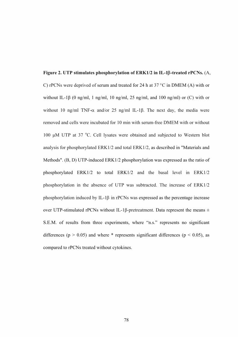
Figure 2. UTP stimulates phosphorylation of ERK1/2 in IL-1β-treated rPCNs. (A,
C) rPCNs were deprived of serum and treated for 24 h at 37 °C in DMEM (A) with or
without IL-1β (0 ng/ml, 1 ng/ml, 10 ng/ml, 25 ng/ml, and 100 ng/ml) or (C) with or
without 10 ng/ml TNF-α and/or 25 ng/ml IL-1β. The next day, the media were
removed and cells were incubated for 10 min with serum-free DMEM with or without
100 μM UTP at 37 oC. Cell lysates were obtained and subjected to Western blot
analysis for phosphorylated ERK1/2 and total ERK1/2, as described in "Materials and
Methods". (B, D) UTP-induced ERK1/2 phosphorylation was expressed as the ratio of
phosphorylated ERK1/2 to total ERK1/2 and the basal level in ERK1/2
phosphorylation in the absence of UTP was subtracted. The increase of ERK1/2
phosphorylation induced by IL-1β in rPCNs was expressed as the percentage increase
over UTP-stimulated rPCNs without IL-1β-pretreatment. Data represent the means ±
S.E.M. of results from three experiments, where “n.s.” represents no significant
differences (p > 0.05) and where * represents significant differences (p < 0.05), as
compared to rPCNs treated without cytokines.
78

A
B
79

C
D
80
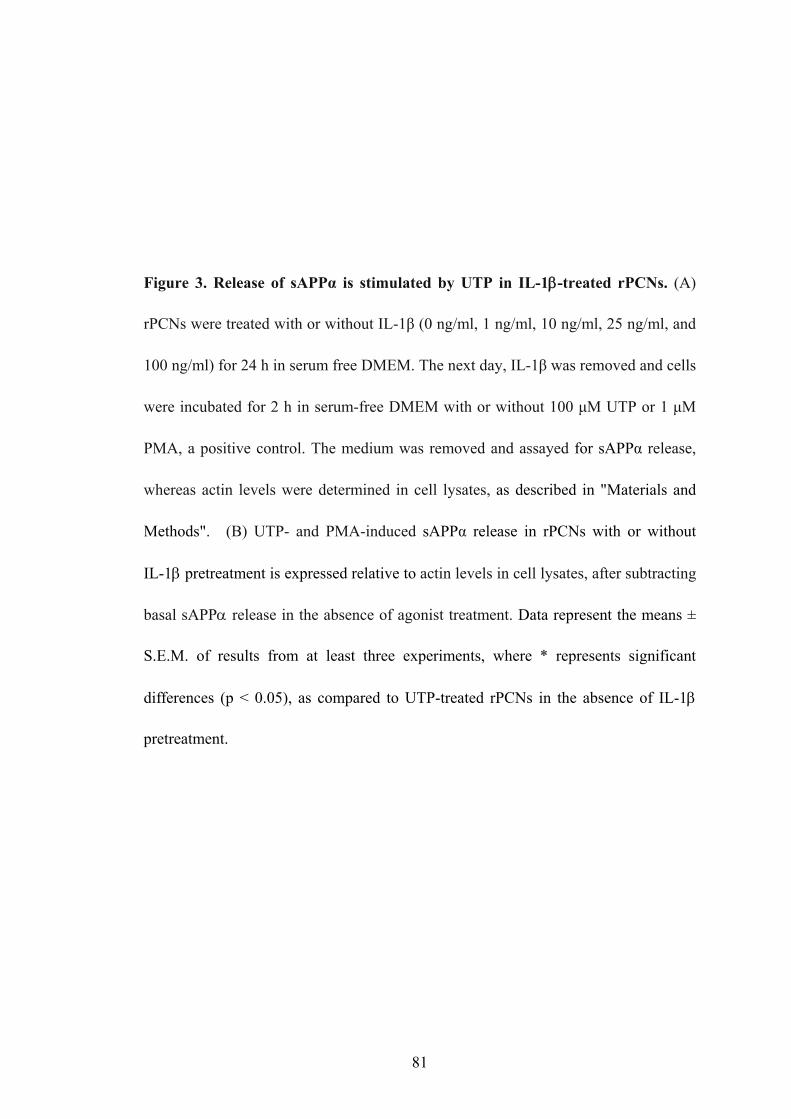
Figure 3. Release of sAPPα is stimulated by UTP in IL-1β-treated rPCNs. (A)
rPCNs were treated with or without IL-1β (0 ng/ml, 1 ng/ml, 10 ng/ml, 25 ng/ml, and
100 ng/ml) for 24 h in serum free DMEM. The next day, IL-1β was removed and cells
were incubated for 2 h in serum-free DMEM with or without 100 μM UTP or 1 μM
PMA, a positive control. The medium was removed and assayed for sAPPα release,
whereas actin levels were determined in cell lysates, as described in "Materials and
Methods". (B) UTP- and PMA-induced sAPPα release in rPCNs with or without
IL-1β pretreatment is expressed relative to actin levels in cell lysates, after subtracting
basal sAPPα release in the absence of agonist treatment. Data represent the means ±
S.E.M. of results from at least three experiments, where * represents significant
differences (p < 0.05), as compared to UTP-treated rPCNs in the absence of IL-1β
pretreatment.
81

A
B
82

Figure 4. P2Y2 receptor expression in rPCNs enhances α-secretase-mediated APP
processing induced by UTP. (A) rPCNs were transfected with human P2Y2R cDNA,
as described in “Materials and Methods”. Cells expressing P2Y2R mRNA (data not
shown) were incubated for 2 h in serum-free DMEM with or without 100 μM UTP or
1 μM PMA, and sAPPα release was determined as described in “Materials and
Methods”, and compared to untransfected (-) controls. Actin levels in cell lysates are
shown as a loading control. (B) sAPPα release is expressed relative to actin levels in
cell lysates. Data represent the means ± S.E.M. of results from three independent
experiments, where * represents significant differences (p < 0.05) between the
indicated sample groups.
83

A
B
84

Figure 5. A metalloprotease inhibitor prevents UTP-induced sAPPα release from
rPCNs. (A) rPCNs were pretreated overnight with 25 ng/ml IL-1β and then incubated
for 30 min with or without TAPI-2 (10 µM) followed by incubation with or without
UTP (100 µM) for 2 h in serum-free DMEM. The release of sAPPα into the medium
was analyzed, as described in “Materials and Methods”. (B) sAPPα release is
expressed relative to actin levels in cell lysates. Data represent the means ± S.E.M. of
results from six experiments, where * represents significant differences (p < 0.05)
between the indicated sample groups.
85

A
B
86

Figure 6. UTP-induced sAPPα release occurs independently of PKC activation in
IL-1β-treated rPCNs. (A) rPCNs were pretreated overnight with 25 ng/ml IL-1β and
then incubated for 30 min with or without GF109203 (10 µM) followed by incubation
with or without UTP (100 µM) or PMA (1 µM) for 2 h in serum-free medium. Then,
sAPPα release into the medium was analyzed, as described in “Materials and
Methods”. (B) sAPPα release is expressed relative to actin levels in cell lysates. Data
represent the means ± S.E.M. of results from three experiments, where “n.s.”
represents no significant differences (p > 0.05) and where * represents significant
differences (p < 0.05) between the indicated sample groups.
87

A
B
88

Figure 7. Inhibition of PI3K prevents UTP-induced sAPPα release in
IL-1β-treated rPCNs. (A) rPCNs were pretreated overnight with 25 ng/ml IL-1β and
then incubated for 30 min with or without LY29004 (1 µM) followed by incubation
with or without UTP (100 µM) for 2 h in serum-free medium. Then, sAPPα release
into the medium was analyzed, as described in “Materials and Methods”. (B) sAPPα
release is expressed relative to actin levels in cell lysates. Data represent the means ±
S.E.M. of results from three experiments, where * represents significant differences (p
< 0.05) between the indicated sample groups.
89

A
B
90

Figure 8. UTP-stimulated sAPPα release in IL-1β-treated rPCNs is partially
dependent on MAPK/ERK1/2 activation. (A) rPCNs were pretreated overnight with
25 ng/ml IL-1β and then incubated for 30 min with or without U0126 (1 µM)
followed by incubation with or without UTP (100 µM) for 2 h in serum-free medium.
Then, sAPPα release into the medium was analyzed, as described in “Materials and
Methods”. (B) sAPPα release is expressed relative to actin levels in cell lysates. Data
represent the means ± S.E.M. of results from three experiments, where * represents
significant differences (p < 0.05) between the indicated sample groups.
91

A
B
92

CHAPTER 4
EXPRESSION AND REGULATION OF P2Y2 NUCLEOTIDE RECEPTORS
IN RODENT BRAINS AND THE EFFECT OF CEREBRAL ISCHEMIA
Unpublished data; Weisman, G.A., et al., Mol Neurobiol 31(1-3): 169-183.
93

ABSTRACT
Large amounts of adenosine 5'-triphosphate (ATP) released from cellular sources
under pathological conditions such as ischemia may activate purinoceptors of the P2X
and P2Y types. Although signaling pathways for P2Y2 receptors are well understood
in both primary glial cultures and cell lines derived from various tissues, less is
known about P2Y2 receptor distribution in the brain. Therefore, in the present study,
in situ hybridization and RT-PCR were utilized to examine the regional expression
patterns of P2Y2 receptors in adult rat/mouse/gerbil brain. P2Y2R mRNA was
expressed at relatively low levels in normal rodent brain, although expression levels
are similar in rat, mouse and gerbil brains. In general, more abundant P2Y2R mRNA
expression was detected in the hippocampus and cerebellum. In the hippocampus,
P2Y2R mRNA was detected in the dentate gyrus and to a lesser extent in the CA1 and
CA3 subregions. Transient global cerebral ischemia (i.e., 5 min occlusion of the CCA)
did not affect P2Y2R mRNA levels in any region of gerbil brain after various
reperfusion times (4 h, 16 h and 48 h), indicating that the P2Y2R is not regulated at
the transcriptional level in this ischemia model.
94

INTRODUCTION
Cerebral ischemia occurs when blood supply to the brain or to regions of the
brain is decreased due to blockage or obstruction of cerebral blood vessels. Ischemia
can result in rapid increases in the intracellular calcium concentration and release of
ATP from pre-synaptic neurons followed by ATP depletion that can lead to neuronal
cell death (Golstein and Kroeme, 2006).
ATP release can activate two types of purinergic receptors: ligand-gated ion
channel P2X receptors and G protein-coupled P2Y receptors (Burnstock, 1980).
Among the known P2 receptor subtypes, P2Y2 and P2X7 nucleotide receptors have
gained recent attention due to their widespread distribution in neuronal cells and their
roles in the regulation of cell proliferation, inflammation, and apoptosis (reviewed in
Franke et al., 2006).
Recent studies showed that brain astrocytes express P2Y2 receptors and that
P2Y2R expression, as opposed to P2Y1R, is upregulated in freshly isolated astrocytes
in hippocampus during development (Zhu and Kimelberg, 2001). Upregulation of
P2Y2Rs also has been shown to occur in many other cell types, tissues and injury
models as previously described (Chapter 3; Turner et al., 1997; Erlinge et al., 1998;
Hou et al., 1999; Seye et al., 1997 & 2002; Shen et al., 2004; Schrader et al., 2005).
Here, in situ hybridization and RT-PCR were utilized to examine the regional
expression patterns of P2Y2 receptor subtypes in adult rodent brain and a gerbil
transient ischemia model was used to study the regulation of P2Y2R mRNA
expression in response to brain ischemia.
95

MATERIALS AND METHODS
Materials
The RNeasy Mini Kit was obtained from Qiagen (Chatsworth, CA, USA). The
cDNA Synthesis Kit and the PCR Kit were obtained from Invitrogen Life Technology
(Carlsbad, California, USA). The Lig'nScribe Reaction Kit was from Ambion (Austin,
TX, USA). The [35S]-NTP RNA Labeling Kit (SP6/T7) was from Roche (Indianapolis,
IN, USA). [35S]-UTP and [35S]-dATP were obtained from NEN (Boston, MA). All
other biochemicals were obtained from Sigma Chemical Co. (St. Louis, MO, USA).
RNA extraction and RT-PCR
Different regions (i.e., hippocampus, cerebellum, cerebral cortex, brain stem and
the rest of the brain) were dissected from adult rodent brain. Total RNA was extracted
from tissues in four steps: a. tissue was reduced from pieces (10-100 mg) to a powder
with a mortar and pestle unit containing liquid nitrogen; b. powdered tissue was
dissolved in 1 ml Trizol reagent and large pieces of undissolved tissue were removed;
c. 200 μl of chloroform was added to the samples, which were vortexed vigorously
and centrifuged for 15 min at 12,000 x g at 4 °C; and d. the upper aqueous phase was
collected and one volume of 70% (v/v) ethanol was added, mixed and RNA was
purified using the Qiagen Rneasy Mini Kit (Qiagen).
cDNA was synthesized by reverse transcription using purified RNA from mouse
tissues (Invitrogen Life Technology). A P2Y2 receptor cDNA fragment was amplified
from the mouse cDNA by PCR using a 35 cycle program (94 °C for 1 min, 60 °C of
the annealing temperature for 30 sec, 72 °C for 1 min; G3PDH was used as a control.
96

with 30 cycles and 55 °C for annealing) in the presence of 50 U/ml Taq polymerase
(ThermoAceTM DNA polymerase from Invitrogen) and two P2Y2R C-terminal primers
(1 μmol/l): gerbil P2Y2 forward: 5’-TGC TGC CCC TGT CCT ACG GCG TGG-3’
(1-24 of AF313448; the sequence of gerbil P2Y2R from
http://www.ncbi.nlm.nih.gov/entrez/viewer.fcgi?db=nuccore&id=12751144), gerbil
P2Y2 reverse: 5’-AGG TTG GTG TAG AAG AGG AAG CGC-3’ (241-218 of
AF313448). The following primers for human G3PDH were used as a control: sense
5'-TGA AGG TCG GAG TCA ACG GAT TTG GT-3', antisense 5'-CAT GTG GGC
CAT GAG GTC CAC CAC-3'. PCR products were resolved by 2% agarose gel
electrophoresis, and P2Y2/G3PDH ratios were determined by densitometry.
Gerbil global cerebral ischemia model
Adult male Mongolian gerbils (60–80 g body weight) (Charles River,
Wilmington, MA) were given free access to water and lab chow and maintained at
22 ± 2 °C with a constant humidity under a 12:12 h light:dark cycle. Gerbils were
randomly divided into a sham control group, an ischemic group and an ischemic
group with resveratrol. For some animals in each group, resveratrol (Sigma, St.
Louis, MO) was dissolved in corn oil and injected intraperitoneally (30 mg/kg body
weight) immediately after and 24 h after induction of ischemia by occlusion of both
common carotid arteries (CCAs), as previously described (Wang et al., 2002). Sham
controls also were injected with the same volume of corn oil and underwent similar
surgical procedures without occlusion of the CCAs. The animal protocol was
approved by the University of Missouri-Columbia Animal Care and Use Committee
97
(Protocol #1741). Experiments were in accordance with the NIH Guidelines for the
Care and Use of Laboratory Animals. To eliminate gerbils that have communicating
arteries, regional cerebral blood flow (rCBF) was monitored before and after
clamping the bilateral CCA using a laser Doppler flowmeter (MBF3D, Moor
Instruments, Devon, UK). Gerbils showing a decrease in rCBF of less than 80%
upon occlusion of CCAs were excluded from subsequent analyses (Wang et al.,
2002).
Tissue sections
Normal/sham/ischemic rodent brains were removed, wrapped in parafilm and
aluminum foil and placed in tightly capped 50 ml conical tubes, unless the brains
were sectioned immediately. Stored tissue was cut into 12 µm-thick sections,
thaw-mounted onto microscope slides, dried briefly on a warm plate (32 °C; 1 min),
and kept in 100-place slide boxes at -80°C until in situ hybridization.
Probes
To generate P2Y2R specific riboprobes for in situ hybridization, a T7 promoter
adapter was ligated to the 247-bp P2Y2R fragment amplified by reverse
transcription-polymerase chain reaction (RT-PCR) with the Lig'nScribe Reaction Kit
(Ambion). Antisense and sense riboprobes were obtained by in vitro transcription with
[35S]-UTP using the [35S]-NTP RNA Labeling Kit (SP6/T7) (Roche). P2Y2R
oligoprobes were prepared by labeling the following oligonucleotides with [35S]-dATP
at the free 3’ hydroxyl end using terminal deoxynucleotidyl transferase: 1. 5’-GAT
GGC GTT GAG GGT GTG GCA ACT GAG GTC AAG TGA TCG GAA GGA-3’;
98

and 2. 5’-CGT TCC AGG TCT TGA GGC GGC ACA GGA AGA TGT AGA)-3’.
In situ hybridization
Hybridization was performed overnight at 55 °C for the riboprobes and 37 °C for
the oligoprobes in a humidified chamber, as described previously (Simonyi et al.,
2002). Briefly, normal rodent brain or ischemic hippocampal sections were fixed in
situ by 4% (v/v) paraformaldehyde for 5 min, then rinsed with 2 × SSC. Next,
sections were dehydrated and delipidated with the following graded series of ethanol
(EtOH) dilutions and chloroform: 1 min in 50% EtOH, 1 min in 70% EtOH, 1 min in
80% EtOH, 2 min in 95% EtOH, 1 min in 100% EtOH, 5 min in Chloroform, 1 min in
100% EtOH, and 1 min in 95% EtOH. The sections then were air-dried. Sections to be
used as negative controls were treated with RNase A solution (10 mg RNase/50 ml,
0.5 M NaCl, 0.01 M Tris, 0.001 M EDTA, pH = 8) at 37 °C for 30 min, then washed
in 1 X SSC three times for 1 min each, dehydrated twice with 70% EtOH for 2 min,
twice with 80% EtOH for 2 min, and twice with 90% EtOH for 2 min. Next, sections
on slides were hybridized under coverslips in a humidified chamber at 55 °C
overnight in 25 μl hybridization buffer containing approximately 1-2 × 10⁶ dpm
radioactive probe (antisense or sense) and 1 μl nonradioactive probe. After sections
were cooled to room temperature, coverslips were removed in 4 × SSC, washed 4
times in 4 × SSC for 15 min each at room temperature, and incubated in RNase buffer
for 30 min at 37 °C to reduce nonspecific background. Then, sections were washed,
dehydrated, and exposed to a radiosensitive film (Kodak Biomax MR-1).
99

Image analysis and data analysis
Using standard autoradiographic techniques, the sections on slides were apposed
to radiosensitive film (Kodak Biomax MR-1) with [14C]-labeled calibration standards
(American Radiolabeled Chemicals, St. Louis, MO) for 1 week and then developed.
The standards comprised of 16 [14C]-labeled standards on glass slides with 0.002 to
3.58 µCi/g radioactivity were used to construct linear calibration curves of
tissue-equivalent radioactivity (µCi/g) relative to the film optical density. Based on
the standard curve, microdensitometry of film autoradiograms was performed on the
signal over different hippocampal subregions using the BIOQUANT True Color
Windows 95 software version 2.50, as previously described (Shelat et al., 2006). The
amount of [35S]-labeled probe radioactivity (µCi/g) within various hippocampal
subregions was quantitated, from which background radioactivity/optical density was
subtracted. The average density measured from various hippocampal subregions fell
within the linear range of the standards.
Data are expressed as means ± S.E.M. The number of animals in each group
varied between 15 and 23 except for the resveratrol-treated groups which contained 7
animals. Statistical differences between sample groups were evaluated by ANOVA
and Student-Newman-Keuls test, where p < 0.05 was considered to be significant..
RESULTS
P2Y2R mRNA expression in rodent brains
P2Y2 receptor riboprobes were designed from a cloned 247 base pair sequence of
100

gerbil P2Y2R cDNA that has high similarity to the rat gene sequence. Two
oligoprobes complementary to different regions of the cDNA were prepared. These
probes and the antisense riboprobe produced identical patterns of hybridization when
tested on adjacent sections (data not shown). Sense riboprobe and RNase treatment
were used as two negative controls for nonspecific binding. Both the RNase-treated
tissue labeled with oligoprobes (or antisense probe) and the untreated ischemic tissue
labeled with sense riboprobe had clean backgrounds, which indicated weak
nonspecific binding. P2Y2 receptor mRNA was expressed at relatively low levels in
various brain regions, but was most abundant in hippocampus and cerebellum in rat,
mouse and gerbil brains (Figure 1). Similar P2Y2R expression patterns in adult rat
brains were demonstrated by RT-PCR (Figure 2A). P2X7 receptor mRNA was
detected in all brain regions (i.e., cerebral cortex, cerebellum, brain stem,
hippocampus and the rest of the brain), but expression was highest in hippocampus,
cerebellum and cortex (Figure 2B). Other P2Y receptor mRNAs were expressed in rat
hippocampus (P2Y1) and cortex (P2Y1 and low levels of P2Y6) (Figure 2C).
P2Y2 mRNA expression in the gerbil hippocampus after transient ischemia
Transient ischemia induced by a 5-minute occlusion of CCAs in the gerbil causes
selective and delayed neuronal death (DND) of CA1 pyramidal neurons associated
with activation of surrounding astrocytes in the hippocampus (Kirino, 1982).
Regulation of P2Y2R mRNA expression in response to ischemia was studied in
hippocampal subregions of gerbil brain after a 5 min CCA occlusion followed by
various reperfusion times (Figure 3). In control hippocampus, the hybridization signal
101

was relatively high in dentate gyrus as compared to the CA1 and CA3 regions (Figure
4).
Since increased expression of P2Y2 receptors has been associated with
inflammation, tissue damage and disease (Weisman et al., 2005; Seye et al., 2006),
P2Y2 mRNA expression was quantified in gerbil hippocampus after ischemic insult/
reperfusion. Statistical analysis showed that there was no significant change of P2Y2
mRNA expression in the CA1 region during early stages of transient ischemic injury
(5 min occlusion + 4 h/16 h/48 h reperfusion; Figure 3 A-C). P2Y2 receptor mRNA
levels were not significantly changed in other hippocampal subregions in response to
CCA occlusion followed by 4-48 h reperfusion (Figure 4). Neurons in the
hippocampal CA1 region were depleted 3 days after occlusion/reperfusion, whereas
microglia and astrocytes proliferated in this brain region. Although P2Y2R mRNA
was hard to detect in the CA1 region (Figure 3D), results appear to indicate that
P2Y2R mRNA expression is not transcriptionally regulated by ischemia in this animal
model.
Resveratrol administration does not affect P2Y2R mRNA levels in normal or ischemic
gerbil brain
Increased oxidative stress has been implicated in the mechanisms of DND
following cerebral ischemic insult (Kirino, 2000). As a polyphenolic compound
enriched in grape and red wine, resveratrol has drawn wide attention lately as an
effective anti-oxidative agent (Ray et al., 1999; Hung et al., 2000; Huang et al., 2001;
Giovannini et al., 2001; Fremont, 2003). Resveratrol has been shown to protect
102

against global cerebral ischemic injury in gerbils (Wang et al., 2002) and
neurotoxicity induced by kainic acid (Wang et al., 2004). Therefore, we tested
whether resveratrol, a polyphenolic antioxidant enriched in grape, has an effect on the
P2Y2R mRNA expression and/or promote neuroprotective signaling through
activation of P2Y2R in gerbil ischemic brain. Mongolian gerbils were divided into
three groups: a sham control group, an ischemic group and ischemic group treated
with resveratrol. Results indicated that injection of resveratrol significantly prevented
DND (data not shown), but had no effect on the P2Y2R mRNA expression levels in
ischemic animals (Figure 5).
DISCUSSION
This study demonstrated similar expression patterns for P2Y2 receptor mRNA in
the brains of adult rats, mice and gerbils. P2Y2 receptor mRNA was expressed at
relatively low levels in various brain regions, but was most abundant in hippocampus
and cerebellum. P2X7 receptor mRNA was detected in all brain regions, but was
expressed at highest levels in hippocampus, cerebellum and cortex.
Rapid release of ATP from pre-synaptic vesicles in the early stages of ischemic
injury can activate P2 nucleotide receptors and is likely to be an important signal in
the ischemic process (Franke and Illes, 2006b). The P2Y2 receptor subtype can
mediate cellular responses typically associated with cell proliferation and
inflammation that may contribute to ischemic responses in cells (Erb et al., 2006).
Moreover, upregulation of the P2Y2R subtype has been found in cells and tissue
sexposed to stress and injury (Koshiba et al., 1997; Turner et al., 1997 and Seye et al.,
103

2002). Thus, it is important to investigate how P2Y2Rs are regulated in neuronal cells
in response to brain ischemia.
Extensive evidence demonstrates that ischemia and reperfusion causes the
upregulation of P2X (P2X2, 4, 7) and P2Y (P2Y1) receptor subtypes in neurons and
glial cells in specific brain regions (Franke and Illes, 2006b) and suggests a direct role
for these receptors in the pathophysiology of cerebral ischemia in vitro and in vivo
(Cavaliere et al., 2002, 2003, 2004; Franke et al., 2004a). In contrast, the present
studies show that P2Y2 receptor mRNA expression is not significantly altered in
gerbil brain hippocampus at different ischemic stages (i.e., 4-48 h of reperfusion after
a 5 min CCA occlusion).
Resveratrol is a polyphenolic antioxidant enriched in grapes and red wine that has
been found to reduce ischemia/reperfusion injury in both non-neural organs such as
rat hearts and kidney (Ray et al., 1999; Hung et al., 2000; Giovannini et al., 2001) and
in the rat focal cerebral ischemia and gerbil transient global cerebral ischemia models
(Huang et al., 2001; Wang et al., 2002). The present studies indicate that resveratrol
injection did not alter P2Y2R mRNA levels in ischemic gerbil brain. Therefore,
resveratrol mediates neuroprotective signaling independent of the transcriptional
activation of P2Y2R in gerbil ischemic brain, although further studies are warranted to
examine the mechanisms underlying transcriptional regulation of P2Y2R expression
and the effect of antioxidants.
104

Figure 1. P2Y2 receptor mRNA expression in the adult rodent brain. The
expression pattern of P2Y2 nucleotide receptor mRNA in gerbil/rat/mouse brains was
identified by in situ hybridization with a [35S]-labeled riboprobe, as described in
“Materials and Methods”. Rnase A-treated sections were used as a negative control.
HI, hippocampus; CE, cerebellum; CO, cerebral cortex.
105

Rnase-treated P2Y2 R
Gerbil
Rat
Mouse
HI
HI
HI
CE
CE
CO
CO
106

Figure 2. Expression of mRNA for P2Y2, P2X7, P2Y1, P2Y4 and P2Y6 nucleotide
receptors in adult rat brain. (A) P2Y2, (B) P2X7, and (C) P2Y1, P2Y4 and P2Y6
receptor mRNA expression levels in different regions of adult rat brain were analyzed
by RT-PCR. Total RNA was prepared from hippocampus (HI), cerebral cortex (CT),
cerebellum, brain stem and the rest of the male Sprague-Dawley rat brain. Human
1321N1 astrocytoma cells expressing specific rat P2Y receptor subtypes were used as
a positive control for each PCR reaction. Expression levels of the housekeeping gene
G3PDH were measured as a control. RT-PCR was performed in the presence (+) or
absence (-) of reverse transcriptase.
107

A
B
C
108

Figure 3. P2Y2R mRNA levels in the gerbil hippocampal CA1 region after
ischemic injury. P2Y2R mRNA expression in the gerbil hippocampal CA1 region
was quantified at different stages of transient cerebral ischemia/reperfusion, as
described in “Materials and Methods”. (A) 5 min occlusion + 4 h reperfusion; (B) 5
min occlusion + 16 h reperfusion; and (C) 5 min occlusion + 48 h reperfusion; and (D)
gerbil hippocampal sections are shown after a 5 min CCA occlusion + 4-72 h
reperfusion.
109

A
B
110
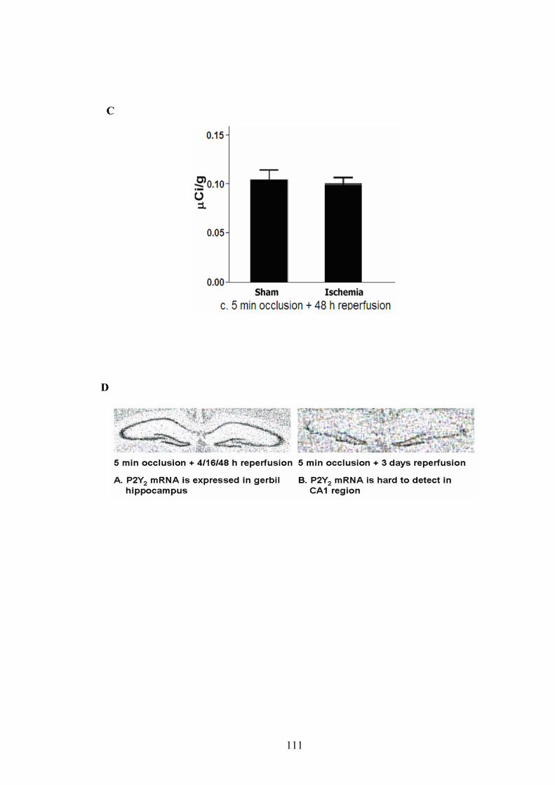
C
D
111

Figure 4. Expression of P2Y2R mRNA in hippocampal subregions after global
cerebral ischemia. P2Y2R mRNA levels were quantified in different hippocampal
regions of sham-operated gerbils (control) and gerbils after a 5 min CCA occlusion
followed by a 16 h reperfusion. CA3: hippocampal CA3 subregion; hilus: also called
CA4 but considered part of the dentate gyrus; DG: dentate gyrus, which contains
upper blade part, lower blade part and the hilus.
112

113

Figure 5. Effect of resveratrol on P2Y2R mRNA expression in the CA1 region of
gerbil brain after transient global cerebral ischemia. Mongolian gerbils were
administered resveratrol by injection (Resv; 30 mg/kg body weight) immediately after
and 24 h after a 5 min CCA occlusion followed by reperfusion (ischemia). A
sham-operated group served as a control. Injection of resveratrol did not significantly
affect P2Y2 receptor mRNA levels in the CA1 region under ischemic conditions (n =
7 animals).
114

115

CHAPTER 5
GENERAL DISCUSSION
ATP and other extracellular nucleotides are released from a variety of cell types
in the central nervous system including neurons, glia, endothelium, and blood cells
(Bergfeld et al., 1992; Ciccarelli et al., 1999; Ahmed et al., 2000; Ostrom et al., 2001).
Nucleotides have been suggested to act as a neuroprotective growth factor by
regulating cell differentiation, proliferation, and survival, as well as a neurotoxic
signal in promoting neuronal degeneration and cell death (Franke and Illes, 2006).
Extracellular nucleotides activate two classes of P2 nucleotide receptors (P2X1-7
ligand-gated ion channels and P2Y1,2,4,6,11-14 G protein-coupled receptors; Sak and
Webb, 2002; Inbe et al., 2004). P2X/Y receptor families are widely distributed and
developmentally expressed in the central nervous system (CNS), as previously
described (reviewed in Franke and Illes, 2006b), which suggests a role for purinergic
signaling in the development of brain structures. Further studies show that P2
nucleotide receptors play important physiological and pathophysiological roles in a
variety of biological processes including neuronal growth, survival and brain injury
(Franke et al, 2006a & b).
The upregulation of P2 nucleotide receptor expression has been reported after
ATP/NGF/cytokine stimulation (Rathbone et al., 1999; D’Ambrosi et al., 2001), and
in tissue injury and disease models of the CNS and other tissues (Collo et al., 1997;
Moore et al., 2000; Seye et al., 2002; Cavaliere et al., 2003 &2004; Parvathenani et
116

al., 2003; Franke et al., 2004). In Alzheimer’s disease and brain ischemia, in vivo and
in vitro studies have shown that P2X2 (in ischemia), P2X4 (in ischemia), P2X7 (in
ischemia and AD) and P2Y1 (in AD) receptor expression is upregulated in specific
cell types, as described in Table 1.
The P2Y2R can be equipotently activated by ATP and UTP to stimulate G
protein-coupled signaling pathways, and to transactivate growth factor receptors (Liu
et al., 2004) and integrins, e.g., αVβ3/β5 (Erb et al., 2001; Wang et al., 2005; Liao et
al., 2007). Studies also indicate that upregulation of the P2Y2R occurs in a variety of
cell and tissue types after injury (Seye et al., 2002; Schrader et al., 2005; Kunzli et al.,
2007). The P2X7R is distinguished from other P2XR subtypes due to its inability to
form heteromeric channels, the presence of an extended C-terminal tail; the high
concentrations of agonist required for activation, and the ability to form pores that
enable the passage of large molecular weight molecules (Sperlagh et al., 2006).
P2X7R functions in the CNS include the regulation of synaptic transmission and
neuronal cell death (Sperlagh et al., 2006).
This dissertation describes the functional expression and regulation of P2Y2
receptor activity by IL-1β in primary neurons and the upregulation of the P2Y2R in
tissue injury models. Results obtained demonstrate that P2Y2Rs mediate the release
of neuroprotective sAPPα, whereas P2X7Rs mediate apoptosis in rat primary cortical
neurons. However, further studies are needed to better understand the roles of P2Y2
and P2X7 nucleotide receptors in neurodegeneration, such as the mechanism whereby
IL-1β induces upregulation of P2Y2R expression, the role of P2Y2Rs in APP
117

translocation/phosphorylation and Aβ production induced by caspase-dependent APP
processing, and the relationship of P2X7R-mediated neuronal apoptosis to the AD
phenotype.
Major contributions and significance
The scientific significance of this dissertation work to the field is summarized
below.
1) P2Y2 receptor expression was identified: a) in rat primary cortical neurons
(Kong et al., 2005 & manuscript in preparation); b) in adult rodent brains; and c) in
brain hippocampus at different stages of cerebral ischemia (unpublished data;
Weisman et al., 2005; Chapter 4). 2) Upregulation of the P2Y2R occurs after
IL-1β treatment in rPCNs and after collar placement around rabbit carotid
arteries. In rat primary cortical neurons, IL-1β treatment increased P2Y2R mRNA
expression and receptor activity in a dose-dependent manner (Kong et al., manuscript
in preparation; Chapter 3). Other studies demonstrate the upregulation of P2Y2Rs in
endothelial and smooth muscle cells of collared rabbit carotid arteries (Appendix-1;
Seye et al., 2002 & 2003; reviewed in Seye et al., 2005). 3) P2Y2R activation
enhances α-secretase-dependent amyloid precursor protein processing in rPCNs.
Previous studies from our lab with the human 1321N1 astrocytoma cell line that does
not express endogenous P2Y2Rs, indicated that transfection with human P2Y2R
cDNA enables UTP to induce α-secretase-dependent APP processing (Camden et al.,
2005). The current studies (Chapter 3) confirmed that P2Y2Rs upregulated by IL-1β
treatment of primary cultures of rat cortical neurons mediate neuroprotective APP
118

processing, and suggest that P2Y2R expression in neurons could be targeted to
suppress neurodegenerative Aβ deposition in Alzheimer’s disease (Kong et al.,
manuscript in preparation). 4) P2X7Rs mediate apoptosis of rPCNs. In rat PCNs,
functional expression of P2X7Rs was detected and activation of the P2X7R was found
to mediate caspase-dependent apoptosis and studies presented identified the signaling
pathways involved (Kong et al., 2005; Chapter 2). 5) P2Y2Rs mediate UTP-induced
intimal hyperplasia in collared rabbit carotid arteries. Our studies demonstrate for
the first time that functional P2Y2R upregulation leads to the development of
neointimal hyperplasia in collared rabbit carotid arteries, a response associated with
atherosclerosis and restenosis after angioplasty (Seye et al., 2002; Appendix-1).
Further studies showed that the P2Y2 receptor also mediates UTP-induced vascular
cell adhesion molecule-1 expression in coronary artery endothelial cells that promotes
the binding of monocytes leading to vascular inflammation (Seye et al., 2003). 6)
Studies in collaboration with other lab members identified P2Y2 receptor
signaling pathways involving integrin interactions and demonstrated that
antagonists of CD39 ectonucleotide triphosphate diphosphohydrolase
(E-NTPDase) activity also inhibit P2Y2R functions (Erb et al., 2001; Gendron et al.,
2002; Wang et al., 2005). Studies with the E-NTPDase inhibitor BG0136 showed that
this compound displays inhibitory activity towards the P2Y2R expressed in human
1321N1 cells, which suggests that BG0136 may represent a novel P2 nucleotide
receptor antagonist (Gendron et al., 2002; Appendix-2). Other studies demonstrate
that the P2Y2 nucleotide receptor interacts with αV integrins through an RGD domain
119

in the first extracellular loop of the P2Y2R. These P2Y2R/αv integrin interactions were
shown to be required for Go-mediated signal transduction that regulates astrocyte
migration (Erb et al., 2001; Appendix-3).
120

APPENDIX
BRIEF REVIEW OF OTHER RESEARCH PROJECTS
121

A-1. FUNCTIONAL P2Y2 NUCLEOTIDE RECEPTORS MEDIATE URIDINE
5’-TRIPHOSPHATE-INDUCED INTIMAL HYPERPLASIA IN COLLARED
RABBIT CAROTID ARTERIES
Seye et al., (2002) Circulation 106(21):2720-2726.
ABSTRACT
Extracellular UTP induces mitogenic activation of smooth muscle cells (SMCs)
through binding to P2Y2 nucleotide receptors. Previous reports also show that P2Y2
receptor mRNA is upregulated in intimal lesions of rat aorta, but it is unclear how
P2Y2 receptors contribute to development of intimal hyperplasia. In the present
studies, we used a silicone collar placed around rabbit carotid arteries to induce
vascular injury and intimal thickening. Collar placement caused rapid upregulation of
P2Y2 receptor mRNA and receptor activity (i.e., UTP-induced increases in the
intracellular calcium concentration) in medial SMCs before appearance of neointima.
Neointimal development was significantly enhanced by perivascular infusion of UTP
(100 µmol/L) and neointima were infiltrated by macrophages. UTP also stimulated
osteopontin expression in situ in collared arteries and in cultured SMCs in a
dose-dependent manner. Furthermore, P2Y2 antisense oligonucleotide inhibited
osteopontin expression induced by UTP suggesting that this response to UTP was
specifically mediated by P2Y2 receptor subtype. In conclusion, these findings indicate
for the first time a role for the UTP/ATP receptor, P2Y2, in development of intimal
hyperplasia associated with atherosclerosis and restenosis.
122

INTRODUCTION
Atherosclerosis is an immunoinflammatory process that involves complex
interactions between the vessel wall and blood components primarily affecting the
large conduit arteries. Endothelial dysfunction is thought to be a key factor in the
development of vascular disease (Ross 1993; Ferns et al., 1991; Wang et al., 1992).
Previous studies suggest that an intact endothelium is important in maintaining a low
proliferative state of smooth muscle cells (SMCs) under normal conditions (Gordon,
1986). In arterial injury, nucleotides are released from endothelial cells, SMCs, and
various blood cells and can bind to P2 receptors to modulate proliferation and
migration of SMCs, which is known to be involved in intimal hyperplasia that
accompanies atherosclerosis and postangioplasty restenosis (Erlinge et al., 1993). The
aim of this study was to determine whether activation of the P2Y2R promotes intimal
hyperplasia under conditions that maintain the vascular endothelium.
MATERIALS AND METHODS
Cell culture
Rabbit SMCs obtained from normal carotid arteries by enzymatic dissociation
were maintained in culture and used from passages 4 to 6.
Phosphorothioate-modified oligonucleotides to rabbit P2Y2R were synthesized and
purified by Integrated DNA Technologies (ADT). Sequences including translation
initiation site were as follows: sense 5'-GGGCAATGGCACTCTTCCCTCACCTG
123
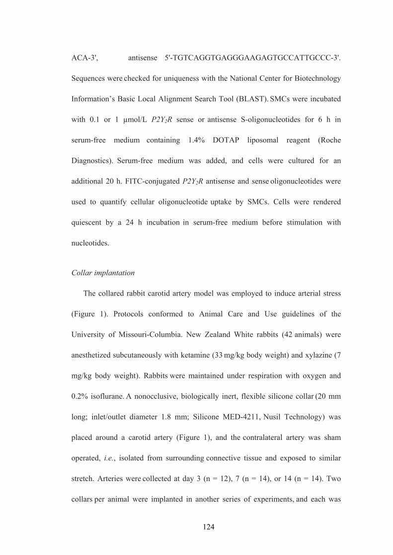
ACA-3', antisense 5'-TGTCAGGTGAGGGAAGAGTGCCATTGCCC-3'.
Sequences were checked for uniqueness with the National Center for Biotechnology
Information’s Basic Local Alignment Search Tool (BLAST). SMCs were incubated
with 0.1 or 1 µmol/L P2Y2R sense or antisense S-oligonucleotides for 6 h in
serum-free medium containing 1.4% DOTAP liposomal reagent (Roche
Diagnostics). Serum-free medium was added, and cells were cultured for an
additional 20 h. FITC-conjugated P2Y2R antisense and sense oligonucleotides were
used to quantify cellular oligonucleotide uptake by SMCs. Cells were rendered
quiescent by a 24 h incubation in serum-free medium before stimulation with
nucleotides.
Collar implantation
The collared rabbit carotid artery model was employed to induce arterial stress
(Figure 1). Protocols conformed to Animal Care and Use guidelines of the
University of Missouri-Columbia. New Zealand White rabbits (42 animals) were
anesthetized subcutaneously with ketamine (33 mg/kg body weight) and xylazine (7
mg/kg body weight). Rabbits were maintained under respiration with oxygen and
0.2% isoflurane. A nonocclusive, biologically inert, flexible silicone collar (20 mm
long; inlet/outlet diameter 1.8 mm; Silicone MED-4211, Nusil Technology) was
placed around a carotid artery (Figure 1), and the contralateral artery was sham
operated, i.e., isolated from surrounding connective tissue and exposed to similar
stretch. Arteries were collected at day 3 (n = 12), 7 (n = 14), or 14 (n = 14). Two
collars per animal were implanted in another series of experiments, and each was
124

connected to an osmotic minipump (Alzet 2 ML2; Alza Corporation) implanted
subdermally in the thoracic region. The pump continuously delivered (5 µl/h) UTP
(100 µmol/l) or PBS (mmol/l: NaCl 154, Na2HPO4 8, NaH2PO4 2, EDTA 0.2) for 3
(n = 14), 7 (n = 14), and 14 (n = 14) days. After surgery, analgesic (Buprenex; 0.1 to
0.5 mg/kg body weight) was administered.
Immunoblotting
Cultured cells were solublized in 2X Laemmli sample buffer (120 mmol/L
Tris-HCl, pH 6.8, 2% SDS, 10% sucrose, 1 mmol/L EDTA, 50 mmol/L
dithiothreitol, 0.003% bromophenol blue). Rabbit aortas were homogenized with a
polytron homogenizer in buffer containing 0.9% NaCl, 20 mmol/L Tris-HCl, pH 7.6,
1 mmol/L PMSF in 0.2% Triton X-100. Equivalent amounts of protein (100 µg)
were subjected to 10% SDS-PAGE and transferred to nitrocellulose membranes for
immunoblotting. Detection of OPN (Chemicon International) was performed with
rat anti-human OPN (1:1000 dilution) and horseradish-peroxidase-conjugated goat
anti-rat IgG (1:1500 dilution) as primary and secondary antibodies, respectively. For
signal normalization, membranes were reprobed with anti-mouse -tubulin antibody
(1:1000 dilution; Santa Cruz Biotechnology).
RESULTS
Effect of local UTP application on OPN expression
Immunoblots of sham-operated arteries indicated lack of OPN expression (Figure
2A, Lane 5), whereas collared arteries at day 14 expressed OPN (Figure 2A, Lane 2).
125

Local UTP infusion greatly increased OPN expression (Figure 4A, Lane 4) compared
with PBS (Figure 2A, Lane 3).
UTP-induced OPN expression is mediated by P2Y2Rs in SMCs
UTP exposure for 8 h stimulated OPN expression in vascular SMCs (Figure 2B) in
a dose-dependent manner (Figure 2C). ATP and 10% FBS (positive control) induced
OPN expression, whereas ADP, 2-MeSATP, and UDP did not (Figure 2B), which
suggests that P2Y2Rs mediate OPN expression. To conclusively demonstrate the role
of P2Y2Rs, SMCs incubated with 0.1 or 1 µg of P2Y2R antisense or sense
oligonucleotides were stimulated with 50 µmol/L UTP for 8 h. Antisense (Figure 2D,
Lanes 5 and 6) but not sense (Figure 2D, Lanes 2 and 3) P2Y2R oligonucleotides
inhibited UTP-induced OPN expression in SMCs.
DISCUSSION
Proliferation of SMCs is a hallmark of vascular diseases such as atherosclerosis
and restenosis following angioplasty (Hanke et al., 1990). Nucleotides released from
various arterial and blood cells (Di Virgilio and Solini, 2002) can bind to P2
nucleotide receptors. In our present studies, in vivo experiments showed that the P2Y2
receptor was functionally upregulated in both SMCs and endothelial cells after collar
implantation (Seye et al., 2002). Moreover, intimal thickening of collared rabbit
carotid arteries was greatly enhanced by in situ UTP application (Seye et al., 2002)
and was closely associated with osteopontin (OPN) expression in medial SMCs
(Figure 2A). In vitro studies showed that both UTP and ATP increased OPN
126

expression in cultured SMCs whereas ADP, UDP, and 2-MeSATP were ineffective
(Figures 2B-C), which suggests a role for the P2Y2R in which ATP and UTP are
equipotent, and is confirmed by the inhibition of UTP-induced OPN expression in
cultured SMCs after P2Y2R antisense oligonucleotide treatment (Figure 2D).
Extracellular nucleotides may also cause cell recruitment by inducing lymphocyte
and macrophage adhesion to human pulmonary artery endothelial cells, as
demonstrated in vitro (Parker et al., 1996). In collared rabbit carotid arteries, we
found that local UTP delivery via an osmotic pump induced intimal accumulation of
macrophages mediated by P2Y2 receptors (data not shown; Seye et al., 2002).
In conclusion, these studies demonstrate for the first time that extracellular UTP
and the P2Y2R contribute to the development of arterial lesions associated with
atherosclerosis and restenosis.
127

Figure 1. Collared rabbit carotid artery model. New Zealand White rabbits (42
animals) were anesthetized and (A) a nonocclusive, biologically inert, flexible
silicone collar (20 mm long; inlet/outlet diameter 1.8 mm; Silicone MED-4211,
Nusil Technology) was (B) placed around a carotid artery, and the contralateral
artery was sham operated, i.e., isolated from surrounding connective tissue and
exposed to similar stretch.
128

129

Figure 2. Nucleotide-induced OPN expression in collared carotid arteries and
SMC cultures. A. Collared arteries connected to an osmotic minipump were
continuously infused with UTP or saline for 14 days. Lane 1, control (kidney); 2,
collar without infusion; 3, collar with PBS, 4, collar with UTP; 5, sham-operated
artery. Blots were reprobed with anti- -tubulin antibody. B-C. SMCs were stimulated
with 10% FBS or (B) 50 µmol/L nucleotide or (C) the indicated UTP concentration.
Immunoblot analysis was performed with anti-OPN antibodies. Data represent 3 (A)
or 5 (B) experiments. D. SMCs incubated with P2Y2R sense or antisense
S-oligonucleotides for 6 h in DMEM containing 1.4% DOTAP liposomal reagent
were cultured for an additional 16 h in serum-free medium before 50 µM UTP
treatment. Cells were incubated with serum-free medium (Lane 1); 0.1 (Lane 2) or 1
(Lane 3) µg of sense oligonucleotide; serum (Lane 4); or 0.1 (Lane 5) or 1 (Lane 6)
µg of antisense oligonucleotide.
130

A
B
C
D
131

A-2. P2Y2 RECEPTOR ACTIVITY IS MODULATED BY THE ECTO-
NUCLEOTIDASE NTPASE (CD39)
Unpublished data; Gendron et al., (2002) Curr Drug Targets. (3):229-245.
ABSTRACT
Extracellular nucleotides initiate various cellular and physiological responses
through P2 nucleotide receptors. Another important factor affecting extracellular
nucleotide signaling is ecto-nucleoside triphosphate diphosphohydrolase (NTPDase;
CD39) now considered as the main ecto-nucleotidase responsible for degrading tri-
and diphosphonucleosides to monophosphate derivatives through sequential
hydrolysis. Since the concentration and duration of extracellular nucleotides are key
regulators of P2 nucleotide receptor function, the influence of ecto-nucleotidases is
always considered as one of the major ways to modulate P2 receptor function. In the
present studies, the NTPDase inhibitor BG0136 and several cell model systems
including COS-7 cells, HEK cells and human 1321N1 astrocytoma cells stably
transfected with P2Y2R cDNA were employed to understand the complex
interrelationship between P2Y2 receptors and ecto-nucleotidases. Treatment with 350
μM BG0136 resulted in a 30-50% decrease in UTP-stimulated ERK1/2 activity in
COS-7, HEK cells and P2Y2R-transfected 1321N1 cells. In contrast, the COS-7/CD39
cell transfectants demonstrated a decrease in UTP-stimulated inositol phosphate
release compared to native COS-7 cells. The variable effects of BG0136 on P2Y2
receptor signal propagation in cells expressing CD39 cDNA observed in our studies
suggests that BG0136 may represent a new P2Y2R antagonist.
132
INTRODUCTION
It was suggested that inhibition of the ecto-nucleotidases responsible for the
hydrolysis of extracellular nucleotides could be an important factor in the modulation
of purinergic signaling. This study concerns the inhibition of NTPDase and its
possible impact on different physiological systems. Several enzymes are involved in
the metabolism and control of extracellular nucleotide concentrations (Beaudoin et al.,
1996 & 2000; Gordon 1986; Zimmermann 1992 & 2000; Ziganshin et al., 1994;
Plesner et al., 1995; Vlajkovic et al., 1998). The enzymes described above may also
play an important role in the dephosphorylation of extracellular nucleotides
(Zimmermann 2000; Beaudoin et al., 1996; Culic et al., 1990). In the past,
modulation of nucleotide-induced cell signaling was focused on nucleotide receptors.
Ecto-nucleotidases, more particularly the E-NTPDases family of ecto-enzymes, are
now considered as potential new targets for P2 receptor modulation. Inhibition or
modulation of E-NTPDases may prolong the effect of nucleotides at their respective
receptors and/or modulate cell-to-cell interaction (Imai et al., 1999; Clifford et al.,
1997; Dzhandzhugazyan et al., 1998). New molecules have been synthesized to
inhibit ecto-nucleotidases and particularly the E-NTPDases, such as P2 receptor
agonists/antagonists and molecules related to the suramin-Evans blue families. Based
on the structure-activity requirements, as described by Wittenburg et al. (Wittenburg
et al., 1996), we report here the possible antagonistic property of BG0136
(1-naphthol-3, 6-disulfonic acid, disodium salt) towards the P2Y2 receptor.
133

MATERIALS AND METHODS
Materials
Fetal bovine serum (FBS) was obtained from Hyclone (Logan, UT, USA).
Dulbecco’s Modified Eagle’s medium (DMEM), penicillin (100 units/ml), and
streptomycin (100 units/ml) were obtained from Gibco-BRL (Carlsbad, CA, USA).
Rabbit anti-rat ERK1/2, horseradish peroxidase (HRP)-conjugated goat anti-rabbit
IgG, and HRP-conjugated goat anti-mouse IgG antibodies were obtained from Santa
Cruz Biotechnology (Santa Cruz, CA, USA). PERK antibody was obtained from Cell
Signaling Technology (Beverly, MA, USA). The Precision Plus Protein Standards and
nitrocellulose membranes (0.45 mm) were obtained from Bio-Rad (Hercules, CA,
USA). LumiGLO chemiluminescent substrates were obtained from New England
Biolabs (Beverly, MA, USA). Nucleotides and all other biochemicals were obtained
from Sigma Chemical Co. (St. Louis, MO, USA).
Cell Culture and Transfection
Human 1321N1 astrocytoma cells (Parr et al., 1994), African Green Monkey
Kidney Fibroblasts (COS-7 cells) and Human Embryonic Kidney (HEK) cells were
cultured in DMEM (GIBCO BRL) containing 5% FBS, 100 U/ml penicillin, and 100
µg/ml streptomycin. Cells were maintained at 37 °C in a humidified atmosphere of
5% CO2 and 95% air. The retroviral vector, pLXSN, was used for stable expression of
the P2Y2R construct in 1321N1 cells as described previously (Erb et al., 1995). In
brief, the recombinant P2Y2R-pLXSN construct was used to transfect PA317
amphotrophic packaging cells for production of viral vectors. Then, 1321N1 cells
134
were infected with the viral vectors and selected for neomycin resistance with 1 mg/ml
G418 (GIBCO BRL).
IP Assay
To measure accumulation of IPs, COS-7 cells with or without expression of
CD39 were plated in 12-well dishes and incubated in the presence or absence of the
specified antibodies for 24 h in 0.7 ml serum-free and inositol-free DMEM containing
2 µCi of [3H]-inositol (20 Ci/mmol). The cells were washed three times and incubated
for 30 min at 37 °C in 1 ml of LiCl solution (15 mM glucose, 25 mM Hepes, 110 mM
NaCl, 5 mM KCl, 1.8 mM CaCl2, 0.9 mM MgCl2, and 30 mM LiCl) followed by
stimulation with 0.1 µM UTP for 10 min. The reactions were stopped by aspiration
and cell extracts were obtained by addition of 0.3 ml of ice-cold 10% perchloric acid
for 15 min. The extracts were neutralized with 0.6 ml of 2 M NaOH and 225 mM
Hepes. The IPs in the neutralized extracts were separated from inositol and
glycerophosphoinositol on Dowex columns, as described previously (Berridge et al.,
1983). Radioactivity in the IP fraction was determined by liquid scintillation
spectrometry using Ecolume scintillation fluid.
Western blot analysis
Cells cultured in 6-well plates were treated with nucleotides in the presence or
absence of inhibitors as indicated. The cells were washed twice with ice-cold PBS,
and lysed for 5 min in 200 ml of ice-cold lysis buffer (20 mM Tris-HCl, pH 7.5, 2%
(w/v) sodium dodecyl sulfate (SDS), and 1 mM sodium orthovanadate). Lysate (30
135

mg of protein) was added to Laemmli sample buffer (187.5 mM Tris-HCl, pH 6.8, 6%
(w/v) SDS, 1.8% (v/v) β-mercaptoethanol and 0.003% (w/v) bromophenol blue).
Samples were heated for 5 min at 96 °C, and subjected to 7.5% (w/v)
SDS-polyacrylamide gel electrophoresis (SDS-PAGE) and transferred to
nitrocellulose membranes for protein immunoblotting. After overnight blocking at 4
°C with 5% (w/v) fat-free milk in TBS-T (10 mM Tris-HCl, pH 7.4, 120 mM NaCl,
and 0.1% (v/v) Tween-20), membranes were incubated with either 1:1000 dilution of
anti-ERK1/2 or anti-phospho-ERK1/2 antibodies for 2 h at room temperature,
followed by incubation with HRP-conjugated anti-rabbit or anti-mouse IgG antibodies
(1:1000 dilution in TBS-T containing 5% (w/v) fat-free milk) for 1 h at room
temperature. Protein immunoreactivity was visualized on autoradiographic film using
the LumiGlo Chemiluminescence System (New England BioLabs), according to the
manufacturer’s instructions. The protein bands detected on X-ray film were quantified
using a computer-driven scanner and Quantity One software (Bio-Rad, Hercules, CA).
The activation levels of ERK1/2 were calculated as a percentage of control (i.e., total
ERK1/2).
RESULTS
To determine the effect of CD39 on P2 nucleotide receptor-mediated signaling
pathways, COS-7 and HEK cells were transfected with CD39. UTP-stimulated
inositol phosphate release (Figure 1A) and ERK phosphorylation (Figure 1B and 2)
were both inhibited in CD39-transfected COS-7 and HEK cells compared to
untransfected cells, indicating that the expression and activation of E-NTPDase by
136

UTP inhibited UTP-stimulated P2Y2 nucleotide receptor signaling pathways. A
recently developed compound, 1-hydroxynaphthalene-3, 6-disulfonic acid (BG0136)
has been shown to inhibit bovine spleen NTPDase with a Ki of 350 μM. However,
BG0136, the inhibitor of E-NTPDase also inhibited UTP-stimulated ERK
phosphorylation in both CD39-transfected COS-7 and HEK cells (Figure 1B and 2),
indicating the possible direct interaction of BG0136 with the P2Y2 nucleotide
receptor.
To investigate whether BG0136 could interact with P2 purinoceptors, human
1321N1 astrocytoma cells which do not express endogenous P2Y2Rs were stably
transfected with human P2Y2R. Pretreatment of cell transfectants with 350 μM
BG0136 resulted in greater than 50% inhibition of ATP- and UTP- stimulated
ERK1/2 phosphorylation in hP2Y2-1321N1 cells (Figure 3). Since this cell line has
been found to have very little ectonucleotidase activity (Gendron et al., 2002), it
appears that BG0136 could be either acting directly on the P2Y2 receptor or on some
other component(s) in the ERK signaling pathway.
Altogether, our results obtained with BG0136 and CD39-transfected COS-7,
HEK and hP2Y2-1321N1 cells showed that transfection with ectonucleotidase inhibits
UTP-stimulated signaling mediated by P2Y2 nucleotide receptors, including inositol
phosphate release and ERK1/2 activation, whereas BG0136 inhibits
UTP/ATP-stimulated ERK phosphorylation mediated by P2Y2Rs, possibly by acting
as a P2Y2 receptor antagonist. Hence, even if BG0136 displays inhibitory activity
towards E-NTPDase, it should be considered as a potential new P2Y2 nucleotide
137

receptor antagonist. However, further studies need to be done with this compound to
determine its molecular specificity for P2 nucleotide receptor subtypes and
ectonucleotidases.
138

Figure 1. Effect of BG0136 on UTP-stimulated inositol phosphate (IP) release
and ERK1/2 activation in COS-7 cells with or without CD39 transfection. African
Green Monkey Kidney Fibroblast (COS-7) Cells with (CD39/COS7) or without
CD39 transfection were incubated with 350 μM BG0136 for 5 min at 37 oC, followed
by stimulation with 10 μM UTP at 37 oC for the indicated time. IP release was
measured by inositol assay and the phosphorylation of ERK1/2 was determined by
Western blot analysis in which total ERK1/2 levels served as a loading control.
139

A
B
140

Figure 2. Effect of BG0136 on UTP-stimulated ERK1/2 activation in HEK cells
with or without CD39 transfection. Human Embryonic Kidney (HEK) cells with or
without CD39 transfection were incubated with 350 μM BG0136 for 5 min at 37 oC,
followed by stimulation with 10 μM UTP at 37 oC for the indicated time, and the
phosphorylation of ERK1/2 was determined by Western blot analysis in which total
ERK1/2 levels served as a loading control.
141

142

Figure 3. Effect of BG0136 on nucleotide-stimulated ERK activation in human
1321N1 astrocytoma cells expressing the human P2Y2 nucleotide receptor.
Human 1321N1 astrocytoma cells, stably transfected with the human P2Y2R cDNA,
were incubated with 350 μM BG0136 for 5 min at 37 oC, followed by stimulation
with 10 μM ATP or UTP at 37 oC for the indicated time. Phosphorylation of ERK1/2
was determined by Western blot analysis in which total ERK levels served as a
loading control.
143
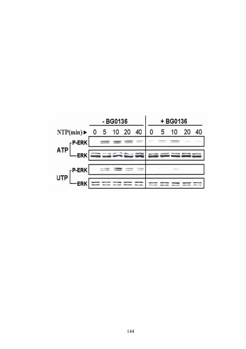
144
A-3. AN RGD SEQUENCE IN THE P2Y2 RECEPTOR INTERACTS WITH
αVß3 INTEGRINS AND IS REQUIRED FOR Go-MEDIATED SIGNAL
TRANSDUCTION
Erb et al., (2001) J Cell Biol. 153(3):491-501.
ABSTRACT
The P2Y2 nucleotide receptor (P2Y2R) contains the integrin-binding domain
arginine-glycine-aspartic acid (RGD) in its first extracellular loop, raising the
possibility that this P2Y2R interacts directly with an integrin through its RGD domain.
In this project, we tested whether an antibody raised against the αV integrin subunit
could interfere with UTP-stimulated P2Y2R signal transduction such as inositol
phosphates accumulation. Human 1321N1 cells expressing the P2Y2R were incubated
in the presence or absence of specific anti-αV integrin antibody for 24 h in serum-free
and inositol-free DMEM containing [3H]-inositol. Cells were then used to measure
UTP-stimulated inositol phosphate (IP) release. The results indicate that overnight
treatment with anti-αV antibody partially inhibits IP formation, consistent with the
partial inhibition of UTP-induced FAK and ERK activation by anti-αV antibody and
the decreased calcium response mediated by RGE mutant P2Y2Rs as compared to
wild-type P2Y2Rs (Erb et al., 2001). In conclusion, these studies suggest that
interaction of αV integrins with the P2Y2R is important for optimum P2Y2R
activation.
145

INTRODUCTION
As a G protein-coupled nucleotide receptor, activation of the P2Y2R stimulates
PLC-dependent production of IP3 and DAG followed by calcium release and PKC
activation. P2Y2Rs can also bind to growth factor receptors (GFRs) through
SH3-binding domains in the C-terminal tail of the P2Y2R and cause transactivation
of GFR signaling pathways. The present studies explore protein complex formation
between integrins and the P2Y2 nucleotide receptor (P2Y2R), a Go- and Gq-coupled
P2 nucleotide receptor subtype that is equipotently stimulated by ATP or UTP and
mediates various cellular responses (Erb et al. 1993; Boarder et al. 1995). RGD has
been found in various integrin ligands, (Clark and Brugge 1995; Frenette and
Wagner 1996) as the core recognition sequence for many integrins, including αVß3,
αVß5, αVß11, α5ß1, and αIIbß3 (Hynes 1992). Studies by Erb et al. (Erb et al., 2001)
with P2Y2R-transfected cells indicated that a mutation of the RGD motif in the
wild-type P2Y2R to RGE decreases receptor colocalization with αV integrins by
10-fold and the RGE-mutant P2Y2R requires 1,000-fold higher agonist
concentrations than the wild-type P2Y2R to phosphorylate FAK, activate ERK, and
initiate the PLC-dependent mobilization of intracellular Ca2+. Furthermore, pertussis
toxin, an inhibitor of Gi/o proteins, partially inhibited Ca2+ mobilization mediated by
the wild-type P2Y2R, but not by the RGE mutant, suggesting that the RGD sequence
is required for P2Y2R-mediated Go but not Gq activation. To address the possibility
that the P2Y2R interacts with a specific integrin, here it was investigated whether an
146

antibody raised against the αV integrin subunit could interfere with UTP-stimulated
P2Y2R signal transduction.
MATERIALS AND METHODS
Materials
Fetal bovine serum (FBS) was obtained from Hyclone (Logan, UT, USA).
Dulbecco’s Modified Eagle’s medium (DMEM), penicillin (100 units/ml), and
streptomycin (100 units/ml) were obtained from Gibco-BRL (Carlsbad, CA, USA).
Anti–human αV monoclonal antibody (mAb) was purchased from Zymed
Laboratories. Anti–human α3 mAb was purchased from Santa Cruz Biotechnology,
Inc. Nucleotides and all other biochemicals were obtained from Sigma Chemical Co.
(St. Louis, MO, USA).
Cell culture and transfection
Human 1321N1 astrocytoma cells (Parr et al., 1994) were cultured in DMEM
(GIBCO BRL) containing 5% FBS, 100 U/ml penicillin, and 100 µg/ml streptomycin.
Cells were maintained at 37 °C in a humidified atmosphere of 5% CO2 and 95% air.
The retroviral vector, pLXSN, was used for stable expression of the P2Y2R constructs
in 1321N1 cells, as described previously (Erb et al., 1995). In brief, the recombinant
P2Y2R-pLXSN construct was used to transfect PA317 amphotrophic packaging cells
for production of the viral vectors. Then, 1321N1 cells were infected with the viral
vectors and selected for neomycin resistance with 1 mg/ml G418 (GIBCO BRL).
IP assay
147

To measure accumulation of IPs, 1321N1 cells expressing the P2Y2R were plated
in 12-well dishes and incubated in the presence or absence of the specified antibodies
for 24 h in 0.7 ml serum-free and inositol-free DMEM containing 2 µCi of
[3H]-inositol (20 Ci/mmol). The cells were washed three times and incubated for 30
min at 37 °C in 1 ml of LiCl solution (15 mM glucose, 25 mM Hepes, 110 mM NaCl,
5 mM KCl, 1.8 mM CaCl2, 0.9 mM MgCl2, and 30 mM LiCl) followed by stimulation
with 0.1 mM UTP for 10 min. The reactions were stopped by aspiration and cell
extracts were obtained by addition of 0.3 ml of ice-cold 10% perchloric acid for 15
min. The extracts were neutralized with 0.6 ml of 2 M NaOH and 225 mM Hepes. The
IPs in the neutralized extracts were separated from inositol and
glycerophosphoinositol on Dowex columns, as described previously (Berridge et al.,
1983). Radioactivity in the IP fraction was determined by liquid scintillation
spectrometry using Ecolume scintillation fluid.
RESULTS
P2Y2R-mediated signaling is inhibited by anti-αV antibodies
We investigated whether an antibody raised against the V integrin subunit
could interfere with UTP-stimulated P2Y2R signal transduction. To analyze the
effect of this antibody on the P2Y2R calcium signaling pathway, we measured IP
formation since inositol 1, 4, 5-trisphosphate (IP3) is the second messenger for
intracellular calcium mobilization (Weisman et al., 1998). The results indicated that
overnight treatment with anti-αV antibody partially inhibited IP formation induced
148

by UTP (Figure 1), consistent with the decreased calcium response seen for the RGE
mutant P2Y2 receptor as compared with the wild-type P2Y2R (data not shown; Erb
et al., 2001). Anti- 3 integrin antibody had no effect on the ability of the P2Y2R to
stimulate calcium signaling (Figure 1). These findings suggest that interaction of V
integrin with the P2Y2R is important for optimum P2Y2R activation.
149
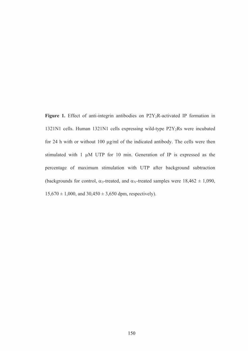
Figure 1. Effect of anti-integrin antibodies on P2Y2R-activated IP formation in
1321N1 cells. Human 1321N1 cells expressing wild-type P2Y2Rs were incubated
for 24 h with or without 100 µg/ml of the indicated antibody. The cells were then
stimulated with 1 µM UTP for 10 min. Generation of IP is expressed as the
percentage of maximum stimulation with UTP after background subtraction
(backgrounds for control, α3-treated, and αV-treated samples were 18,462 ± 1,090,
15,670 ± 1,000, and 30,450 ± 3,650 dpm, respectively).
150
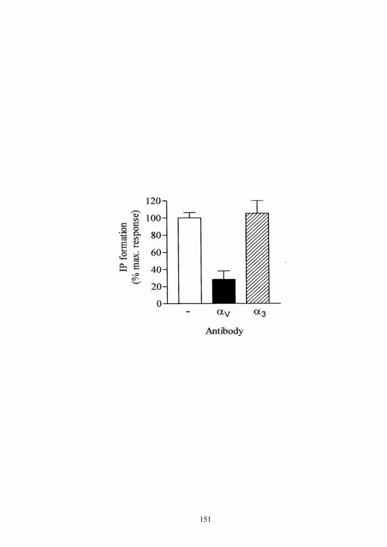
151

REFERENCES
Abbracchio MP, Boeynaems JM, Barnard EA, Boyer JL, Kennedy C, Miras-Portugal
MT, King BF, Gachet C, Jacobson KA, Weisman GA, Burnstock G. (2003)
Characterization of the UDP-glucose receptor (renamed here the P2Y14 receptor)
adds diversity to the P2Y receptor family. TiPS 24:52-55.
Abbracchio MP, Burnstock G, Boeynaems JM, Barnard EA, Boyer JL, Kennedy C,
Miras-Portugal MT, King BF, Gachet C, Jacobson KA and Weisman GA.
(2004) The Recently Deorphanized GPR80 (GPR99) proposed to be the P2Y15
Receptor is not a Genuine P2Y Receptor. TiPS 26: 8-9.
Ahmed SM, Rzigalinski BA, Willoughby KA, Sitterding HA, Ellis EF. (2000)
Stretch-induced injury alters mitochondrial membrane potential and cellular
ATP in cultured astrocytes and neurons. J Neurochem 74(5): 1951–1960.
Allinson TM, Parkin ET, Turner AJ, Hooper NM. (2003) ADAMs family members as
amyloid precursor protein alpha-secretases. J Neurosci Res 74:342–352.
Anderson JP, Refolo LM, Wallace W, Mehta P, Krishnamurthi M, Gotlib J, Bierer L,
Haroutunian V, Perl D, and Robakis NK. (1989) Differential brain expression of
the Alzheimer's amyloid precursor protein. EMBO J 8(12): 3627–3632.
Atkinson L, Batten TF, Moores TS, Varoqui H, Erickson JD, Deuchars J. (2004)
Differential co-localization of the P2X7 receptor subunit with vesicular
glutamate transporters VGLUT1 and VGLUT2 in rat CNS. Neuroscience
123:761-768.
152

Beaudoin AR, Sévigny J, Picher M. (1996) In: Biomembranes Vol.5. (Lee, AG; Ed.).
JAI Press Inc. Greenwich, pp. 367-399.
Beaudoin AR, Grondin G, Enjyoji K, Robson SC, Sévigny J, Fischer B, Gendron FP.
(2000) in: Ecto-ATPases and related ectonucleotidases. (Vanduffel L. and
Lemmens R; Eds), Shaker Publishing B.V.; The Netherlands, pp.125-135.
Beigi RD, Kertesy SB, Aquilina G, Dubyak GR. (2003) Oxidized ATP (oATP)
attenuates proinflammatory signaling via P2 receptorindependent mechanisms.
Br J Pharmacol 140:507-519.
Benham CD. (1990) ATP-gated channels in vascular smooth muscle cells. Ann N Y
Acad Sci 603:275-285.
Bennett GC, Ford AP, Smith JA, Emmett CJ, Webb TE, Boarder MR. (2003) P2Y
receptor regulation of cultured rat cerebral cortical cells: calcium responses and
mRNA expression in neurons and glia. Br J Pharmacol 139(2):279-88.
Bergfeld GR and Forrester T. (1992) Release of ATP from human erythrocytes in
response to a brief period of hypoxia and hypercapnia. Cardiovasc Res 26(1):
40–47.
Berridge MJ, Dawson RMC, Downes CP, Heslop JP, Irvine RF. (1983) Changes in
the level of inositol phosphates after agonist-dependent hydrolysis of membrane
phosphoinositides. Biochem J 212:473-482.
Bianchi BR, Lynch KJ, Touma E, Niforatos W, Burgard EC, Alexander KM, Park HS,
Yu H, Metzger R, Kowaluk E, Jarvis MF, van Biesen T. (1999) Pharmacological
153

characterization of recombinant human and rat P2X receptor subtypes. Eur J
Pharmacol 376:127-38.
Black RA, Rauch CT, Kozlosky CJ, Peschon JJ, Slack JL, Wolfson MF, Castner BJ,
Stocking KL, Reddy P, Srinivasan S, Nelson N, Boiani N, Schooley KA,
Gerhart M, Davis R, Fitzner JN, Johnson RS, Paxton RJ, March CJ, Cerretti DP.
(1997) A metalloproteinase disintegrin that releases tumor-necrosis factor-alpha
from cells. Nature 385(6618):729-733.
Boarder MR, Weisman GA, Turner JT, Wilkinson GF. (1995) G protein-coupled P2
purinoceptors: from molecular biology to functional responses.Trends
Pharmacol Sci 16(4):133-9. Review.
Bogdanov YD, Wildman SS, Clements MP, King BF, Burnstock G. (1998) Molecular
cloning and characterization of rat P2Y4 nucleotide receptor. Br J Pharmacol
124(3):428-430.
Brugg B, Dubreuil YL, Huber G, Wollman EE, Delhaye-Bouchaud N, Mariani J.
(1995) Inflammatory processes induce beta-amyloid precursor protein changes
in mouse brain. Proc Natl Acad Sci U S A 92(7):3032-3035.
Burnstock G, Cocks T, Kasakov L, Wong HK. (1978) Direct evidence for ATP
release from non-adrenergic, non-cholinergic (“purinergic”) nerves in the
guinea-pig taenia coli and bladder. Eur J Pharmacol 49:145-149.
Burnstock G. (1980) Purinergic nerves and receptors. Prog Biochem Pharmacol
16:141-154.
Burnstock G and Wood JN. (1996) Purinergic receptors: their role in nociception and
154

primary afferent neurotransmission. Curr Opin Neurobiol 6(4): 526–532.
Burnstock G. (1999) Release of vasoactive substances from endothelial cells by shear
stress and purinergic mechanosensory transduction. J Anat 194:335–342
Burnstock G. (2000) P2X receptors in sensory neurons. Br. J. Anaesth. 84(4):476-488.
Cacquevel M, Lebeurrier N, Cheenne S, Vivien D. (2004) Cytokines in
neuroinflammation and Alzheimer's disease. Curr Drug Targets (6):529-34.
Review.
Camden JM, Schrader AM, Camden RE, Gonzalez FA, Erb L, Seye CI, Weisman GA.
(2005) P2Y2 nucleotide receptors enhance alpha-secretase-dependent amyloid
precursor protein processing. J Biol Chem 280(19):18696- 18702.
Canet-Aviles RM, Anderton M, Hooper NM, Turner AJ, Vaughan PF. (2002)
Muscarine enhances soluble amyloid precursor protein secretion in human
neuroblastoma SH-SY5Y by a pathway dependent on protein kinase C(alpha),
src-tyrosine kinase and extracellular signal-regulated kinase but not
phospholipase C.Brain Res Mol Brain Res102(1-2):62-72.
Cavaliere F, Sancesario G, Bernardi G, Volonte C. (2002). Extracellular ATP and
nerve growth factor intensify hypoglycemia induced cell death in primary
neurons: role of P2 and NGFRp75 receptors. J Neurochem 83(5):1129–1138.
Cavaliere F, Florenzano F, Amadio S, Fusco FR, Viscomi MT, D’Ambrosi N, et al.
(2003). Up-regulation of P2X2, P2X4 receptor and ischemic cell death:
prevention by P2 antagonists. Neuroscience 120(1):85–98.
Cavaliere F, Amadio S, Sancesario G, Bernardi G, Volonte C. (2004) Synaptic P2X7
155

and oxygen/glucose deprivation in organotypic hippocampal cultures. J Cereb
Blood Flow Metab 4:392-398.
Chang K, Hanaoka M, Kumada M, Takuwa Y. (1995) Molecular cloning and
functional analysis of a novel P2 nucleotide receptor. J Biol Chem.
44:26152–26158
Checler F. (1995) Processing of the beta-amyloid precursor protein and its regulation
in Alzheimer's disease. J Neurochem 65:1431–1444.
Chessell IP, Simon J, Hibell AD, Michel AD, Barnard EA, Humphrey PP. (1998)
Cloning and functional characterization of the mouse P2X7 receptor. FEBS Lett;
439: 26-30.
Cheung KK and Burnstock G. (2002). Localization of P2X3 receptors and
coexpression with P2X2 receptors during rat embryonic neurogenesis. J Comp
Neurol 443(4):368–382.
Cheung KK, Ryten M, Burnstock G. (2003) Abundant and dynamic expression of G
protein-coupled P2Y receptors in mammalian development. Dev Dyn.
228(2):254-66.
Ciccarelli R, Di Iorio P, Giuliani P, D'Alimonte I, Ballerini P, Caciagli F, Rathbone
MP. (1999) Rat cultured astrocytes release guaninebased purines in basal
conditions and after hypoxia/hypoglycemia. Glia 25: 93–98.
Ciccarelli R, Ballerini P, Sabatino G, Rathbone MP, D'Onofrio M, Caciagli F, Di Iorio
P. (2001) Involvement of astrocytes in purine-mediated reparative processes in
the brain. Int J Dev Neurosci. 19(4):395-414.
156

Clark EA and Brugge JS. (1995) Integrins and signal transduction pathways: the road
taken. Science 268:233-238.
Clifford EE, Martin KA, Dalal P, Thomas R, Dubyak GR. (1997) Stage-specific
expression of P2Y receptors, ecto-apyrase, and ecto-5'-nucleotidase in myeloid
leukocytes. Am. J. Physiol. 42:C973-C987.
Collo G, Neidhardt S, Kawashima E, Kosco-Vilbois M, North RA, Buell G. (1997).
Tissue distribution of the P2X7 receptor. Neuropharmacology 36(9):1277– 1283.
Coutinho-Silva R, Persechini PM, Da Cunha Bisaggio R, Perfettini J, Torres de Santo
AC, Kanellopoulos JM, Mottaly I, Dautry-Varsat A, Ojcius DM (1999)
P2Z/P2X7 receptor-dependent apoptosis of dendritic cells. Am J Physiol 276:
C1139–C1147
Culic O, Sabolic I, Zanic-Grubisic T. (1990) The stepwise hydrolysis of adenine
nucleotides by ectoenzymes of rat renal brush-border membranes. Biochim.
Biophys. Acta 1030:143-151.
D’Ambrosi N, Murra B, Cavaliere F, Amadio S, Bernardi G, Burnstock G, Volonte C.
(2001). Interaction between ATP and nerve growth factor signaling in the
survival and neuritic outgrowth from PC12 cells. Neuroscience 108(3):527–
534.
de la Torre. (2000) Impaired cerebromicrovascular perfusion. Summary of evidence
in support of its causality in Alzheimer's disease. Ann N Y Acad Sc. 924:
136-152
DeLeo J, Toth L, Schubert P, Rudolphi K, Kreutzberg GW. (1987) Ischemia-induced
157

neuronal cell death, calcium accumulation, and glial response in the
hippocampus of the Mongolian gerbil and protection by propentofylline (HWA
285). J Cereb Blood Flow Metab (6):745-51.
Denlinger LC, Fisette PL, Sommer JA, Watters JJ, Prabhu U, Dubyak GR, Proctor
RA, Bertics PJ. (2001) Cutting edge: The nucleotide receptor P2X7 contains
multiple protein- and lipid-interaction motifs including a potential binding site
for bacterial lipopolysaccharide. J Immunol 167:1871-1876.
Desdouits-Magnen J, Desdouits F, Takeda S, Syu LJ, Saltiel AR, Buxbaum JD,
Czernik AJ, Nairn AC, Greengard P. (1998) Regulation of secretion of
Alzheimer amyloid precursor protein by the mitogen-activated protein kinase
cascade. J Neurochem 70(2):524-530.
Deuchars SA, Atkinson L, Brooke RE, Musa H, Milligan CJ, Batten TF, Buckley NJ,
Parson SH, Deuchars J. (2001) Neuronal P2X7 receptors are targeted to
presynaptic terminals in the central and peripheral nervous systems. J Neurosci
21:7143-7152.
Di Virgilio F, Zanovello P, Zambon A, Bronte V, Pizzo P, Murgia M (1995) Cell
membrane receptors for extracellular ATP: a new family of apoptosis-signalling
molecules. Fundam Clin Immuno 3:80–81.
Di Virgilio F, Chiozzi P, Ferrari D, Falzoni S, Sanz JM, Morelli A, Torboli M,
Bolognesi G, Baricordi OR. (2001a) Nucleotide receptors: An emerging family
of regulatory molecules in blood cells. Blood 97: 587-600.
158
Di Virgilio F, Vishwanath V, Ferrari D. (2001b) On the role of the P2X7 receptor in
the immune system. In Abbracchio MP,WilliamsM(eds): Handbook of
Experimental Pharmacology: Purinergic and Pyrimidinergic Signalling Vol.
151/II, Berlin Heidelberg New York: Springer 355-377.
Di Virgilio F and Solini A (2002) P2 receptors: new potential players in
atherosclerosis. Br J Pharmacol 135(4):831–842.
Drouet B, Pincon-Raymond M, Chambaz J, Pillot T. (2000) Molecular basis of
Alzheimer's disease. Cell Mol Life Sci 57(5):705-15. Review.
Duan S, Anderson CM, Keung EC, Chen Y, Chen Y, Swanson RA. (2003) P2X7
receptor-mediated release of excitatory amino acids from astrocytes. J Neurosci
23: 1320-1328.
Dzhandzhugazyan KN, Kirkin AF, Straten PT, Zeuthen J. (1998) Ecto-ATP
diphosphohydrolase/CD39 is overexpressed in differentiated human melanomas.
FEBS Lett 430, 227-230.
Eikelenboom P, Rozemuller AJ, Hoozemans JJ, Veerhuis R, van Gool WA. (2000)
Neuroinflammation and Alzheimer disease: clinical and therapeutic implications.
Alzheimer Dis Assoc Disord 1:S54-61.
Erb L, Lustig KD, Ahmed AH, Gonzalez FA, Weisman GA. (1990) Covalent
incorporation of 30-O-(4-benzoyl) benzoyl-ATP into a P2 purinoceptor in
transformed mouse fibroblasts. J Biol Chem 265: 7424-7431.
Erb L, Lustig KD, Sullivan DM, Turner JT, Weisman GA. (1993) Functional
159

expression and photoaffinity labeling of a cloned P2U purinergic receptor. Proc
Natl Acad Sci USA 90:10449-10453.
Erb L, Garrad R, Wang Y, Quinn T, Turner JT, Weisman GA. (1995) Site-directed
mutagenesis of P2U purinoceptors. Positively charged amino acids in
transmembrane helices 6 and 7 affect agonist potency and specificity. J Biol
Chem 270(9):4185-4188.
Erb L, Liu J, Ockerhausen J, Kong Q, Garrad RC, Griffin K, Neal C, Krugh B,
Santiago-Perez LI, Gonzalez FA, Gresham HD, Turner JT, Weisman GA. (2001)
An RGD sequence in the P2Y2 receptor interacts with αVβ3 integrins and is
required for Go-mediated signal transduction. J Cell Biol 153(3):491-501.
Erb L, Liao Z, Seye CI and Weisman GA. (2006) P2 receptors: intracellular signaling.
Pflugers Arch - Eur J Physiol 452: 552–562.
Erlinge D, Yoo H, Edvinsson L, Reis DJ, Wahlestedt C. (1993) Mitogenic effects of
ATP on vascular smooth muscle cells vs. other growth factors and sympathetic
cotransmitters. Am J Physiol 265: H1089–H1097.
Erlinge D, Hou M, Webb TE, Barnard EA, Möller S. (1998) Phenotype changes of
the vascular smooth muscle cell regulate P2 receptor expression as measured
by quantitative RT-PCR. Biochem Biophys Res Commun 248:864–870.
Esler WP and Wolfe MS. (2001) A portrait of Alzheimer secretases--new features and
familiar faces. Science 293(5534):1449-1454.
160

Ferns GA, Reidy MA, Ross R. (1991) Balloon catheter de-endothelialization of the
nude rat carotid: response to injury in the absence of functional T-lymphocytes.
Am J Pathol 138:1045–1057.
Ferrari D, Chiozzi P, Falzoni S, Hanau S, Di Virgilio F. (1997) Purinergic modulation
of interleukin-1 beta release from microglial cells stimulated with bacterial
endotoxin. J Exp Med 185: 579-582.
Ferrari D, Los M, Bauer MK, Vandenabeele P, Wesselborg S, Schulze-Osthoff K
(1999) P2Z purinoreceptor ligation induces activation of caspases with distinct
roles in apoptotic and necrotic alterations of cell death. FEBS Lett 447(1):71–75.
Franke H, Gunther A, Grosche J, Schmidt R, Rossner S, Reinhardt R,
Faber-Zuschratter H, Schneider D, Illes P. (2004) P2X7 receptor expression after
ischemia in the cerebral cortex of rats. J Neuropathol Exp Neurol
63(7):686–699.
Franke H, Krugel U, Illes P. (2006a) P2 receptors and neuronal injury. Pflugers Arch.
452(5):622-644.
Franke H and Illes P. (2006b) Involvement of P2 receptors in the growth and survival
of neurons in the CNS. Pharmacology & Therapeutics 109:297 – 324.
Fremont L. (2000) Biological effects of resveratrol. Life Sci 66: 663–673.
Frenette PS and Wagner DD. (1996) Adhesion molecules. N Engl J Med
334:1526-1529.
161
Fu W, Lu C, Mattson MP. (2002) Telomerase mediates the cell survival-promoting
actions of brain-derived neurotrophic factor and secreted amyloid precursor
protein in developing hippocampal neurons. J Neurosci 22(24):10710-10719
Fumagalli M, Brambilla R, D’Ambrosi N, Volonté C, Matteoli M, Verderio C,
Abbracchio MP. (2003) Nucleotide-mediated calcium signaling in rat cortical
astrocytes: role of P2X and P2Y receptors. Glia 43: 218-230.
Garcia JH. (1975) The neuropathology of stroke. Hum Pathol 6(5):583-598.
Gendron FP, Benrezzak O, Krugh BW, Kong Q, Weisman GA, Beaudoin AR. (2002)
Purine signaling and potential new therapeutic approach: possible outcomes of
NTPDase inhibition. Curr Drug Targets 3(3):229-45. Review.
Gendron FP, Neary JT, Theiss PM, Sun GY, Gonzalez FA, Weisman GA. (2003)
Mechanisms of P2X7 receptor-mediated ERK1/2 phosphorylation in human
astrocytoma cells. Am J Physiol Cell Physiol 284: C571-581.
Giovannini L, Migliori M, Longoni BM, Das DK, Bertelli AA, Panichi V, Filippi C,
Bertelli A. (2001) Resveratrol, a polyphenol found in wine, reduces ischemia
reperfusion injury in rat kidneys. J Cardiovasc Pharmaco 37:262–270.
Golstein P, Kroemer G. (2006) Cell death by necrosis: towards a molecular definition.
Trends Biochem Sci 32(1):37-43.
Gordon JL. (1986) Extracellular ATP: effects, sources and fate. Biochem J 233:
309–319.
Grafe P, Mayer C, Takigawa T, Kamleiter M, Sanchez-Brandelik R. (1999) Confocal
162
calcium imaging reveals an ionotropic P2 nucleotide receptor in the paranodal
membrane of rat Schwann cells. J Physiol 515:377-383.
Grammas P and Ovase R. (2001) Inflammatory factors are elevated in brain
microvessels in Alzheimer's disease. Neurobiol Aging 22(6):837-842.
Guerra AN, Fisette PL, Pfeiffer ZA, Quinchia-Rios BH, Prabhu U, Aga M, Denlinger
LC, Guadarrama AG, Abozeid S, Sommer JA, Proctor RA, Bertics PJ. (2003)
Purinergic receptor regulation of LPS-induced signaling and pathophysiology.
Endotoxin Res 9:256-263.
Guzzetta F, Deodato F, Rando T. (2000) Brain ischemic lesions of the newborn.
Childs Nerv Syst 16(10-11):633-637.
Hanke H, Strohschneider T, Oberhoff M, Betz E, Karsch KR. (1990) Time course of
smooth muscle cell proliferation in the intima and media of arteries following
experimental angioplasty. Circ Res 67(3):651-9.
Hauss-Wegrzyniak B, Dobrzanski P, Stoehr JD, Wenk GL. (1998) Chronic
neuroinflammation is a contributor to the pathophysiology of Alzheimer’s
disease Brain Res 780(2):294-303.
Hou M, Moller S, Edvinsson l, Erlinge D. (2000) Cytokines induced upregulation of
Vascular P2Y2 receptors and increased mitogenic responses to UTP and ATP.
Arterioscler Thromb Vasc Biol 20(9):2064-2069.
Hu Y, Fisette PL, Denlinger LC, Guadarrama AG, Sommer JA, Proctor RA, Bertics
PJ (1998) Purinergic receptor modulation of lipopolysaccharide signaling and
inducible nitric-oxide synthase expression in RAW 264.7 macrophages. J Biol
163

Chem 273 (42):27170 –27175.
Huang SS, Tsai MC, Chih CL, Hung LM, Tsai SK. (2001) Resveratrol reduction of
infarct size in Long-Evans rats subjected to focal cerebral ischemia. Life Sci
69:1057–1065.
Humphreys BD, Rice J, Kertesy SB, Dubyak GR. (2000) Stress-activated protein
kinase/JNK activation and apoptotic induction by the macrophage P2X7
nucleotide receptor. J Biol Chem 275: 26792-26798.
Hung LM, Chen JK, Huang SS, Lee RS, Su MJ. (2000) Cardioprotective effect of
resveratrol, a natural antioxidant derived from grapes. Cardiovasc Res
47:549–555.
Hynes RO. (1992) Integrins: versatility, modulation, and signaling in cell adhesion.
Cell 69:11-25.
Illes P and Ribeiro AJ. (2004) Neuronal P2 receptors of the central nervous system.
Eur J Pharmacol 483:5-17.
Imai M, Takigami K, Guckelberger O, Enjyoji K, Neal Smith R, Lin Y, Csizmadia E,
Sévigny J, Rosenberg RD, Bach FH, Robson SC. (1999) Modulation of
nucleoside [correction of nucleotide] triphosphate diphosphohydrolase-1
(NTPDase-1)cd39 in xenograft rejection. Mol Med 5:743-752.
Inbe H, Watanabe S, Miyawaki M, Tanabe E, Encinas JA. (2004) Identification and
characterization of a cell-surface receptor, P2Y15, for AMP and adenosine. J
Biol Chem 279(19): 19,790–19,799.
164

Inoue K. (2002) Microglial activation by purines and pyrimidines. Glia 40:156-63.
Iwatsubo, T. Odaka, A, Suzuki N, Mizusawa H, Nukina N, Ihara Y. (1994)
Visualization of AB42(43) and AB40 in senile plaques with end-specific AB
monoclonals: evidence that an initially deposited species is AB42(43). Neuron
13: 45-53.
Jacobson KA, Hoffmann C, Cattabeni F, Abbracchio MP (1999) Adenosine- induced
cell death: evidence for receptor-mediated signalling. Apoptosis 4(3):197–211.
Jacques-Saliva MC, Rodnight R, Lenz G, Liao Z, Kong Q, Tran M, Kang Y, Gonzalez
FA, Weisman GA, Neary JT. (2004) P2X7 receptors stimulate AKT
phosphorylation in astrocytes. Br J Pharmacol 141(7):1106-1117.
Kaczmarek E, Erb L, Koziak K, Jarzyna R, Wink MR, Guckelberger O, Blusztajn JK,
Trinkaus-Randall V, Weisman GA, Robson SC. (2005) Modulation of
endothelial cell migration by extracellular nucleotides: involvement of focal
adhesion kinase and phosphatidylinositol 3-kinase- mediated pathways. Thromb
Haemost 93(4):735-42.
Kaiser D, Freyberg MA, Friedl P (1997) Lack of hemodynamic forces triggers
apoptosis in vascular endothelial cells. Biochem Biophys Res Commun
231(3):586–590.
Kapus´cin´ski J, Skoczylas B. (1978) Fluorescent complexes of DNA with DAPI 40,
6-diamidine-2-phenyl indole. 2HCl or DCI 40,6-dicarboxyamide-2-phenyl
indole. Nucleic Acids Res 5: 3775-3799.
Kaup M, Dassler K, Weise C, Fuchs H. (2002) Shedding of the transferrin receptor is
165

mediated constitutively by an integral membrane metalloprotease sensitive to
tumor necrosis factor alpha protease inhibitor-2. J Biol Chem
277(41):38494-38502.
Khakh BS, Burnstock G, Kennedy C, King BF, North RA, Séguéla P, Voigt M,
Humphrey PP. (2001) International Union of Pharmacology. XXIV. Current
status of the nomenclature and properties of P2X receptors and their subunits.
Pharmacol Rev 53: 107-118.
Khera M, Somogyi GT, Kiss S, Boone TB, Smith CP. (2004) Botulinum toxin A
inhibits ATP release from bladder urothelium after chronic spinal cord injury.
Neurochem Int 45: 987-993.
Khreiss T, Jozsef L, Hossain S, Chan JS, Potempa LA, Filep JG. (2002) Loss of
pentameric symmetry of C-reactive protein is associated with delayed apoptosis
of human neutrophils. J Biol Chem 277: 40775-40781.
Kidd EJ, Grahames CBA, Simon J, Michel AD, Barnard EA, Humphrey PPA. (1995)
Localization of P2X purinoceptor transcripts in the rat nervous system. Mol
Pharmacol 48(4), 569–573.
Kidd EJ, Miller KJ, Sansum AJ, Humphrey PP. (1998) Evidence for P2X3 receptors
in the developing rat brain. Neuroscience 87(3), 533–539.
Kirazov L, Loffler T, Schliebs R, Bigl V. (1997) Glutamate-stimulated secretion of
amyloid precursor protein from cortical rat brain slices. Neurochem Int 30(6):
557-563.
166

Kitaguchi N, Takahashi Y, Tokushima Y, Shiojiri S, Ito H. (1988) Novel precursor of
Alzheimer's disease amyloid protein shows protease inhibitory activity. Nature
331(6156):530-532.
Kirino T. (1982) Delayed neuronal death in the gerbil hippocampus following
ischemia. Brain Res 239(1):57-69.
Koike H, Tomioka S, Sorimachi H, Saido TC, Maruyama K, Okuyama A,
Fujisawa-Sehara A, Ohno S, Suzuki K, Ishiura S. (1999) Membrane-anchored
metalloprotease MDC9 has an alpha-secretase activity responsible for
processing the amyloid precursor protein. Biochem J 343(2):371-375.
Kong Q, Wang M, Liao Z, Camden JM, Yu S, Simonyi A, Sun GY, Gonzalez FA, Erb
L, Seye CI, Weisman GA. (2005) P2X7 nucleotide receptors mediate
caspase-8/9/3-dependent apoptosis in rat primary cortical neurons. Purinergic
Signalling 1:337-347.
Koshiba M, Apasov S, Sverdlov V, Chen P, Erb L, Turner JT, Weisman GA, Sitkovsky
MV. (1997) Transient up-regulation of P2Y2 nucleotide receptor mRNA
expression is an immediate early gene response in activated thymocytes. Proc
Natl Acad Sci 94(3):831-836.
Kunzli BM, Berberat PO, Giese T, Csizmadia E, Kaczmarek E, Baker C, Halaceli I,
Buchler MW, Friess H, Robson SC. (2007) Upregulation of CD39/NTPDases
and P2 receptors in human pancreatic disease. Am J Physiol Gastrointest Liver
Physiol. 292(1):G223-30.
Kuwana T, Smith JJ, Muzio M, Dixit V, Newmeyer DD, Kornbluth S. (1998)
167

Apoptosis induction by caspase-8 is amplified through the mitochondrial release
of cytochrome c. J Biol Chem 273: 16589-16594.
Lahiri DK, Lewis S, Farlow MR. (1994) Tacrine alters the secretion of the
beta-amyloid precursor protein in cell lines. J Neurosci Res 37:777–787.
Lammas DA, Stober C, Harvey CJ, Kendrick N, Panchalingam S, Kumararatne DS
(1997) ATP-induced killing of mycobacteria by human macrophages is
mediated by purinergic P2Z (P2X7) receptors. Immunity 7(3):433–444.
Lammich S, Kojro E, Postina R, Gilbert S, Pfeiffer R, Jasionowski M, Haass C,
Fahrenholz F. (1999) Constitutive and regulated alpha-secretase cleavage of
Alzheimer's amyloid precursor protein by a disintegrin metalloprotease. Proc
Natl Acad Sci 96(7):3922-3927.
Le Stunff H, Auger R, Kanellopoulos J, Raymond MN. (2004) The Pro-451 to Leu
polymorphism within the C-terminal tail of P2X7 receptor impairs cell death but
not phospholipase D activation in murine thymocytes. J Biol Chem 279:
16918-26.
LeBlanc AC, Chen HY, Autilio-Gambetti L, Gambetti P. (1991) Differential APP gene
expression in rat cerebral cortex, meninges, and primary astroglial, microglial
and neuronal cultures. FEBS Lett 292(1-2):171-8.
LeBlanc AC, Papadopoulos M, Belair C, Chu W, Crosato M, Powell J, Goodyer CG.
(1997) Processing of amyloid precursor protein in human primary neuron and
astrocyte cultures. J Neurochem 68(3):1183-90.
LeBlanc AC, Koutroumanis M, Goodyer CG. (1998) Protein kinase C activation
168

increases release of secreted amyloid precursor protein without decreasing Aβ
production in human primary neuron cultures. J Neurosci 18(8):2907-2913.
Lenz G, Gottfried C, Luo Z, Avruch J, Rodnight R, Nie WJ, Kang Y, Neary JT. (2000)
P2Y purinoceptor subtypes recruit different mek activators in astrocytes. Br J
Pharmacol 129(5):927-936.
Li H, Zhu H, Xu CJ, Yuan J. (1998) Cleavage of BID by caspase 8 mediates the
mitochondrial damage in the Fas pathway of apoptosis. Cell 94: 491-501.
Li HL, Roch JM, Sundsmo M, Otero D, Sisodia S, Thomas R, Saitoh T. (1997)
Defective neurite extension is caused by a mutation in amyloid beta/A4 (A beta)
protein precursor found in familial Alzheimer's disease. J Neurobiol
(5):469-480.
Li P, Nijhawan D, Budihardjo I, Srinivasula SM, Ahmad M, Alnemri ES, Wang X.
(1997) Cytochrome c and dATPdependent formation of Apaf-1/caspase-9
complex initiates an apoptotic protease cascade. Cell 91: 479-89.
Liu J, Liao Z, Camden J, Griffin KD, Garrad RC, Santiago-Perez LI, Gonzalez FA,
Seye CI, Weisman GA, Erb L. (2004) Src homology 3 binding sites in the P2Y2
nucleotide receptor interact with Src and regulate activities of Src, proline-rich
tyrosine kinase 2, and growth factor receptors. J Biol Chem 279(9):8212-8218
Loesch A, Glass R. (2006) Electron microscopy and in situ hybridization: Expression
of of P2Y2 receptor mRNA in the cerebellum. Methods Mol Biol 326:151-62.
Lundy PM, Hamilton MG, Mi L, Gong W, Vair C, Sawyer TW, Frew R. (2002)
169

Stimulation of Ca2+ influx through ATP receptors on rat brain synaptosomes:
Identification of functional P2X7 receptor subtypes. Br J Pharmacol 135:
1616-1626.
Luo JJ, Wallace MS, Hawver DB, Kusiak JW, Wallace WC. (2001) Characterization
of the neurotrophic interaction between nerve growth factor and secreted
alpha-amyloid precursor protein. J Neurosci Res 63(5):410-420.
Luo X, Budihardjo I, Zou H, Slaughter C, Wang X. (1998) Bid, a Bcl2 interacting
protein, mediates cytochrome c release from mitochondria in response to
activation of cell surface death receptors. Cell 94: 481-90.
Martins RN, Robinson PJ, Chleboun JO, Beyreuther K, Masters CL. (1991) The
molecular pathology of amyloid deposition in Alzheimer's disease. Mol
Neurobiol 5(2-4):389-398.
Mattson MP. (1997) Cellular actions of beta-amyloid precursor protein and its soluble
and fibrillogenic derivatives. Physiol Rev 77(4):1081-132.
Mehlhorn G, Hollborn M, Schliebs R. (2000) Induction of cytokines in glial cells
surrounding cortical beta-amyloid plaques in transgenic Tg2576 mice with
Alzheimer pathology. Int J Dev Neurosci 18(4-5):423-431.
Mills J, Laurent Charest D, Lam F, Beyreuther K, Ida N, Pelech SL, Reiner PB.
(1997) Regulation of amyloid precursor protein catabolism involves the
mitogen-activated protein kinase signal transduction pathway. J Neurosci 17(24):
9415-22.
170

Mills J and Reiner PB. (1999) Regulation of amyloid precursor protein cleavage. J
Neurochem 72(2):433-460.
Moore D, Iritani S, Chambers J, Emson P. (2000). Immunohistochemical localization
of the P2Y1 purinergic receptor in Alzheimers disease. NeuroReport
11(17):3799–3803.
Moss ML, Jin SL, Milla ME, Bickett DM, Burkhart W, Carter HL, Chen WJ, Clay
WC, Didsbury JR, Hassler D, Hoffman CR, Kost TA, Lambert MH, Leesnitzer
MA., McCauley P, McGeehan G, Mitchell J, Moyer M, Pahel G, Rocque W,
Overton LK, Schoenen F, Seaton T, Su JL, Becherer JD. (1997) Cloning of a
disintegrin metalloproteinase that processes precursor tumour-necrosis
factor-alpha. Nature. 385(6618):733-736. Erratum in: (1997) Nature
386(6626):738.
Muscella A, Elia MG, Greco S, Storelli C, Marsigliante S. (2003) Activation of P2Y2
receptor induces c-FOS protein through a pathway involving mitogen- activated
protein kinases and phosphoinositide 3-kinases in HeLa cells. J Cell Physiol.
195(2):234-240.
National Center for Biotechnology Information. Basic Local Alignment Search Tool
(BLAST). Available at: http://www.ncbi.nlm.nih. gov/BLAST.
Neary JT, Rathbone MP, Cattabeni F, Abbracchio MP, Burnstock G. (1996) Trophic
actions of extracellular nucleotides and nucleosides on glial and neuronal cells.
Trends Neurosci 19(1): 13–18.
Nguyen T, Erb L, Weisman GA, Marchese A, Heng HHQ, Garrad RC, George
171

SR, Turner JT, O'Dowd BF. (1995) Cloning, expression and chromosomal
localization of the human uridine nucleotide receptor gene. J Biol Chem 270:
30845–30848
Nitsch RM, Slack BE, Wurtman RJ, Growdon JH. (1992) Release of Alzheimer
amyloid precursor derivatives stimulated by activation of muscarinic
acetylcholine receptors. Science 258:304–307.
Nitsch RM, Slack BE, Farber SA, Schulz JG, Deng M, Kim C, Borghesani PR,
Korver W, Wurtman RJ, Growdon JH. (1994) Regulation of proteolytic
processing of the amyloid beta-protein precursor of Alzheimer's disease in
transfected cell lines and in brain slices. J Neural Transm Suppl 44: 21–27.
Nitsch RM, Wurtman RJ, Growdon JH. (1995) Regulation of proteolytic processing
of the amyloid beta-protein precursor by first messengers. A novel potential
approach for the treatment of Alzheimer's disease. Arzneim Forsch 45: 435–438.
Nitsch RM, Deng A, Wurtman RJ, Growdon JH. (1997) Metabotropic glutamate
receptor subtype mGluR1alpha stimulates the secretion of the amyloid
beta-protein precursor ectodomain. J Neurochem 69(2):704-712.
Norenberg W and Illes P. (2000) Neuronal P2X receptors: Localisation and functional
properties. Naunyn-Schmiedeberg’s Arch Pharmacol 362: 324-39.
North RA and Surprenant A. (2000) Pharmacology of cloned P2X receptors. Annu
Rev Pharmacol Toxicol 40:563-80.
Ostrom RS, Gregorian C, Drenan RM, Gabot K, Rana BK, Insel PA. (2001) Key role
for constitutive cyclooxygenase-2 of MDCK cells in basal signaling and
172

response to released ATP. Am J Physiol Cell Physiol 281(2): C524–C531.
Panenka W, Jijon H, Herx LM, Armstrong JN, Feighan D, Wei T, Yong VW,
Ransohoff RM, MacVicar BA. (2001) P2X7-like receptor activation in
astrocytes increases chemokine monocyte chemoattractant protein-1 expression
via mitogen-activated protein kinase. J Neurosci 21:7135-42.
Papp L, Vizi ES, Sperlagh B. (2004) Lack of ATP-evoked GABA and glutamate
release in the hippocampus of P2X7 receptor-/- mice. NeuroReport 15: 2387-91.
Parihar MS and Hemnani T. (2004) Alzheimer’s disease pathogenesis and therapeutic
interventions J Clinical Neurosci 11(5):456-467
Parker AL, Likar LL, Dawicki DD, Rounds S (1996) Mechanism of ATP-induced
leukocyte adherence to cultured pulmonary artery endothelial cells. Am J
Physiol 270:L695–L703.
Parkin ET, Trew A, Christie G, Faller A, Mayer R, Turner AJ, Hooper NM. (2002)
Structure-activity relationship of hydroxamate-based inhibitors on the secretases
that cleave the amyloid precursor protein, angiotensin converting enzyme, CD23,
and pro-tumor necrosis factor-alpha. Biochemistry (Mosc) 41: 4972–4981.
Parr CE, Sullivan DM, Paradiso AM, Larowski ER, Burch LH, Olsen JC, Erb L,
Weisman GA, Boucher RC, Turner JT. (1994) Cloning and expression of a
human P2U nucleotide receptor, a target for cystic fibrosis pharmacotherapy.
Proc Nat Acad Sci USA 91:3275-3279.
Parvathenani LK, Tertyshnikova S, Greco CR, Roberts SB, Robertson B, Posmantur
R. (2003) P2X7 mediates superoxide production in primary microglia and is
173

up-regulated in a transgenic mouse model of Alzheimer’s disease. J Biol Chem
278:13309-17.
Parvathy S, Hussain I, Karran EH, Turner AJ, Hooper NM. 1998. Alzheimer’s
amyloid precursor protein alpha-secretase is inhibited by hydroxamic acid-based
zinc metalloprotease inhibitors: similarities to the angiotensin converting
enzyme secretase. Biochemistry 37:1680–1685.
Plesner L. (1995) Ecto-ATPases: identities and functions. Int Rev Cytol. 158: 141-214.
Queiroz G, Meyer DK, Meyer A, Starke K, von Kügelgen I. (1999) A study of the
mechanism of the release of ATP from rat cortical astroglial cells evoked by
activation of glutamate receptors. Neuroscience 91: 1171-1181.
Rassendren F, Buell GN, Virginio C, Collo G, North RA, Surprenant A. (1997) The
permeabilizing ATP receptor, P2X7. Cloning and expression of a human cDNA.
J Biol Chem 272:5482-5486.
Rathbone MP, Middlemiss PJ, Gysbers JW, Andrew C, Herman MA, Reed JK,
Ciccarelli R, Di Iorio P, Caciagli F. (1999) Trophic effects of purines in neurons
and glial cells. Prog Neurobiol 59(6):663– 690.
Ray PS, Maulik G, Cordis GA, Bertelli AA, Bertelli A, Das DK. (1999) The red wine
antioxidant resveratrol protects isolated rat hearts from ischemia reperfusion
injury. Free Radical Biol Med 27:160–169.
Rice PL, Beard KS, Driggers LJ, Ahnen DJ. (2004) Inhibition of extracellular- signal
regulated kinases 1/2 is required for apoptosis of human colon cancer cells in
174

vitro by Sulindac metabolites. Cancer Res 64:8148-8151.
Robert SJ, Zugaza JL, Fischmeister R, Gardier AM, Lezoualc’h F. (2001) The human
serotonin 5-HT4 receptor regulates secretion of non-amyloidogenic precursor
protein. J Biol Chem 276(48):44881-44888.
Rochette MJ, Murphy MP. (2002) Gamma-secretase: substrates and inhibitors. Mol
Neurobiol 26(1):81-95.
Ross R. (1993) The pathogenesis of atherosclerosis: a perspective for the 1990s.
Nature 362:801–809.
Sak K and Webb TE. (2002) A retrospective of recombinant P2Y receptor subtypes
and their pharmacology. Arch Biochem Biophys 397(1): 131–136.
Salvesen GS and Dixit VM. (1997) Caspases: Intracellular signaling by proteolysis.
Cell 91: 443-446.
Sasaki Y, Hoshi M, Akazawa C, Nakamura Y, Tsuzuki H, Inoue K, Kohsaka S. (2003)
Selective expression of Gi/o-coupled ATP receptor P2Y12 in microglia in rat
brain. Glia 44(3):242-50.
Schendel SL, Azimov R, Pawlowski K, Godzik A, Kagan BL, Reed JC. (1999) Ion
channel activity of the BH3 only Bcl-2 family member, BID. J Biol Chem
274:21932-21936.
Schrader AM, Camden JM, Weisman GA. (2005) P2Y2 nucleotide receptor
up-regulation in submandibular gland cells from the NOD.B10 mouse model of
Sjogren's syndrome. Arch Oral Biol 50(6):533-540.
175

Schulze-Lohoff E, Hugo C, Rost S, Arnold S, Gruber A, Brune B, Sterzel RB. (1998)
Extracellular ATP causes apoptosis and necrosis of cultured mesangial cells via
P2Z/P2X7 receptors. Am J Physiol 275:F962–F971.
Seye CI, Gadeau AP, Daret D, Dupuch F, Alzieu P, Capron L, Desgranges C. (1997)
Overexpression of the P2Y2 purinoceptor in intimal lesion of the rat aorta.
Arterioscler Thromb Vasc Biol 17: 3602–3610.
Seye CI, Kong Q, Erb L, Garrad RC, Krugh B, Wang M, Turner JT, Sturek M,
Gonzalez FA, Weisman GA. (2002) Functional P2Y2 nucleotide receptors
mediate uridine 5'-triphosphate-induced intimal hyperplasia in collared rabbit
carotid arteries. Circulation 106(21):2720-2726.
Seye CI, Yu N, Jain R, Kong Q, Minor T, Newton J, Erb L, Gonzalez FA, Weisman
GA. (2003) The P2Y2 nucleotide receptor mediates UTP-induced vascular cell
adhesion molecule-1 expression in coronary artery endothelial cells. J Biol
Chem 278(27):24960-24965.
Seye CI, Yu N, Gonzalez FA, Erb L, Weisman GA. (2005) The P2Y2 nucleotide
receptor mediates vascular cell adhesion molecule-1 expression through
interaction with VEGF receptor-2 (KDR/Flk-1). J Biol Chem
279(34):35679-35686.
Seye CI, Kong Q, Yu N, Gonzalez FA, Erb L, Weisman GA (2006) P2 receptors in
atherosclerosis and postangioplasty retenosis. Purinergic Signalling 2:471-480
Shen J, Seye CI, Wang M, Weisman GA, Wilden PA, Sturek M. (2004) Cloning,
up-regulation, and mitogenic role of porcine P2Y2 receptor in coronary artery
176

smooth muscle cells. Mol Pharmacol 66(5):1265-1274.
Sim JA, Young MT, Sung HY, North RA, Surprenant A. (2004) Reanalysis of P2X7
receptor expression in rodent brain. J Neurosci 24: 6307-14.
Sims JE, Gayle MA, Slack JL, Alderson MR, Bird TA, Giri JG, Colotta F, Re F,
Mantovani A, Shanebeck K, et al. (1993) Interleukin 1 signaling occurs
exclusively via the type I receptor. Proc Natl Acad Sci U S A 90:6155–6159.
Simonyi A, Woods D, Sun AY, Sun GY. (2002) Grape polyphenols inhibit chronic
ethanol-induced COX-2 mRNA expression in rat brain. Alcohol Clin Exp Res
26(3):352-357.
Slee EA, Harte MT, Kluck RM, Wolf BB, Casiano CA, Newmeyer DD, Wang HG,
Reed JC, Nicholson DW, Alnemri ES, Green DR, Martin SJ. (1999) Ordering
the cytochrome c-initiated caspase cascade: Hierarchical activation of
caspases-2, -3, -6, -7, -8, and -10 in a caspase-9-dependent manner. J Cell Biol
144: 281-92.
Solano DC, Sironi M, Bonfini C, Solerte SB, Govoni S, Racchi M. (2000) Insulin
regulates soluble amyloid precursor protein release via phosphoatidyl inositol 3
kinase-dependent pathway. FASEB J 14:1015-1022.
Solini A, Chiozzi P, Falzoni S, Morelli A, Fellin R, Di Virgilio F (2000) High glucose
modulates P2X7 receptor-mediated function in human primary fibroblasts.
Diabetologia 43(10):1248–1256.
Sperlagh B, Kofalvi A, Deuchars J, Atkinson L, Milligan CJ, Buckley NJ, Vizi ES.
(2002) Involvement of P2X7 receptors in the regulation of neurotransmitter
177

release in the rat hippocampus. J Neurochem 81:1196-211.
Sperlagh B, Vizi ES, Wirkner K, Illes P. (2006) P2X7 receptors in the nervous system.
Prog Neurobiol 78(6):327-346.
Stein TD, Anders NJ, DeCarli C, Chan SL, Mattson MP, Johnson JA. (2004)
Neutralization of transthyretin reverses the neuroprotective effects of secreted
amyloid precursor protein (APP) in APPSW mice resulting in tau
phosphorylation and loss of hippocampal neurons: support for the amyloid
hypothesis. J Neurosci 24(35):7707-7717.
Stella N, Estelles A, Siciliano J, Tence M, Desagher S, Piomelli D, Glowinski J,
Premont J. (1997) Interleukin-1 enhances the ATP-evoked release of arachidonic
acid from mouse astrocytes. J Neurosci 17(9):2939-2946.
Suen KC, Lin KF, Elyaman W, So KF, Chang RC, Hugon J. (2003) Reduction of
calcium release from the endoplasmic reticulum could only provide partial
neuroprotection against beta-amyloid peptide toxicity. J Neurochem 87:1413-26.
Sugiyama T, Kobayashi M, Kawamura H, Li Q, Puro DG. (2004) Enhancement of
P2X7-induced pore formation and apoptosis: An early effect of diabetes on the
retinal microvasculature. Invest Ophthalmol Vis Sci 45: 1026-1032.
Surprenant A, Rassendren F, Kawashima E, North RA, Buell G. (1996) The cytolytic
P2Z receptor for extracellular ATP identified as a P2X receptor (P2X7). Science
272: 735-738.
Tanaka S, Shiojiri S, Takahashi Y, Kitaguchi N, Ito H, Kameyama M, Kimura J,
178

Nakamura S, Ueda K. (1989) Tissue-specific expression of three types of
beta-protein precursor mRNA: enhancement of protease inhibitor-harboring
types in Alzheimer's disease brain. Biochem Biophys Res Commun
165(3):1406-1414.
Tang D, Lahti JM, Kidd VJ. (2000) Caspase-8 activation and bid cleavage contribute
to MCF7 cellular execution in a caspase-3-dependent manner during
staurosporine-mediated apoptosis. J Biol Chem 275:9303-9307.
Traystman RJ. (2003) Animal models of focal and global cerebral ischemia. ILAR J
44(2):85-95.
Turner JT, Weisman GA, Camden JM. (1997) Upregulation of P2Y2 nucleotide
receptors in rat salivary gland cells during short-term culture. Am J Physiol
273(3 Pt 1):C1100-1107.
Turner JT, Park M, Camden JM, Weisman GA. (1998) Salivary gland nucleotide
receptors: Changes in expression and activity related to development and tissue
damage. Ann N Y Acad Sci 842:70-75.
Unal-Cevik I, Kilinc M, Gursoy-Ozdemir Y, Gurer G, Dalkara T. (2004) Loss of
NeuN immunoreactivity after cerebral ischemia does not indicate neuronal cell
loss: A cautionary note. Brain Res 1015:169-174.
Virginio C, MacKenzie AB, North RA, Surprenant A. (1999) Kinetics of cell lysis,
dye uptake and permeability changes in cells expressing the rat P2X7 receptor. J
Physiol 519:335-346.
179

Viswanath V, Wu Y, Boonplueang R, Chen S, Stevenson FF, Yantiri F, Yang L, Beal
MF, Andersen JK. (2001) Caspase-9 activation results in downstream caspase-8
activation and bid cleavage in 1-methyl-4-phenyl-1,2,3,6 –tetrahydropyridine
-induced Parkinson’s disease. J Neurosci 21:9519-9528.
Vitolo OV, Ciotti MT, Galli C, Borsello T, Calissano P. (1998) Adenosine and ADP
prevent apoptosis in cultured rat cerebellar granule cells. Brain Res 809(2):
297–301.
von Kugelgen I and Wetter A. (2000) Molecular pharmacology of P2Y receptors.
Naunyn-Schmiedeberg’s Arch Pharmacol 362:310-323.
Vlajkovic SM, Thorne PR, Hously GD, Munoz DJB, Kendrick IS. (1998)
Ecto-nucleotidases terminate purinergic signalling in the cochlear
endolymphatic compartment. Neuroreport 9:1559-1565.
Wang DJ, Huang NN, Heppel LA. (1992) Extracellular ATP and ADP stimulate
proliferation of porcine aortic smooth muscle cells. J Cell Physiol 153: 221–223.
Wang M, Kong Q, Gonzalez FA, Sun GY, Erb L, Seye CI, Weisman GA. (2005) P2Y2
nucleotide receptor interation with αv integrin mediates astrocyte migration. J
Neurochem 95:630-640.
Wang Q, Xu J, Rottinghaus GE, Simonyi A, Lubahn D, Sun GY, Sun AY. (2002)
Resveratrol protects against global cerebral ischemic injury in gerbils, Brain Res
958:439–447.
Wang Q, Wang L, Feng YH, Li X, Zeng R, Gorodeski GI (2004) P2X7 receptor-
180

mediated apoptosis of human cervical epithelial cells. Am J Physiol Cell Physiol
287(5):C1349–C1358.
Wang X, Arcuino G, Takano T, Lin J, Peng WG, Wan P, Li P, Xu Q, Liu QS,
Goldman SA, Nedergaard M. (2004) P2X7 receptor inhibition improves
recovery after spinal cord injury. Nat Med 10:821-827.
Webb TE, Henderson DJ, Roberts JA, Barnard EA. (1998) Molecular cloning and
characterization of the rat P2Y4 receptor. J Neurochem 71(4):1348-57.
Wei MC, Zong WX, Cheng EH, Lindsten T, Panoutsakopoulou V, Ross AJ, Roth KA,
MacGregor GR, Thompson CB, Korsmeyer SJ. (2001) Proapoptotic BAX and
BAK: A requisite gateway to mitochondrial dysfunction and death. Science 292:
727-730.
Weisman GA, De´ BK, Friedberg I, Pritchard RS, Heppel LA. (1984) Cellular
responses to external ATP which precede an increase in nucleotide permeability
in transformed cells. J Cell Physiol 119: 211-219.
Weisman GA, Gonzalez FA, Erb L, Garrad RC, Turner JT. (1998) The cloning and
expression of G protein-coupled P2Y nucleotide receptors. In Turner J.T.,
Weisman G.A., Fedan J.S., eds. P2 Nucleotide Receptors Totowa, NJ, Humana
Press, 63-79.
Weisman GA, Wang M, Kong Q, Chorna NE, Sun GY Gonzalez FA, Seye CI, Erb L.
(2005) Molecular determinants of P2Y2 nucleotide receptor function. Molecular
Neurobiology 31:169-183.
181

Wen LT, Caldwell CC, Knowles AF (2003) Poly (ADP-ribose) polymerase activation
and changes in Bax protein expression associated with extracellular
ATP-mediated apoptosis in human embryonic kidney 293-P2X7 cells. Mol
Pharmacol 63(3):706–713.
Wesche H, Korherr C, Kracht M, Falk W, Resch K, Martin MU. (1997) The
interleukin-1 receptor accessory protein (IL-1RAcP) is essential for
IL-1-induced activation of interleukin-1 receptor-associated kinase (IRAK) and
stress-activated protein kinases (SAP kinases). J Biol Chem 272:7727–7731.
Wittenburg H, Bültmann R, Pause B, Ganter C, Kurz G, Starke K. (1996)
P2-purinoceptor antagonists: II. Blockade of P2-purinoceptor subtypes and
ecto-nucleotidases by compounds related to Evans blue and trypan blue.
Naunyn-Schmiedeberg’s Arch Pharmacol 354, 491-497.
Wolf BB and Green DR. (1999) Suicidal tendencies: Apoptotic cell death by caspase
family proteinases. J Biol Chem 274: 20049-20052.
Wyss-Coray T, Lin C, Yan F, Yu GQ, Rohde M, McConlogue L, Masliah E, Mucke
L. (2001) TGF-beta1 promotes microglial amyloid-beta clearance and reduces
plaque burden in transgenic mice. Nat Med 7(5):612-618.
Younkin SG. (1998) The role of A beta 42 in Alzheimer's disease. Journal of
Physiology Paris 92: 289-292.
Zanovello P, Bronte V, Rosato A, Pizzo P, Di Virgilio F (1990) Responses of mouse
lymphocytes to extracellular ATP. II. Extracellular ATP causes cell
type-dependent lysis and DNA fragmentation. J Immunol 145(5):1545–1550.
182

Zheng LM, Zychlinsky A, Liu CC, Ojcius DM, Young JD (1991) Extracellular ATP
as a trigger for apoptosis or programmed cell death. J Cell Biol 112(2):279–288.
Zhu Y and Kimelberg HK. (2001) Developmental expression of metabotropic P2Y1
and P2Y2 receptors in freshly isolated astrocytes from rat hippocampus. J
Neurochem 77(2):530-41.
Zhu Y and Kimelberg HK. (2004) Cellular expression of P2Y and beta-AR receptor
mRNAs and proteins in freshly isolated astrocytes and tissue sections from the
CA1 region of P8-12 rat hippocampus. Brain Res Dev Brain Res 148(1):77-87.
Zhuang S, Lynch MC, Kochevar IE. (1999) Caspase-8 mediates caspase-3 activation
and cytochrome c release during singlet oxygen-induced apoptosis of HL-60
cells. Exp Cell Res 250:203-212.
Ziganshin AU, Hoyle CHV, Burnstock G. (1994) Ecto-enzymes and. metabolism of
extracellular ATP Drug Dev Res 32:134-146.
Zimmermann H. (1992) 5'-Nucleotidase: molecular structure and functional
aspects.Biochem J 285:345-365.
Zimmermann H. (2000) Extracellular metabolism of ATP and other nucleotides.
Naunyn-Schmiedeberg’s Arch Pharmacol 362:299-309.
183

VITA
Qiongman Kong was born on August 25, 1979, in Hubei Province, People's
Republic of China. After attending public schools in Xianning, Hubei Province, she
started her college studies in the Special Class for the Gifted Young at the University
of Science and Technology of China in Hefei, Anhui Province. In July of 1999, she
graduated with a B.S. degree in Molecular and Cellular Biology from the School of
Life Sciences. She then joined the graduate program in the Interdisciplinary
Neuroscience Program at the University of Missouri-Columbia. As of September
2007, she will start her postdoctoral training in the Department of Neuroscience at
The Ohio State University in Columbus, Ohio.
184


