Embed Size (px)
Citation preview
Review
Understanding IAP function and regulation: a view fromDrosophila
BA Hay*,1
1 Division of Biology, MC 156-29, California Institute of Technology, 1200 EastCalifornia Boulevard, Pasadena, CA 91125, USA
* Corresponding author: Division of Biology, MC 156-29, California Institute ofTechnology, 1200 East California Boulevard, Pasadena, CA 91125, USA.Tel: 626-395-3399; Fax: 626-449-0756; E-mail: [email protected]
Received 31.7.00; revised 20.8.00; accepted 23.8.00Edited by S Kumar
AbstractApoptosis is an active form of cell suicide that results in theorderly death and phagocytosis of cells during normaldevelopment and in the adult. Many death signals lead to theactivation of members of a family of cysteine proteases knownas caspases. These proteins act to transduce death signalsfrom different cellular compartments and they cleave anumber of cellular proteins, leading ultimately to many ofthe biochemical and morphological events associated withdeath. Many mechanisms act to inhibit cell death upstream ofcaspase activation. However, only one family of cellularproteins, the inhibitorsof apoptosis (IAPs), hasbeen identifiedthat inhibit caspase activation and/or activity. The observa-tions that IAP function is essential for cell survival inDrosophila, and that IAP expression is deregulated in manyforms of cancer in humans, argue that IAPs are important celldeath inhibitors and that deregulation of their function is likelytobe important inhumandisease.HerewereviewIAPfunction,with particular reference to insights that study of theDrosophila IAPs has provided. We also discuss somedirections for future study. Cell Death and Differentiation(2000) 7, 1045 ± 1056.
Keywords: caspase; IAP; inhibitor of apoptosis; Drosophila;Reaper; Hid
Abbreviations: IAP, inhibitor of apoptosis; CARD, caspaserecruitment domain; BIR, baculovirus IAP repeat
Discovery of the IAP family of cell deathinhibitors
In 1991 Lois Miller and colleagues described the identificationof a baculovirus protein, p35, from the Autographa californicanucleopolyhedrovirus, that functioned to block lepidopteranhost cell death in the context of viral infection.1 Over the yearsp35 has been shown to inhibit cell death in a variety oforganisms, in many different contexts (reviewed in refer-
ences2,3). A mechanistic basis for this inhibition was providedwhen it was discovered that p35 acts as a broad specificityinhibitor of members of the caspase family of proteases,which act in many contexts as cell death signal transducersand executioners.4,5 Baculovirus genomes have acquiredmany genes from other genomes, presumably the hostcellular genome or genomes of other viruses (reviewed inreference6). In Autographa californica, p35 occurs in anonconserved region, suggesting that it came from anothergenome, perhaps its insect host. Recently a very divergentp35 homolog, Sl-p49, was isolated from the Spodopteralittoralis nucleopolyhedrovirus (SINPV).7 A comparison of p35and Sl-p49 sequences identifies conserved regions that onemight hope would provide the basis for identification of cellularp35 homologs. However, searches have thus far failed toidentify such proteins, leaving the origins and extent of thisfamily of caspase inhibitors unclear.
Following up on their identification of p35 as abaculovirus encoded cell death suppressor, Miller andcolleagues carried out a screen to identify baculovirusproteins that could suppress the death of lepidopteran cellsinfected with a p35-deleted Autographa californica nuclearpolyhedrosis virus (AcMNPV) baculoviral strain. Death-suppressing genes from two different viruses, Cydiapomonella granulovirus (CpGV), and Orgyia pseudo-tsugata nucleopolyhedrovirus (OpNPV) were identified.8 ± 10
The encoded proteins, Cp-IAP and Op-IAP, had nosimilarity with p35, but showed a high degree of similarityto each other, thus defining the first members of theinhibitor of apoptosis (IAP) family of proteins. Theseproteins contain two N-terminal repeats of an *70-aminoacid motif known as a baculovirus IAP repeat (BIR). Asdiscussed below, this motif plays an essential role in deathinhibition by all IAPs. In addition, Op-IAP and Cp-IAP alsocontain a C-terminal RING finger domain. This domain isrequired for death inhibition of some, but not all IAPs. RINGfingers have been found in proteins that function in anumber of different contexts. For some of these proteinsthe RING domain confers ubiquitin ligase activity.11
Since the original identification of baculoviral IAPs, anumber of cellular proteins have been identified inorganisms ranging from yeast to humans that sharesimilarity with the baculoviral IAPs, based on the presenceof one or more copies of the BIR motif (reviewed inreferences12 ± 15) (Figure 1). A number of these BIR repeat-containing proteins (BIRPs) have been shown to functionas cell death inhibitors using various assays, usuallyoverexpression. In mammals these include XIAP (MIHA,hILP), c-IAP1 (MIHB), c-IAP2 (MIHC), NAIP and Survivin(TIAP in the mouse) (reviewed in references13,15). DIAP1,DIAP2 and Deterin have been shown to act as cell deathinhibitors in Drosophila.16,17 In addition, two different
Cell Death and Differentiation (2000) 7, 1045 ± 1056ã 2000 Macmillan Publishers Ltd All rights reserved 1350-9047/00 $15.00
www.nature.com/cdd
lepidopteran IAPs, SfIAP and Tn-IAP1, also inhibit celldeath.18,19 The most convincing data arguing that IAPsnormally function as cell death inhibitors comes fromobservations showing that the Drosophila IAP DIAP1 isrequired for the survival of many cell types in the fly.16,20 ± 22
Interestingly, however, not all BIRPs function as cell deathinhibitors. Some viral IAPs do not block death and theirfunction is unknown (reviewed in reference23). BIRPS thatdo not (or are unlikely to) inhibit apoptosis have also beenidentified in C. elegans and yeast (reviewed in reference12).The C. elegans protein Bir-1 is required for cytokinesis.24
The Saccharomyces cerevisiae protein BIR1 is required formeiosis and mitosis, and the Schizosaccharomyces pombegene Bir1p is required for chromosome segregation at themetaphase/anaphase transition.25 ± 28 These proteins, aswell as the mammalian cell death inhibiting BIRP Survivin,which also appears to play a role in cell cycle regula-tion,29,30 have BIRs that form a distinct structural
subgroup.12 Thus, while all members of the IAP family ofcell death inhibitors contain one or more BIR repeats bydefinition, not all proteins with BIR repeats are IAPs.
The Drosophila IAPs
The Drosophila IAPs were among the first cellular IAPsisolated. Drosophila IAP1 (DIAP1), the product of the thread(th) locus, was identified from a genetic screen for cell deathsuppressors.16 Since this screen forms the basis for many ofthe assays used to characterize IAP function in the fly wedescribe the screen in some detail.
Many but not all apoptotic cell deaths in the fly requirethe functions of one or more of three genes, reaper (rpr),31
head involution defective (hid),32 and grim,33 located in the75C region of the genome. These genes are transcription-ally or post-transcriptionally activated in many cells that die.The death they induce is inhibitable by p35 or tetrapeptidecaspase inhibitors, and is thus caspase-dependent.16,32 ± 35
In addition, overexpression of any one of these genes issufficient to induce caspase-dependent death in many cellsthat normally live (reviewed in references36,37).
The Drosophila eye has proved to be a good system inwhich to screen for genes important in a number of cellularprocesses.38 It is particularly well suited for the study of celldeath because the eye is nonessential for viability orfertility. Thus increased eye cell death can be scored asviable flies that have small eyes. Finally, proteins can beexpressed specifically in the eye using the P elementtransposon vector GMR.39 Expression of rpr, hid or grim inthe eye using the GMR vector (e.g. GMR-rpr flies) leads toincreased retinal cell death, which manifests itself as flieswith small eyes (Figure 2). These flies, in which cell deathsignaling has been hyperactivated, constitute a sensitizedgenetic background. In this background a twofold reduction(making the fly heterozygous for a loss-of-functionmutation) in the activity of other genes in the activateddeath pathway may result in a change in eye phenotype.For example, dominant suppressors of the small eyephenotype may identify genes required to carry out celldeath, while dominant enhancers may identify genes thatnormally act to antagonize cell death signaling. Thus, forscreening purposes, one can often think of the eye as aliving 96 well plate in which the level of cell death signalingis read out as a function of change in eye size.
It is important to note, however, that a dominant modifierscreen such as that described above is limited to identifyingdeath regulators from among the subset of genes that areexpressed in the eye, and that are rate limiting for thehyperactivated pathway. To identify death regulators thatnormally function in other tissues, in an eye-based screen,a different approach must be used. One strategy that mayoften work involves screening for modifiers that result fromtargeted eye-specific gene overepression using a transpo-son mutagen that carries an eye-specific enhancer.40
We screened a collection of deficiencies that uncoversabout 70% of the genome for dominant enhancers andsuppressors of GMR-rpr- and GMR-hid-dependent celldeath, and identified one region, at 72D, that acted as avery strong enhancer (Figure 2). Lethal mutations in the
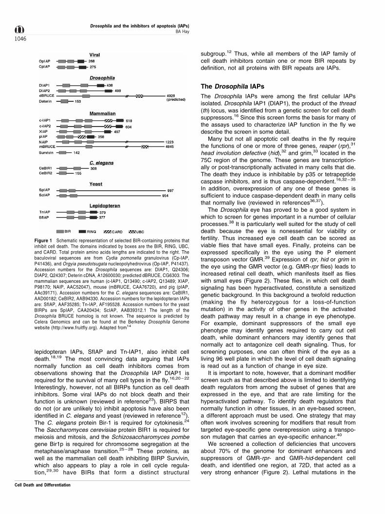
Figure 1 Schematic representation of selected BIR-containing proteins thatinhibit cell death. The domains indicated by boxes are the BIR, RING, UBC,and CARD. Total protein amino acids lengths are indicated to the right. Thebaculoviral sequences are from Cydia pomonella granulovirus (Cp-IAP,P41436), and Orgyia pseudotsugata nucleopolyhedrovirus (Op-IAP, P41437).Accession numbers for the Drosophila sequences are: DIAP1, Q24306;DIAP2, Q24307; Deterin cDNA, A12600030; predicted dBRUCE, CG6303. Themammalian sequences are human (c-IAP1, Q13490; c-IAP2, Q13489; XIAP,P98170; NAIP, AAC52047), mouse (mBRUCE, CAA76720), and pig (pIAP,AAc39171). Accession numbers for the C. elegans sequences are: CeBIR1,AAD00182; CeBIR2, AAB94330. Accession numbers for the lepidopteran IAPsare: SfIAP, AAF35285; Tn-IAP, AF195528. Accession numbers for the yeastBIRPs are SpIAP, CAA20434; ScIAP, AAB39312.1 The length of theDrosophila BRUCE homolog is not known. The sequence is predicted byCelera Genomics and can be found at the Berkeley Drosophila Genomewebsite (http://www.fruitfly.org). Adapted from14
Drosophila and the inhibitors of apoptosis (IAPs)BA Hay
1046
Cell Death and Differentiation
thread (th) locus, which also maps to this region, showed asimilar phenotype (Figure 2). Cloning of thread showed thatit encoded a protein, DIAP1, with a high degree of similarityto the viral IAPs, in that it contained two N-terminal BIRrepeats and a C-terminal RING finger.16 A number ofDIAP1 mutants have also been isolated more recently indominant modifier screens for enhancers and suppressorsof GMR-rpr-, GMR-hid-, and GMR-grim-dependent celldeath.21,22 We and others also identified a secondDrosophila IAP, referred to here as DIAP2, that hasthree N-terminal BIR repeats and a C-terminal RINGfinger.16,41 ± 43 The th locus and hid were also identified ina later eye-based gene overexpression screen for celldeath regulators.40
DIAP1 blocks normally occurring cell death in the flyeye,16 as well as that induced by GMR-driven expression ofrpr, hid16,21,22,44,45 or grim.22 DIAP2 overexpression alsoresults in suppression of these deaths,16,44 though resultswith respect to grim depend on the expression system
(Vernooy and Hay unpublished, GMR-grim, yes;44, GMR-GAL4-UAS grim, no). More recent observations haveshown that DIAP1 and DIAP2 can inhibit cell death inresponse to a variety of stimuli in lepidopteran cells,46 ± 50
and in mammalian cell culture.51
It was observed early on that fragments of DIAP1 andDIAP2 consisting of the BIR repeats, but lacking the RINGfinger, retained the ability to inhibit a number of differentcell deaths.16 These observations provided the firstevidence that the DIAP1 BIR repeats alone were sufficientto inhibit cell death. For DIAP1, the BIR repeats without theRING provided greater protection from several differentdeath stimuli in the fly eye (X-ray irradiation and GMR-hid)than did the full-length protein. This, in conjunction with theobservation that expression of the RING domain in isolationinduced retinal cell death, suggested that the RING domainmight negatively regulate in some way DIAP1's BIR-dependent death inhibition function. More recent observa-tions in lepidopteran cells confirm that the BIR repeatsalone are better death protectors than the full length proteinwith respect to hid,49,50 and that expression of the RINGalone promotes cell death.46 However, in other contexts theDIAP1 BIR repeats protect less well than the full lengthprotein.46,47 Together, these results suggest that DIAP1and DIAP2 inhibit cell death through multiple mechanisms.These are discussed in more detail below.
Overexpression of DIAP1 or DIAP2 is sufficient to blockcell death in the fly, but do these proteins normally performthis function? In the case of DIAP1 the answer is veryclearly yes. DIAP1 is expressed at high levels throughoutembryogenesis16 and in a number of other tissues,including the eye22 and ovary.52 Expression in many othertissues has simply not been examined. Decreasing thedose of DIAP1 by twofold in the eye increases rpr-, hid-,and grim-dependent cell death, indicating that theendogenous levels of DIAP1 are sufficient to act as abrake on induced death. Most striking, however, areobservations of cells homozygous for DIAP1 loss-of-function mutations. Early observations that clones ofDIAP1 mutant cells could not be obtained suggested thatDIAP1 was required for the survival of cells in the eye andovary.16 More recent observations showed that zygoticDIAP1 expression is in fact required for the survival ofprobably all cells in the embryo.20 ± 22 Together, theseobservations suggest that, at least during development,DIAP1 function, or perhaps that of DIAP2 in certaincases,53 is required for the survival of all cells. Inlepidopteran cells, overexpressed DIAP1 and DIAP2 arelocalized in a punctate cytoplasmic pattern.48,49 What thesesites are, and their physiological significance, is unknown.
The normal functions of DIAP2 are more enigmatic.DIAP2 blocks cell death due to overexpression of rpr, hid orgrim very efficiently when overexpressed in the eye or inlepidopteran cells, yet deficiencies that remove this locushave no effect on GMR-rpr- or GMR-hid-dependent retinalcell death.16 This suggests that DIAP2 is either notexpressed in the fly eye (and thus twofold reductions inits genetic dose have no consequence) or that its activity isnot rate limiting, perhaps due to a large excess of DIAP1.Alternatively, endogenous DIAP2 may have other preferred
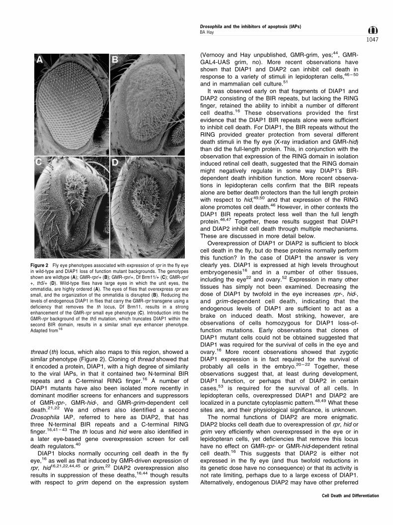
Figure 2 Fly eye phenotypes associated with expression of rpr in the fly eyein wild-type and DIAP1 loss of function mutant backgrounds. The genotypesshown are wildtype (A); GMR-rpr/+ (B); GMR-rpr/+, Df Brm11/+ (C); GMR-rpr/+, th5/+ (D). Wild-type flies have large eyes in which the unit eyes, theommatidia, are highly ordered (A). The eyes of flies that overexpress rpr aresmall, and the organization of the ommatidia is disrupted (B). Reducing thelevels of endogenous DIAP1 in flies that carry the GMR-rpr transgene using adeficiency that removes the th locus, Df Brm11, results in a strongenhancement of the GMR-rpr small eye phenotype (C). Introduction into theGMR-rpr background of the th5 mutation, which truncates DIAP1 within thesecond BIR domain, results in a similar small eye enhancer phenotype.Adapted from16
Cell Death and Differentiation
Drosophila and the inhibitors of apoptosis (IAPs)BA Hay
1047
targets. The most notable feature of DIAP2 expressiondescribed thus far is that it is transcriptionally regulated aspart of a coordinated, ecdysone-dependent response thatleads to cell death of the larval salivary gland.53 DIAP1 andDIAP2 transcription are also regulated during ovariandevelopment and the death of the germline nurse cells.52
Two other proteins with BIR repeats have recently beenidentified in Drosophila. Deterin is a small protein with asingle N-terminal BIR repeat and a C-terminal region thatmay form a coiled coil.17 It is most similar in overallstructure, and within the BIR repeat, to mammalianSurvivin. Deterin has been shown to block rpr-dependentcell death, as well as death due to the act of celltransfection in a Drosophila cell culture system. Thecontexts in which Deterin functions in the fly remain to bedetermined. dBRUCE14 is a predicted protein homolog ofmammalian BRUCE.54 BRUCE and dBRUCE are intriguing,if somewhat daunting proteins. They are most notable fortheir size (predicted to be greater than 500 kD), and thefact that they each have an N-terminal BIR repeat and a C-terminal domain that is predicted to have E2 ubiquitinconjugation activity. In the case of BRUCE this activity hasbeen demonstrated.54 The dBRUCE BIR repeat isnoteworthy because it is much more similar to the BIRrepeat of its mammalian counterpart than the BIR repeatsof other evolutionarily distant IAPs are to each other. As ofyet there is no evidence linking BRUCE or dBRUCEfunction to cell death regulation.
IAPs as caspase inhibitors
Mammalian IAPs Most if not all cells express caspasessuf®cient to carry out apoptosis. Since proteolytic activation isirreversible, and caspases have the ability to engage inamplifying cascades of proteolysis, their activation andactivity must presumably be tightly inhibited in cells thatnormally live. IAPs have been shown to interact with a numberof different proteins (reviewed in references13,15). Amechanism by which a number of these might function toinhibit cell death was originally suggested by the observationsthat several mammalian IAPs inhibit caspase activation oractivity. XIAP, c-IAP1 and c-IAP2 bind and inhibit theactivation of the apical caspase caspase-9. In addition theyalso inhibit active forms of caspases 3 and 7, including anintermediate form in which the large subunit still contains theprodomain and a C-terminal linker that forms the junctionbetween large and small subunits.55 ± 58 XIAP, c-IAP1, and c-IAP2 appear not to interact with caspases-1, -6, -8 or -10.NAIP inhibits cell death in some contexts,42,59,60 but it has notbeen shown to interact with any caspases. It is possible theNAIP caspase partner has not been identi®ed or tested. It hasrecently been suggested that NAIP mediates at least some ofits death protective effects through interactions withHippocalcin, a calcium-binding protein that it binds throughthe third NAIP BIR.61 Several mammalian IAPs, including c-IAP1, c-IAP2 and pIAP have CARD domains (Figure 1). Thesigni®cance of these for caspase inhibition is unclear sinceXIAP, which inhibits multiple caspases, lacks a CARD.Perhaps the IAP CARD mediates interactions with other
apoptotic regulators that have CARD domains (reviewed inreference62).
Survivin inhibits caspase-dependent cell death in anumber of contexts (reviewed in references13,15,63), andhas been immunoprecipitated from cells or reticulocytelysates in association with active caspase-3 and -7.64,65
More recently, others have failed to observe interactionsbetween recombinant Survivin and caspase-3, or any abilityof Survivin to inhibit caspase-3.66 However, these latterobservations leave open the possibility that Survivin in vivois modified in ways that promote its function as a caspaseinhibitor (c.f. reference67). Other models of Survivin functionare presented in the discussion of Deterin function (below).
Drosophila IAPs Drosophila encodes 7 functional caspasesand there is ample evidence that cell death in Drosophila iscaspase-dependent (reviewed in reference14, Kumar et al,this issue). Of the four Drosophila BIRPs, only DIAP1 hasbeen shown to inhibit caspase activity. DIAP1 inhibits thedeath of yeast that express full length DCP-1,68 aconstitutively activated form of drICE in which the large andsmall subunits are rearranged into an active conformation,20
or full length DRONC.69 Since yeast lack a describedapoptotic pathway, it seems likely that DIAP1's ability toinhibit caspase-dependent yeast cell death is due to itsfunction as a caspase inhibitor. Supporting this argument,recombinant DIAP1 has also been shown to inhibit thecaspase activity of recombinant DCP-1,68 as well as theactivity of three other Drosophila caspases, DECAY, DREAMand DAYDREAM (SJ Yoo, unpublished) in in vitro assays. Inaddition, DIAP1 has been shown to bind to certain forms ofdrICE in cell extracts,47 and to interact with the DRONCprodomain, as well as processed forms of DRONC.70
Together, these results suggest that DIAP1 may be able toinhibit the activation or activity of all Drosophila caspases.However, as discussed below in the section on IAPs asubiquitin-protein ligases, it is important to keep in mind that invivo IAPs may inhibit caspase activity through multiplemechanisms. Interactions between DIAP2 and Drosophilacaspases, including DCP-1, drICE and DRONC, have notbeen detected (CJ Hawkins, unpublished).
The best evidence supporting the idea that DIAP1 has aprosurvival function in vivo as a caspase inhibitor comesfrom the observation that DIAP1 mutant embryos, in whichall cells die, have greatly increased levels of caspaseactivity that is DIAP1 inhibitable.20 In addition, DIAP1 hasalso been shown to block DRONC-dependent cell death inthe fly eye,69,70 and drICE-, Sf-caspase-1- and mammaliancaspase-3-dependent lepidopteran cell death.47
What are DIAP1's caspase targets in vivo? In addres-sing this question it is useful to include observations madein lepidopteran cells as well. The Drosophila caspase fieldis still at a very early stage, and possible pathways ofcaspase activation are only just being identified (reviewedin Kumar et al, this volume). In one major pathway ofcaspase activation in mammals, cellular stress of varioussorts leads to the release of mitochondrial cytochrome-c,which binds the cytosolic adaptor protein Apaf-1. Cyto-chrome-c-bound Apaf-1 binds to and activates caspase-9.CARD domains present in Apaf-1 and caspase-9 mediate
Cell Death and Differentiation
Drosophila and the inhibitors of apoptosis (IAPs)BA Hay
1048
interactions between the two proteins (reviewed inreference71). DRONC is likely to be the Drosophilacaspase-9 ortholog because it is the only Drosophilacaspase with a CARD domain,72 and it binds theDrosophila Apaf-1.73 The fact that dominant negativeforms of DRONC inhibit rpr-, hid-, and grim-dependentcell deaths in the fly argues, though not definitively, thatDRONC is an important apical cell death caspase actingdownstream of these genes.69,70 DRONC has severalunusual features. Unlike other caspases, it cleaves afterglutamate as well as aspartate.69 In addition, it is p35-insensitive.69,70 Cell death induced in lepidopteran cells byviral infection or actinomycin D also involves a p35-insensitive proteolytic event.74 ± 76 The caspase thatmediates this cleavage is unknown. Active versions ofDRONC and the DRONC-like lepidopteran caspasepresumably process and activate effector caspases withshort prodomains. Based on patterns of caspase cleavageobserved in vitro,69,70 and in lepidopteran cells,75,76 drICEand Sf-caspase-1 are good candidates, respectively. Otherinsect short prodomain caspases have not been tested.
An important site of DIAP1 prosurvival function is likelyto be inhibition of the p35-insensitive apical caspase.DIAP1 inhibits GMR-DRONC-dependent retinal celldeath.69,70 Since DIAP1 binds the DRONC prodomainand inhibits DRONC-dependent killing of yeast, it seemslikely that DIAP1's effects on DRONC activity in vivo aredirect. These observations, together with those showingthat dominant negative DRONC blocks cell death in flies,suggest that inhibition of DRONC activation and/or activityis likely to be an important DIAP1 prosurvival function. Thesteps at which DIAP1 acts to inhibit DRONC function areunknown. The baculovirus IAPs Op-IAP and Cp-IAP alsoact on a p35-insensitive apical caspase, perhaps aDRONC-like caspase-9 homolog. Thus, Op-IAP and Cp-IAP block the activating cleavage of Sf-caspase-1 betweenthe large and small subunits in lepidopteran cells, while p35fails to do so.75,76 In addition, the lepidopteran IAP, SfIAP,has been shown to inhibit the activation of caspase-9 inmammalian cells, but shows no activity towards the effectorcaspases caspase-3 and caspase-7.18 While SfIAP'sactivity with respect to inhibition of Sf-caspase-1 activationhas not been tested, these results suggest that it (andpresumably also the closely related lepidopteran IAP Tn-IAP1) inhibits the lepidopteran caspase-9 homolog.
DIAP1 may also inhibit the activity of Drosophilacaspases activated by DRONC, or by other longprodomain caspases such as DCP-2/DREDD or DREAM.This point is suggested in Drosophila by the fact that DIAP1inhibits the activity of many Drosophila caspases in vitro. Itis also suggested by the fact that in lepidopteran cellsDIAP1 binds to an active (large-small subunit cleaved)version of drICE generated when the full-length drICEcoding region is introduced into cells experiencing a deathsignal. Interestingly however, DIAP1 does not bind eitherthe full length drICE precursor or active drICE resultingfrom the introduction into cells of plasmids that expressmature large and small subunits.47 These observationssuggest that in vivo DIAP1 inhibits the activity of a drICEprocessing intermediate. Consistent with this hypothesis,
we observed that versions of drICE that lack prodomainsequences are not inhibited by DIAP1,68 while versions thatcontain these sequences are inhibited.20
The fact that DIAP1 is unable to inhibit cell deathinduced by prodomainless GMR-DRONC, or the activity offully mature drICE, indicates that there are points at whichcaspase signaling and effector function can be madeindependent of DIAP1 activity. It will be interesting to seeif the cleavage events that generate these DIAP1-insensitive caspases define points of cell death regulationin vivo.
Functional interactions between DIAPs, Reaper,Hid and Grim and caspases
Death-inhibiting IAPs have been shown to bind a number ofdifferent cellular proteins other than caspases (reviewed inreference13,15). Some of these may identify mechanisms bywhich IAPs inhibit apoptosis, or by which IAP activity isregulated. Others may point towards roles of IAPs in signaltransduction. Here we focus on the Drosophila IAP interactingproteins, Reaper, Hid and Grim, and the mammalian proteinSmac/Diablo, because the functions of these proteins areclearly linked to regulation of cell death. Interactions ofDrosophila IAPs with Doom, a Drosophila-derived apoptosisinducer that binds baculovirus IAPs,77 and with the ThickVeins receptor,78 are not discussed.
Reaper, Hid and Grim promote caspase-dependent celldeath, and they each contain a short region (about 15amino acids) of homology at their N-termini. This sequenceis sufficient to allow binding to DIAP1 and DIAP2.48,49
These sequences also mediate Reaper and Grim binding toc-IAP1 from mammals.79 Domain analysis of Hid and Grimindicates that the presence of these N-terminal sequenceson proteins is sufficient to induce apoptosis in lepidopterancells.49 These observations suggested a model in whichReaper, Hid and Grim promote apoptosis by inhibitingDIAP1 function as a caspase inhibitor, thereby unleashinglatent caspase activity (Figure 3).47 Several observations
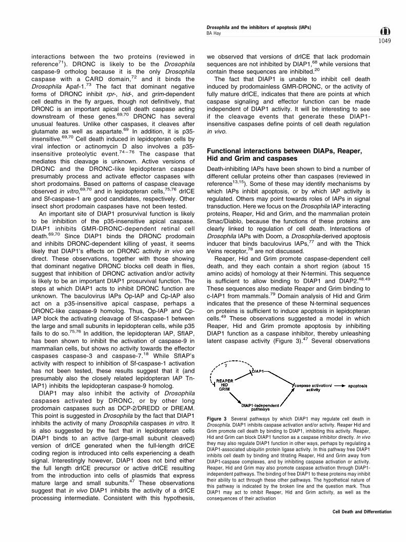
Figure 3 Several pathways by which DIAP1 may regulate cell death inDrosophila. DIAP1 inhibits caspase activation and/or activity. Reaper Hid andGrim promote cell death by binding to DIAP1, inhibiting this activity. Reaper,Hid and Grim can block DIAP1 function as a caspase inhibitor directly. In vivothey may also regulate DIAP1 function in other ways, perhaps by regulating aDIAP1-associated ubiquitin protein ligase activity. In this pathway free DIAP1inhibits cell death by binding and titrating Reaper, Hid and Grim away fromDIAP1-caspase complexes, and by inhibiting caspase activation or activity.Reaper, Hid and Grim may also promote caspase activation through DIAP1-independent pathways. The binding of free DIAP1 to these proteins may inhibittheir ability to act through these other pathways. The hypothetical nature ofthis pathway is indicated by the broken line and the question mark. ThusDIAP1 may act to inhibit Reaper, Hid and Grim activity, as well as theconsequences of their activation
Cell Death and Differentiation
Drosophila and the inhibitors of apoptosis (IAPs)BA Hay
1049
strongly support this model. First and foremost, in in vitroassays containing purified DIAP1 and DCP-1, full lengthHid or an N-terminal cell killing Hid fragment are able toblock DIAP1's ability to inhibit caspase activity.20 Reaperand Grim, Hid and the N-terminal cell killing Hid fragmentalso block DIAP1's ability to inhibit caspase-dependentyeast cell death. Finally, a peptide corresponding to the first18 residues of Grim prevents the lepidopteran IAP SfIAPfrom inhibiting the activation of downstream caspases in amammalian cell extract system.18 These observations allargue that Reaper, Hid and Grim can block DIAP1 andSfIAP function as caspase inhibitors. But do they performthis function in vivo? Reaper, Hid and Grim can induce thedeath of cells that normally live. If inhibition of DIAP1constitutes a primary mechanism by which these genes act,then loss of DIAP1 in cells that normally live should lead toa similar phenotype: cell death associated with increasedcaspase activity. As discussed above, at least in theembryo this is in fact the case.20 Finally, this model predictsthat versions of DIAP1 that do not bind Reaper, Hid or Grimshould be relatively insensitive to the death-promotingactivity of these proteins. Such mutants should beidentified in eye-based dominant modifier screen as deathsuppressors. Several such mutants have in fact beenidentified.21 While it is possible that these mutant proteinsare simply better caspase inhibitors than the wildtypeDIAP1, these results are consistent with the idea that themutant DIAP1 proteins are more potent suppressors of rprand grim-dependent cell death because they interact withthese proteins less well.
Apoptotic cell death is an evolutionarily old process.Thus it is expected that molecules and mechanisms of celldeath regulation found in one group of organisms may bepresent in others as well. Recent observation suggest thatthis is true as well for regulation of productive IAP-caspaseinteractions. Thus, several groups recently identified aprotein known as Smac,80 or DIABLO,81 that binds anumber of different mammalian and baculoviral IAPs.Smac/Diablo is initially localized to mitochondria. Followingan apoptotic stimulus it is released into the cytoplasm alongwith cytochrome c, where it binds IAPs. In addition,overexpression of Smac/Diablo sensitizes cells to differentapoptotic stimuli. These observations argue strongly thatSmac/Diablo, like Reaper, Hid and Grim, suppresses IAPfunction. How Smac/Diablo achieves this is unclear, but theobservation that Smac/DIABLO, like Reaper, Hid and Grim,inhibits the ability of IAPs to block caspase-dependentyeast cell death, suggests their mechanism(s) of actionmay be similar.81 (See Note added in proof.) Theidentification of Smac/Diablo leaves open the possibilitythat there are additional mammalian proteins that inhibitIAP function. There are several reasons to consider this.First, Smac/Diablo has a restricted tissue distribution.Second, Smac/Diablo is localized to mitochondria. Fromthis site it is expected to promote apoptosis as a part of thecaspase-9 activation cascade, inhibiting IAP functions thatwould antagonize cytochrome-c and Apaf-1-dependentcaspase-9 activation. However, it seems very reasonableto imagine that there will exist other, perhaps cytoplasmicproteins that perform a similar function in the context of
other signal transduction pathways that regulate cell death.Hid orthologs are a particularly attractive possibility sinceHid is regulated both transcriptionally82 and post-transcrip-tionally45 by the Ras/Map kinase pathway, which playsmany roles in regulating decisions of cell fate and survival.
The above observations suggest that overexpression ofDIAP1 inhibits cell death through several mechanisms: byacting as a sink for Reaper, Hid and Grim, titrating themaway from interactions with DIAP1 bound to caspase, andby directly inhibiting caspase activation or activity. DIAP1binding to Reaper, Hid and Grim may also serve to inhibitother proapoptotic functions of these proteins (Figure 3).Peptides corresponding to the N-termini of Reaper andGrim have been shown to inhibit the activation of potassiumchannels.83 This might promote depolarization-dependentcell death. Binding of Reaper, Hid or Grim to cytoplasmicDIAP1 via their N-terminal sequences might be expected toprevent functional interactions of these same sequenceswith membrane channel proteins. Reaper, Hid and Grimalso interact in a Xenopus extract system with the Scytheprotein.84 These interactions do not require the conservedReaper, Hid, Grim N-terminal sequences. In the case of atleast Reaper, binding to Scythe is associated with releaseof a cytoplasmic factor that promotes the release ofcytochrome c from mitochondria. Drosophila has a Scythehomolog,14 suggesting that one or more of Reaper, Hid orGrim may promote apoptosis through a Scythe-dependentpathway. At this point it is unclear if binding of Reaper, Hidor Grim to DIAP1 prevents productive interactions withScythe. However, as discussed below, the observation thatRING finger-containing IAPs can have ubiquitin ligaseactivity towards bound substrates suggests anothermechanism by which DIAP1 or DIAP2 might inhibitReaper-, Hid- or Grim-dependent cell death: by promotingtheir ubiquitination and degradation.
IAPs as ubiquitin-protein ligases
Many proteins are modified by the covalent attachment of oneor more copies of ubiquitin, a 76-amino acid protein. In manycases ubiquitination leads to subsequent degradation of themodified protein by the 26S proteosome (reviewed inreference85). Attachment of ubiquitin to a substrate typicallyinvolves the action of three enzymes, a ubiquitin activatingenzyme (E1), a ubiquitin carrier protein (E2), and a ubiquitin-protein ligase (E3). Free ubiquitin is bound to E1 by a thioesterbond. This activated ubiquitin is then transferred to E2 througha second thioester bond formed between ubiquitin and E2. Ina third step, E2, in conjunction with substrate bound E3,transfers ubiquitin to target proteins. There is one E1, butthere are many E2s, E3s or E3 multiprotein complexes.Recently it has been recognized that RING fingers of anumber of different proteins have E3 ubiquitin-protein ligaseactivity (reviewed in references11,86). Several IAPs, c-IAP1and XIAP, are among these. Thus, in the presence of E1 andE2, c-IAP1 and XIAP are able in vitro to catalyze auto-ubiquitination. In addition, deletion or mutation of the RINGsuppresses IAP ubiquitination and degradation in vivo.Intriguingly, full length, but not RING mutated versions ofthese IAPs disappear in thymocytes induced to die,
Cell Death and Differentiation
Drosophila and the inhibitors of apoptosis (IAPs)BA Hay
1050
suggesting that stimulation of IAP auto-ubiquitination definesa mechanism for promoting cell death.87 The nature of thesignals that promote IAP auto-ubiquitination in thymocytes areunknown. c-IAP1 has also recently been shown to mono-ubiquitinate caspase-3 and caspase-7 in vitro.88 Thephysiological significance of this reaction is unclear, but itdoes suggest the possibility that RING-containing IAPs mayubiquitinate and promote the degradation of bound proapop-totic substrates. This would provide a mechanism wherebyIAPs could inhibit the activity of apoptosis inducerscatalytically, rather than stoichometrically.
At this point it is not known if DIAP1 has E3 activity. Butit seems likely that it does. There are several nonexclusivemodels for how RING-dependent ubiquitin ligase activitymight regulate DIAP1 function. One possibility is that RING-dependent DIAP1 auto-ubiquitination, as with c-IAP1 andXIAP, promotes DIAP1 degradation. Stimuli that promotethis activity would then have the effect of decreasing IAPlevels, thus sensitizing cells to lower levels of inducers orcaspase activity. What might define an auto-ubiquitination-promoting stimulus? Perhaps Reaper, Hid or Grim bindingto DIAP1. DIAP1 E3 activity might also promote thedegradation of bound apoptosis inducers such as Reaper,Hid, Grim or caspases. In this model IAP ubiquitin ligaseactivity would promote DIAP1's prosurvival function. In thecontext of this hypothesis it is interesting to note that inlepidopteran cells expression of Op-IAP or Cp-IAP isassociated with the formation of a ladder of highermolecular weight forms of Reaper and Hid.48,49 Finally, ina substrate choice model DIAP1 may have both activities.DIAP1 might, for example auto-ubiquitinate itself when it isfree, but preferentially ubiquitinate other substrates whenthey are DIAP1-bound. In this way DIAP1 levels would beregulated according to the cell's current levels ofproapoptotic DIAP1-interacting proteins. Stimuli that alterthe ratio of cis to trans ubiquitination would promote orsuppress apoptosis, respectively. These models are allspeculative, but they are also eminently testable.
Deterin cell death while Survivin the cell cycle
The small, single BIR mammalian IAP Survivin has excitedmuch interest recently because its transcription is upregulatedin many human cancers.89 In normal cells Survivin expressionis upregulated in G2/M, and it is essentially absent inpostmitotic cells. Survivin inhibits cell death in culture inresponse to a number of different stimuli, including expressionof caspases. In addition, antisense Survivin promotescaspase activity and cell death (reviewed in reference13).Finally, as noted above, Survivin has been found by some, butnot others, to bind directly to caspases. Thus, Survivin is atumor marker, and potentially a good therapeutic cancertarget since it is not appreciably expressed in normal postmitotic cells. However, there is also good evidence thatSurvivin may not function only as a cell death inhibitor(reviewed in reference63). The Survivin BIR repeat is mostclosely related to those found in BIRPs of C. elegans, S.cerevisiae and S. pombe. Genetic analysis indicates thatthese proteins promote cytokinesis and chromosomesegregation . The C. elgans protein BIR1 does not inhibit
cell death,24 and it seems unlikely that the yeast proteins doeither. Survivin can partly rescue the C. elegans cytokinesisdefect in bir1 mutants, suggesting some conservation infunction. Survivin also localizes to the mitotic spindle inanaphase,30 as does the S. cerevisiae protein Bir1p.25 Finally,loss of Survivin function leads to a number of cell divisiondefects, including hyperploidy, multinucleation, and extracentrosomes, all consistent with defects in cytokinesis.30
A domain analysis of Survivin indicates that it localizesto spindle microtubules via interactions with its C-terminalcoiled-coil domain. The importance of this localization issuggested by the fact that deletion of the coiled-coil domainleads to a loss of antiapoptotic function in at least somecontexts. Mutation of the BIR domain also leads to a loss ofantiapoptotic function, and creates a molecule that hasdominant negative activity, in that its expression promotescell death and displaces wild-type Survivin away fromspindles.29
How is Survivin regulating cell death? One possibility isthat caspase activity occurs as a normal part of the cellcycle, and that Survivin functions to regulate this activity.This caspase activity might be required for some aspect ofcell division. Other observations suggest that Survivin mightregulate caspase activity indirectly. Survivin has beendescribed as translocating to the nucleus in response tosome death signals, there binding the cyclin-dependentkinase 4 (CDK4).90 CDK4 itself can bind the CDK inhibitor,p21. p21 has been shown to have antiapoptotic activity in anumber of systems (reviewed in reference91). While at leastsome of this activity may derive from p21's ability to inhibitproapoptotic activities associated with inappropriate CDKactivation, p21 has also been described as an inhibitor ofthe activation of mitochondrially localized procaspase-3.92
Based on these observations it has been argued thatSurvivin might promote survival by promoting the release ofp21 from CDK4. Survivin may also play a checkpoint role inmonitoring the status of spindle assembly. In this model,defective spindle assembly would lead to delocalization ofSurvivin from microtubules, and thus loss of its ability toinhibit caspase activity. The fact that a fraction of caspase-3 is localized to centrosomes (where Survivin is alsopresent) in a Survivin-dependent manner supports thispossibility.30 Alternatively, or in addition, Survivin may bean essential part of the machinery that carries out celldivision. In this model, loss of Survivin function leads to cellcycle defects that indirectly promote caspase activation.
There are a number of important unanswered questions.Survivin is clearly able to inhibit cell death. Thus far thisactivity has only been shown in cycling cells in culture. Doesit have activity in noncycling cells, or is it active in only onepart of the cell cycle? What are the proteins Survivininteracts with that mediate its localization to spindles andcentrosomes? Is caspase activation necessary for cell cycleprogression? What is the source of the caspase activityfound in cells with decreased Survivin function? Is it theresult of loss of Survivin the caspase inhibitor or asecondary consequence of Survivin-dependent cell cycledefects? Drosophila provides a convenient system in whichto address these questions. But is Drosophila Deterin aSurvivin homolog? Several observations suggest that it may
Cell Death and Differentiation
Drosophila and the inhibitors of apoptosis (IAPs)BA Hay
1051
be. In early and mid embryogenesis Deterin mRNAexpression shows no correlation with cell proliferation.However, somewhat later Deterin becomes upregulated inthe developing gonad, a proliferative tissue. Survivinexpression in other proliferative tissues, in particular theimaginal discs, has not been examined. The subcellularlocalization of the Deterin protein has also not beenexamined. However, Deterin clearly functions as a celldeath inhibitor, at least in cells in culture. In addition, adomain analysis of Deterin suggests that, like Survivin, boththe N-terminal BIR domain and the C-terminal domain arerequired for optimal function, and that mutation or removal ofthe BIR leads to the creation of a protein with dominantnegative activity. Thus far, all manipulations of Survivin andDeterin function have been carried out in cell culture.However, ultimately it is important to examine the functionof these proteins in cells functioning as a part of a tissue.Overexpression of the wild-type Deterin protein and thedominant negative version, as well as the use of RNAiknockout technology, should lead very quickly to anunderstanding of the consequences of manipulating Deterinlevels in the fly. A characterization of these phenotypes inthe presence and absence of caspase inhibitors should bevery illuminating in uncovering roles Deterin plays as a cellcycle regulator and as a cell death inhibitor.
Structure-function analysis of the mammalian IAPs
The sequence requirements for caspase inhibition have beenexplored in mammals for XIAP and c-IAP1. Full length XIAPand c-IAP1 and c-IAP2 inhibit caspase-3 and caspase-7, andthe activation of caspase-9.55 ± 57 A fragment consisting of
BIR2 and about 30 residues N- and C-terminal (R124-P260) issufficient for this activity.93 Inhibition of caspase-9 activationby XIAP requires both BIR3 as well as the RING finger (A243-end).58 Since the caspase activity assays used do not supportRING-dependent ubiquitination, the RING must play a bindingor structural role in this context. A short sequence N-terminalto BIR2 (E134-Y154) plays a critical role in XIAP-dependentcaspase inhibition.94 This N-terminal extension contains apotential caspase target site (DISD), the P4 aspartate ofwhich is required for BIR2-dependent caspase inhibition(Figure 4). Several lines of evidence suggest a model inwhich this fragment binds caspase-3 in the active site(presumably via the DISD residues), perhaps in conjunctionwith a lower affinity caspase binding interaction mediated bythe BIR domain. However, this simple interpretation iscomplicated by the fact that mutation of the putative P1aspartate in the DISD sequence to alanine (DISA) causesonly a threefold decrease in activity as a caspase inhibitor. Inaddition, the P1 aspartate is not conserved in other IAPs,leaving the generality of the proposed mechanism unclear. Anumber of other IAPs, including DIAP1, Op-IAP, Cp-IAP andDIAP2, have a conserved aspartate just N-terminal to the BIR(DIAP1 D40). However, the significance of this aspartate orothers nearby in the linker region has not been tested. AnNMR structural analysis of a fragment of c-IAP1 consisting ofBIR3 and a C-terminal linker has suggested that a conservedaspartate (D302) positioned within a BIR surface loop mightalso mediate interactions with caspases.95
Insect and viral IAPs It has been clear for some time thatwhile insect and baculoviral IAP have prosurvival functions ascaspase inhibitors, they also have important functional
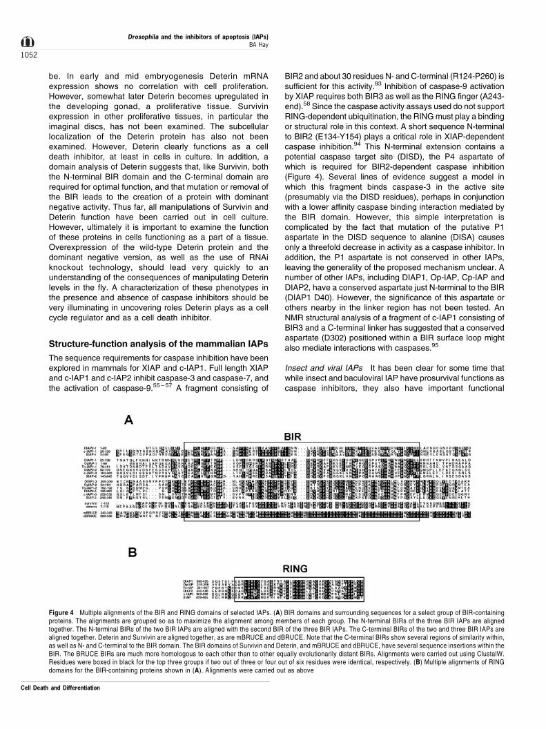
Figure 4 Multiple alignments of the BIR and RING domains of selected IAPs. (A) BIR domains and surrounding sequences for a select group of BIR-containingproteins. The alignments are grouped so as to maximize the alignment among members of each group. The N-terminal BIRs of the three BIR IAPs are alignedtogether. The N-terminal BIRs of the two BIR IAPs are aligned with the second BIR of the three BIR IAPs. The C-terminal BIRs of the two and three BIR IAPs arealigned together. Deterin and Survivin are aligned together, as are mBRUCE and dBRUCE. Note that the C-terminal BIRs show several regions of similarity within,as well as N- and C-terminal to the BIR domain. The BIR domains of Survivin and Deterin, and mBRUCE and dBRUCE, have several sequence insertions within theBIR. The BRUCE BIRs are much more homologous to each other than to other equally evolutionarily distant BIRs. Alignments were carried out using ClustalW.Residues were boxed in black for the top three groups if two out of three or four out of six residues were identical, respectively. (B) Multiple alignments of RINGdomains for the BIR-containing proteins shown in (A). Alignments were carried out as above
Cell Death and Differentiation
Drosophila and the inhibitors of apoptosis (IAPs)BA Hay
1052
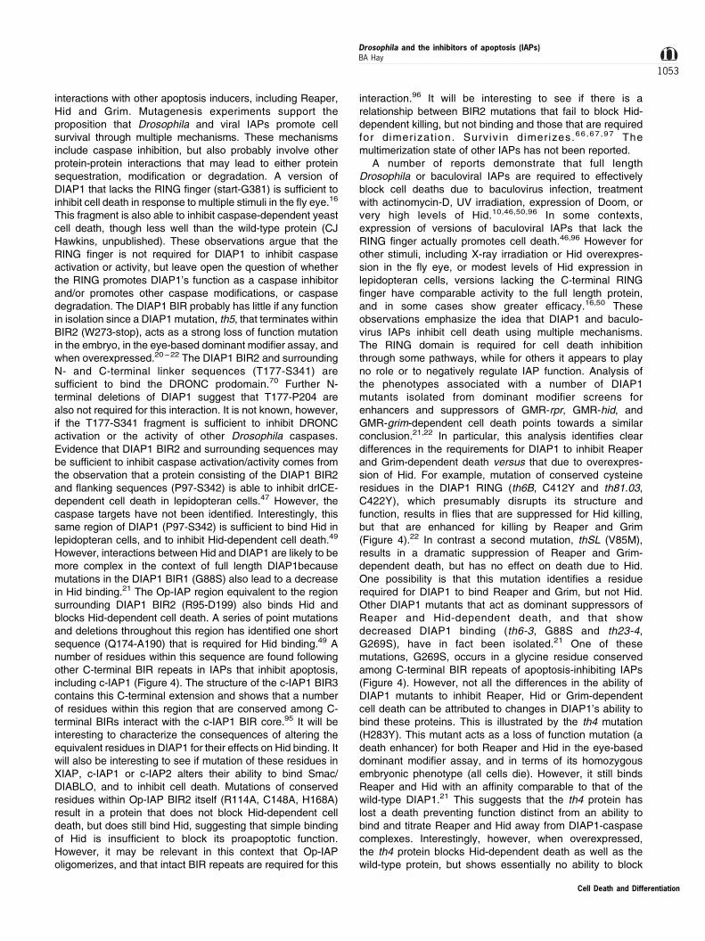
interactions with other apoptosis inducers, including Reaper,Hid and Grim. Mutagenesis experiments support theproposition that Drosophila and viral IAPs promote cellsurvival through multiple mechanisms. These mechanismsinclude caspase inhibition, but also probably involve otherprotein-protein interactions that may lead to either proteinsequestration, modi®cation or degradation. A version ofDIAP1 that lacks the RING ®nger (start-G381) is suf®cient toinhibit cell death in response to multiple stimuli in the ¯y eye.16
This fragment is also able to inhibit caspase-dependent yeastcell death, though less well than the wild-type protein (CJHawkins, unpublished). These observations argue that theRING ®nger is not required for DIAP1 to inhibit caspaseactivation or activity, but leave open the question of whetherthe RING promotes DIAP1's function as a caspase inhibitorand/or promotes other caspase modi®cations, or caspasedegradation. The DIAP1 BIR probably has little if any functionin isolation since a DIAP1 mutation, th5, that terminates withinBIR2 (W273-stop), acts as a strong loss of function mutationin the embryo, in the eye-based dominant modi®er assay, andwhen overexpressed.20 ± 22 The DIAP1 BIR2 and surroundingN- and C-terminal linker sequences (T177-S341) aresuf®cient to bind the DRONC prodomain.70 Further N-terminal deletions of DIAP1 suggest that T177-P204 arealso not required for this interaction. It is not known, however,if the T177-S341 fragment is suf®cient to inhibit DRONCactivation or the activity of other Drosophila caspases.Evidence that DIAP1 BIR2 and surrounding sequences maybe suf®cient to inhibit caspase activation/activity comes fromthe observation that a protein consisting of the DIAP1 BIR2and ¯anking sequences (P97-S342) is able to inhibit drICE-dependent cell death in lepidopteran cells.47 However, thecaspase targets have not been identi®ed. Interestingly, thissame region of DIAP1 (P97-S342) is suf®cient to bind Hid inlepidopteran cells, and to inhibit Hid-dependent cell death.49
However, interactions between Hid and DIAP1 are likely to bemore complex in the context of full length DIAP1becausemutations in the DIAP1 BIR1 (G88S) also lead to a decreasein Hid binding.21 The Op-IAP region equivalent to the regionsurrounding DIAP1 BIR2 (R95-D199) also binds Hid andblocks Hid-dependent cell death. A series of point mutationsand deletions throughout this region has identi®ed one shortsequence (Q174-A190) that is required for Hid binding.49 Anumber of residues within this sequence are found followingother C-terminal BIR repeats in IAPs that inhibit apoptosis,including c-IAP1 (Figure 4). The structure of the c-IAP1 BIR3contains this C-terminal extension and shows that a numberof residues within this region that are conserved among C-terminal BIRs interact with the c-IAP1 BIR core.95 It will beinteresting to characterize the consequences of altering theequivalent residues in DIAP1 for their effects on Hid binding. Itwill also be interesting to see if mutation of these residues inXIAP, c-IAP1 or c-IAP2 alters their ability to bind Smac/DIABLO, and to inhibit cell death. Mutations of conservedresidues within Op-IAP BIR2 itself (R114A, C148A, H168A)result in a protein that does not block Hid-dependent celldeath, but does still bind Hid, suggesting that simple bindingof Hid is insuf®cient to block its proapoptotic function.However, it may be relevant in this context that Op-IAPoligomerizes, and that intact BIR repeats are required for this
interaction.96 It will be interesting to see if there is arelationship between BIR2 mutations that fail to block Hid-dependent killing, but not binding and those that are requiredfor dimer izat ion. Surviv in dimerizes.66 ,6 7 ,97 Themultimerization state of other IAPs has not been reported.
A number of reports demonstrate that full lengthDrosophila or baculoviral IAPs are required to effectivelyblock cell deaths due to baculovirus infection, treatmentwith actinomycin-D, UV irradiation, expression of Doom, orvery high levels of Hid.10,46,50,96 In some contexts,expression of versions of baculoviral IAPs that lack theRING finger actually promotes cell death.46,96 However forother stimuli, including X-ray irradiation or Hid overexpres-sion in the fly eye, or modest levels of Hid expression inlepidopteran cells, versions lacking the C-terminal RINGfinger have comparable activity to the full length protein,and in some cases show greater efficacy.16,50 Theseobservations emphasize the idea that DIAP1 and baculo-virus IAPs inhibit cell death using multiple mechanisms.The RING domain is required for cell death inhibitionthrough some pathways, while for others it appears to playno role or to negatively regulate IAP function. Analysis ofthe phenotypes associated with a number of DIAP1mutants isolated from dominant modifier screens forenhancers and suppressors of GMR-rpr, GMR-hid, andGMR-grim-dependent cell death points towards a similarconclusion.21,22 In particular, this analysis identifies cleardifferences in the requirements for DIAP1 to inhibit Reaperand Grim-dependent death versus that due to overexpres-sion of Hid. For example, mutation of conserved cysteineresidues in the DIAP1 RING (th6B, C412Y and th81.03,C422Y), which presumably disrupts its structure andfunction, results in flies that are suppressed for Hid killing,but that are enhanced for killing by Reaper and Grim(Figure 4).22 In contrast a second mutation, thSL (V85M),results in a dramatic suppression of Reaper and Grim-dependent death, but has no effect on death due to Hid.One possibility is that this mutation identifies a residuerequired for DIAP1 to bind Reaper and Grim, but not Hid.Other DIAP1 mutants that act as dominant suppressors ofReaper and Hid-dependent death, and that showdecreased DIAP1 binding (th6-3, G88S and th23-4,G269S), have in fact been isolated.21 One of thesemutations, G269S, occurs in a glycine residue conservedamong C-terminal BIR repeats of apoptosis-inhibiting IAPs(Figure 4). However, not all the differences in the ability ofDIAP1 mutants to inhibit Reaper, Hid or Grim-dependentcell death can be attributed to changes in DIAP1's ability tobind these proteins. This is illustrated by the th4 mutation(H283Y). This mutant acts as a loss of function mutation (adeath enhancer) for both Reaper and Hid in the eye-baseddominant modifier assay, and in terms of its homozygousembryonic phenotype (all cells die). However, it still bindsReaper and Hid with an affinity comparable to that of thewild-type DIAP1.21 This suggests that the th4 protein haslost a death preventing function distinct from an ability tobind and titrate Reaper and Hid away from DIAP1-caspasecomplexes. Interestingly, however, when overexpressed,the th4 protein blocks Hid-dependent death as well as thewild-type protein, but shows essentially no ability to block
Cell Death and Differentiation
Drosophila and the inhibitors of apoptosis (IAPs)BA Hay
1053
Reaper-dependent death. Why might this be? Mutationalanalysis of Reaper and Grim argues strongly that theseproteins promote apoptosis through multiple pathways.Thus, deletion of the Reaper- or Grim N-terminus, whichmediates binding to DIAP1, results in proteins that can stillkill.44,49,98 In contrast, the results of experiments inlepidopteran cells argue that the N-terminus of Hid is bothnecessary and sufficient to mediate Hid's proapoptoticfunction.49 This suggests that Hid acts through a morelimited set of pathways. One obvious possibility is that theth4 protein lacks the ability to inhibit caspase activityactivated downstream of Reaper and Grim functions thatpromote caspase activation through DIAP1-independentpathways. This scenario would also explain why the th4protein acts as a dominant enhancer of Hid-dependentdeath in the eye, and as a loss of function mutant in theembryo: it is unable to inhibit caspase activation/activityoccurring downstream of high level Hid expression in theeye, or the activation/activity of caspases present in theearly embryo.
In summary, mutational analysis of DIAP1 and baculo-virus IAPs demonstrates that these proteins are functionallycomplex, and shows that there are distinct sequencerequirements for inhibition of different death signals. Asdiscussed above, these IAPs may inhibit apoptosis bysequestering apoptosis inducers and/or blocking caspaseactivation/activity. The RING domain may participate as astructural element in these activities. In addition, it maymediate ubiquitination of bound substrates. Complicatingthe mutational analysis even further, the RING mayregulate (presumably negatively) IAP function throughauto-ubiquitination, which may or may not itself beregulated by the presence of bound substrates. Geneticscreens provide a powerful approach to identifyingfunctionally important DIAP1 mutations. However, anumber of different assays, including binding, caspaseactivity, ubiquitination, expression level and cellularlocalization will be needed to turn these mutants into clearmechanistic models of IAP function and regulation.
Where do we go from here?
It is clear that IAPs regulate cell death through multiplemechanisms. Many act as caspase inhibitors. However forothers such as Survivin and NAIP, this has not been shown.They may regulate death through other mechanisms. Thefinding that some RING-containing IAPs have E3 ubiquitinligase has opened up a new and exciting area of study. Thepotential exists for ubiquitination to both positively andnegatively regulate IAP function. In addition, it is importantto keep in mind that ubiquitination requires the activity of anE2 ubiquitin carrier protein, of which there are a growingnumber. The ubiquitin pathway is also regulated by the activityof a number of deubiquitinating enzymes. Therefore, it seemslikely that new and exciting regulatory circuits involving IAPsand ubiquitination will emerge over the next few years. Thestudy of insect and baculoviral IAPs has led the way inidentifying proteins, Reaper, Hid and Grim, that regulate IAPfunction. These proteins can suppress the ability of IAPs toinhibit caspase activity. However, given the functional
complexity of the insect IAPs (and presumably theirmammalian counterparts), it is very possible that they alterIAP function in other ways as well. A further characterizationof these interactions is likely to lead to new mechanisticinsights into IAP function and regulation. Finally, since thisarticle focuses on Drosophila, it is important to point out thepotential of Drosophila genetics to uncover new aspects ofIAP function. The dominant modifier screen that led to theoriginal identification of DIAP1, and to the isolation of anumber of interesting DIAP1 mutants is a powerful toolbecause it is a function-based screen. There is every reasonto believe that further genetic screens for enhancers andsuppressors of Reaper, Hid or Grim-dependent cell death willuncover new aspects of IAP function.
Note added in proofSeveral recent reports99,100 demonstrate that, as with Reaper, Hid andGrim in Drosophila, Smac/DIABLO promote the activation and activity ofmultiple caspases by inhibiting IAP function.
AcknowledgmentsSpecial thanks to S Vernooy for help with the ®gures as well as commentson the text. This work was supported by grants from a BurroughsWellcome Fund New Investigator Award in the pharmacologicalSciences, the Ellison Medical Foundation, and NIH grant GM057422-01.
References
1. Clem RJ, Fechneimer M and Miller LK (1991) Prevention of apoptosis by a
baculovirus gene during infection of insect cells. Science 254: 1388 ± 1390
2. Clem RJ, Hardwick JM and Miller LK (1996) Anti-apoptotic genes of
baculoviruses. Cell Death Differ. 3: 9 ± 16
3. Ekert PG, Silke J and Vaux DL (1999) Caspase inhibitors. Cell Death Differ. 6:
1081 ± 1086
4. Bump NJ, Hackett M, Hugunin M, Seshagiri S, Brady K, Chen P, Ferenz C,Franklin S, Ghayur T, Li P, Licari P, Mankovich J, Shi LF, Greenberg AH, Miller
LK and Wong WW (1995) Inhibition of ICE family proteases by baculovirus
antiapoptotic protein p35. Science 269: 1885 ± 1888
5. Xue Dand Horvitz HR (1995) Inhibition of the Caenorhabditiselegans cell-death
protease CED-3 by a CED-3 cleavage site in baculovirus p35 protein. Nature
377: 248 ± 251
6. Ayres MD, Howard SC, Kuzio J, Lopez-Ferber M and Possee RD (1994) The
complete DNA sequence of Autographa californica nuclear polyhedrosis virus.
Virology 202: 586 ± 605
7. Du Q, Lehavi D, Faktor O, Qi Y and Chejanovsky N (1999) Isolation of an
apoptosis suppressor gene of the Spodoptera littoralis Nucleopolyhedrovirus.
J. Virol. 73: 1278 ± 1285
8. Crook NE, Clem RJ and Miller LK (1993) An apoptosis-inhibiting baculovirus
gene with a zinc finger like motif. J. Virol. 67: 2168 ± 2174
9. Birnbaum MJ, Clem RJ and Miller LK (1994) An apoptosis-inhibiting gene from a
nuclear polyhedrosis virus encoding a polypeptide with Cys/His sequence
motifs. J. Virol. 68: 2521 ± 2528
10. Clem RJ and Miller LK (1994) Control of programmed cell death by thebaculoviral genes p35 and iap. Mol. Cell. Biol. 14: 5212 ± 5222
11. Freemont PS (2000) Ubiquitination: RING for destruction? Curr. Biol. 10: 84 ±
87
12. Uren AG, Coulson EJ and Vaux DL (1998) Conservation of baculovirus inhibitor
of apoptosis repeat proteins (BIRPs) in viruses, nematodes, vertebrates and
yeasts. Trends Biochem. Sci. 23: 159 ± 162
Cell Death and Differentiation
Drosophila and the inhibitors of apoptosis (IAPs)BA Hay
1054
13. Deveraux QL and Reed JC (1999) IAP family proteins ± suppressors of
apoptosis. Genes Dev. 13: 239 ± 252
14. Vernooy SY, Copeland J, Ghaboosi N, Griffin EE, Yoo SJ and Hay BA (2000)
Cell death regulation in Drosophila: conservation of mechanism and unique
insights. J. Cell Biol. 150: F69 ± F75
15. LaCasse EC, Baird S, Korneluk RG and MacKenzie AE (1998) The inhibitors of
apoptosis (IAPs) and their emerging role in cancer. Oncogene 17: 3247 ± 3259
16. Hay BA, Wassarman DA and Rubin GM (1995) Drosophila homologs ofbaculovirus inhibitor of apoptosis proteins function to block cell death. Cell 83:
1253 ± 1262.
17. Jones G, Jones D, Zhou L, Steller H and Chu Y (2000) Deterin, a new
inhibitor of apoptosis from Drosophila melanogaster. J. Biol. Chem. 275:
22157 ± 22165
18. Huang Q, Deveraux QL, Maeda S, Salvesen GS, Stennicke HR, Hammock BD
and Reed JC (2000) Evolutionary conservation of apoptosis mechanisms:
Lepidopteran and baculoviral inhibitor of apoptosis proteins are inhibitors of
mammalian caspase-9. Proc. Natl. Acad. Sci. USA 97: 1427 ± 1432
19. Seshagiri S, Vucic D, Lee J and Dixit VM (1999) Baculovirus-based genetic
screen for antiapoptotic genes identifies a novel IAP. J. Biol. Chem. 274:
36769 ± 36773
20. Wang SL, Hawkins CJ, Yoo SJ, Muller HA and Hay BA (1999) The Drosophila
caspase inhibitor DIAP1 is essential for cell survival and is negatively regulated
by HID. Cell 98: 453 ± 463
21. Goyal L, McCall K, Agapite J, Hartwieg E and Steller H (2000) Induction of
apoptosis by Drosophila reaper, hid and grim through inhibition of IAP function.
EMBO J. 19: 589 ± 59722. Lisi S, Mazzon L and White W (2000) Diverse domains of THREAD/DIAP1 are
required to inhibit apoptosis induced by REAPER and HID in Drosophila.
Genetics 154: 669 ± 678
23. Clem RJ and Duckett CS (1997) The iap genes: unique arbitors of cell death.
Trends Cell Biol. 7: 337 ± 339
24. Fraser AG, James C, Evan GI and Hengartner MO (1999) Caenorhabditis
elegans inhibitor of apoptosis protein (IAP) homologue BIR-1 plays a
conserved role in cytokinesis. Curr. Biol. 9: 292 ± 301
25. Uren AG, Beilharz T, O'Connell MJ, Bugg SJ, van Driel R, Vaux DL and Lithgow
T (1999) Role for yeast inhibitor of apoptosis (IAP)-like proteins in cell division.
Proc. Natl. Acad. Sci. USA 96: 10170 ± 10175
26. Rajagopalan S and Balasubramanian MK (1999) S-pombe Pbh1p: an inhibitor
of apoptosis domain containing protein is essential for chromosome
segregation. FEBS Letters 460: 187 ± 190
27. Yoon HJ and Carbon J (1999) Participationof BIR1p, amemberof the inhibitor of
apoptosis family, in yeast chromosome segregation events. Proc. Natl. Acad.
Sci. USA 96: 13208 ± 13213
28. Li F, Flanary PL, Altieri DC and Dohlman HG (2000) Cell division regulation byBIR1, a member of the inhibitor of apoptosis family in yeast. J. Biol. Chem. 275:
6707 ± 6711
29. Li F, Ambrosini G, Chu EY, Plescia J, Tognin S, Marchisio PC and Altieri DC
(1998) Control of apoptosis and mitotic spindle checkpoint by survivin. Nature
396: 580 ± 584
30. Li F, Ackermann EJ, Bennett CF, Rothermel AL, Plescia J, Tognin S, Villa A,
Marchisio PC and Altieri DC (1999) Pleiotropic cell-division defects and
apoptosis induced by interference with survivin function. Nature Cell Biol. 1:
461 ± 466
31. White K, Grether ME, Abrams JM, Young L, Farrell K and Steller H (1994)
Genetic control of programmed cell death in Drosophila. Science 264: 677 ±
683
32. Grether ME, Abrams JM, Agapite J, White K and Steller H (1995) The head
involutiondefective gene of Drosophila melanogaster functions in programmed
cell death. Genes Dev. 9: 1694 ± 1708
33. Chen P, Nordstrom W, Gish B and Abrams JM (1996) grim, a novel cell death
gene in Drosophila. Genes Dev. 10: 1773 ± 1782
34. Pronk GJ, Ramer K, Amiri P and Williams LT (1996) Requirement of an ICE-like
protease for induction of apoptosis and ceramide generation by REAPER.Science 271: 808 ± 810
35. White K, Tahaoglu E and Steller H (1996) Cell killing by the Drosophila gene
reaper. Science 271: 805 ± 807
36. Abrams JM (1999) An emerging blueprint for apoptosis in Drosophila. Trends
Cell Biol. 9: 435 ± 440
37. Bangs P and White K (2000) Regulation and execution of apoptosis during
Drosophila development. Developmental Dynamics 218: 68 ± 79
38. Thomas BI and Wassarman DA (1999) A fly's eye view of biology. Trends
Genetics 15: 184 ± 190
39. Hay BA, Wolff T and Rubin GM (1994) Expression of baculovirus P35 prevents
cell death in Drosophila. Development 120: 2121 ± 2129
40. Hay BA, Maile R and Rubin GM (1997) P element insertion-dependent
gene activation in the Drosophila eye. Proc. Natl. Acad. Sci. USA 94:
5195 ± 5200
41. Duckett CS, Nava VE, Gedrich RW, Clem RJ, Van Dongen JL, Gilfillan MC,Shiels H, Hardwick JM and Thompson CB (1996) A conserved family of cellular
genes related to the baculovirus iap gene and encoding apoptosis inhibitors.
EMBO J. 15: 2685 ± 2694
42. Liston P, Roy N, Tamai K, Lefebvre C, Baird S, Cherton-Horvat G, Farahani R,
McLean M, Ikeda JE, MacKenzie A and Korneluk RG (1996) Suppression of
apoptosis in mammalian cells by NAIP and a related family of IAP genes. Nature
379: 349 ± 353
43. Uren AG, Pakusch M, Hawkins CJ, Puls KL and Vaux DL (1996) Cloning and
expression of apoptosis inhibitory protein homologs that function to inhibit
apoptosis and/or bind tumor necrosis factor receptor-associated factors. Proc.
Natl. Acad. Sci. USA 93: 4974 ± 4978
44. Wing JP, Zhou L, Schwartz LM and Nambu JR (1998) Distinct cell killing
properties of the Drosophila reaper, head involution defective, and grim genes.
Cell Death Differ. 5: 930 ± 939
45. Bergmann A, Agapite J, McCall K and Steller H (1998) The Drosophila gene hid
is a direct molecular target of Ras-dependent survival signaling. Cell 95: 331 ±
341
46. Harvey AJ, Soliman H, Kaiser WJ and Miller LK (1997) Anti-and proapoptoticactivities of baculovirus and Drosophila IAPs in an insect cell line. Cell Death
Differ. 4: 733 ± 744
47. Kaiser WJ, Vucic D and Miller LK (1998) The Drosophila inhibitor of apoptosis D-
IAP1 suppresses cell death induced by the caspase drICE. FEBS Lett. 440:
243 ± 248
48. Vucic D, Kaiser WJ, Harvey AJ and Miller LK (1997) Inhibition of reaper-induced
apoptosis by interaction with inhibitor of apoptosis proteins (IAPs). Proc. Natl.
Acad. Sci. USA 94: 10183 ± 10188
49. Vucic D, Kaiser WJ and Miller LK (1998) Inhibitor of apoptosis proteins
physically interact with and block apoptosis induced by Drosophila proteins HID
and GRIM. Mol. Cell. Biol. 18: 3300 ± 3309
50. Vucic D, Kaiser WJ and MillerLK (1998) A mutational analysis of the baculovirus
inhibitor of apoptosis Op-IAP. J. Biol. Chem. 273: 33915 ± 33921
51. Hawkins CJ, Ekert PG, Uren AG, Holmgren SP and Vaux DL (1998) Anti-
apoptotic potential of insect cellular and viral IAPs in mammalian cells. Cell
Death Differ. 5: 569 ± 576
52. Foley K and Cooley L (1998) Apoptosis in late stage Drosophila nurse cells does
not require genes within the H99 deficiency. Development 125: 1075 ± 108253. Jiang CA, Lamblin AFJ, Steller H and Thummel CS (2000) A steroid-triggered
transcriptional hierarchy controls salivary gland cell death during Drosophila
metamorphosis. Mol. Cell 5: 445 ± 455
54. Hauser HP, Bardroff M, Pyrowolakis G and Jentsch S (1998) A giant ubiquitin-
conjugating enzyme related to IAP apoptosis inhibitors. J. Cell Biol. 141: 1415 ±
1422
55. Deveraux QL, Takahashi R, Salvesen GS and Reed JC (1997) X-linked IAP is a
direct inhibitor of cell death proteases. Nature 388: 300 ± 304
56. Roy N, Deveraux QL, Takahashi R, Salvesen GS and Reed JC (1997) The c-
IAP-1 and c-IAP-2 proteins are direct inhibitors of specific caspases. EMBO J.
16: 6914 ± 6925
57. Deveraux QL, Roy N, Stennicke HR, Van Arsdale T, Zhou Q, Srinivasula SM,
Alnemri ES, Salvesen GS and Reed JC (1998) IAPs block apoptotic events
induced by caspase-8 and cytochrome c by direct inhibition of distinct
caspases. EMBO J. 17: 2215 ± 2223
58. Deveraux QL, Leo E, Stennicke HR, Welsh K, Salvesen GS and Reed JC (1999)
Cleavage of human inhibitor of apoptosis protein XIAP results in fragments with
distinct specificities for caspases. EMBO J. 18: 5242 ± 5251
59. Xu DG, Crocker SJ, Doucet JP, StJean M, Tamai K, Hakim AM and Ikeda JE(1997) Elevation of neuronal expression of NAIP reduces ischemic damage in
the rat hippocampus. Nat. Med. 3: 997 ± 1004
60. Simons M, Beinroth S, Gleichmann M, Liston P, Korneluk RG, MacKenzie AE,
Bahr M, Klockgether T, Robertson GS, Weller M and Schulz JB (1999)
Adenovirus-mediated gene transfer of inhibitors of apoptosis protein delays
apoptosis in cerebellar granule neurons. J. Neurochem. 72: 292 ± 301
Cell Death and Differentiation
Drosophila and the inhibitors of apoptosis (IAPs)BA Hay
1055
61. Mercer EA, Korhonen L, Skoglosa Y, Olsson P-A, Kukkonen JP and Lindholm D
(2000) NAIP interacts with hippocalcin and protects neurons against calcium-
induced cell death through caspase-3 dependent and -independent pathways.
EMBO J. 19: 3597 ± 3607
62. Hofmann K (1999) The modular nature of apoptotic signaling proteins. Cell. Mol.
Life Sci. 55: 1113 ± 1128
63. Reed JC and Reed SI (1999) Survivin' cell-separation anxiety. Nat. Cell Biol. 1:
E199 ± E20064. Tamm I, Wang Y, Sausville E, Scudiero DA, Vigna N, Oltersdorf T and Reed JC
(1998) IAP-family protein survivin inhibits caspase activity and apoptosis
induced by Fas (CD95), Bax, caspases, and anticancer drugs. Cancer Res. 58:
5315 ± 5320
65. Kobayashi K, Hatano M, Otaki M, Ogasawara T and Tokuhisa T (1999)
Expression of a murine homologue of the inhibitor of apoptosis protein is related
to cell proliferation. Proc. Natl. Acad. Sci. USA 96: 1457 ± 1462
66. Verdecia MA, Huang H-k, Dutil E, Kaiser DA, Hunter T and Noel JP (2000)
Structure of the human anti-apoptotic protein survivin reveals a dimeric
arrangement. Nat. Struct. Biol. 7: 602 ± 608
67. Muchmore SW, Chen J, Jakob C, Zakula D, Matayoshi ED, Wu W, Zhang H, Li F,
Ng S-C and Altieri DC (2000) Crystal structure and mutagenic analysis of the
inhibitor-of-apoptosis protein Survivin. Mol. Cell 6: 173 ± 182
68. HawkinsCJ, Wang SLand Hay BA (1999)A cloningmethod to identifycaspases
and their regulators in yeast: identification of Drosophila IAP1 as an inhibitor of
the Drosophila caspase DCP-1. Proc. Natl. Acad. Sci. USA 96: 2885 ± 2890
69. Hawkins CJ, Yoo SJ, Peterson EP, Wang SL, Vernooy SY and Hay BA (2000)
The Drosopohila caspase DRONC is a glutamate/aspartate protease whoseactivity is regulated by DIAP1, HID and GRIM. J. Biol. Chem. 275: 27084 ±
27093
70. Meier P, Silke J, Leevers SJ and Evan GI (2000) The Drosophila caspase
DRONC is regulated by DIAP1. EMBO J. 19: 598 ± 611
71. Budihardjo I, Oliver H, Lutter M, Luo X and Wang X (1999) Biochemical
pathways of caspase activation during apoptosis. Annu. Rev. Cell Biol. 15:
269 ± 290
72. Dorstyn L, Colussi PA, Quinn LM, Richardson H and Kumar S (1999) DRONC,
an ecdysone-inducible Drosophila caspase. Proc. Natl. Acad. Sci. USA 96:
4307 ± 4312
73. Kanuka H, Sawamoto K, Inohara N, Matsuno K, Okano H and Miura M (1999)
Controll of the cell death pathway by Dapaf-1, a Drosophila Apaf-1/CED-4-
related caspase activator. Mol. Cell 4: 757 ± 769
74. Manji GA, Hozak RR, LaCount DJ and Friesen PD (1997) Baculovirus inhibitor
of apoptosis functions at or upstream of the apoptotic suppressor P35 to prevent
programmed cell death. J. Virol. 71: 4509 ± 4516
75. Seshagiri S and Miller LK (1997) Baculovirus inhibitors of apoptosis (IAPs)
block activation of Sf- caspase-1. Proc. Natl. Acad. Sci. USA 94: 13606 ± 1361176. LaCount DJ, Hanson SF, Schneider CL and Friesen PD (2000) Caspase
inhibitor and inhibitor of apoptosis Op-IAP block in vivo proteolytic activation of
an effector caspase at different steps. J. Biol. Chem. 275: 15657 ± 15664
77. Harvey AJ, Bidwai AP and Miller LK (1997) Doom, a product of the Drosophila
mod(mdg4) gene, induces apoptosis and binds to baculovirus inhibitor-of-
apoptosis proteins. Mol. Cell Biol. 17: 2835 ± 2843
78. Oeda E, Oka Y, Miyazono K and Kawabata M (1998) Interaction of Drosophila
inhibitors of apoptosis with Thick Veins, a type I serine/threonine kinase
receptor for Decapentaplegic. J. Biol. Chem. 273: 9353 ± 9356
79. McCarthy JV and Dixit VM (1998) Apoptosis induced by Drosophila Reaper and
Grim in a Human system. J. Biol. Chem. 273: 24009 ± 24015
80. Du C, Fang M, Li Y, Li L and Wang X (2000) Smac, a mitochondrial protein that
promotes cytochrome c-dependent caspase activation by eliminating IAP
inhibition. Cell 102: 33 ± 42
81. Verhagen AM, Ekert PG, Pakusch M, Silke J, Connolly LM, Reid GE, Moritz RL,
Simpson RJand Vaux DL (2000) Identification ofDIABLO,a mammalian protein
that promotes apoptosis by binding to and antagonizing IAP proteins. Cell 102:
43 ± 53
82. Kurada P and White K (1998) Ras promotes cell survival in Drosophila
melanogaster by downregulating hid expression. Cell 95: 319 ± 329
83. Avdonin V, Kasuya J, Ciorba MA, Kaplan B, Hoshi T and Iverson L (1998)
Apoptotic proteins Reaper and Grim induce stable inactivation in voltage-gated
K+ channels. Proc. Natl. Acad. Sci. USA 95: 11703 ± 11708
84. Thress K, Evans EK and Kornbluth S (1999) Reaper-induced dissociation of a
scythe-sequestered cytochrome c- releasing activity. EMBO J. 18: 5486 ± 5493
85. Hershko A and Ciechanover A (1998) The ubiquitin system. Annu. Rev.Biochem. 67: 425 ± 479
86. Deshaies RJ (1999) SCF and cullin/RING H2-based ubiquitin ligases. Annu.
Rev. Cell Dev. Biol. 15: 435 ± 467
87. Yang Y, Fang S, Jensen JP, Weissman AM and Ashwell JD (2000) Ubiquitin
protein ligase activity of IAPs and their degradation in proteosomes in response
to apoptotic stimuli. Science 288: 874 ± 877
88. Huang H-k, Joazeiro CAP, Bonfoco E, Kamada S, Leverson JD and Hunter T
(2000) The inhibitor cIAP1, functions as a ubiquitin-protein ligase and promotes
in vitro mono-ubiquitination of caspases-3 and -7. J. Biol. Chem. 275: 26661 ±
26664
89. Velculescu VE, Madden SL, Zhang L, Lash AE, Yu J, Rago C, Lal A, Wang CJ,
Beaudry GA, Ciriello KM, Cook BP, Dufault MR, Ferguson AT, Gao YH, He TC,
Hermeking H, Hiraldo SK, Hwang PM, Lopez MA, Luderer HF, Mathews B,
Petroziello JM, Polyak K, Zawel L, Zhang W, Zhang XM, Zhou W, Haluska FG,
Jen J, Sukumar S Landes GM, Riggins GJ, Vogelstein B and Kinzler KW (1999)
Analysis of human transcriptomes. Nat. Genet. 23: 387 ± 388
90. Suzuki A, Ito T, Kawano H, Hayashida M, Hayasaki Y, Tsutomi Y, Akahane K,
Nakano T, Miura M and Shiraki K (2000) Survivin initiates procaspase 3/p21complex formation as a result of interaction with Cdk4 to resist Fas-mediated
cell death. Oncogene 19: 1346 ± 1353
91. Guo Mand Hay BA (1999) Cell proliferation and apoptosis. Curr. Opin. Cell. Biol.
11: 745 ± 752
92. Suzuki A, Tsutomi Y, Akahane K, Araki T and Miura M (1998) Resistance to Fas-
mediated apoptosis: activation of caspase 3 is regulated by cell cycle regulator
p21WAF1 and IAP gene family ILP. Oncogene 17: 931 ± 939
93. Takahashi R, Deveraux QL, Tamm I, Welsh K, Assa-Munt N, Salvesen GS and
Reed JC (1998) A single BIR domain of XIAP sufficient for inhibiting caspases.
J. Biol Chem. 273: 7787 ± 7790
94. Sun C, Cai M, Gunasekera AH, Meadows RP, Wang H, Chen J, Zhang H, Wu W,
Xu N, Ng SC and Fesik SW (1999) NMR structure and mutagenesis of the
inhibitor-of-apoptosis protein XIAP. Nature 401: 818 ± 822
95. Hinds MG, Norton RS, Vaux DL and Day CL (1999) Solution structure of a
baculoviral inhibitor of apoptosis (IAP) repeat. Nat. Struct. Biol. 6: 648 ± 651
96. Hozak RE, Manji GA and Friesen PD (2000) The BIR motifs mediate dominant
interference and oligomerization of inhibitor of apoptosis Op-IAP. Mol. Cell.
Biol. 20: 1877 ± 188597. Chantalat L, Skoufias DA, Kleman J-P, Jung B, Diderberg O and Margolis RL
(2000) Crystal structure of human Survivin reveals a bow tie-shaped dimer with
two unusual a-helical extensions. Mol. Cell 6: 183 ± 189
98. Chen P, Lee P, Otto L and Abrams J (1996) Apoptotic activity of REAPER is
distinct from signaling by the tumor necrosis factor receptor 1 death domain. J.
Biol. Chem. 271: 25735 ± 25737
99. Chai JJ, Du CY, Wu JW, Kyin S, Wang XD and Shi YG (2000) Structural and
biochemical basis of apoptotic activation of Smac/DIABLO. Nature 406: 855 ±
862.
100. Srinivasula SM, Datta P, Fan X-J, Fernandez-Alnemri T, Huang Z and Alnemri
ES (2000) Molecular determinants of the caspase-promoting activity of Smac/
DIABLO and its role in the Death receptor pathway. J. Biol. Chem. (in press).
Cell Death and Differentiation
Drosophila and the inhibitors of apoptosis (IAPs)BA Hay
1056


