Embed Size (px)
Citation preview

Vol.:(0123456789)1 3
Medical Oncology (2018) 35:127 https://doi.org/10.1007/s12032-018-1191-7
ORIGINAL PAPER
Reduced HBV cccDNA and HBsAg in HBV-associated hepatocellular carcinoma tissues
Anchalee Tantiwetrueangdet1 · Ravat Panvichian2 · Pattana Sornmayura3 · Natthaporn Sueangoen1 · Surasak Leelaudomlipi4
Received: 17 July 2018 / Accepted: 8 August 2018 / Published online: 16 August 2018 © The Author(s) 2018
AbstractApproximately 50% of hepatocellular carcinoma (HCC) is attributable to chronic infection with hepatitis B virus (HBV). Serum hepatitis B surface antigen (HBsAg) is an important diagnostic marker of HBV infection, whereas intrahepatic HBV covalently closed circular DNA (cccDNA) is a surrogate marker of HBV persistence. This study aimed to investigate relationships between serum HBsAg, intrahepatic HBsAg, and intrahepatic cccDNA in HBV-associated HCC. Intrahepatic HBsAg was determined by immunohistochemistry in matched non-cancerous and HCC tissues from 88 patients; 56 patients (63.64%) were serum HBsAg positive. In serum HBsAg-positive group, intrahepatic HBsAg was positive staining in 73.2% of non-cancerous tissues, but only in 10.7% of HCC tissues. Significant correlation between serum HBsAg and intrahepatic HBsAg was observed in non-cancerous tissues (p < 0.001), but not in HCC tissues (p = 0.415). Absolute quantification of intrahepatic cccDNA was performed by droplet digital PCR in tissues from 30 patients; 18 patients (60%) were serum HBsAg positive. In serum HBsAg-positive group, intrahepatic cccDNA was detected in 66.66% of non-cancerous tissues, but only in 5.55% of HCC tissue; intrahepatic cccDNA levels in non-cancerous tissues were significantly higher than those in HCC tissues (p < 0.001), and correlated with serum HBsAg (p < 0.01). Significant correlations between intrahepatic HBsAg and intrahepatic cccDNA were found in both non-cancerous tissues (p < 0.01) and HCC tissues (p < 0.05). We concluded that HBV cccDNA and intrahepatic HBsAg in HBV-associated HCC tissues were significantly reduced, as compared with matched non-cancerous tissues. This warrants further investigation into the impacts and the cause(s) of cccDNA reduction in HBV-associated HCC tissues, which might yield novel immune-related therapy for HBV-associated HCC.
Keywords cccDNA · ddPCR · HBsAg · HBV · HCC
Introduction
Hepatocellular carcinoma (HCC) is the fifth most com-mon cancer in men and the ninth in women, as well as the second leading cause of cancer-related death globally [1, 2]. Almost 50% of all cases of HCC are associated with
chronic infection with hepatitis B virus (HBV) [3, 4]. HBV belongs to a family of viruses known as Hepadnaviridae and encodes only four genes in a highly compact viral genome: the surface gene (S), the core gene (C), the X gene (X), and the polymerase gene (P) [5]. Hepatitis B surface anti-gen (HBsAg) is an important diagnostic marker of hepatitis
* Ravat Panvichian [email protected]
Anchalee Tantiwetrueangdet [email protected]
Pattana Sornmayura [email protected]
Natthaporn Sueangoen [email protected]
Surasak Leelaudomlipi [email protected]
1 Research Center, Faculty of Medicine, Ramathibodi Hospital, Mahidol University, Bangkok, Thailand
2 Division of Medical Oncology, Department of Internal Medicine, Faculty of Medicine, Ramathibodi Hospital, Mahidol University, Rama 6 Road, Rajthevi, Bangkok 10400, Thailand
3 Department of Pathology, Faculty of Medicine, Ramathibodi Hospital, Mahidol University, Bangkok, Thailand
4 Department of Surgery, Faculty of Medicine, Ramathibodi Hospital, Mahidol University, Bangkok, Thailand
Medical Oncology (2018) 35:127
1 3
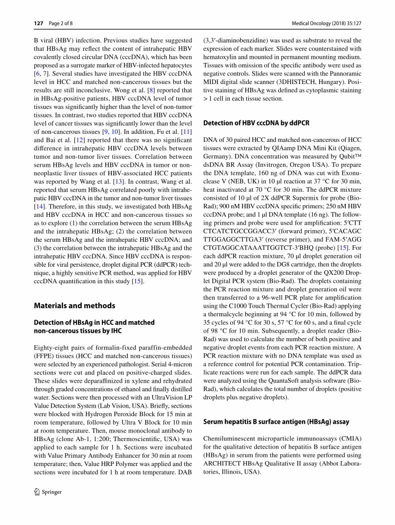
127 Page 2 of 8
B viral (HBV) infection. Previous studies have suggested that HBsAg may reflect the content of intrahepatic HBV covalently closed circular DNA (cccDNA), which has been proposed as a surrogate marker of HBV-infected hepatocytes [6, 7]. Several studies have investigated the HBV cccDNA level in HCC and matched non-cancerous tissues but the results are still inconclusive. Wong et al. [8] reported that in HBsAg-positive patients, HBV cccDNA level of tumor tissues was significantly higher than the level of non-tumor tissues. In contrast, two studies reported that HBV cccDNA level of cancer tissues was significantly lower than the level of non-cancerous tissues [9, 10]. In addition, Fu et al. [11] and Bai et al. [12] reported that there was no significant difference in intrahepatic HBV cccDNA levels between tumor and non-tumor liver tissues. Correlation between serum HBsAg levels and HBV cccDNA in tumor or non-neoplastic liver tissues of HBV-associated HCC patients was reported by Wang et al. [13]. In contrast, Wang et al. reported that serum HBsAg correlated poorly with intrahe-patic HBV cccDNA in the tumor and non-tumor liver tissues [14]. Therefore, in this study, we investigated both HBsAg and HBV cccDNA in HCC and non-cancerous tissues so as to explore (1) the correlation between the serum HBsAg and the intrahepatic HBsAg; (2) the correlation between the serum HBsAg and the intrahepatic HBV cccDNA; and (3) the correlation between the intrahepatic HBsAg and the intrahepatic HBV cccDNA. Since HBV cccDNA is respon-sible for viral persistence, droplet digital PCR (ddPCR) tech-nique, a highly sensitive PCR method, was applied for HBV cccDNA quantification in this study [15].
Materials and methods
Detection of HBsAg in HCC and matched non‑cancerous tissues by IHC
Eighty-eight pairs of formalin-fixed paraffin-embedded (FFPE) tissues (HCC and matched non-cancerous tissues) were selected by an experienced pathologist. Serial 4-micron sections were cut and placed on positive-charged slides. These slides were deparaffinized in xylene and rehydrated through graded concentrations of ethanol and finally distilled water. Sections were then processed with an UltraVision LP Value Detection System (Lab Vision, USA). Briefly, sections were blocked with Hydrogen Peroxide Block for 15 min at room temperature, followed by Ultra V Block for 10 min at room temperature. Then, mouse monoclonal antibody to HBsAg (clone Ab-1, 1:200; Thermoscientific, USA) was applied to each sample for 1 h. Sections were incubated with Value Primary Antibody Enhancer for 30 min at room temperature; then, Value HRP Polymer was applied and the sections were incubated for 1 h at room temperature. DAB
(3,3′-diaminobenzidine) was used as substrate to reveal the expression of each marker. Slides were counterstained with hematoxylin and mounted in permanent mounting medium. Tissues with omission of the specific antibody were used as negative controls. Slides were scanned with the Pannoramic MIDI digital slide scanner (3DHISTECH, Hungary). Posi-tive staining of HBsAg was defined as cytoplasmic staining > 1 cell in each tissue section.
Detection of HBV cccDNA by ddPCR
DNA of 30 paired HCC and matched non-cancerous of HCC tissues were extracted by QIAamp DNA Mini Kit (Qiagen, Germany). DNA concentration was measured by Qubit™ dsDNA BR Assay (Invitrogen, Oregon USA). To prepare the DNA template, 160 ng of DNA was cut with Exonu-clease V (NEB, UK) in 10 µl reaction at 37 °C for 30 min, heat inactivated at 70 °C for 30 min. The ddPCR mixture consisted of 10 µl of 2X ddPCR Supermix for probe (Bio-Rad); 900 nM HBV cccDNA specific primers; 250 nM HBV cccDNA probe; and 1 µl DNA template (16 ng). The follow-ing primers and probe were used for amplification: 5′CTT CTC ATC TGC CGG ACC 3′ (forward primer), 5′CAC AGC TTG GAG GCT TGA 3′ (reverse primer), and FAM-5′AGG CTG TAG GCA TAA ATT GGTCT-3′BHQ (probe) [15]. For each ddPCR reaction mixture, 70 µl droplet generation oil and 20 µl were added to the DG8 cartridge, then the droplets were produced by a droplet generator of the QX200 Drop-let Digital PCR system (Bio-Rad). The droplets containing the PCR reaction mixture and droplet generation oil were then transferred to a 96-well PCR plate for amplification using the C1000 Touch Thermal Cycler (Bio-Rad) applying a thermalcycle beginning at 94 °C for 10 min, followed by 35 cycles of 94 °C for 30 s, 57 °C for 60 s, and a final cycle of 98 °C for 10 min. Subsequently, a droplet reader (Bio-Rad) was used to calculate the number of both positive and negative droplet events from each PCR reaction mixture. A PCR reaction mixture with no DNA template was used as a reference control for potential PCR contamination. Trip-licate reactions were run for each sample. The ddPCR data were analyzed using the QuantaSoft analysis software (Bio-Rad), which calculates the total number of droplets (positive droplets plus negative droplets).
Serum hepatitis B surface antigen (HBsAg) assay
Chemiluminescent microparticle immunoassays (CMIA) for the qualitative detection of hepatitis B surface antigen (HBsAg) in serum from the patients were performed using ARCHITECT HBsAg Qualitative II assay (Abbot Labora-tories, Illinois, USA).
Medical Oncology (2018) 35:127
1 3
Page 3 of 8 127
Serum alpha‑fetoprotein (AFP) assay
Electrochemiluminescence immunoassays (ECLIA) for the in vitro quantitative determination of alpha-fetoprotein (AFP) in serum from the patients were performed using the AFP kit with a cobas e601 analyzer (Roche Diagnostics Limited GmbH, Mannheim, Germany).
Statistical analysis
Statistical analyses were performed with SPSS v.11.5 (SPSS Inc., Chicago, Illinois, USA) or GraphPad Prism 7 (version 7.03). Correlation was determined using Chi-square test. The p values less than 0.05 were considered statistically significant.
Results
Correlation between serum HBsAg and intrahepatic HBsAg
Intrahepatic HBsAg was determined in 88 pairs of HCC and matched non-cancerous tissues by IHC. The data of serum HBsAg were collected from patient records. The clinicopathological features are illustrated in Table 1. Of 88 patients included in this study, serum HBsAg was deter-mined as positive in 56 cases (63.64%) and negative in 32 cases (36.36%). In serum HBsAg-positive group, intra-hepatic HBsAg was positive staining in 73.2% (41/56) of matched non-cancerous tissues but only in 10.7% (6/56) of HCC tissues, as shown in Fig. 1a. In serum HBsAg-negative group, intrahepatic HBsAg was positive staining in 18.7% (6/32) of matched non-cancerous tissues and only in 3.1% (1/32) of HCC tissues, as shown in Fig. 1b. The significant correlation between serum HBsAg and intrahepatic HBsAg in matched non-cancerous tissues was observed (p < 0.001) but there was no correlation between serum HBsAg and intrahepatic HBsAg in HCC tissues (p = 0.415), as shown in Table 2.
Correlation between serum HBsAg and intrahepatic HBV cccDNA
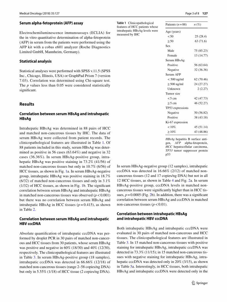
Absolute quantification of intrahepatic cccDNA was per-formed by droplet PCR in 30 pairs of matched non-cancer-ous and HCC tissues from 30 patients, whose serum HBsAg was positive and negative in 60% (18/30) and 40% (12/30), respectively. The clinicopathological features are illustrated in Table 3. In serum HBsAg-positive group (18 samples), intrahepatic cccDNA was detected in 66.66% (12/18) of matched non-cancerous tissues (range 2–58 copies/ng DNA) but only in 5.55% (1/18) of HCC tissue (2 copies/ng DNA).
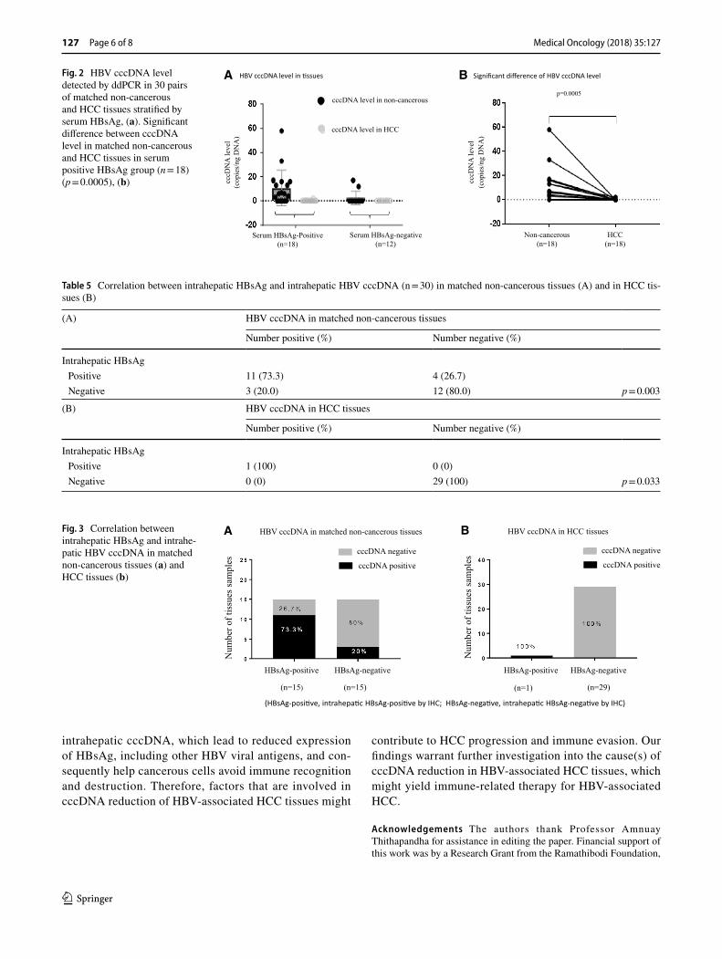
In serum HBsAg-negative group (12 samples), intrahepatic cccDNA was detected in 16.66% (2/12) of matched non-cancerous tissues (12 and 17 copies/ng DNA) but not in all 12 HCC tissues, as shown in Table 4 and Fig. 2a. In serum HBsAg-positive group, cccDNA levels in matched non-cancerous tissues were significantly higher than in HCC tis-sues, p = 0.0005 (Fig. 2b). In addition, there was a significant correlation between serum HBsAg and cccDNA in matched non-cancerous tissues (p < 0.01).
Correlation between intrahepatic HBsAg and intrahepatic HBV cccDNA
Both intrahepatic HBsAg and intrahepatic cccDNA were evaluated in 30 pairs of matched non-cancerous and HCC tissues. The clinicopathological features are illustrated in Table 3. In 15 matched non-cancerous tissues with positive staining for intrahepatic HBsAg, intrahepatic cccDNA was detected in 73.3% (11/15); in 15 matched non-cancerous tis-sues with negative staining for intrahepatic HBsAg, intra-hepatic cccDNA was detected only in 20% (3/15), as shown in Table 5a. Interestingly, in HCC tissues, both intrahepatic HBsAg and intrahepatic cccDNA were detected only in the
Table 1 Clinicopathological features of HCC patients whose intrahepatic HBsAg levels were measured by IHC
HBsAg hepatitis B surface anti-gen, AFP alpha-fetoprotein, HCC hepatocellular carcinoma, TP53 tumor suppressor protein p53
Patients (n = 88) n (%)
Age (years) < 50 25 (28.4) ≥ 50 63 (71.6)
Sex Male 75 (85.23) Female 13 (14.77)
Serum HBsAg Positive 56 (63.64) Negative 32 (36.36)
Serum AFP < 500 ng/ml 62 (70.46) ≥ 500 ng/ml 24 (27.27) Unknown 2 (2.27)
Tumor size < 5 cm 42 (47.73) ≥ 5 cm 46 (52.27)
TP53 expressions Negative 50 (56.82) Positive 38 (43.18)
Ki-67 expression < 10% 45 (51.14) ≥ 10% 43 (48.86)

Medical Oncology (2018) 35:127
1 3
127 Page 4 of 8
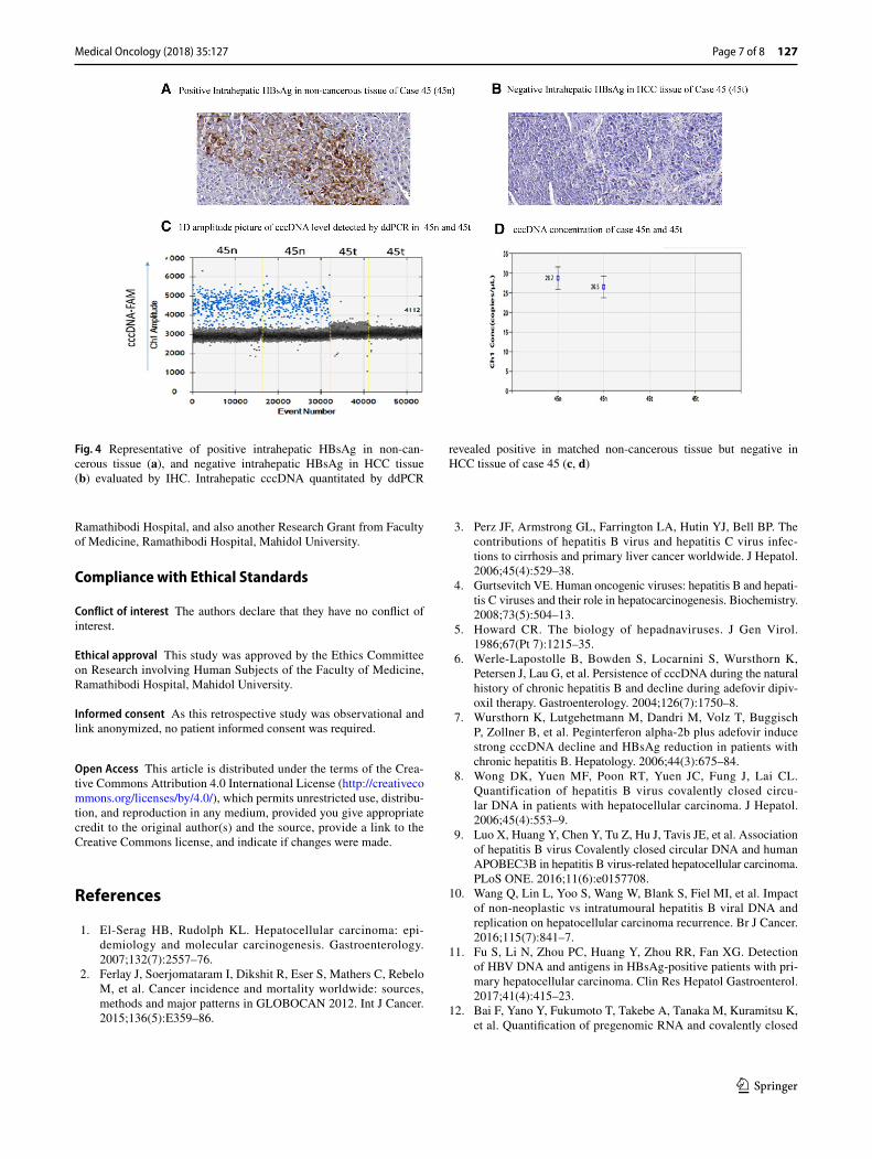
same 1 HCC tissue (2 copies/ng DNA) but not in the other 29 HCC tissues, as shown in Table 5b and Fig. 3. Repre-sentative of intrahepatic HBsAg and cccDNA is shown in Fig. 4. Significant correlations between intrahepatic HBsAg and cccDNA were found in both matched non-cancerous (p < 0.01) and HCC tissues (p < 0.05).
Discussion
Serum HBsAg is an important diagnostic marker for HBV infection. However, some studies suggest that HBV DNA can be detected in the liver tissue of HBsAg seroclearance or patients with occult HBV, mainly in the form of cccDNA, and these patients can continue to develop HCC [16, 17]. In the present study, we investigated intrahepatic HBsAg in both matched non-cancerous and HCC tissues by IHC, and found that serum HBsAg was significantly associated with intrahepatic HBsAg only in matched non-cancerous tis-sues. In contrast to matched non-cancerous tissues, no asso-ciation between serum HBsAg and intrahepatic HBsAg in
HCC tissues was observed. In serum HBsAg-positive group, positive intrahepatic HBsAg was detected in matched non-cancerous and HCC tissues at the frequency of 73.2 and 10.7%, respectively. Our results showed that intrahepatic HBsAg was reduced in most HCC tissues, which is con-sistent with the finding of Tian et al. [18], Jing et al. [19] and Wang [20], but the exact mechanism is still unclear. The finding that intrahepatic HBsAg could not be detected in HCC tissues by IHC from patients with positive serum HBsAg might be explained as follows, (1) HBV in HCC tis-sues could not express HBsAg, (2) HBsAg in HCC tissues could not be detected by the antibody used in this study, (3) HBV DNA is no longer present in HCC tissues. Therefore, in this study, so as to explore whether the decreased expres-sion of HBsAg in HCC tissues is caused by HBV cccDNA reduction, intrahepatic HBV cccDNA was quantitated by ddPCR in both negative intrahepatic HBsAg and positive intrahepatic HBsAg tissues.
In the present study, the significant correlation between serum HBsAg and intrahepatic cccDNA was found only in matched non-cancerous tissues. This is different from
Fig. 1 Intrahepatic HBsAg detected in non-cancerous and HCC tissues in serum HBsAg-positive group, n = 56 (a), and serum HBsAg-negative group, n = 32 (b)
A B
Intrahepatic HBsAg-positive
Intrahepatic HBsAg-negative
Non-cancerous(n=56)
Non-cancerous(N=32)
HCC(n=56)
HCC(N=32)
Num
ber o
f tis
sue
sam
ples
Num
ber o
f tis
sue
sam
ples
Serum HBsAg-positive group (n=56) Serum HBsAg-negative group (n=32)
73.2%
26.8%
10.7%
89.3%
18.7%
81.3%
3.1%
96.9%
Intrahepatic HBsAg-positive
Intrahepatic HBsAg-negative
Table 2 Correlation between serum HBsAg and intrahepatic HBsAg (n = 88) in matched non-cancerous tissues (A) and HCC tissues (B)
(A) Intrahepatic HBsAg in matched non-cancerous tissues
Number positive (%) Number negative (%)
Serum HBsAg Positive 41 (73.2) 15 (26.8) p < 0.0001 Negative 6 (18.7) 26 (81.3)
(B) Intrahepatic HBsAg in HCC tissues
Number positive (%) Number negative (%)
Serum HBsAg Positive 6 (10.7) 50 (89.3) p = 0.415 Negative 1 (3.1) 31 (96.9)
Medical Oncology (2018) 35:127
1 3
Page 5 of 8 127
previous studies; as Fu et al. reported that there was no cor-relation between serum HBsAg and intrahepatic cccDNA in HBV-related hepatocellular carcinoma [11], but Wang et al. [13] reported correlation between serum HBsAg and intrahe-patic cccDNA in both tumor and non-neoplastic liver tissues of HBV-associated HCC patients. The reasons for variety of results are inconclusive. But the possible contributing factor
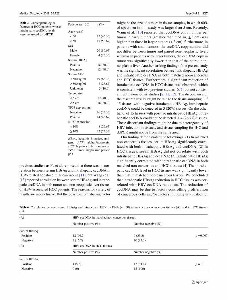
might be the size of tumors in tissue samples, in which 60% of specimen in this study was larger than 5 cm. Recently, Wang et al. [10] reported that cccDNA copy number per tumor in early tumors (smaller than median, ≤ 3 cm) was higher than those in larger tumors (> 3 cm); furthermore, in patients with small tumors, the cccDNA copy number did not differ between tumor and paired non-neoplastic liver, whereas in patients with larger tumors, the cccDNA copy in tumor was significantly lower than that of the paired non-neoplastic liver. Another striking finding of the present study was the significant correlation between intrahepatic HBsAg and intrahepatic cccDNA in both matched non-cancerous and HCC tissues. Furthermore, a significant reduction of intrahepatic cccDNA in HCC tissues was observed, which is consistent with two previous studies [6, 7] but not consist-ent with some other studies [8, 11, 12]. The discordance of the research results might be due to the tissue sampling. Of 15 tissues with negative intrahepatic HBsAg, intrahepatic cccDNA could be detected in 3 (20%) tissues. On the other hand, of 15 tissues with positive intrahepatic HBsAg, intra-hepatic cccDNA could not be detected in 4 (26.7%) tissues. These discordant findings might be due to heterogeneity of HBV infection in tissues, and tissue sampling for IHC and ddPCR might not be from the same area.
Our finding demonstrated the followings: (1) In matched non-cancerous tissues, serum HBsAg significantly corre-lated with both intrahepatic HBsAg and cccDNA; (2) In HCC tissues, serum HBsAg did not correlate with both intrahepatic HBsAg and cccDNA; (3) Intrahepatic HBsAg significantly correlated with intrahepatic cccDNA in both matched non-cancerous and HCC tissues; (4) The intrahe-patic cccDNA level in HCC tissues was significantly lower than that in matched non-cancerous tissues. We concluded that intrahepatic HBsAg reduction in HCC tissues was cor-related with HBV cccDNA reduction. The reduction of cccDNA may be due to factors controlling proliferation of cancerous cells and/or factors inducing eradication of
Table 3 Clinicopathological features of HCC patients whose intrahepatic cccDNA levels were measured by ddPCR
HBsAg hepatitis B surface anti-gen, AFP alpha-fetoprotein, HCC hepatocellular carcinoma, TP53 tumor suppressor protein p53
Patients (n = 30) n (%)
Age (years) < 50 13 (43.33) ≥ 50 17 (56.67)
Sex Male 26 (86.67) Female 4 (13.33)
Serum HBsAg Positive 18 (60.0) Negative 12 (40.0)
Serum AFP < 500 ng/ml 19 (63.33) ≥ 500 ng/ml 8 (26.67) Unknown 3 (10.0)
Tumor size < 5 cm 12 (40.0) ≥ 5 cm 18 (60.0)
TP53 expressions Negative 16 (53.33) Positive 14 (46.67)
Ki-67 expression < 10% 8 (26.67) ≥ 10% 22 (73.33)
Table 4 Correlation between serum HBsAg and intrahepatic HBV cccDNA (n = 30) in matched non-cancerous tissues (A), and in HCC tissues (B)
(A) HBV cccDNA in matched non-cancerous tissues
Number positive (%) Number negative (%)
Serum HBsAg Positive 12 (66.7) 6 (33.3) p = 0.007 Negative 2 (16.7) 10 (83.3)
(B) HBV cccDNA in HCC tissues
Number positive (%) Number negative (%)
Serum HBsAg Positive 1 (5.6) 17 (94.4) p = 1.0 Negative 0 (0) 12 (100)
Medical Oncology (2018) 35:127
1 3
127 Page 6 of 8
intrahepatic cccDNA, which lead to reduced expression of HBsAg, including other HBV viral antigens, and con-sequently help cancerous cells avoid immune recognition and destruction. Therefore, factors that are involved in cccDNA reduction of HBV-associated HCC tissues might
contribute to HCC progression and immune evasion. Our findings warrant further investigation into the cause(s) of cccDNA reduction in HBV-associated HCC tissues, which might yield immune-related therapy for HBV-associated HCC.
Acknowledgements The authors thank Professor Amnuay Thithapandha for assistance in editing the paper. Financial support of this work was by a Research Grant from the Ramathibodi Foundation,
Fig. 2 HBV cccDNA level detected by ddPCR in 30 pairs of matched non-cancerous and HCC tissues stratified by serum HBsAg, (a). Significant difference between cccDNA level in matched non-cancerous and HCC tissues in serum positive HBsAg group (n = 18) (p = 0.0005), (b)
A B
Serum HBsAg-Positive(n=18)
Serum HBsAg-negative(n=12)
Non-cancerous (n=18)
HCC (n=18)
p=0.0005
cccD
NA
leve
l (c
opie
s/ng
DN
A)
cccD
NA
leve
l (c
opie
s/ng
DN
A)
cccDNA level in non-cancerous
cccDNA level in HCC
HBV cccDNA level in �ssues Significant difference of HBV cccDNA level
Table 5 Correlation between intrahepatic HBsAg and intrahepatic HBV cccDNA (n = 30) in matched non-cancerous tissues (A) and in HCC tis-sues (B)
(A) HBV cccDNA in matched non-cancerous tissues
Number positive (%) Number negative (%)
Intrahepatic HBsAg Positive 11 (73.3) 4 (26.7) Negative 3 (20.0) 12 (80.0) p = 0.003
(B) HBV cccDNA in HCC tissues
Number positive (%) Number negative (%)
Intrahepatic HBsAg Positive 1 (100) 0 (0) Negative 0 (0) 29 (100) p = 0.033
Fig. 3 Correlation between intrahepatic HBsAg and intrahe-patic HBV cccDNA in matched non-cancerous tissues (a) and HCC tissues (b)
A HBV cccDNA in matched non-cancerous tissues HBV cccDNA in HCC tissues
(n=15) (n=15) (n=1) (n=29)
Num
ber o
f tis
sues
sam
ples
Num
ber o
f tis
sues
sam
ples
(HBsAg-posi�ve, intrahepa�c HBsAg-posi�ve by IHC; HBsAg-nega�ve, intrahepa�c HBsAg-nega�ve by IHC)
HBsAg-positive HBsAg-positiveHBsAg-negative HBsAg-negative
cccDNA negative
cccDNA positivecccDNA negative
cccDNA positive
B
Medical Oncology (2018) 35:127
1 3
Page 7 of 8 127
Ramathibodi Hospital, and also another Research Grant from Faculty of Medicine, Ramathibodi Hospital, Mahidol University.
Compliance with Ethical Standards
Conflict of interest The authors declare that they have no conflict of interest.
Ethical approval This study was approved by the Ethics Committee on Research involving Human Subjects of the Faculty of Medicine, Ramathibodi Hospital, Mahidol University.
Informed consent As this retrospective study was observational and link anonymized, no patient informed consent was required.
Open Access This article is distributed under the terms of the Crea-tive Commons Attribution 4.0 International License (http://creat iveco mmons .org/licen ses/by/4.0/), which permits unrestricted use, distribu-tion, and reproduction in any medium, provided you give appropriate credit to the original author(s) and the source, provide a link to the Creative Commons license, and indicate if changes were made.
References
1. El-Serag HB, Rudolph KL. Hepatocellular carcinoma: epi-demiology and molecular carcinogenesis. Gastroenterology. 2007;132(7):2557–76.
2. Ferlay J, Soerjomataram I, Dikshit R, Eser S, Mathers C, Rebelo M, et al. Cancer incidence and mortality worldwide: sources, methods and major patterns in GLOBOCAN 2012. Int J Cancer. 2015;136(5):E359–86.
3. Perz JF, Armstrong GL, Farrington LA, Hutin YJ, Bell BP. The contributions of hepatitis B virus and hepatitis C virus infec-tions to cirrhosis and primary liver cancer worldwide. J Hepatol. 2006;45(4):529–38.
4. Gurtsevitch VE. Human oncogenic viruses: hepatitis B and hepati-tis C viruses and their role in hepatocarcinogenesis. Biochemistry. 2008;73(5):504–13.
5. Howard CR. The biology of hepadnaviruses. J Gen Virol. 1986;67(Pt 7):1215–35.
6. Werle-Lapostolle B, Bowden S, Locarnini S, Wursthorn K, Petersen J, Lau G, et al. Persistence of cccDNA during the natural history of chronic hepatitis B and decline during adefovir dipiv-oxil therapy. Gastroenterology. 2004;126(7):1750–8.
7. Wursthorn K, Lutgehetmann M, Dandri M, Volz T, Buggisch P, Zollner B, et al. Peginterferon alpha-2b plus adefovir induce strong cccDNA decline and HBsAg reduction in patients with chronic hepatitis B. Hepatology. 2006;44(3):675–84.
8. Wong DK, Yuen MF, Poon RT, Yuen JC, Fung J, Lai CL. Quantification of hepatitis B virus covalently closed circu-lar DNA in patients with hepatocellular carcinoma. J Hepatol. 2006;45(4):553–9.
9. Luo X, Huang Y, Chen Y, Tu Z, Hu J, Tavis JE, et al. Association of hepatitis B virus Covalently closed circular DNA and human APOBEC3B in hepatitis B virus-related hepatocellular carcinoma. PLoS ONE. 2016;11(6):e0157708.
10. Wang Q, Lin L, Yoo S, Wang W, Blank S, Fiel MI, et al. Impact of non-neoplastic vs intratumoural hepatitis B viral DNA and replication on hepatocellular carcinoma recurrence. Br J Cancer. 2016;115(7):841–7.
11. Fu S, Li N, Zhou PC, Huang Y, Zhou RR, Fan XG. Detection of HBV DNA and antigens in HBsAg-positive patients with pri-mary hepatocellular carcinoma. Clin Res Hepatol Gastroenterol. 2017;41(4):415–23.
12. Bai F, Yano Y, Fukumoto T, Takebe A, Tanaka M, Kuramitsu K, et al. Quantification of pregenomic RNA and covalently closed
Fig. 4 Representative of positive intrahepatic HBsAg in non-can-cerous tissue (a), and negative intrahepatic HBsAg in HCC tissue (b) evaluated by IHC. Intrahepatic cccDNA quantitated by ddPCR
revealed positive in matched non-cancerous tissue but negative in HCC tissue of case 45 (c, d)
Medical Oncology (2018) 35:127
1 3
127 Page 8 of 8
circular DNA in hepatitis B virus-related hepatocellular carci-noma. Int J Hepatol. 2013;2013:849290.
13. Wang Q, Luan W, Warren L, Fiel MI, Blank S, Kadri H, et al. Serum hepatitis B surface antigen correlates with tissue covalently closed circular DNA in patients with hepatitis B-associated hepa-tocellular carcinoma. J Med Virol. 2016;88(2):244–51.
14. Wang M, Qiu N, Lu S, Xiu D, Yu J, Wang XT, et al. Serum hepatitis B surface antigen is correlated with intrahepatic total HBV DNA and cccDNA in treatment-naive patients with chronic hepatitis B but not in patients with HBV related hepatocellular carcinoma. J Med Virol. 2013;85(2):219–27.
15. Mu D, Yan L, Tang H, Liao Y. A sensitive and accurate quanti-fication method for the detection of hepatitis B virus covalently closed circular DNA by the application of a droplet digital poly-merase chain reaction amplification system. Biotechnol Lett. 2015;37(10):2063–73.
16. Pollicino T, Squadrito G, Cerenzia G, Cacciola I, Raffa G, Craxi A, et al. Hepatitis B virus maintains its pro-oncogenic
properties in the case of occult HBV infection. Gastroenterology. 2004;126(1):102–10.
17. Yuen MF, Wong DK, Fung J, Ip P, But D, Hung I, et al. HBsAg seroclearance in chronic hepatitis B in Asian patients: replica-tive level and risk of hepatocellular carcinoma. Gastroenterology. 2008;135(4):1192–9.
18. Tian X, Li J, Ma ZM, Zhao C, Wan DF, Wen YM. Role of hepa-titis B surface antigen in the development of hepatocellular car-cinoma: regulation of lymphoid enhancer-binding factor 1. J Exp Clin Cancer Res. 2009;28:58.
19. Jing YY, Liu WT, Guo SW, Ye F, Fan QM, Yu GF, et al. Hepatitis B virus (HBV) receptors: deficiency in tumor results in scant HBV infection and overexpression in peritumor leads to higher recur-rence risk. Oncotarget. 2015;6(40):42952–62.
20. Wang Y, Wu MC, Sham JS, Tai LS, Fang Y, Wu WQ, et al. Differ-ent expression of hepatitis B surface antigen between hepatocel-lular carcinoma and its surrounding liver tissue, studied using a tissue microarray. The Journal of pathology. 2002;197(5):610–6.


![STATISTICAL ANALYSISPLAN Efficacy, Safety, and ...negative HBV infection (hepatitis B surface antigen [HBsAg]-positive for at least 6months; with HBV DNA 10 5 copies/mL,ALT 1.5 ×ULN](https://img.dokumen.tips/doc/110x75/60da3b26730d43190e266c87/statistical-analysisplan-efficacy-safety-and-negative-hbv-infection-hepatitis.jpg)