Embed Size (px)
Citation preview

Zoologica Scripts, Vol. 15, No. 4, pp. 275-293,1986 Printed in Great Britain
0300-3256/86 $3.00+ .00 Pergamon Journals Ltd.
The Norwegian Academy of Science and Letters
Redescription of a brooding nemertine, Cyanophthalma obscura (Schultze) gen. et comb.n., with observations on its biology and discussion of the species of Prostomatella and related taxa*t
JON NORENBURG
Accepted 22 January 1986
Norenburg, J. 1986. Redescription of a brooding nemertine, Cyanophthalma obscura (Schultze) gen. et. comb, n., with observations on its biology and discussion of the species ot Prostomatella and related taxa.—Zool. Scr. 15:275-293.
A new genus, Cyanophthalma, is erected with the type species Tetrastemma obscura Schultze, 1851. Cyanophthalma obscura is transferred from Prostomatella and is redescribed from observa- tions of more than 300 living specimens and serial sections of 59 specimens from the northeast coast of North America. Comparison is made with serial sections of C. obscura from the Baltic and with serially sectioned specimens of three other described species of Prostomatella. Two of these, P. enteroplecta and P. merula, do not fit either genus and are provisionally placed in the aggregate genus Tetrastemma, whose present diagnosis they do fit. Prostomatella remains with only its type species, P. arenieola. Amphiporus cordiceps is also transferred to Cyanophthalma. Some of the characters that form the basis for classifying Monostilifera are assessed for phylogenetic signifi- cance. Cyanophthalma obscura in Nova Scotia lives in salt marshes. Young are brooded through the winter while ice covers most of the marsh.
Jon Norenburg, Mount Desert Island Biological Laboratory, Salsbury Cove, Maine 04672, U.S.A.
Introduction
Cyanophthalma obscura (Schultze, 1851) gen. et coihb.n., previously known from the Baltic Sea and the Black Sea, is here reported from salt marshes in New England and Nova Scotia. A detailed investigation of its anatomy has resolved several questions and points of disagreement raised in the brief descriptions by Karling (1934) and Friedrich (1935a). Cyanophthalma obscura is compared with the other three species of Prostomatella, which were also re-examined. Comparison of these with other monostiliferans forms a basis for proposing a new genus, Cyanophthalma, with the type species C. obscura, and for following Friedrich's (1970) suggestion and provisionally placing P. merula Correa, 1954 and P. enteroplecta Cor- rea, 1954 in the aggregate genus Tetrastemma. Prosto- matella remains with only its type species, P. arenieola Friedrich, 1935. Recent, extensive additions to our know- ledge of the anatomy of the closely related Prostoma (Gibson & Moore 1976) and Tetrastemma (Kirsteuer 1963), a redescription of Prostomatella arenieola (Mock 1981) and personal observations on a number of addi- tional amphiporids and tetrastemmids provide the back- ground for emending the diagnoses of Tetrastemma merula, T. enteroplecta and P. arenieola, Amphiporus
* Contribution No. 106 from the Marine Science Institute, Northeastern University, Nahant, Massachusetts, U.S.A. t Supported in part by National Research Council of Canada Operating Grant A2009 to J. Sherman Bleakney, Acadia University, Wolfville, Nova Scotia, and a Smithsonian Institution Postdoctoral Fellowship.
cordiceps (Jensen, 1878) (sensu Friedrich 1933, 1935a) appears closely related to C. obscura and is referred to Cyanophthalma.
The recent descriptions of many nemertines have greatly expanded the data base of morphological charac- ters available. Thus, it is possible to begin exploring the phylogenetic relationships among nemertines as a basis for classification, as opposed to a classification based on arbitrary weighting of characters and subjective evalua- tion of the 'total aggregates' of characters. I have adopted Pfennig's (1966) phylogenetic principles as a paradigm for discussing some of the characters that underlie our present classification of the Monostilifera. These include: probos- cis insertion, anterior diverticula of the intestinal caecum, number of ocelli and proboscis nerves, construction of the nephridial system, vascular system and gonads.
Material and methods
Specimens of Cyanophthalma obscura were obtained throughout the year from salt marsh pools in Nova Scotia near Kingsport and Pickett's Wharf, which border the Minas Basin of the Bay of Fundy in Canada. Additional specimens were collected during summer and late autumn from salt marshes near Ipswich and Rowley, Massachusetts, U.S.A.
The following description is based on observations of more than 300 living specimens, of which 59 were sectioned. For histological examina- tion worms were routinely anaesthetized in 7% MgCk, and occasionally in propylene phenoxitol (Owen 1955) or chloral hydrate. Fixation was routinely in Bouin's or Hollande's cupri-picri-formal-acetic fluid, and occasionally in Helly's or Zenker's fluid. Specimens were embedded in polyester wax (Steedman 1957) and sectioned in transverse, horizontal or sagittal planes at 5 and 7 jim. Routine staining was by Heidenhain's azan and Gomori's trichrome. Descriptions of anatomical features are based on confirmation from at least 20 specimens, unless noted diffe- rently, and from specimens representing each month except August.
275 Zoologica Scripta 15

276 7. Norenburg
Fig. 1. Cyanophthalma obscura. Female in dorsolateral view. Scale 0.2 mm.
Emended diagnoses for Tetrastemma merula, T. enteroplecto and Prostomatella arenicola are compiled from previous descriptions (Correa 1954, 1961; Friedrich 1935a; Mock 1981) and examination of section series. One set of serial sections for T. merula and one for T. enteroplecto were provided by Prof. D.D. CorrSa, along with additional fixed specimens of each species from Brazil and Florida. Two of each from each locale were serially sectioned in tranverse and longitudinal planes and stained by Heidenhain's azan and Heidenhain's iron hematoxylin techniques. The section series of P. arenicola provided by Prof. H. Friedrich is apparently the source for the original description.
Description
Cyanophthalma obscura (Schultze, 1851) gen. et comb.n. (Figs. W8)
Type. Unknown. Voucher specimens. Section series of 3 males and one female bearing
young deposited with the American Museum of Natural History (AMNH285) and section series of one male and one immature specimen deposited with the National Museum of Canada (NNCIC1983-0599, NMCIC1983-0600).
External features
Living animals, when fully extended, measure 0.3 x 2 mm for the smallest juvenile to a maximum of 2.5 x 40 mm for a gravid female. Mean length of 310 specimens is 12.8 mm
(S.D. = 6.7 mm). Females tend to be larger than males. The maximum length recorded for a male is 12 mm, while some non-gravid females were longer than 30 mm. The cephalic region is broadly rounded and slightly flattened and is indistinctly demarcated from the rest of the body by a cephalic furrow that encircles the body just behind the cerebral ganglia. The body is widest shortly posterior to this furrow and then tapers to a narrow, rounded caudal tip (Fig. 1). The dorsal side of the animal is rounded and yellowish-brown to dark olive-green. The ventral surface is flattened and usually pale. Density of color is due to green pigment concentrated primarily along the rhyn- chocoel wall and to brown pigment granules located in the epidermis. The rhynchocoel reaches the caudal end and the proboscis is as long as the body, although it usually lies loosely coiled within the anterior half of the rhynchocoel.
There are four double-cup ocelli present (Figs. 1, 17, 18), as noted by Friedrich (1935a). The anterior ocelli of newly released juveniles are still single cups and the navy-blue pigment of all the ocelli is restricted to the eye cups. Within two days this pigment radiates outwards from the cups of the posterior pair of ocelli (Riser, pers. commun.), In large specimens there are usually pig- mented ocellar fragments near the posterior ocelli (Fig. 2), as was also noted by Karling (1934). The shallow, crescent-shaped, vertical cephalic furrows of the cerebral
Fig. 2. Cyanophthalma obscura. Internal organization of anterior portion in lateral view. Schematic reconstruction from serial sections made by projecting the organs of the left side of the body onto a median-sagittal plane. Scale 0.4 mm.
Zoologica Scripta 15
Cyanophthalma obscura gen. et. comb.n. and related taxa 277
organs lie between the anterior and posterior pairs of ocelli. Eight to twelve single or clustered bristles, 25-55 ixm long, located about the frontal organ, are readily observed in juvenile specimens, but were never observed in larger, older specimens. Although Schultze (1851) described a terminal proboscis pore and a slightly sub- terminal mouth (it is likely that he confused the former with a frontal organ), the proboscis and alimentary canal share a subterminal rhynchodaeal pore. Karling (1934) implies the presence of a normal frontal organ and Fried- rich (1935a) states that there is a small one present. In the present study it could be observed in only a few living specimens.
Gonads approaching maturity can be readily discerned through the body wall and alternate with the light-colored intestinal diverticula. Mature testes are dark, bilobate and have a reticulate appearance. The large outline and characteristic translucent nucleus (germinal vesicle) of maturing ova readily identifies the female.
Body wall, musculature and parenchyma
Thickness of the epidermis in histological sections is generally between 30 and 50 /xm for specimens that are 10-20 mm long. This variation is primarily a function of fixation. In squeeze-preparations of living worms about 10 mm long the epidermis is about 20 /xm thick, with that of the cephalic tip and dorsal surface 2-5 /im thicker.
As is typical for hoplonemertines, ciliated cells are distributed over the entire body surface and are interspersed with two types of gland cells (Fig. 20) (Noren- burg 1985). Slender, flask-shaped gland cells contain granular and/or globular orange- to red-stained material (azan) or homogeneous, strongly azanophilous secretion. The other gland cells are large, goblet-shaped cells which contain foamy, weakly cyanophilous material. The cephalic tip contains relatively few gland cells and they are lacking entirely from the anterior cephalic furrows (Fig. 13), these being lined by cells with cilia 6-10 /im long, as opposed to 4-6 /xm over the rest of the body surface. Conspicuous brown granules occur in the basal third of the epidermis and appear to be intercellular as well as intracellular in the ciliated cells. They seem to be un- affected by fixation and staining procedures, their num- bers appear to increase with age and they lead to the characteristic darkening of the integument during starva- tion shrinkage.
A distinct basal lamina was not observed, the epidermis appearing to rest directly on the dcrmis (Norenburg 1985). The dermis also varies in thickness within and between specimens and is profoundly influenced by the fixative. The dermis consistently is thinner and optically more dense after Bouin's fixation. Short, irregularly orientated connective tissue fibers, nerve fibers, assorted cell bodies and nuclei, as well as gland cell necks, are observed within the dermis (Norenburg 1985). At the cephalic tip the dermis is a thin, fenestrated lamina, through which pass many of the cephalic gland cell necks (Figs. 3,9).
Body wall musculature (Figs. 14, 20) is comparably developed to that of Prostoma species, but is much weaker
than is the general case for species of Tetrastemma. It consists of outer circular muscle, 4-8 fibers thick, a sparse network of diagonal fibers and a layer of longitudinal muscle, which, as described by Friedrich (1935a), is usu- ally twice the thickness of the circular muscle. Both of these latter muscle layers reach the cephalic tip (Figs. 3, 4), as noted by Friedrich (1935a), but the longitudinal layer is very thin and dispersed in front of the cerebral organs. At the posterior level of the cerebral ganglia the innermost fibers split away and converge medially as approximately 20-30 bundles (Figs. 6-8, 17) and pass close to the anterior face of the cerebral ganglia to form the proboscis insertion (Figs. 6,7). The peripheral portion of the proboscis insertion is of the 'dissolved' type, although in the central portion the individual bundles converge to form a closed face. A short wedge of paren- chyma (Figs. 8, 18) separates the inner from the outer portion of the longitudinal muscle layer. The latter gives rise precerebrally to anteromedially directed cephalic retractors which extend into the parenchyma and cephalic gland, reaching to the cephalic tip. Just dorsal to the rhynchodaeum and behind the anterior ocelli, fibers of circular muscle extend transversely through the paren- chyma and mesh with each other and the cephalic retrac- tors in the formation of a horizontal septum. Dorsoventral muscle bundles, 25-30 /urn in diameter, extend from the body wall circular muscle (Fig. 14) over the anterior and posterior faces of the intestinal diverticula. These muscles pass close to and occasionally mesh with the circular muscle of the rhynchocoel. A few, thin, dorsoventral muscles pass along the stomach and pylorus, extending from the body wall circular muscle to the circular muscle of the rhynchocoel. Also, several transverse muscles extend laterally from the dermal circular muscle sheath to the stomach and pylorus, diverging above and below, thereby anchoring the stomach when the esophagus is everted during feeding.
Parenchyma, consisting primarily of extracellular matrix, is abundant throughout the body of sexually immature specimens and about the foregut and cerebral ganglia in sexually mature specimens (Figs. 8, 10). Sub- muscular gland cells are common in the cephalic paren- chyma, where they penetrate the musculature either singly or in clusters (Figs. 5, 17). These cells have a cyanophilous to purplish reaction with azan. They also occur throughout the rest of the body, but are rare and buried in the longitudinal musculature. They, perhaps, are akin to the acidophilic gland cells described by Moore & Gibson (1985).
Friedrich (1935a, 1936) describes the cephalic gland (Figs. 2,4,9) as primarily a dorsal lobe not quite reaching the brain. However, in addition to this dominant lobe, the present observations show that there are two smaller ventrolateral lobes which extend to the cerebral organs, while the dorsal lobe is limited by the proboscis insertion and may reach midway over the cerebral ganglia. In each lobe the necks of the gland cells unite to form a duct extending to the region of the frontal organ. The latter is not always distinct. The gland cells are 25 ju.m or more in diameter and have a foamy, weakly cyanophilous content that may be liberated at the site of the frontal organ or more peripherally through individual cell processes pene-
Zoologica Scripta 15

278 J. Norenburg
Be- % L -sSK
>%£
^#%
dg
dc
%
»vc vg
1
ilm
, MJttft •''.'- '!%>' ,W,v%k
eg W
oc
Zoologica Scripta 15

Cyanophthalma obscura gen. et. comb. n. and related taxa 279
Fig. 16. Cyanophthalma obscura. Anterior portion of nervous system in dorsal view. Schematic reconstruction from serial sections. Scale 0.2 mm.
trating the fenestrated 'basal lamina' of the cephalic tip (Figs. 3, 18). Nerve and muscle fibers occur throughout the gland.
Nervous system
The dorsal and ventral ganglia (Figs. 8, 16-18) are fused anteriorly with their neuropils meshed, while they are demarcated externally by a relatively deep furrow distally. As noted by Karling (1934), the dorsal neuropil charac- teristically has a marginal lobe. The dorsal and ventral ganglia are of approximately similar volumes, but the dorsal are more globose (Fig, 16). Maximum cross- sectional width of the left gangliar corpus (determined for 11 specimens between 10 and 25 mm long) is 18-25% (mean = 21%) of body width. The ventral commissure lies below the dorsal one and is 2-3 times its diameter. The ventral ganglia elongate posteriorly, giving rise to the lateral nerve cords which unite posteriorly in a supra-anal anastomosis. Each nerve cord consists of a single neuropil ensheathed in a ganglion cell layer. A strand of 3-7 longitudinal muscle fibers is located centrally within the neuropil of each nerve cord.
Three or four major and minor nerves emanate from the anterior face of each cerebral lobe (Fig. 16). One or more of these may supply the anterior ocelli, whose innervation appears diffuse and could not be traced. Friedrich's (1935a) description indicates only a single nerve arising from the anterior face of each brain lobe and suggests that it is the anterior ocellar nerve. A pair of stout
nerves arises medially from the lateral face of each cere- bral lobe (Fig. 16); as noted by Friedrich, one innervates the posterior ocelli (Fig. 18) and the other a cerebral organ. The cerebral organ nerve arises from the ventral portion of the dorsal neuropil. The ventral commissure gives rise to a pair of foregut nerves that proceed post- eriorly along the stomach (Fig. 16). In two section series they appear to be linked by a transverse connective in the stomach wall. At each end of the ventral commissure a stout nerve arises that enters the longitudinal musculature of the proboscis at its insertion (Fig. 16). Three nerves arise posteriorly from the ventrolateral surface of each ventral ganglion and proceed to the ventral surface of the stomach (Fig. 16). A nerve arising from the medioventral surface of each ventral ganglion extends along the dorso- lateral surface of the stomach. A dorsomedian nerve is not present.
Sense organs
There are two pairs of double-cup ocelli (Figs. 1,15, 17, 18), as noted by Friedrich (1935a). The ocellar fragments in some large specimens may include an innervated, single-cup ocellus. The posterior ocelli (Figs. 2, 18) are dorsolateral and anterior to the cerebral ganglia, while the anterior ocelli (Figs. 1,17) are close to the cephalic tip and lateral to the rhynchodaeum. A single layer of epithelium containing navy-blue pigment forms the pig- ment cup and partially encloses the photoreceptive cells. Several cytoplasmic tracts also containing pigment granules radiate from the cup (Fig. 15). The double-cup ocelli measure 30-90 fim along their long axis and are 30-60 /im deep.
As previously noted by Friedrich (1935a, 1936), the cerebral organs (Figs. 2, 17, 19) reach to just under the cerebral ganglia and have a glandular component small in comparison with Protosoma species. Each organ is ensheathed in connective tissue continuous with the dermis. A thin wedge of connective tissue penetrating into the medial side of the organ effectively creates a large glandular lobe posteriorly and a smaller lobe anteriorly. The latter contains a cluster of gland cells and bipolar nerve cells. 'Axons' of these nerve cells proceed post- eriorly as the cerebral organ nerve. Most of the gland cells of both lobes are large-bodied cells with foamy, cyanophilous material and long necks leading to the blind end of the ciliated canal (Fig. 19). Dorsal and ventral to the canal are two clusters of cells that appear vesiculate and have intensely stained nuclei. The ciliated canal is relatively straight; a slight outward flexure anteriorly leads to the cephalic grooves. The canal lumen is 6-10 fim in diameter and is lined by ciliated cuboidal epithelium with granular nuclei appressed to the basal border. The
Figs. 3-15. Cyanophthalma obscura. Transverse sections.—3-7. Pre-cerebral region. Scale 50 fim.—3. Anterior tip with rhychodaeal pore and perforated 'basal lamina'.—4. Anterior ocelli, cephalic gland and rhynchodaeum.—5. Just posteriad to separation of rhynchodaeum and esophagus.—6. Septum formed of longitudinal muscle with rhynchodaeal circular muscle giving rise to outer circular muscle of proboscis sheath and inner circular muscle of proboscis.—7. Section through rhynchodaeal sphincter and muscle septum just anterior to section in Fig. 6.—8. Mid-region of cerebral ganglia with ventral and dorsal commissures. Bundles of inner longitudinal muscle lie in surrounding parenchyma. Scale 50 /nm.—9. Frontal section of cephalic gland and its pore (Frontal organ). Scale 50 fim.—10. Stomach region with abundant parenchyma. Scale 50 /urn.—//. Intestine, diverticula and mature ovary. Scale 50 fim.—12. Intestine with gregarines; bilobed testis with efferent duct. Scale 25 /im.— 13. Anterior cephalic furrow. Scale 10 fim.—14. Dorsoventral muscle inserting into circular muscle. Scale 5 /um.—15. Double-cup ocellus. Scale 10 ^m.
Zoologka Scripta 15

280 /. Norenburg
# '•'CSfciBISBSS'*'*
Inc.
! •-/-•
•A ^
oc -A&
, , ^: pcsr* dg
, f •P^Ci^jSSSi
Fr'gi. 17—IS. Cyanophthalma obscura. Frontal sections through cephalic lobe. Scale 50 pm.—17. Ventral cerebral ganglia, cerebral organs and anterior ocelli.—18. Dorsal cerebral ganglia and posterior ocelli.
cilia of the mediolateral side are longer than the remain- der. The blind end of the canal is non-ciliated. Length of the cerebral organ is roughly related to body length, whereas width relative to section width is variable. Width of the cerebral organ relative to body width for seven well fixed specimens ranged from 7-20%. Karling (1934) recorded a value of 13%. Small blood spaces, nephridial tubules and, occasionally, cephalic gland cells occur in close proximity to the cerebral organ.
Proboscis and rhynchocoel
The rhynchodaeal pore is subterminal. The rhyn- chodaeum (Figs. 2,4,5) widens as its proceeds posteriorly and terminates with an abrupt constriction, the rhyn- chodaeal sphincter (Fig. 7), which is 15-20 /u.m thick. The esophagus opens into the rhynchodaeum. Anterior to this juncture the rhynchodaeum is enclosed within longitudi- nal musculature (Fig. 4), while posteriorly there is only a
Fig. 19. Cyanophtkulmu obscura. Cerebral organ. Schematic diagram made with camera-lucida of mid-horizontal section. Scale 0.1 mm.
layer of circular muscle (Figs. 5, 6). The latter is not clearly continuous with the circular muscle of the rhyn- chocoel.
Karling (1934) and Friedrich (1935a) commented only that there was nothing unusual about the proboscis. The proboscis is as long as or longer than the body and has a diameter of 12-35% of body width, with a mean of 25% (measured in 19 specimens in the stomach region). It is divisible into anterior chamber, diaphragm, vesicle and posterior chamber, to which is attached a retractor muscle (Figs. 22-32). The proboscis will be described in its everted state.
The epithelium of the anterior chamber is raised into many conical papillae (Figs. 23, 29), 60-100 jam tall, which when everted have a functional abfrontal face; the frontal face consisting primarily of supporting connective tissue covered by a thin epithelium. The abfrontal face consists of azanophilous cells with a broad, basal cell body having a narrow distal process extending to the surface and broad, mucus producing, cyanophilous cells. The latter are largely nestled among the cell necks of the former cell type. Some of the azanophilous cells contain only coarse, densely staining granules; several granules often protrude distally from the cell neck and appear as small barbs 1-2 ^im long (Fig. 30). Several tall, club- shaped cells in each papilla contain azanophilous globules. A small nerve extends from the proboscis nerve plexus to the tip of each papilla, where it appears to be in contact with a cluster of external, ciliary bristles (Fig. 32); probably, this actually represents a collar cell with direct innervation.
The connective tissue below the epithelium is 20-36 /xm thick in the everted portion of several proboscides, but is only half as thick in non-everted portions. Below this connective tissue is a thin, outer circular muscle layer one or two fibers thick. It is separated by underlying connec- tive tissue from a well-developed longitudinal muscle layer. The latter consists of numerous bundles, each wrapped in connective tissue. The longitudinal muscle layer is divided into an inner and a thicker, outer cylinder by an extensive nerve plexus (Fig. 23) also ensheathed by connective tissue. A thin endothelium facing the rhyn- chocoel fluid and lining the proboscis overlies a sparse, inner layer of circular muscle fibers. The pair of proboscis nerves from the cerebrum ramify shortly after entering the proboscis, giving rise to the 10-16 proboscideal nerves
Zoologica Scnpia 15
Cyanophthalma obscura gen. et. comb. n. and related taxa 281
Table I. Number ofproboscideal nerves found in C. obscura
No. nerves No. worms
10 3
11 3
11-12 4
12 6
12-13 10
13 7
14 16 1 1
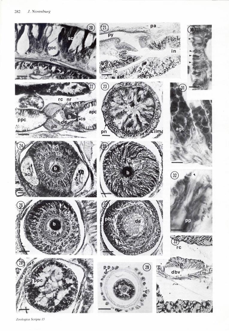
and their associated nerve plexus (Fig. 23). Table I lists the number of nerves for 33 specimens in which they could be accurately counted. Karling (1934) recorded 13 nerves for a single specimen. The number of proboscideal nerves may be constant throughout the length of the proboscis or may vary between regions of the same proboscis, e.g. 11 and 12 or 12 and 13 (Table I). The proboscideal nerves are interconnected in the diaphragm by a slim ring commis- sure and a thicker one in the vesicle (Figs. 22,26,27),
The morphology of the proboscis diaphragm and vesicle is in general agreement with that described for other Monostilifera (cf. Mclntosh 1873/74; Burger 1895; Strieker 1981,1985). Two accessory stylet sacs each con- tain 4-6 stylets (Fig. 24) and retain communication with the anterior proboscis chamber via a small duct and pore in the face of the diaphragm. The central basis is set in a deep, cup-shaped depression of the epithelium of the ductus ejaculatorius, which passes through the bulbous vesicle, connecting the anterior and posterior proboscis chambers (Figs. 22, 24-27). A circular muscle sphincter encloses each end of the duct, the anterior one encircling the middle of the basis. Large, strongly azanophilous, glandular cells extend radially from the basis (Figs. 24- 26). The basis figured by Friedrich (1935a, fig. 30a) is typical, but the stylet he depicts appears disproportion- ately small. Specimens of 9-13 mm length in the present study had bases 90-108 fim long with stylets 63-82 /im long. Juvenile specimens 2.5-4.5 mm long had bases 47-63 nm long and stylets 33-41 /*m long. These figures are in close agreement with those reported by Schultze (1851). The length ratio of stylet to basis for 20 adults and juveniles is 71% (range = 63-81%), with a coefficient of correlation of +0.96. In juveniles the basis tapers smoothly from a broad, rounded posterior end to a narrow apex that barely encompasses the stylet head. As the worm matures, the apical region of the basis thickens, becoming equal to, and sometimes exceeding, the post- erior width, while a median constriction remains.
The posterior chamber (Figs. 28, 29, 31) has an epithelium of tall azanophilous and cyanophilous cells j as well as a spacious lumen containing homogeneous, cyanophilous material and azanophilous granules (Fig. 28). Most of the cells contain azanophilous globules (Fig. 31) basally with smaller, granular material more distally; some contain only granules. Cells containing foamy cyanophilous material make up about one-fourth to one- third of the total cell number. A thin basal lamina sepa- rates the epithelium from a thin layer of longitudinal muscle and sparse circular muscle fibers. The latter is followed by the endothelial proboscis lining. The probos- cis retractor muscle extends from the longitudinal muscle fibers of the proboscis to those of the rhynchocoel wall at about the posterior third of the animal.
The rhynchocoel extends to the caudal end of the body. It is wide anteriorly, with most of the proboscis lying loosely coiled in this region, and tapers sharply posteriorly (Fig. 1). The lumen is lined by an endothelium that is
continuous with that of the proboscis and is often greatly thickened in the posterior and anterior extremities, where it contains much green pigment material (Fig. 36). The endothelium overlies longitudinal muscle fibers. A layer of circular muscle, 2-5 fibers thick, surrounds the latter.
Alimentary canal
Karling (1934) and Friedrich (1935a) comment only that the gut has an unpaired intestinal caecum with deep lateral diverticula. The esophagus (Figs. 2, 5-8) extends from the rhynchodaeum to under the cerebral ring. It is 20-70 jxm in diameter and lined principally by non- ciliated, vacuolate cells surrounded by longitudinal mus- cle fibers (Figs. 5-7). A few widely separated, large, ciliated, cyanophilous cells appear along the anterior esophagus and increase in number dorsally and post- eriorly, eventually leading to the fully ciliated epithelium of the stomach. A circular muscle sphincter is occasionally obvious at the rhynchodaeal juncture.
The stomach (Figs. 2,10,35) is a bulbous chamber with a deeply folded wall. Its thick epithelium is densely ciliated and consists of large cyanophilous and smaller azanophilous gland cells. The entire stomach is enclosed within a connective tissue sheath, which in turn is tightly invested by dorsal and lateral muscle fibers. The pylorus (Figs. 2, 21) is 2-3 times the stomach length and is not demarcated by change in epithelial cell types but by progressive posterior thinning of the wall and a decreasing portion of gland cells. In some cases the juncture with the intestine (Fig. 21) is distinguished by a band of non- ciliated epithelium underlain by a small cluster of muscle fibers and connective tissue. In others, the narrow pyloric pore is simply succeeded abruptly by intestinal epithelium without an associated musculature.
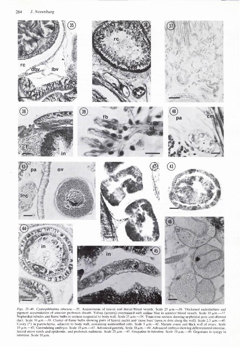
The caecum (Figs. 2, 21) extends forward from the intestine and has 2 or 3 pairs of deep, unbranched diver- ticula similar to those of the intestine. As noted by Friedrich (1935a), this caecum reaches forward no further than mid-stomach. He states that it has a broadly-rounded end, although in several of the present specimens there is a trace of bifurcation, the resultant pockets never exceed- ing 120 /am in length. The former condition appears to be the usual one. The intestine (Figs. 11, 12, 21) also has regularly arranged pairs of deep, unbranched diverticula and its epithelium consists of many elongate, azano- philous gland cells and a few attenuated ciliated cells. Gregarines (Figs. 12,45,46) are common in the intestinal lumen of most specimens and frequently fill the lumen in mature males.
Blood-vascular system
The blood-vascular system (Figs. 5-8, 33-36) was previ- ously described by Karling (1934) and Friedrich (1935a) basically as two lateral blood vessels united anteriorly by a cephalic loop and posteriorly by a supra-anal anatomosis which includes the dorsal vessel. The trunks of the cephalic loop pass medially through the cerebral ring. The dorsal vessel arises from either the left or the right (10 and 6 specimens, respectively) lateral vessel (Fig. 35) just behind the ventral commissure. In most cases, this origin
Zooiogica Scripta 15
282 J. Norenburg
Cyanophthalma obscura gen. et. comb.n. and related taxa 283
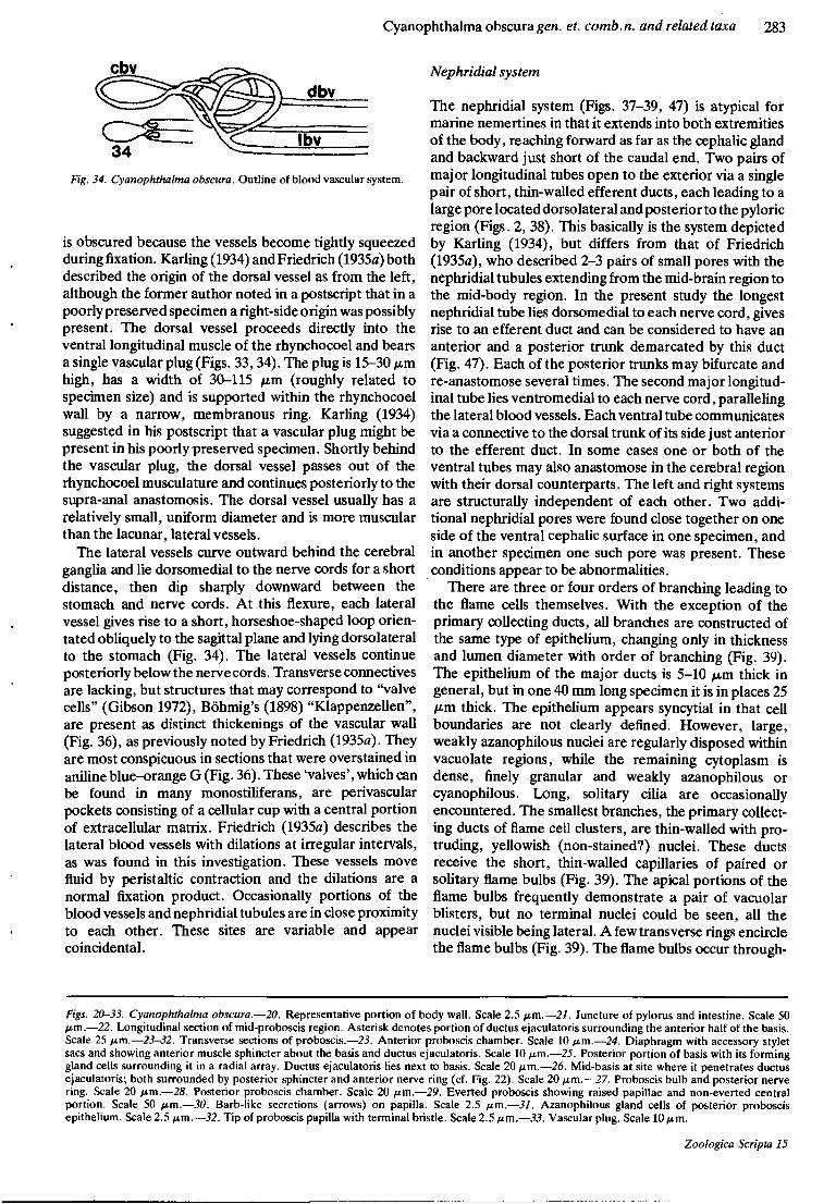
cbv
Fig. 34. Cyanophthalma obscura. Outline of blood vascular system.
is obscured because the vessels become tightly squeezed during fixation. Karling (1934) and Friedrich (1935a) both described the origin of the dorsal vessel as from the left, although the former author noted in a postscript that in a poorly preserved specimen a right-side origin was possibly present. The dorsal vessel proceeds directly into the ventral longitudinal muscle of the rhynchocoel and bears a single vascular plug (Figs. 33,34). The plug is 15-30 fim high, has a width of 30-115 pm (roughly related to specimen size) and is supported within the rhynchocoel wall by a narrow, membranous ring. Karling (1934) suggested in his postscript that a vascular plug might be present in his poorly preserved specimen. Shortly behind the vascular plug, the dorsal vessel passes out of the rhynchocoel musculature and continues posteriorly to the supra-anal anastomosis. The dorsal vessel usually has a relatively small, uniform diameter and is more muscular than the lacunar, lateral vessels.
The lateral vessels curve outward behind the cerebral ganglia and lie dorsomedial to the nerve cords for a short distance, then dip sharply downward between the stomach and nerve cords. At this flexure, each lateral vessel gives rise to a short, horseshoe-shaped loop orien- tated obliquely to the sagittal plane and lying dorsolateral to the stomach (Fig. 34). The lateral vessels continue posteriorly below the nerve cords. Transverse connectives are lacking, but structures that may correspond to "valve cells" (Gibson 1972), Bohmig's (1898) "Klappenzellen", are present as distinct thickenings of the vascular wall (Fig. 36), as previously noted by Friedrich (1935a). They are most conspicuous in sections that were overstained in aniline blue-orange G (Fig. 36). These 'valves', which can be found in many monostiliferans, are perivascular pockets consisting of a cellular cup with a central portion of extracellular matrix. Friedrich (1935a) describes the lateral blood vessels with dilations at irregular intervals, as was found in this investigation. These vessels move fluid by peristaltic contraction and the dilations are a normal fixation product. Occasionally portions of the blood vessels and nephridial tubules are in close proximity to each other. These sites are variable and appear coincidental.
Nephridial system
The nephridial system (Figs. 37-39, 47) is atypical for marine nemertines in that it extends into both extremities of the body, reaching forward as far as the cephalic gland and backward just short of the caudal end. Two pairs of major longitudinal tubes open to the exterior via a single pair of short, thin-walled efferent ducts, each leading to a large pore located dorsolateral and posterior to the pyloric region (Figs. 2, 38). This basically is the system depicted by Karling (1934), but differs from that of Friedrich (1935a), who described 2-3 pairs of small pores with the nephridial tubules extending from the mid-brain region to the mid-body region. In the present study the longest nephridial tube lies dorsomedial to each nerve cord, gives rise to an efferent duct and can be considered to have an anterior and a posterior trunk demarcated by this duct (Fig. 47). Each of the posterior trunks may bifurcate and re-anastomose several times. The second major longitud- inal tube lies ventromedial to each nerve cord, paralleling the lateral blood vessels. Each ventral tube communicates via a connective to the dorsal trunk of its side just anterior to the efferent duct. In some cases one or both of the ventral tubes may also anastomose in the cerebral region with their dorsal counterparts. The left and right systems are structurally independent of each other. Two addi- tional nephridial pores were found close together on one side of the ventral cephalic surface in one specimen, and in another specimen one such pore was present. These conditions appear to be abnormalities.
There are three or four orders of branching leading to the flame cells themselves. With the exception of the primary collecting ducts, all branches are constructed of the same type of epithelium, changing only in thickness and lumen diameter with order of branching (Fig. 39). The epithelium of the major ducts is 5-10 /im thick in general, but in one 40 mm long specimen it is in places 25 /im thick. The epithelium appears syncytial in that cell boundaries are not clearly defined. However, large, weakly azanophilous nuclei are regularly disposed within vacuolate regions, while the remaining cytoplasm is dense, finely granular and weakly azanophilous or cyanophilous. Long, solitary cilia are occasionally encountered. The smallest branches, the primary collect- ing ducts of flame cell clusters, are thin-walled with pro- truding, yellowish (non-stained?) nuclei. These ducts receive the short, thin-walled capillaries of paired or solitary flame bulbs (Fig. 39). The apical portions of the flame bulbs frequently demonstrate a pair of vacuolar blisters, but no terminal nuclei could be seen, all the nuclei visible being lateral. A few transverse rings encircle the flame bulbs (Fig. 39). The flame bulbs occur through-
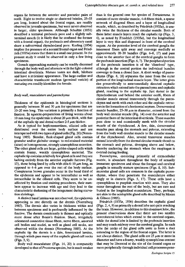
Figs. 20-33. Cyanophthalma obscura.—20. Representative portion of body wall. Scale 2.5 /u.m.—21. Juncture of pylorus and intestine. Scale 50 Mm-—22. Longitudinal section of mid-proboscis region. Asterisk denotes portion of ductusejaculatoris surrounding the anterior half of the basis. Scale 25 /im.—23-32. Transverse sections of proboscis.—23. Anterior proboscis chamber. Scale 10 ptm.—24. Diaphragm with accessory stylet sacs and showing anterior muscle sphincter about the basis and ductus ejaculatoris. Scale 10 p.m.—25. Posterior portion of basis with its forming gland cells surrounding it in a radial array. Ductus ejaculatoris ties next to basis. Scale 20 p.m.—26. Mid-basis at site where it penetrates ductus ejaculatoris; both surrounded by posterior sphincter and anterior nerve ring (cf. Fig. 22). Scale 20 /xm.—27. Proboscis bulb and posterior nerve ring. Scale 20 /im.—28. Posterior proboscis chamber. Scale 20 jam.—29. Everted proboscis showing raised papillae and non-everted central portion. Scale 50 fim.—30. Barb-like secretions (arrows) on papilla. Scale 2.5 /im.—31. Azanophilous gland cells of posterior proboscis epithelium. Scale 2.5 fim.—32. Tip of proboscis papilla with terminal bristle. Scale 2.5 fim.—33. Vascular plug. Scale 10 fim.
Zoologica Scripta 15
284 J. Norenburg
• MM ^_13B
'^m WB&'''~
*•.»«, •*?•*/_:>'
Figs. 35-46. Cyanophthalma obscura.—35. Anastomosis of lateral and dorsal blood vessels. Scale 25 ^im.—36. Thickened endothelium and pigment accumulation of anterior proboscis sheath. Valves (arrows) overstained with aniline blue in anterior blood vessels. Scale 10 ju.m.—37. Nephridial tubules and flame bulbs in section tangential to body wall. Scale 25 fim.—38. Transverse section showing nephridial pore and efferent duct. Scale 10 ftm,—39. Cluster of flame bulbs showing pairs of lateral nuclei and 'cross bars' (seen as dots along the wall). Scale 2.5 /xm.—40. Cavity (*) in parenchyma, adjacent to body wall, containing unidentified cells. Scale 5 ^.m.—41. Mature ovum and thick wall of ovary. Scale 10 fj.m.—42. Gastrulating embryos. Scale 10 ju.m.—43. Advanced gastrula. Scale 10 /um.—44. Advanced cmbrvo showing differentiated intestine, lateral nerve cords and epidermis, and proboscis rudiment. Scale 25 p.m.—45. Gregarine in intestine. Scale 10 /um.—46. Gegarines in syzygy in intestine. Scale 10 /im.
Cyanophthalma obscura gen. et. comb.n. and related taxa 285
=n2 np «d dnt
m vnt
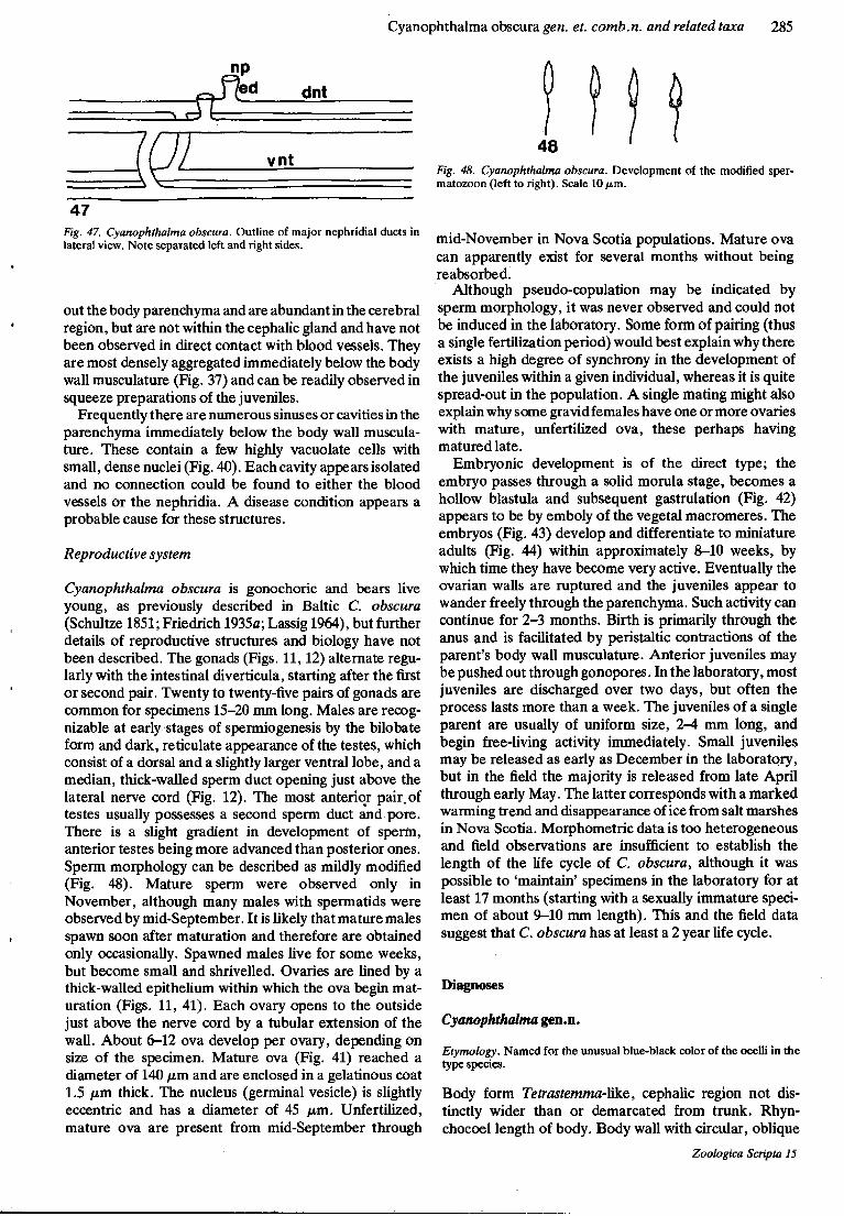
47 Fig. 47. Cyanophthalma obscura. Outline of major nephridial ducts in lateral view. Note separated left and right sides.
out the body parenchyma and are abundant in the cerebral region, but are not within the cephalic gland and have not been observed in direct contact with blood vessels. They are most densely aggregated immediately below the body wall musculature (Fig. 37) and can be readily observed in squeeze preparations of the juveniles.
Frequently there are numerous sinuses or cavities in the parenchyma immediately below the body wall muscula- ture. These contain a few highly vacuolate cells with small, dense nuclei (Fig. 40). Each cavity appears isolated and no connection could be found to either the blood vessels or the nephridia. A disease condition appears a probable cause for these structures.
Reproductive system
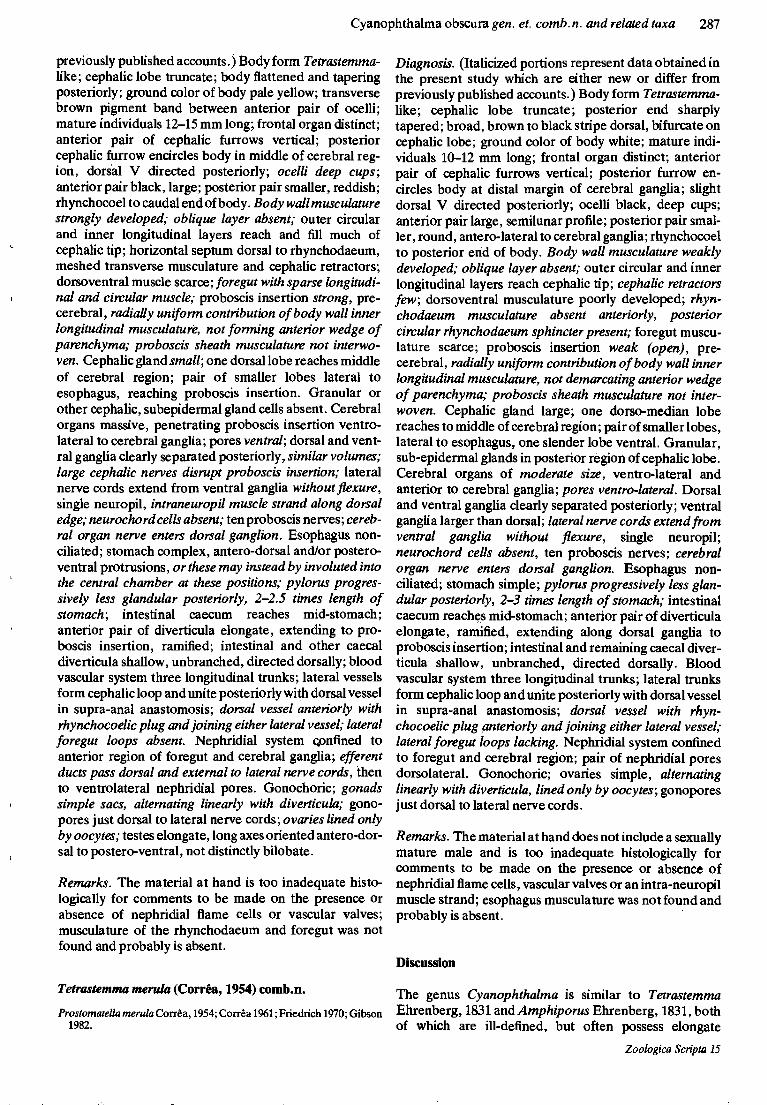
Cyanophthalma obscura is gonochoric and bears live young, as previously described in Baltic C, obscura (Schultze 1851; Friedrich 1935a; Lassig 1964), but further details of reproductive structures and biology have not been described. The gonads (Figs. 11,12) alternate regu- larly with the intestinal diverticula, starting after the first or second pair. Twenty to twenty-five pairs of gonads are common for specimens 15-20 mm long. Males are recog- nizable at early stages of spermiogenesis by the bilobate form and dark, reticulate appearance of the testes, which consist of a dorsal and a slightly larger ventral lobe, and a median, thick-walled sperm duct opening just above the lateral nerve cord (Fig. 12). The most anterior pair, of testes usually possesses a second sperm duct and pore. There is a slight gradient in development of sperm, anterior testes being more advanced than posterior ones. Sperm morphology can be described as mildly modified (Fig. 48). Mature sperm were observed only in November, although many males with spermatids were observed by mid-September. It is likely that mature males spawn soon after maturation and therefore are obtained only occasionally. Spawned males live for some weeks, but become small and shrivelled. Ovaries are lined by a thick-walled epithelium within which the ova begin mat- uration (Figs. 11, 41). Each ovary opens to the outside just above the nerve cord by a tubular extension of the wall. About 6-12 ova develop per ovary, depending on size of the specimen. Mature ova (Fig. 41) reached a diameter of 140 fim. and are enclosed in a gelatinous coat 1.5 jum thick. The nucleus (germinal vesicle) is slightly eccentric and has a diameter of 45 fim. Unfertilized, mature ova are present from mid-September through
48 Fig. 48. Cyanophthalma obscura. Development of the modified sper- matozoon (left to right). Scale 10/*m.
mid-November in Nova Scotia populations. Mature ova can apparently exist for several months without being reabsorbed.
Although pseudo-copulation may be indicated by sperm morphology, it was never observed and could not be induced in the laboratory. Some form of pairing (thus a single fertilization period) would best explain why there exists a high degree of synchrony in the development of the juveniles within a given individual, whereas it is quite spread-out in the population. A single mating might also explain why some gravid females have one or more ovaries with mature, unfertilized ova, these perhaps having matured late.
Embryonic development is of the direct type; the embryo passes through a solid morula stage, becomes a hollow blastula and subsequent gastrulation (Fig. 42) appears to be by emboly of the vegetal macromeres. The embryos (Fig. 43) develop and differentiate to miniature adults (Fig. 44) within approximately 8-10 weeks, by which time they have become very active. Eventually the ovarian walls are ruptured and the juveniles appear to wander freely through the parenchyma. Such activity can continue for 2-3 months. Birth is primarily through the anus and is facilitated by peristaltic contractions of the parent's body wall musculature. Anterior juveniles may be pushed out through gonopores. In the laboratory, most juveniles are discharged over two days, but often the process lasts more than a week. The juveniles of a single parent are usually of uniform size, 2-4 mm long, and begin free-living activity immediately. Small juveniles may be released as early as December in the laboratory, but in the field the majority is released from late April through early May. The latter corresponds with a marked warming trend and disappearance of ice from salt marshes in Nova Scotia. Morphometric data is too heterogeneous and field observations are insufficient to establish the length of the life cycle of C. obscura, although it was possible to 'maintain' specimens in the laboratory for at least 17 months (starting with a sexually immature speci- men of about 9-10 mm length). This and the field data suggest that C. obscura has at least a 2 year life cycle.
Diagnoses
Cyanophthalma gen.n.
Etymology. Named for the unusual blue-black color of the ocelli in the type species.
Body form Tetrastemma-Mke, cephalic region not dis- tinctly wider than or demarcated from trunk. Rhyn- chocoel length of body. Body wall with circular, oblique
Zoologica Scripta 15

286 J. Norenburg
and longitudinal muscle layers; inner longitudinal muscle forms proboscis insertion; intestinal region with dorso- lateral muscle bundles; proboscis sheath musculature not interwoven. Cerebral organs and cephalic gland present. Lateral nerve cords with single neuropil, neurochord cells lacking. Anterior intestinal diverticula not longer than others. Blood vascular system three longitudinal trunks; valves present; dorsal blood vessel with single lateral anastomosis and single vascular plug. Single pair of nephridial pores. Gonads alternating regularly with intes- tinal diverticula, gonopores dorsal to lateral nerve cords.
cerebral region to posterior end of body; two pairs of lateral trunks, one dorsal and one ventral to lateral nerve cord; flame bulbs single or paired, with pair of lateral nuclei. Gonochoric; viviparous; males smaller, testes, except first two pairs, distinctly bilobate; ovary wall includes non-oocyte cells.
Prostomatella arenicola Friedrich, 1935
Prostomatella arenicola Friedrich, 1935s; Friedrich 1936, 1955; Kirsteuer 1971; Mock 1981; Gibson 1982.
Cyanophthalma obscura (Schultze, 1851) comb.n.
Tetrastemma obscurum Schultze, 1851; Diesing 1862; Gibson 1972, folia obscura (part.); Beneden 1861. Prosorhockmus obscurus; Montgomery 1894. Prosioma obscurum; Burger 1904; Gering 1914; Segerstr&le 1933,1955,
1960; Karling 1934; Friedrich 1935a, 1936; Smidt 1944; Thorson 1946; Lassig 1964; Remane & Schlieper 1971; Riser 1974.
Prostomatella obscurum; Friedrich 1955,1979; Gibson 1982, Prostoma obscura; Brunberg 1964. Prostomatella obscura; Multer 1966; Moore & Gibson 1985; Norenburg 1985.
Diagnosis. Cephalic region truncate; body tapering to broad tip posteriorly; ground color yellowish-brown to dark olive-green, lateral outlines of rhynchocoel wall appearing as a pair of dark green stripes; mature indi- viduals 10-30 mm long; frontal organ often indistinct; anterior cephalic furrows vertical, shallow crescents; posterior cephalic furrow encircles body at posterior of cerebral region, dorsal V directed posteriorly; ocelli blue- black, shallow, double-cups; anterior pair near cephalic tip; posterior pair dorsolateral and close to cerebral ganglia, often associated with ocellar fragments or, rarely, an additional single-cup ocellus. Body wall musculature moderately developed; outer circular and inner longi- tudinal layers reach cephalic tip; horizontal septum of meshed cephalic retractors and transverse muscles above rhynchodaeum; pre-esophageal portion of rhyn- chodaeum with longitudinal muscle, posteriorly with cir- cular muscle only; esophagus and stomach with longitudi- nal musculature; stomach fixed by dorsoventral and lat- eral muscles arising from body wall circular muscle; stout dorsoventral muscles between all diverticula; proboscis insertion radially uniform contribution of body wall inner longitudinal musculature, demarcating an anterior wedge of parenchyma. Cephalic gland large, discharging by fron- tal organ and numerous cephalic pores; dorsomedian lobe large, reaches dorsal commissure; ventrolateral lobes small, reaching anterior cephalic furrows. Sub-epidermal gland cells in posterior cephalic region. Cerebral organs moderate size, ventrolateral and close to cerebral ganglia; pores lateral. Dorsal and ventral ganglia clearly separated posteriorly, similar volumes; lateral nerves extending from ventral ganglia without flexure; cerebral organ nerve enters dorsal ganglion; 10-16 proboscideal nerves. Esophagus non-ciliated; stomach simple; pylorus progres- sively less glandular posteriorly, 2.5 times length of stomach; intestinal caecum reaches to mid-stomach; anterior pair of diverticula not elongate, but reaching cerebral ganglia; caecal and intestinal diverticula simple, shallow, directed dorsolaterally. Nephridia extend from
Zoologica Scripta 15
Diagnosis. (Italicized portions represent data obtained in the present study which are either new or differ from previously published accounts.) Body form thread-like; rose colored; approximately 10-12 mm long and 0.3 mm thick; 4 reddish-brown ocelli, deep cups; rhynchocoel reaches posterior end of body; cephalic furrow pre- cerebral; ciliary bristles along cephalic margin and on caudal tip; frontal organ present. Body wall musculature strongly developed, reaching and filling much of cephalic tip; oblique layer absent; cephalic retractors present; weak dorsoventral musculature between intestinal diverticula; esophagus with scarce longitudinal muscle; rhynchodaeum with circular muscle; stomach without musculature; proboscis insertion of radially symmetric array of fascicles contributed by body wall inner longitudinal muscle, forms a closed face centrally; proboscis sheath musculature not interwoven. Proboscideal nerves seven. Cephalic gland weakly developed. Cephalic subepidermal glands few. Cerebral organs moderately developed, not reaching cerebral ganglia; innervation from dorsal ganglion; pores lateral. Dorsal and ventral cerebral ganglia demarcated posteriorly, similar volumes; numerous cephalic nerves; lateral nerve cords extending from ventral ganglia without flexure, single neuropil; neurochord cells absent. Esophagus non-ciliated; stomach simple; pylorus about twice length of stomach, rapidly decreasing number of gland cells posteriorly; intestinal caecum reaches mid- stomach, without anterior bifurcation or diverticula; intestinal diverticula shallow. Blood vascular system with three longitudinal trunks; lateral vessels form cephalic loop and unite posteriorly with dorsal vessel in supra-anal anastomosis; dorsal vessel joins a single lateral vessel, has a rhynchocoelic connection and a small vascular plug; lateral foregut loops absent. Nephridial system restricted to foregut region; several pores on each side.
Remarks. Only a single, very old section series was avail- able for study. The specimen had ejected the proboscis and was greatly contracted. Where possible, the re- description of P. arenicola by Mock (1981) is the source for much of the above.
Tetrastemma enteroplecta (Correa, 1954) comb.n.
Prostomatella enteroplecta Correa, 1954; Correa 1961; Friedrich 1970; Gibson 1982.
Diagnosis. (Italicized portions represent data obtained in the present study which are either new or differ from
Cyanophthalma obscura gen. et. comb.n. and related taxa 287
previously published accounts.) Body form Tetrastemma- like; cephalic lobe truncate; body flattened and tapering posteriorly; ground color of body pale yellow; transverse brown pigment band between anterior pair of ocelli; mature individuals 12-15 mm long; frontal organ distinct; anterior pair of cephalic furrows vertical; posterior cephalic furrow encircles body in middle of cerebral reg- ion, dorsal V directed posteriorly; ocelli deep cups; anterior pair black, large; posterior pair smaller, reddish; rhynchocoel to caudal end of body. Body wall musculature strongly developed; oblique layer absent; outer circular and inner longitudinal layers reach and fill much of cephalic tip; horizontal septum dorsal to rhynchodaeum, meshed transverse musculature and cephalic retractors; dorsoventral muscle scarce; foregut with sparse longitudi- nal and circular muscle; proboscis insertion strong, pre- cerebral, radially uniform contribution of body wall inner longitudinal musculature, not forming anterior wedge of parenchyma; proboscis sheath musculature not interwo- ven. Cephalic gland small; one dorsal lobe reaches middle of cerebral region; pair of smaller lobes lateral to esophagus, reaching proboscis insertion. Granular or other cephalic, subepidermal gland cells absent. Cerebral organs massive, penetrating proboscis insertion ventro- lateral to cerebral ganglia; pores ventral; dorsal and vent- ral ganglia clearly separated posteriorly, similar volumes; large cephalic nerves disrupt proboscis insertion; lateral nerve cords extend from ventral ganglia without flexure, single neuropil, intraneuropil muscle strand along dorsal edge; neurochord cells absent; ten proboscis nerves; cereb- ral organ nerve enters dorsal ganglion. Esophagus non- ciliated; stomach complex, antero-dorsal and/or postero- ventral protrusions, or these may instead by involuted into the central chamber at these positions; pylorus progres- sively less glandular posteriorly, 2-2.5 tones length of stomach; intestinal caecum reaches mid-stomach; anterior pair of diverticula elongate, extending to pro- boscis insertion, ramified; intestinal and other caecal diverticula shallow, unbranched, directed dorsally; blood vascular system three longitudinal trunks; lateral vessels form cephalic loop and unite posteriorly with dorsal vessel in supra-anal anastomosis; dorsal vessel anteriorly with rhynchocoelic plug and joining either lateral vessel; lateral foregut loops absent. Nephridial system cpnfined to anterior region of foregut and cerebral ganglia; efferent ducts pass dorsal and external to lateral nerve cords, then to ventrolateral nephridial pores. Gonochoric; gonads simple sacs, alternating linearly with diverticula; gono- pores just dorsal to lateral nerve cords; ovaries lined only by oocytes; testes elongate, long axes oriented antero-dor- sal to postero-ventral, not distinctly bilobate.
Remarks. The material at hand is too inadequate histo- rically for comments to be made on the presence or absence of nephridial flame cells or vascular valves; musculature of the rhynchodaeum and foregut was not found and probably is absent.
Diagnosis. (Italicized portions represent data obtained in the present study which are either new or differ from previously published accounts.) Body form Tetrastemma- like; cephalic lobe truncate; posterior end sharply tapered; broad, brown to black stripe dorsal, bifurcate on cephalic lobe; ground color of body white; mature indi- viduals 10-12 mm long; frontal organ distinct; anterior pair of cephalic furrows vertical; posterior furrow en- circles body at distal margin of cerebral ganglia; slight dorsal V directed posteriorly; ocelli black, deep cups; anterior pair large, semilunar profile; posterior pair smal- ler, round, antero-lateral to cerebral ganglia; rhynchocoel to posterior end of body. Body wall musculature weakly developed; oblique layer absent; outer circular and inner longitudinal layers reach cephalic tip; cephalic retractors few; dorsoventral musculature poorly developed; rhyn- chodaeum musculature absent anteriorly, posterior circular rhynchodaeum sphincter present; foregut muscu- lature scarce; proboscis insertion weak (open), pre- cerebral, radially uniform contribution of body wall inner longitudinal musculature, not demarcating anterior wedge of parenchyma; proboscis sheath musculature not inter- woven. Cephalic gland large; one dorso-median lobe reaches to middle of cerebral region; pair of smaller lobes, lateral to esophagus, one slender lobe ventral. Granular, sub-epidermal glands in posterior region of cephalic lobe. Cerebral organs of moderate size, ventro-lateral and anterior to cerebral ganglia; pores ventro-lateral. Dorsal and ventral ganglia clearly separated posteriorly; ventral ganglia larger than dorsal; lateral nerve cords extend from ventral ganglia without flexure, single neuropil; neurochord cells absent, ten proboscis nerves; cerebral organ nerve enters dorsal ganglion. Esophagus non- ciliated; stomach simple; pylorus progressively less glan- dular posteriorly, 2-3 times length of stomach; intestinal caecum reaches mid-stomach; anterior pair of diverticula elongate, ramified, extending along dorsal ganglia to proboscis insertion; intestinal and remaining caecal diver- ticula shallow, unbranched, directed dorsally. Blood vascular system three longitudinal trunks; lateral trunks form cephalic loop and unite posteriorly with dorsal vessel in supra-anal anastomosis; dorsal vessel with rhyn- chocoelic plug anteriorly and joining either lateral vessel; lateral foregut loops lacking. Nephridial system confined to foregut and cerebral region; pair of nephridial pores dorsolateral. Gonochoric; ovaries simple, alternating linearly with diverticula, lined only by oocytes; gonopores just dorsal to lateral nerve cords.
Remarks. The material at hand does not include a sexually mature male and is too inadequate histologically for comments to be made on the presence or absence of nephridial flame cells, vascular valves or an intra-neuropil muscle strand; esophagus musculature was not found and probably is absent.
Discussion
Tetrastemma merula (Correa, 1954) comb.n.
Prostomatetta merula CorrSa, 1954; CorrSa 1961; Friedrich 1970; Gibson 1982.
The genus Cyanophthalma is similar to Tetrastemma Ehrenberg, 1831 and Amphiporus Ehrenberg, 1831, both of which are ill-defined, but often possess elongate
Zoologica Scripta 15
288 /. Norenburg
anterior diverticula (Friedrich 1955) not found in C. obscura. Among the included species, C. obscura appears to be most similar to Amphiporus cordiceps (sensu Fried- rich 1933,1935a).
Cyanophthalma obscura is generally found in low densities in the Baltic Sea and adjacent waters (Friedrich 1936; Remane & Schlieper 1971), the Black Sea (Muller 1966) and is reported here from salt marshes along the Gulf of Maine and Bay of Fundy. Based on section series and description of Baltic specimens (Karling 1934; Fried- rich 1935a), the North American and Baltic populations are considered to be conspecific, in that they agree in every character that could be compared in sectioned specimens.
The present study suggests intraspecific variation of several characters. The presence or absence of a frontal organ cannot always be clearly determined, although it would seem unlikely that the actual presence of this structure might be variable. The irregular occurrence of additional or fragmentary ocelli with increasing age is not uncommon among nemertines. Dimensions, such as (apparent) thickness of epidermis, dermis and muscula- ture are extremely variable; much of this is related to fixation protocol. Although various measurements were made of the cephalic gland and cerebral organs, these figures proved meaningless, even in a relative sense, again probably due to fixation artifact, as evidenced by highly variable distortion of the entire cephalic region during fixation. Sundberg (1979), in a statistical analysis of Tetra- stemma laminariae (Uschakow, 1928) encountered simi- lar results. He noted that statistical analysis of such struc- tures requires one to distinguish natural variation from fixation artifact. In addition, one would have to make several difficult assumptions concerning allometry of these organs and its expression in terms of age and developmental history (epigenetic influences). No one has yet reported on such investigations. Despite the prob- lems of contraction, certain relative associations, such as those of the stomach, pylorus and caecum, have a reason- able degree of predictability in C. obscura
The present observations of North American and Baltic specimens do not support Karling's (1934) description that the lateral nerve cords arise from a distinct lateral lobe on the ventral ganglia, nor Friedrich's (1935A) view that the cerebral organ nerve is functionally divided; no portion of this nerve enters the neuropil of the ventral cerebral ganglion. The present findings also contradict Friedrich's (1935a) statement that the nephridia have an intimate association with the blood vessels. It appears that he had not even observed the flame bulbs.
Although C. obscura has been aligned with the family Tetrastemmidae [spelling—i.e. not Tetrastemmatidae— follows Hubrecht (1879) and the guidelines in the Inter- national Code of Zoological Nomenclature (1985), article 35] by Gibson (1982), it has strong similarities to members of two other families, Amphiporidae and Pro- sorhochmidae, the latter primarily because members of its type genus, Prosorhochmus Keferstein, 1862, have a propensity to live-bearing of young. The recent descrip- tion of Prosorhochmus claparedii Keferstein, 1862, the type species (Gibson & Moore 1985), allows us to suggest that C. obscura is more closely related to members of the
first two families, on grounds such as cephalic morphology and absence of neurochords. However, I must agree with Gibson & Moore (1985) and others who have noted how tenuous and arbitrary existing familial diagnoses are in the monostiliferans. This is not surprising; Berg (1973), for instance, pointed out the difficulty of distinguishing and defining even the type genera, Amphiporus and Tetrastemma. At present, a number of anatomical fea- tures of C. obscura described here have been described only sporadically for other taxa. However, failure to mention a given feature cannot be taken as evidence of absence, or an implied presence. Examination of many other monostiliferans suggests that some features are more common than evidenced by the literature. Authors interpret other features differently and, accordingly, may give them more or less systematic value.
Several features that have received varying degrees of importance in taxonomic work dealing with monostilife- rans will not be treated in considering the supra-specific relationships of C. obscura. Relative size of cerebral organs or cephalic glands only take on significance when the differences are of extremes. Similarly, the major blood vessels of all monostiliferans probably are muscular to some degree and the 'lacunar' nature of these vessels may be strongly influenced by fixation. Also, the valves, which are not cells perse, appear to occur to some extent in most monostiliferans. The actual structural basis for distinguishing between the 'valves' and 'extra-vascular pouches' as used by Moore & Gibson (1981, and sub- sequently) is not evident to me, though the distinction may be appropriate. As structure and function are prob- ably intimately related in the nephridial system, it seems premature to use its cytological characters, obtained with light microscopy, in higher taxonomy. The presence of a ciliary bristle at the tip of proboscis papillae is seen in other nemertines also (pers. obs.) (In the proboscis of interstitial cephalothricids these bristles are long and readily distinguished from neighboring, so-called nemato- cyst-like structures), and may be a normal, but easily overlooked, feature. Projections from the proboscis papillae, as described by Hubrecht (1875) and Gibson (1983), of two drepanophorid species appear to be par- ticularly large ciliary tufts. * Burger (1897-1907) noted the presence of a strand of longitudinal muscle fibers in the lateral nerve cords of several Hetero- and Hoplonemer- tina, but this has received only sporadic mention since then (cf. Corrda 1948; Kirsteuer 1973, 1974; Gibson & Moore 1985). The relative position may have taxonomic implications; e.g. in Ototyphlonemertes Diesing (1863) the muscle strand is always at the periphery of the neuropil (pers. obs.), whereas it is central in C. obscura. Despite Stiasny-Wijnhoff s (1923) study of the cephalic region, cephalic musculature, such as that of the rhynchodaeum,
* Hubrecht {1875, p. 252) states "the proboscis ... is covered with thickly-set papillae, which are composed of fine translucent rods sticking together. They produce a gelatinous mucus. . .".Gibson (1983) appears to take part of this as referring to the papilla's projection and that he and Hubrecht, therefore, are describing "quite different" structures. I read Hubrecht's statement as describing the columnar cells, which comprise the bulk of the papilla, in a section that probably was relatively thick and poorly preserved. I would view the two papillae as being very similar and consider the pronounced apical projection as substantiation.
Zoologica Scripta 15
Cyanophthalma obscura gen. et. comb.n. and related taxa 289
esophagus and proboscis insertion, has received little attention since. The presence of a zone of oblique muscu- lature in the body wall probably has taxonomic signifi- cance, but, as in C. obscura, it may readily be overlooked, even in sections tangential to the body wall. When they are evident in transverse section as are the two oblique layers in Prosorhochmus claparedii (Gibson & Moore 1985, plate lb), they should not be glossed over as not forming "a discrete layer" (ibid.); clearly they do. To me, the presence or absence (even if lost) of an attribute is at least as important as its absolute degree of expression.
In considering the possible generic affinities of C. obscura, it is accepted a priori as distinct from genera displaying tetraneury, having a proboscis insertion formed of the outer longitudinal musculature or having interwoven musculature in the proboscis sheath. The important characters for Friedrich (1935a, 1970) that distinguish Prostomatella from Tetrastemma are the form of the proboscis insertion and the anterior diverticula of the intestinal caecum. According to Friedrich (1970) Prostomatella has an 'open' proboscis insertion consisting of few and distinct fixators and the anterior pair of diver- ticula does not extend to the cerebral ganglia and is not more elongate than the remaining diverticula, whereas Tetrastemma (sensu Friedrich, 1955) has a 'closed' probos- cis insertion and the anterior diverticula reach the back of the cerebral ganglia. Correa's (1954) two species of Prostomatella, according to her, have an 'open' proboscis insertion, but have elongate anterior diverticula and Friedrich (1970) recommended returning them to Tetra- stemma for the time being.
Kirsteuer (1974) demonstrated that the architecture of the proboscis insertion offers taxonomically useful charac- ters and that the concepts of 'open' and 'closed' may be useful when applied to a precerebral septum (i.e. fibers of the outer longitudinal muscle layer contributing to the proboscis insertion). However, the present results confirm that these concepts are ineffectual when applied to proboscis insertions formed only by the inner longitud- inal muscle. Section series of P. arenicola, T. merula, T. enteroplecta, Oerstedia dorsalis (Abildgaard, 1806) and several Tetrastemma spp. all demonstrate a proboscis insertion that is basically a radial array of muscle extend- ing from the proboscis, in front of the cerebral ganglia, directly to the body wall or posteriorly along the cerebral ganglia and then inserting in the body wall longitudinal muscle (pers. obs.). Of these, only in O. dorsalis (see also O. wijnhoffi Friedrich, 1935a, fig. 25; and Zygonemertes fragariae Correa, 1954, fig. 52) is this insertion consis- tently formed of distinct fascicles. Prostomatella arenicola may also approach this condition, based on Friedrich's specimen in this study and on Mock's (1981) redescrip- tion. In the others, the 'cone' of musculature forming the insertion is a uniform sheet as it approaches the proboscis. In the many section series of C. obscura one can find the entire range of proboscis insertions, from open' to 'closed', Such variability is primarily a function of speci- men size and muscle robustness. Some fixation proce- dures collapse the extracellular matrix, leading to an impression of muscle fascicles. In each species there is an inherent range of robustness to the proboscis insertion, analogous to robustness of the body wall musculature.
The architecture of a proboscis insertion, such as sites of origin of the contributing muscle fibers, is probably an important feature at higher taxonomic levels, but robust- ness appears to be a questionable taxonomic character, even at the species level.
Friedrich (1970) has made the lack of elongation of the anterior diverticula in Prostomatella the primary feature distinguishing it from Tetrastemma. Among the amphiporids and tetrastemmids, the most frequently encountered condition is a caecum with the anterior pair of diverticula more elongate than the other diverticula. It seems a reasonable hypothesis that this condition is derived from 'normal' length diverticula; i.e. the latter would be plesiomorphous. As such, it would be inappro- priate as a test of relationship (Hennig 1966), although it may still be used to support the separation of C. obscura from Tetrastemma and Amphiporus. Mock (1981) has confirmed that P. arenicola lacks elongate diverticula, but also demonstrated that this species belongs to the special- ized interstitial fauna. It is common among interstitial nemertines for the caecum and all of the diverticula to be reduced or lacking (pers. obs.).
Cyanophthalma obscura possesses a suite of characters that are less well-known or distinctly unusual among monostiliferans. These may offer some clues to potential taxonomic affinities of C. obscura. These features include: double-cup ocelli, extensive distribution of the nephridial system, cells of the cephalic gland opening through the epidermis peripheral to the frontal organ, lateral stomach muscles, a pair of dorsolateral blood vascular loops in the foregut region, a single rhynchocoelic vascular plug, bi- lobed testes and viviparity.
Although it is well-known that number of ocelli can be variable within certain nemertine species (Gibson 1972), they may be indicative of relationships when used in conjunction with other characters. Double-cup ocelli are documented for Amphiporus cordiceps, Communoporus hagmeieri (Friedrich, 1938), Tetrastemma aseptata (Fried- rich, 19356), Tetrastemma cruciatum Burger, 1895 and Tetrastemma insolens Iwata, 1952; the last with only the posterior pair double and the others with four double-cup ocelli. In C. obscura these ocelli arise ontogenetically by duplication of four single-cup ocelli. In several other species there is even greater multiplication, although superficially there appear to be only four ocelli. This is documented for Tetrastemma aberrans Coe, 1901, Tetra- stemma esbenseni Wheeler, 1934, Tetrastemma stanleyi Wheeler, 1934 and Amphiporus super bus (Girard, 1854) (sensu Berg 1973). Those that are adequately described can all be separated from C. obscura by characters such as tetraneury, elongate anterior diverticula or lack of an intestinal caecum. The ontogeny and several, apparently independent, occurrences of double-cup ocelli support viewing four single-cup ocelli as plesiomorphous for a large group of monostiliferans. The frequent occurrence in four patches of the more numerous ocelli of amphiporids supports this as well. Such an interpretation of plesiomorphy would invalidate the possession of four ocelli as the primary unifying factor (i.e. an autapomor- phy) for Tetrastemma as currently used. The ocelli of C. obscura, in terms of number, shape and pigment, at this time offer only a species character and support a general
Zoologica Scripta 15

290 J. Norenburg
association with the tetrastemmids, amphiporids and prosorhochmids.
Similar arguments might be applied to number of prob- oscis nerves. This again appears to be a variable character, within and between species, but does show certain trends, the tetrastemmids generally have low numbers, about 10-12, while amphiporids tend to have 14 or more. How- ever, as this is an internal feature, it is unknown for too many species to confidently speculate further. Cyanophthalma obscura again falls somewhere in the middle-ground with its range of proboscis nerves, but tending toward an amphiporid condition in number and degree of variability. It appears that we may safely regard a pair of proboscis nerves as plesiomorphic for the phylum; it is the general condition for anoplans (Hubrecht 1880) and a pair of cerebral nerves gives rise, by branch- ing, to the multiple proboscis nerves of Drepanophorus according to Hubrecht (1880), C. obscura, Tetrastemma bilineatum, T. merula, T. enteroplecta, Prostoma sp. and other monostiliferans (pers. obs.). Hence, parsimony would suggest that the greater number of proboscis nerves also is apomorphic, although reversal cannot be ruled out.
The general and detailed structure of nephridial sys- tems in Monostilifera can be a difficult feature to use at a generic level, because it is so strongly linked to environ- mental considerations and certainly few systematists would use environment as a generic diagnostic. However, as shown by Moore & Gibson (1981) and Moore (1985) it may be very useful when extensive comparable data is available. All monostiliferans that have a nephridial system that extends throughout the body are found in habitats that may be considered osmotically stressful (fresh water, brackish water, supra-littoral and terres- trial). Of interest here are: the freshwater genus Pros- toma; the supra-littoral/terrestrial "Geonemertes" com- plex (sensu Fantin 1969); A. cordiceps (sensu Friedrich 1955) and the monotypic genera, Sacconemertes Karling, 1934, Sacconemertopsis Iwata, 1970 and Sacconemertella Iwata, 1970, all found in brackish water; Campbellone- merles Moore & Gibson, 1972 and Potamonemertes Moore & Gibson, 1973, both from fresh water. The "Geonemertes" complex has recently been treated by Moore & Gibson (1981) and none of the species appears to have a generic relationship to C, obscura.
The general, and apparently plesiomorph, condition in marine monostiliferans finds the nephridia confined to the foregut region with only a single pair of efferent pores. The brackish-water forms mentioned above are remark- ably similar to C. obscura in general construction of the system (pattern of the ducts) and have only a single pair of dorsolateral pores (Iwata 1970), but few details are available. The species of Prostoma have 3-14 pairs of efferent pores, and those of Campbellonemertes and Potamonemertes have even more, while the terrestrial nemertines have hundreds to thousands of pores (Moore & Gibson 1973). Prostomatella arenicola, also an inhabi- tant of brackish water, has nephridia restricted to the foregut region, but has 2-3 pairs of efferent pores (Fried- rich 1935a, 1955; Mock 1981). The upper littoral Pro- sorhochmus claparedii appears to be similar in these respects (Pantin 1969; Gibson & Moore 1985). Thus there is evidence for two separate grades of primary, sequential
Zoologica Scripta 15
modifications in osmotically stressed environments; increase in number and extent of nephridia or increase in number of pores (or both). With respect to these grades, C. obscura and P. arenicola actually show an inverted relationship, which may be used as a strong negative character for separating the genera.
Moore & Gibson (1985) note that presence of a frontal organ, into which the cephalic glands discharge, appears to be the primitive condition for marine hoplonemertines, but that in genera of one group of terrestrial nemertines the frontal organ has been lost and the cephalic glands open by peripheral improvised openings. In two other genera of terrestrial nemertines these occur in addition to a frontal organ (ibid,). Although there are good reasons not to suppose any special relationship with C. obscura, we see here one more interesting parallel between species that occupy osmotically stressful environments.
Of the various arrangements of gut musculature, only the presence or absence of dorsoventral musculature has been routinely recorded in anatomical accounts of nemer- tines. Musculature of the esophagus, rhynchodaeum and stomach may have supra-specific implications, but has been treated only sporadically (cf. Stiasny-Wijnhoff 1923). Friedrich (1935c) describes lateral stomach mus- cles for Oerstediella similiformis Friedrich, 1935, and Lineus (=Heterolineus) pseudoruber (Friedrich, 1938) is described as having horizontal stomach muscles (without specifying lateral or longitudinal). Sacconemertella possesses laterally directed stomach muscles, which Iwata (1970) describes as inserted in the body wall longitudinal muscle. This would be a significant departure from C. obscura, where the lateral fixators insert in the circular muscle, as appears to be the norm for dorsoventral muscles (pers. obs.). The presence or absence of splanchnic musculature has significant potential for taxonomic applications.
Again, there are few detailed examinations of the blood vascular system of monostiliferans. A single vascular plug is found in C. obscura, Tetrastemma, Prosorhochmus (Pantin 1969), Pantinonemertes, Geonemertes (Moore & Gibson 1981) and amphiporids that have been adequately studied. The variable left or right origin of the dorsal blood vessel in C. obscura supports Berg's (1973) opinion that this is probably not always a good species character. Kirsteuer (1965) notes that Africanemertes swakopmundi Stiasny-Wijnoff, 1942 has a pair of lateral blood vascular loops in the foregut region. These may be similar to those in C. obscura, but there is not enough known of the distribution of this feature to comment further.
Friedrich (1938) documented the occurrence of bilobed testes for Amphiporella baltica Friedrich, 1938, Com- munoporus hagmeieri Friedrich, 1938 and Amphiporus burgeri Isler, 1900. A similar condition has been described for the polystiliferan Curranemertes natans Kirsteuer, 1973. This condition is easily overlooked and may have a wider distribution.
The large genera Tetrastemma and Amphiporus include many inadequately described species on the basis of a few characters. It seems likely that among these will be found the close relatives of C. obscura. In fact, A. cordiceps and C. obscura are so similar that they may well be considered sister-species. Amphiporus cordiceps resembles C. obs-

Cyanophthalma obscura gen. et. comb.n. and related taxa 291
cura in its double-cup ocelli, body wall musculature, structure of the intestinal caecum, proboscis insertion and distribution of the nephridial system (Friedrich 1933; Brunberg 1964). The body wall oblique musculature of A. cordiceps is much more well-defined than in C. obscura. I concur with Friedrich (1955) that A. cordiceps should be removed from Amphiporus, but his suggestion of resur- recting Cosmocephala Stimpson, 1857, cannot be accepted because its type species is a synonym of Amphiporus lactifloreus (Coe 1901, p. 41). Hence, A. cordiceps (sensu Friedrich 1933) here is referred to Cyanophthalma. Brunberg (1964) suspected that Tetra- stemma vittatum Verrill, 1874 (found along the New England Coast) is a synonym of Amphiporus (= Cyanophthalma) cordiceps. Riser (pers. commun.) has compared the two and confirms this. Thus, C. cordiceps and C. obscura would have identical known geographic ranges (i.e. Baltic Sea, Black Sea and northwest Atlantic) with ecological ranges that overlap, although neither fully encompasses the other. However, T. vittatum has been found only in fully marine conditions (salinity c. 32%o) (Verrill 1874; Riser pers. commun.). There is also some doubt that the Baltic C. cordiceps are conspecific with the A. cordiceps originally described by Jensen (1878) from the west coast of Norway (Brunberg 1964). Although the three cordiceps populations need to be compared from new material, their identity is strongly supported by the combination of such unusual features as double-cup ocelli and the radial stripe pattern on the cephalic tip. Section series of two T. vittatum (from Cape Cod Bay) showed large nephridial tubules, but were inadequate for clear observation of the remainder of the nephridial system (pers. obs.). We know nothing of the effect that the ambient salinity has on developmental morphogenesis of the nemertine nephridial system. Both C. obscura and C. cordiceps may provide interesting test material in this respect. Although structure of a nephridial system may provide useful or important taxonomic data (cf. Moore & Gibson 1981), this is not yet possible with the species of Cyanophthalma.
Several monostiliferans brood their larvae (for review see Friedrich 1979), but none of these appear to have any other special characters in common with C. obscura and it is unlikely that brooding is a synapomorphy for the several genera represented. Rather, it appears that several com- binations of many factors may relate to the evolution of brooding in metazoans (Jablonski & Lutz 1983, and refer- ences therein). Two factors would appear to be of poten- tial significance in the case of C. obscura.
Although C. obscura appears to be an osmoconformer (pers. obs.), internal fertilization and brooding may offer short-term protection during potentially large fluctuations of salinity in the salt marsh. Similarly, loss of gametes and juveniles is minimized during high tides in the salt marsh. At the present time, neither factor appears to be particu- larly important for populations of C. obscura in the Baltic Sea or Black Sea. The juveniles probably obtain signifi- cant nourishment from the parent. Yolk reserves appear to be exhausted by the time the juveniles are 1 mm in length, while length at birth may be 4 mm. The young of a species of 'prosorhochmid' (found along the Indian River near Fort Pierce, Florida) hatch from their egg capsules at
a similar size and state of development, but the egg starts at about 23 times the volume (400 /im vs. 140 fim in diameter) of C. obscura eggs (pers. obs.). Thus brooding may also have its own more general justification (i.e. viviparity).
Moore & Gibson (1973) argue properly "that it is the total assemblage which must be used for taxonomy, rather than arbitrarily chosen characters". I believe that most taxonomists would agree with the sentiment, but also accept that sampling all characters (morphological, electrophoretical, physiological, etc.) is impractical for most organisms, and characters may vary in their taxonomic utility or phylogenetic significance within and among taxa. For instance, Gibson & Moore (1985) state summarily that "the number of eyes or proboscis nerves or the size and position of the cerebral organs, are now known to have little or no taxonomic significance at the specific level and are certainly of no value in a familial diagnosis". In the generic diagnosis of Prosorhochmus they do include number of eyes, size and relative position of cerebral organs and other structures (Gibson & Moore 1985). I think that at the 'bottom fine', they agree that we are not in a position to arbitrarily discard characters (particularly those that are convenient to obtain). They have also shown elegantly that careful searching for 'new' and better documentation of 'old' characters confers rewards in dispersing some of our taxonomic 'lumps' (cf. Gibson & Moore 1976; Moore & Gibson 1981). There seems to be a good probability that the presence of four eyes is indeed an autapomorphy for a large segment of the Monostilifera. This may be strengthened by a correlated low number of proboscis nerves; the polarity of this character is supported by the proboscis autapomorphy of two proboscis nerves for the phylum. However, for number of proboscis nerves to be useful a statistically meaningful statement is necessary in species descriptions.
Acknowledgements
Appreciation is expressed to Drs J. Sherman Bleakney and Nathan Riser for their support and valuable suggestions during this research. Discussions with Drs Ray Gibson and Ernst Kirsteuer and others have contributed significantly to the writing of this paper. Special thanks are extended to Drs Diva Correa, Hermann Friedrich, Tor Karling and Nathan Riser for loan of section series and for providing additional specimens to section. Thanks also to two reviewers for their substantive efforts and to Dr Mary Rice and the Smithsonian Marine Station at Link Port for support during the writing.
Abbreviations used in the figures
acs accessory stylet sac age acidophilic gland cell ape anterior proboscis chamber b basis be basis cup bpc bipolar cell cbv cephalic blood vessel eg cephalic gland cm circular muscle CO cerebral organ coc cerebral organ canal con cerebral organ nerve cs central stylet d dermis dbv dorsal blood vessel dc dorsal commissure
Zoologica Scripta 15
292 J. Norenburg
de ductus ejaculated us dg dorsal ganglion dm dorsovential muscle dnt dorsal nephridial tube e epidermis ed efferent (excretory) duct end endothelium env esophageal nerve eso esophagus fl> flame bulb fo frontal organ gd gonoduct goc goblet cell grg gregarine ic intestinal caecum id intestinal diverticulum Urn inner longitudinal muscle in intestine Ibv lateral blood vessel Im longitudinal muscle Inc lateral nerve cord np nephridiopore nr nerve ring nt nephridial tube oc ocellus ocn ocellar nerve ov ovary pa parenchyma pb proboscis bulb pcs precerebral septum pd proboscis diaphragm pn proboscis nerve PP proboscis papilla ppc posterior proboscis chamber py pylorus re rhynchocoel rd rhynchodaeum sc serous cell St stomach stn stomach nerve t testis vc ventral commissure vg ventral ganglion vnt ventral nephridial tube vp vascular plug vsc vesicular cell
References
Abildgaard, P. 1806. Zoologica Danica 4:1-46. Havniae. Beneden, P. van, 1861. Recherches sur la Fauna littorale de Belgique.
Turbellaries.—Mim. Acad. r, Sci. Belg. 32:1-56. Berg, G. 1973. On morphology and distribution of some hoplonemer-
tean species along Scandinavian coasts.—Zool. Scr. 2:63-70. Bohmig, L. 1898. Beitrage zur Anatomic und Histologie der Nemerti-
nen.—Z. wiss. Zool. 64:479-564. Brunberg, L. 1964. On the nemertean fauna of Danish waters.—Ophelia
1:77-111. Burger, 0.1895. Nemertinen.—/buna Flora Golf. Neapel22:1-743. Burger, O, 1897-1907. Bronn's Klassen und Ordnungen des Tierreichs
4, Suppl. Nemertini. C. F. Winter, Leipzig. Burger, 0.1904. Das Tierreich, Vol. 20. Nemertini. R. Friedlaender &
Sohn, Berlin. Coe, W. 1901. Papers from the Harriman Alaska Expedition. XX.
Nemerteans.—Proc. Wash. Acad. Sci. 3:1-110. Correa, D. 1948. Ototyphlonemertes from the Brazilian coast.—Comun.
Zool. Mus. Hist. not. Montev. 2(49): 1-12. Correa, D 1954. Nemertinos do literal brasileiro.—Bolm. Fac. Filos.
Ciinc. Univ. S Paulo 19:1-122. CorrSa, D. 1961. Nemerteans from Florida and Virgin Islands.—Bull.
mar. Sci. Gulf. Caribb. 11:1-44. Diesing, K. 1862. Revision der Turbellarien. Abteilung Rhabdo-
coelen.—Akad. Wiss., Wien S. B. Math. nat. Kl. 45:247-307. Diesing, K. 1863. Nachtrage zur Revision der Turbellarien.—Akad.
Wiss., Wien S. B. Math. nat. Kl. 46:173-188. Ehrenberg, C. 1831. Symbolae physicae seu icones et descriptions
corporum naturalium novorum aut minus cognitorum. Phytozoa turbellaria.—Abh. Akad. Berlin: 1831.
Friedrich, H, 1933. Morphologische Studien an Nemertinen der Kieler Bucht, I und II.—Z. wiss. Zool. 144:496-509.
Friedrich, H. 1935a. Studien zur Morphologic, Systematic und Okologie der Nemertinen der Kieler Bucht.—Arch. Naturgesch. N.E. 4:293-375.
Friedrich, H. 19356. Neue Hoplonemertinen der Kieler Bucht.—Schr. naturw. Ver. Schlesw, -Hoist. 21:10-19.
Friedrich, H. 1936. Nemertini.—TierweltN.- u. Ostsee4(d): 1-69. Friedrich, H. 1938. Einige neue Hoplonemertinen aus der Ostsee.—
Kieler Meeresforsch. 3:233-251. Friedrich, H. 1955. Beitrage zu einer Synopsis der Gattungen der
Nemertini monostilifera nebst Bestimmungsschlussel.—Z. wiss. Zool. 158:133-192.
Friedrich, H. 1970. Nemertinen aus Chile.—-Sarsia 40:1-80. Friedrich, H. 1979. Morphogenese der Tiere, Vol. 3: D5-1. Nemertini.
Gustav Fischer Verlag, Stuttgart, New York. Gering, G. 1914. Ostpreussische Nemertinen.—Schr.phys.-okon. Ges.
Konigsb. 54:292-295. Gibson, R. 1972. Nemerteans. Hutchinson & Co. Ltd., London. Gibson, R. 1982. Nemertea. In Synopsis and classification of living
organisms. Vol. 1 (ed. S. P. Parker): 823-846. McGraw-Hill, New York.
Gibson, R. 1983. Nemerteans of the Great Barrier Reef. 6. Enopla Hoplonemertea (PolystiHfera: Reptantia).—Zool. J. Lin.. Soc. 78: 73-104.
Gibson, R. & Moore, J. 1976. Freshwater nemerteans.—Zool. J. Linn. Soc. 58:177-218.
Gibson, R. & Moore, J. 1985. The genus Prosorhochmus Keferstein, 1862 (Hoplonemertea).—J. Zool, Lond. A206:145-162.
Girard.C. 1854. In: Stimpson, W. 1854. Synopsis of the marine Inverte- brata of Grand Manan.—Smithson. Contr. Zool. 6:28.
Hennig.W. 1966. Phylogenetic Systematic!. University of Illinois Press, Urbana.
Hubrecht, A. 1875. Some remarks about the minute anatomy of Mediterranean Nemerteans.—Q. J. microsc. Sci. 15:249-256.
Hubrecht, A. 1879. The genera of European nemerteans critically revised, with description of several new species.—Notes Leyden Mus. 1:193-232.
Hubrecht, A. 1880. Zur Anatomie und Physiologie des Nervensystems der Nemertinen.—Nat. Verh. K. Akad. 20:1-47.
Isler, E. 1900. Beitrage zur Kenntnis der Nemertinen.—Zool. Anz. 23: 177-180.
Iwata, F. 1952. Nemertini from the coast of Kyusyu.—/. Fac. Sci. Hokkaido Univ. Ser. 6 Zool. 11:126-148.
Iwata, F. 1970. On the brackish-water nemerteans from Japan, provided with special circulatory and nephridial organs useful for osmoregula- tion.—Zool. Anz. 184:133-154.
Jablonski, D. & Lutz, R. A. 1983. Larval ecology of marine benthic invertebrates: Paleo-biological implications.—Biol. Rev. 58:21-89.
Jensen, O. 1878. Turbellaria ad Litora Norvegiae occidentalia. Bergen. Karling, T. 1934. Ein Beitrag zur Kenntnis der Nemertinen des Finni-
schen Meerbusens.—Memo. Soc. Fauna Flora fenn. 10:76-90. Keferstein, W. 1862. Untersuchungenuber niedere Seetiere.—Z. wiss.
Zool. 12:51-90. Kirsteuer, E. 1963. Beitrag zur Kenntnis der Systematik und Anatomie
der adriatischen Nemertinen (Genera Tetrastemma, Oerstedia, Oerstediella).—Zool. Jb. Anat. 80: 555-616.
Kirsteuer, E. 1965. Uber das Vorkommen von Nemertinen in einem tropischen Korallenriff. 4. Hoplonemertini Monostilifera.—Zool. /fc.Syst. 92:289-326.
Kirsteuer, E. 1971. The interstitial nemertean fauna of marine sand.— Smithson. Contr. Zool. 76:17-19.
Kirsteuer, E. 1973. A new polystiliferous hoplonemertean, Currane- mertes natans gen. et sp.n., from the Carribbean Sea (Nemertina, PolystiHfera Reptantia).—Zool. Scr. 2:125-140.
Kirsteuer, E. 1974. Description of Poseidonemertes caribensis sp.n., and discussion of other taxa of Hoplonemertini Monostilifera with divided longitudinal musculature in the body wall.—Zool. Scr. 3: 153-166.
Lassig, J. 1964. Notes on the occurrence and reproduction of Prostoma obscurum Schultze (Nemertini) in the inner Baltic.—Annls zool. fenn. 1:146.
Mclntosh, W. 1873/74. A monograph of the British annelids. Part I. The Nemerteans.—Roy Soc. Publ. 22:1-218.
Mock, H. 1981. Zur Moprhologie von Prostomatella arenicola (Hoplo- nemertini, Monostilifera.—Helg. Meeresunters. 34:491-496.
Montgomery, T. 1894. Uber die Stilette der Nemertinen—ZW. Anz. 17:298-302.
Moore, J. 1985. The distribution and evolution of terrestrial nemer- tines.—Am. Zoologist 25:15-21.
Moore, J. & Gibson, R. 1972. On a new genus of freshwater hoplo- nemertean from Campbell Island.—Freshwat. Biol. 2:187-202.
Moore, J. & Gibson, R. 1973. A new genus of freshwater hoplonemer- tinefrom New Zealand.—Zool. J. Linn. Soc. 52:141-157.
Zoologica Scripta 15
Cyanophthalma obscura gen. et. comb.n. and related taxa 293
Moore, J. & Gibson, R. 1981. The Geonemertes problem.—/. Zool., Lond. 194:175-201.
Moore, J. & Gibson, R. 1985. The evolution and comparative physi- ology of terrestrial and freshwater nemerteans.—Biol. Rev. 60: 257-312.
Miiller, B. 1966. Analiza zoogeograrica a Faunei de Nemertieni din Marea Neagri.—Hidrobiologia 7:131-140.
Norenburg, J. 1985. Structure of the nemertine integument with consid- eration of its ecological and phylogenetic significance.—Am. Zoologist 25:37-51.
Owen, G. 1955. Use of propyiene phenoxitol as a relaxing agent.— Nature, Lond. 175:434.
Pantin, C. 1969. The genus Geonemertes.—Bull. Br. Mus. nat. Hist. (Zool.) 18:263-310.
Remane A. & Schlieper, C. 1971. Biology of brackish water. Wiley Interscience, New York.
Riser, N. 1974. Nemertinea. In Reproduction of marine invertebrates, Vol. 1 (eds. A. C. Giese and J. S. Pearse): 359-389. Academic Press, New York.
Schuitze, "M. 1851. Beitrage zur naturgeschichte der Turbellarien.— Greifswald: 59-66.
Segerstrale. S. 1933. Studien iiber die Bodentierwelt in sudfinnlandi- schen Kustengewassern. II. Ubersicht iiber die Bodentierwelt, mit besonderer Beriicksichtigung der Produktionsverhaltnisse.—Com- mentat. biol. 4(9): 1-77.
Segerstrale, S. 1955. Uusia tietoja eraiden selkarangattomien levinneisyydesta rannikko- ja sisavesissamme.—Luonnon Tutk. 59: 21-23.
Segerstrale, 1960. Havaintoja Perameren elaimistosta.—Luonnon Tutk. 64:19-20.
Smidt, E. 1944. Biological studies of the invertebrate fauna of the Harbour of Copenhagen.—Vidensk. Meddr dansk naturh. Foren. 707:235-316.
Steedman, H. 1957. Polyester wax. A new ribboning embedding medium for histology.—Nature, Lond. 179:1345.
Stiasny-Wijnhoff, B. 1923. Die Entstehung des Kopfes bei den Nemerti- nen.—Acta zool, Stockh. 4:223-240.
Stiasny-Wijnhoff, G. 1942. Nemertinen derwestafrikanischen Kuste.— Zool. Jb. Syst. 75:121-194.
Stimpson, W. 1857. Prodromus descriptions animalium evertebra- torum, quae in Expeditions ad Oceanum Pacificum Septemtrio- nalem a Republics Federata missa, Cadwaladaro Ringgold et Johanne Rodgers Ducibus, observavit et descripsit.—Proc. Acad. nat. Set. Philad. 9:159-165.
Strieker, S. 1981. The stylet apparatus of the nemertean Paranemertes peregrina: its ultrastructure and role in prey capture.—Zoo- morpnologie 97:205-223.
Strieker, S. 1985. The stylet apparatus of monostitiferous hoplonemer- teans.—Am. Zoologist25:87-97.
Sundberg, P. 1979. Statistical analysis of variation in characters in Tetrastemma laminariae (Nemertini), with a ^description of the species.—/. Zool, Lond. 189:39-56.
Thorson, G. 1946. Reproduction and larval development of Danish marine bottom invertebrates with special reference to the planktonic larvae in the sound (Oeresund).—Meddr Kommn Havunders. 4: 1-523.
Uschakow, P. 1928. Beschreibung einiger neuer Nemertinenarten vom BarentsMeere, Weissen Meere u. Nowaja Semlja.—Zool. Jb. Syst. 54:407-424.
Verrill. A. 1874. Brief contributions to zoology from the Museum of Yale College. No. XXVI. Results of recent dredging expeditions on the coast of New England. No. 4.—Am. J. Sci. 7:38-46.
Wheeler, J. 1934. Nemerteans from the South Atlantic and Southern Oceans.—'Discovery' Rep. 9:215-294.
Zoologica Scripta 15