Embed Size (px)
Citation preview
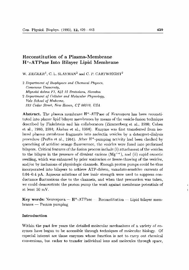
Gen. Physiol. Biophys. (1993), 12, 429—443 429
Reconsti tution of a Plasma-Membrane H+-ATPase Into Bilayer Lipid Membrane
W. ZIEGLER1, C. L. SLAYMAN2 and C. P. CARTWRIGHT2
2 Department of Biophysics and Chemical Physics, Comenius University, Mlynská dolina Fl, 842 15 Bratislava, Slovakia
2 Department of Cellular and Molecular Physiology, Yale School of Medicine, 333 Cedar Street, New Haven, CT 06510, USA
A b s t r a c t . The plasma membrane H + -ATPase of Neurospora has been reconsti
tu ted into planar lipid bilayer membranes by means of the vesicle-fusion technique
described by Finkelstein and his collaborators (Zimmerberg et al., 1980; Cohen
et al., 1980, 1984; Akabas et al., 1984). Enzyme was first transferred from iso
lated plasma membrane fragments into asolectin vesicles by a detergent-dialysis
procedure (Perlin et al., 1984). After H + -pumping activity had been checked by
quenching of acridine orange fluorescence, the vesicles were fused into preformed
bilayers. Critical features of the fusion process include (i) a t tachment of the vesicles
to the bilayer in the presence of divalent cations ( M g + + ) , and (ii) rapid osmotic
swelling, which was enhanced by prior sonication or freeze-thawing of the vesicles,
and /o r by inclusions of physiologic channels. Enough proton pumps could be thus
incorporated into bilayers to achieve ATP-driven, vanadate-sensitive currents of
0.04-0.4 pA. Aqueous solutions of low ionic strength were used to suppress con
ductance fluctuations due to the channels, and when that precaution was taken,
we could demonstrate the proton pump the work against membrane potentials of
at least 50 mV.
K e y words : Neurospora — H + -ATPase — Reconstitution — Lipid bilayer membranes — Proton pumping
I n t r o d u c t i o n
Within the past few years the detailed molecular mechanisms of a variety of en
zymes have begun to be accessible through techniques of molecular biology. Of
especial interest are those enzymes whose function is not to carry out chemical
conversions, but rather to transfer individual ions and molecules through space,

430 Ziegler et al.
from one cell compar tment to another or between the cell interior and exterior,
thus carrying out osmotic work. In the broadest sense, these"Transport-ases" or
"Translocases" are of three classes, depending on the form of energ\ which is cou
pled to the osmotic movement. Most general and wide-spread are the ion-coupled
co- and counter-transport systems, which drain the chemical concentration gradi
ent (or electrochemical gradient) of one ionic species (usually N a + or H + ) in order
to build gradients for other species (e.g. sugars, aminoacids, K + , phosphate, etc.).
The prototype of this class is the lactose permease in the plasma membrane of
Escherichia coh. The second class comprises redox pumps found in the membranes
of energy-conserving organelles and certain bacterial envelope membranes, which
couple the movement of protons (and in some cases sodium ions) to electron flow;
the best understood example of this class is probably the reaction center protein
from photosynthetic bacteria. And finally, the third class comprises a variety of
chemically distinct enzymes which couple ion movement to the energy of phosphate
anhydride bonds (usually via ATP hydrolysis). These are located in virtually every
kind of biological membrane, but are divided chemically into three distinct sub
classes, typified by a) the Fo-Fi ATPase of the mitochondrial inner membrane, b)
the vacuolar ATPase of plant cells, and c) the N a + , K + -ATPase in the plasma
membranes of most animal cells.
Wi th the advent of molecular biological techniques for detailed structural ana
lysis of "Transport-ases", a clear need has also developed to find conditions for
studying the physical and physiological function of these enzymes in isolation. At
present the most at t ract ive condition is that of incorporation into artificial bi
layer lipid membranes, which has been widely used to study ion-channel proteins.
In order to develop bilayer incorporation techniques for pump proteins, we have
focused on one system which appears simplest among the class of ATP-coupled
transporters: the plasma-membrane H + -ATPase from the fungus Neurospora. This
enzyme, whose complete amino-acid sequence has been determined (from the nu
cleotide sequence of its cloned gene (Hager et al. 1986; Addison 1986)), contains
920 amino acids with a total molecular weight of 99.886 and carries out its major
t ransport function as a single unassociated protein monomer (Goormaghtigh et al.
1986). In terms of bo th structure and chemical reactivity, this enzyme is closely
allied with the N> + . K+-ATPase of animal cell membranes, but it functions to
pump (outward ;u .oss the cell membranes) only a single H + ion for each molecule
ATP split. Electrophvsiological experiments have shown that in vivo this pump
can generate currents of approx. 20 / /A/cm 2 (short-circuit conditions; Gradmann
et al. 1978) or can support membrane potentials as large as —350 mV (open circuit
conditions; Blatt et al. 1987). Complementary experiments on plasma membrane
vesicles (Scarborough 1976) and on enzymes reconstituted into liposomes (Perlin et
al. 1984) have shown the enzyme to be electrogenic under those conditions as well.
As compared with channel proteins, pump proteins are difficult to reconstitute

Proton Pumping in a Phospholipid Bilayer 431
satisfactorily into bilayer membranes, because of their low turnover numbers. T h e minimal current which can be measured reliably in biological membrane systems are approx. 0.1 pA, which means 6 x 10 5 charges/s. The estimated turnover number for the Nexihispora H + pump in vivo is near 200 ions/s and per molecule of enzyme, meaning t h a t 3 x 10 3 molecules need t o function for minimally detectable reconst i tut ion. (By comparison, channel proteins are clearly measurable at densities of one molecule/bilayer, and bilayer-vesicle fusion experiments normally yield only a few dozen events/bilayer.) Adequate reconstitution of pumps requires, therefore, extreme care in preserving enzymatic activity throughout the steps of purification and incorporation, substantial enhancement of fusion frequencies, and caution t o make the measuring amplifiers as quiet and sensitive as possible.
A b b r e v i a t i o n s : AO: acridine orange; ATP: adenosine-5'-triphosphate; B T P : 1,3-bis[tris(hydroxymethyl)-methylaminol-propane; C C C P : carbonyl cyanide m-chloro-phenyl-hydrazone; D O C : 7-deoxycholic acid, sodium salt; EDTA: ethylenediamine t e t r a acetic acid; H Ľ P E S : N-2-[hydroxyethyl] piperazine-N'-[2-ethanesulfonic acid]
M a t e r i a l s and M e t h o d s
Preparation of vesicles
Plasma membrane fragments were isolated from Neurospora by the technique of Bowman et al. (1981): partial digestion of the cell walls with snail-gut enzyme, disruption of the cells by homogenization and sonication, fractionation of the membranes by differential centrifugation, resuspension in 1 mmol/1 EGTA-Tris at pH 7.2, and storage at —70 °C. Reformed vesicles were prepared according to the method of Perlin et al. (1984), by first diluting aliquots of the above suspension (containing ca. 3 mg protein) into HEPES-buffered KCl/asolectin: 2 ml final volume, with 10 mmol/1 HEPES-KOH at pH 7.2, 1 mmol/1 NaoEDTA, 150 mmol/1 KC1, and 10 mg of acetone-washed asolectin. Purified DOC was added, to a final concentration of 0.6%, and the suspension was claiified by vigorous homogenization. The resultant solution was rapidly applied to a Bio-Gel P-10 column and eluted with HEPES buffer (similar to above, but with 0.1 mmol/1 Na2EDTA and 500 mmol/1 KC'l) at 1 ml/min. The cloudy fractions were pooled, diluted 40-fold in further HEPES/KC1 buffer, and centrifuged at 100,000 x g for 1 hour. The resultant vesicles (pellet) were resuspended in the same buffer and assayed for their ability to generate a pH gradient by means of AO quenching.
Formation of bilayers
Conventional bilayer lipid membranes were formed over a 0.4 mm hole in a teflon septum (0.1 mm thick) which had been pretreated with bilayer-forming solution prioi to immersion in the recording medium. The bilayer-forming solution consisted of 45 mg/ml of soybean lecithin (SIGMA, type IV-S) and 5 mg/ml soybean lysolecithin (SIGMA) dissolved in spectroscopic grade decane. One microliter of this solution was pipetted onto the teflon septum and allowed to fill the hole. A black film usually formed within 2-5 min. yielding

432 Ziegler et al.
a trans-membrane resistance > 4 x 10¹⁰ Ohms and a capacitance of ca. 1-3x10 F. Standard recording medium contained 10 mmol/1 HEPES-KOH at pH 7.2 and 10 mmol/1 or 100 mmol/1 KC1.
I0GÍ1
command voltage
Figure 1. Block diagram of the circuit used to attach the patch-clamp amplifier to the bilayer chamber. The cis compartment of the chamber was grounded, and the trans compartment connected to the head-stage of the amplifier. Membrane potential determined as trans minus cis, with positive clamp current flowing from the trans to the cis compartment. ATP-driven cis-trans current through the pump is thus negative, opposite to the usual in vivo electrophysiological convention.
Electrical circuits
A Yale "Mark-V" patch clamp amplifier (design of Dr. D.P. Corey) equipped with a 10 GOhm feed-back resistor was used to measure membrane current. The cis compartment of the recording chamber, to which all additions were made, was grounded; the trans compartment was connected to the high-resistance input of the headstage amplifier, as diagrammed in Fig. 1. Reported voltages therefore represent the potential of the trans compartment relative to that of the cis compartment, and positive clamp current is defined as flowing from the trans to the cis side. Amplifier output was filtered by an 8 pole, low-pass Bessel filter set at 30 Hz for routine work and 1 or 2 Hz for measuring the pump current.
Fusion and testing
Reformed plasma-membrane vesicles were fused to bilayers generally in the manner described by Finkelstein and co-workers (Zimmerberg et al. 1980; Cohen et al. 1980). MgCl2 (4 mmol/1) was first added to the cis chamber (total volume 3.5 ml) to supply the divalent ions needed for vesicle attachment and the Mg2+ needed for ATPase activation. A dense suspension of hyperosmotic vesicles (12 fil volume, containing 500 mmol/1 KC1 as described above) was lightly sonicated and then injected from a micropipette placed 0.5 mm in front of the lipid membrane. A small DC potential (2-10 mV, usually trans side negative) was imposed on the membrane, as noted in the figure legends below. Fusion
Proton Pumping in a Phospholipid Bilayer 433
5 r
< O.
a. E o
-2
t t b c
t t d e
0 1 2 3
Time/min 0 1 2 3
Time/min
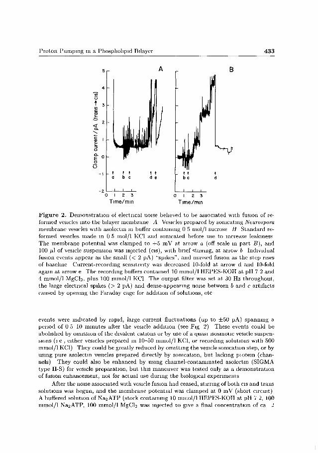
Figure 2. Demonstration of electrical noise believed to be associated with fusion of reformed vesicles into the bilayer membrane A Vesicles prepared by sonicating Neurospora membrane vesicles with asolectm in buffer containing 0 5 mol/1 sucrose B Standard reformed vesicles made m 0 5 mol/l KC1 and sonicated before use to increase leakiness The membrane potential was clamped to +5 mV at arrow a (off scale in part B), and 100 fú of vesicle suspension was injected (cis), with brief stirring, at arrow b Individual fusion events appear as the small (< 2 pA) "spikes", and massed fusion as the step rises of baseline Current-recording sensitivity was decreased 10-fold at arrow d and 10-fold again at arrow e The recording buffers contained 10 mmol/1 HEPES-KOH at pH 7 2 and 4 mmol/1 MgCb, plus 100 mmol/1 KC1 The output filter was set at 30 Hz throughout, the large electrical spikes (> 2 pA) and dense-appearing noise between b and c artifacts caused by opening the Faraday cage for addition of solutions, etc
events were indicated by rapid, large current fluctuations (up to ±50 pA) spanning a period of 0 5 10 minutes after the vesicle addition (see Fig 2) These events could be abolished by omission of the divalent cations or by use of a quasi isosmotic vesicle suspensions (i e , either vesicles prepared in 10-50 mmol/1 KC1, or recording solutions with 500 mmol/1 KC1) They could be greatly reduced by omitting the vesicle somcation step, or by using pure asolectm vesicles prepared directly by somcation, but lacking piotein (channels) They could also be enhanced by using channel-contaminated asolectm (SIGMA type II-S) for vesicle preparation, but this maneuver was tested only as a demonstration of fusion enhancement, not for actual use during the biological experiments
After the noise associated with vesicle fusion had ceased, stirring of both cis and trans solutions was begun, and the membrane potential was clamped at 0 mV (short circuit) A buffered solution of Na2ATP (stock containing 10 mmol/1 HEPES-KOH at pH 7 2, 100 mmol/1 Na2ATP, 100 mmol/1 MgCb was injected to give a final concentration of ca 2

434 Ziegler et al.
mmol/1, and the resultant short-circuit current was recorded. In a few experiments ATP solution (4 mmol/1) was injected close to the membrane without stirring, in the same manner as the vesicle suspension was applied. The pump inhibitor orthovanadate (100 /uniol/1) was applied both by addition to the bulk solute and by injection directly at the membrane. Possible binding artifacts associated with ATP/bilayer experiments (see e.g. Hyman 1977) were checked by adding ATP to fusion bilayers formed with ATPase-free liposomes. Such tests generated no significant short-circuit currents.
Results
Demonstration of proton pumping in reformed vesicles
As has already been shown (Perlin et al. 1984), reformed vesicles from Neurospora
plasma membrane are studded with particles - made visible by freeze-fracture
electron microscopy - of appropriate size and density to be the plasma-membrane
ATPase. They hydrolyze ATP and accumulate protons at the rate of one H +
ion pumped per ATP molecule split (Perlin et al. 1986). Vesicles maintained in
the presence of other permeant ions (i.e. Cl~) build steady-state pH differences
of 2 units (interior acid), as assayed by quenching of the fluorescent pH-indicator
acridine orange (AO; see Fig. 3^4), and those maintained in the absence of other
MgClg V0 4 MgCI2 V0 4 CCCP
Figure 3. ATP-driven proton uptake by isolated and reformed plasma membrane vesicles from Neurospora, assayed by quenching of AO fluorescence, in (̂ 4) HEPES buffer + 10 mmol/1 KC1, or (B) HEPES buffer + 500 mmol/1 KC1. Vesicles were pre-equilibrated with 2 //mol/1 acridine orange; 1.5 mmol/1 ATP (final concentration, neutralized with BTP) was added 30-60 s before these tracks commence. ATP hydrolysis was initiated by injecting 1.5 mmol/1 MgCb, and hydrolysis was stopped by addition of 100 fj,mol/\ orthovanadate. Because the quench was slow to reverse in 500 mmol/1 KC1, 20 /iinol/1 CCCP was injected to restore the control fluorescence level. 30 mg membrane protein in the vesicles of part A, and 35 mg of protein in part B.
Proton Pumping in a Phospholipid Bilayer 435
permeant ions (i.e., acetate as the major anion) rapidly develop equivalent mem
brane potentials (assayed by oxonol-V fluorescence; da ta not shown).
Fluorescence quenching of AO was checked routinely on all vesicles prepara
tions to be used for bilayei fusion experiments. The rate and extent of quenching
varied with the exact preparative procedure, and also with the ionic composition
of the test medium. In particular (see Fig. 2>B) the initial rate of quenching was
50-65% slower in HEPES buffer containing 500 mmol/1 KCl than it was in buffer
with 100 mmol/1 KCl, but total quenching at steady-state was normally about 15%
greater for the higher salt concentration. A simple interpretation of the fluores
cence steady-state is tha t the H + difference across vesicle membranes is limited by
outward leak of protons (Perlin et al. 1986); and the overall date indicate tha t
elevated KCl can separately reduce both the maximal rate of pumping and the
leakiness of the liposomal membrane. (We have so far no explanation of why the
leak appears diminished at higher salt (Cl~) concentrations.)
Occurrence of channels in fused bilayers
In a long series of experiments with variously prepared vesicles, a general correlation
was noted between the amount of fusion noise, the ability of post-fusion bilayers to
produce a current in response to ATP, and the simultaneous presence of discrete
channels in the bilayer. This led to the notion tha t aqueous permeability of the
vesicles might be a controlling factor for fusion events (see also Woodbury and Hall
1987), and also revealed a treacherous experimental problem: to obtain maximal
fusion without introducing enough channel proteins to overcome the low-level pump
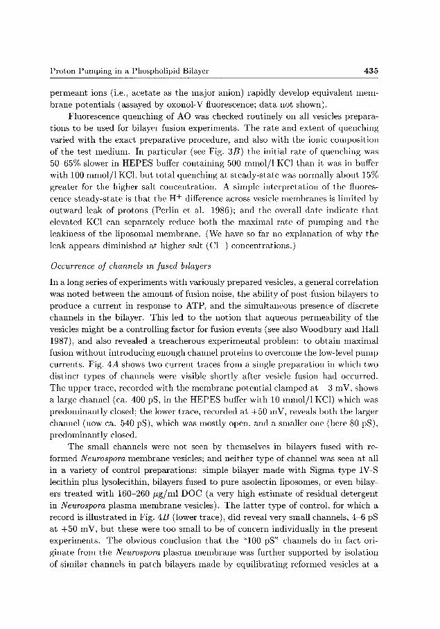
currents. Fig. 4^4 shows two current traces from a single preparation in which two
distinct types of channels were visible shortly after vesicle fusion had occurred.
The upper trace, recorded with the membrane potential clamped at —3 mV, shows
a large channel (ca. 400 pS, in the HEPES buffer with 10 mmol/1 KCl) which was
predominantly closed; the lower trace, recorded at +50 mV, reveals both the larger
channel (now ca. 540 pS), which was mostly open, and a smaller one (here 80 pS),
predominantly closed.
The small channels were not seen by themselves in bilayers fused with re
formed Neurospora membrane vesicles; and neither type of channel was seen at all
in a variety of control preparations: simple bilayer made with Sigma type IV-S
lecithin plus lysolecithin, bilayers fused to pure asolectin liposomes, or even bilay
ers t reated with 160-260 / ig /ml DOC (a very high estimate of residual detergent
in Neurospora plasma membrane vesicles). The latter type of control, for which a
record is illustrated in Fig. 4 5 (lower trace), did reveal very small channels, 4-6 pS
at +50 mV, but these were too small to be of concern individually in the present
experiments. The obvious conclusion that the "100 pS" channels do in fact ori
ginate from the Neurospora plasma membrane was further supported by isolation
of similar channels in patch bilayers made by equilibrating reformed vesicles at a
436 Ziegler et al.
B
5
0
1 -5 c -10
=> 100 o O-E
^ 50 1-
+4~
o
tr*f' ~t <j p r̂y"*"
Jb
-inimtfY-uj-"
—W*^*^^^lŕ'r'^>***'^rs,* i ŕr,f'M'r
+ 50 p mV
- 5 0 20
-20 0 I 2
I -
0 10 20 Time/s
WUjf-WU-L 0 .30 60
Time/s
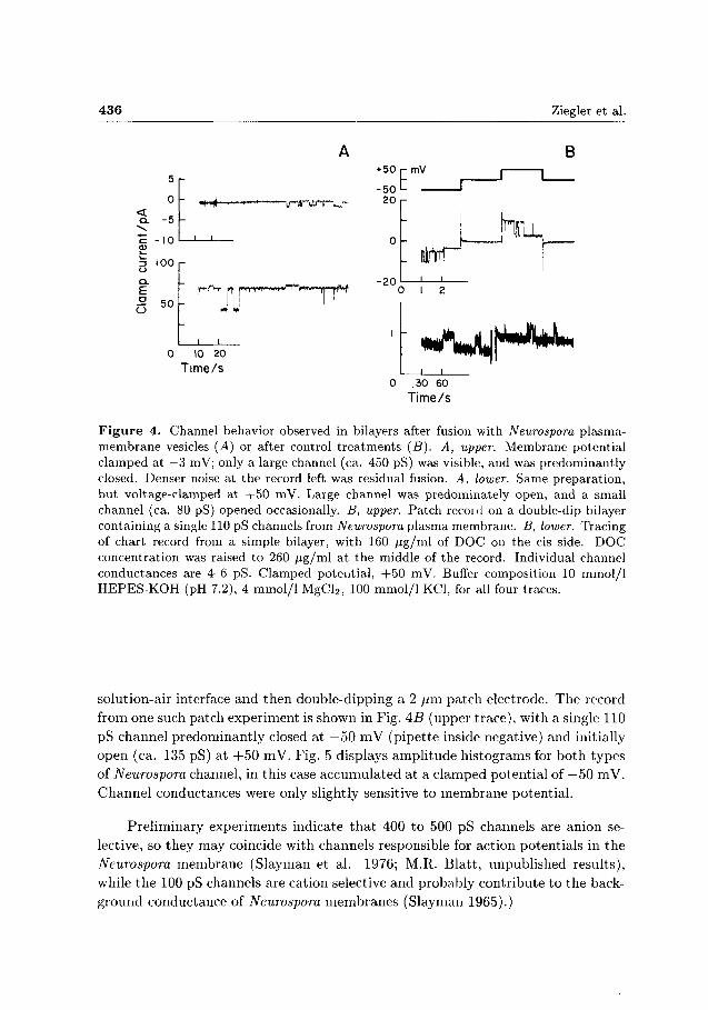
Figure 4. Channel behavior observed in bilayers after fusion with Neurospora plasma-membrane vesicles (A) or after control treatments (B). A, upper. Membrane potential clamped at —3 mV; only a large channel (ca. 450 pS) was visible, and was predominantly closed. Denser noise at the record left was residual fusion. A, lower. Same preparation, but voltage-clamped at +50 mV. Large channel was predominately open, and a small channel (ca. 80 pS) opened occasionally. B, upper. Patch recoid on a double-dip bilayer containing a single 110 pS channels from Neurospora plasma membrane. B, lower. Tracing of chart record from a simple bilayer, with 160 /ig/ml of DOC on the cis side. DOC concentration was raised to 260 /ig/ml at the middle of the record. Individual channel conductances are 4-6 pS. Clamped potential, +50 mV. Buffer composition 10 mmol/1 HEPES-KOH (pH 7.2), 4 mmol/1 MgCl2, 100 mmol/1 KCl, for all four traces.
solution-air interface and then double-dipping a 2 /im patch electrode. The record from one such patch experiment is shown in Fig. AB (upper trace), with a single 110 pS channel predominantly closed at —50 mV (pipette inside negative) and initially open (ca. 135 pS) at + 5 0 mV. Fig. 5 displays amplitude histograms for both types of Neurospora channel, in this case accumulated at a clamped potential of —50 mV. Channel conductances were only slightly sensitive to membrane potential.
Preliminary experiments indicate t h a t 400 to 500 pS channels are anion selective, so they may coincide with channels responsible for action potentials in the Neurospora membrane (Slayman et al. 1976; M.R. Blatt, unpublished results), while the 100 pS channels are cation selective and probably contribute t o the background conductance of Neurospora membranes (Slayman 1965).)

Proton Pumping in a Phospholipid Bilayer 437
0 6
0 5
o c a) 04 3 o o o
Frac
tiona
l O
O
0 1
-
-
1
A
1 1
B
r—i 300 400 500 600 80 120 160
Channel conductance/pS 260 300
Figure 5. Typical amplitude histograms for the large (A) and small (B) channels described in Fig. 4A. Data from a single experiment accumulated over about 20 minutes, with membrane potential clamped at —50 mV. The events at 260-280 pS (B) were assigned to the small channel since they represented openings, not closings, at —50 mV (see text).
Recording of pump currents
The simplest way to augment fusion of reformed plasma membrane vesicles to
bilayer membranes proved to be brief sonication of the vesicle suspension before
addition to the bilayer chamber. (Freeze-thawing of the suspension also yielded
large fusion noise, but tented to inactivate the enzyme.) In such preparations,
ATPase activity remained at 90-95% of the control values, but the initial AO
quenching rate was reduced to 50% or less of the control rate, which may be taken
as at least a crude measure of the vesicle's increased leakiness.
After fusion with such vesicles, bilayers always displayed channel activity, pre
dominantly of the small variety described in Fig. 4 above. Recording of pump
currents required a stable baseline and therefore was possible only when the num
ber of open channels was constant, or nearly so. Fortunately, substi tution of the
normal buffer solution with 10 mmol/1 HEPES containing only 10 mmol/1 KCl
(plus 4 mmol/1 M g C ^ ) served both to reduce individual channel conductance and
to reduce channel switching frequency.
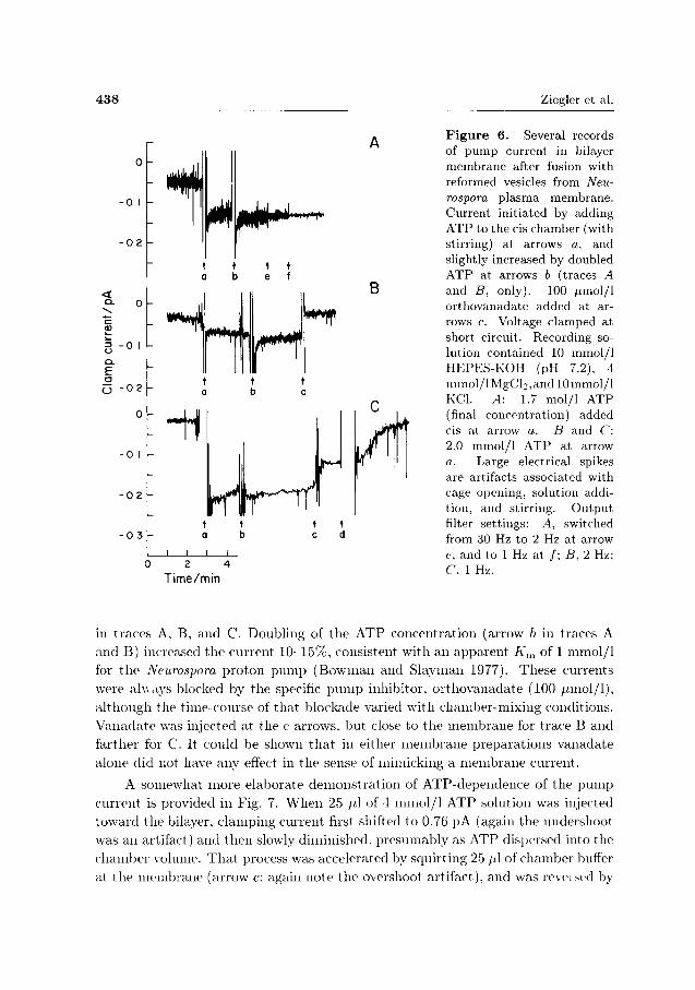
Fig. 6 shows pump activation in several records from bilayers in which chan
nel behaviour had been nearly suppressed, during short-circuit clamping. In each
experiment, ca. 2 mmol/1 ATP (added to the cis compartment at arrow a) pro
duced a negative current, at steady levels of 0.08, 0.03, and 0.22 pA, respectively
438 Ziegler et al.
0 -
- 0 I
- 0 2
< °- 0
3 " 0 I
E • Č3 - 0 2
B
0
- 0 1
- 0 2
- 0 3
- TI
• W\ t i a t
i i i i
Time/min
Figure 6. Several records of pump current in bilayer membrane after fusion with reformed vesicles from Neurospora plasma membrane. Current initiated by adding ATP to the cis chamber (with stirring) at arrows a, and slightly increased by doubled ATP at arrows b (traces A and B, only). 100 /xmol/1 orthovanadate added at arrows c. Voltage clamped at short circuit. Recording solution contained 10 mmol/1 HEPES-KOH (pH 7.2), 4 mmol/lMgCl2,and 10mmol/l KCl. A: 1.7 mol/1 ATP (final concentration) added cis at arrow a. B and C: 2.0 mmol/1 ATP at arrow a. Large electrical spikes are artifacts associated with cage opening, solution addition, and stirring. Output filter settings: A, switched from 30 Hz to 2 Hz at arrow e. and to 1 Hz at /; B, 2 Hz: C, 1 Hz.
in traces A. B, and C. Doubling of the ATP concentration (arrow b in traces A and B) increased the current 10-15%, consistent with an apparent A',„ of 1 mmol/1 for t h e Neurospora proton pump (Bowman and Slayman 1977). These currents were always blocked by the specific p u m p inhibitor, orthovanadate (100 iimol/1), a l though the time-course of that blockade varied with chamber-mixing conditions. Vanadate was injected at the c arrows, but close to the membrane for trace B and farther for C. It could be shown t h a t in either membrane preparations vanadate alone did not have any effect in the sense of mimicking a membrane current.
A somewhat more elaborate demonstrat ion of ATP-dependence of the pump current is provided in Fig. 7. When 25 /il of 4 mmol/1 ATP solution was injected toward the bilayer, clamping current first shifted to 0.76 pA (again the undershoot was an artifact) and then slowly diminished, presumably as ATP dispersed into the chamber volume. T h a t process was accelerated by squirting 25 /A of chamber buffer at the membrane (arrow c; again note the overshoot artifact), and was r e v e l e d by

Proton Pumping in a Phospholipid Bilayer 439
Figure 7. Variation of pump current with ATP supply. Continuous record from a single bilayer preparation, to which ATP was added by injection from a micropipette directly in front of the membrane (see Methods). Arrow a: 25 /tl of 4 mmol/1 ATP added; b: pipette removed, with slight stirring; c: ATP-free buffer squirted at membrane; d: addition of ATP (1.7 mmol/1, final concentration) remote from the membrane, with stirring; e: further stirring, which restored pump current to the initial level. The large current undershoot on first injection (a) and spikes along the record are all artifacts. Output filter setting, 30 Hz. Recording solution as in Fig. 6.
- 1 0 -
- 2 0
E o
- 2 0 -
t t
0 2 4 Time/min
a second pulse of ATP (arrow d).
Bilayers loaded with reformed vesicles of Neurospora plasma membrane tented
to be fragile under either mechanical or electrical stress, and could not be reliable
clamped at potentials larger than about 50 mV. Nevertheless, polarizations up to
this level ( + / —) could be imposed, and pump activation could be observed when a
"bucking" current was applied to the recorder in order to offset the background clue
to average channel opening. One such recording is illustrated in Fig. 8. Baseline
noise in this record represents multiple switching of the 100 pS channels (see Fig. 4).
t t c d
2 min
Figure 8. Demonstration of the ability of bilayer-reconstituted pumps to operate against a membrane potential. Membrane potential clamped at +50 mV, with baseline of open channels requiring ca. 50 pA offset current. Arrow a: 2 mmol/1 ATP added cis, with stirring: arrow c: 100 /imol/1 orthovanadate added, b and d: additional stirring. Recording solution as in Fig. 6. Output filter at 1 Hz: baseline noise represents switching of several small (100 pS) channels.

440 Ziegler et al.
After addition of ATP to the bulk solution in the cis chamber (arrow a) and a second
stirring (arrow b), c lamp current increased by —0.17 pA. Addition of 100 /xmol/1
vanadate to the solution (arrow c, with stirring at d) abolished the ATP-stimulated
current. Although for technical reasons we do not have a direct comparison of ATP
effects on a single membrane in the presence and absence of imposed polarization,
the result in Fig. 8 makes clear at least tha t a 50 mV opposing voltage does not
suppress the proton pump. This result is in keeping with observations on whole cells
of Neurospora, which have found the reversal potential for the plasma membrane
proton pump to be near —400 mV and pump current to be nearly insensitive to the
actual membrane potential , in the range —100 to 0 mV (Gradmann et al. 1978).
Overall, the results described above have shown tha t protein incorporated
into bilayer membranes by fusion with reformed Neurospora membrane vesicles can
generate t ransmembrane currents of a magnitude expected for ca. 104 functioning
enzyme molecules (for numerical estimates see Introduction). Furthermore, the
currents are abolished by orthovanadate, a highly-specific inhibitor of "E1-E2"-
type cation pumps, and they are insensitive to small shifts of membrane potential,
as also would be expected for the Neurospora plasma membrane proton pump.
Discuss ion
We regard the da ta presented above as a minimal demonstrat ion of the feasibility
for functional reconstitution of ion pumps into flat bilayer lipid membranes, and
the procedures used as a starting point for more effective reconstitution. ATP-
stimulated and vanadate-inhibited currents produced by the Neurospora plasma
membrane ATPase are of the same magnitude as those recently reported in bilayers
containing cytochrome c oxidase from E. coli (Hamamoto et al. 1985) or the Fo~
F i ATPase from the thermophilic bacterium PS3 (Hirata et al. 1986). However,
because steady-state currents obtainable from all three enzymes are near the limit of
present recording methods, it is obvious that production-level experiments on these
enzymes in bilayers will require much methodological improvement. (Borisova et
al. (1984) have also reported currents of 0.2-1.0 pA for Streptococcus fecahs F0-F1
ATPase in artificial bilayer membranes, but serious doubt about that work arises
because of contradictions between the conductance and current values given.) As
an operational target, we are aiming for currents 10-fold larger than have been
obtained thus far, i.e. 1-4 pA. This range compares with 10-40 pA steady current
seen for bacterial rhodopsin incorporated into bilayers (Bamberg et al. 1981), or
similar transient currents for N a + , K + -ATPase at tached onto bilayers (Fendler et
al. 1985; Nagel et al. 1987).
The most likely route for further enhancing bilayer reconstitution of the Neu
rospora plasma-membrane ATPase is via purified ATPase which has been reincor
porated into liposomes. As was noted above, bilayers fused to reformed plasma-

Proton Pumping in a Phospholipid Bilayer 441
membrane vesicles become fragile, and indeed usually break spontaneously when
fusion noise is substantially more intense than tha t illustrated in Fig. 2. It is rea
sonable to suppose tha t fragility arises from protein loading, and since the ATPase
is only 5-10% of total protein in the reformed vesicles, a 10-20 fold improvement
of current might be achieved with purified enzyme in liposomes. In other words,
we might expect incorporation of ca. 105 functional pump molecules per bilayer
instead 104 molecules/bilayer or fewer. An important fringe benefit of purification
may be an almost complete abolition of the channel noise observed upon fusion
of reformed plasma-membrane vesicles. This in turn might increase measuring
sensitivity several-fold.
Several other factors hopefully will also give improvements, albeit smaller than
expected for purified enzyme: a) More gentle purification (i.e., with different de
tergents and lipid protection), since only about 50% of pump molecules in the
reformed vesicles are reckoned to be active (K. E. Allen and C. W. Slayman, un
published experiments), b) Alteration of the osmoticum loaded into the vesicles
(by sonication) just before fusion; in preliminary experiments vesicles loaded with
0.25-1.0 mol/1 sucrose were able to fuse with bilayers (judged by the recording
noise), but did not yield measurable currents. Since the hydrolytic activity of su
crose vesicles was equal to that of control vesicles, the nature of pump inhibition
in the bilayers is unclear. It may be related to ionic strength effects of the usual
osmoticum (500 mmol/1 KCl; see Bowman and Slayman, 1977), and bears seri
ous examination, c) Selection of lipids - in the bilayer and /o r the vesicles - with
critical phase behaviour as a function of temperature or ambient pH. Such exper
iments are suggested by burgeoning biochemical work of the past two years (see,
e.g., Abstracts of US. Biophysical Society Meeting, New Orleans, March 1987) on
the relation of lipid phase transitions to vesicle-vesicle fusion efficiency.
Ideally, of course, we would like to obtain bilayers containing purified pumps
at densities similar to those found in normal cell membranes: for the Neurospora
ATPase ca. 3000 /tmi2, so tha t comfortable electrical signals could be obtained from
cell-sized pieces of membrane (i.e., 100-1000 / im2) , which could be electrically iso
lated in large patch electrodes. Whether such densities can be obtained by physical
reconstitution is unclear. Test- tube biosynthesis of single-protein membranes may
be a more feasible possibility in the future.
Acknowledgements. The authors are indebted to Mr. Kenneth Allen and Dr. Gerald Zuckier for crucial technical assistance, and to Dr. Carolyn Slayman for much helpful advice. The work was supported by research grant GM15858 from the National Institute of General Medical Sciences.
R e f e r e n c e s
Addison R. (1986): Primary structure of the Neurospora plasma membrane H+-ATPase deduced from the gene sequence. J. Biol. Chem. 261, 14896—14901

442 Ziegler et al.
Akabas M. H., Cohen F. S., Finkelstein A. (1984): Separation of the osmotically driven fusion event from vesicle-planar membrane attachement in a model system for exocytosis. J. Cell Biol. 98, 1063—1071
Bamberg E., Dencher N. A., Fahr A., Heyn M. P. (1981): Trans- membraneous incorporation of photoelectrically active bacteriorhodopsin in planar lipid bilayers. Proc. Nat. Acad. Sci. USA 78, 7502—7506
Blatt M. R., Rodriguez-Navarro A., Slayman C. L. (1987): The potassium-proton symport in Neurospora: Kinetic control by pH and membrane potential. J. Membrane Biol. 98, 169—189
Borisova M. P., Babakov A. V., Kolomytkin O. V. (1984): H+-ATPase in a planar lipid bilayer. Biol. Membrány 1, 187—190
Bowman B. J., Slayman C. W. (1977): Characterization of plasma membrane adenosine triphosphatase of Neurospora crassa. J. Biol. Chem. 252, 3357—3363
Bowman E. J., Bowman B. J., Slayman C. W. (1981): Isolation and characterization of plasma membranes from wild type Neurospora crassa. J. Biol. Chem. 256, 12336— 12342
Cohen F. S., Zimmerberg J., Finkelstein A. (1980): Fusion of phospholipid vesicles with planar phospholipid bilayer membranes. II. Incorporation of a vesicular membrane marker into the planar membrane. J. Gen. Physiol. 75, 251—270
Cohen F. S., Akabas M. H., Zimmerberg J., Finkelstein A. (1984): Parameters affecting the fusion of unilamellar phospholipid vesicles with planar bilayer membranes. J. Cell Biol. 98, 1054—1062
Fendler K., Grell E., Haubs M., Bamberg E. (1985): Pump currents generated by the purified Na+ , K+-ATPase from kidney on black lipid membranes. EMBO J. 4, 3079—3085
Goormaghtigh E., Chadwick C , Scarborough G. A. (1986): Monomers of the Neurospora plasma membrane H+-ATPase catalyse efficient proton translocation. J. Biol. Chem. 261, 7466—7471
Gradmann D., Hansen U.-P., Long W. S., Slayman C. L., Warncke J. (1978): Current-voltage relationships for the plasma membrane and its principal electrogenic pump in Neurospora crassa. I. Steady-state conditions. J. Membrane Biol. 39, 333—367
Hager K. M., Mandala S. M., Davenport J. W., Speicher D. W., Benz E. J., Slayman C. W. (1986): Amino acid sequence of the plasma membrane ATPase of Neurospora crassa: Deduction from genomic and cDNA sequences. Proc. Nat. Acad. Sci. USA 83, 7693—7697
Hamamoto T., Carrasco N., Matsushita K., Kaback H. R., Montal M. (1985): Direct measurment of the electrogenic activity of o-type cytochrome oxidase from Escherichia coli reconstituted into planar lipid bilayers. Proc. Nat. Acad. Sci. USA 82, 2570—2573
Hirata H., Ohmo K., Shone N., Kagawa Y., Hamamoto T. (1986): Direct measurment of the electrogenicity of the H+-ATPase from thermophilic bacterium PS3 reconstituted in planar phospholipid bilayers. J. Biol. Chem. 261, 9839—9843
Hyman E. S. (1977): Electrogenesis from an ATPase-ATP-sodium pseudo pump. J. Membrane Biol. 37, 263—275
Nagel G., Bamberg E., Fendler K., Grell E. (1987): Na+ currents generated by the purified Na+ , K+-ATPase on planar lipid membranes. Biochim. Biophys. Acta 901, 239-249

Proton Pumping in a Phospholipid Bilayer 443
Perlin D. S., Kasamo K., Brooker R. J., Slayman C. W. (1984): Electrogenic H + translocation by the plasma membrane ATPase of Neurospora: Studies on plasma membrane vesicles and reconstituted enzyme. J. Biol. Chem. 259, 7884 7892
Perlin D. S., San Francisco M. J. D., Slayman C. W., Rosen B. P. (1986): H+/ATP stoichiometry of proton pumps from Neurospora crassa and Escherichia coh. Arch. Biochem. Biophys. 248, 53—61
Scarborough G. A. (1976): The Neurospora plasma membrane ATPase is an electrogenic pump. Proc. Nat. Acad. Sci. USA 73, 1485—1488
Slayman C. L. (1965): Electrical properties of Neurospora ciassa: Effect of external cations on the intracellular potential. J. Gen. Physiol. 49, 69—92
Slayman C. L., Long W. S., Gradmann D. (1976): "Action potential" in Neurospora crassa, a mycelial fungus Biochim. Biophys. Acta 426, 732—744
Woodbury D. J., Hall J. E. (1987): Porin channels greatly enhance osmotically induced fusion of vesicles with planar bilayers. Biophys. J. 51 , 45a item M-PM-E9
Zimmerberg J., Cohen F. S., Finkelstein A. (1980): Fusion of phospholipid vesicles with planar phospholipid bilayer membranes. I. Discharge of \esicle contents across the planar membrane. J. Gen. Physiol. 75, 241—250
Final version accepted October 6, 1993

444 Ziegler et al.