Embed Size (px)
Citation preview

Volume 13 Number 13 1985 Nucleic Acids Research
Molecular recognition of B-DNA by Hoechst 33258+
Keith D.Harshman and Peter B.Dervan*
Division of Chemistry and Chemical Engineering, California Institute of Technology, Pasadena,CA 91125, USA
Received 18 March 1985; Revised and Accepted 27 May 1985
A13STRACTThe binding sites of Hoechst 33258, netropsin and distamycin on three DNA restrictionfragments from plasmid pBR32? were compared by footprinting with methidiumpropyl-EDTA-Fe(ll) [ MPE.Fe(lI)]. Hoechst, netropsin and distamycin share common binding sitesthat are five ± one bo in size and rich in A*T DNA base pairs. The five base pairprotection patterns for Hoechst may result from a central three base pair recognition sitebound by two bisbenzimidazole NHs forming a bridge on the floor of the minor groovebetween adjacent adenine N3 and thymine 02 atoms on opposite helix strands.Rvdroohobic interaction of the flanking phenol and N-methylpiperazine rings would afforda steric blockade of one additional base pair on each side.
1NTROrMJCTIONHoechst dye 33258 (H) is a bisbenzimidazole that binds double helical DNA rich in
adenine (A) and thymine (T) base pairs (1-2). Martin and Holmes have shown that the
binding of Hoechst 33258 on DNA requires a minimum of four consecutive A.T base pairs
(2). Hoechst is of similar size and shape to the di- and tripeptides, netropsin and
distamycin, which show a marked preference for A-T rich DNA (3-13). From a recent X-
ray analysis of the complex of netropsin with the B-DNA dodecamer of sequence
CGCGAATTCG(CG, Dickerson and coworkers orovide a molecular basis for the sequence
specific recognition of nNA by netropsin, and, by extension, distamycin (14). They find
that the crescent shaped netropsin sits symmetrically in the center of the minor groove of
right-handed DNA and displaces the water molecules of the spine of the hydration (14).Fach of its three amide NH groups forms a bridge between adjacent adenine N3 or
thymine 02 atoms on opposite helix strands (14). This explains recent data that
distamycin-like analogues having n-amides characteristicallv bind to n+1 successive base
pairs (15-17). The inside edge of the crescent framework of Hoechst has potential NHrecogniton elements similar in disposition in space to the carboxamide NHs of netropsin
and distamycin. Mikhailov and coworkers have suggested that the bisbenzimidazole
framework of Hoechst forms a helix isogeometric to B-form DNA similar to netropsin and
distamycin (1). A crystal structure of Hoechst dye bound to DNA does not yet exist.
However, with the availability of the netropsin:DNA dodecamer crystal structure (14), it
© I R L Press Limited, Oxford, England. 4825
Nucleic Acids ResearchVolume 13 Number 13 1985

Nucleic Acids Research
would seem appropriate to ask by footprinting methods whether Hoechst, netropsin, anddistamycin, molecules similar in shape though different in chemical structure, share
common binding sites on B-form DNA. Accurate resolution of the binding site sizes mayprovide insight on the number of hydrophobic, hydrogen bonding, and electrostaticrecognition elements for each molecule. We report here a comparison of the binding sites
of Hoechst 3325R, netropsin, and distamycin on three 32P end-labeled restriction
fragments from plasmid p%R322 by footprinting methods. We use the synthetic DNA
cleaving agent, methidiumpropyl-EDTA.Fe(II) (MPF..Fe(II)) which has been shown to
resolve binding site sizes more accurately than the enzyme ONase I (18-19).
MATERIALS AND METHODS
Reagents and Enzymes. nistamycin A was obtained from Boehringer M-annheim.Hoechst dye 33258 was obtained from Calbiochem. Netropsin was a gift of D. Patel.
MUethidiumoropyl-EnTA (M4PE) was synthesized and purified as described by Hertzberg andDervan (20-21). Purities were determined by thin layer chromatography. Concentrations
were determined spectroscopically. D)ithiothreitol (DTT) was obtained from Calbiochem.Ferrous ammonium sulfate, Fe(NH4)2(S04)2*6 H20 was obtained from Baker. Restrictionendonucleases and the Klenow fragment of DNA polymerase I were obtained from NewEngland Biolabs. Bacterial alkaline phosphatase and T4 kinase were obtained from BRL.
DNA Restriction Fragments. Three restriction fragments from plasmid pBR322
were prepared. Superhelical plasmid pBR322 was first digested with restriction endo-
nuclease B3am HI and labeled at the 3'-end with a-32P dATP and the Klenow fragment of
1DNA polymerase I. A second enzymatic digest with Eco RI yielded the 3' end-labeled 381
bp fragment which was isolated according to the procedures of Maxam-Gilbert (21). The5' end-labeled 381 hp fragment was obtained by treatment of Banm HI digested plasmidpBR322 first with bacterial alkaline phosphatase and then with y-32P ATP and T4 kinase
prior to restriction with Eco RI. The plasmid pBR322 was labeled at the 5' and 3' end at
the Eco RI site, followed by restriction with Rsa I to yield the end-labeled 167 and 517 bp
fragments.Footprinting. The MPE.Fe(II) cleavage reactions were run in a buffer (TN)
containing 10 mM Tris, pH 7.4 and 50 mM NaCI. To 4 iL solution containing 2.5 x TNbuffer, >600 cpm 32P end-labeled restriction fragment, and 250 vM (base pair) sonicated
calf thymus DNA was added 2 viL of the inhibiting compound of the appropriateconcentration (see Fig. 2 for final concentrations). This solution was incubated in thedark for 30 min at 370C. Next, 2 pL of a freshly prepared solution containing 12.5 iMUMPE and 25 pM Fe(NJH4)2(S04)2 was added. This was incubated for an additional 15 minat 370C. The cleavage reaction was initiated by the addition of 2 jiL freshly prepared20 mM dithiothreitol, bringing the total reaction volume to 10 jL. The cleavage reactionwas allowed to continue for 15 min at 370C before stopping by freezing in dry ice. The
4826

Nucleic Acids Research
samples were then lyooholyzed and resuspended in 4 iL of formamide loading buffer.
Sequencing Gels. Cleavage inhibition patterns were determined by electrophoresison 0.4 mm thick, 40 cm long, 9% polyacrylamide (167 and 517 bp fragments) or 5%
polyacrvlamide (381 bp fragment), 1:20 crosslinked sequencing gels containing 50% urea.
Electrophoresis was carried out at 1000 volts for 7.5 h (381 bo fragment) or 4 h (167 and
517 bp fragments). Autoradiography was carried out at -700C without the use of an
intensification screen. An 8" x 10" copy of the original autoradiogram was scanned at 485nm with the incident beam collimated to a width of 0.2 nm on a Cary 219 spectrophoto-meter. The data was recorded as absorbance relative to the film base density.
RFSULTSFootprints produced by partial cleavage of three nNA restriction fragments from
plasmid pIlR322 protected by Hoechst, netropsin and distamycin were examined. The 381-bp (Ram HI-Eco RI), 167 bp (Eco RI-Rsa I), and 517 bp (Eco RI-Rsa I) DNA fragments haveseveral strong Hoechst, netropsin and distamycin binding sites. The nNA fragments
labeled at the 5' (or 3') end with 32p were allowed to equilibrate with Hoechst, netropsin
and distamycin at ratios of ligand to DNA base pair of 0.06 and 0.03. Then MPE-Fe(II) was
added to afford final ratio of MPE-Fe(II) to MNA base pair of 0.025. The reaction was
initiated by the additon of dithiothreitol (MTT) at 4 mM concentrations. Cleavage byMPE.e(TI) was stopped after 15 min (370C) by freezing, lyophilization, and resuspension
in a formamide buffer. The 32P end-labeled DNA products were analyzed by high
resolution denaturing gel electrophoresis capable of resolving DNA fragments differing in
length by one nucleotide. The autoradiograms for MPE.Fe(II) footprinting on the 381, 167
and 517 bp fragments are shown in Figures 2.
0
Figure 1. (Left to right) Hoechst 33258, netropsin and distamycin.
4827

Nucleic Acids Research
av
.ii . 3... A.
WI-.,
s | ..|$
,. EiT.... .z...
__y.._.
::y.3S'r Z
|M,.C.Z,; .N'
ik M
3
.
.. ..R t...t.X..l
..>.. ... ..w.,- ,*_
,w
to.5'.'' 3_.
:X:'w_1
w min........_ .. .. .. .. ...:
:, . _s
U~~~~~~~~~i i7H:
Ely .....<. .aw.&.P
.,,4V: . g.:
..
ei F
'.
#1....;i...
IN a "tII...01M ...
W.=.
:L0
C-LC2
2
'::
0
c-
I
rWt
! 2
f-
0,,..
L7!^*1. '.. :.
i.
iTi,.
_4U.v":I'
CD
C
2
2
zM
L_
rO 4IL.2F4;h
I
.\ S
C.Ul
4828
*.
,i
.,~it
.1-.:-f'
K
z;!
Wp-. .. B....; .11.1111t.. mill' 11 I
vemg.v.4kiWIrI"1111 .....................
1
1
i
i.
i
i
i
i
I
r
I
i.P,11
IiI
I
:,:i
,I

Nucleic Acids Research
C H H N N D D GI I
H H N N D D G
I
345 678 9 1011 12131415161718
.....
_0
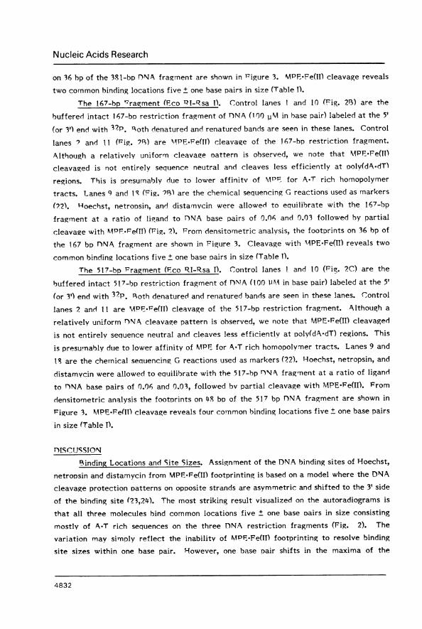
Figure 2. (A) Autoradiogram of 381 bp TINA restriction fragment, (Ps) 167 bp DNArestriction fragrnent, and (C) 517 bD PDNA restriction fragment. Lanes 1-9 and 10-18 areDTNA labeled with 32P at the 5' and 3' ends, respectively, lanes I and 10, intact DNA;lanes ? and II, MAPF-Fe(II) cleavage of unprotected DNA; lanes 3-8 and 12-17, MPli.Fe(ll)footDrinting with Hoechst at 6.3 vAM (lanes 3 and 12) and 3.1 lvM (lanes 4 and 13),netronsin at 6.3 v1M (lanes 5 and 14), at 3.1 viM lanes (lanes 6 and 15) distamycin at6.3 pvM (lanes 7 and 16) at 3.1tiM (lanes S and 17); lanes9 and I8 are the Maxam-Gilbertchemical sequencing G-specific reaction. riottom to middle of the autoradiogram is theseauence left to right in Figure 3.
4829
1 2
do

Nucleic Acids Research
H _
5'GCGC ATAG CAT CAACGCA GCTAGC3'CGCGTA GT AGTTGCGTj GCGATCG
N
5'GCGCAT G A GCATCAACGCA IGCGCTAGC3'C GC G TA TC TT T AACGTA GT T GC G ATC GC GA TC G
D
5'GCGCATAGCATCAACGC CGCTAGC3'CGCGTA GT AGTTGC _GCGATCG
BH
5'CGGTTA-CACAGTT AAATTIGCTAACGC AGTCA3'G CCATICAAATAGTG TCAAITTTAAjCG ATTGCGTCAGT
N _
5'CGGTAGTTTATCACAGTTAATTIGCTAACGCAGTCA3GC CATCIAAATAGTGTC AAMITTAAJC G ATTGCGTCAGT
D w5'CGGT TTTATCCAGTTGCTAACGCAGTCA3'GCCAT AAATAGTGTCAAIITTATCGATTGCGTC AGT
CH
5'CGCCTICCTTATITATAGG AT ATGATAATATGG CTTAGACG3GCGG ATCC TACA TACTCA_AT A AAAGAATCTGC
N
5'CGCCTIATTTT1TATAGGTTAATIGTCATGAT ATAITGGITTTCTTIAGACG3'GCGGA AAAAATATCC A CAGTACTA_ AC A_ ITCTGC
w -~Nw
D
5CGCCTAfTTFATAGGTTAATGTCATGATAATAATGGTTTCTTAGACG3SGCGGATAAAA TATCC AATTACAGTACTATTAT TACCAIAAGAAT CTGC
Figure 3. FootDrints of Hoechst (H), netropsin (N) and distamycin (D) at 6.3 Mconcentration on (A) the 381 bp restriction fragments, bp 264-229 of pBR322 (Fig. 2), (B)the 167 bp restriction fragment bp 40-75 of pBR322 (Fig. 3) and (C) the 517 bp restrictionfragment bp 4333-4286 of pBR322 (Fig. 4). The MPIF.Fe(II) footprints (light regions in thegel autoradiograr, Fig. 2) are shown as histograms. The height is proportional to thereduction of cleavage at each nucleotide compared with MPF.-Fe(II) cleavage of unpro-tected 1)NA (control lanes ? and I 1, Fig. 2). The top strand patterns are for 5' end-labeledFINJA; the bottom strand patterns are for 3' end-labeled 1)NA. Boxes are the Hoechst,netropsin and distamycin binding sites, assigned by the asymmetric MPE.Fe(TI) footprint-ing model (16,23,24).
4830

Nucleic Acids Research
Table 1. Binding Sites of Hoescht 33258 (H), Netropsin (N) and T)istamycin (D)
Capital letters denote the sequence (5'-3') of the presumedlower case letters show the neighboring nucleotides.
binding sites. The
The 381 hD Fragment (Bam HI-Fco RI). Control lanes 1 and 10 (Fig. 2A) are thebuffered intact 381 bp restriction fragment of Y)NA (100 1M in base pair), labeled at the
5' (or 3') end with 32p. Both denatured and renatured bands are seen in these lanes.Control lanes 2 and 11 (Fig. 2A) are MPE.Fe(II) cleavage of the 381-bp restriction
fragment. Although a relatively uniform DNA cleavage pattern is observed, we note thatMPE-Fe(II) cleavaged is not entirely sequence neutral and cleaves less efficiently at
poly(dA-dT) regions. This is presumably due to lower affinity of MPE for A-T richhomopolymer tracts. Lanes 9 and 18 (Fig. 2A) are the Maxam-Gilbert chemical
sequencing G reactions used as markers (22). Hoechst, netropsin and distamycin were
allowed to equilibrate with the 381 bp nNA fragment at ratios of ligand to DNA base pairof 0.06 (Fig. 2A, lanes 3, 5, 7, 12, 14, 16) and 0.03 (Fig. 2A, lanes 4, 6, 8, 13, 15, 17)
followed by partial cleavage with MPE-Fe(II). From densitometric analysis, the footprints
4831
Restriction BindingFragment Site Site Size
381 H AAATT 5N aAATT 4n AAATT 5
H aTATA 4N aTATA 4n ATATA 5
1'57 H GTTT.AT 6N gTTTAT 5rG-TTTA t 5
H AAATT 5N AAATT 5F) AAATT 5
517 H ATTTTt 5N ATTTTt 5D) aTTTTT 5
H TTAATG 6N TTAATg 5I) TTAATg 5
H1 AATAA 5N AATAA 5F) AATAA 5
H TTTCTta 5N TTTCTTa 6D- tTTCTTA 6
Nucleic Acids Research
on 36 bp of the 381-bp DNA fragmnent are shown in Figure 3. MPF Fe(II) cleavage reveals
two common binding locations five ± one base pairs in size (Table I).
The 167-bp '-ragment (Fco PI-Rsa I). Control lanes I and 10 (Fig. 21) are the
buffered intact 167-bp restriction fragment of DNA (100 11M in base pair) labeled at the 5'
(or 3') end with 32P. qoth denatured and renatured bands are seen in these lanes. Control
lanes I and ItI (Pig. VP>) are MAPE-Fe(I) cleavage of the i67-bp restriction fragment.
Although a relatively uniform cleavage oattern is observed, we note that MPF,.Fe(HI)
cleavaged is not entirely sequence neutral and cleaves less efficiently at rooly(dA.dT)
regions. This is presumably due to lower affinitv of MPF for A-T rich homopolymer
tracts. Lanes 9 and iS (Fig. ?13) are the chemical sequencing G reactions used as markers
(02). 4oechst, netroDsin, and distamycin were allowed to equilibrate with the 167-bp
fragment at a ratio of ligand to DNA base pairs of 0.06 and 0.03 followed by partial
cleavage with 4PF.Pe(II) (Fig. 2). From densitometric analysis, the footprints on 36 bp of
the 167 br DNA fragment are shown in Figure 3. Cleavage with MPE.Fe(lI) reveals two
common binding locations five ± one base pairs in size (Table 1).
The 517-bp Fragment (Fco RI-Rsa I). KControl lanes 1 and 10 (Fig. 2C) are the
buffered intact 517-bp restriction fragment of DNIA (100 1AM in base pair) labeled at the 5'
(or 3') end with 32P. Roth denatured and renatured bands are seen in these lanes. Control
lanes 2 andl 11 are MPEFe(II) cleavage of the 517-bp restriction fragment. Although a
relatively uniform DNA cleavage pattern is observed, we note that MPE.Fe(II) cleavaged
is not entirely sequence neutral and cleaves less efficiently at poly(dA.dT) regions. This
is presumably due to lower affinity of MPF for A.T rich homopolymer tracts. Lanes 9 and
18 are the chemical sequencing G reactions used as markers (22). Hoechst, netropsin, and
distamycin were allowed to equilibrate with the 517-bp DTNA fragment at a ratio of ligand
to D1NA base pairs of 0.06 and 0.03, followed bv partial cleavage with MPE.Fe(II). From
densitometric analysis the footorints on 48 bs of the 517 bp DNA fragment are shown in
Figtire 3. MPE-Fe(II) cleavage reveals four common binding locations five ± one base pairs
in size (Table I).
DISCUSSON
Binding Locations and Site Sizes. Assignment of the DNA binding sites of Hoechst,
netronsin and distamycin from MPF.Fe(II) footprinting is based on a model where the DNA
cleavage protection patterns on opoosite strands are asymmetric and shifted to the 3' side
of the binding site (23,24). The most striking result visualized on the autoradiograms is
that all three molecules bind common locations five ± one base Dairs in size consisting
mostlv of A-T rich sequences on the three DNA restriction fragments (Fig. 2). The
variation may simply reflect the inabilitv of MAPF.Fe(II) footprinting to resolve binding
site sizes within one base pair. However, one base pair shifts in the maxima of the
4832

Nucleic Acids Research
-N0
H2N
3 5
-,N -HNI-N
0
14H~~~~~~~~~H5 /N
3' N 3N H
Figure 4. Model for distamycin and Hoechst 33258 in the minor groove of B-DNA. Thisis a refineinent of the AMikhailov model (1) and is based on the X-ray structure of netropsin(14). Circles with two dots represent lone pairs of electrons on NJ3 of adenine and 02 ofthymine at the edoes of the base pairs on the floor of the minor groove of the DNA helix.Dotted lines are bridged hydrogen bonds to the bisbenzimidazole NH.
cleavage inhibition patterns from MIPE-Fe(II) footprinting can be easily identified from the
densitometric traces and are the basis for our assignments here. From affinity cleavingexperiments on these same DNA restriction fragments, we know that distamycin has a
binding site size of five base pairs (15-17). Distamycin binds these five base pair sites
with two orientations (15-17). A footprint at a discrete location mav be the sum of two
orientations protecting that site. If both orientations bind the same base pairs, a
minimum binding site size from footorinting will be observed. However, if the two
orientations bind different bases within a common location, then the footprint from an
MPF.Fe(IT) cleavage experiment should be larger than the minimum binding site size.Using M4PE.Fe(IT) footprints of distamycin as a guide, we find the maxima of the
asymmetric inhibition natterns on opposite D)NA strands are typically separated by onebase pair. We assign the Hoechst and netropsin binding site size as five base pairs if themaxima of the asymmetric inhibition patterns are the same as distamycin, and four or sixbase pairs if the separation of the maxima is one base pair smaller or larger, respectively.
Of the eight common binding locations analyzed on the three restriction fragments,
Hoechst has a binding site size of five base pairs on 5 of the 8 sites, netropsin has a
binding site size of five base pairs on 5 of the 8 sites and distamycin has a binding site
size of five base pairs on 7 of the 8 sites (Table I). Although all binding sites for Hoechst,netroDsin and distamycin are in common locations, not all sites are identical. The sites
that are identical for all three molecules are (5'-3') AAATT and AATAA (Table I). Atseveral locations there are variations of one to two base pairs. For example, on the 167
bp DNA fragment Hoechst, netropsin and distamycin bind (5'-3') GTTTAT, gTTTAT, andlGTTTAt, respectively. On the 517 bp DNA fragment Hoechst, netropsin and distamycin
4833

Nucleic Acids Research
bind (5'-3') ATTTTt, ATTTTt, and aTTTTT, respectively, at one site, and TTTCTta,
TTTCTTa, and tTTCTTA, at another (Fig. 3). Finally, we note that Hoechst, like
netropsin and distamycin, will bind A.T rich sequences containing GGC base pairs (Table I)
(0,16,17).Molecular Origin of the Specificity of Hoechst. The MAPF.Fe(TI) footprinting data
presented here reveals that Hoechst binds similar locations on DNA as netropsin and
distamycin. If the snecificity of these crescent shaped molecules for these common
locations on DNA are of similar molecular origin, it mav not be unreasonable to refine the
Mikhailov model of Hoechst based on the crystal structure of netropsin bound to DNA
(2,14). Similar to netronsin and distamycin, we presume that the binding of Hoechst to
homopolvmer A.T rich regions involves the displacement of water molecules in the spine
of hydration (12,14). Like netropsin and distamycin, the electrostatic interaction of the
cationic end and the negative potential in the minor groove of DNA undoubtedly
contrihute to the binding stabilitv of Hoechst (13). From inspection of the models,Hoechst has two possible NH recognition elements on the bisbenzimidazoles capable of
bridging adjacent adenine N3 or thymine 0? atoms on opposite helix strands in the minor
groove of B-DNA. Therefore, based on the crystal structure for netroosin (14) and the n+l
rule for 2jjgo-N-rnethylpyrrolecarboxamides (13-15), one might expect a binding site size
for Hoechst of three base pairs due to bisbenzimidazole recognition alone. However, a
binding site size of five ± one base pairs is observed. One possibility is that the Hoechst
protection pattern on DNA results from more than one binding mode, such as two
orientations that use some but not all common base pairs. The alternative explanation is
that a single common central three base pair binding location is utilized by the
bisbenzimidazole and in addition, the phenol and N-methyloiperazine rings flanking the
bisbenzimidazole add a steric blockade of one base pair on each side of the A-T hydrogenbinding site. This would afford overall Drotection from MPE.Fe(II) cleavage of five base
pairs (Fig. 4). Similar to netropsin binding, perhaps Hoechst sits in the center of the
minor groove with its four rings twisted noncoplanar, so that each ring is parallel to the
walls of the groove to afford a good steric fit in the right-handed helix (Fig. 4). Why the
binding by oligo-N-methylpyrrolecarboxamides and bisbenzimidazole in the minor groove
of B-DNA helix is centered at the same locations must be due to the local micro-
environment of these particular A.T rich sequences (Table I). Dickerson has pointed out
that the sequence specific recognition of certain A-T rich regions may be the result of
hydrophobic interactions in the minor groove and the NH hydrogen bonding elements
simoly align the inside edge of the crescent-shaped molecule on the floor of the minor
groove of the helix (14). With regard to the design of synthetic sequence specific DNA
binding molecules, a comparison of the Hloechst 33258, netropsin and distamycin
structures suggests that flat aromatic rings twisted in a screw sense to match the walls of
4834

Nucleic Acids Research
the DNA helix and oriented on the floor of the helix by one or more bridged hydrogen
bonds may be a general feature of B-form D)NA recognition at A.T rich sequences.
ACKNOWLEDGEMENTSSupported by the American Cancer Society research grant Number NP-428.
*To whom correspondence should be addressed
+Contribution no. 7162 from Division of Chemistry and Chemical Engineering, California Institute ofTechnology, Pasadena, CA 91125, USA
RF,FER NCFS1. Mikhailov, M. V., 7asedatelev, A. S., Krylov, A. S., & Gurskii, G. V. (1981) Mol.
Biol. (F.ngl. Trans.) 15, 541-553.2. Martin, R. F., &k Holmes, N. (1983) Nature 302, 452-454.3. 7immer, Ch. "Progress in Nucleic Acids Research and Molecular Biology"; Cohn,
W. E., Ed.; Academic Press: New York, 1975, p. 285-318.4. Krey, A. K. "Progress in Molecular and Subcellular Biology"; Hahn, F. E., Ed.;
Springer-Verlag: New York, 1980, Vol. 7, p. 43.5. McGhee, J. n. (1976) Biopolymers 15, 1345-1375.6. Luck, G., Zimmer, Ch., Reinert, K. E., &' Arcamone, F. (1977) Nucl. Acids Res. 4,
2655-2670.7. Patel, n. J., & Canuel, L. L. (1977) Proc. Natl. Acad. Sci., U.S.A. 74, 5207-521 1.8. Berman, H. M., Neidle, S., Zimmer, Ch., & Thrum H. (1979) Biochem. Biophys. Acta
561, 124-131.9. Krylov, A. S.; Grokhovsky, S. L., Zasedatelev, A. S., Zhuze, A. L., Gursky, G. V., &c
Gottikh, B. P. (1979) Nucl. Acids Res. 6, 289-304.10. Van Dyke, M. W., Hertzberg, R. P., &c nervan, P. B. (1982) Proc. Natl. Acad. Sci.,
U.S.A. 79, 5470-5474.11. GTursky, G. V., Zasedatelev, A. L., Zhuze, A. L., Khorlin, A. A., Grokhovsky, S. L.,
Streltsov, S. A., Surovaya, A. N., Nikitin, S. M., Krylov, A. S., Retchinsky, V. 0.,MVikhailov, M. V., Beabealashvili, R. S., & Gottikh, B. P. (1982) Cold Spring HarborSvmP. Ouant. #liol.47, 367-378.
12. Marky, L. A., Plumenfeld, K. S., & Breslauer, K. J. (1983) Nucl. Acids Res. 11, 2857-2870.
13. Zakrzewska, K., Lavery, R., &c Pullman, B. (1983) Nucl. Acids Res. 11, 8825-8839.14. Kopka, M. L., Yoon, C., Goodsell, 1)., Pjura, P., & Dickerson, R. E. (1985) Proc.
Natl. Acad. Sci. USA 82, 1376-1 380.15. Iavior, J. chultz, P. G., & Dervan, P B. (1984) Tetrahedron 40, 457-465.16. 'Schultz, P. G., &r Dervan, P. B. (1984) Biomolecular ¶TETUre and Dynamics 1,
1.133-1147.17. Youngquist, R. S., & Dervan, P. B. (1985) Proc. Natl. Acad. Sci., USA 82, 2565-2569.18. Van Dyke, M. W., & Dervan, P. B. (1983) Nucl. Acids Res. 11, 5555-5567.19. Van Dyke, M. W., DDervan, P. B. (1984) Science 225, 1127-1 127.20. Hertzberg, R. P., & Dervan, P. B. (1982) J. Am. Chem. Soc. 104, 313-315.21. Hertzberg, R. P., & Dervan, P. B. (1984) Biochemistry 23, 3934-3945.22. Maxam, A. M., & Gilbert, W. (1980) MethodcEnzymol. 65, 499-560.23. Van Dyke, M. W., Ac Dervan, P. B. 1983Cold Srin Harbor Symposium 47, 347-
353.24. Van Dyke, M. W., & Dervan, P. B. (1983) Biochemistry 22, 2373-2377.
4835


![Prezentace18 [režim kompatibility] - Univerzita Karlovakfrserver.natur.cuni.cz/lide/schwarze/bunka_prakt/...• Princip vizualizace pomocí barviva Hoechst 33258, SYTO barviva (web](https://img.dokumen.tips/doc/110x75/60e56b8f0e3d7563012d7d43/prezentace18-reim-kompatibility-univerzita-a-princip-vizualizace-pomoc.jpg)


![Kirin-Amgen v Hoechst (TKT) - FICPI Canada · J.A. KEMP & CO. FICPI ABC Meeting QUEBEC CITY June 2005 Kirin-Amgen v Hoechst (TKT) [2004] UKHL 46 Alan Senior](https://img.dokumen.tips/doc/110x75/5b3cd6247f8b9a5e1f8dad96/kirin-amgen-v-hoechst-tkt-ficpi-ja-kemp-co-ficpi-abc-meeting-quebec.jpg)