Embed Size (px)
Citation preview

36 A. RADUNZ UND G. H. SCHMID
Reactions of Antisera to Lutein and Plastoquinone with Chloroplast Preparations and their Effects on
Photosynthetic Electron Transport
A l f o n s R a d u n z u n d G e o r g H . S c h m id
Max-Planck-Institut für Züchtungsforschung (Erwin-Baur-Institut) Abt. Menke,Köln-Vogelsang
(Z. Naturforsdi. 28 c, 36—44 [1973]; received November 3, 1972)
Antibodies, lutein, plastoquinone, chloroplasts, photosystem II
An antiserum to lutein inhibits photosynthetic electron transport between water and potassium ferricyanide in chloroplasts from green Nicotiana tabacum var. John William’s Breadleaf. However, electron transport between diphenylcarbazide and potassium ferricyanide is not impaired. From this it is concluded that the photochemically active carotenoid should feed its electrons into the photosynthetic electron transport chain before the site from which diphenylcarbazide donates electrons. The inhibition of the ferricyanide H ill reaction in chloroplasts by antibodies to lutein depends on the accessibility of the carotenoid antigen in the thylakoid membrane. In fresh preparations the accessibility is greater in chloroplasts in which photosynthetic electron transport is coupled to photophosphorylation. Concomitantly the antiserum to lutein agglutinates only such chloroplast preparations in which the H i l l reaction is impaired by the antiserum.
An antiserum to plastoquinone inhibits ferricyanide photoreduction of chloroplasts regardless whether driven by water or diphenylcarbazide as the electron donors. Typical photosystem-I-reactions are not influenced by the antiserum. In a certain type of chloroplast preparations the antiserum does not inhibit PMS-mediated photophosphorylation inferring that plastoquinone, eventually involved in this reaction, is either not accessible to antibodies, or that this cyclic electron flow does not necessarily pass through plastoquinone.
In a previous paper we showed that antibodies to chlorophyll are specifically adsorbed onto the lamellar system of chloroplasts from Nicotiana tabacum and Antirrhinum majus 1. Depending on the condition in which the lamellar system is in, agglutination occurs either directly or after the addition of rabbit-anti-y- globulin from the goat. It was demonstrated that the antibodies to chlorophyll inhibit photosynthetic electron transport on the photosystem II side. From the experiments it was concluded that part of the reaction centre chlorophyll of photosystem II was situated in an accessible location in the thylakoid membrane. K o e n ig
and co-workers describe antibodies to a photosystem II-activity exhibiting preparation, namely antibodies to a particle preparation that did not exhibit any photosystem I activity 2. These antibodies impair electron transport on the photosystem-II-side between water and diphenylcarbazide. B r a u n and G o v in d je e 3
independently report on an antiserum which, according to the presented data, inhibits photosynthetic electron flow at probably the same site as that obtained by
K o e n ig et al. These reports are to our knowledge the only ones using the serological approach to characterize photosystem II. In view of the question of energy transfer from carotenoids to chlorophyll4 or from chlorophyll to carotenoids 5>8 and the participation of carotenoids in the primary photochemical activity of photosystem II 7> 8 we have prepared an antiserum to lutein. With an antiserum to this chloroplast pigment we expected to obtain information as to whether carotenoids are located on the outside or inside face of the thylakoid membrane. The eventual effect of the antiserum on photosynthetic electron transport was supposed to characterize the possible role of carotenoids in photosynthesis. Plastoquinone, a component of the photosynthetic electron transport chain, is easily extractable from chloroplast preparations as shown by many authors 7> 9_u. The component is thought to participate in PMS-mediated photophosphorylation 12>13 and W it t suggests that a plastoquinone complexed with the reaction centre chlorophyll of photosystem II (Chlaii) serves as the primary electron acceptor to
Requests for reprints should be sent to Dr. Georg H. Abbreviations: DCPIP, 2-6-dichlorophenol-indophenol; Sc h m id , M ax-Planck-Institut für Züchtungsforschung PMS, phenazine methosulfate; DCMU, 3,4-dichloro-
(Erwin-Baur-Institut), D-5000 Köln 30. phenyl-l,l-dim ethylurea.
This work has been digitalized and published in 2013 by Verlag Zeitschrift für Naturforschung in cooperation with the Max Planck Society for the Advancement of Science under a Creative Commons Attribution-NoDerivs 3.0 Germany License.
On 01.01.2015 it is planned to change the License Conditions (the removal of the Creative Commons License condition “no derivative works”). This is to allow reuse in the area of future scientific usage.
Dieses Werk wurde im Jahr 2013 vom Verlag Zeitschrift für Naturforschungin Zusammenarbeit mit der Max-Planck-Gesellschaft zur Förderung derWissenschaften e.V. digitalisiert und unter folgender Lizenz veröffentlicht:Creative Commons Namensnennung-Keine Bearbeitung 3.0 DeutschlandLizenz.
Zum 01.01.2015 ist eine Anpassung der Lizenzbedingungen (Entfall der Creative Commons Lizenzbedingung „Keine Bearbeitung“) beabsichtigt, um eine Nachnutzung auch im Rahmen zukünftiger wissenschaftlicher Nutzungsformen zu ermöglichen.

ANTIBODIES TO PHOTOSYSTEM -II-REACTIONS IN CHLOROPLASTS 37
photosystem I I 14 which according to him might be identical to the photosystem II fluorescence quencher Q 5. This quencher was thought to be located between the reaction centre chlorophyll of photosystem II (Chlaii) and plastoquinone ls. We felt that an antiserum to plastoquinone should give, as in the case of lutein, information as to whether plastoquinone is situated in an accessible location in the thylakoid membrane. In this case we expected interference of the antibodies to plastoquinone with photosynthetic electron transport.
Indeed, our experiments demonstrate that the antisera to lutein and plastoquinone do interfere with photosynthetic electron transport on the photosystemII side.
Materials and Methods
Isolation of lutein: The main part of the carotenoids and the chlorophylls were eluted over Florisil columns from ether soluble Urtica lipids 16. In order to separate from the chlorophylls the pigment mixture was hydrolysed at room temperature by addition of 5 %> sodium ethylate according to the methods of B r u n n e r and G r o b 17. The carotenoids were dissolved in ether. Isolation of lutein was achieved by thin layer chromatography on silica gel-G plates with petrolether, benzene, ethanol (100, 20, 12.5) as solvent according to the method of E ic h e n b e r g e r and G r o b 18. Subsequently lutein was rechromatographed on plates of CaCOa, MgO, Ca(OH)2 according to H a g e r and M e y e r - B e r t e n r a t h 19. The solvent used in this case was benzene (b. p. 100—140 °C), acetone, chloroforme (50, 50, 40). The obtained lutein was twice recristal- lized from a benzene/methanol mixture. Analyzed by thin layer chromatography, it was pure.
Plastoquinone: 2,3-dimethyl-5-solanesyl 1,4 benzo- quinone was purchased from Hoffmann-La-Roche & Co Ltd (Basel, Switzerland). It was rechromatographed on silica gel-G plates with benzene as solvent. Immunisation with lutein and plastoquinone: 2 mg antigen were dissolved in 5 ml ethanol and 5 ml water were added to this solution. The ethanol was completely removed by concentrating the suspension to 0.5 ml. Subsequently, 1 ml rabbit serum and 1 ml of a 2 °/o sodium chloride solution were mixed together and emulsified by ultrasonication. This emulsion was injected into the ear vein of rabbits on every second day. In a parallel immunisation tests we also used a solution of 0.1 % methylated bovine serum albumin instead of rabbit serum. In order to test for the formation of immunoglobulins, blood was withdrawn from the animals before treatment after the 6th and then after every further third injection. The serum was stored at —16 °C.
Agglutination tests: The passive heme-agglutination test as well as the protein decomposition by subtilisin
have been described earlier 2°-22. For the antiglobulin test, equal amounts of antiserum and stroma-freed chloroplasts (1.5 mg/ml) suspended in physiological saline (0.8 °/o) were mixed together. After 12 hours the chloroplasts were washed 5 times in physiological saline and centrifuged each time at 2000 g. The incubated and washed chloroplasts were suspended in phosphate buffer pH 7.4 and added to the same volume of anti-rabbit-y-globulin from the goat of type IgG or type IgM. The reactions were followed under the light microscope. Anti-rabbit-y-globulin, type IgG and IgM were obtained from Miles-Yeda LTD, Kiryat Weiz- man, Rehovot, Israel. The membrane fragments of the ultrasonic supernatant were washed 8 times in water after incubation with the respective antisera and subsequently centrifuged at 75000 g. The sediment was suspended by ultrasonication. Rabbit sera, withdrawn from the animals before treatment and antisera to the methylated bovine serum albumin gave negative results in the respective control assays.Chloroplast preparations: Stroma-containing chloroplasts from Nicotiana tabacum var. John William’s Broadleaf were prepared according to H o m a n n and S c h m id 23.
Pigment analyses, light measurements and photoreactions were carried out as described by R a d u n z et a l .1 and K o e n ig et al. 2.
Electron microscopy: The specimens for electron microscopy were prepared according to the procedures described earlier 23. Sections were cut on a LKB U ltratome III using glass knives, double stained with uranyl- acetate and with lead citrate and examined under an Elmiskope I (Siemens) at 80 kv.
Reactions of the antisera to lutein and plastoquinone with chloroplast preparations
Rabbits were immunised with lutein and plastoquinone in order to obtain antisera. The antigens were emulsified in rabbit blood serum from the respective animals or with methylated bovine serum albumin solutions. After 12 injections, all 4 treated animals produced antibodies. If a suspension of stroma-freed chloroplasts is added to lutein or plastoquinone antiserum, no agglutination is observed (Table I). How ever, a green agglutinate is formed if rabbit anti-y- globulin type IgG is added according to the method of C o o m b s et a l.24. Addition of rabbit anti-y-globulins type IgM does not yield an agglutinate. The same results were obtained, if the non bound serum proteins are removed from the chloroplast suspension by washing with physiological sodium chloride, before the addition of rabbit anti-y-globulins. These results indicate that although a direct agglutination is not obtained, the immunoglobulins to lutein and plastoquinone are

38 A. RADUNZ UND G. H. SCHMID
Table I. Reactions of the antisera to lutein and plastoquinone with different diloroplast preparations.
ChloroplastPreparation
Antiserumto
AgglutinationTest
Antiserum to Rabbit-y-Globulin
of Type
AntiglobulinTest
stroma-freedchloroplastsfrom Antirrhinum
lutein no agglutinate IgGIgM
green agglutinate no agglutinate
stroma-freedchloroplastsfrom Antirrhinum
plastoquinone no agglutinate IgGIgM
green agglutinate no agglutinate
ultrasonic sediment from Antirrhinum
luteinplastoquinone
no agglutinate no agglutinate
ultrasonic supernatant from Antirrhinum
lutein no precipitate IgGIgM
green precipitate no precipitate
ultrasonic supernatant from Antirrhinum
plastoquinone no precipitate IgGIgM
green precipitate no precipitate
chloroplasts from Antirrhinum and N. tabacum in
luteinplastoquinone
green agglutinate green agglutinate
0.4 m sucrose,0.05 M tris buffer pH 7.8 and 0.01 m saline
specifically adsorbed onto the lamellar system. Furthermore, the antiglobulin test shows that immunoglobulins of the type IgG are formed against plastoquinone and lutein.
After partial protein decomposition by subtilisin, stroma-freed chloroplasts are agglutinated by the antiserum to lutein and plastoquinone. Thus, the two antisera react with stroma-freed chloroplasts in a similar manner to the antiserum to chlorophyll1 and the antisera to the two anionic chloroplast lipids sulphoquino- vosyl diglyceride 20 and phosphatidyl glycerol21. Free thylakoid stacks (ultrasonic sediment), as well as fragments of the thylakoid membrane (ultrasonic supernatant), obtained from stroma-freed chloroplasts by ultrasonication and subsequent centrifugation at 34000 g 2S, are not agglutinated by the antisera to lutein and plastoquinone. However, the immunoglobulins to lutein and to plastoquinone are adsorbed onto the membrane fragments of the ultrasonic supernatant (Table I), as demonstrated by the antiglobulin test according to the C o o m b s method 24. A precipitation of the membrane fragments occurs after incubation with the antisera only if rabbit anti-j/-globulins type IgG are added. Consequently, the membrane fragments behave like chloroplasts towards the lutein and plasto
quinone antisera. At this point it should be noted that the antiserum to chlorophyll agglutinates directly the ultrasonic sediment. From the agglutination tests we may draw the conclusion that the antigenic determinants of the lutein and plastoquinone are accessible in the thylakoid membranes of stroma-freed chloroplasts, as are those of chlorophyll1, sulpholipid 20 and phosphatidyl glycerol 21. These determinants should be located in depressions between protein molecules. In our earlier paper we presented evidence that chloroplasts from Antirrhinum and Nicotiana 23, prepared according to a certain method in a sucrose, tris buffer solution are directly agglutinated by antisera to chlorophyll1, sulpholipid 20 and phosphatidyl glycerol 21. The same chloroplast preparations are also agglutinated by antisera to lutein and plastoquinone. As a possible explanation for the direct agglutinability of these chloroplasts, we had previously proposed that the thylakoids of these chloroplasts may swell considerably due to water uptake (Fig. I* )21. A displacement of the lutein and plastoquinone molecules nearer towards the outer surface could take place, due to an expansion of the thylakoid membrane.
* Figs 1 and 4 see Table page 40a.
ANTIBODIES TO PHOTOSYSTEM -II-REACTIONS IN CHLOROPLASTS 39
On the other hand, it is possible that inner parts of the partitions become accessible to immunoglobulins due to a swelling of the thylakoids (Fig. 1). Accessibility should also become increased by ultrasonication. However, as the ultrasonic sediment is not agglutinated by the antisera to lutein and plastoquinone in contrast to the antiserum to chlorophyll, the following conclusions may be drawn: Lutein and plastoquinone molecules are located in depressions of the thylakoid membrane in both the intergrana regions and the partitions. On the other hand chlorophyll molecules are situated in different locations in the intergrana and grana regions. Thus, chlorophyll molecules would be located in the intergrana regions equally in depressions but in the grana regions they should be located more towards the outer face of the thylakoid membrane. However, it should be emphasized that these suggestions are only conclusive if no change in molecule orientation is taking place during ultrasonication.
It should be mentioned, that the antigens lutein and plastoquinone did not yield a positive reaction in the passive heme-agglutination test26. Therefore it was not possible to investigate the specificity of the two antisera.
Effect of the antiserum to lutein on photosynthetic electron transport
The antiserum to lutein inhibits the ferricyanide H i l l reaction of chloroplasts prepared according to H o m a n n and Schm id 23. Stroma-freed chloroplasts from Antirrhinum 1 are not inhibited. I t appears that photosynthetic electron flow coupled to photophos-
Time [m in]-----*■
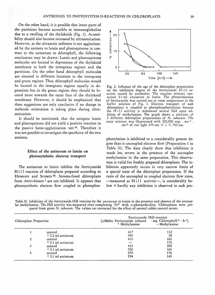
Fig. 2. Influence of the age of the chloroplast preparation on the inhibition degree of the ferricyanide H i l l reaction caused by antibodies. The reaction mixture contained 0.1 ml antiserum to lutein. The photoreduction of ferricyanide was carried out at room temperature in the buffer solution of Fig. 1. Electron transport of such chloroplasts is coupled to photophosphorylation because the H i l l activity is stimulated several fold upon addition of methylamine. The graph shows a selection of 3 different chloroplast preparations of N . tabacum. The assay mixture was illuminated with 300,000 ergs . sec-1 .
cm-2 of red light 575 nm < A < 700 nm.
phorylation is inhibited to a considerably greater degree than is uncoupled electron flow (Preparation 1 in Table II). The data clearly show that inhibition is much less severe in the presence of the uncoupler methylamine in the same preparation. This observation is valid for freshly prepared diloroplasts. The inhibition apparently occurs in very narrow limits of a special state of the chloroplast preparation. If the ratio of the uncoupled to coupled electron flow rates, —measured as H i l l activity—, is considerably below 4 hardly any inhibition is observed in such pre
Table II. Inhibition of the ferricyanide H ill reaction by the antiserum to lutein in the presence and absence of the uncoupler methylamine. The Hill activity was measured after complexing Fe2+ w ith o-phenanthroline. Chloroplasts were pre
pared from green N. tabacum. The values are corrected for the effect of normal rabbit control serum.
Ferricyanide Hill-reactionChloroplast Prepartion [aMoles Ferricyanide reduced • mg Chlorophyll-1 • h_1]
+ Methylamine - Methylamine
1 control 417 112+ 0.2 ml antiserum 395 38
2 control 415 165+ 0.1 ml antiserum — 170
3 control 415 200+ 0.1 ml antiserum 350 165
4 control 370 176+ 0.1 ml antiserum 334 145

40 A. RADUNZ UND G. H . SCHMID
Table III. Photophosphorylation mediated by K3 Fe(CN)# or PMS in diloroplasts from green N. tabacum in the presence of antibodies to lutein and plastoquinone. The assays were carried out in 120 000 lux white light at 15 °C. The chloro- plast preparations type 1 and 2 differed only in their phosphorylating activities but were both prepared according to the
same procedure.
Photophosphorylation [//Moles ATP formed-mg ChlorophylH-h-1]
Serum Ks Fe(CN)e [PMS] PMSChloroplast Chloroplast Chloroplast
Type 1 Type 1 Type 2
Control without Serum 34 95 245 + 35Antiserum to Lutein 46 160 320 + 15Control Serum 37 95 320 ±15Antiserum to Plastoquinone 35 160 318 ±30Control Serum 31 95 393 ±25
parations (Preparations 2-4 in Table II). Furthermore the relative inhibition by the antiserum is lost in the course of aging of the chloroplast preparations (Fig. 2). At the point were no inhibition of the ferri- cyanide Hill rection is observed in a given chloroplast preparation, the agglutinability of the chloroplast preparation is equally lost. It is inferred that thylakoid membranes alter in the course of aging, resulting in a change of the molecular structure of the thylakoid membrane. Ferricyanide-mediated photophosphorylation appears to be slightly stimulated in the presence of antiserum to lutein (Table III). No inhibition of this typical system II reaction is observed in different types of chloroplasts regardless wether the control rate is high or low (Table III). When testing the effect of the antiserum to lutein on the chloroplast preparation, four effects are to be considered: Firstly, the rate of H i l l activity of the control experiment decreases; secondly the antiserum to lutein inhibits ferricyanide photoreduction; thirdly the control serum might stimulate this photoreduction and finally the observed degree of inhibition becomes smaller in the course of the experiment (Fig. 2). From this it is obvious that it is difficult to give a reliable value for the maximal degree of inhibition of the ferricyanide Hill reaction in chloroplasts from green tobacco. Maximal degrees of inhibition varied in 10 experiments between 10 and 66 %>. In addition it should be mentioned that in several experiments up to a 65 % inhibition was also observed immediately upon the addition of methyl- amine. Thus in the coupled state and in the transition from the coupled to the uncoupled condition, up to 65 °/o of lutein, which plays a role in photosynthetic electron transport, must be accessible to antibodies to lutein. The position of the carotenoid in the electron
transport chain is tentatively placed on the side from which electrons are donated to the reaction centre of photosystem II, rather than on the acceptor side. An argument in favor of this view is that the antiserum to lutein does not inhibit electron transport by chloroplasts between diphenylcarbazide, hydroxylamine or hydrazine and ferricyanide respectively (Table V). Hence, the inhibition site must be located before diphenylcarbazide. If one visualizes only sensitizer functions of the carotenoid in question, then the presented data mean, that binding of antibodies to the carotenoid would quench this energy transfer to chlorophyll an .
The binding of antibodies to the carotenoid might lead to an alteration of the molecular structure of the thylakoid membrane, resulting in an increase of the mean distance between chlorophyll and carotenoid molecules. It would be difficult, although possible, to interpret the results of Table V in this context, but this would require speculations. From this it becomes obvious that we should make fluorescence measurements.
Table IV. Anthraquinone-2-sulfonate M e h 1 e r reaction with the DCPIP/ascorbate donor couple in red light in the presence of antibodies to plastoquinone and lutein. Chloroplasts were prepared from N. tabacum. The oxygen uptake was measured with an oxygen electrode as described previously(2>. The assays were carried out in the
presence 10-5 m DCMU.
Serum[//Moles
Oxygen Uptake • mg Chlorophyll-1 • h-1]
Antiserum to Lutein 585Control Serum 550
Antiserum to Plastoquinone 664Control Serum 540

A lfons R adunz und G eorg H. SchmxD, Reactions of Antisera to Lutein and Plastoquinone with Chloroplast Preparations and their Fffects on Photosynthetic Electron Transport (S. 36)
Fig. 1. Lamellar system of a chloroplast from green N. tabacum in 0.4 m sucrose, 0.05 m tricine, and 0.01 m saline, fixed in the dark in glutaraldehyde. a) Fixation5 min after suspension of the chloroplasts in the tricine buffer. Mag. 39 500 :1. b) Fixation 45 min after suspen
sion of the chloroplasts in the tricine buffer. Mag.39 500 : 1.
Fig. 4. Lamellar system of a chlorplast from green N . ta- bacum under the conditions of Fig. la but in the presence of 6.7 • 10-2 m methylamine. Mag. 39 500 : 1. The electron transport of such chloroplasts is in the range of 500—1200 //moles ferricyanide reduced • mg chlorophyll-1 • h_1, because of uncoupling from photophosphorylation.
Zeitschrift für Naurforsdiung 28 c, Seite 40 a.

ANTIBODIES TO PHOTOSYSTEM -II-REACTIONS IN CHLOROPLASTS 41
Table V. Photoreduction of ferricyanide in chloroplasts from green N. tabacum in the presence of antibodies to lutein. The assay was carried out at 300 000 ergs • sec-1 • cm-2 of red light 575 nm < k < 700 nm at room temperature
The rates are initial rates measured after 2 min of illumination.
Electron Donor SerumPhotoreduction of Ferricyanide
[^wMoles Ferricyanide reduced- mg ChlorophylH-h-1]
h ,o antiserum to lutein 208(coupled electron flow) control serum 287
h 2o antiserum to lutein 460(methylamine uncoupled electron flow)
control serum 455
Diphenyl antiserum to lutein 373carbazide control serum 330Hydroxylamine antiserum to lutein 94
control serum 77
Typical photosystem I reactions (such as PMS- mediated cyclic photophosphorylation Table III),
NADP-photoreduction and anthraquinone-2-sulpho- nate photoreduction (Table IV) with the D CPIP/ ascorbate couple as electron donors are not inhibited by the antiserum to lutein. In view of the suggestion of T. H iya m a and B. K e 27 that a carotenoid (P 430) may be a candidate for the position of primary acceptor of photosystem I, we tested the antiserum to lutein in the anthraquinone-2-sulphonate M e h l e r reaction (Table IV). Our data show that the M e h -1 e r reaction mediated by the DCPIP-ascorbate couple in the presence of DCMU is not influenced by the antiserum.
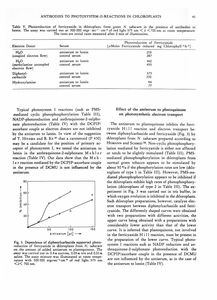
A n t i s e r u m [ |x l ] -----►
Fig. 3. Dependence of diphenylcarbazide supported photoreduction of ferrcyanide in diloroplasts from N. tabacum on the amount of added antiserum to plastoquinone. The assay was carried out in 0.4 m sucrose, 0.05 m tris and 0.01m saline. The assay mixture was illuminated at room temperature with 300 000 ergs-sec_1-cm“* of red light 575 nm < a < 700 nm.
Effect of the antiserum to plastoquinone on photosynthetic electron transport
The antiserum to plastoquinone inhibits the ferricyanide H i l l reaction and electron transport between diphenylcarbazide and ferricyanide (Fig. 3) by chloroplasts from N. tabacum prepared according to H o m a n n and S c h m id 23. Non-cyclic photophosphorylation mediated by ferricyanide is either not affected or tends to be slightly stimulated (Table III). PMS- mediated photophosphorylation in chloroplasts from normal green tobacco appears to be stimulated by about 50 °/o if the phosphorylation rates are low (chloroplasts of type 1 in Table III). However, PMS-mediated photophophorylation appears to be inhibited if the chloroplasts exhibit high rates of photophosphorylation (chloroplasts of type 2 in Table III). The experiment in Fig. 3 was carried out in tris buffer, in which oxygen evolution is inhibited in the chloroplasts. Such chloroplast preparations, however, catalyze electron transport between diphenylcarbazide and ferricyanide. The differently shaped curves were obtained with two preparations with different activities, the upper curve being obtained with a preparations with considerably lower activity than that of the lower curve. It is inferred that plastoquinone, not involved in the ferricyanide H i l l reaction, must be present in the preparation of the lower curve. Typical photosystem I reactions such as NADP reduction and an- thraquinone-2-sulphonate photoreduction with the DCPIP/ascorbate couple in the presence of DCMU are not influenced by the antiserum, as in the case of the antiserum to lutein (Table IV).

42 A. RADUNZ UND G. H. SCHMID
Discussion
Our experiments present evidence that a carotenoid, probably lutein, can actively participate in photosynthetic electron transport. According to the data of Tables II and V, we suggest electron donor properties before the site from which diphenylcarbazide donates electrons to the reaction centre of photosystem II. However, inhibition could also, be due to a conformational change, of proteins for example, in the thylakoid membrane, induced by the binding of antibodies to carotenoid. This would only be possible if the carotenoid in question is bound to a protein or a suitable cofactor molecule. In this case no direct participation of the carotenoid in photosynthetic electron transport would be necessary. The observation that the relative degree of inhibition of the ferricyanide H i l l reaction is lost, during the short period in which the absolute chloroplast activity decreases by only 30 °/o, clearly shows that the chloroplast membrane is undergoing a change. Concomitantly, the agglutinability of the chloroplast preparation is lost, inferring that the binding of antibodies to this site results in both inhibition of the ferricyanide H i l l reaction and agglutination. The observation that chloroplasts in which photosynthetic electron transport is still coupled to photophosphorylation, are inhibited to a larger degree by the antiserum is reminiscent of a suggestion by P a c k e r ,
namely that the molecular structure of thylakoid membranes undergoes changes in the presence of uncouplers of photophosphorylation 28. In context with our observation that a carotenoid seems to transfer energy to the reaction centre of photosystem II, work by H a r n is c h f e g e r and G a f f r o n should be mentioned 4>29. These workers found that the H i l l activity of tobacco chloroplasts may be much lower in blue light than in red ligh t29. This lower rate in blue light could be partly brought back to that in red light by increasing the osmotic pressure of the medium, for example, by adding more sucrose to an originally hypertonic medium. From their studies with monochromatic light, they postulated that the initial transfer of energy from blue absorbing accessory pigments to chlorophyll is interrupted in the first phase of their effect4. Our observation gives support to this interpretation. Also very recent work by O k a y a m a and B u t l e r has demonstrated the participation of ^-carotene in photosynthetic electron transport on the photosystem II side7. These workers found that “a carotenoid, —in their
case ^-carotene—, is essential for the primary photochemical activity of photosystem II” 7. Due to the fact that we were unable to test the specificity of the antiserum to lutein we cannot be certain that our antiserum is specific for lutein only.
The maximal degree of inhibition is difficult to fix as has been shown. However, in two preparations a maximal degree of inhibition of the ferricyanide H i l l reaction of 65 % was observed. If one accepts the assumption that antibodies do not enter the partitions, it must be concluded from this 65 % inhibition that photosystem II is also or even preferentially present in the intergrana region.
This is seemingly contradictory to the earlier suggestion of H o m a n n and S c h m id 23, and to all those reports who have supported and confirmed this suggestion 3°-38. It was suggested that a fully, active photosystem I could be associated with single unfolded thy- lakoids whereas a fully functioning photosystem II seemed to require the close packing of at least two thylakoids. The dependence of the quantum efficiency of PMS-mediated DCMU-insensitive photophosphorylation on wavelength together with fluorescence data clearly suggested the presence of some inactive (with respect to O a-evolution) photosystem II in single unfolded thylakoids or intergrana regions 23. However, it should be borne in mind that a condition of the lamellar system, such as that presented in Figs la and b would easily explain that 65°/o of the total carot- enoids are accessible to antibodies if these were indeed mainly located in the partitions. From this it appears that a statement that an antigen of the thylakoid membrane is accessible to antibodies or not, is not meaningful if the condition in which the thylakoid membrane is actually in, is neither understood nor defined.
Consequently, antigenic determinants of the chloroplast lamellar system which appear not accessible to antibodies might not only be located on the inside face of the thylakoid membrane but also in partition regions, whenever partitions are present in the respective chloroplast preparation or chloroplast condition (Fig.4). With this interpretation in mind, results of B r ia n - t a is and P ic a u d 39 could be brought in line with reports of others 1-3. However, localisation of antigenic determinants on the thylakoid membrane is only meaningful if the antigens from which the respective sera are derived are pure and their chemical nature therefore being known. B y means of an antiserum to a chloroplast particle fraction it might appear quite

ANTIBODIES TO PHOTOSYSTEM -II-REACTIONS IN CHLOROPLASTS 43
plausible that the primary acceptor of photosystem I is located on the outside face 39 but the remainder of photosystem I or components associated with photosystem I might well be located elsewhere 2.
Summarizing our results we would like to emphasize again that we have made 3 observations which at the first appear to be completely contradictory. Firstly, we observed that the coupled chloroplasts are inhibited to a greater extend than uncoupled ones in fresh preparations. Secondly, the relative degree of this inhibition is lost in the course of approximately2 hours (Fig. 2). Thirdly, in aged chloroplast preparations in which the rate of electron flow has decreased to less than 50 °/o, the first-mentioned relationship seems to be reversed, i. e. methylamine uncoupled chloroplasts are occasionally inhibited to a higher degree than coupled ones.
An obvious explanation is that two effects are involved: Namely changes of the morphological structure of the lamellar system and alterations of the molecular structure of the thylakoid membranes. Especially in the case of lutein it clearly appears that there are at least two kinds of lutein molecules both of which are accessible to antibodies but only one kind being involved in electron transport or the functioning of photosystem II. Whether a molecule plays a role in electron transport depends on its location in the thylakoid membrane which is demonstrated by the agglutinability.
In context with the morphological structure of the lamellar system we would like to quote I z a w a and G o o d 40>41. These workers have demonstrated that the addition of amine uncouplers causes shrinkage of the chloroplasts and the lamellar system 40>41. The state in Fig. 1 (0.4 m sucrose, 0.05 M tricine, 0.01 m NaCl, no illumination) is comparable to their observations in isotonic medium. Chloroplasts in the same medium but containing 0.06 m methylamine-HCl appear to have a lamellar system with a more grana-like structure (Fig. 4) just as observed by Iz a w a and G o d d 40> 41. Besides the fact that the described situation can account for the accessibility of partition regions in the initially uncoupled state, it further explains that under conditions of excessive electron transport (uncoupled), or when the chloroplast have aged (< 2 hours), the degree of accessibility of the carotenoid in the thylakoid membrane may be lower. Besides this argument, the involvement of an alternation of the molecular structure should not be neglected.
It is difficult to find out from the literature where plastoquinone should be placed in the electron trans
port chain. Some workers think that plastoquinone could be the primary electron acceptor of photosystem II 5>14 others think that it functions behind the still unknown primary electron acceptor Q 42. D u y - se n s and co-workers for example were able to show that the DCMU sensitive inhibition site is located between the quencher designated Q and the main plastoquinone p o o l15. Experiments of B ö h m e and C r a m e r
1971 with the plastoquinone antagonist dibromo- thymoquinone place plastoquinone in the photosynthetic electron transport chain between cytochrome b559 and cytochrome / 43.
The question whether plastoquinone participates in cyclic photophosphorylation is also not clear. On the one hand it was shown that extracted chloroplasts need plastoquinone for the reconstitution of PMS- mediated cyclic photophosphorylation 12>14 whereas on the other hand workers report that PMS-mediated photophosphorylation was plastoquinone independent 44̂ «.
Our experiments with the plastoquinone antiserum contribute only little to the clarification of this confusing situation. We were able to show that the DCMU sensitive ferricyanide Hill reaction of chloroplasts is inhibited by antibodies to plastoquinone, whereas typical photosystem I reactions such as the NADP- reduction with the electron donor couple D CPIP/ ascorbate are not. It should be emphasised that all system I reactions were routinely carried out in the presence of DCMU in order to block electron flow from photosystem II. The fact that electron transport is also inhibited between diphenylcarbazide and ferricyanide (Fig. 3) clearly shows that the inhibition site is different to that of the carotenoid antiserum. It is obvious from our results that the inhibition site must be on the photosystem II side. However, there seems to be more plastoquinone present than is involved in mere electron transport between diphenylcarbazide and ferricyanide (Fig. 3), because some chloroplast preparations needed unusual high amounts of antiserum to block electron flow.
The observation that PMS-mediated cyclic photophosphorylation is stimulated by the antiserum up to 50 °/o is easily explained as being due to the fact that electron flow from photosystem II to photosystem I is reduced by the antiserum 47>48. However, that PMS-mediated photophosphorylation in a certain type of chloroplasts is not inhibited by the antiserum shows that either the eventual plastoquinone involved in this cyclic electron flow is not accessible to antibodies or that this cyclic electron flow does not pass through

44 ANTIBODIES TO PHOTOSYSTEM -II-REACTIONS IN CHLOROPLASTS
plastoquinone (chloroplast of type 1 in Table III). This observation may have preliminary character because especially high rates of PMS-mediated photophosphorylation are inhibited by the antiserum to plastoquinone. However, both observations thereby support the contention of T r e b st namely that plastoquinone is an obligatory component on the photo-
1 A. R a d u n z , G. H . Sc h m id and W . M e n k e , Z. N atu r- forsch. 26 b, 435 [1971].
2 F. K o e n ig , W. M e n k e , H . C r a u b n e r , G. H . Sc h m id and A. R a d u n z , Z. Naturforsch. 27b, 1225 [1972],
* B. Z. B r a u n and G o v in d j e e , FEBS Letters 25, 143 [1972].
4 G . H a r n is c h f e g e r and H . G a f f r o n , Planta [Berl.] 93, 89 [1970].
5 H . T. W i t t , Q uarterly Reviews of Biophysics 4, 365[1971].
9 K. W i t t and C h . W o l f , Z. Naturforsch. 25 b, 387[1970].
7 S. O k a y a m a and W . L. B u t l e r , Plant Physiol. 49, 769[1972].
8 L. N. D u y s e n s , paper read at Wageningen on April 12 [1972].
9 N. J. B is h o p , Proc. nat. Acad. Sei. USA 45, 696 [1959].10 P. M. W o o d , H . N. B h a g a v a n and F. L. C r a n e , Plant
Physiol. 41, 633 [1966].11 D . J. A r n o n and A A. H a r t o n , Acta chem. scand. 17,
5135 [1963].12 D . W . K r o g m a n and K . O l iv e r o , J. biol. Chemistry
237, 3292 [1962].13 F. R. W h a t l e y and A. A. H a r t o n , Acta chem. scand.
17, 5140 [1963].14 H. H . St ie h l and H . T. W i t t , Z. Naturforsch. 24 b,
1588 [1969].15 L. N . M. D u y s e n s , Prog. Biophys. Mol. Biol. 14, 1
[1964].16 A. R a d u n z , Hoppe-Seyler’s Z. physiol. Chem. 349, 303
[1968].17 B. B. B r u n n e r and E . G . G ro b (personal com m unicat
ion).18 W . E ic h e n b e r g e r and E . G . G r o b , Helv. chim. Acta 45,
974 [1962].19 A. H a g e r and T. M e y e r -B e r t e n r a t h , Planta 69, 198
[1966],20 A. R a d u n z and R . Be r z b o r n , Z. Naturforsch. 25b, 412
[1970].21 A. R a d u n z , Z. Naturforsch. 26 b, 916 [1971].22 A. R a d u n z , Z. Naturforsch. 27b, 822 [1972].23 P. H . H o m a n n and G. H . Sc h m id , Plant Physiol. 42,
1619 [1967].24 R. R. A. C o o m b s , M. H . G l e e s ö n -W h it e and J. G .
H a l l , Brit. J. exp. Pathol. 32, 195 [1951].25 C . G . K a n n a n g a r a , D . van W yk and W . M e n k e , Z.
Naturforsch. 25 b, 613 [1970].
system II side of non cyclic electron flow but not of the cyclic one42.
The skillful technical assistance of Mrs. K. E s s m a n n , P. M . L ü r sse n and M . Russo is acknowledged. The authors thank Prof. W. M e n k e for stimulating discussions and Dr. G. A. C o d d for help in the preparation of the manuscript.
26 J. B. K w a pin sk i, Methods of Serological Res. 210, J. Wiley G. Cons, New York [1965].
27 T. H iyama and B. K e , Proc. nat. Acad. Sei. USA 68, 1010 [1971].
28 L. P a c k er , Book of Abstracts VI International Congress on Photobiology, Bochum, Ed. G. O. Sehende, abstract num ber 30 [1972].
29 G . H arnisch feg er an d H . G a ffr o n , P la n ta 89, 385[1969].
50 K. C. W o o , J. M. A n d e r s o n , N. K. B o a rd m a n , W. J. S. D o w n to n , G. B. O sm o n d , and S. W . T h o r n e , Proc. nat. Acad. Sei. USA 67, 18 [1970].
31 P . V. Sane and R. B. P ark , Biochim. biophysica Acta [Amsterdam] 253, 208 [1971].
32 M . M e r r e t t , A rch. M ik ro b io l. 65, 1 [1969],33 G. J a c o b i, Ber. dtsch. bot. Ges. 83, 451 [1970].34 ^ W iessner and F. A melunxen , Arch. M ikrobiol. 67,
357 [1969].85 W. J. S. D ow TO N and N . A. P y lio t is , Canad. J. Bot. 49,
179 [1971].38 B. J. R eger and R . W. K rauss, Plant Physiol. 46, 568
[1970].37 E. M. Balla ntin e and B. J. F o r d e , Amer. J. Bot. 57,
1150 [1970].38 J. H . A rg y g ro u d i- A k o y u n o g lö u , Z. F e le k i an d G.
A k o y u n o g lo u , B iochem . b iophysic . R es. C o m m u n . 45, 606 [1971].
39 J. M. Bria n ta is and M. P icaud , FEBS Letters 20, 100[1972].
40 S. Izaw a and N. E. G o o d , Plant Physiol. 41, 533 [1966].41 S. I z a w a and N. E. G o o d , Plant Physiol. 41, 544 [1966].42 A. T rebst , Ind. International Congress on Photosyn
thesis, Stresa 1971, Ed. G. F orti and M. A v r o n , D. W. Junk N. V. Publishers (The Hague) Vol. 1, 399, 1972.
43 H. Bö hm e and W. A. C ramer, FEBS L e tte rs 15, 349[1971].
44 C. J. A r n tze n , J. N eu m a n n and R. A. D illey , J. Bioenergetics 2, 73 [1971].
45 B. R u m b e rg and H. T. W i t t , Z. Naturforsch. 19 b, 693[1964].
49 H. Bö h m e , S. R ein er and A. T rebst, Z. Naturforsch.26 b, 341 [1971].
47 K. T a g aw a , H. Y. T su jim o to and D. J. A r n o n , Proc. n a t. A cad . Sei. USA 49, 567 [1963].
48 U. H eber , Progress in Photosynthesis Res., Edit. H . M e t z n e r , Vol. 2, 1082 [1969].