Embed Size (px)
Citation preview

392 http://www.journal-imab-bg.org / J of IMAB. 2013, vol. 19, issue 4/
RARE CASE OF MULTIFOCAL EPITHELIAL
HYPERPLASIA OF ORAL MUCOSA
Elitsa G. Deliverska1, Maria Dencheva2
1)Department of Oral and Maxillofacial surgery,2)Department of Oral Imaging and oral diagnostic,Faculty of dental medicine, Medical University, Sofia, Bulgaria
Journal of IMAB - Annual Proceeding (Scientific Papers) 2013, vol. 19, issue 4ISSN: 1312-773X (Online)
ABSTRACT:Introduction: Multifocal papilloma virus epithelial
hyperplasia (MPVEH) is a rare disease of the oral mucosaassociated with the human papilloma viruses (HPV) 13 and32. It occurs at any age, but children have the highestincidence.
Purpose: To present a case of oral multifocalpapilloma virus epithelial hyperplasia with palatal tumourand multiple metachronic primary cancers.
Material and methods: The medical history shows a60 years female patient with complaints of asymptomaticmass on the palatal mucosa, with a two years’ development.The patient has been treated surgically for previous thyroidcancer, endometrial cancer and cancer of the mammarygland. An incisional biopsy has been performed in her leftbuccal mucosa 10 years ago and histopathologicalexamination confirmed the diagnosis of MPVEH.
The palatal tumour was removed surgically byelectroexcision and the histological result was pleomorphicadenoma of a small palatal salivary gland.
Conclusion: Papillomatosis is considered as a benignneoplasm with an unpredictable course and with a lowprobability of malignancy.We report of an unusual case ofMPVEH persistent for more than 30 years withoutregression and with significant impact on a patient‘s qualityof life.
Key words: Multifocal papilloma virus epithelialhyperplasia, palatal tumour, multiple primary tumours.
INTRODUCTION:Multifocal epithelial hyperplasia (MEH) is also
described as verrucae of the oral cavity, focal epithelialhyperplasia, Heck’s disease, and multifocal papilloma virusepithelial hyperplasia. The condition was first noted in theSpanish literature by March in 1881 and later by Stern [7]and Helms.[9] In 1961, Heck came across the conditionagain and the earliest English publications were by Archandet al.[1] and Witkop and Niswander [15] in 1965. Hecknoted multiple papules spread throughout the oral mucosain an 11-year-old Navajo girl.[1, 4] A series of 19 pediatric
patients with intraoral papules was reported, subsequentlywith the authors coining the term “focal epithelialhyperplasia” to describe the intraoral papules found inaffected Indian children from the Unites States and Braziland an Eskimo child from Alaska.[1] In 1966, Hettwer andRodgers[8] noted focal epithelial hyperplasia in a Polynesiangirl and named the condition “Heck’s disease” with theeponym enduring in the dental literature; however the nameof “multifocal epithelial hyperplasia” is the mostappropriate, as it describes the major features of the disease(the presence of multiple lesions widespread on the oralmucosa and the microscopically prominent hyperplasia ofthe oral epithelium Multifocal epithelial hyperplasia (MEH)is a rare disease of the oral mucosa usually associated withthe human papilloma viruses (HPV) 13 and 32.[11] Itpresents as multiple nodular or papular lesions with a sessilebase, ranging in diameter from 0.1 cm to 1.0 cm, frequentlycoalescing. Lesion colour varies from red to white,depending on the presence and degree of frictionalkeratinisation. The condition occurs at any age, but childrenhave the highest incidence. Multiple epithelial hyperplasiaappears mainly in childhood and adolescence between 3 and18 years of age and tends to spontaneously regress, althoughit can persist for many years. The condition is rare in peopleliving in urban society, with most cases noted in tribalcommunities from remote Greenland and North, Central, andSouth America.[9] It is more common among females.[14]The lesions are usually painless, unless traumatized, and arefrequently found on the buccal mucosa, lower lip, andcommissures but also found on the mucosa of the upper lip,tongue, hard palate, and gingival. The etiological factorsand pathogenesis of MEH are speculative, but mainlyassociated with the human papilloma viruses (HPV) 13 and32 type.
The histologic appearance of epithelial hyperplasiavaries with basal epithelial club-shaped projections thatoccasionally anastomose horizontally (“bronze age battle-axe” appearance), koliocytes, and other intracellular changesindicative of viral infection such as hyperchromatism andenlargement of the nuclei. Human papillomavirus 13 and 32DNA have been consistently detected in these lesions. The
http://dx.doi.org/10.5272/jimab.2013194.392
/ J of IMAB. 2013, vol. 19, issue 4/ http://www.journal-imab-bg.org 393
degenerating epithelial cells resemble cells in various phasesof mitosis and are called mitosoid cells.[7] These are mostlyfound in the upper part of the epithelium and in youngerlesions but are not consistently present, with dilatedcapillaries and some lymphocytes evident in the connectivetissue.[12]
The differential diagnoses include condylomaaccuminatum, florid oral papillomatosis, Cowden’ssyndrome, Crohn’s disease, Darier disease, focal dermalhypoplasia (Goltz syndrome), and white spongy naevus.[7]
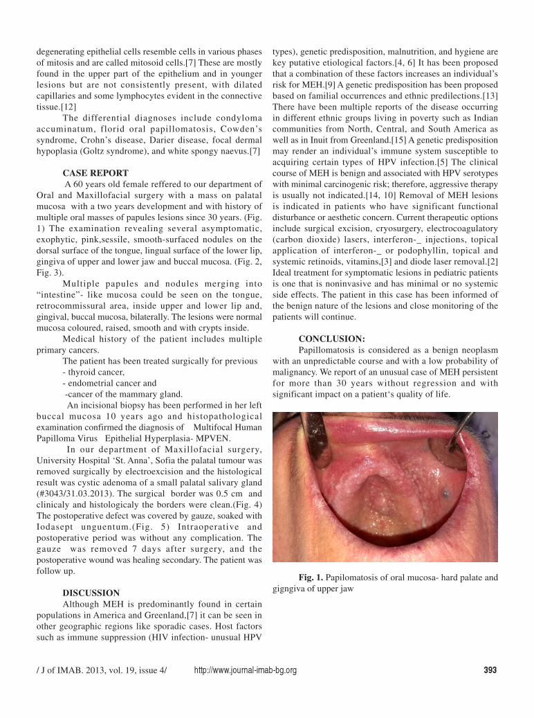
CASE REPORT A 60 years old female reffered to our department of
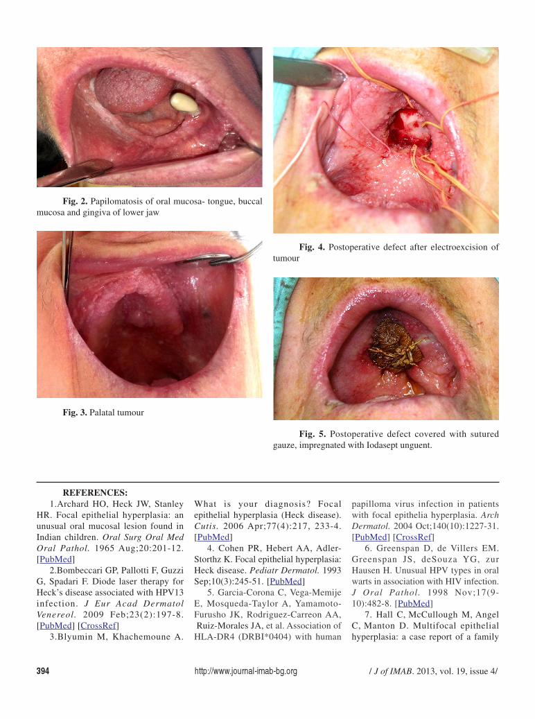
Oral and Maxillofacial surgery with a mass on palatalmucosa with a two years development and with history ofmultiple oral masses of papules lesions since 30 years. (Fig.1) The examination revealing several asymptomatic,exophytic, pink,sessile, smooth-surfaced nodules on thedorsal surface of the tongue, lingual surface of the lower lip,gingiva of upper and lower jaw and buccal mucosa. (Fig. 2,Fig. 3).
Multiple papules and nodules merging into“intestine”- like mucosa could be seen on the tongue,retrocommissural area, inside upper and lower lip and,gingival, buccal mucosa, bilaterally. The lesions were normalmucosa coloured, raised, smooth and with crypts inside.
Medical history of the patient includes multipleprimary cancers.
The patient has been treated surgically for previous- thyroid cancer,- endometrial cancer and -cancer of the mammary gland. An incisional biopsy has been performed in her left
buccal mucosa 10 years ago and histopathologicalexamination confirmed the diagnosis of Multifocal HumanPapilloma Virus Epithelial Hyperplasia- MPVEN.
In our department of Maxillofacial surgery,University Hospital ‘St. Anna’, Sofia the palatal tumour wasremoved surgically by electroexcision and the histologicalresult was cystic adenoma of a small palatal salivary gland(#3043/31.03.2013). The surgical border was 0.5 cm andclinicaly and histologicaly the borders were clean.(Fig. 4)The postoperative defect was covered by gauze, soaked withIodasept unguentum.(Fig. 5) Intraoperative andpostoperative period was without any complication. Thegauze was removed 7 days after surgery, and thepostoperative wound was healing secondary. The patient wasfollow up.
DISCUSSIONAlthough MEH is predominantly found in certain
populations in America and Greenland,[7] it can be seen inother geographic regions like sporadic cases. Host factorssuch as immune suppression (HIV infection- unusual HPV
types), genetic predisposition, malnutrition, and hygiene arekey putative etiological factors.[4, 6] It has been proposedthat a combination of these factors increases an individual’srisk for MEH.[9] A genetic predisposition has been proposedbased on familial occurrences and ethnic predilections.[13]There have been multiple reports of the disease occurringin different ethnic groups living in poverty such as Indiancommunities from North, Central, and South America aswell as in Inuit from Greenland.[15] A genetic predispositionmay render an individual’s immune system susceptible toacquiring certain types of HPV infection.[5] The clinicalcourse of MEH is benign and associated with HPV serotypeswith minimal carcinogenic risk; therefore, aggressive therapyis usually not indicated.[14, 10] Removal of MEH lesionsis indicated in patients who have significant functionaldisturbance or aesthetic concern. Current therapeutic optionsinclude surgical excision, cryosurgery, electrocoagulatory(carbon dioxide) lasers, interferon-_ injections, topicalapplication of interferon-_ or podophyllin, topical andsystemic retinoids, vitamins,[3] and diode laser removal.[2]Ideal treatment for symptomatic lesions in pediatric patientsis one that is noninvasive and has minimal or no systemicside effects. The patient in this case has been informed ofthe benign nature of the lesions and close monitoring of thepatients will continue.
CONCLUSION:Papillomatosis is considered as a benign neoplasm
with an unpredictable course and with a low probability ofmalignancy. We report of an unusual case of MEH persistentfor more than 30 years without regression and withsignificant impact on a patient‘s quality of life.
Fig. 1. Papilomatosis of oral mucosa- hard palate andgigngiva of upper jaw
394 http://www.journal-imab-bg.org / J of IMAB. 2013, vol. 19, issue 4/
1.Archard HO, Heck JW, StanleyHR. Focal epithelial hyperplasia: anunusual oral mucosal lesion found inIndian children. Oral Surg Oral MedOral Pathol. 1965 Aug;20:201-12.[PubMed]
2.Bombeccari GP, Pallotti F, GuzziG, Spadari F. Diode laser therapy forHeck’s disease associated with HPV13infection. J Eur Acad DermatolVenereol. 2009 Feb;23(2):197-8.[PubMed] [CrossRef]
3.Blyumin M, Khachemoune A.
What is your diagnosis? Focalepithelial hyperplasia (Heck disease).Cutis. 2006 Apr;77(4):217, 233-4.[PubMed]
4. Cohen PR, Hebert AA, Adler-Storthz K. Focal epithelial hyperplasia:Heck disease. Pediatr Dermatol. 1993Sep;10(3):245-51. [PubMed]
5. Garcia-Corona C, Vega-MemijeE, Mosqueda-Taylor A, Yamamoto-Furusho JK, Rodriguez-Carreon AA, Ruiz-Morales JA, et al. Association ofHLA-DR4 (DRBI*0404) with human
Fig. 2. Papilomatosis of oral mucosa- tongue, buccalmucosa and gingiva of lower jaw
Fig. 3. Palatal tumour
Fig. 4. Postoperative defect after electroexcision oftumour
Fig. 5. Postoperative defect covered with suturedgauze, impregnated with Iodasept unguent.
REFERENCES:papilloma virus infection in patientswith focal epithelia hyperplasia. ArchDermatol. 2004 Oct;140(10):1227-31.[PubMed] [CrossRef]
6. Greenspan D, de Villers EM.Greenspan JS, deSouza YG, zurHausen H. Unusual HPV types in oralwarts in association with HIV infection.J Oral Pathol. 1998 Nov;17(9-10):482-8. [PubMed]
7. Hall C, McCullough M, AngelC, Manton D. Multifocal epithelialhyperplasia: a case report of a family
/ J of IMAB. 2013, vol. 19, issue 4/ http://www.journal-imab-bg.org 395
of Somalian descent living in Australia.Oral Surg Oral Med Oral Pathol OralRadiol Endod. 2010 Jan;109(1):e20-24. [PubMed] [CrossRef]
8.Hettwer KJ, Rodgers MS. Focalepithelial hyperplasia (Heck’s disease)in a Polynesian. Oral Surg Oral MedOral Pathol. 1966 Oct;22(4):466-70.[PubMed]
9. Ledesma-Montes C, Garces-OrtizM, Hernandez-Guerrero JC. Clinico-pathological and immunocytochemicalstudy of multifocal epithelialhyperplasia. J Oral Maxillofac Surg.2007 Nov;65(11): 2211-7. [PubMed][CrossRef]
10.March CJ. Multiple papillary
tumours of the labial, buccal andglossal mucous membrane. DentalCosmos 1881;23:165.
11. Parkin DM, Almonte M, BruniL, Clifford G, Curado MP, Piñeros M.Burden and Trends of Type-SpecificHuman Papillomavirus Infections andRelated Diseases in the Latin Americaand Caribbean Region, Vaccine. 2008Aug 19;26 Suppl 11:L1-15. [PubMed][CrossRef]
12. Requena L, Requena C.[Histopathology of the more commonviral skin infections]. [in Spanish]Actas Dermosifiliogr. 2010 Apr;101(3):201-16. [PubMed] [CrossRef]
13. Segura-Saint-Gerons R, Toro-
Rojas M, Ceballos-Salobreña A,Aparicio-Soria JL, Fuentes-VaamondeH. Focal epithelial hyperplasia. A raredisease in our area. Med Oral PatolOral Cir Bucal. 2005 Mar-Apr;10(2):128-31. [PubMed]
14.Vera-Iglesias E, Garcia-Arpa M,Sanchez-Caminero P, Romero-Aguilera G, Cortina de la Calle P. Focalepithelial hyperplasia. ActasDermosifiliogr. 2007 Nov;98(9):621-3.[PubMed] [CrossRef]
15.Witkop CJ Jr, Niswander JD.Focal epithelial hyperplasia in Centraland South American Indians andLadinos. Oral Surg Oral Med OralPathol. 1965 Aug;20:213-7. [PubMed]
Address for correspondence:dr Elitsa Deliverska,Department of Oral and Maxillofacial surgery, Faculty of Dental Medicine,1, Georgi Sofiyski blvd., 1431 Sofia, Bulgaria; tel.+359 888949740;email: [email protected],


![Endometrium presentation - Dr Wright[1] · Endometrial Hyperplasia Simple hyperplasia Complex hyperplasia (adenomatous) Simple atypical hyperplasia ... Progression of Hyperplasia](https://img.dokumen.tips/doc/110x75/5b8a421e7f8b9a50388bc13d/endometrium-presentation-dr-wright1-endometrial-hyperplasia-simple-hyperplasia.jpg)