Embed Size (px)
Citation preview
© JAPI • VOL. 55 • NOVEMBER 2007 www.japi.org 797
Case Report
Progressive Multifocal Leukoencephalopathy – As A Presenting Manifestation of AIDSS Raina*, SS Kaushal**, D Gupta***, P Himral*, N Sawal+, V Sood+, A Goyal+
AbstractProgressive multifocal leukoencephalopathy (PML) is an opportunistic demyelinating disease caused by the ubiquitous, usually non pathogenic JC Polyomavirus. We report a case of PML as a presenting manifestation of AIDS in a forty five year old man on the basis of clinical features and neuroradiology. ©
recent as well as past events as narrated by attendants. For the last one day patient was having altered sensorium in the form of aggression, agitation and abnormal behaviour with history of urinary and fecal incontinence. There was no history of any motor weakness or sensory loss. No history suggestive of cranial nerves involvement. There was no history of seizures, fever, vomiting, headache, head injury, jaundice and ear discharge. There was no history of any prior medical illness. He was a smoker and a social drinker. He was married and having two children. On examination patient was afebrile. He was agitated, uncooperative and disoriented to time, place and person. Cranial nerve examination was normal. Speech appeared to be slurred. He was moving all the four limbs spontaneously. The tone and deep tendon reflexes were normal. The plantar reflex was bilaterally extensor. The patient was responding to painful stimulus. There were no meningeal signs present. Skull and spine examination was normal. Rest of the examination was normal.
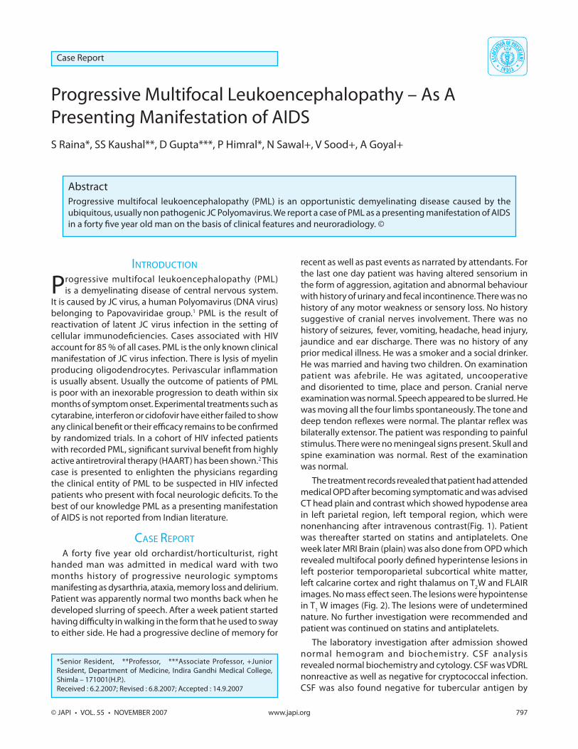
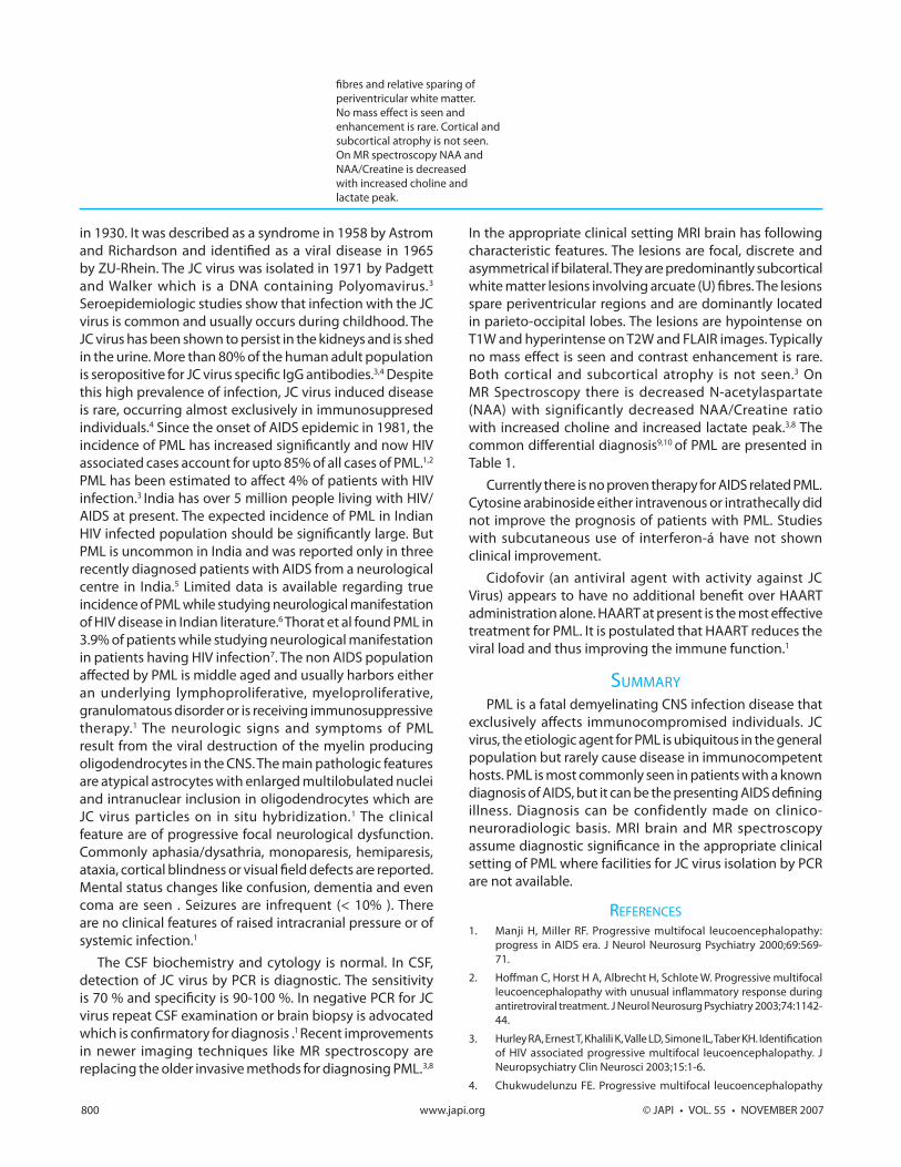
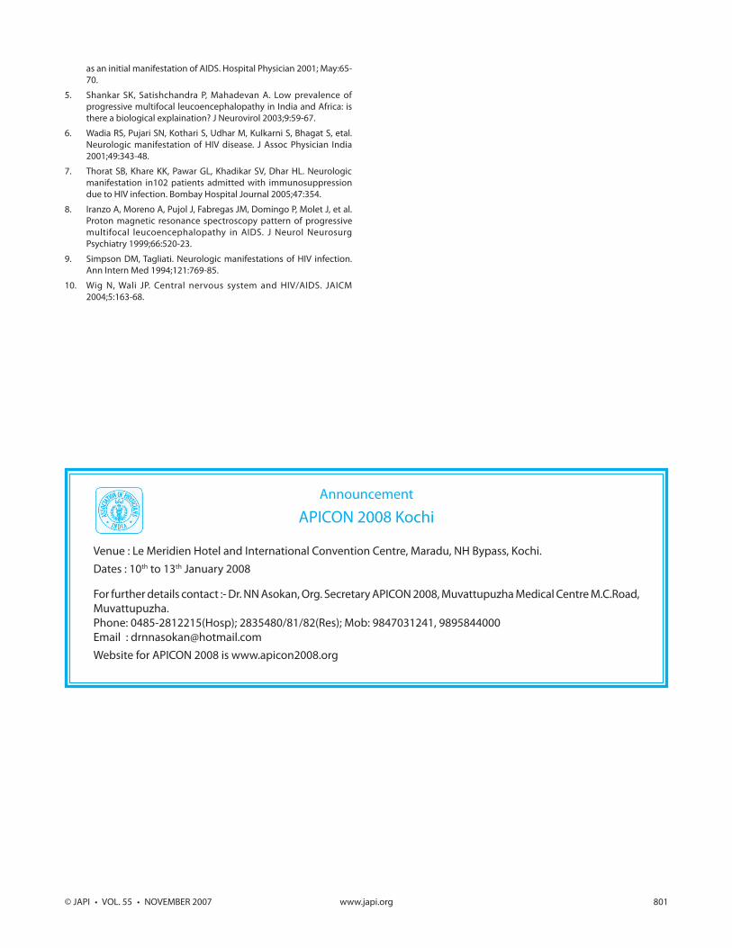
The treatment records revealed that patient had attended medical OPD after becoming symptomatic and was advised CT head plain and contrast which showed hypodense area in left parietal region, left temporal region, which were nonenhancing after intravenous contrast(Fig. 1). Patient was thereafter started on statins and antiplatelets. One week later MRI Brain (plain) was also done from OPD which revealed multifocal poorly defined hyperintense lesions in left posterior temporoparietal subcortical white matter, left calcarine cortex and right thalamus on T2W and FLAIR images. No mass effect seen. The lesions were hypointense in T1 W images (Fig. 2). The lesions were of undetermined nature. No further investigation were recommended and patient was continued on statins and antiplatelets.
The laboratory investigation after admission showed normal hemogram and biochemistry. CSF analysis revealed normal biochemistry and cytology. CSF was VDRL nonreactive as well as negative for cryptococcal infection. CSF was also found negative for tubercular antigen by
*Senior Resident, **Professor, ***Associate Professor, +Junior Resident, Department of Medicine, Indira Gandhi Medical College, Shimla – 171001(H.P.).Received : 6.2.2007; Revised : 6.8.2007; Accepted : 14.9.2007
INTRODUCTION
Progressive multifocal leukoencephalopathy (PML) is a demyelinating disease of central nervous system.
It is caused by JC virus, a human Polyomavirus (DNA virus) belonging to Papovaviridae group.1 PML is the result of reactivation of latent JC virus infection in the setting of cellular immunodeficiencies. Cases associated with HIV account for 85 % of all cases. PML is the only known clinical manifestation of JC virus infection. There is lysis of myelin producing oligodendrocytes. Perivascular inflammation is usually absent. Usually the outcome of patients of PML is poor with an inexorable progression to death within six months of symptom onset. Experimental treatments such as cytarabine, interferon or cidofovir have either failed to show any clinical benefit or their efficacy remains to be confirmed by randomized trials. In a cohort of HIV infected patients with recorded PML, significant survival benefit from highly active antiretroviral therapy (HAART) has been shown.2 This case is presented to enlighten the physicians regarding the clinical entity of PML to be suspected in HIV infected patients who present with focal neurologic deficits. To the best of our knowledge PML as a presenting manifestation of AIDS is not reported from Indian literature.
CASE REPORTA forty five year old orchardist/horticulturist, right
handed man was admitted in medical ward with two months history of progressive neurologic symptoms manifesting as dysarthria, ataxia, memory loss and delirium. Patient was apparently normal two months back when he developed slurring of speech. After a week patient started having difficulty in walking in the form that he used to sway to either side. He had a progressive decline of memory for
798 www.japi.org © JAPI • VOL. 55 • NOVEMBER 2007
Fig. 1 : Cranial CT plain(right) and contrast showing nonenhancing hypodense area in left temporoparietal lobe.
Fig. 2 : Cranial MRI showing hypointense area on TIW image (right) and hyperintense on T2W image in left posterior temporoparietal
subcortical white matter.
Fig. 3(a) : Cranial MRI showing hypointense area on T1W image (right) and hyperintense on T2W image in left temporoparietal and right
thalamus.
Fig. 3(b) : Cranial MRI showing hyperintense area on FLAIR (right) and nonenhancing on contrast image in left temporoparietal and right
thalamus.
polymerase chain reaction. Contrast enhanced MRI brain done on second day of
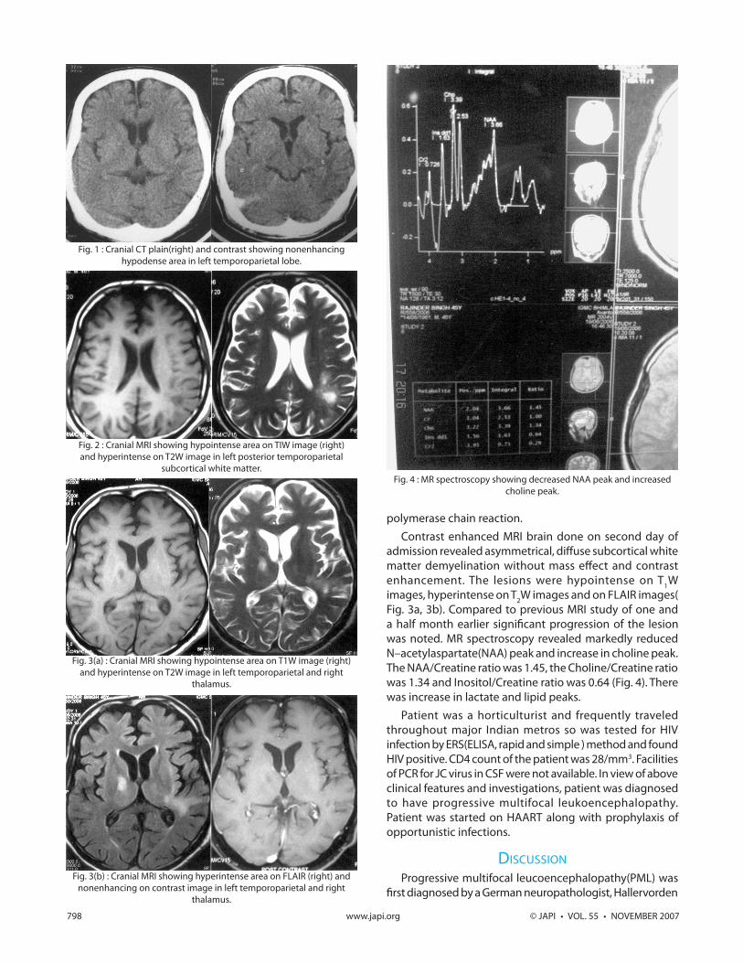
admission revealed asymmetrical, diffuse subcortical white matter demyelination without mass effect and contrast enhancement. The lesions were hypointense on T1W images, hyperintense on T2W images and on FLAIR images( Fig. 3a, 3b). Compared to previous MRI study of one and a half month earlier significant progression of the lesion was noted. MR spectroscopy revealed markedly reduced N–acetylaspartate(NAA) peak and increase in choline peak. The NAA/Creatine ratio was 1.45, the Choline/Creatine ratio was 1.34 and Inositol/Creatine ratio was 0.64 (Fig. 4). There was increase in lactate and lipid peaks.
Patient was a horticulturist and frequently traveled throughout major Indian metros so was tested for HIV infection by ERS(ELISA, rapid and simple ) method and found HIV positive. CD4 count of the patient was 28/mm3. Facilities of PCR for JC virus in CSF were not available. In view of above clinical features and investigations, patient was diagnosed to have progressive multifocal leukoencephalopathy. Patient was started on HAART along with prophylaxis of opportunistic infections.
DISCUSSIONProgressive multifocal leucoencephalopathy(PML) was
first diagnosed by a German neuropathologist, Hallervorden
Fig. 4 : MR spectroscopy showing decreased NAA peak and increased choline peak.
© JAPI • VOL. 55 • NOVEMBER 2007 www.japi.org 799
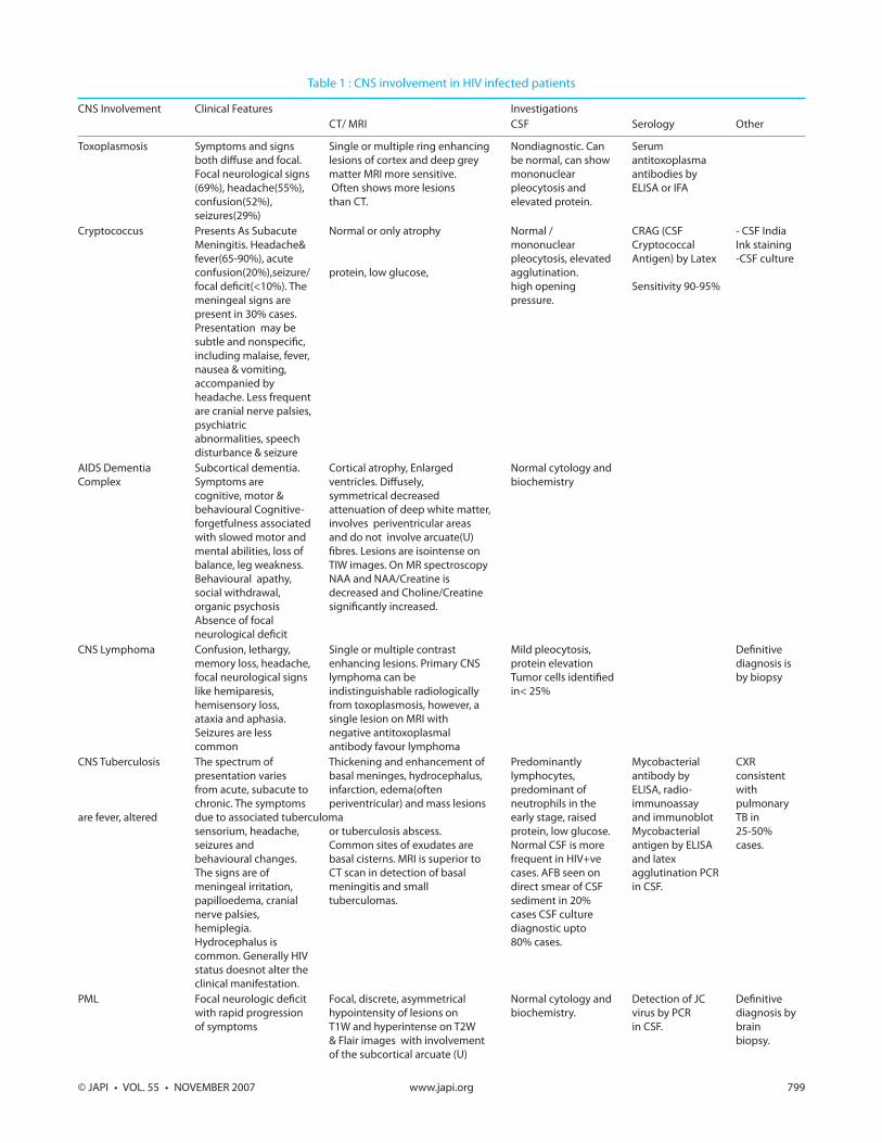
Table 1 : CNS involvement in HIV infected patients
CNS Involvement Clinical Features Investigations CT/ MRI CSF Serology Other
Toxoplasmosis Symptoms and signs Single or multiple ring enhancing Nondiagnostic. Can Serum both diffuse and focal. lesions of cortex and deep grey be normal, can show antitoxoplasma Focal neurological signs matter MRI more sensitive. mononuclear antibodies by (69%), headache(55%), Often shows more lesions pleocytosis and ELISA or IFA confusion(52%), than CT. elevated protein. seizures(29%) Cryptococcus Presents As Subacute Normal or only atrophy Normal / CRAG (CSF - CSF India Meningitis. Headache& mononuclear Cryptococcal Ink staining fever(65-90%), acute pleocytosis, elevated Antigen) by Latex -CSF culture confusion(20%),seizure/ protein, low glucose, agglutination. focal deficit(<10%). The high opening Sensitivity 90-95% meningeal signs are pressure. present in 30% cases. Presentation may be subtle and nonspecific, including malaise, fever, nausea & vomiting, accompanied by headache. Less frequent are cranial nerve palsies, psychiatric abnormalities, speech disturbance & seizure AIDS Dementia Subcortical dementia. Cortical atrophy, Enlarged Normal cytology and Complex Symptoms are ventricles. Diffusely, biochemistry cognitive, motor & symmetrical decreased behavioural Cognitive- attenuation of deep white matter, forgetfulness associated involves periventricular areas with slowed motor and and do not involve arcuate(U) mental abilities, loss of fibres. Lesions are isointense on balance, leg weakness. TIW images. On MR spectroscopy Behavioural apathy, NAA and NAA/Creatine is social withdrawal, decreased and Choline/Creatine organic psychosis significantly increased. Absence of focal neurological deficit CNS Lymphoma Confusion, lethargy, Single or multiple contrast Mild pleocytosis, Definitive memory loss, headache, enhancing lesions. Primary CNS protein elevation diagnosis is focal neurological signs lymphoma can be Tumor cells identified by biopsy like hemiparesis, indistinguishable radiologically in< 25% hemisensory loss, from toxoplasmosis, however, a ataxia and aphasia. single lesion on MRI with Seizures are less negative antitoxoplasmal common antibody favour lymphoma CNS Tuberculosis The spectrum of Thickening and enhancement of Predominantly Mycobacterial CXR presentation varies basal meninges, hydrocephalus, lymphocytes, antibody by consistent from acute, subacute to infarction, edema(often predominant of ELISA, radio- with chronic. The symptoms periventricular) and mass lesions neutrophils in the immunoassay pulmonary are fever, altered due to associated tuberculoma early stage, raised and immunoblot TB in sensorium, headache, or tuberculosis abscess. protein, low glucose. Mycobacterial 25-50% seizures and Common sites of exudates are Normal CSF is more antigen by ELISA cases. behavioural changes. basal cisterns. MRI is superior to frequent in HIV+ve and latex The signs are of CT scan in detection of basal cases. AFB seen on agglutination PCR meningeal irritation, meningitis and small direct smear of CSF in CSF. papilloedema, cranial tuberculomas. sediment in 20% nerve palsies, cases CSF culture hemiplegia. diagnostic upto Hydrocephalus is 80% cases. common. Generally HIV status doesnot alter the clinical manifestation. PML Focal neurologic deficit Focal, discrete, asymmetrical Normal cytology and Detection of JC Definitive with rapid progression hypointensity of lesions on biochemistry. virus by PCR diagnosis by of symptoms T1W and hyperintense on T2W in CSF. brain & Flair images with involvement biopsy. of the subcortical arcuate (U)
800 www.japi.org © JAPI • VOL. 55 • NOVEMBER 2007
in 1930. It was described as a syndrome in 1958 by Astrom and Richardson and identified as a viral disease in 1965 by ZU-Rhein. The JC virus was isolated in 1971 by Padgett and Walker which is a DNA containing Polyomavirus.3 Seroepidemiologic studies show that infection with the JC virus is common and usually occurs during childhood. The JC virus has been shown to persist in the kidneys and is shed in the urine. More than 80% of the human adult population is seropositive for JC virus specific IgG antibodies.3,4 Despite this high prevalence of infection, JC virus induced disease is rare, occurring almost exclusively in immunosuppresed individuals.4 Since the onset of AIDS epidemic in 1981, the incidence of PML has increased significantly and now HIV associated cases account for upto 85% of all cases of PML.1,2
PML has been estimated to affect 4% of patients with HIV infection.3 India has over 5 million people living with HIV/AIDS at present. The expected incidence of PML in Indian HIV infected population should be significantly large. But PML is uncommon in India and was reported only in three recently diagnosed patients with AIDS from a neurological centre in India.5 Limited data is available regarding true incidence of PML while studying neurological manifestation of HIV disease in Indian literature.6 Thorat et al found PML in 3.9% of patients while studying neurological manifestation in patients having HIV infection7. The non AIDS population affected by PML is middle aged and usually harbors either an underlying lymphoproliferative, myeloproliferative, granulomatous disorder or is receiving immunosuppressive therapy.1 The neurologic signs and symptoms of PML result from the viral destruction of the myelin producing oligodendrocytes in the CNS. The main pathologic features are atypical astrocytes with enlarged multilobulated nuclei and intranuclear inclusion in oligodendrocytes which are JC virus particles on in situ hybridization.1 The clinical feature are of progressive focal neurological dysfunction. Commonly aphasia/dysathria, monoparesis, hemiparesis, ataxia, cortical blindness or visual field defects are reported. Mental status changes like confusion, dementia and even coma are seen . Seizures are infrequent (< 10% ). There are no clinical features of raised intracranial pressure or of systemic infection.1
The CSF biochemistry and cytology is normal. In CSF, detection of JC virus by PCR is diagnostic. The sensitivity is 70 % and specificity is 90-100 %. In negative PCR for JC virus repeat CSF examination or brain biopsy is advocated which is confirmatory for diagnosis .1 Recent improvements in newer imaging techniques like MR spectroscopy are replacing the older invasive methods for diagnosing PML.3,8
fibres and relative sparing of periventricular white matter. No mass effect is seen and enhancement is rare. Cortical and subcortical atrophy is not seen. On MR spectroscopy NAA and NAA/Creatine is decreased with increased choline and lactate peak.
In the appropriate clinical setting MRI brain has following characteristic features. The lesions are focal, discrete and asymmetrical if bilateral. They are predominantly subcortical white matter lesions involving arcuate (U) fibres. The lesions spare periventricular regions and are dominantly located in parieto-occipital lobes. The lesions are hypointense on T1W and hyperintense on T2W and FLAIR images. Typically no mass effect is seen and contrast enhancement is rare. Both cortical and subcortical atrophy is not seen.3 On MR Spectroscopy there is decreased N-acetylaspartate (NAA) with significantly decreased NAA/Creatine ratio with increased choline and increased lactate peak.3,8 The common differential diagnosis9,10 of PML are presented in Table 1.
Currently there is no proven therapy for AIDS related PML. Cytosine arabinoside either intravenous or intrathecally did not improve the prognosis of patients with PML. Studies with subcutaneous use of interferon-á have not shown clinical improvement.
Cidofovir (an antiviral agent with activity against JC Virus) appears to have no additional benefit over HAART administration alone. HAART at present is the most effective treatment for PML. It is postulated that HAART reduces the viral load and thus improving the immune function.1
SUMMARYPML is a fatal demyelinating CNS infection disease that
exclusively affects immunocompromised individuals. JC virus, the etiologic agent for PML is ubiquitous in the general population but rarely cause disease in immunocompetent hosts. PML is most commonly seen in patients with a known diagnosis of AIDS, but it can be the presenting AIDS defining illness. Diagnosis can be confidently made on clinico-neuroradiologic basis. MRI brain and MR spectroscopy assume diagnostic significance in the appropriate clinical setting of PML where facilities for JC virus isolation by PCR are not available.
REFERENCES1. Manji H, Miller RF. Progressive multifocal leucoencephalopathy:
progress in AIDS era. J Neurol Neurosurg Psychiatry 2000;69:569-71.
2. Hoffman C, Horst H A, Albrecht H, Schlote W. Progressive multifocal leucoencephalopathy with unusual inflammatory response during antiretroviral treatment. J Neurol Neurosurg Psychiatry 2003;74:1142-44.
3. Hurley RA, Ernest T, Khalili K, Valle LD, Simone IL, Taber KH. Identification of HIV associated progressive multifocal leucoencephalopathy. J Neuropsychiatry Clin Neurosci 2003;15:1-6.
4. Chukwudelunzu FE. Progressive multifocal leucoencephalopathy
© JAPI • VOL. 55 • NOVEMBER 2007 www.japi.org 801
as an initial manifestation of AIDS. Hospital Physician 2001; May:65-70.
5. Shankar SK, Satishchandra P, Mahadevan A. Low prevalence of progressive multifocal leucoencephalopathy in India and Africa: is there a biological explaination? J Neurovirol 2003;9:59-67.
6. Wadia RS, Pujari SN, Kothari S, Udhar M, Kulkarni S, Bhagat S, etal. Neurologic manifestation of HIV disease. J Assoc Physician India 2001;49:343-48.
7. Thorat SB, Khare KK, Pawar GL, Khadikar SV, Dhar HL. Neurologic manifestation in102 patients admitted with immunosuppression due to HIV infection. Bombay Hospital Journal 2005;47:354.
8. Iranzo A, Moreno A, Pujol J, Fabregas JM, Domingo P, Molet J, et al. Proton magnetic resonance spectroscopy pattern of progressive multifocal leucoencephalopathy in AIDS. J Neurol Neurosurg Psychiatry 1999;66:520-23.
9. Simpson DM, Tagliati. Neurologic manifestations of HIV infection. Ann Intern Med 1994;121:769-85.
10. Wig N, Wali JP. Central nervous system and HIV/AIDS. JAICM 2004;5:163-68.
Announcement
APICON 2008 Kochi
Venue : Le Meridien Hotel and International Convention Centre, Maradu, NH Bypass, Kochi. Dates : 10th to 13th January 2008
For further details contact :- Dr. NN Asokan, Org. Secretary APICON 2008, Muvattupuzha Medical Centre M.C.Road, Muvattupuzha. Phone: 0485-2812215(Hosp); 2835480/81/82(Res); Mob: 9847031241, 9895844000Email : [email protected] for APICON 2008 is www.apicon2008.org


![Case Report Progressive Multifocal Leukoencephalopathy in ... · tors have also been implicated [ ]. Bone marrow studies of ... T.Weber,C.Trebst,S.Fryeetal., Analysisofthesystemicand](https://img.dokumen.tips/doc/110x75/60e713b25f32486a7f72a80b/case-report-progressive-multifocal-leukoencephalopathy-in-tors-have-also-been.jpg)