Embed Size (px)
Citation preview
Rapid Publication
Rearrangement of the Genes for the Beta and GammaChains of the T CellReceptor Is Rarely Observed in Adult B CellLymphoma and Chronic Lymphocytic LeukemiaAlan C. Aisenberg, Barbara M. Wilkes, and Joseph 0. JacobsonHematology/Oncology Unit of the Department of Medicine, Massachusetts General Hospital, Boston, Massachusetts 02114
AbstractWe determined the configuration of the genes for the beta
(Tb,.) and gamma (T,,,.) chains of the T cell receptor inDNAfrom 100 consecutive cases of B cell lymphoma and Bcell chronic lymphocytic leukemia (B-CLL), and compared thefindings with those in 18 T cell neoplasms. In 7 of the 100 Bcell specimens, a single nongermline band was detected afterdigestion with the restriction enzyme BamHI, but the rear-
rangement could be confirmed with a second restriction en-
zyme in only two. The B cell fragments were small in size andof limited size diversity when compared with the T cell cases,and germline bands of equal intensity were present. A rear-
rangement of the T... gene was never seen in a B cell sample.In contrast, T cell specimens usually rearranged both alleles ofTb,. (15 of 18), the rearrangement could be confirmed with a
second restriction enzyme (17 of 18), both alleles of the firstconstant region gene segment of Tb,.t always underwent eitherrearrangement or deletion, and the TV. gene was also rear-
ranged or deleted (17 of 18). Weconclude that ordered rear-
rangement of the T cell receptor is a rare event in B cell lym-
phoma and B-CLL. T cell receptor gene studies allow B and Tcell lymphomas to be distinguished from each other and fromcommon acute lymphoblastic leukemia antigen-positivenon-T, non-B acute lymphoblastic leukemia.
Introduction
When clonally rearranged T cell receptor genes were first de-tected in T cell malignancies (1-3), it was hoped that the find-ing would prove a hallmark of neoplasms of this cell lineage.However, within a year of that discovery, several laboratoriesreported rearranged T cell receptor gammachain (Tgamma), andless frequently, T cell receptor beta chain (Tbeta) genes innon-T cell, non-B cell, acute lymphoblastic leukemia (non-T-, non-T-ALL)' (3-5), a disorder of pre-B cell lineage (6). It
Reprint requests should be addressed to Dr. Aisenberg, MassachusettsGeneral Hospital, Boston, MA02114.
Received for publication 23 February 1987 and in revised form 6
May 1987.
1. Abbreviations used in this paper: ALL, acute lymphoblastic leuke-mia; B- and T-ALL, B and T cell ALL; ATL, adult T cell leukemia;CLL, chronic lymphocytic leukemia; B- and T-CLL, B and T cell CLL;
became important to establish if such rearranged T cell recep-tor genes were restricted to this pre-B cell lineage (ALL), orwhether they could be demonstrated in mature B cell popula-tions as well. Wehave therefore examined the status of the Tcell receptor genes in 100 consecutive specimens of B celllymphoma and chronic leukemia, and report here our failureto find a single adult B cell neoplasm with convincing rear-rangement of both Tb,, and Tgammagenes.
Methods
Patients and surface marker analysis. An earlier publication (7) ana-lyzed surface markers and Ig genes in 100 consecutive adult patientswith non-Hodgkin's lymphoma or B cell chronic lymphocytic leuke-mia (B-CLL). Biopsy specimens were subclassified according to a mod-ified Rappaport system for non-Hodgkin's lymphoma (8), and thediagnosis of B-CLL was based on conventional morphologic criteria.The 100 cases were comprised of 32 of B-CLL, 23 of follicular lym-phoma (lymphocytic or mixed), 23 of diffuse large cell lymphoma, 14of diffuse poorly differentiated lymphocytic lymphoma, and 8 miscel-laneous lymphomas. Lymphoblastic lymphoma and leukemias of Tcell lineage were specifically excluded from this consecutive series. Inall cases, lymphocyte surface markers were determined on suspensionsprepared from fresh biopsy material (lymphoma) or Ficoll-Hypaquegradient-purified blood cells (leukemia).
Surface immunoglobulin (SIg) was assessed with a fluorescencemicroscope and fluorescein-conjugated heteroantisera specific forhuman IgM and IgG heavy chains, and the kappa and lambda lightchains (Meloy Laboratories Inc., Springfield, VA). The suspensionswere also evaluated with the following monoclonal antibodies: OKT4(inducer-helper T cells); OKT8(cytotoxic-suppressor T cells); OKT1 1(sheep cell receptor); and B I (B cells). Except B l, which was a productof Coulter Diagnostics (Hialeah, FL), the monoclonal antibodies wereobtained from the Ortho Pharmaceutical (Raritan, NJ). The SIg was ofkappa light chain type in 57 cases and lambda light chain type in 26: inthe remainder a predominant light chain could not be determined.Details of the surface marker techniques employed are available in theearlier publication (7).
A second series of cases, comprised of 18 consecutive patients withvarious T cell malignancies, was studied in similar fashion. The Tbt.gene findings in three of these patients, case I in the cutaneous T celllymphoma (CTCL), adult T cell leukemia (ATL), and T CLL catego-ries, were described in an earlier report (2).
Southern blot analysis of genomic DNA. High molecular weightDNAwas prepared from each of the immunotyped specimens, and6-10 1Ag was digested with appropriate restriction enzymes (New En-gland Biolabs, Beverly, MA) and subjected to electrophoresis on 0.8%
CTCL, cutaneous T cell lymphoma; JH, heavy chain joining region;SIg, surface immunoglobulin; Tb,,, T cell receptor beta chain; T...,T cell receptor gammachain.
T Cell Receptor Genes in B Cell Lymphoma 1209
J. Clin. Invest.© The American Society for Clinical Investigation, Inc.0021-9738/87/10/1209/06 $2.00Volume 80, October 1987, 1209-1214
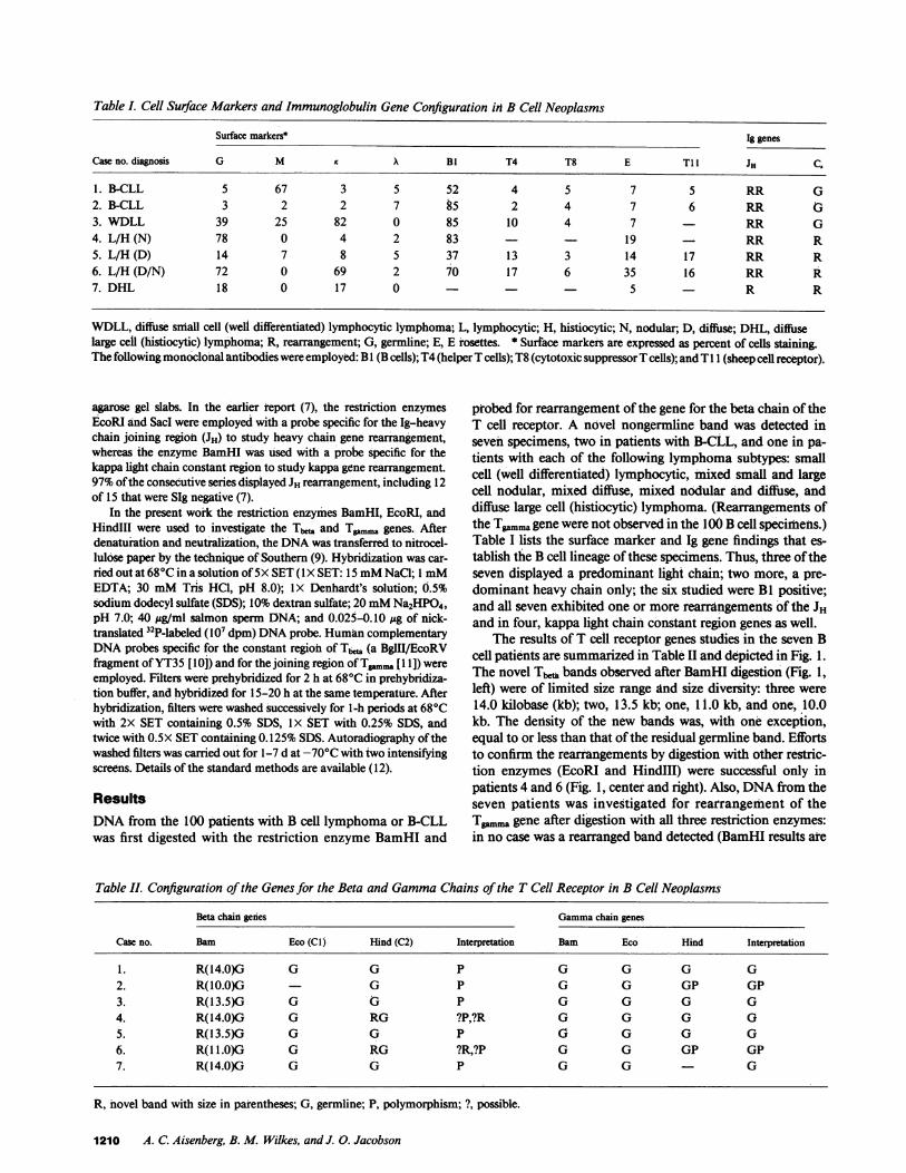
Table I. Cell Surface Markers and Immunoglobulin Gene Configuration in B Cell Neoplasms
Surface markers* Ig genes
Case no. diagnosis G M X BI T4 T8 E T11 JH C.
1. B-CLL 5 67 3 5 52 4 5 7 5 RR G2. B-CLL 3 2 2 7 85 2 4 7 6 RR G3. WDLL 39 25 82 0 85 10 4 7 RR G4. L/H (N) 78 0 4 2 83 19 - RR R5. L/H (D) 14 7 8 5 37 13 3 14 17 RR R6. L/H (D/N) 72 0 69 2 7O 17 6 35 16 RR R7. DHL 18 0 17 0 - 5 R R
WDLL, diffuse small cell (well differentiated) lymphocytic lymphoma, L, lymphocytic; H. histiocytic; N, nodular; D, diffuse; DHL, diffuselarge cell (histiocytic) lymphoma; R, rearrangement; G, germline; E, E rosettes. * Surface markers are expressed as percent of cells staining.The following monoclonal antibodies were employed: B I (B cells); T4 (helper T cells); T8 (cytotoxic suppressor T cells); and T 1 i (sheep cell receptor).
agarose gel slabs. In the earlier report (7), the restriction enzymesEcoRI and Sacd were employed with a probe specific for the Ig-heavychain joining region (JH) to study heavy chain gene rearrangement,whereas the enzyme BamHI was used with a probe specific for thekappa light chain constant region to study kappa gene rearrangement.97%of the consecutive series displayed JH rearrangement, including 12of 15 that were SIg negative (7).
In the present work the restriction enzymes BamHI, EcoRI, andHindIII were used to investigate the Tbta and To.. genes. Afterdenaturation and neutralization, the DNAwas transferred to nitrocel-lulose paper by the technique of Southern (9). Hybrdization was car-ried out at 680C in a solution of 5X SET (I X SET: 15 mMNaCl; 1 mMEDTA; 30 mMTris HC1, pH 8.0); 1X Denhardt's solution; 0.5%sodium dodecyl sulfate (SDS); 10%dextran sulfate; 20 mMNa2HPO4,pH 7.0; 40 ,ug/ml salmon sperm DNA; and 0.025-0.10 pg of nick-translated 32P-labeled (107 dpm) DNAprobe. HumancomplementaryDNAprobes specific for the constant region of Tbea (a BglII/EcoRVfragment of YT35 [10]) and for the joining region of T... [1 1]) wereemployed. Filters were prehybridized for 2 h at 680C in prehybridiza-tion buffer, and hybridized for 15-20 h at the same temperature. Afterhybridization, filters were washed successively for 1-h periods at 680Cwith 2X SET containing 0.5% SDS, IX SET with 0.25% SDS, andtwice with 0.5X SET containing 0.125% SDS. Autoradiography of thewashed filters was carried out for 1-7 d at -70'C with two intensifyingscreens. Details of the standard methods are available ( 12).
ResultsDNAfrom the 100 patients with B cell lymphoma or B-CLLwas first digested with the restriction enzyme BamHI and
probed for rearrangement of the gene for the beta chain of theT cell receptor. A novel nongermline band was detected inseven specimens, two in patients with B-CLL, and one in pa-tients with each of the following lymphoma subtypes: smallcell (well differentiated) lymphocytic, mixed small and largecell nodular, mixed diffuse, mixed nodular and diffuse, anddiffuse large cell (histiocytic) lymphoma. (Rearrangements ofthe Tgammagene were not observed in the 100 B cell specimens.)Table I lists the surface marker and Ig gene findings that es-tablish the B cell lineage of these specimens. Thus, three of theseven displayed a predominant light chain; two more, a pre-dominant heavy chain only; the six studied were BI positive;and all seven exhibited one or more rearrangements of the JHand in four, kappa light chain constant region genes as well.
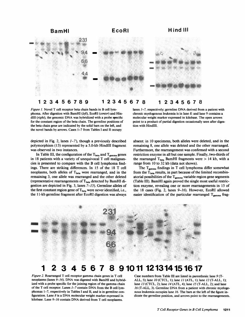
The results of T cell receptor genes studies in the seven Bcell patients are summarized in Table II and depicted in Fig. 1.The novel Tb bands observed after BamHI digestion (Fig. 1,left) were of limited size range and size diversity: three were14.0 kilobase (kb); two, 13.5 kb; one, 1 1.0 kb, and one, 10.0kb. The density of the new bands was, with one exception,equal to or less than that of the residual germline band. Effortsto confirm the rearrangements by digestion with other restric-tion enzymes (EcoRI and HindIII) were successful only inpatients 4 and 6 (Fig. 1, center and right). Also, DNAfrom theseven patients was investigated for rearrangement of theTg.. gene after digestion with all three restriction enzymes:in no case was a rearranged band detected (BamHI results ate
Table II. Configuration of the Genes for the Beta and GammaChains of the T Cell Receptor in B Cell Neoplasms
Beta chain genes Gammachain genes
Case no. Bam Eco (Cl) Hind (C2) Interpretation Bam Eco Hind Interpretation
1. R(14.0)G G G P G G G G2. R(l0.0)G G P G G GP GP3. R(13.5)G G 0 P G G G G4. R(14.0)G G RG ?P,?R G G G 05. R(1 3.5)G G G P CT G G G6. R( L.O)G G RG ?R,?P G G GP GP7. R(14.0)G G G P G G - G
R, novel band with size in parentheses; G, germline; P, polymorphism; ?, possible.
1210 A. C. Aisenberg, B. M. Wilkes, and J. 0. Jacobson
BamHI- ~~~~~~23:1
_ _* .-: la*.
Pi~
s .6
* 440
1 2 3 4 5 6 7 8 9 1 23 4 5 6 7 8 1 2 3 4 5 6 7 8Figure 1. Novel T cell receptor beta chain bands in B cell lym-phoma. After digestion with BamHI (left), EcoRI (center) and Hin-dIII (right), the genomic DNAwas hybridized with a probe specificfor the constant region of the beta chain. The germline positions ofthe beta chain gene are indicated by the solid bars on the left, andthe novel bands by arrows. Cases 1-7 from Tables I and II occupy
depicted in Fig. 2, lanes 1-7), though a previously describedpolymorphism (13) represented by a 5.0-kb HindIII fragmentwas observed in two instances.
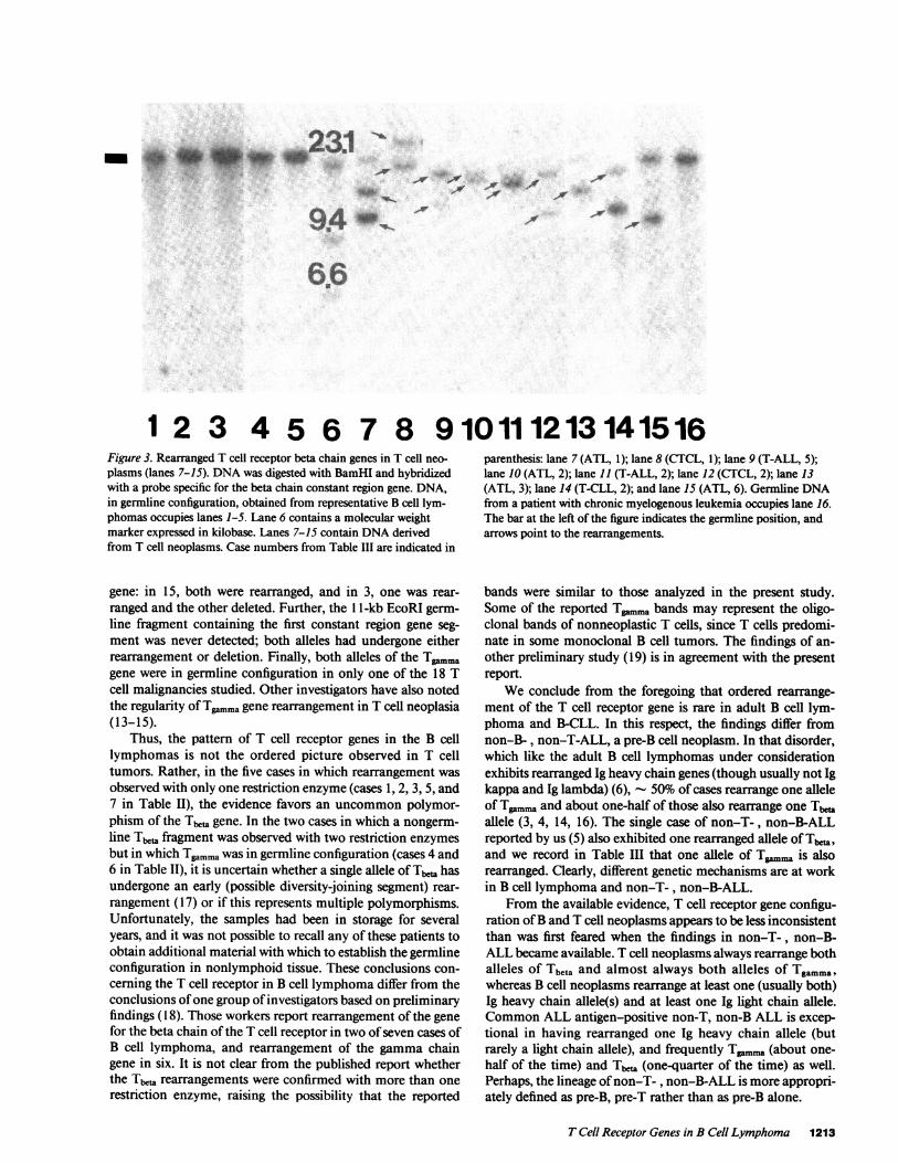
In Table III, the configuration of the Tb and TgamMagenesin 18 patients with a variety of unequivocal T cell malignan-cies is presented to compare with the B cell lymphoma find-ings. There are striking differences. In 15 of the 18 T cellneoplasms, both alleles of Tb,, were rearranged, and in theremaining 3, one allele was rearranged and the other deleted(representative rearrangements of Tbe, detected by BamHI di-gestion are depicted in Fig. 3, lanes 7-15). Germline alleles ofthe first constant region gene of Tb,, were never identified, i.e.,the 11-kb germline fragment after EcoRI digestion was always
lanes 1-7, respectively; germline DNAderived from a patient withchronic myelogenous leukemia is in lane 8; and lane 9 contains amolecular weight marker expressed in kilobase. The open arrowspoint to a product of partial digestion occasionally seen after diges-tion with HindIll.
absent: in 10 specimens, both alleles were deleted, and in theremaining 8, one allele was deleted and the other rearranged.Furthermore, the rearrangement was confirmed with a secondrestriction enzyme in all but one sample. Finally, two-thirds ofthe rearranged Tbet. BamHI fragments were > 14 kb, with arange from 10 to 32 kb (data not shown).
The T.mma findings in T cell lymphoma differ somewhatfrom the Tb,, results, in part because of the limited recombin-atorial possibilities of the Tgmmavariable region gene segments(Table III). BamHI again proved the single most useful restric-tion enzyme, revealing one or more rearrangements in 15 ofthe 18 cases (Fig. 2, lanes 9-16). However, EcoRI allowedeasier identification of the particular rearranged Tg,,ma frag-
WV
23
O.4
s6o46a6 0
I
1 2 3 4 5 6 7 8 91011 121314151617Figure 2. Rearranged T cell receptor gammachain genes in T cellneoplasms (lanes 9-16). DNAwas digested with BamHI and hybrid-ized with a probe specific for the joining region of the gammachainof the T cell receptor. Lanes 1-7 contain DNAfrom the B cell lym-
phomas 1-7, respectively in Tables I and II, and is in germline con-
figuration. Lane 8 is a DNAmolecular weight marker expressed inkilobase. Lane 9-16 contain DNAderived from T cell neoplasms.
Case numbers from Table III are listed in parenthesis: lane 9 (T-ALL, 5); lane 10 (CTCL, 1); lane 11 (ATL, 1); lane 12 (T-ALL, 1);lane 13 (CTCL, 2); lane 14 (ATL, 4); lane 15 (T-ALL, 2); and lane16 (T-ALL, 3). Germline DNAfrom a patient with chronic myeloge-nous leukemia occupies lane 16. The bars at the left of the figure in-dicate the germline position, and arrows point to the rearrangements.
T Cell Receptor Genes in B Cell Lymphoma 1211
EcoRi Hind III
*-0*-041
W.-
.. .-,A- 40i".-W
.. 40 4b
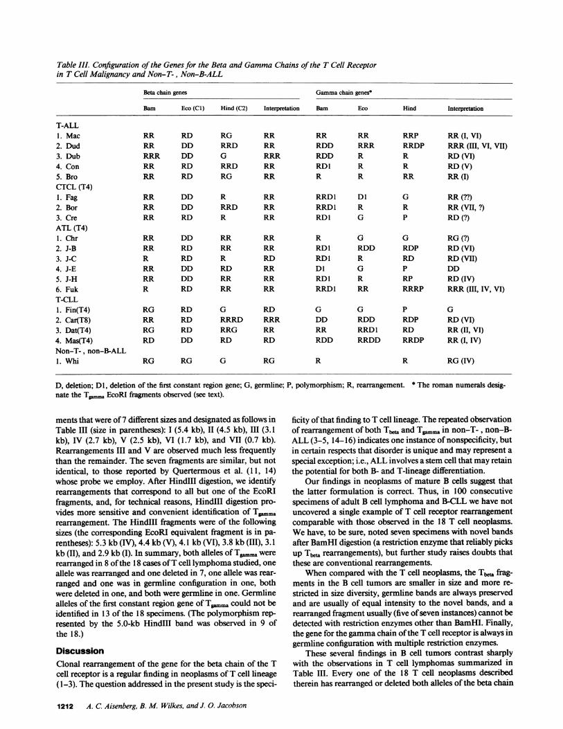
Table III. Configuration of the Genes for the Beta and GammaChains of the T Cell Receptorin T Cell Malignancy and Non-T-, Non-B-ALL
Beta chain genes Gammachain genes*
Barn Eco (CI) Hind (C2) Interpretation Bam Eco Hind Interpretation
T-ALL1. Mac RR RD RG RR RR RR RRP RR(I, VI)2. Dud RR DD RRD RR RDD RRR RRDP RRR(III, VI, VII)
3. Dub RRR DD G RRR RDD R R RD(VI)
4. Con RR RD RRD RR RDI R R RD(V)5. Bro RR RD RG RR R R RR RR(I)CTCL (T4)1. Fag RR DD R RR RRD1 Dl G RR(??)2. Bor RR DD RRD RR RRD1 R R RR(VII, ?)3. Cre RR RD R RR RDI G P RD(?)ATL (T4)1. Chr RR DD RR RR R G G RG(?)2. J-B RR RD RR RR RD1 RDD RDP RD(VI)
3. J-C R RD R RD RD1 R RD RD(VII)4. J-E RR DD RD RR Dl G P DD5. J-H RR DD RR RR RDI R RP RD(IV)6. Fuk R RD RR RR RRDI RR RRRP RRR(III, IV, VI)T-CLL1. Fin(T4) RG RD G RD G G P G2. Car(T8) RR RD RRRD RRR DD RDD RDP RD(VI)3. Dat(T4) RG RD RRG RR RR RRD1 RD RR(II, VI)
4. Mas(T4) RD DD RD RD RDD RRDD RRDP RR(I, IV)Non-T-, non-B-ALL1. Whi RG RG G RG R R RG(IV)
D, deletion; Dl, deletion of the first constant region gene; G, germlinnate the Tpmm, EcoRI fragments observed (see text).
ments that were of 7 different sizes and designated as follows inTable III (size in parentheses): I (5.4 kb), 11 (4.5 kb), III (3.1kb), IV (2.7 kb), V (2.5 kb), VI (1.7 kb), and VII (0.7 kb).Rearrangements III and V are observed much less frequentlythan the remainder. The seven fragments are similar, but notidentical, to those reported by Quertermous et al. (11, 14)whose probe we employ. After HindIII digestion, we identifyrearrangements that correspond to all but one of the EcoRIfragments, and, for technical reasons, HindIII digestion pro-vides more sensitive and convenient identification of T.rn..arearrangement. The HindIII fragments were of the followingsizes (the corresponding EcoRI equivalent fragment is in pa-rentheses): 5.3 kb (IV), 4.4 kb (V), 4.1 kb (VI), 3.8 kb (III), 3.1kb (II), and 2.9 kb (I). In summary, both alleles of Tgmmawererearranged in 8 of the 18 cases of T cell lymphoma studied, oneallele was rearranged and one deleted in 7, one allele was rear-ranged and one was in germline configuration in one, bothwere deleted in one, and both were germline in one. Germlinealleles of the first constant region gene of Tgaimra could not beidentified in 13 of the 18 specimens. (The polymorphism rep-resented by the 5.0-kb HindIII band was observed in 9 ofthe 18.)
DiscussionClonal rearrangement of the gene for the beta chain of the Tcell receptor is a regular finding in neoplasms of T cell lineage(1-3). The question addressed in the present study is the speci-
e; P, polymorphism; R, rearrangement. * The roman numerals desig-
ficity of that finding to T cell lineage. The repeated observationof rearrangement of both Tbe and TPmmain non-T-, non-B-ALL (3-5, 14-16) indicates one instance of nonspecificity, butin certain respects that disorder is unique and may represent aspecial exception; i.e., ALL involves a stem cell that may retainthe potential for both B- and T-lineage differentiation.
Our findings in neoplasms of mature B cells suggest thatthe latter formulation is correct. Thus, in 100 consecutivespecimens of adult B cell lymphoma and B-CLL we have notuncovered a single example of T cell receptor rearrangementcomparable with those observed in the 18 T cell neoplasms.Wehave, to be sure, noted seven specimens with novel bandsafter BamHI digestion (a restriction enzyme that reliably picksup Tba rearrangements), but further study raises doubts thatthese are conventional rearrangements.
Whencompared with the T cell neoplasms, the Tb frag-ments in the B cell tumors are smaller in size and more re-stricted in size diversity, germline bands are always preservedand are usually of equal intensity to the novel bands, and arearranged fragment usually (five of seven instances) cannot bedetected with restriction enzymes other than BamHI. Finally,the gene for the gammachain of the T cell receptor is always ingermline configuration with multiple restriction enzymes.
These several findings in B cell tumors contrast sharplywith the observations in T cell lymphomas summarized inTable III. Every one of the 18 T cell neoplasms describedtherein has rearranged or deleted both alleles of the beta chain
1212 A. C. Aisenberg, B. M. Wilkes, and J. 0. Jacobson
.._ool~~~~~~~~WI%.
a9|4 I-V
-,.r-0 AV,Alr .10 .:
6.6
1 2 3 4 5 6 7 8 910111213141516Figure 3. Rearranged T cell receptor beta chain genes in T cell neo-plasms (lanes 7-15). DNAwas digested with BamHI and hybridizedwith a probe specific for the beta chain constant region gene. DNA,in germline configuration, obtained from representative B cell lym-phomas occupies lanes 1-5. Lane 6 contains a molecular weightmarker expressed in kilobase. Lanes 7-15 contain DNAderivedfrom T cell neoplasms. Case numbers from Table III are indicated in
gene: in 15, both were rearranged, and in 3, one was rear-ranged and the other deleted. Further, the 11-kb EcoRI germ-line fragment containing the first constant region gene seg-ment was never detected; both alleles had undergone eitherrearrangement or deletion. Finally, both alleles of the T .magene were in germline configuration in only one of the 18 Tcell malignancies studied. Other investigators have also notedthe regularity of Tgmmagene rearrangement in T cell neoplasia(13-15).
Thus, the pattern of T cell receptor genes in the B celllymphomas is not the ordered picture observed in T celltumors. Rather, in the five cases in which rearrangement wasobserved with only one restriction enzyme (cases 1, 2, 3, 5, and7 in Table II), the evidence favors an uncommon polymor-phism of the Tb,.a gene. In the two cases in which a nongerm-line Tbeta fragment was observed with two restriction enzymesbut in which Tgammawas in germline configuration (cases 4 and6 in Table II), it is uncertain whether a single allele of Tb,, hasundergone an early (possible diversity-joining segment) rear-rangement (17) or if this represents multiple polymorphisms.Unfortunately, the samples had been in storage for severalyears, and it was not possible to recall any of these patients toobtain additional material with which to establish the germlineconfiguration in nonlymphoid tissue. These conclusions con-cerning the T cell receptor in B cell lymphoma differ from theconclusions of one group of investigators based on preliminaryfindings (18). Those workers report rearrangement of the genefor the beta chain of the T cell receptor in two of seven cases ofB cell lymphoma, and rearrangement of the gamma chaingene in six. It is not clear from the published report whetherthe Tb,,. rearrangements were confirmed with more than onerestriction enzyme, raising the possibility that the reported
parenthesis: lane 7 (ATL, 1); lane 8 (CTCL, 1); lane 9 (T-ALL, 5);lane 10 (ATL, 2); lane 11 (T-ALL, 2); lane 12 (CTCL, 2); lane 13(ATL, 3); lane 14 (T-CLL, 2); and lane 15 (ATL, 6). Germline DNAfrom a patient with chronic myelogenous leukemia occupies lane 16.The bar at the left of the figure indicates the germline position, andarrows point to the rearrangements.
bands were similar to those analyzed in the present study.Some of the reported Tgamma bands may represent the oligo-clonal bands of nonneoplastic T cells, since T cells predomi-nate in some monoclonal B cell tumors. The findings of an-other preliminary study (19) is in agreement with the presentreport.
Weconclude from the foregoing that ordered rearrange-ment of the T cell receptor gene is rare in adult B cell lym-phoma and B-CLL. In this respect, the findings differ fromnon-B-, non-T-ALL, a pre-B cell neoplasm. In that disorder,which like the adult B cell lymphomas under considerationexhibits rearranged Ig heavy chain genes (though usually not Igkappa and Ig lambda) (6), 50% of cases rearrange one alleleof Tpmmaand about one-half of those also rearrange one Tbaallele (3, 4, 14, 16). The single case of non-T-, non-B-ALLreported by us (5) also exhibited one rearranged allele of Tb,and we record in Table III that one allele of Tgmn is alsorearranged. Clearly, different genetic mechanisms are at workin B cell lymphoma and non-T-, non-B-ALL.
From the available evidence, T cell receptor gene configu-ration of B and T cell neoplasms appears to be less inconsistentthan was first feared when the findings in non-T-, non-B-ALL became available. T cell neoplasms always rearrange bothalleles of Tbeta and almost always both alleles of Tgamma,whereas B cell neoplasms rearrange at least one (usually both)Ig heavy chain allele(s) and at least one Ig light chain allele.CommonALL antigen-positive non-T, non-B ALL is excep-tional in having rearranged one Ig heavy chain allele (butrarely a light chain allele), and frequently Tgamma (about one-half of the time) and Tb,, (one-quarter of the time) as well.Perhaps, the lineage of non-T- , non-B-ALL is more appropri-ately defined as pre-B, pre-T rather than as pre-B alone.
T Cell Receptor Genes in B Cell Lymphoma 1213
Acknowledgments
Weare grateful to Dr. Thomas Quertermous for the gift of the T...probe and to Dr. Tak Mak for the gift of Tb,,.
This work was supported by a research grant CA30020-05 from theNational Institutes of Health.
References
1. Minden, M. D., B. Toyanaga, K. Ha, Y. Yanagi, B. Chin, E.Gelfan, and T. Mak. 1985. Somatic rearrangement of T-cell antigenreceptor gene in human T-cell malignancy. Proc. Natl. Acad. Sci. USA.82:1224-1227.
2. Aisenberg, A. C., T. G. Krontiris, T. W. Mak, and B. M. Wilkes.1985. Rearrangement of the gene for the beta chain of the T-cellreceptor in T-cell chronic lymphocytic leukemia and allied disorders.N. Engl. J. Med. 313:529-533.
3. Minden, M. D., and T. W. Mak. 1986. The structure of the T cellantigen receptor genes in normal and malignant T cells. Blood.68:327-336.
4. Tawa, A., N. Hozumi, M. Minden, T. W. Mak, and E. W.Gelfand. 1985. Rearrangement of the T cell receptor beta chain gene innon-T, non-B acute lymphoblastic leukemia of childhood. N. Engl. J.Med. 313:1033-1037.
5. Aisenberg, A. C., and B. M. Wilkes. 1985. The genotype andphenotype of T cell and non-T, non-B acute lymphocytic leukemia.Blood. 66:1215-1218.
6. Korsmeyer, S. J., A. Arnold, A. Bakshin, J. V. Ravetch, U.Siebenlist, P. A. Hieter, S. 0. Sharrow, T. W. Le Bien, J. H. Kersey,D. G. Poplack, P. Leder, and T. A. Waldmann. 1983. Immunoglobulingene rearrangement and cell surface antigen expression in acute lym-
phocytic leukemias of T cell and B cell precursor origin. J. Clin. Invest.71:301-313.
7. Aisenberg, A. C., B. M. Wilkes, J. Jacobson, and N. L. Harris.1987. Immunoglobulin gene rearrangements in adult non-Hodgkin'slymphoma. Am. J. Med. 82:738-744.
8. The Non-Hodgkin's Lymphoma Classification Project. 1982.National Cancer Institute Study of Classification of Non-Hodgkin'sLymphomas. Summary and description of a Working Formulation forclinical usage. Cancer. 49:2112-2135.
9. Southern, E. M. 1975. Detection of specific sequences amongDNA fragments separated by gel electrophoresis. J. Mol. Biol.98:503-517.
10. Minden, M. D., B. Toyanaga, K. Ha, Y. Yanagi, B. Chin, E.Gelfan, and T. Mak. 1985. Somatic rearrangement of T-cell antigenreceptor gene in human T-cell malignancies. Proc. Natl. Acad. Sci.USA. 82:1224-1227.
11. Quertermous, T., C. Murre, D. Dialynas, A. D. Duby, J. L.Strominger, T. A. Waldmann, and J. G. Seidman. 1986. HumanT-cellgamma chain genes: organization, diversity and rearrangement.Science (Wash. DC). 231:252-255.
12. Maniatis, T., E. F. Fritsch, and J. Sambrook. 1982. MolecularCloning: a Laboratory Manual. Cold Spring Harbor Laboratory. ColdSpring Harbor, NewYork. 545 pp.
13. Lefranc, M.-P., and T. H. Rabbitts. 1985. Two tandemly orga-nized human genes encoding the T-cell gammaconstant-region se-quences show multiple rearrangement in different T-cell types. Nature(Lond.). 316:464-466.
14. Greenberg, J. M., T. Quertermous, J. G. Seidman, and J. H.Kersey. 1986. Human T cell gamma-chain gene rearrangements inacute lymphoid and nonlymphoid leukemia: comparison with the T-cell receptor beta-chain gene. J. Immunol. 137:2043-2049.
15. Goorha, R., N. Bunin, J. Mirro, S. B. Murphy, A. H. Cross,F. G. Behm, T. Quertermous, J. Seidman, and G. R. Kitchingman.1986. Provocative patterns of the genes for the gamma- and beta-chains of the T-cell receptor in childhood acute lymphoblastic leuk-mia. Blood. 68(Suppl. 1):256.
16. Pelicci, P.-G., D. M. Knowles, and R. D. Favera. 1985. Lym-phoid tumors displaying rearrangements of both immunoglobulin andT cell receptor genes. J. Exp. Med. 162:1015-1024.
17. Yancopoulos, G. D., and F. W. Alt. 1986. Regulation of theassembly and expression of variable-region genes. Annu. Rev. Im-munol. 4:339-368.
18. Griesser, H., A. Feller, K. Lennert, M. Tweedale, H. A.Messner, J. Zalcberg, M. D. Minden, and T. W. Mak. 1986. Thestructure of the T cell gammachain gene in lymphoproliferative dis-orders and lymphoma cell lines. Blood. 65:592-594.
19. Suber, M., P. G. Pelicci, A. Neri, D. Littman, D. M. Knowles,and R. Dalla-Favera. 1986. Different T-gamma rearrangement pat-terns in different cell types. Blood. 68(Suppl. 1):267.
1214 A. C. Aisenberg, B. M. Wilkes, and J. 0. Jacobson