Embed Size (px)
Citation preview

Rabbit and Rodent
DENTISTRYH A N D B O O K
VITTORIO CAPELLO, DVM
Dipl ECZM-Smal l Mammals, Dipl ABVP-ECM
With Margherita Gracis, DVM, Dipl AVDC and EVDC
Edited by Angela M. Lennox, DVM, Dipl ABVP-Avian, ECM
Zoological Education Network
0-Frontmatter-Preface-Foreword-RL.qxd 4/5/2005 2:14 PM Page i

iiPREFACE
Although great care has been taken to provide accurate and current information, nei-ther the authors nor the publisher nor the editor shall be liable for any loss, damage orliability directly or indirectly caused or alleged to be caused by this book. The materialcontained herein is not intended to provide specific advice or recommendation for anyspecific situation.
Library of Congress A catalog record of this book is available from the Library of Congress(http://catalog.loc.gov)
ISBN: 0-9706395-1-1
Zoological Education Network, Inc.PO Box 541749, Lake Worth, Florida 33454-1749 USAwww.exoticdvm.com
Creative and Project Director: Richard Larson
©2005 Zoological Education Network, Inc. All rights reserved.
Neither this book nor any part may be reproduced or transmitted in any form or by anymeans, electronic or mechanical, including photocopying, microfilming and recording,or by any information storage and retrieval system, without written permission from thepublisher.
Current printing (last digit):
10 9 8 7 6 5 4 3 2 1
Printed in Canada
0-Frontmatter-Preface-Foreword-RL.qxd 4/5/2005 2:14 PM Page ii
iiiPREFACE
The authors thank:
Akiteru Amimoto, DVM, PhDAmica Pet Clinic, Ube
Yamaguchi, Japan
Cristiano Colombo, DVMBusto Arsizio (Va), Italy
David A. Crossley, DVMAnimal Medical Centre Referral Services
Manchester, UK
Gabriele Ghisleni, DVMMilano, Italy
Sonia Giola, DVMMilano, Italy
Cristina Pieralisi, DVMCentro Diagnostico Veterinario
Milano, Italy
Alexander Reiter, DVM, Dipl Tzt, Dipl AVDC, Dipl EVDCDepartment of Clinical Studies
Veterinary Hospital of the University of PennsylvaniaPhiladelphia, USA
Ayako Okuda, DVM, PhD, Dipl AVDCVettec Dentistry, Sumida-ku
Tokyo, Japan
Giuseppe Ripamonti, DVMVarese, Italy
Germana Scerbanenco, DVMMilano, Italy
Yosuke Sugiura, DVMDepartment of Anatomy, Faculty of Veterinary Medicine
Azabu University, SagamiharaKanagawa, Japan
for their contributions to this handbook.
(contributors are listed alphabetically)
0-Frontmatter-Preface-Foreword-RL.qxd 4/5/2005 2:14 PM Page iii

vPREFACE
Foreword David Crossley
Preface Vittorio Capello
1Introduction to Dentistry in Exotics and Exotic Mammals 1Vittorio Capello
2Anatomy of the Skull and Teeth 3Vittorio Capello and Margherita Gracis
LAGOMORPHS 7THE RABBIT 8
Anatomy of the Skull and Teeth 8Dental Formula 10Superficial Muscles 14Deep Muscles, Arteries, Veins and Nerves 15Medial Muscles 16Nasolacrimal Duct 17
RODENTS 18Porcupine-like (Hystrychomorph) Rodents 19Rat-like (Myomorph) Rodents 19Squirrel-like (Sciuromorph) Rodents 20
THE GUINEA PIG 21Anatomy of the Skull and Teeth 21Dental Formula 22Superficial and Deep Muscles 26
THE CHINCHILLA 27Anatomy of the Skull and Teeth 27Dental Formula 28
THE GOLDEN HAMSTER 30Anatomy of the Skull and Teeth 30Dental Formula 32
THE RUSSIAN HAMSTER 32Anatomy of the Skull and Teeth 32Dental Formula 33
THE RAT 34Anatomy of the Skull and Teeth 34Dental Formula 35Superficial and Deep Muscles 36
THE PRAIRIE DOG 37Anatomy of the Skull and Teeth 37Dental Formula 38
SIZE COMPARISON OF THE SKULLS 41
3Oral Physiology 43Vittorio Capello
LAGOMORPHS 43Jaw at Rest 44Jaw Retraction 44Jaw Protrusion 45Chewing Movements 46
RODENTS 47
4The Clinical Exam 49Vittorio Capello
THE RABBIT 49Common Presenting Symptoms
Associated with Dental Disease 49Common Presenting Signs
Associated with Dental Disease 50Common Presenting Primary
Dental Disease 50Common Presenting Diseases
Secondary to Dental Disease 50Facial and Oral Findings Associated
with Oro-dental Disease 51Normal Incisor Teeth 52
THE GUINEA PIG 55Normal Incisor Teeth 55
THE CHINCHILLA 56Normal Incisor Teeth 56
THE HAMSTER 59Normal Incisor Teeth 59
THE PRAIRIE DOG 60Normal Incisor Teeth 60
DENTAL RECORDS 60
C O N T E N T S
0-Frontmatter-Preface-Foreword-RL.qxd 4/5/2005 2:14 PM Page v

viPREFACE
5Radiology of the Skull and Teeth 65Vittorio Capello and Margherita Gracis
EQUIPMENT AND INSTRUMENTS 65RADIOGRAPHIC PROJECTIONS 67
Skull Radiographs 67Use of Non-screen Dental Films 69
THE RABBIT 70Skull Radiographs 70Contrast Radiography of the Nasolacrimal Duct 74Dental Radiographs 75
THE GUINEA PIG 76Skull Radiographs 76
THE CHINCHILLA 80Skull Radiographs 80
THE DEGU 84Skull Radiographs 84
THE GOLDEN HAMSTER 87Skull Radiographs 87
THE RUSSIAN HAMSTER 89Skull Radiographs 89
THE RAT 90Skull Radiographs 90
THE PRAIRIE DOG 93Skull Radiographs 93Dental Radiographs 97
THE CHIPMUNK 98
SIZE COMPARISON OF RADIOGRAPHIC IMAGES 98
6Endoscopy 101Vittorio Capello and Margherita Gracis
INSTRUMENTS 101THE RABBIT 102THE GUINEA PIG 104THE CHINCHILLA 105THE DEGU 106THE GOLDEN HAMSTER 106THE PRAIRIE DOG 107
7Other Diagnostics 109Vittorio Capello
HEMATOLOGY AND BIOCHEMISTRY 109BACTERIAL CULTURE AND SENSITIVITY 109
HISTOPATHOLOGY 110COMPUTERIZED TOMOGRAPHIC
SCANNING 110
8Dental Diseases 113Vittorio Capello
THE RABBIT 113DENTAL DISEASES OF INCISOR TEETH 113
Congenital 113Traumatic Injuries 114Mandibular Prognathism and Maxillary
Brachygnathism 116Malocclusion of Cheek Teeth 118Metabolic Bone Disease 118Stages of Incisor Malocclusion 119
PATHOPHYSIOLOGY OF DENTAL DISEASE 122Improper and/or Insufficient Wearing 123Metabolic Bone Disease 124
DENTAL DISEASES OF CHEEK TEETH 126Stages of Cheek Teeth Malocclusion
and Acquired Dental Disease 126Spurs and Malocclusion of Mandibular
Cheek Teeth 129Longitudinal Fractures of Cheek Teeth 132Spurs and Malocclusion of Maxillary
Cheek Teeth 133End Stage Acquired Dental Disease
of Cheek Teeth 135THE GUINEA PIG 139
Dental Diseases of Incisor Teeth 139Dental Diseases of Cheek Teeth 141
THE CHINCHILLA 145
Dental Diseases of Incisor Teeth 145Dental Diseases of Cheek Teeth 146
THE GOLDEN HAMSTER 152
Dental Diseases of Incisor Teeth 152Dental Diseases of Cheek Teeth 154
THE RUSSIAN HAMSTER 155
Dental Diseases of Incisor Teeth 155Dental Diseases of Cheek Teeth 156
THE GERBIL 156THE RAT 157THE PRAIRIE DOG 158
Dental Diseases of Incisor Teeth 158Pseudo-odontoma 159Dental Diseases of Cheek Teeth 161
THE CHIPMUNK 163
0-Frontmatter-Preface-Foreword-RL.qxd 4/5/2005 2:14 PM Page vi
viiPREFACE
9Secondary Diseases 165Vittorio Capello
THE DENTAL DISEASE “SYNDROME” 165THE RABBIT 165
Systemic Disease 165Skin Disease 166Ocular Disease 166Gastrointestinal Disease 168Lesions of the Tongue and Oral Mucosa 169Facial Abscesses and Osteomyelitis 171
THE GUINEA PIG 180Gastrointestinal Disease 180Periapical Abscesses and Osteomyelitis 181
THE CHINCHILLA 183THE HAMSTER 184
10Medical Treatment 187Vittorio Capello
ANTIBIOTIC THERAPY 187ANALGESIC THERAPY 190SUPPORTIVE THERAPY 191OTHER MEDICAL THERAPIES 192
11Dental Instruments and Equipment 193Vittorio Capello and Margherita Gracis
DIAGNOSIS 193OTHER INSTRUMENTS 201MECHANICAL TRIMMING 202MANUAL TRIMMING 205DENTAL EXTRACTION 207
SURGICAL TREATMENT OF ABSCESSES OF DENTAL ORIGIN 211
12Dental Procedures 213Vittorio Capello and Margherita Gracis
REDUCTION OF INCISOR HEIGHT IN RABBITS 213
PARTIAL PULPECTOMY AND DIRECT PULP CAPPING 217
REDUCTION OF INCISOR HEIGHT IN RODENTS 221
OCCLUSAL ADJUSTMENT:
Cheek Teeth in Rabbits 222Cheek Teeth in Guinea Pigs 226Cheek Teeth in Chinchillas 227
EXTRACTION OF ARADICULAR HYPSODONT TEETH: 229
Mandibular Incisor Teeth 229Maxillary Incisor Teeth 233
Complications 236Cheek Teeth, Intraoral 238Cheek Teeth, Extraoral 243
EXTRACTION OF ARADICULAR HYPSODONT TEETH IN PORCUPINE-LIKE RODENTS 246
13Surgical Treatment of Periapical Abscessations 249Vittorio Capello and Margherita Gracis
PATIENT SELECTION AND DECISION MAKING 249
MARSUPIALIZATION 250Postoperative Debridement 254Postoperative Follow-up 255Long-term Follow-up 258
INTRODUCTION OF PMMA BEADS 259
CALCIUM HYDROXIDE PACKING 261
DIFFUSE OSTEOMYELITIS 263
DEBRIDEMENT OF MAXILLARY ABSCESSATION 264
ABSCESSATION OF THE NASOLACRIMAL DUCT 266
CHEEK TEETH ABSCESSATION:In the Guinea Pig 268In the Chinchilla 271
References 274
0-Frontmatter-Preface-Foreword-RL.qxd 4/5/2005 2:14 PM Page vii

viiiPREFACE
F O R E W O R D
There are many books available on rabbitand rodent medicine but few include aserious look at dental disease, and with a
couple of exceptions, those that do tend to bepoorly illustrated. It is therefore very nice to seethis book which brings together basic scientificknowledge and presents it along with graphicalillustration of the range of pathology and practi-cal treatment methods.
Owners now expect to have their animals, ofwhatever species, treated appropriately placingincreasing pressures on their veterinarians.Luckily an increasing proportion of these own-ers realize that it costs money to obtain the nec-essary specialized equipment and skills to treatthe less frequently seen species, and that theyhave to pay a professional fee for veterinarytreatment. Long gone are the days when thelimit to what an owner would pay was the cost ofa new animal.
Whilst there were some dedicated “rabbit” sur-gical and dental instruments available at thetime I qualified (27 years ago) the selection wasvery limited: a mouth gag, a cheek dilator and atotally inappropriate rasp for grinding overlongcheek teeth. Without the right equipment we are
severely limited in the quality of work we canoffer. This has changed dramatically over thelast 10 years. An increasing number of dedicat-ed instruments are being produced as the resultof work done by a small number of pioneers ofgood “exotic” animal dentistry. At the last countthere were 11 different commercial designs ofrabbit and rodent incisor elevators and luxatorson the market, one new design per year sincethe first was introduced.
Medicine advances rapidly, the amount ofknowledge doubling every 5-10 years. Generaltechnological advances occur at a similar rateand with each advance the cost of equipmentfalls in real terms. Air driven dental units, ultra-sound scanners and video-endoscopic instru-mentation are now affordable by most veterinaryclinics, opening up the possibility of their use inall species, not just cats and dogs. Endoscopesare particularly useful for examining inside smallopenings, such as the mouths of rabbits andsmall rodents. As can be seen from the illustra-tions in Chapter 6, these are very useful both fordiagnostic examination and for observation dur-ing treatment.
David Crossley
0-Frontmatter-Preface-Foreword-RL.qxd 4/5/2005 2:14 PM Page viii

ixPREFACE
P R E FA C E
Exotic or non-traditional pet species areincreasing in popularity worldwide. As aresult, veterinarians interested in the
medicine and surgery of these special speciesare faced with challenges to provide the highestquality care.
More and more continuing education seminarsand publications describe advanced exotic medi-cine and surgery topics such as endoscopy,orthopedics, surgery and others. Recently, thesubject of rabbit and rodent dentistry hasreceived considerable attention.
In the past, dental disease of rabbits wasdescribed simply as “slobbers,” “lumps,” or sim-ply “malocclusion” with little thought to under-lying etiology. Today, the many differing clinicalsigns, symptoms, and etiologies are well recog-nized and more properly grouped as a syndrome,including primary dental disease, and dental dis-ease secondary to other underlying conditions.
In addition, dental disease is being recognizedin rodent species as well, and it is clear thesespecies with their unique anatomical and physi-ological differences often require a slightly dif-ferent diagnostic and treatment approach.
The primary goal of the Rabbit and RodentDentistry Handbook is to provide the practitioner
with a useful tool to aid in the diagnosis and
treatment of dental disease in these various
species. Veterinarians new to exotic mammal
medicine will appreciate sections focusing on
gross and radiographic anatomy, physiology, and
visual and endoscopic examination techniques.
Another chapter describes dental instrumenta-
tion and equipment. Of primary importance are
chapters on the manifestations of dental disease
and treatment recommendations. Many proce-
dures are described in step-by-step detail.
The handbook utilizes a modern visual style
with over 1000 full color photos and accompany-
ing text, a style well recognized by those famil-
iar with Exotic DVM magazine and the various
excellent publications produced by Zoological
Education Network.
We owe a special acknowledgement to Dr. David
Crossley, our friend and mentor, who has kindly
provided many charts, illustrations and photo-
graphs for this handbook. We would also like to
thank Dr. Ayako Okuda, who provided addition-
al much needed support for this project.
Vittorio Capello
0-Frontmatter-Preface-Foreword-RL.qxd 4/5/2005 2:14 PM Page ix

xPREFACE
VITTORIO CAPELLODVM, Dipl ECZM-SM, Dipl ABVP-ECM
Dr. Vittorio Capello graduated in1989 from the School of VeterinaryMedicine of the University of Milano,Italy. He has practiced exotic animalmedicine exclusively since 1993,providing professional services fortwo veterinary clinics in Milano. Dr.Capello’s focus has been the medi-cine and surgery of small exoticpets, in particular rabbits, rodentsand ferrets.
Dr. Capello has lectured, publishedand taught exotic animal coursesand practical laboratories through-out Italy and other parts of Europe,and is a frequent guest lecturer atthe International Conference onExotics, where he was voted mostappreciated speaker two years in arow. He has written articles forExotic DVM Magazine and theJournal of Exotic Mammal Medicineand Surgery. Other works includethe Small Rodent Surgeries sectionin “The Exotic Guidebook” (ZoologicalEducation Network) and an educa-tional CD for Italian veterinarianson the medicine and surgery of thepet hamster. Dr. Capello is a memberof the advisory board of Exotic DVMmagazine.
MARGHERITA GRACIS DVM, Dipl AVDC, Dipl EVDC
Dr. Margherita Gracis graduated in1993 from the Veterinary School ofthe University of Milano, Italy. Afterworking in private practice for a fewyears, she completed a Residency inVeterinary Dentistry at the Veteri-nary School of the University ofPennsylvania, Philadelphia (USA).From 1998 until 2000 she worked atthe same institution as a lecturer inVeterinary Dentistry. Since 2000, Dr.Gracis has been working at tworeferral clinics in Milano (Italy) andin Monza (Milano, Italy), dedicated todentistry and oral surgery. She is aDiplomate of both the American(AVDC) and the European (EVDC)Veterinary Dental Colleges. Dr. Gracisis the current Past President of theEuropean Veterinary Dental Society(EVDS) and the Italian VeterinaryDental Society (SIODOV).
ANGELA M. LENNOX DVM, Dipl ABVP-Avian, ECM
Dr. Angela Lennox graduated in1989 from Purdue University Schoolof Veterinary Medicine, and has prac-ticed avian and exotic animal medi-cine exclusively since 1991. She isthe owner of the Avian and ExoticAnimal Clinic of Indianapolis. Dr.Lennox was awarded board certifica-tion in avian medicine by theAmerican Board of Veterinary Practi-tioners in 2004, and is an adjunctprofessor at Purdue University whereshe teaches courses in exotic petmedicine to both veterinary and vet-erinary technician students. She cur-rently serves as the President of theAssociation of Exotic Mammal Veteri-narians. Dr. Lennox is a frequentguest lecturer at the InternationalConference on Exotics and theConference of the Association ofAvian Veterinarians, and has servedas editor for numerous publications,including Seminars in Exotic PetMedicine, Veterinary Clinics of NorthAmerica, Exotic DVM magazine andthe Journal of Exotic MammalMedicine and Surgery. She is a mem-ber of the advisory board of ExoticDVM magazine.
0-Frontmatter-Preface-Foreword-RL.qxd 4/5/2005 2:16 PM Page x

C H A P T E R 2
Anatomy of theSkull and Teeth
VITTORIO CAPELLOMARGHERITA GRACIS
Table 2.1. Dental and Periodontal Anatomic Glossary (Figures 2.1, 2.2)
The distinctive anatomy of rabbits and rodents must be thoroughly appreciated in order to successfully diag-nose and treat dental disease. A discussion of dental anatomy is more meaningful with an understandingof precise terms used in dentistry.
Alveolarcrest
The most coronal portion of the alveolarbone
Apex The termination or end of a tooth root. Thisterm is also used to define the termination ofaradicular (see elodont) teeth. It is normally“open” (with a single large foramen) inaradicular hypsodont teeth and immaturebrachyodont teeth, “closed” (with an apicaldelta) in adult brachyodont teeth
Apicaldelta
A group of fine channels at the apex of abrachyodont tooth root through which thepulp blood vessels and nerves pass
Cemento-enamel junction
Where enamel and cementum meet (corresponds to the neck of brachyodontteeth)
Crown oranatomicalcrown
Portion of the tooth covered by enamel
Crown,clinical
Exposed portion of the tooth within themouth, above the gingival margin
Crown,reserve
In a hypsodont tooth, the part of the crownlocated below the gingival margin
Furcation In a brachyodont tooth with multiple roots,the area where roots diverge
Gingival margin
The most coronal portion of the gingiva
Gingival sulcus
The shallow space between the gingiva andthe tooth, measured from the gingival marginto the gingival attachment to the tooth surface (junctional epithelium)
Laminadura
The wall of the dental alveolus or socket, visible radiographically as a radiopaque line
Neck The portion of the brachyodont toothbetween the crown and the root
Periodontalspace
The space between the tooth and the alveolar bone occupied by periodontal ligament fibers
Pulp cavity The pulp chamber and root canal in a tooth,containing pulp tissue
Root Portion of the brachyodont tooth covered bycementum. The reserve crown of aradicularhypsodont teeth is often improperly referredto as “root”
2-Anatomy of the Skull and Teeth-RLnew.qxd 4/5/2005 1:08 PM Page 3
8CHAPTER 2 RABBIT AND RODENT DENTISTRY HANDBOOK
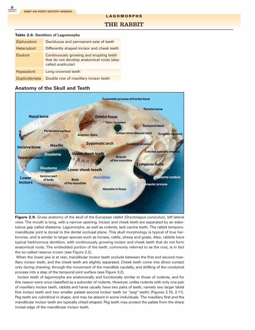
THE RABBIT
Figure 2.9. Gross anatomy of the skull of the European rabbit (Oryctolagus cuniculus), left lateralview. The mouth is long, with a narrow opening. Incisor and cheek teeth are separated by an eden-tulous gap called diastema. Lagomorphs, as well as rodents, lack canine teeth. The rabbit temporo-mandibular joint is dorsal to the dental occlusal plane. This skull morphology is typical of true her-bivores, and is similar to larger species such as horses, cattle, sheep and goats. Also, rabbits havetypical herbivorous dentition, with continuously growing incisor and cheek teeth that do not formanatomical roots. The embedded portion of the teeth, commonly referred to as the root, is in factthe so-called reserve crown (see Figure 2.2).When the lower jaw is at rest, mandibular incisor teeth occlude between the first and second max-
illary incisor teeth, and the cheek teeth are slightly separated. Cheek teeth come into direct contactonly during chewing, through the movement of the mandible caudally, and shifting of the condyloidprocess into a step of the temporal joint surface (see Figure 3.2).
Incisor teeth of lagomorphs are anatomically and functionally similar to those of rodents, and forthis reason were once classified as a suborder of rodents. However, unlike rodents with only one pairof maxillary incisor teeth, rabbits and hares usually have two pairs of teeth, namely two larger labialfirst incisor teeth and two smaller palatal second incisor teeth (or “peg” teeth) (Figures 2.10, 2.11).Peg teeth are cylindrical in shape, and may be absent in some individuals. The maxillary first and themandibular incisor teeth are typically chisel-shaped. Peg teeth may protect the palate from the sharpincisal edge of the mandibular incisor teeth.
Diphyodont Deciduous and permanent sets of teeth
Heterodont Differently shaped incisor and cheek teeth
Elodont Continuously growing and erupting teeththat do not develop anatomical roots (alsocalled aradicular)
Hypsodont Long crowned teeth
Duplicidentata Double row of maxillary incisor teeth
Table 2.6. Dentition of Lagomorphs
L A G O M O R P H S
Anatomy of the Skull and Teeth
2-Anatomy of the Skull and Teeth-RLnew.qxd 4/5/2005 1:10 PM Page 8

9CHAPTER 2ANATOMY OF THE SKULL AND TEETH
T H E R A B B I T
Figure 2.11. Close up of incisor teeth on live animal, lateralview.
Figure 2.10. Close up of incisor teeth, gross specimen, lat-eral view.
Figure 2.12. Dorsal view of the skull. Figure 2.13. Ventral view of the skull, mandible removed.
Figure 2.14. Ventral view of the skull, with the mandible inplace.
2-Anatomy of the Skull and Teeth-RLnew.qxd 4/5/2005 1:11 PM Page 9
10CHAPTER 2 RABBIT AND RODENT DENTISTRY HANDBOOK
T H E R A B B I T
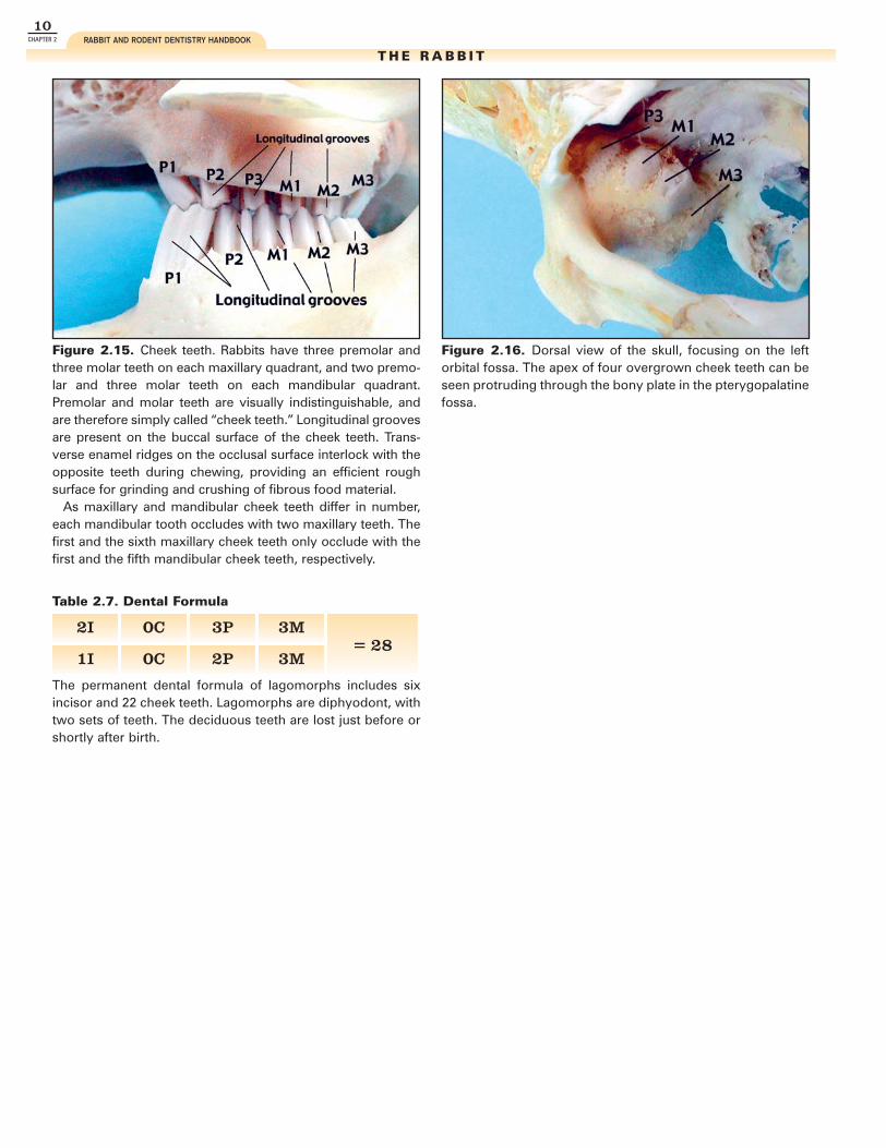
Figure 2.15. Cheek teeth. Rabbits have three premolar andthree molar teeth on each maxillary quadrant, and two premo-lar and three molar teeth on each mandibular quadrant.Premolar and molar teeth are visually indistinguishable, andare therefore simply called “cheek teeth.” Longitudinal groovesare present on the buccal surface of the cheek teeth. Trans-verse enamel ridges on the occlusal surface interlock with theopposite teeth during chewing, providing an efficient roughsurface for grinding and crushing of fibrous food material.
As maxillary and mandibular cheek teeth differ in number,each mandibular tooth occludes with two maxillary teeth. Thefirst and the sixth maxillary cheek teeth only occlude with thefirst and the fifth mandibular cheek teeth, respectively.
Figure 2.16. Dorsal view of the skull, focusing on the leftorbital fossa. The apex of four overgrown cheek teeth can beseen protruding through the bony plate in the pterygopalatinefossa.
2I 0C 3P 3M= 28
1I 0C 2P 3M
The permanent dental formula of lagomorphs includes sixincisor and 22 cheek teeth. Lagomorphs are diphyodont, withtwo sets of teeth. The deciduous teeth are lost just before orshortly after birth.
Table 2.7. Dental Formula
2-Anatomy of the Skull and Teeth-RLnew.qxd 4/5/2005 1:12 PM Page 10

15CHAPTER 2ANATOMY OF THE SKULL AND TEETH
T H E R A B B I T
Deep Muscles, Arteries, Veins and Nerves
Figure 2.28. Diagram of the deep muscles, arteries, veins and nerves of the head of the rabbit, lateral view.
Reprinted from A Colour Atlas of Anatomy of Small Laboratory Animals, Vol. I, Popesko P, Rajtovà V, Horàk J, pp 32, 35, 39, 40, 1992, with permission from Elsevier.
2-Anatomy of the Skull and Teeth-RLnew.qxd 4/5/2005 1:16 PM Page 15

23CHAPTER 2ANATOMY OF THE SKULL AND TEETH
T H E G U I N E A P I G
Figures 2.41a,b. a) Diagram of cheek teeth of guinea pigs, skyline view. b) Close-up of a rostral view of the cheek teeth, inci-sors removed. The oblique occlusal plane is clearly visible. Guinea pigs are anisognathic, with the mandible much wider thanthe maxilla. The maxillary dental arcades are therefore closer to each other than the mandibular arcades. The cheek teeth ofguinea pigs normally come in full occlusion when the jaw is at rest. The mandibular teeth are curved with a pronounced buc-cal convexity, and the maxillary teeth with a prominent palatal convexity. This results in a 30 degree oblique occlusal plane thatslopes from buccal to lingual, dorsal to ventral.
Illustration modified from Crossley DA: Clinical aspects of rodent dental anatomy, J Vet Dent 1995, 12: 131-135 with permission.
a b
Figures 2.40a,b. Cheek teeth. Guinea pigs and other porcupine-like rodents have one premolar and three molar teeth on theleft and right maxilla and mandible. Premolar and molar teeth have no anatomical or physiological differences, and are there-fore simply called “cheek teeth.” Therefore, guinea pigs have four mandibular and four maxillary cheek teeth on each side, fora total of 16 cheek teeth. Deep longitudinal grooves are present on the buccal surface of the cheek teeth (Figure 2.43). Eachmandibular cheek tooth occludes with one single opposing maxillary tooth. Mandibular and maxillary teeth of each arcade arealigned to form a straight line angled obliquely in a disto-mesial, bucco-palatal direction. In particular, right and left maxillaryarcades converge rostrally to the point that the first cheek teeth closely meet.
a b
2-Anatomy of the Skull and Teeth-RLnew.qxd 4/5/2005 1:22 PM Page 23

C H A P T E R 5
Radiology of the Skull and Teeth
VITTORIO CAPELLOMARGHERITA GRACIS
Figure 5.1. Good quality skull radiographs can be obtainedwith the use of standard radiographic equipment and screenfilms.
Figure 5.2. High-resolution mammography x-ray films areparticularly advantageous.
EQUIPMENT AND INSTRUMENTS
Radiographic examination of the skull and teeth isan essential diagnostic tool in cases of suspecteddental disease in lagomorphs and rodents. Multipleviews are necessary for a full evaluation; the diagno-sis should not be based on any single radiographicimage. The radiographic series should alwaysinclude a lateral skull view, two lateral oblique skullviews, and a ventrodorsal or dorsoventral skull view.
A rostral skull view and one or more intraoral den-tal views may also be useful. Deep sedation or gen-eral anesthesia is usually necessary for perfect posi-tioning. For some anesthetic procedures, larger rab-bits and rodents are intubated. However, for the pur-poses of skull radiography, an endotracheal tubemay interfere with the image.
5-Radiology of the skull and teeth-RL.qxd 4/5/2005 12:31 PM Page 65

71CHAPTER 5RADIOLOGY OF THE SKULL AND TEETH
T H E R A B B I T
Skull Radiographs: Lateral View, Radiographic Anatomy
Figures 5.14a,b. The apex of the mandibular incisor teeth (a, red circle) normally extends tothe level of the ipsilateral first cheek tooth. Maxillary incisor teeth are more curved thanmandibular incisors, and their apex is normally located half the length of the diastema, at somedistance from the corresponding radiopaque hard palate (a, yellow circle). The tip of the radi-olucent pulp system of normal incisor teeth usually extends to the level of the alveolar ridge,or just above it (green circles). The maxillary second incisor teeth (peg teeth) are short andsmall, with a slight curvature. The buccal alveolar margins of both maxillary first and mandibu-lar incisor teeth are more apical than the palatal and lingual margins. Note the regular, smoothpalisade formed by the cheek teeth. The mandibular cheek teeth apexes are at some distancefrom the ventral cortex of the mandible.
a
b
5-Radiology of the skull and teeth-RL.qxd 4/5/2005 12:37 PM Page 71

126CHAPTER 8 RABBIT AND RODENT DENTISTRY HANDBOOK
T H E R A B B I T
DENTAL DISEASES OF CHEEK TEETH
Many different patterns of abnormalities from mildto severe may be recognized in cases of acquireddental disease of cheek teeth (ADD). The severity ofthe pathologic changes can be staged. Both radiog-raphy and endoscopy are important for staging ofADD, and endoscopy in particular allows a moredetailed examination of the oral cavity. Thoroughevaluation and diagnosis are particularly impor-
tant, as the patient’s clinical signs may be mild ormay be absent.Early diagnosis is the key to early treatment and
resolution of lesions involving soft tissues of thegingiva, tongue and oral mucosa (see Chapter 12),which can be a source of constant pain in the petrabbit. Prompt treatment will also prevent the pro-gression of dental disease.
Stages of Cheek Teeth Malocclusion and Acquired Dental Disease
Figure 8.35. The earliest stage of acquired dental disease(ADD) of cheek teeth in rabbits is elongation of the crowns.Because both the reserve crown and the clinical crown beginto take up more space, abnormalities related to increasedpressure begin to occur. The zig-zag pattern of the cheek teethocclusal plane is still normal (see Figure 5.13), but the radiolu-cent line is less visible when the mandible is at rest, even in aproper lateral projection. Pressure on the reserve crownsbegins to increase when the animal chews. Since there is notanother tooth cranial to the first premolars, they begin tocurve, with increasing mesial convexity (red arrow).
In some early cases, slight deformation of the ventralmandibular cortical bone due to the increased pressure maybe visible (yellow arrow). Due to the abnormal convexity,interproximal space of mandibular cheek teeth begins towiden (blue arrows). Malocclusion of incisor teeth is usuallynot present at this stage.
Figure 8.36. ADD of cheek teeth, later stage. Abnormalchanges to the occlusal plane due to excessive and irregularcrown elongation are clearly visible, with height differencesbetween adjacent molars of up to a few millimeters. An irreg-ular zig-zag radiolucent line or the superimposition of two dif-ferent zig-zag lines are present. This abnormal occlusal planeis called “wave mouth.” Mandibular cheek teeth root deformi-ties are also visible. Malocclusion of incisor teeth is not stillpresent.
8-Dental Diseases-RL.qxd 4/5/2005 3:40 PM Page 126

Dental ProceduresVITTORIO CAPELLO
MARGHERITA GRACIS
C H A P T E R 1 2
Figure 12.1. Simple amputation of overgrown incisor teethwith nail clippers or other similar instruments must be dis-couraged, as this technique is associated with patient discom-fort and a high rate of potentially severe complications,including vertical fractures. Fractures and the application ofthese types of forces to the teeth can lead to damage of theapical germinal tissues. Clipping does not allow the restora-tion of a normal incisal edge, and creates rough surfaces thatcan produce secondary injuries to the tongue and lips.
Figure 12.2. The pulp system of elongated incisor teethoften extends beyond the gingival margin and may be seenas a pink discoloration of the clinical crown.
REDUCTION OF INCISOR HEIGHT IN RABBITSElongated incisor teeth may require reduction toallow normal food prehension and in many cases,to help restore the normal occlusion of the cheekteeth. However, aradicular hypsodont teeth oftengrow very quickly, at an average rate of 2 mm perweek for maxillary incisor teeth and 2.4 mm per
week for mandibular incisor teeth. Therefore, thisprocedure may need to be repeated frequently.Incisor reduction is indicated when malocclusion ismild and can be readily corrected. Extraction maybe more appropriate in case of severe malocclusion.
INCORRECT
12-Dental procedures-RL.qxd 4/5/2005 11:20 AM Page 213
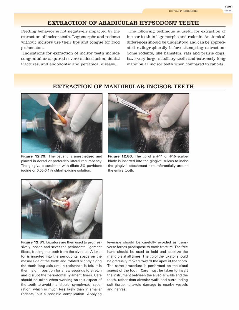
229CHAPTER 12DENTAL PROCEDURES
Figure 12.79. The patient is anesthetized andplaced in dorsal or preferably lateral recumbency.The gingiva is scrubbed with dilute 2% povidoneiodine or 0.05-0.1% chlorhexidine solution.
Figure 12.80. The tip of a #11 or #15 scalpelblade is inserted into the gingival sulcus to incisethe gingival attachment circumferentially aroundthe entire tooth.
Figure 12.81. Luxators are then used to progres-sively loosen and sever the periodontal ligamentfibers, freeing the tooth from the alveolus. A luxa-tor is inserted into the periodontal space on themesial side of the tooth and rotated slightly alongthe tooth long axis until a resistance is felt. It isthen held in position for a few seconds to stretchand disrupt the periodontal ligament fibers. Careshould be taken when working on this aspect ofthe tooth to avoid mandibular symphyseal sepa-ration, which is much less likely than in smallerrodents, but a possible complication. Applying
leverage should be carefully avoided as trans-verse forces predispose to tooth fracture. The freehand should be used to hold and stabilize themandible at all times. The tip of the luxator shouldbe gradually moved toward the apex of the tooth.The same procedure is performed on the distalaspect of the tooth. Care must be taken to insertthe instrument between the alveolar walls and thetooth, rather than alveolar walls and surroundingsoft tissue, to avoid damage to nearby vesselsand nerves.
EXTRACTION OF MANDIBULAR INCISOR TEETH
EXTRACTION OF ARADICULAR HYPSODONT TEETHFeeding behavior is not negatively impacted by theextraction of incisor teeth. Lagomorphs and rodentswithout incisors use their lips and tongue for foodprehension.Indications for extraction of incisor teeth include
congenital or acquired severe malocclusion, dentalfractures, and endodontic and periapical disease.
The following technique is useful for extraction ofincisor teeth in lagomorphs and rodents. Anatomicaldifferences should be understood and can be appreci-ated radiographically before attempting extraction.Some rodents, like hamsters, rats and prairie dogs,have very large maxillary teeth and extremely longmandibular incisor teeth when compared to rabbits.
12-Dental procedures-RL.qxd 4/5/2005 11:39 AM Page 229
250CHAPTER 13 RABBIT AND RODENT DENTISTRY HANDBOOK
Figure 13.1. Abnormalities seen on this lateral radiographare consistent with mandibular osteomyelitis. A fragment ofmandibular CT1 (arrow) is visible in the circular radiolucentlesion that likely represents purulent material. Right and leftmaxillary premolar teeth had been previously extracted.
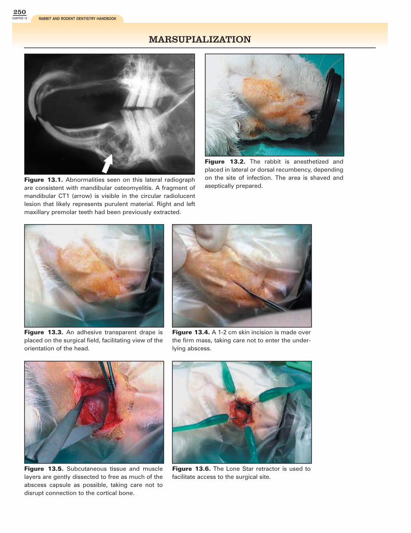
Figure 13.2. The rabbit is anesthetized andplaced in lateral or dorsal recumbency, dependingon the site of infection. The area is shaved andaseptically prepared.
Figure 13.3. An adhesive transparent drape isplaced on the surgical field, facilitating view of theorientation of the head.
Figure 13.4. A 1-2 cm skin incision is made overthe firm mass, taking care not to enter the under-lying abscess.
Figure 13.5. Subcutaneous tissue and musclelayers are gently dissected to free as much of theabscess capsule as possible, taking care not todisrupt connection to the cortical bone.
Figure 13.6. The Lone Star retractor is used tofacilitate access to the surgical site.
MARSUPIALIZATION
13-Periapical Abscessations-RL.qxd 4/4/2005 10:35 PM Page 250

R E F E R E N C E S
1. Aiken SA: Small mammal dentistry(Part II). In Quesenberry KE,Carpenter JW (eds): Ferrets,Rabbits and Rodents: ClinicalMedicine and Surgery 2nd ed. WBSaunders Co, imprint of ElsevierScience, 2004, pp 379-382.
2. Amimoto A, Hachimura H, NoguchiM, et al: Repeated reduction of theincisor crowns in a rabbit sufferingfrom malocclusion. J Anim ClinMed 6(4):35-40, 1998.
3. Bennet RA: Management ofabscesses of the head in rabbits.Proc No Am Vet Conf, 1999, pp822-823.
4. Blood DC, Stuttert VP: SaundersComprehensive VeterinaryDictionary 2nd ed. WB Saunders Co,1999.
5. Böhmer E: Röntgendiagnostik beizahnsowie kiefererkrankungen derhasenartigen und nager. Teil 1: tier-artspezifische zahn- und kiefer-anatomie sowie pathologie, indika-tionen fur röntgendiagnostik. (X-raydiagnosis of tooth and jaw impair-ment in lagomorphs and rodents.Part 1: Overall anatomy and pathol-ogy of the head area, indicationsfor x-ray diagnosis). (German)Tierärztl Prax 29:316-327, 2001.
6. Brown SA: Surgical removal of inci-sors in the rabbit. J Sm Exot AnimMed 1(4):150-153, 1992.
7. Burling K, Murphy CJ, da SilvaCuriel, et al: Anatomy of the rabbitnasolacrimal duct and its clinicalimplications. Prog Vet CompOphthal 1:33-40, 1991.
8. Capello V: Incisor extraction toresolve clinical signs of odontomain a prairie dog. Exotic DVM 4(1):9,2002.
9. Capello V: Dental diseases and sur-gical treatment in pet rodents.Exotic DVM 5(3):21-27, 2003.
10. Capello V: Endoscopic assessmentand treatment of cheek teeth mal-occlusion in pet rabbits. ExoticDVM 6(2):37-40, 2004.
11. Capello V: Extraction of cheek teethand surgical treatment of periodon-tal abscessation in pet rabbits withacquired dental disease. ExoticDVM 6(4):31-38, 2004.
12. Capello V: Extraction of incisorteeth in pet rabbits. Exotic DVM6(4):23-30, 2004.
13. Capello V, Lennox AM (ed): Smallrodent surgeries. The ExoticGuidebook. Lake Worth, ZoologicalEducation Network, 2004.
14. Capello V: Diagnosis and treatmentof dental disease in pet rabbits androdents: A review. J Exot MammalMed Surg 2(2):12-19, 2004.
15. Carpenter JW, Mashima TY, RupiperDJ: Exotic Animal Formulary 2nd ed.Philadelphia, WB Saunders Co,2001.
16. Crossley DA: Clinical aspects ofrodent dental anatomy. J Vet Dent12(4):131-135, 1995.
17. Crossley DA: Clinical aspects oflagomorph dental anatomy: Therabbit (Oryctolagus cuniculus). J VetDent 12(4):137-140, 1995.
18. Crossley DA, Dubielzig RR, BensonKG: Caries and odontoclasticresorptive lesions in a chinchilla(Chinchilla lanigera). Vet Rec141:337-339, 1997.
19. Crossley DA, Jackson A, Yates J, etal: Use of computed tomography toinvestigate cheek tooth abnormali-ties in chinchillas (Chinchillalaniger). J Sm Anim Pract 39:385-
389, 1998.20. Crossley DA, Roxburgh G: The site
of obstruction of the lacrimaldrainage system in chinchillas(Chinchilla lanigera) with “wet eye.”Proc Brit Sm Anim Vet Assoc, 1999.
21. Crossley DA, Okuda A: Clinical den-tistry of rodents and lagomorphs.(Japanese) Farmpress, 1999.
22. Crossley DA: Rodent and rabbitradiology. In DeForge DH, ColmeryBH III (eds): An Atlas of VeterinaryDental Radiology. Ames, Iowa StateUniversity Press, 2000, pp 247-260.
23. Crossley DA: Dental disease inlagomorphs and rodents. InBonagura JD (ed): Kirk’s CurrentVeterinary Therapy XIII: SmallAnimal Practice. Philadelphia, WBSaunders Co, 2000, pp 1133-1137.
24. Crossley DA, del Mar Miguélez M:Skull size and cheek-tooth length inwild-caught and captive-bred chin-chillas. Archiv Oral Biol 46:919-928,2001.
25. Crossley DA: The risk of pulp expo-sure when trimming rabbit incisorteeth. Proc 10th Europ Vet DentCong, 2001, pp 21-22.
26. Crossley DA: Small mammal den-tistry (Part I). In Quesenberry KE,Carpenter JW (eds): Ferrets,Rabbits and Rodents: ClinicalMedicine and Surgery 2nd ed. WBSaunders, imprint of ElsevierScience, 2004, pp 370-379.
27. Crossley DA: Dental disease inchinchillas in the UK. J Small AnimPract 42:12-19, 2001.
28. Crossley DA: Oral biology and dis-orders of lagomorphs. Vet Clin NoAm Exot Anim Pract 6:629-659,2003.
29. Delany MJ: Rodents. InEncyclopedia of Animals. Barnes &Noble Books, 2002, pp 214-218.
30. Delany MJ: Lagomorph. InEncyclopedia of Animals. Barnes &Noble Books, 2002, pp 229-231.
31. Divers SJ: Mandibular abscesstreatment using antibiotic-impreg-nated beads. Exotic DVM 2(5):15-18, 2000.
32. Emily PP, Penman S: Problemspeculiar to continually eruptingteeth. Handbook of Small AnimalDentistry 2nd ed. Oxford, PergamonPress, 1994.
33. Ethell MT, Bennet RA, Brown MP, etal: In vitro elution of gentamicin,amikacin, and ceftiofur from poly-methylmethacrylate and hydroxyap-atite cement. Vet Surg 29:375-382,2000.
34. Fish EW, Harris LJ: The effects ofvitamin C deficiency on tooth struc-ture in guinea pigs. Brit Dent J58:3-20, 1935.
35. Greenberg T: Premolar extraction inthe domestic rabbit. Exotic DVM2(4):11, 2000.
36. Harcourt-Brown FM: Calcium defi-ciency, diet and dental disease inpet rabbits. Vet Rec 139:567-571,1996.
37. Harcourt-Brown FM: Treatment offacial abscesses in rabbits. ExoticDVM 1(3):83-88, 1999.
38. Harcourt-Brown FM: Honey to treatrabbit abscesses. Exotic DVM3(6):13-14, 2002.
39. Harcourt-Brown FM, Baker SJ:Parathyroid hormone, haematologi-cal and biochemical parameters inrelation to dental disease and hus-bandry in rabbits. J Small AnimPract 42:130-136, 2001.
40. Harcourt-Brown FM: Dental dis-
ease. In Harcourt-Brown FM:Textbook of Rabbit Medicine.Butterworth-Heinemann, imprint ofElsevier Science, 2002, pp 165-205.
41. Harcourt-Brown FM: Ophthalmicdiseases. In Harcourt-Brown FM:Textbook of Rabbit Medicine.Butterworth-Heinemann, imprint ofElsevier Science, 2002, pp 292-306.
42. Harcourt-Brown FM: Update onmetabolic bone disease in rabbits.Exotic DVM 4(3):43-46, 2002.
43. Harcourt-Brown FM: Dacryocystitisin rabbits. Exotic DVM 4(3):47-49,2002.
44. Harkness JE, Wagner JE: TheBiology and Medicine of Rabbitsand Rodents 4th ed. Philadelphia,Lea & Febiger, imprint of Williams &Wilkins, 1995.
45. Hernandez-Divers SJ: Molar dis-ease and abscesses in rabbits.Exotic DVM 3(3):65-69, 2001.
46. Kertesz P: Dental anatomy. In AColour Atlas of Veterinary Dentistryand Oral Surgery. Aylesbury, WolfePublishing, 1993, pp 31-34.
47. Legendre LFJ: Oral disorders ofexotic rodents. Vet Clin No Am ExotAnim Pract 6:601-628, 2003.
48. Lightfoot T, Bartlett L: Rabbit androdent dental techniques. ExoticCompanion Animal Surgeries CD.Lake Worth, FL, ZoologicalEducation Network, 1999.
49. Mathews KA, Binnington AG:Wound management using sugar.Comp Cont Ed 24(1):41-50, 2002.
50. Mathews KA, Binnington AG:Wound management using honey.Comp Cont Ed 24(1):53-60, 2002.
51. Morimoto T, Inoue T, Nakamura T etal: Characteristics of rhythmic jawmovements of the rabbit. Arch OralBiol 30:673-677. 1985.
52. Murray MJ: Application of rigidendoscopy in small exotic mam-mals. Exotic DVM 2(3):13-18, 2000.
53. Nelson WB: Rabbit and dental care:Protocols for rabbit incisor extrac-tion. Exotic DVM 2(4):12, 2000.
54. Okuda A, Sugiura Y, Takahashi M,et al: Skull growth, occlusion andanatomy of unique tongue structurein the guinea pig. Proc Eurp Col VetDent, 2004, pp 55-57.
55. Popesko P, Rjtovà V, Horàk J: AColour Atlas of Anatomy of SmallLaboratory Animals Vol I: Rabbit,Guinea pig; Vol II: Rat, Mouse,Hamster. London, Wolfe PublishingLtd, 1992.
56. Punyasingh JT, Hoffmann S, HarrisSS, et al: Effect of vitamin A defi-ciency on rat incisor formation. JOral Path 13:40-51, 1984.
57. Rasmussen P: Effect of extreme cal-cium deprivation on degree of min-eralization of alveolar bone, dentinand enamel in rats. Scand J DentRes 80:327-333, 1972.
58. Rasmussen P: Quantitative observa-tion on teeth during calcium depri-vation in rats. Scand J Dent Res85:348-354, 1977.
59. Remeeus PGK, Verbeek M: The useof calcium hydroxyde in the treat-ment of abscesses in the cheek ofthe rabbit resulting from a dentalperiapical disorder. J Vet Dent12:19-22, 1995.
60. Rosenfield ME: Successful eradica-tion of severe abscesses in rabbitswith long term administration ofpenicillin G benzathine/penicillin Gprocaine. Available at: www.unix.oit.umass.edu/~jwmoore/bicillin/bicillin.htm
61. Rosenthal, KL: Therapeutic con-traindications in exotic pets. SemAvian Exot Pet Med 13(1):44-48,2004.
62. Schour J, Hoffmann MM, SmithMC: Changes in the incisor teeth ofalbino rats with vitamin A deficien-cy and the effects of replacementtherapy. Am J Path 17:529-561,1941.
63. Schour J, Medak H: Experimentalincrease in rate of eruption andgrowth of rat incisor by eliminatingattrition. J Dent Res 30:521, 1951.
64. Silverman S, Tell LA: Radiology ofRodents, Rabbits and Ferrets: AnAtlas of Normal Anatomy andPositioning. WB Saunders, imprintof Elsevier, 2005.
65. Slootweg PJ, Kuijpers MHM, van deKooij AJ: Rat odontogenic tumorsassociated with disturbed tootheruption. J Oral Patol Med 25:481-483, 1996.
66. Taglinger K, König HE:Makroskopisch-anatomische unter-suchungen der zahne deskanichens (Oryctolagus cuniculus).(Macroscopic-anatomical studies onthe teeth of rabbits (Oryctolaguscuniculus). (German) Wien TierärztlMschr 86:129-135, 1999.
67. Taylor M: A wound packing tech-nique for rabbit dental abscesses.Exotic DVM 5(3):28-31, 2003.
68. Taylor M: Endoscopy as an aid toexamination and treatment of theoropharyngeal disease of small her-bivorous mammals. Sem AvianExotic Pet Med 8(3):139-141, 1999.
69. Tyrrel KL, Citron DM, Jenkins JR, etal: Periodontal bacteria in rabbitmandibular and maxillary abscess-es. J Clin Microbiol 40:1044-1047,2002.
70. Van der Woerdt A: Ophthalmologicdiseases in small pet mammals. InQuesenberry KE, Carpenter JW(eds): Ferrets, Rabbits and Rodents:Clinical Medicine and Surgery 2nd
ed. WB Saunders, imprint ofElsevier Science, 2004, pp 421-428.
71. Verstraete FMJ: Advances in diag-nosis and treatment of small exoticmammal dental disease. Sem AvianExot Pet Med 12(1):37-48, 2003.
72. Wagner RA, Garman RH, CollinsBM: Diagnosing odontomas inprairie dogs. Exotic DVM 1:7-10,1999.
73. Wagner R, Johnson D: Rhinotomyfor treatment of odontoma in prairiedogs. Exotic DVM 3(5):29-34, 2001.
74. Weijs WA, Brugman P, GrimbergenCA: Jaw movements and muscleactivity during mastication in grow-ing rabbits. Anat Rec 224:407-416,1989.
75. Weismen DL, Olmstead ML,Kowalski JJ: In vitro evaluation ofantibiotic elution from polymethyl-metacrylate (PMMA) and mechani-cal assessment of antibiotic-PMMAcomposites. Vet Surg 29:245-251,2000.
76. Wiggs RB, Lobprise HB: Dental dis-ease in rodents. J Vet Dent 7(3):6-8,1990.
77. Wiggs RB, Lobprise HB: Dental andoral disease in lagomorphs. J VetDent 8(2):11-17, 1991.
78. Wiggs RB, Lobprise HB: Dentistry inrabbits and rodents. In: CrossleyDA, Penman S: Manual of SmallAnimal Dentistry, 2nd ed.,Cheltenham, UK, Brit Sm Anim VetAssoc, 1995, pp 68-92.
14-references-RL.qxd 4/5/2005 10:57 AM Page 1