Embed Size (px)
Citation preview
Thangasamy et al. BMC Cancer 2010, 10:282http://www.biomedcentral.com/1471-2407/10/282
Open AccessR E S E A R C H A R T I C L E
Research articleQuercetin abrogates chemoresistance in melanoma cells by modulating ΔNp73Thilakavathy Thangasamy1, Sivanandane Sittadjody1, Geoffrey C Mitchell2, Erin E Mendoza1, Vijayababu M Radhakrishnan3, Kirsten H Limesand1 and Randy Burd*1
AbstractBackground: The alkylating agent Dacarbazine (DTIC) has been used in the treatment of melanoma for decades, but when used as a monotherapy for cancer only moderate response rates are achieved. Recently, the clinical use of Temozolomide (TMZ) has become the more commonly used analog of DTIC-related oral agents because of its greater bioavailability and ability to cross the blood brain barrier. The response rates achieved by TMZ are also unsatisfactory, so there is great interest in identifying compounds that could be used in combination therapy. We have previously demonstrated that the bioflavonoid quercetin (Qct) promoted a p53-mediated response and sensitized melanoma to DTIC. Here we demonstrate that Qct also sensitizes cells to TMZ and propose a mechanism that involves the modulation of a truncated p53 family member, ΔNp73.
Methods: DB-1 melanoma (p53 wildtype), and SK Mel 28 (p53 mutant) cell lines were treated with TMZ (400 μM) for 48 hrs followed by Qct (75 μM) for 24 hrs. Cell death was determined by Annexin V-FITC staining and immunocytochemical analysis was carried out to determine protein translocation.
Results: After treatment with TMZ, DB-1 cells demonstrated increased phosphorylation of Ataxia telangiectasia mutated (ATM) and p53. However, the cells were resistant to TMZ-induced apoptosis and the resistance was associated with an increase in nuclear localization of ΔNp73. Qct treatment in combination with TMZ abolished drug insensitivity and caused a more than additive induction of apoptosis compared to either treatment alone. Treatment with Qct, caused redistribution of ΔNp73 into the cytoplasm and nucleus, which has been associated with increased p53 transcriptional activity. Knockdown of ΔNp73 restored PARP cleavage in the TMZ treated cells, confirming its anti-apoptotic role. The response to treatment was predominantly p53 mediated as the p53 mutant SK Mel 28 cells showed no significant enhancement of apoptosis.
Conclusion: This study demonstrates that Qct can sensitize cells to TMZ and that the mechanisms of sensitization involve modulation of p53 family members.
BackgroundMelanoma has been categorized as the most aggressiveform of skin cancer [1] and its incidence has increasedworldwide over the last 50 years. In the United States,this form of cancer is the fifth and sixth most commoncancer in men and women, respectively, and has an esti-mated average lifetime risk of 1 in 75 [2,3]. Dacarbazine(DTIC) is considered one of the most effective chemo-therapies for metastatic melanoma, with response ratesranging between 10-20%; however, lower response rates
(7-8%) and a 6-year survival rate of 2% have beenreported [4]. Unfortunately, the poor response of mela-noma to chemotherapy is also accompanied by systemictoxicities that lead to poor quality of life for patients.
Temozolomide (TMZ), a DTIC derivative, is a second-generation imidazotetrazine alkylating agent that ishydrolyzed to the active metabolite 5-(3,3-methyltriazen-1-yl) imidazole-4-carboxamide which further decom-poses into a DNA methylating species [5]. TMZ repre-sents a new analogue of DTIC with more desirableproperties because it can enter the cerebrospinal fluidand does not require hepatic metabolism for activation. Ithas the same cytotoxic activity as DTIC, which results
* Correspondence: [email protected] Department of Nutritional Sciences, University of Arizona, Tucson, AZ 85721, USAFull list of author information is available at the end of the article
© 2010 Thangasamy et al; licensee BioMed Central Ltd. This is an Open Access article distributed under the terms of the Creative Com-mons Attribution License (http://creativecommons.org/licenses/by/2.0), which permits unrestricted use, distribution, and reproduc-tion in any medium, provided the original work is properly cited.
Thangasamy et al. BMC Cancer 2010, 10:282http://www.biomedcentral.com/1471-2407/10/282
Page 2 of 10
from its ability to add a methyl group to the O6 position ofguanine in genomic DNA [6]. TMZ has been approvedfor the treatment of brain metastasis and has demon-strated clinical activity against melanoma, but overall ityields response rates similar to that of DTIC.
To improve response rates without increasing toxici-ties, various biological therapies have been considered foruse in combination with this class of chemotherapy. Poly-phenol compounds are of particular interest in combina-tion therapies because they can be readily activated byoxidases overexpressed in many tumors [7,8]. Quercetin,for example, is a naturally occurring polyphenol thatbecomes activated in tyrosinase expressing cells such asmelanoma. Qct is an established anticancer compoundthat exhibits anti-proliferative properties in numerouscancer cell lines [9] and animal models [10]. Enzymaticactivation of Qct by tyrosinase specifically enhances itsanti-tumor activity in melanoma cells [11] and increasesthe effectiveness of additional cytotoxic compounds [12].
The chemosensitizing effect of Qct has yet to be uti-lized clinically, but its use as an adjuvant to conventionalchemotherapy could potentially enhance the therapeuticratio in melanoma cells by increasing tumor cell kill intyrosinase expressing cells while having little effect onnormal tissue toxicity. The mechanism of tumor cell killby chemotherapeutic drugs is in part through the induc-tion of apoptosis [13]. Apoptosis is largely mediated bythe tumor suppressor gene p53, and numerous cancer cellmodels indicate that chemosensitivity is positively corre-lated with the induction of p53. In most melanoma cellsthe p53 gene is wildtype, which further supports the useof apoptosis inducing agents in the treatment of mela-noma.
In melanoma, p53 protein levels increase with tumori-genesis and development [14], and despite the presenceof functional p53, melanoma is generally regarded as achemoresistant tumor type. One possible explanation forthe development of the resistant phenotype could bethrough the upregulation of p53 antagonists, such astruncated p53 family members [14]. The p73 protein is ahomolog of p53, and has antitumor effects in various can-cerous cells, which are mediated through cell cycle arrestand the induction of pro-apoptotic target genes [15].However, several isoforms of p73 exist, including a trun-cated form that act as a p53 antagonist. The N terminaltruncated form (ΔNp73) acts as an antagonist to p53 bylocalizing to the nucleus and preventing transcription ofp53-responsive genes, such as Bax [16]. Here, we demon-strate that ΔNp73 is induced by TMZ and prevents p53-mediated apoptosis and cell death. Chemoresistance isreversed by Qct, and we therefore propose a mechanismby which Qct abrogates the inhibitory effects of ΔNp73by modulating the protein and altering its localization.
MethodsDB-1 melanoma cells were developed from lymph nodebiopsies from metastatic patients at Thomas JeffersonUniversity, Philadelphia [17]. The cells were grown in α-minimum essential medium (MEM) complete medium ina 5% CO2 incubator at 37°C and stably express thepcDNA3 vector as previously described [12]. SK Mel 28(mutant for p53) and SK Mel 5 (wild type for p53) mela-noma cell lines were obtained from American Tissue Cul-ture Collections (Rockville, MD, USA). Quercetin (3, 3, 4,5, 7-pentahydroxy flavone), α-MEM and dimethylsulfox-ide (DMSO) were purchased from Sigma, (St. Louis, MO,USA). TMZ was a kind gift from The DevelopmentalTherapeutics Program, National Cancer Institute(Bethesda, MD, USA). Antibodies for Bax, p53 and Tyro-sinase for western blotting were obtained from SantaCruz Biotechnology (Santa Cruz, CA, USA). Antibodiesfor phosphorylated p53 (at ser 15, 37, 392), phosphory-lated ATM (ser 1981), DNApk and PARP were obtainedfrom Cell Signaling (Danvers, MA, USA). Antibody forGAPDH was purchased from Millipore-Chemicon (SanFrancisco, CA, USA). p73 antibody for western blottingand immunocytochemistry (ICC) was obtained fromIMGENEX (San Diego, CA, USA). Sterile DMSO (0.1%)dissolved in α-MEM complete medium was used as vehi-cle. Quercetin and TMZ were prepared in sterile filteredDMSO.
TMZ and Qct treatmentTMZ (20 mg/ml) was dissolved in DMSO and then dis-solved in α-MEM complete medium and sterile filteredafter adjusting the pH to 7.4. For combination treatmentsthe cell lines were treated with TMZ 400 μM for 48 hr fol-lowed by Qct 75 μM for 24 hr.
Western blottingCell lysates were electrophoresed in 7 and 10% NUPAGEgels (Invitrogen Corp., CA, USA). Separated proteinswere electrophoretically transferred to Hybond PVDFmembrane (Amersham Pharmacia Biotech, UK) and themembrane was blocked for 1 hr by incubating the mem-brane in I-block (Tropix kit, Applied Biosystems, CA,USA). Primary antibodies were used at the dilutionswhich the manufacturers suggested. ALP conjugated goatanti-rabbit IgG was used at a dilution of 1:10000 for anti-bodies for phospho-p53, DNApk, Bax and PARPwhereas, anti-mouse IgG was used for Total p53, Totalp73, phospho-ATM and GAPDH at a dilution of 1:10000.Western detection was carried out using CDP star fromTropix kit, Applied Biosystems, CA, USA.
Annexin V-FITC stainingThe p53 wild type and mutant cell lines were grown up to50% confluency and were treated as mentioned above.

Thangasamy et al. BMC Cancer 2010, 10:282http://www.biomedcentral.com/1471-2407/10/282
Page 3 of 10
Apoptosis was determined using Fluorescein isothiocya-nate-conjugated Annexin V (Annexin V-FITC)/Propid-ium Iodide (PI) apoptosis detection kit (R&D systems,Minneapolis, MN, USA) as per manufacturer's instruc-tions. Approximately 5 × 10⁵ cells were resuspended in100 μl of 1× binding buffer, 1 μl of Annexin V-FITC and10 μl of propidium iodide. After 15 min incubation atroom temperature in the dark, 400 μl of 1× binding bufferwas added and the cells positive for Annexin V-FITCand/or PI were analyzed using a BD FACS flow cytome-ter.
RNA isolation and RT-PCRHomogenization of cells and isolation of RNA were per-formed using QIAshredder spin columns and an RNeasyKit as instructed by the manufacturer (Qiagen, Valencia,CA). 1 μg of RNA was reverse transcribed using a SuperScript III Kit as instructed by the manufacturer (Invitro-gen, Carlsbad, CA) and diluted 1:5 for subsequent analy-sis. The following PCR reaction mix was used: 5 ul ofdiluted cDNA, 1 ul of mixed forward and reverse primers(10 uM each), 12.5 ul SYBR Green (Qiagen), and nucle-ase-free water to a final volume of 25 ul. For non-quanti-tative PCR, cDNA was amplified for thirty cycles. Fortycycles of quantitative PCR were performed (95°C for 15seconds, 54°C for 30 seconds, 72°C for 30 seconds) usingan iQ5 Real-Time PCR Detection System (BioRad, Her-cules, CA) and run on a 1% agarose gel. Real-time PCRswere run in triplicate for each cDNA sample using an iQ5Real-Time PCR Detection System. Forty cycles of PCRwere performed (as described above) with fluorescencedetection during the 72°C step at each cycle. The datawere analyzed using the 2-ΔΔCt method [18], and resultswere normalized to S15, which remains unchanged inresponse to treatment. Normalized values were plotted asrelative fold over untreated. The following primers werepurchased from Integrated DNA Technologies (Coral-ville, IA, USA): S15 [19] transcriptionally active p73(TAp73) [20] and ΔNp73 [21].
ImmunocytochemistryThe cells were grown on cover slips in 100 mm tissue cul-ture dishes and were treated with TMZ and Qct as men-tioned above. The cover slips were placed in 6 well dishesand washed with PBS and fixed with 95% ethanol and 5%glacial acetic acid for 5 min. The slides were rinsed withPBS and were incubated with 0.5% of Triton X-100 in PBSfor 10 min to permeabilize the membranes and rinsedagain. After blocking the endogenous peroxidase with 3%hydrogen peroxide (H2O2) in PBS for 20 min the coverslips were processed according to staining procedure ofthe manufacturer's protocol for Histostain plus kits,Zymed Laboratories (Invitrogen, CA, USA). Total p73antibody was used in the dilution of 1:250.
siRNA transfectionsiRNA transfection was carried out according to manu-facturer's protocol (Invitrogen, CA, USA). Cells weregrown up to 50% confluence in antibiotic free medium in100 mm dishes. Stealth RNAi for p73 at varying concen-trations (1 nM-50 nM) was diluted in 1.5 ml OPTI-MEMI reduced serum. 30 μl of Lipofectamine™ 2000 wasdiluted in 1.5 ml OPTI-MEM I reduced serum medium,mixed gently. After 5 min incubation at RT, diluted oli-gomer was combined with diluted Lipofectamine™ 2000,mixed gently and incubated at room temp for 20 min.The oligomer-Lipofectamine™ 2000 complexes wereadded to each plate in OPTI-MEM I reduced serummedium by mixing gently and by rocking the plate backand forth. After 6 hrs incubation in a 5% CO2 incubator at37°C, the plates were subjected to TMZ treatment for 48hrs without removing the complexes. BLOCK-iT Fluores-cent oligomer was used as positive control.
Transient transfection with TyrosinaseDB-1 cells were transiently transfected with 6 μg of tyro-sinase or pcDNA3 DNA using Lipofectamine™ 2000 inserum free OPTI-MEM medium for 5 hr followed byleaving the complex in Neomycin containing α MEMcomplete medium for at least 18 hrs as demonstrated pre-viously [12].
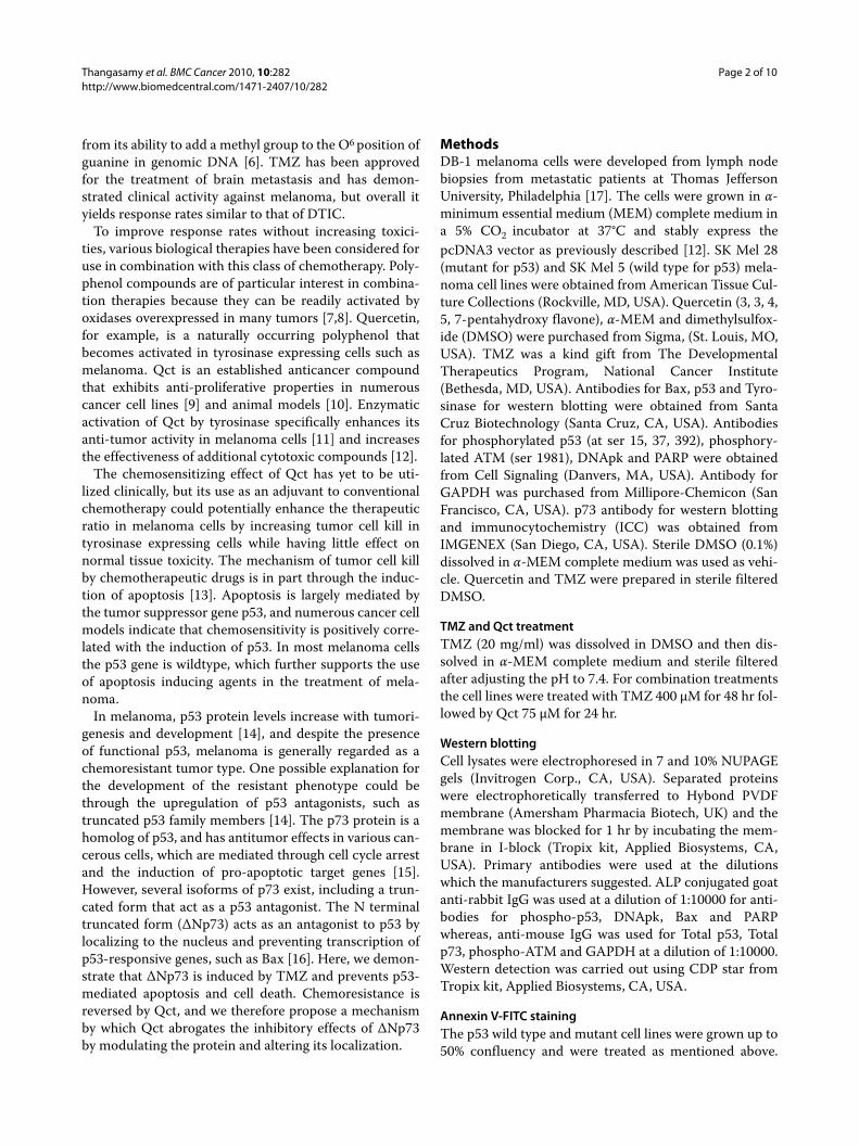
ResultsTMZ and Qct induce Cell DeathTo establish a dose response to TMZ, DB-1 cells weretreated with varying concentrations of TMZ (0-400 μM)for 48 hrs and the amount of cell death (apoptosis andnecrosis) was determined by Annexin V-FITC/PI doublestaining and flow cytometry analysis. Consistent with achemoresistant melanoma phenotype, DB-1 cells wereresistant to TMZ-induced cell death at concentrationslower than 100 μM (Figure 1A). In contrast, Qct treat-ment (0-100 μM) resulted in dose dependent cell death atall concentrations studied (Figure 1B).
For subsequent combination studies, a concentration of400 μM TMZ plus 75 μM Qct was used as those concen-trations individually resulted in a distinguishable degreeof apoptotic cell death (Figure 1C). To treat cells in com-bination TMZ was administered for 48 hrs followed by 24hrs of Qct. In DB-1 cells treatment with TMZ or Qctinduced 9.2% (± 0.4%) or 11.4% (± 0.4%) apoptosis (lowerright quadrant of flow-cytometeric scatter plot, Figure1E), respectively (Figure 1C). However, the combinationtreatment of TMZ plus Qct induced 34.25% (± 2.6%)apoptosis, which was greater than additive in these celllines. Late apoptosis or necrosis (upper right quadrant offlow-cytometeric scatter plot, Figure 1E) was alsoincreased in a more than additive manner. Apoptosisanalysis was also carried out in p53 mutant SK Mel 28
Thangasamy et al. BMC Cancer 2010, 10:282http://www.biomedcentral.com/1471-2407/10/282
Page 4 of 10
Figure 1 The induction of apoptosis by TMZ, Qct or combination treatment. DB-1 cell lines were subjected to varying dose of A) TMZ treatment (0-400 μM) for 48 hrs or B) Qct treatment for 24 hrs (0-100 μM). The cells were subjected to apoptotic analysis by Annexin/FITC staining by using BD FACS flow cytometer and the percentage of apoptosis was determined. C) The stacked percentages of apoptotic cells after TMZ treatment in DB-1 or D) SK Mel 28 cell lines Results are mean of duplicate experiments. E) Representative flow cytometric scatter plots of DB-1 cells. Early apoptotic cells can be visualized in the lower right quadrant, while late apoptotic/necrotic cells are shown in the upper right.
E

Thangasamy et al. BMC Cancer 2010, 10:282http://www.biomedcentral.com/1471-2407/10/282
Page 5 of 10
cells and there was no significant increase in cell deathacross treatments (Figure 1D). Representative flowcytometeric scatter plots for DB-1 cells can be seen inFigure 1E.
DNA Damage ResponseWe next investigated the effect of Qct and TMZ on theDNA damage response. ATM is a protein kinase that israpidly activated by DNA double strand breaks (DSBs)and is known to phosphorylate downstream target sub-strates such as p53. An increase in ATM phosphorylationat Ser 1981 (p-ATM) was observed in DB-1 cell linestreated with TMZ and a slight increase in p-ATM wasobserved by Qct treatment (Figure 2). The greatestincrease in p-ATM was observed in the combinationtreatment. A similar increase in p-ATM was observed inSK Mel 28 cells, but Qct did not measurably activate thisprotein. Induction of DNApk, another DNA repair pro-tein, was also observed in all the treatment groups (Figure2).
Because ATM was activated we investigated the effectof treatment on post-translational modification of p53 byevaluating phosphorylation of its serine moieties. In theDB-1 cells phosphorylation of p53 at serine 15, 37 and392 was increased following treatment with TMZ orTMZ plus Qct (Figure 3A). The expression of total p53also increased accordingly. We confirmed the sensitiza-tion effect in SK Mel 5, another p53 wild type cell line(Figure 3B). However, there was a minimal activation ofp53 at serine 15 in SK Mel 28 cells (Figure 3C) and phos-phorylation at other sites could not be detected (data notshown). No changes in the levels of total p53 weredetected in the SK Mel 28 cells.
Abrogation of apoptosis by ΔNp73The effect of the treatments on the proteins downstreamof p53, including Bax and cleaved PARP were also investi-
gated (Figure 4). The levels of apoptosis (Figure 1C),cleaved PARP and caspase 3 (Figure 4A and 4B) did notcorrelate with the increased levels of p-ATM and phos-pho-p53 (Figures 2 and 3), indicating that signalingbetween p53 and its downstream targets was attenuated
Figure 2 The effect of TMZ, Qct or combination treatment on DNA Damage Response proteins. Western blot analysis after treat-ment with vehicle (DMSO), TMZ 400 μM for 48 hrs followed by Qct 75 μM for 24 hrs. A) DB-1 or B) SK Mel 28 cell lines.
DNApk
p-ATM
GAPDH
DM
SO
TM
Z +
Qct
TM
Z
Qct
DB-1 cells SK Mel 28 cells
BA
DM
SO
TM
Z +
Qct
TM
Z
Qct
Figure 3 The effect of TMZ, Qct or combination treatment on to-tal p53 and phospho-p53. Western blot analysis after treatment with vehicle (DMSO), TMZ 400 μM for 48 hrs followed by Qct 75 μM for 24 hrs. A) DB-1 or B) SK Mel 5 and C) SK Mel 28 cell lines. *indicates loading control for the different blots shown.
GAPDH
p-p53 (S-15)
DB-1 cells
p53 (total)
p-p53 (S-15)
p-p53 (S-392)
p-p53 (S-37)
GAPDH**
DM
SO
TM
Z +
Qct
TM
Z
Qct
SK Mel 28 cells
GAPDH*
p-p53 (S-392)
p53 (total)
GAPDH
SK Mel 5 cells
DM
SO
TM
Z +
Qct
TM
Z
Qct
C
BA
Figure 4 The effect of TMZ, Qct or combination treatment on the levels of Bax and cleaved PARP. Western blots of A) DB-1 B) SK Mel 5 and C) SK Mel 28 cells after treatment with vehicle (DMSO), TMZ 400 μM for 48 hrs followed by Qct 75 μM for 24 hrs. *indicates loading con-trol for the different blots shown.
BAX
GAPDH
DM
SO
TM
Z +
Qct
Qct
DB-1 cells
SK Mel 28 cells
A
PARPClvd PARP
SK Mel 5 cells
TM
Z
DM
SO
TM
Z +
Qct
Qct
TM
Z
BAX
BAX
PARPClvd PARP
PARPClvd PARP
B
C
GAPDH*
GAPDH**
Casp 3
GAPDH
Thangasamy et al. BMC Cancer 2010, 10:282http://www.biomedcentral.com/1471-2407/10/282
Page 6 of 10
and thus blocked apoptosis. In the SK Mel 28 mutant celllines, there were no changes in total p53 levels (data notshown) and levels of cleaved PARP (Figure 4C).
To investigate the abrogation of apoptosis in the cellstreated with TMZ we examined the role of p53 familymembers, which have been shown to have inhibitoryeffects on p53 function. The p73 gene is a homologue offull length p53 and is involved in the transactivation ofp53 target genes and thereby causes an induction inapoptosis and inhibition of cell proliferation [22]. Tran-scriptionally active or TAp73 is the active isoform of p73frequently expressed in human tumors [23] and inhibitedby either N terminally truncated p63 (ΔNp63) or ΔNp73.ΔNp73 can also antagonize p53. We performed RT-PCRfor TAp73 and ΔNp73 to determine the isoformsexpressed in DB-1 and SK-Mel28 cells. The only detect-able isoform in DB-1 cells was ΔNp73 (Figure 5A) so DB-1 cells were used to further investigate the role of the p73deletion mutant on apoptosis and chemoresistance. Fol-lowing treatment with TMZ or the TMZ plus Qct combi-nation there was an increase in the transcription of onlyΔNp73 (Figure 5B). Western blot analysis also revealed asingle band of increased protein levels of p73 in the DB-1cells treated with TMZ alone or in combination with Qctand there was no change in protein levels of p73 in the SKMel 28 cells (Figure 5C).
Qct alters distribution of ΔNp73The localization of p73 plays a major role in its activity sothe effect of Qct on localization of p73 was investigated.The full length transcriptionally active form of p73(TAp73) is normally found in the nucleus and can inducethe transcription of downstream p53 target genes. How-ever, when the ΔN form is in the nucleus, it can antago-nize p53, which could be the reason why the levels of Bax(Figure 4) did not correlate with the increased levels ofphospho-p53 (Figure 3) and the abrogation of apoptosisin the TMZ-treated cells. Consistent with previousreports of p73 localization, immunocytochemistry (ICC)analysis revealed the nuclear staining in the untreatedcells that increased following treatment with TMZ (Fig-ure 5D). In contrast, Qct treatment caused a re-distribu-tion of ΔNp73 from the nucleus into cytoplasm as well asin the nucleus. The re-distribution was also observedwith the combination treatment in DB-1 cell lines (Figure5D) and indicates that the redistribution induced by Qctcan reduce the antagonist effect of ΔNp73.
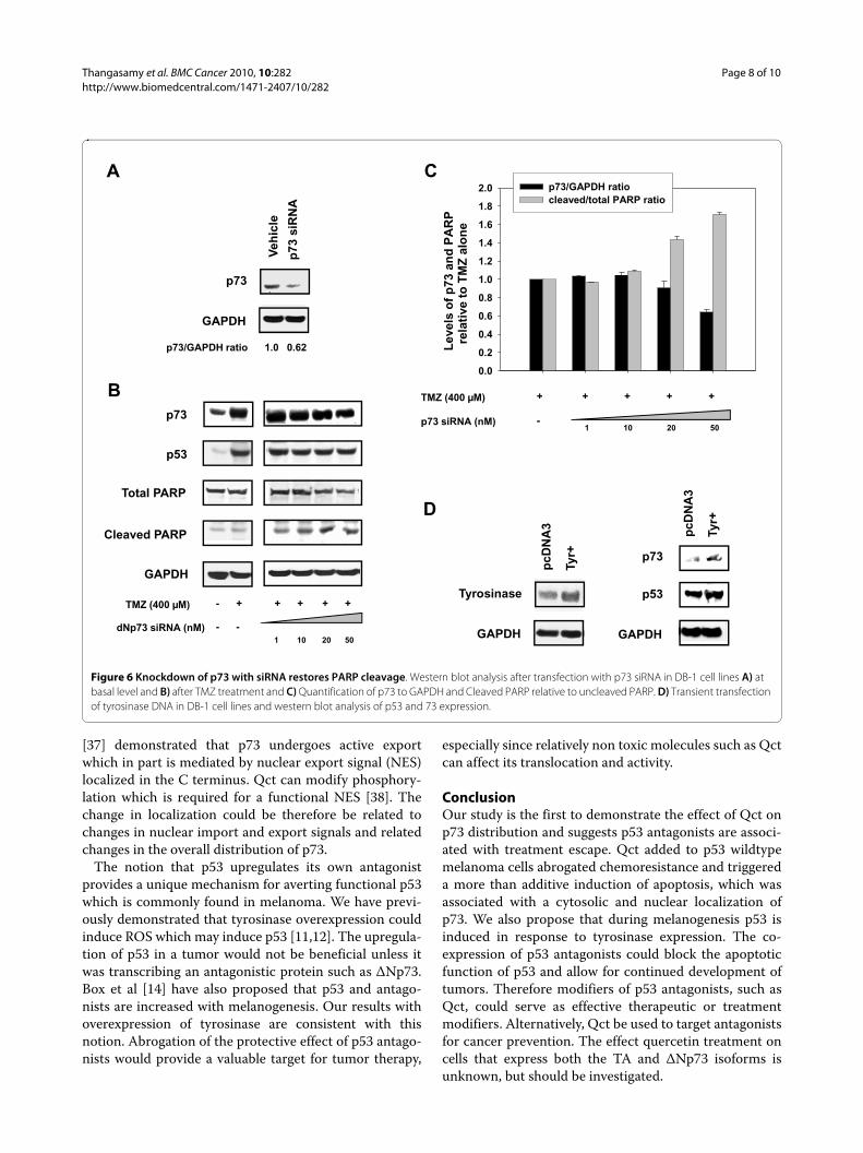
To confirm the role of ΔNp73 in the attenuation of theapoptotic response siRNA experiments were performed.Naumann et al. [24] identified PARP cleavage as a hall-mark of TMZ activity, so we investigated the effect of p73siRNA on PARP levels. Treatment with TMZ resulted inno significant cleavage of PARP compared to control (Fig-ure 6B). However, incubation with p73 siRNA in TMZ-
treated cells knocked down the ΔNp73 protein levels andresulted in an increase in the ratio of cleaved to uncleavedPARP (Figure 6C). Taken together, these results con-firmed the role of ΔNp73 as a p53 antagonist in theresponse to TMZ and suggests that Qct promotes apop-tosis by inducing the translocation of ΔNp73 out of thenucleus.
Melanogenesis and ΔNp73Tyrosinase activity is increased as melanoma tumorsdevelop and we have previously shown that tyrosinaseactivity is associated with an increase in p53 [11,12].Because p53 can transcriptionally activate p73, we inves-tigated if tyrosinase overexpression could induce theexpression of p73. We transiently overexpressed tyrosi-nase (Figure 6D) and observed an increase in the expres-sion of p53 and its transcriptional target p73 (Figure 6D).Therefore, tumorigenesis in melanoma, which coincideswith the overexpression of tyrosinase, could also be asso-ciated with an increase in transcription of p53 antago-nists and inducing resistance to chemotherapies.Targeting these antagonists through compounds such asquercetin could serve as an effective therapeutic or treat-ment modifier by restoring p53 activity.
DiscussionDNA Damage ResponseWe have shown for the first time that Qct can affect thecellular distribution of ΔNp73 and abrogate its anti-apop-totic effects. We also demonstrated in melanoma that Qctcan affect the phosphorylation of p53, which is a key fac-tor in mediating TMZ-induced apoptosis and a majordeterminant of cancer cell response to TMZ [25]. TMZand DTIC methylate DNA at O6 methyl guanine residuesand cause mispairing with thymine and activation ofMutSa-dependent mismatch repair resulting in apoptosis[26,27]. TMZ treatment during the present study causedan induction in total p53 and phosphorylation of its ser-ine residues at 15, 37 and 392. Studies by Mhaidat NM etal. [28], illustrate that the sensitivity of melanoma cells toTMZ was dependent on their p53 status and associatedG2/M arrest. We did confirm the central role of p53 withTMZ treatment and further demonstrated TMZ is astrong activator of ATM. ATM activation was enhancedby Qct, which provides a mechanism for the activationand stabilization of p53 while modulation of p73 likelycontributes to increased p53 activity.
Even though apoptosis is considered the major mecha-nism of death by TMZ, [29] Qct also increased necrosisand indicates that apoptosis might not be the only thera-peutic outcome when using combination treatments. Celldeath reported here consists of an apoptotic fraction anda second fraction of cells that consisted of late apoptosisor necrosis. Necrosis as a mechanism of cell death can

Thangasamy et al. BMC Cancer 2010, 10:282http://www.biomedcentral.com/1471-2407/10/282
Page 7 of 10
not be ruled out and is line with another study in malig-nant melanoma cells [30] that concluded TMZ-inducedO6-methyl guanine triggers the apoptotic as well asnecrotic pathway through the formation of DSBs. How-ever, these pathways appear in part mediated by p53 asthe SK Mel 28 cells were relatively void of treatment-induced cell death.
Abrogation of apoptosis by ΔNp73Another mechanism of action of Qct appears to bethrough the change in distribution of p73, allowing for anincrease in p53 nuclear activity. p73 belongs to the p53family of proteins that exhibit sequence homology [31]and they include p53, p63 and p73. Endogenous TAp73 isupregulated in response to DNA damage or treatmentwith chemotherapeutic drugs and provides antitumoractivity, while the upregulation of ΔNp73 promotes resis-
tance to these drugs [32]. The functionality of the p53family members primarily depends on the nuclear local-ization of p73. Studies demonstrate that the export of thetruncated form, ΔNp73, to the cytoplasm is a majorinducer of p53 functionality [33]. In earlier studies withhepatocellular and cholangiocellular carcinomas, p73 wasreported to be confined to the nucleus [34,35]. It was alsofound to be localized mainly in the nucleus of undifferen-tiating neuroblastomas [36].
Our ICC study demonstrates that expression of ΔNp73was confined to the nucleus in control melanoma cellsand this expression was even increased in the nucleus fol-lowing TMZ treatment. Re-localization to the cytoplasmcould indicate a shift in the balance of p53 to p73 in thenucleus and allow for transcription of p53 target genes, ormay indicate a functional change in p73 itself. Inoue et al.
Figure 5 Qct causes changes in p73 distribution. A) Non-quantitative PCR in DB-1 cells for TAp73 (Lane 1), ΔNp73 (Lane 2) and TAp73 positive con-trol using SK Mel 28 cells (Lane 3) following treatment with TMZ. TAp73 was undetectable in the DB-1 cell line. B) Real time RT-PCR for ΔNp73 in DB-1 cell lines treated with TMZ 400 μM for 48 hrs followed Qct 75 μM for 24 hrs. Results are Mean ± SEM of triplicate experiments in DB-1 cell lines. C) p73 expression by western blot analysis in DB-1 and SK Mel 28 cell lines treated with vehicle (DMSO), TMZ 400 μM for 48 hrs followed by Qct 75 μM for 24 hrs. D) Immunocytochemical analysis of p73 localization in DB-1 cell lines treated with vehicle (DMSO), TMZ 400 μM for 48 hrs followed by Qct 75 μM for 24 hrs. Solid arrowheads indicate nuclear staining in cells without Qct treatment, while line-type arrowheads indicate cytoplasmic staining following Qct treatment.
DB-1 cells
GAPDH
p73
DM
SO
TMZ+
75 Q
TMZ
400
μM
75 μ
M Q
ct
DM
SO
TMZ+
75 Q
TMZ
400
μM
75 μ
M Q
ct
SK Mel 28 cells
DC
DMSO TMZ Qct TMZ+Qct
Fold
Exp
ress
(Vs.
UT)
0
5
10
15
20
25
30B
TAp73 �Np73
TAp73positive control
A
Thangasamy et al. BMC Cancer 2010, 10:282http://www.biomedcentral.com/1471-2407/10/282
Page 8 of 10
[37] demonstrated that p73 undergoes active exportwhich in part is mediated by nuclear export signal (NES)localized in the C terminus. Qct can modify phosphory-lation which is required for a functional NES [38]. Thechange in localization could be therefore be related tochanges in nuclear import and export signals and relatedchanges in the overall distribution of p73.
The notion that p53 upregulates its own antagonistprovides a unique mechanism for averting functional p53which is commonly found in melanoma. We have previ-ously demonstrated that tyrosinase overexpression couldinduce ROS which may induce p53 [11,12]. The upregula-tion of p53 in a tumor would not be beneficial unless itwas transcribing an antagonistic protein such as ΔNp73.Box et al [14] have also proposed that p53 and antago-nists are increased with melanogenesis. Our results withoverexpression of tyrosinase are consistent with thisnotion. Abrogation of the protective effect of p53 antago-nists would provide a valuable target for tumor therapy,
especially since relatively non toxic molecules such as Qctcan affect its translocation and activity.
ConclusionOur study is the first to demonstrate the effect of Qct onp73 distribution and suggests p53 antagonists are associ-ated with treatment escape. Qct added to p53 wildtypemelanoma cells abrogated chemoresistance and triggereda more than additive induction of apoptosis, which wasassociated with a cytosolic and nuclear localization ofp73. We also propose that during melanogenesis p53 isinduced in response to tyrosinase expression. The co-expression of p53 antagonists could block the apoptoticfunction of p53 and allow for continued development oftumors. Therefore modifiers of p53 antagonists, such asQct, could serve as effective therapeutic or treatmentmodifiers. Alternatively, Qct be used to target antagonistsfor cancer prevention. The effect quercetin treatment oncells that express both the TA and ΔNp73 isoforms isunknown, but should be investigated.
Figure 6 Knockdown of p73 with siRNA restores PARP cleavage. Western blot analysis after transfection with p73 siRNA in DB-1 cell lines A) at basal level and B) after TMZ treatment and C) Quantification of p73 to GAPDH and Cleaved PARP relative to uncleaved PARP. D) Transient transfection of tyrosinase DNA in DB-1 cell lines and western blot analysis of p53 and 73 expression.
C
pcD
NA
3
Tyr+
p73
GAPDH
p53
A
D
GAPDH
Tyrosinase
pcD
NA
3
Tyr+
GAPDH
p73V
ehic
le
p73
siR
NA
B
p73/GAPDH ratio 1.0 0.62
1 10 20 50
+ + + +- +
- -
TMZ (400 µM)
dNp73 siRNA (nM)
p73
p53
Total PARP
Cleaved PARP
GAPDH
Lev
els
of
p73
an
d P
AR
P
rel
ativ
e to
TM
Z a
lon
e
0.0
0.2
0.4
0.6
0.8
1.0
1.2
1.4
1.6
1.8
2.0 p73/GAPDH ratio cleaved/total PARP ratio
1 10 20 50
+ + + ++
-
TMZ (400 µM)
p73 siRNA (nM)

Thangasamy et al. BMC Cancer 2010, 10:282http://www.biomedcentral.com/1471-2407/10/282
Page 9 of 10
AbbreviationsATM: Ataxia telangiectasia mutated; ALP: Alkaline phosphatase; Bax: Bcl-2-asso-ciated X protein; DMSO: Dimethyl sulphoxide; DNApk: DNA dependent proteinkinase; DTIC: Dacarbazine; FITC: Fluorescein isothiocyanate; GAPDH: Glyceral-dehyde 3 phosphate dehydrogenase; ICC: Immunohistochemistry; α-MEM: αMinimum essential medium; NQO1: Quinone oxidoreductase 1; PARP:Poly(ADP-Ribose) Polymerase; PI3KK: Phosphatidylinositol 3-kinase-like kinase;Qct: Quercetin; ROS: Reactive Oxygen Species; RT PCR: Real time polymerasechain reaction; TMZ: Temozolomide.
Competing interestsThe authors declare that they have no competing interests.
Authors' contributionsTT and SS carried out molecular and cell biology experiments, data analysis,participated in the design of the studies and drafted the manuscript. GCM par-ticipated in molecular and cell biology experiments, data analysis, interpreta-tion of results and written revisions. VRM contributed to the design ofexperiments and analysis of data. EEM participated in data analysis, interpreta-tion of results and written revisions for the resubmission. KHL an RB conceivedthe studies and participated in design of the experiments. RB oversaw andcoordinated the studies and finalized writing of the manuscript. All authorsread and approved the final manuscript.
AcknowledgementsThis work was financially supported by in part by USDA 2006-38411-17037 (RB).
Author Details1Department of Nutritional Sciences, University of Arizona, Tucson, AZ 85721, USA, 2Cancer Biology Interdisciplinary Program, University of Arizona, Tucson, AZ 85721, USA and 3Arizona Cancer Center, University of Arizona, Tucson, AZ 85721, USA
References1. Gray-Schopfer V, Wellbrock C, Marais R: Melanoma biology and new
targeted therapy. Nature 2007, 445:851-7.2. Ibrahim N, Haluska FG: Molecular pathogenesis of cutaneous
melanocytic neoplasms. Annu Rev Pathol 2009, 4:551-579.3. Jemal A, Murray T, Samuels A, Ghafoor A, Ward E, Thun MJ: Cancer
statistics, 2003. CA Cancer J Clin 2003, 53:5-26.4. Bedikian AY, Millward M, Pehamberger H, Conry R, Gore M, Trefzer U,
Pavlick AC, DeConti R, Hersh EM, Hersey P, Kirkwood JM, Haluska FG: Bcl-2 antisense (oblimersen sodium) plus dacarbazine in patients with advanced melanoma: the Oblimersen Melanoma Study Group. J Clin Oncol 2006, 24:4738-4745.
5. Newlands ES, Stevens MF, Wedge SR, Wheelhouse RT, Brock C: Temozolomide: a review of its discovery, chemical properties, pre-clinical development and clinical trials. Cancer Treat Rev 1997, 23:35-61.
6. Tentori L, Graziani G: Recent approaches to improve the antitumor efficacy of temozolomide. Curr Med Chem 2009, 16:245-257.
7. Metodiewa D, Jaiswal AK, Cenas N, Dickancaite E, Segura-Aguilar J: Quercetin may act as a cytotoxic prooxidant after its metabolic activation to semiquinone and quinoidal product. Free Radic Biol Med 1999, 26:107-116.
8. Simonova M, Wall A, Weissleder R, Bogdanov A Jr: Tyrosinase mutants are capable of prodrug activation in transfected nonmelanotic cells. Cancer Res 2000, 60:6656-6662.
9. Larocca LM, Teofili L, Maggiano N, Piantelli M, Ranelletti FO, Leone G: Quercetin and the growth of leukemic progenitors. Leuk Lymphoma 1996, 23:49-53.
10. Elangovan V, Sekar N, Govindasamy S: Chemopreventive potential of dietary bioflavonoids against 20-methylcholanthrene-induced tumorigenesis. Cancer Lett 1994, 87:107-113.
11. Thangasamy T, Sittadjody S, Lanza-Jacoby S, Wachsberger P, Limesand K, Burd R: Quercetin Selectively Inhibits Bioreduction and Enhances Apoptosis in Melanoma Cells that Overexpress Tyrosinase. Nutrition and cancer 2007, 59:1-11.
12. Thangasamy T, Sittadjody S, Limesand KH, Burd R: Tyrosinase overexpression promotes ATM-dependent p53 phosphorylation by quercetin and sensitizes melanoma cells to dacarbazine. Cell Oncol 2008, 30:371-387.
13. Soengas MS, Lowe SW: Apoptosis and melanoma chemoresistance. Oncogene 2003, 22:3138-3151.
14. Box NF, Terzian T: The role of p53 in pigmentation, tanning and melanoma. Pigment Cell Melanoma Res 2008, 21:525-533.
15. Ozaki T, Nakagawara A: p73, a sophisticated p53 family member in the cancer world. Cancer Sci 2005, 96:729-737.
16. Moll UM, Slade N: p63 and p73: roles in development and tumor formation. Mol Cancer Res 2004, 2:371-386.
17. Wahl ML, Owen JA, Burd R, Herlands RA, Nogami SS, Rodeck U, Berd D, Leeper DB, Owen CS: Regulation of intracellular pH in human melanoma: potential therapeutic implications. Mol Cancer Ther 2002, 1:617-628.
18. Livak KJ, Schmittgen TD: Analysis of relative gene expression data using real-time quantitative PCR and the 2(-Delta Delta C(T)) Method. Methods 2001, 25:402-8.
19. Limesand KH, Schwertfeger KL, Anderson SM: MDM2 is required for suppression of apoptosis by activated Akt1 in salivary acinar cells. Mol Cell Biol 2006, 26:8840-56.
20. Stiewe T, Tuve S, Peter M, Tannapfel A, Elmaagacli AH, Putzer BM: Quantitative TP73 transcript analysis in hepatocellular carcinomas. Clin Cancer Res 2004, 10:626-633.
21. Tomkova K, Belkhiri A, El-Rifai W, Zaika AI: p73 isoforms can induce T-cell factor-dependent transcription in gastrointestinal cells. Cancer Res 2004, 64:6390-6393.
22. Melino G, De L, Vousden KH: p73: Friend or foe in tumorigenesis. Nat Rev Cancer 2002, 2:605-615.
23. DeYoung MP, Ellisen LW: p63 and p73 in human cancer: defining the network. Oncogene 2007, 26:5169-5183.
24. Naumann SC, Roos WP, Jost E, Belohlavek C, Lennerz V, Schmidt CW, Christmann M, Kaina B: Temozolomide- and fotemustine-induced apoptosis in human malignant melanoma cells: response related to MGMT, MMR, DSBs, and p53. Br J Cancer 2009, 100:322-333.
25. Roos WP, Batista LF, Naumann SC, Wick W, Weller M, Menck CF, Kaina B: Apoptosis in malignant glioma cells triggered by the temozolomide-induced DNA lesion O6-methylguanine. Oncogene 2007, 26:186-97.
26. Kaina B, Haas S, Kappes H: A general role for c-Fos in cellular protection against DNA-damaging carcinogens and cytostatic drugs. Cancer Res 1997, 57:2721-2731.
27. Kaina B, Christmann M, Naumann S, Roos WP: MGMT: key node in the battle against genotoxicity, carcinogenicity and apoptosis induced by alkylating agents. DNA Repair (Amst) 2007, 6:1079-1099.
28. Mhaidat NM, Zhang XD, Allen J, Avery-Kiejda KA, Scott RJ, Hersey P: Temozolomide induces senescence but not apoptosis in human melanoma cells. Br J Cancer 2007, 97:1225-1233.
29. Naumann SC, Roos WP, Jost E, Belohlavek C, Lennerz V, Schmidt CW, Christmann M, Kaina B: Temozolomide- and fotemustine-induced apoptosis in human malignant melanoma cells: response related to MGMT, MMR, DSBs, and p53. Br J Cancer 2009, 100:322-333.
30. Naumann SC, Roos WP, Jost E, Belohlavek C, Lennerz V, Schmidt CW, Christmann M, Kaina B: Temozolomide- and fotemustine-induced apoptosis in human malignant melanoma cells: response related to MGMT, MMR, DSBs, and p53. Br J Cancer 2009, 100:322-333.
31. Yang A, Kaghad M, Caput D, McKeon F: On the shoulders of giants: p63, p73 and the rise of p53. Trends Genet 2002, 18:90-95.
32. Muller M, Schilling T, Sayan AE, Kairat A, Lorenz K, Schulze-Bergkamen H, Oren M, Koch A, Tannapfel A, Stremmel W, Melino G, Krammer PH: TAp73/Delta Np73 influences apoptotic response, chemosensitivity and prognosis in hepatocellular carcinoma. Cell Death Differ 2005, 12:1564-1577.
33. Di VA, Sessa F, Casciano I, Banelli B, Franzi F, Brigati C, Allemanni G, Russo P, Dominioni L, Romani M: Different intracellular compartmentalization of TA and DeltaNp73 in non-small cell lung cancer. Int J Oncol 2009, 34:449-456.
34. Tannapfel A, Wasner M, Krause K, Geissler F, Katalinic A, Hauss J, Mossner J, Engeland K, Wittekind C: Expression of p73 and its relation to histopathology and prognosis in hepatocellular carcinoma. J Natl Cancer Inst 1999, 91:1154-1158.
Received: 18 November 2009 Accepted: 11 June 2010 Published: 11 June 2010This article is available from: http://www.biomedcentral.com/1471-2407/10/282© 2010 Thangasamy et al; licensee BioMed Central Ltd. This is an Open Access article distributed under the terms of the Creative Commons Attribution License (http://creativecommons.org/licenses/by/2.0), which permits unrestricted use, distribution, and reproduction in any medium, provided the original work is properly cited.BMC Cancer 2010, 10:282
Thangasamy et al. BMC Cancer 2010, 10:282http://www.biomedcentral.com/1471-2407/10/282
Page 10 of 10
35. Tannapfel A, Engeland K, Weinans L, Katalinic A, Hauss J, Mossner J, Wittekind C: Expression of p73, a novel protein related to the p53 tumour suppressor p53, and apoptosis in cholangiocellular carcinoma of the liver. Br J Cancer 1999, 80:1069-1074.
36. Douc-Rasy S, Barrois M, Echeynne M, Kaghad M, Blanc E, Raguenez G, Goldschneider D, Terrier-Lacombe MJ, Hartmann O, Moll U, Caput D, Benard J: DeltaN-p73alpha accumulates in human neuroblastic tumors. Am J Pathol 2002, 160:631-639.
37. Inoue T, Stuart J, Leno R, Maki CG: Nuclear import and export signals in control of the p53-related protein p73. J Biol Chem 2002, 277:15053-15060.
38. Quelo I, Gauthier C, St-Arnaud R: Casein kinase II phosphorylation regulates alphaNAC subcellular localization and transcriptional coactivating activity. Gene Expr 2005, 12:151-163.
Pre-publication historyThe pre-publication history for this paper can be accessed here:http://www.biomedcentral.com/1471-2407/10/282/prepub
doi: 10.1186/1471-2407-10-282Cite this article as: Thangasamy et al., Quercetin abrogates chemoresistance in melanoma cells by modulating ?Np73 BMC Cancer 2010, 10:282


![Quercetin attenuates reduced uterine perfusion pressure ...Quercetin could be widely found in vegetables, fruits, and soybeans [9]. Various studies reported the effect of quercetin](https://img.dokumen.tips/doc/110x75/60fc3df128e11010ab38e9f6/quercetin-attenuates-reduced-uterine-perfusion-pressure-quercetin-could-be-widely.jpg)